

295,00 € – 895,00 €
Product details
Synonyms = A inhibin subunit; IHA; inhA; Inhibin alpha chain; Inhibin alpha subunit
Antibody type = Recombinant Rabbit monoclonal / IgG
Clone = MSVA-561R
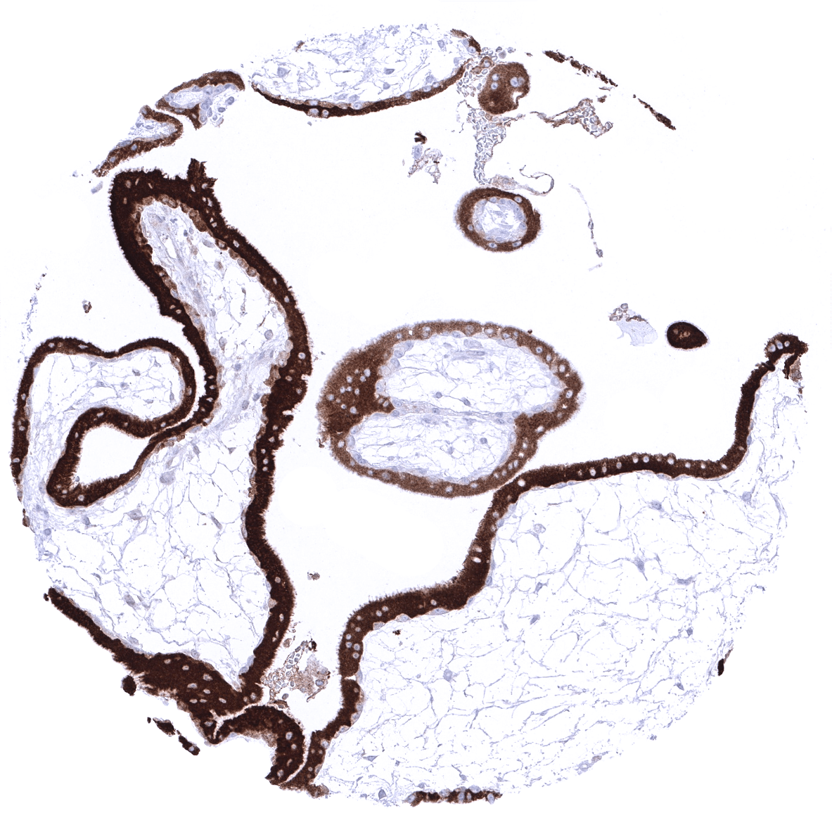
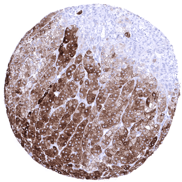
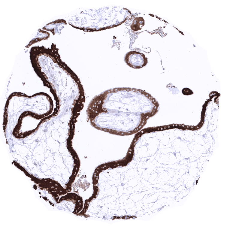
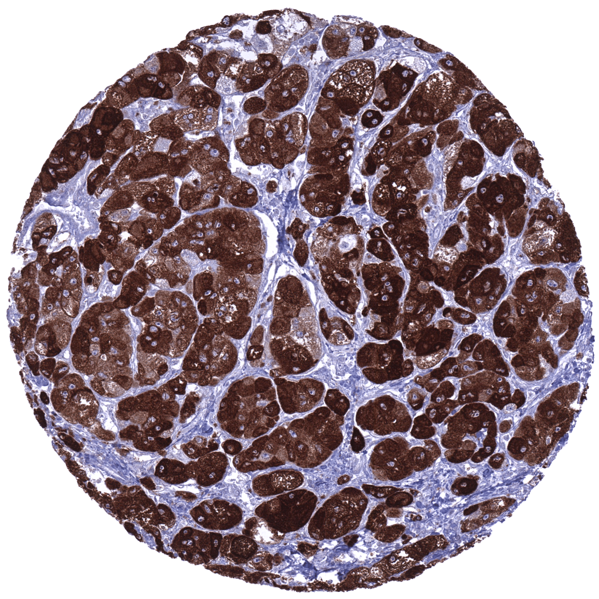
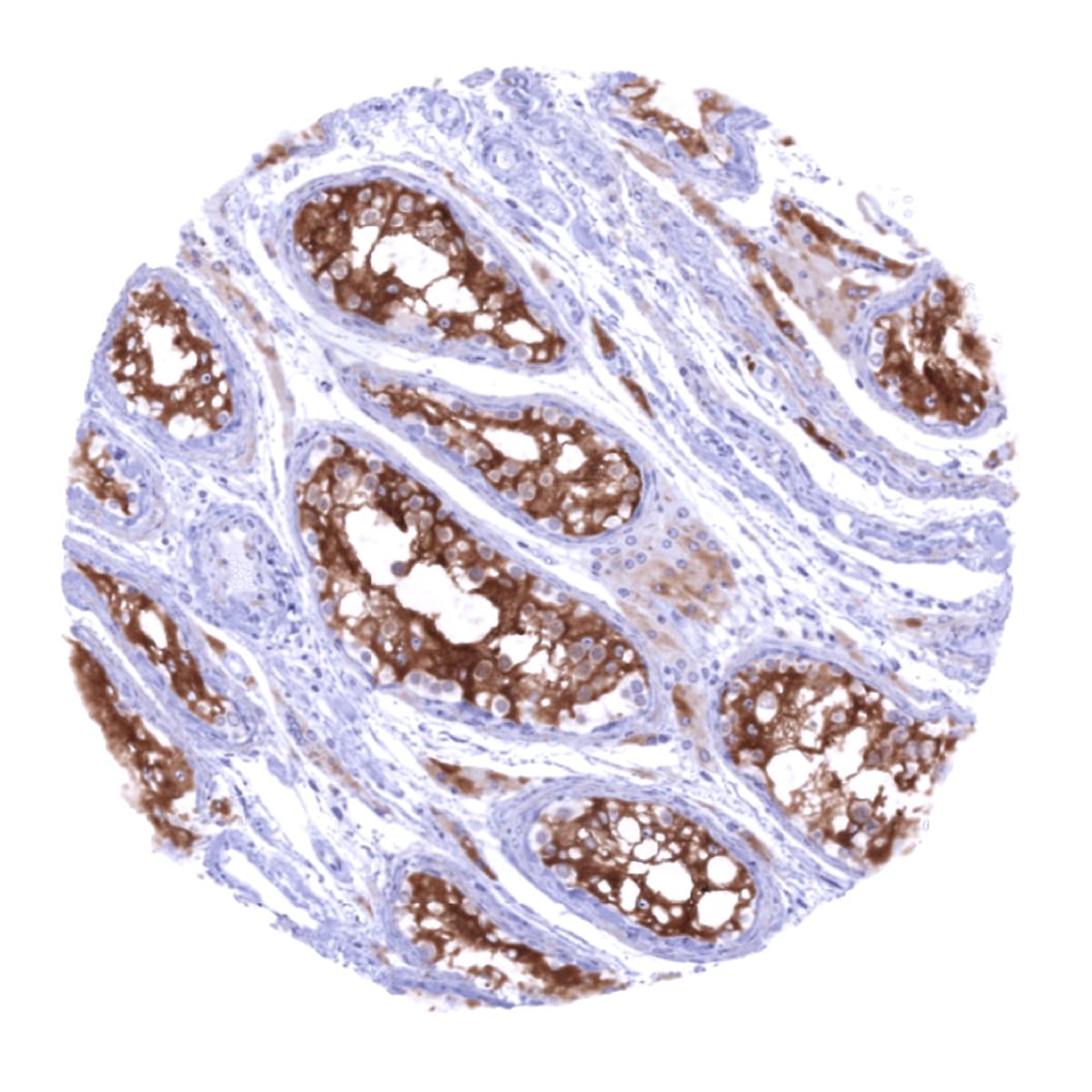
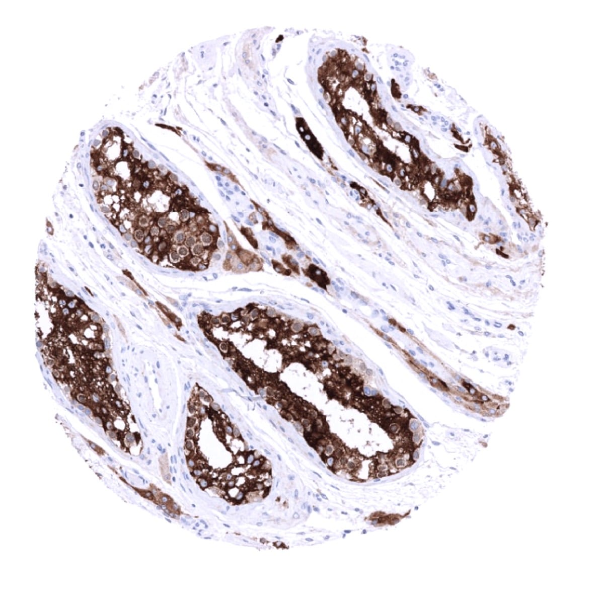
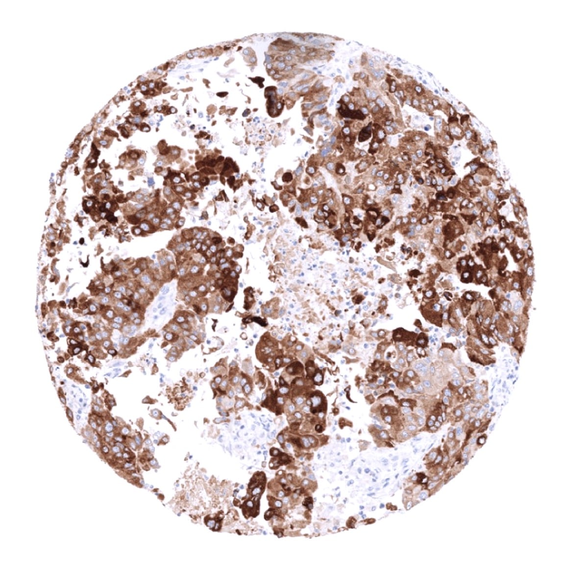
Positive control = Adrenal gland: A moderate to strong INHA positivity should be seen in adrenocortical cells.
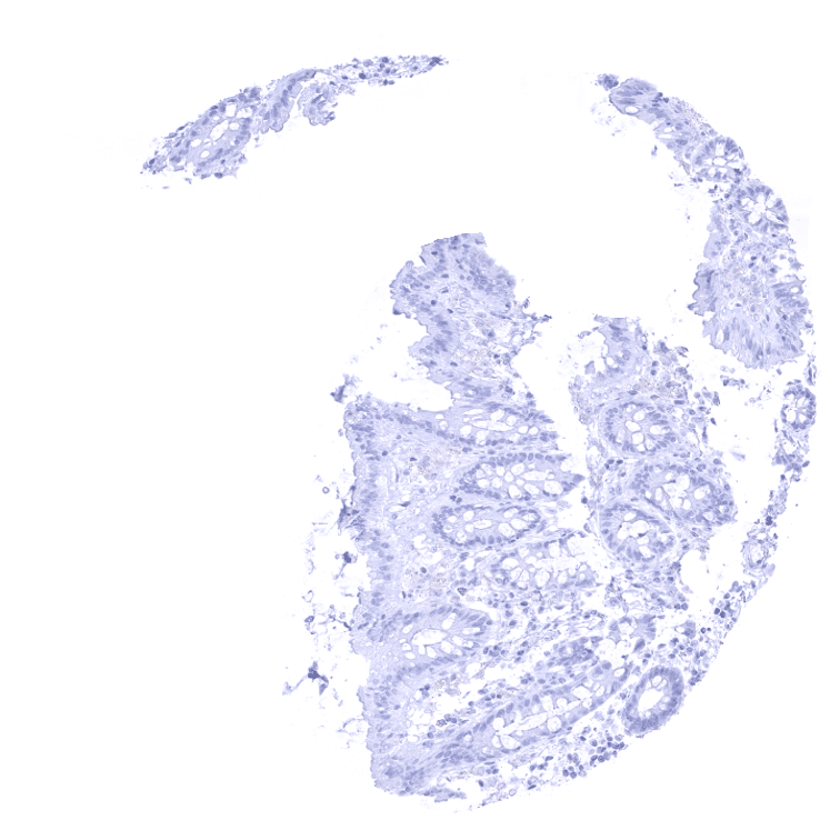
Negative control = Colon: INHA immunostaining should be completely absent.
Cellular localization = Cytoplasmic
Reactivity = Human
Application = Immunohistochemistry
Dilution = 1:100
Intended Use = Research Use Only
Relevance of Antibody
Inhibin alpha is expressed in adrenocortical cells and several rare tumor entities.
Biology Behind
The inhibin alpha chain protein is one of several proteins encoded by the INHA gene at human chromosome 2q35. The preprotein translated from INHA is proteolytically cleaved into multiple peptide products including the alpha chains of the inhibin A and inhibin B subcomplexes. These complexes were originally termed inhibins after their ability to inhibit follicle stimulating hormone (FSH) secreted from the pituitary gland. Chemically, inhibins are members of the TGF-beta (transforming growth factor-beta) superfamily, and as such, they are also involved in the regulation of cell growth and proliferations. Accordingly, a tumor suppressive role of INHA has been suggested. Clinical testing of inhibin is performed to monitor fertility.
Staining Pattern in Normal Tissues
Inhibin Alpha staining pattern in Normal Tissues with antibody MSVA-561R (images are shown in our “Normal Tissue Gallery”)
| Brain | Cerebrum | Negative. |
| Cerebellum | Negative. | |
| Endocrine Tissues | Thyroid | Negative. |
| Parathyroid | Negative. | |
| Adrenal gland | Strong inhibin alpha staining of adrenocortical cells. | |
| Pituitary gland | Negative. | |
| Respiratory system | Respiratory epithelium | Negative. |
| Lung | Negative. | |
| Gastrointestinal Tract | Salivary glands | Negative. |
| Esophagus | Negative. | |
| Stomach | Negative. | |
| Duodenum | Negative. | |
| Small intestine | Negative. | |
| Appendix | Negative. | |
| Colon | Negative. | |
| Rectum | Negative. | |
| Liver | Negative. | |
| Gallbladder | Negative. | |
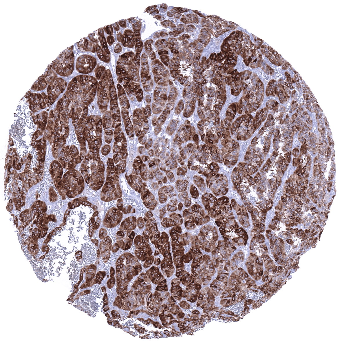
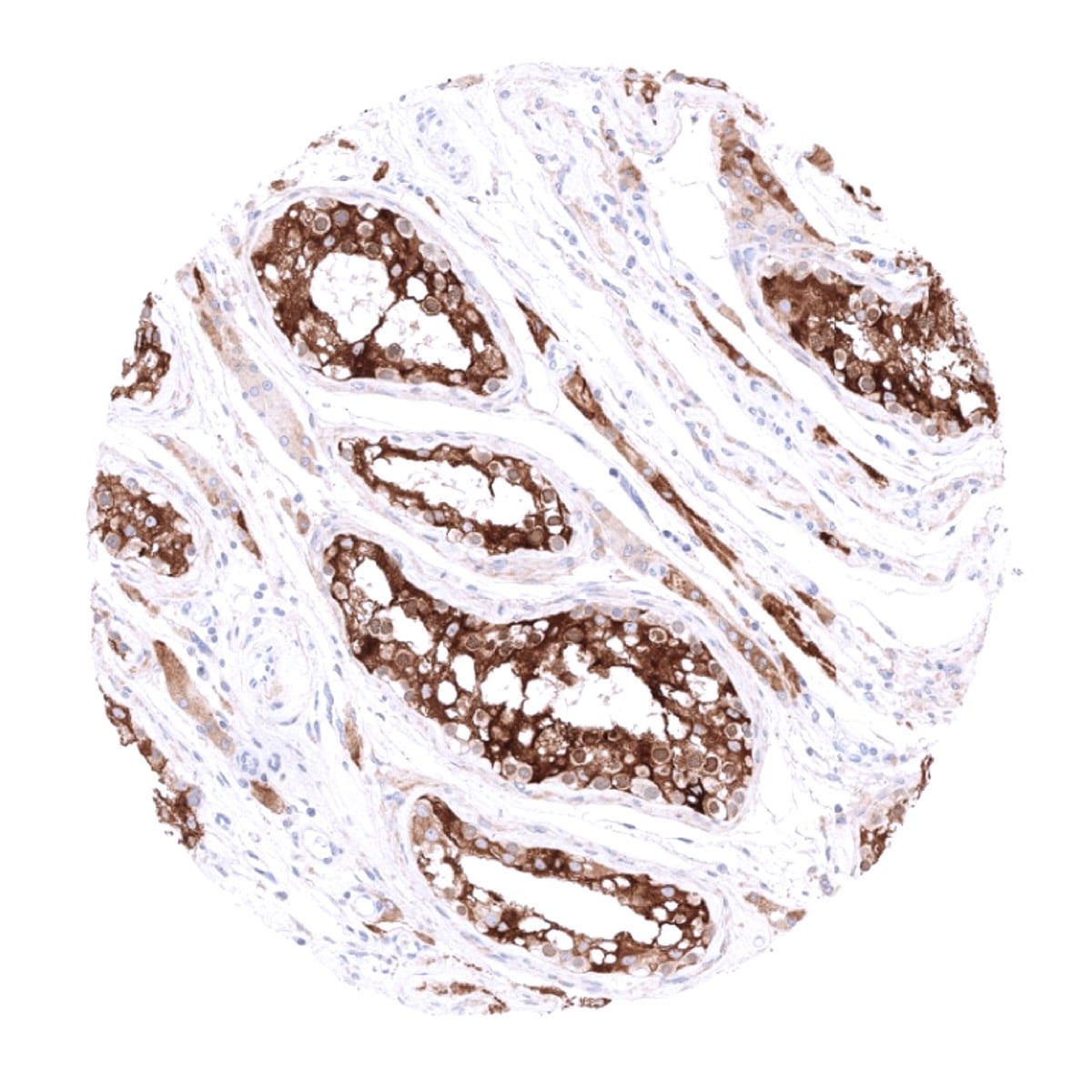
| Pancreas | Weak to moderate inhibin alpha staining of scattered acinar cells (not in all samples). | |
| Genitourinary | Kidney | Negative. |
| Urothelium | Negative. | |
| Male genital | Prostate | Negative. |
| Seminal vesicles | Negative. | |
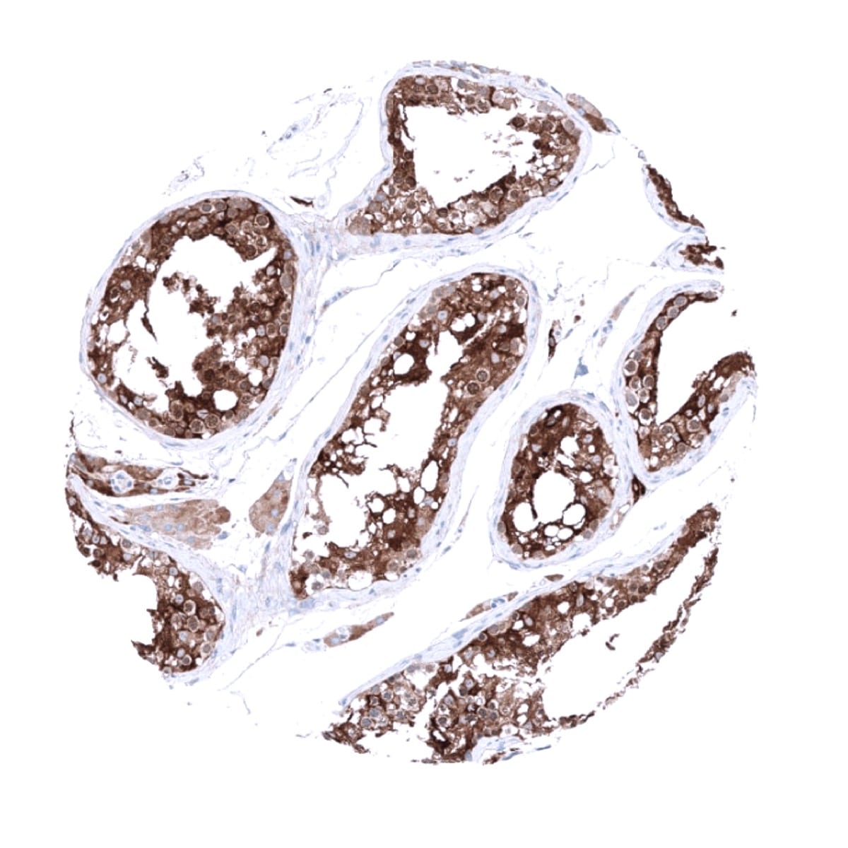
| Testis | Strong inhibin alpha staining of Sertoli and Leydig cells. | |
| Epididymis | Negative. | |
| Female genital | Breast | Negative. |
| Uterus, myometrium | Negative. | |
| Uterus, ectocervix | Negative. | |
| Uterus endocervix | Negative. | |
| Uterus, endometrium | Negative. | |
| Fallopian Tube | Negative. | |
| Ovary | Strong inhibin alpha staining of corpus luteum cells. Weak to moderate (or even strong) inhibin alpha staining of follicular and granulosa cells as well in some further stroma cells. | |
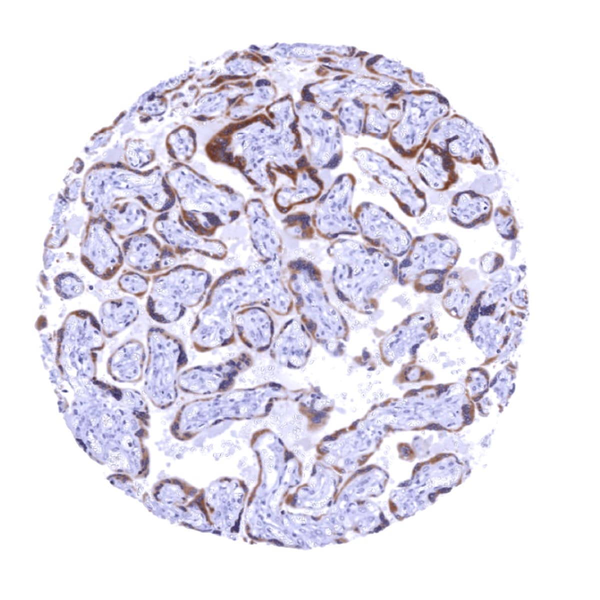
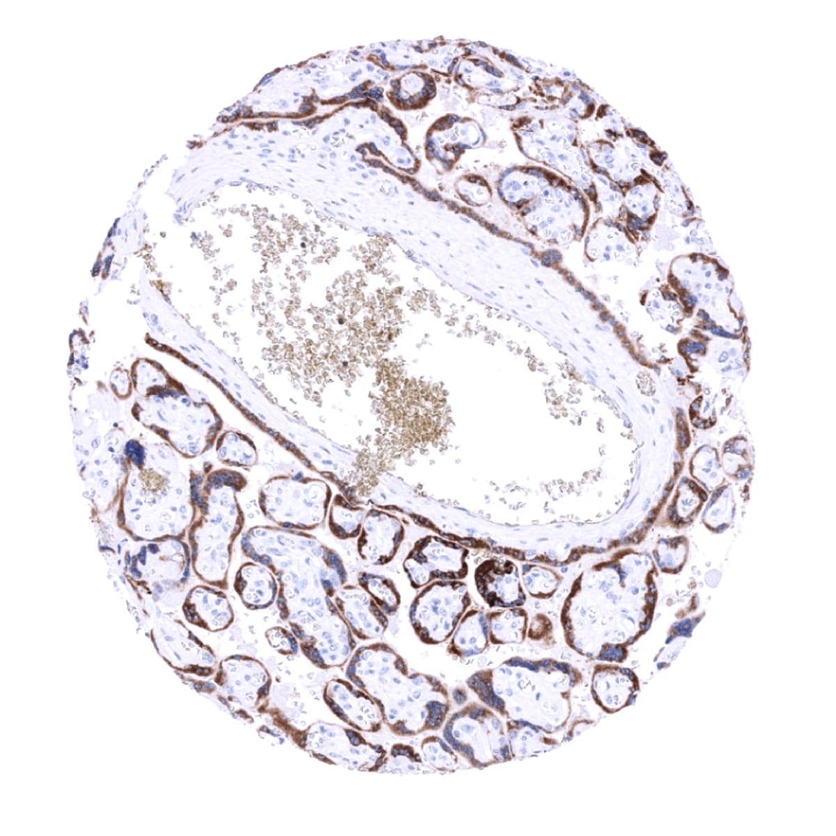
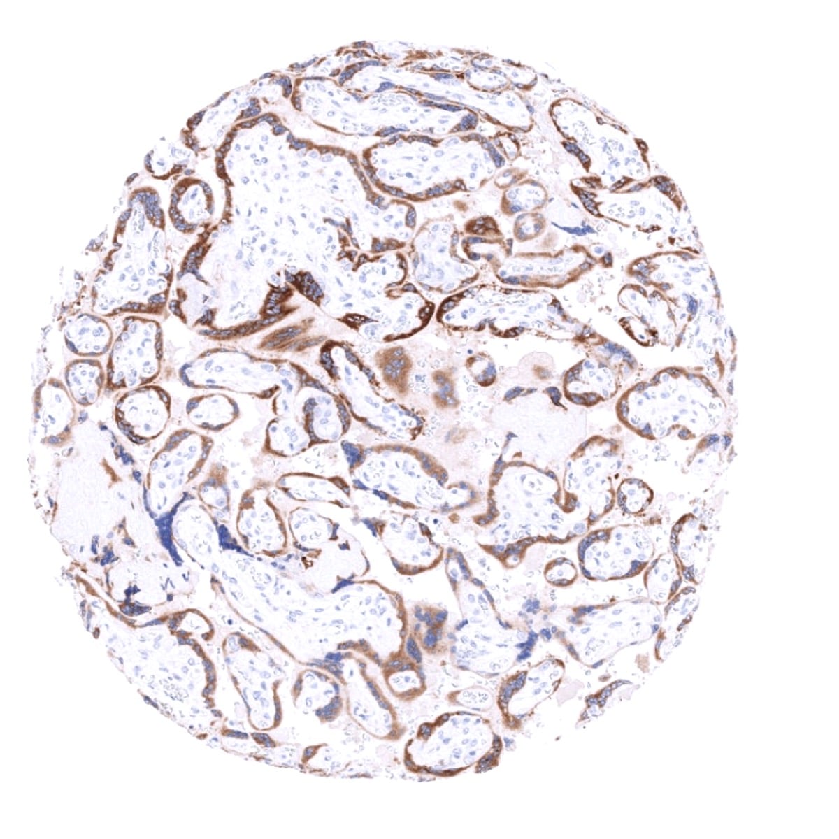
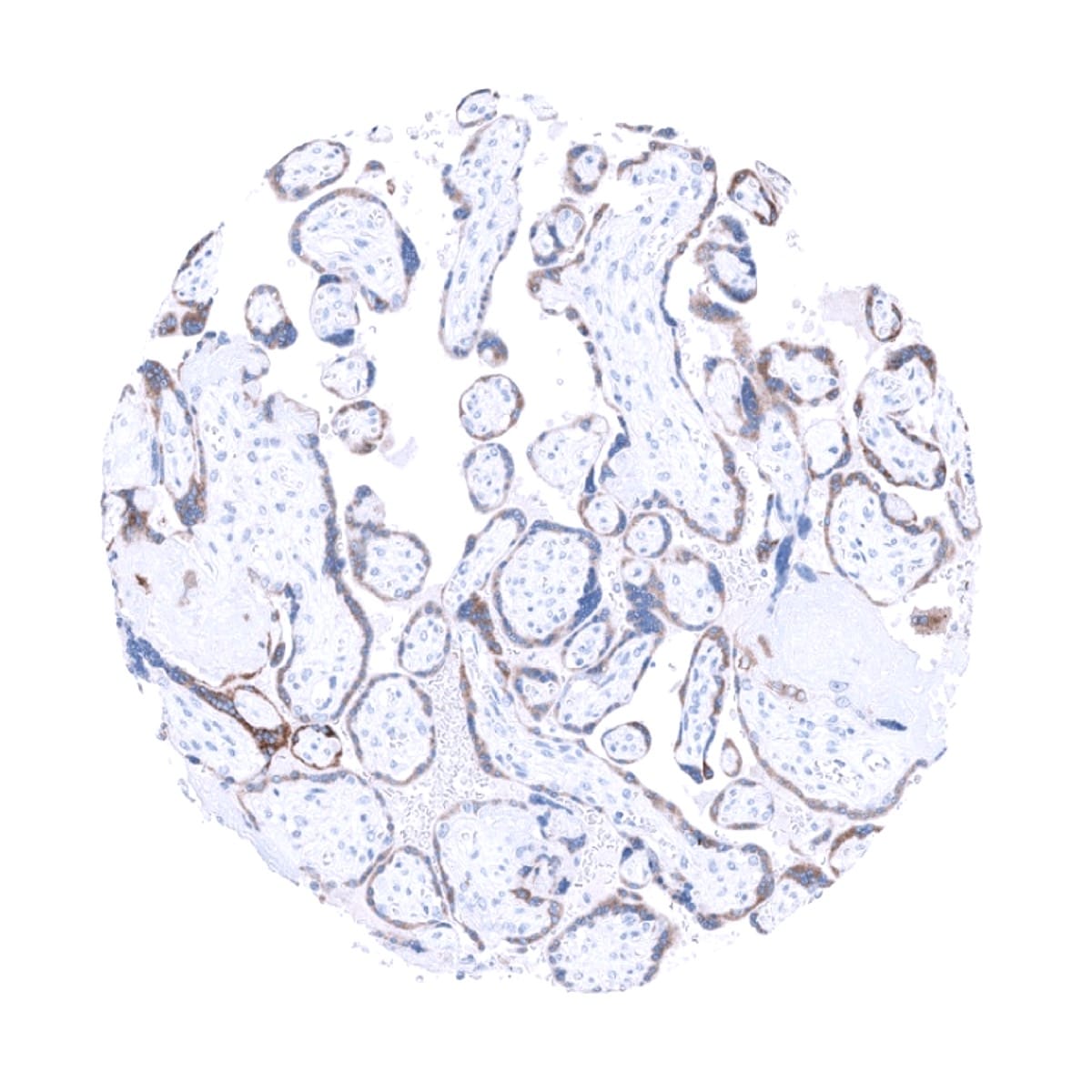
| Placenta early | Strong inhibin alpha staining of cyto- and syncytiotrophoblast. | |
| Placenta mature | Weak to moderate inhibin alpha staining of cyto- and syncytiotrophoblast and of decidua cells. | |
| Amnion | Weak to moderate inhibin alpha staining of amnion cells. | |
| Chorion | Strong inhibin alpha staining of chorion cells. | |
| Skin | Epidermis | Negative. |
| Sebaceous glands | Negative. | |
| Muscle/connective tissue | Heart muscle | Negative. |
| Skeletal muscle | Negative. | |
| Smooth muscle | Negative. | |
| Vessel walls | Negative. | |
| Fat | Negative. | |
| Stroma | Negative. | |
| Endothelium | Negative. | |
| Bone marrow/ lymphoid tissue | Bone marrow | Negative. |
| Lymph node | Negative. | |
| Spleen | Negative. | |
| Thymus | Negative. | |
| Tonsil | Negative. | |
| Remarks | Inhibin alpha staining is cytoplasmic. |
These findings are largely consistent with the RNA and protein data summarized in the Human Protein Atlas (Tissue expression Inhibin alpha) although a heart muscle staining as suggested by the RNA data of the human protein atlas could not be found by using MSVA-561R.
Positive control: Adrenal gland: A moderate to strong INHA positivity should be seen in adrenocortical cells.
Negative control: Colon: INHA immunostaining should be completely absent.
Staining Pattern in Relevant Tumor Types
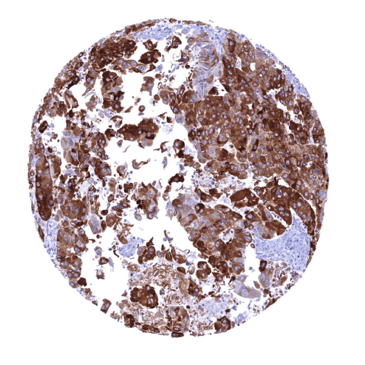
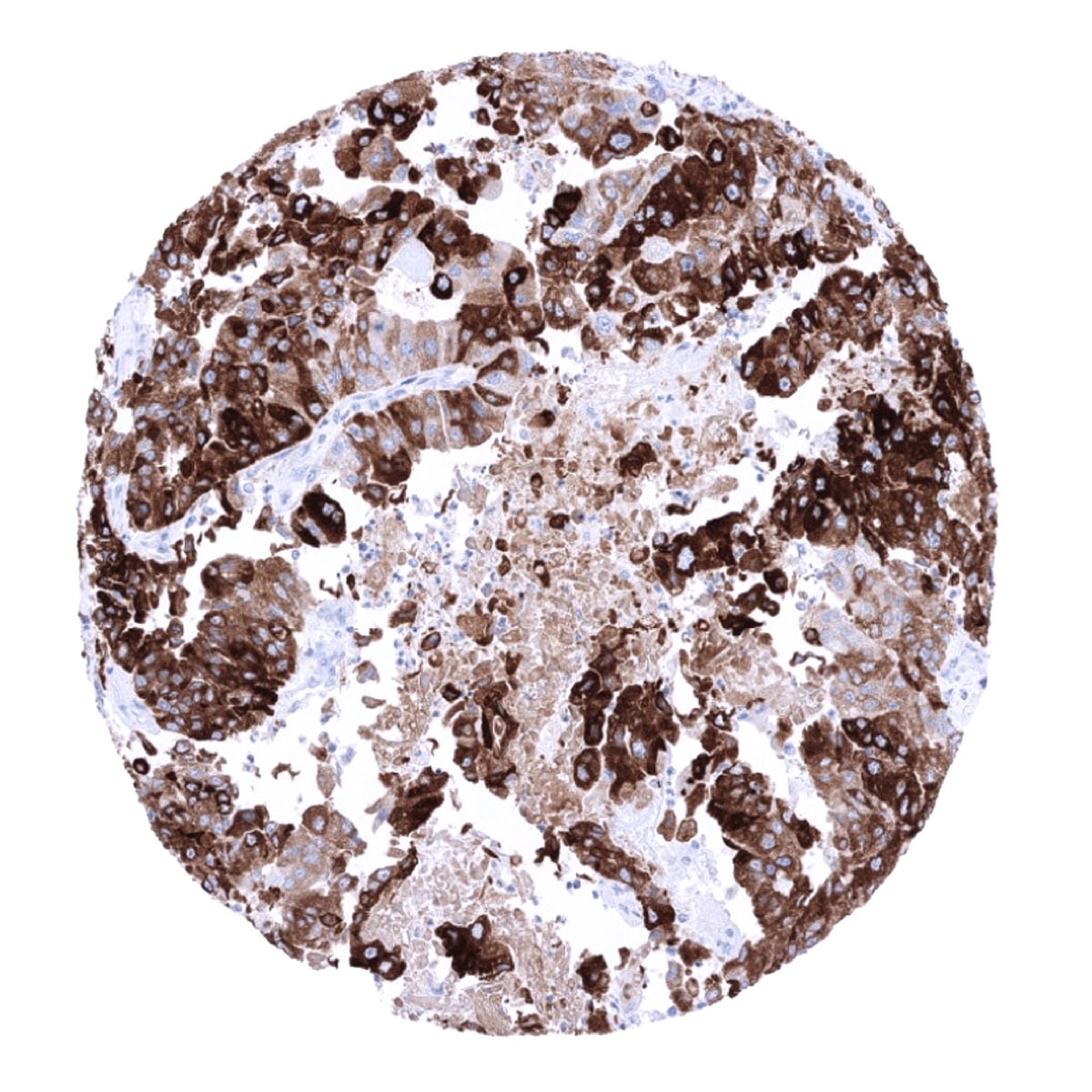
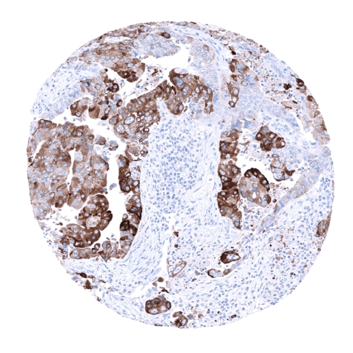
A positive INHA immunostaining is usually seen in adrenocortical tumors, Sertoli cell tumors, Leydig cell tumors, granulosa cell tumors, other sex-cord stromal tumors, and granular cell tumors. INHA positivity can at lower frequency and lower intensity also occur in a variety of other tumor entities.
The TCGA findings on Inhibin alpha RNA expression in different tumor categories have been summarized in the Human Protein Atlas.
Compatibility of Antibodies
Inhibin alpha (MSVA-561R) publication summary
Relevant publication: Weidemann et al. “Inhibin Alpha Expression in Human Tumors: A Tissue Microarray Study on 12,212 Tumors” Published in Biomedicines 2022 Oct 7;10(10):2507. PMID: 36289769
A total of 12’212 tumors from 134 different tumor categories were successfully analyzed by using the following protocol: Heat-induced antigen retrieval for 5 minutes in an autoclave at 121°C in pH 7,8 Target Retrieval Solution buffer. MSVA-561R at a dilution of 1:100 at 37°C for 60 minutes. Visualization of bound antibody by the EnVision Kit (Dako, Agilent). This protocol was also used for all stainings depicted in our tumor and normal tissue galleries.
Overall, 72 (54%) of 134 tumor categories showed detectable INHA expression in at least one case and 26 (19%) tumor categories included at least one case with strong INHA positivity. The highest prevalence of INHA staining and the highest staining levels were found in various types of sex cord stromal tumors of the testis and the ovary (100% positive), granular cell tumors (100%), granulosa cell tumors of the ovary (100%), adrenal cortical adenomas (93%) and carcinomas (80%), and in acinar cell carcinomas of the pancreas (80%). In the ovary, tumor cell nests were often surrounded by a conspicuous layer of INHA positive stromal cells. The distribution of positive staining results is shown in an “organ-systematic” (Figure 1) and in a “ranking order” figure (Figure 2) below (images based on data from Weidemann et al.). Data on associations with histopathological and clinical parameters of tumor aggressiveness in several cancer types are also summarized below (Figure 3; based on data described by Weidemann et al.).
Authors conclusions on diagnostic utility of INHA IHC with respect to the distinction of different tumor entities (Weidemann et al.):
- A strong INHA staining of a large fraction of tumor cells supports a diagnosis of adrenocortical tumors, granular cell tumors, acinar cell carcinoma of the pancreas, and sex cord stromal tumors of the testis and the ovary.
- Because clear cell renal cell carcinomas can (rarely) be strongly INHA positive, INHA immunohistochemistry should not be used as a stand-alone tool for the distinction of normal or neoplastic adrenal tissue from clear cell RCC.
- A focal weak to moderate INHA immunostaining should not be diagnostically overinterpreted because this can occur in many different tumor entities.
Authors conclusions on the prognostic role of INHA immunostaining results (Weidemann et al.):
- INHA positivity was marginally related with few parameters of cancer aggressiveness in neuroendocrine tumors, clear cell renal cell carcinoma, and colorectal adenocarcinoma. The authors thus speculated on a possible tumor-promoting effect of INHA neo-expression in cancer, possibly through a paracrine activity of secreted INHA.
Figure 1. Inhibin Alpha staining in cancer (“organ-systematic”; according to Weidemann et al.)

Figure 2. Inhibin Alpha staining in cancer (“ranking list”; according to Weidemann et al.)

Protocol Recommendations
IHC users have different preferences on how the stains should look like. Some prefer high staining intensity of the target stain and even accept some background. Others favor absolute specificity and lighter target stains. Factors that invariably lead to more intense staining include higher concentration of the antibody and visualization tools, longer incubation time, higher temperature during incubation, higher temperature and longer duration of the heat induced epitope retrieval (slide pretreatment). The impact of the pH during slide pretreatment has variable effects and depends on the antibody and the target protein.
All images and data shown here and in our image galleries are obtained by the manual protocol described below. Other protocols resulting in equivalent staining are described as well.
Manual protocol
Freshly cut sections should be used (less than 10 days between cutting and staining). Heat-induced antigen retrieval for 5 minutes in an autoclave at 121°C in pH 7,8 Target Retrieval Solution buffer. Apply MSVA-561R at a dilution of 1:100 at 37°C for 60 minutes. Visualization of bound antibody by the EnVision Kit (Dako, Agilent) according to the manufacturer’s directions.
Agilent / Dako – Autostainer Link 48
Pretreatment in PT-Link for 30 minutes at 95°C (pH high); FLEX peroxidase blocking for 5 minutes (room temperature), MSVA-561R 1:100 for 20 minutes (room temperature), FLEX+ mouse/rabbit (LINKER) for 15 minutes (room temperature), horseradish peroxidase (HRP) for 20 minutes (room temperature), FLEX DAB+Sub-Chromo for 10 minutes (room temperature), FLEX hematoxylin for 5 minutes (room temperature).
These images reflect stainings by the protocol described above. It is of note that a comparable staining result can also be obtained by different protocols. In general, a longer pretreatment, a longer incubation time of the primary antibody, a higher antibody concentration, and a longer incubation time of FLEX+LINKER result in stronger staining, potentially at the cost of more background staining. Modifications of the protocol with a strengthening effect on staining intensity in combination with changes of other parameters that result in lower staining intensity can result in a comparable result as shown above.
Leica – BOND RX
Dewax at 72°C for 30 seconds; Pretreatment in Bond Epitope Retrieval Solution (ER2 – EDTA pH9) for 20 minutes at 100°C; Peroxidase blocking for 5 minutes (room temperature), MSVA-561R 1:100 for 15 minutes (room temperature), Post primary (rabbit anti mouse) for 8 minutes (room temperature), Polymer (goat anti rabbit) for 8 minutes (room temperature), mixed DAB refine for 10 minutes (room temperature), hematoxylin for 5 minutes (room temperature).
These images reflect stainings by the protocol described above. It is of note that a comparable staining result can also be obtained by different protocols. In general, a longer pretreatment, a longer incubation time of the primary antibody, a higher antibody concentration, a higher temperature during incubation, and a longer incubation time of Post primary and or the Polymer result in stronger staining, potentially at the cost of more background staining. Modifications of the protocol with a strengthening effect on staining intensity in combination with changes of other parameters that result in lower staining intensity can result in a comparable result as shown above.
Roche – Ventana Discovery ULTRA
Pretreatment for 64 minutes at 100°C (pH 8,4); CM peroxidase blocking for 12 minutes (room temperature), MSVA-561R 1:50 for 20 minutes at 36°C, secondary antibody (anti-rabbit HQ) for 12 minutes at 36°C, anti-HQ HRP for 12 minutes at room temperature, DAB at room temperature, hematoxylin II at room temperature for 8 minutes, bluing reagent at room temperature for 4 minutes.
These images depict staining results obtained by the protocol described above. It is of note, that the Ventana machines generally require higher antibody concentrations than other commonly used autostainers because the antibodies are automatically diluted during the procedure. Various other protocols can result in an identical result as shown above. A longer pretreatment, a longer incubation time of the primary antibody, a higher antibody concentration, a higher temperature during incubation, and a longer incubation time of secondary antibody and or the anti-HQ HRP result in stronger staining, potentially at the cost of more background staining.
Potential Research Applications
- A comprehensive study analyzing INHA in various tumor entities would be helpful to better assess the diagnostic significance of INHA IHC.
- The clinical/biological significance of INHA expression in tumors derived from cell types that are not expressing INHA is unclear.
- The functional role of INHA in cancer cells is not fully understood.
- A possible role of INHA as a paracrine factor for tumor stimulation has been proposed.
