
195,00 € – 695,00 €
Product details
Synonyms = GH-N; GH1; Growth hormone 1; HG1; hGH-N; IGHD1B; Pituitary growth hormone; RNGHGP; Somatotropin
Antibody type = Mouse monoclonal / IgG
Clone = MSVA-405M
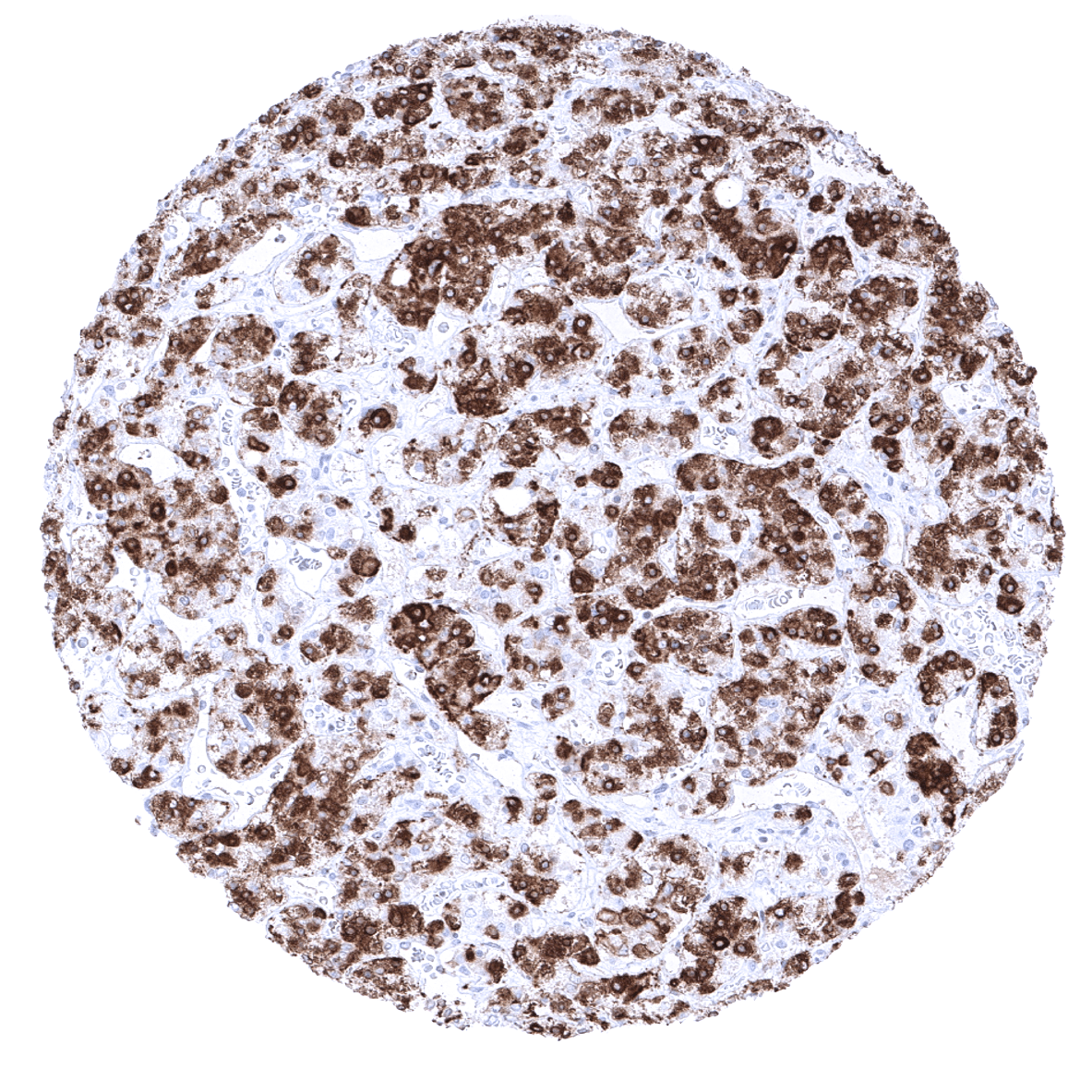
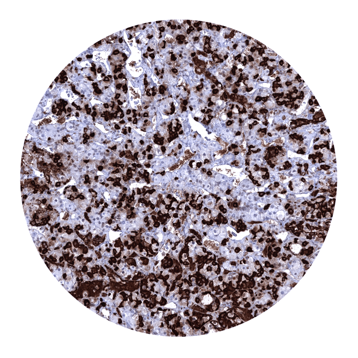
Positive control = Placenta: Trophoblast cells should stain positive.
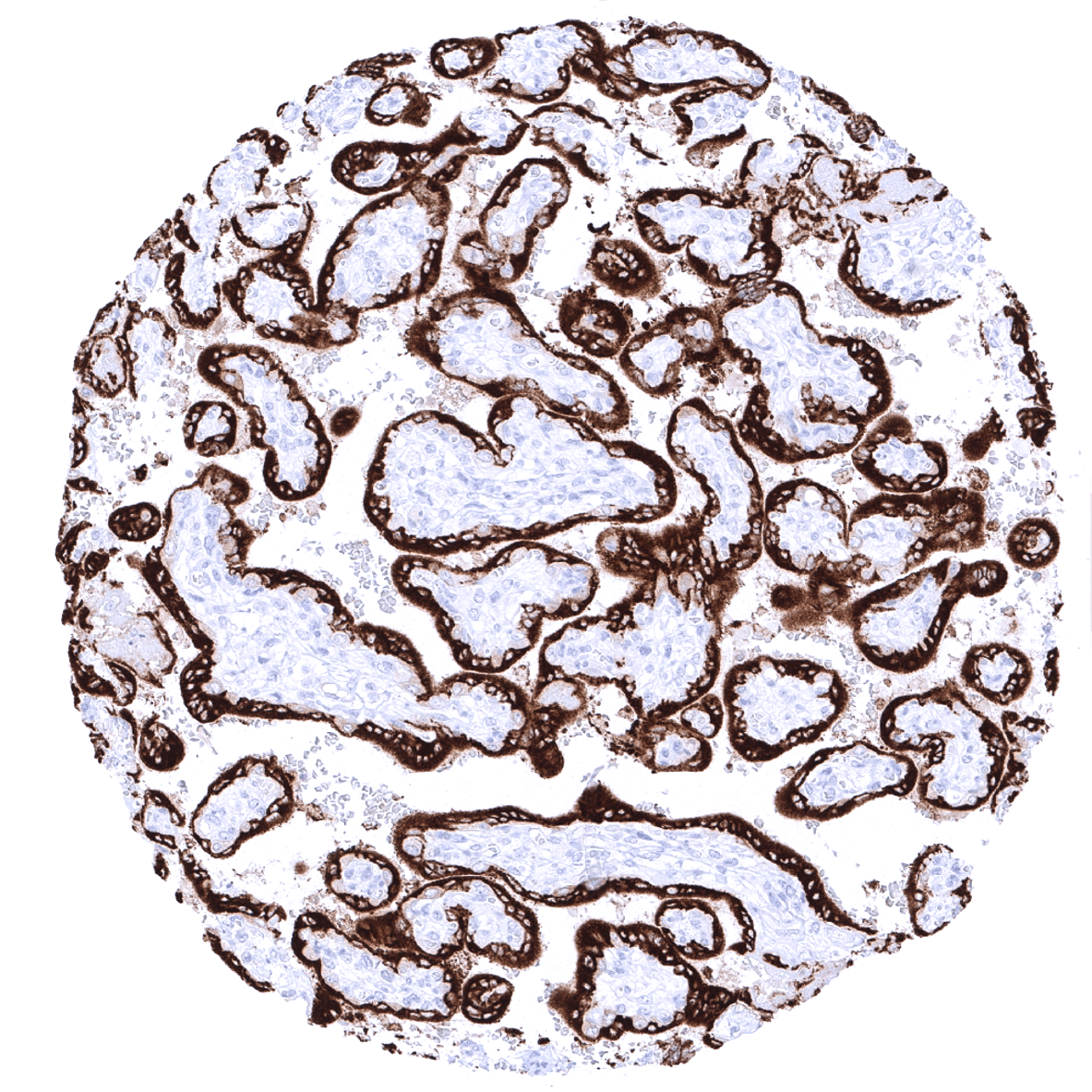
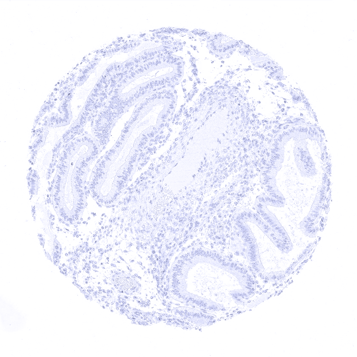
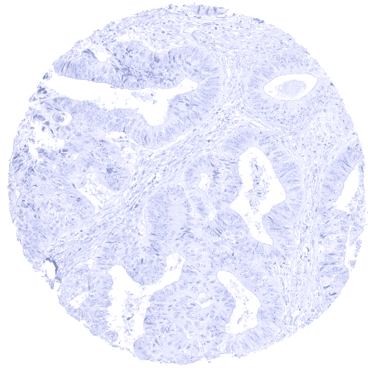
Negative control = Colon: GH staining should be absent in all epithelial and stromal cells.
Cellular localization = Cytoplasmic
Reactivity = Human
Application = Immunohistochemistry
Dilution = 1:100-200
Intended Use = Research Use Only
Relevance of Antibody
Growth Hormone is expressed in a subset of cells in the pituitary gland.
Biology Behind
Growth hormone (GH), also called somatotropin, is a polypeptide hormone secreted mainly by the somatotrophs of the anterior pituitary gland. It belongs to the 4-helical bundle chain hematopoietic super family (same family as most of the cytokines and hematopoietic growth factors) and is composed in its three-dimensional structure of 4 α-helices, non-helical chains, a hydrophobic core, and 2 disulfite bridges. The two genes of h-GH (human GH), GH1 and GH2 are located in a cluster of 5 very similar genes on 17q 23.3. The transcription of GH1 occurs in the pituitary gland and gives rise to two isoforms of 20 kDa (the most abundant) and an alternate splicing of 22 kDa. These two hormones exert an endocrine role at the peripheral level. The transcription of the GH2 gene only takes place in the syncytiotrophoblast of the placenta during gestation. It gives rise to a 22kDA protein called hGH-V (human GH-variant) which plays a major role in materno-fetal development. Growth hormone plays a major role in postnatal growth but is also essential for metabolic regulation. Its peripheral receptors are almost ubiquitous, and its main signalling pathway is mediated through JAK2 activation. Secretion of pituitary growth hormone (GH) is stimulated by the hypothalamic GH-releasing hormone (GHRH).
Staining Pattern in Normal Tissues
A strong cytoplasmic growth hormone staining is seen in trophoblastic cells of the placenta and in a variable number of epithelial cells of the adenohypophysis (30 to 70%). Growth hormone immunostaining is absent in all other tissues.
These findings are largely consistent with the RNA and protein data described in the Human Protein Atlas (Tissue expression Growth Hormone) describing RNA and protein expression to only occur in the pituitary gland and the placenta.
Positive control:Placenta: Trophoblast cells should stain positive.
Negative control: Colon: GH staining should be absent in all epithelial and stromal cells.

Staining Pattern in Relevant Tumor Types
A GH immunostaining is seen in growth hormone producing pituitary gland tumors. Ectopic GH expression appears to occur only very rarely in other tumor entities.
The TCGA findings on Growth Hormone RNA expression in different tumor categories have been summarized in the Human Protein Atlas.

Compatibility of Antibodies
No data available at the moment
Protocol Recommendations
IHC users have different preferences on how the stains should look like. Some prefer high staining intensity of the target stain and even accept some background. Others favor absolute specificity and lighter target stains. Factors that invariably lead to more intense staining include higher concentration of the antibody and visualization tools, longer incubation time, higher temperature during incubation, higher temperature and longer duration of the heat induced epitope retrieval (slide pretreatment). The impact of the pH during slide pretreatment has variable effects and depends on the antibody and the target protein.
All images and data shown here and in our image galleries are obtained by the manual protocol described below. Other protocols resulting in equivalent staining are described as well.
-Manual protocol
Freshly cut sections should be used (less than 10 days between cutting and staining). Heat-induced antigen retrieval for 5 minutes in an autoclave at 121°C in pH 7,8 Target Retrieval Solution buffer. Apply MSVA-405M at a dilution of 1:150 at 37°C for 60 minutes. Visualization of bound antibody by the EnVision Kit (Dako, Agilent) according to the manufacturer’s directions.
Potential Research Applications
- Growth hormone is discussed as a potential autocrine/paracrine factor in various cancer types.
Evidence for Antibody Specificity in IHC
There are two ways how the specificity of antibodies can be documented for immunohistochemistry on formalin fixed tissues. These are: 1. comparison with a second independent method for target expression measurement across a large number of different tissue types (orthogonal strategy), and 2. Comparison with one or several independent antibodies for the same target and showing that all positive staining results are also seen with other antibodies for the same target (independent antibody strategy).
Orthogonal validation: For the antibody MSVA-405M specificity is suggested by the complete concordance of the immunostaining data with data from three independent RNA screening studies, including the Human Protein Atlas (HPA) RNA-seq tissue dataset, the FANTOM5 project, and the Genotype-Tissue Expression (GTEx) project, which are all summarized in the Human Protein Atlas (Tissue expression Growth Hormone). Immunostaining by using MSVA-405M was exclusively detected in the two organs with documented RNA expression: pituitary gland and placenta.
Comparison of antibodies: True expression of GH in pituitary gland and placenta is also suggested by similar findings obtained by the antibody HPA043715 used in the protein atlas analysis. The respective images are depicted in the protein atlas for:
- The pituitary gland
- the placenta