
145,00 € – 595,00 €
Product details
Synonyms = Not Known
Antibody type = Mouse monoclonal / IgG
Clone = MSVA-OCE5
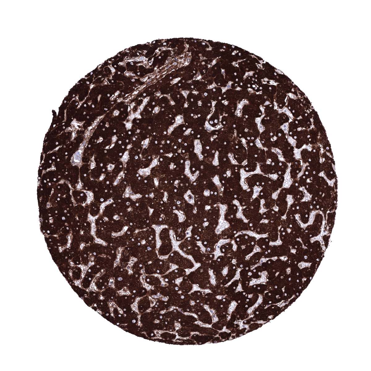
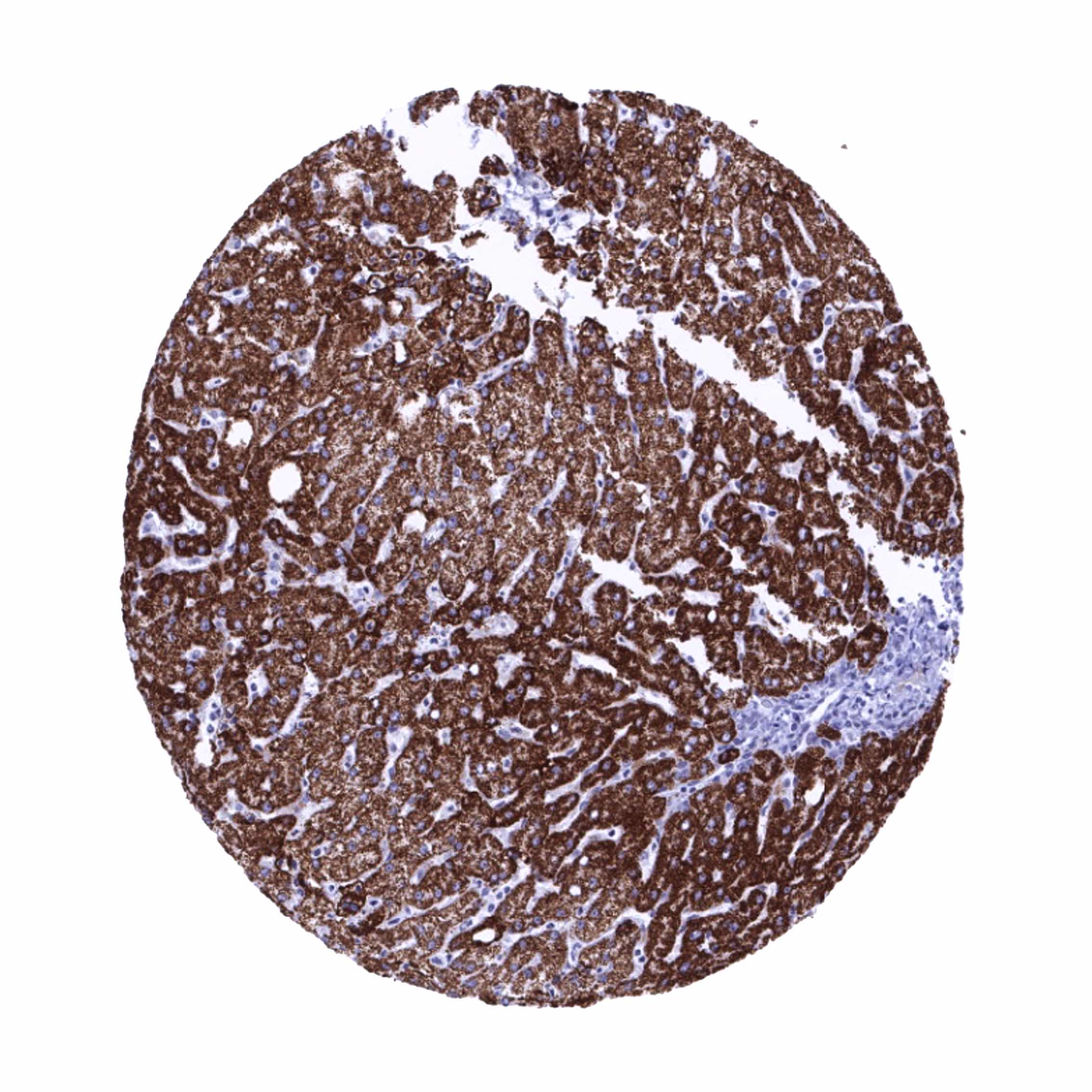
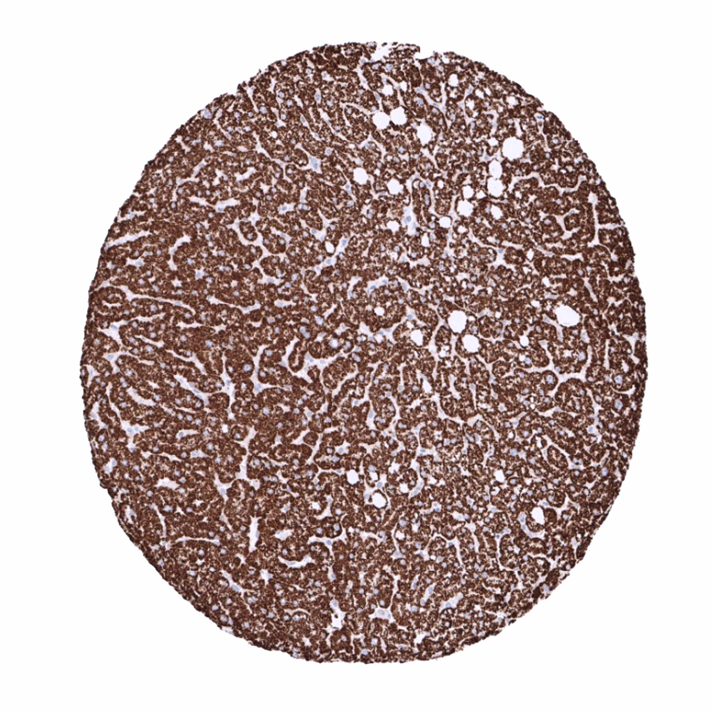
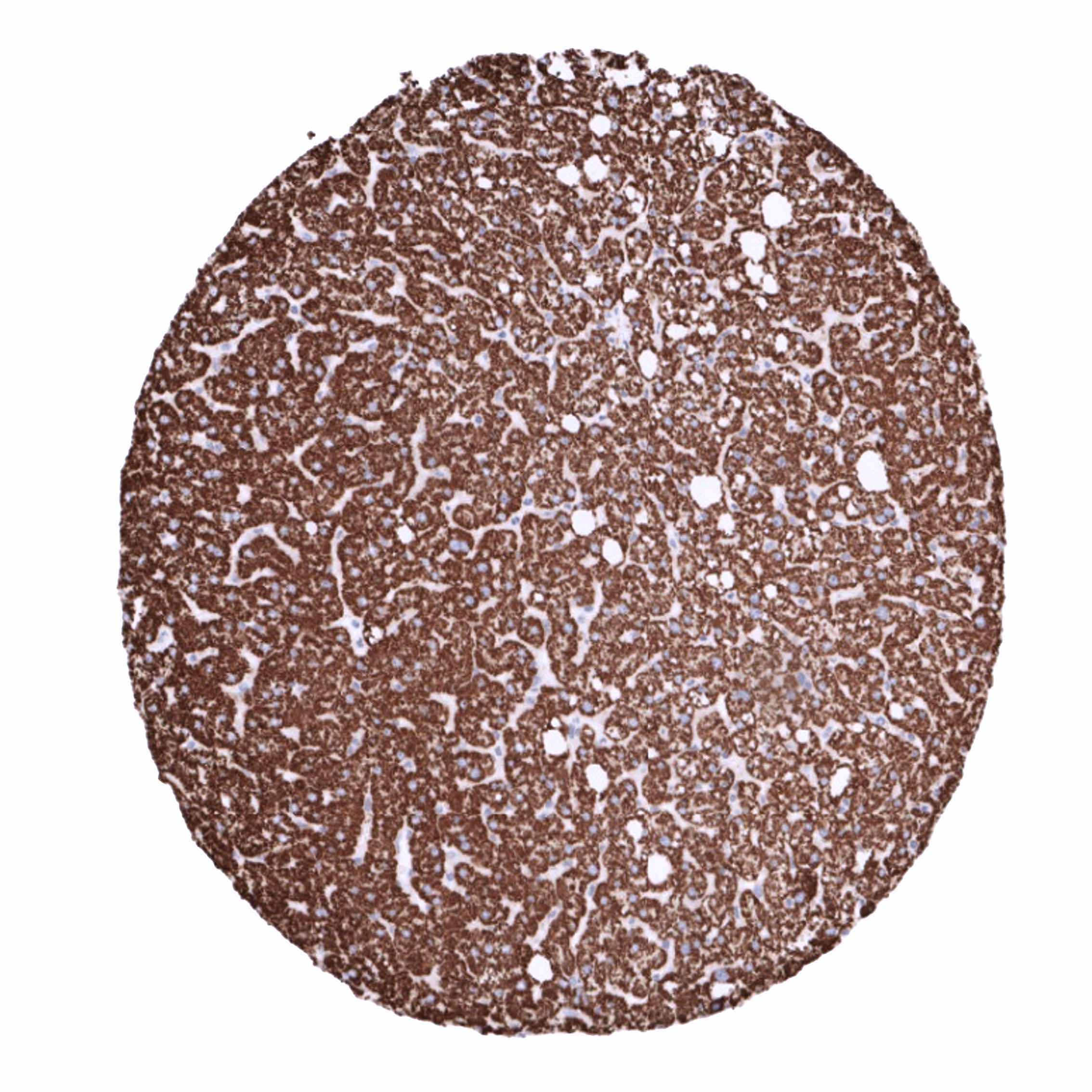
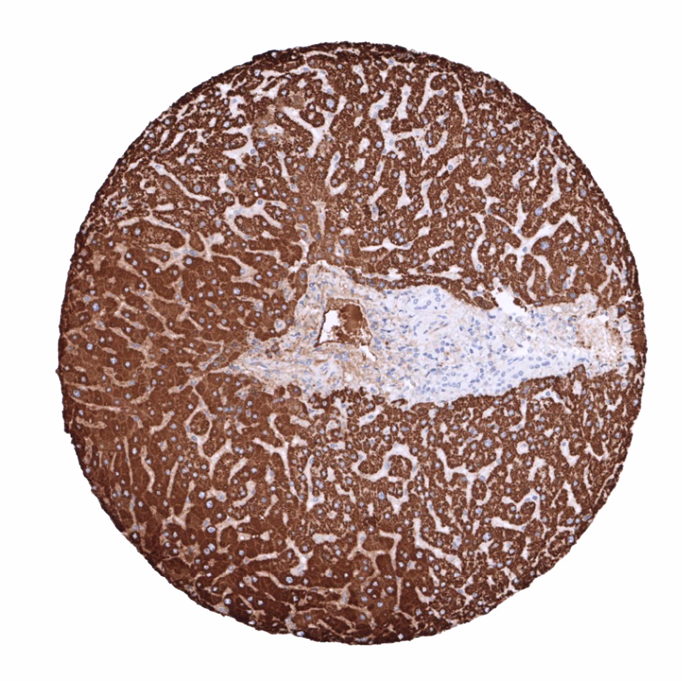
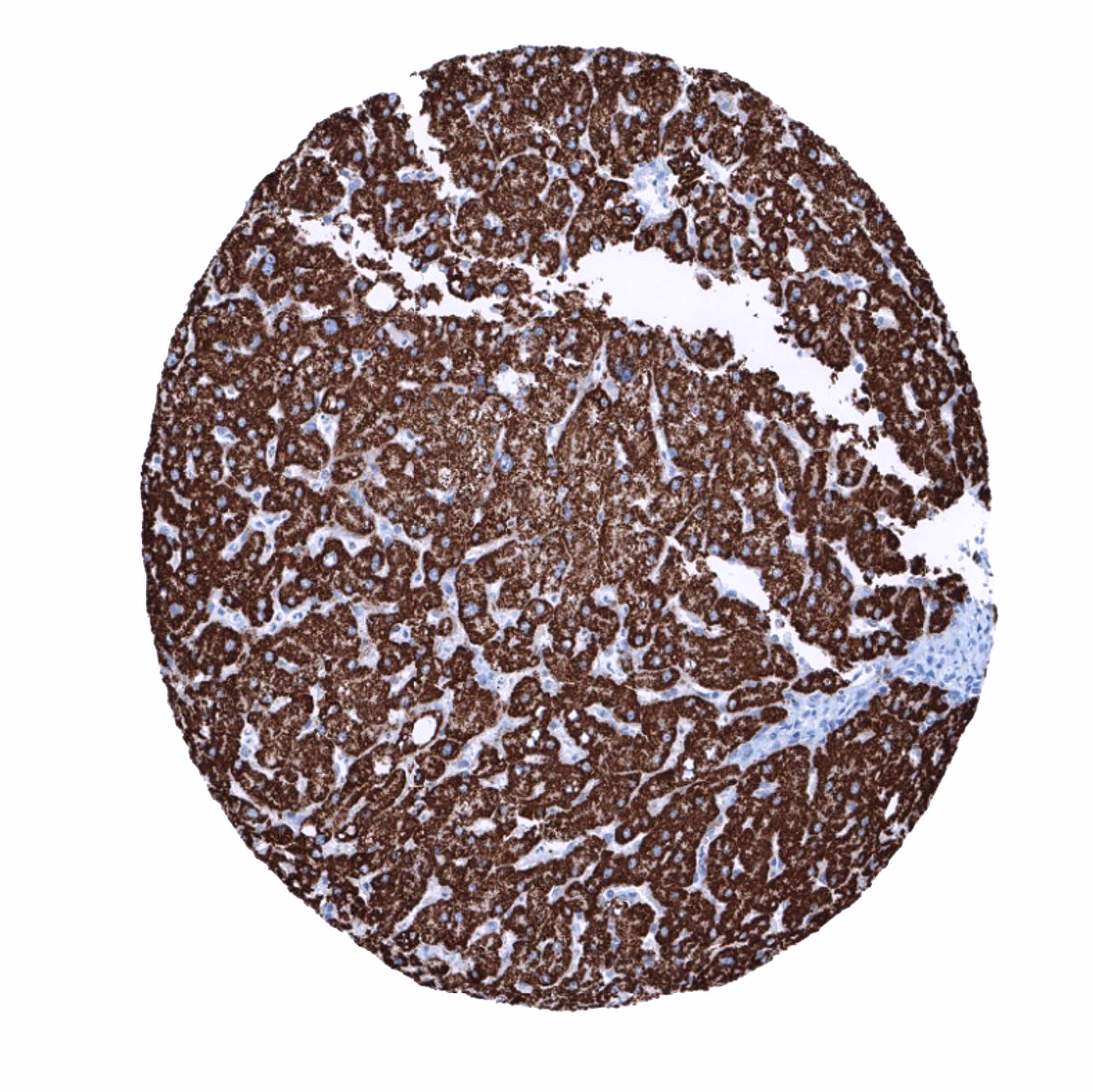
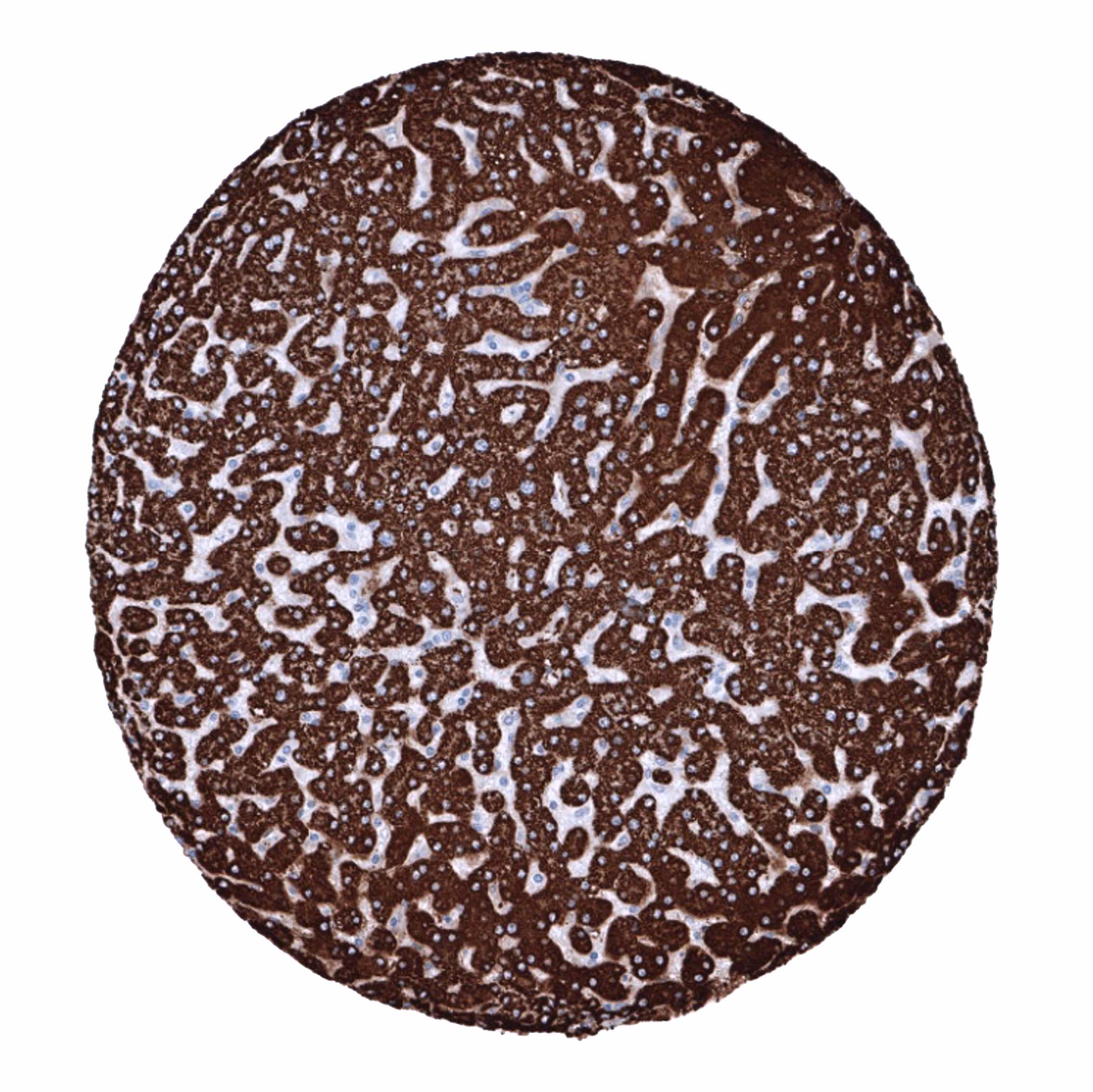
Positive control = Liver: Virtually all hepatocytes should show an at least moderate granular cytoplasmic staining.
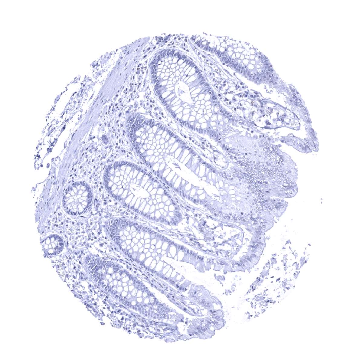
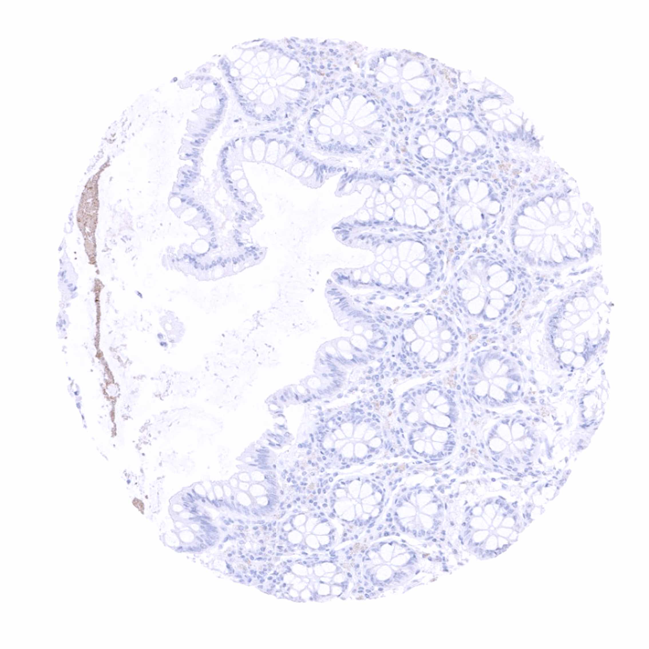
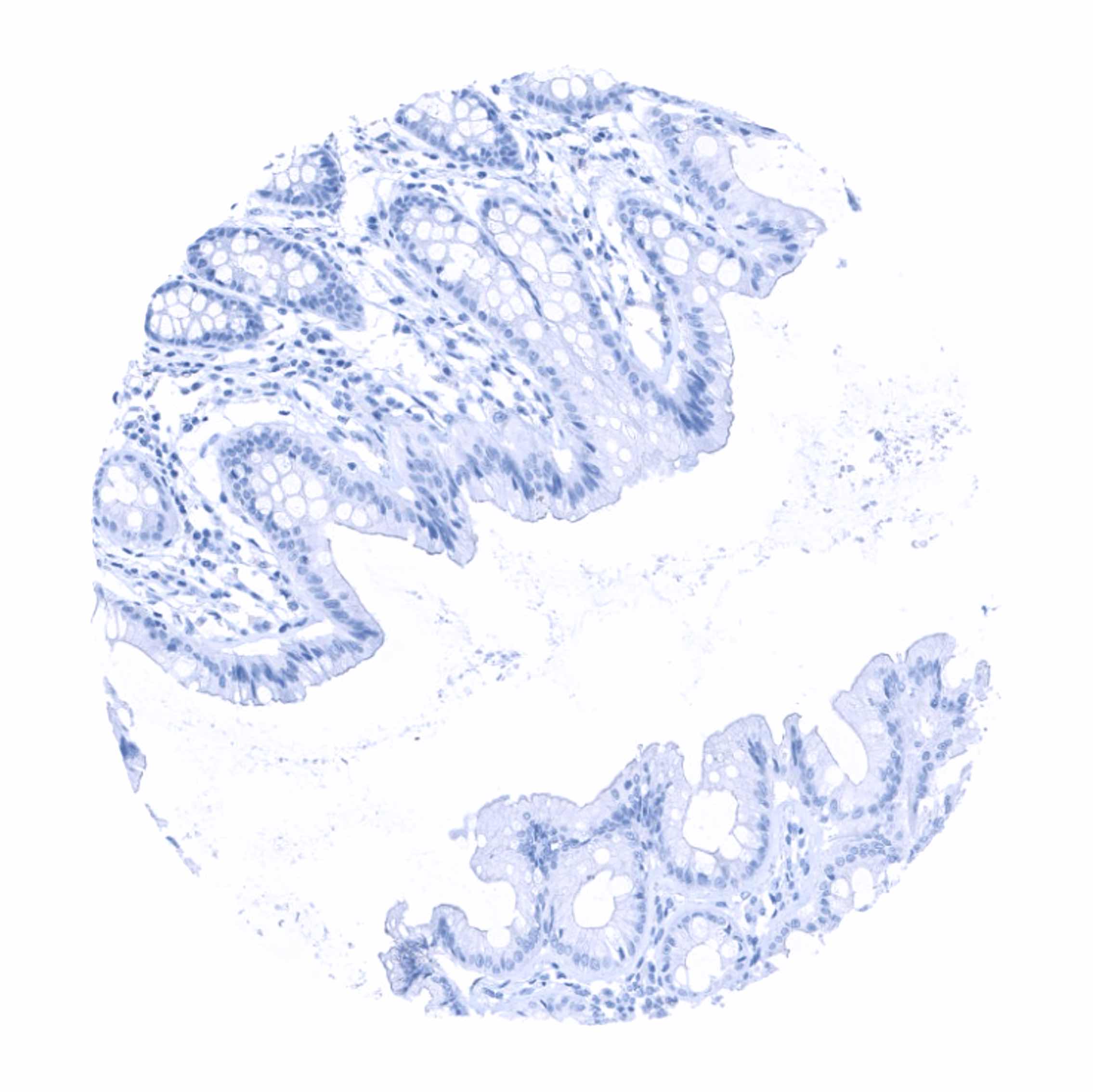
Negative control = Tonsil: No staining should be seen.
Cellular localization = Finely granular cytoplasmic
Reactivity = Human
Application = Immunohistochemistry
Dilution = 1:100 – 1:200
Intended Use = Research Use Only
Relevance of Antibody
The antibody binds to an unspecified protein occurring in normal and neoplastic hepatocytes.
Biology Behind
The “hepatocyte specific” monoclonal antibody Hepatocyte Paraffin 1 (Hep-Par-1) was produced in mice using tissue from a failed allograft liver (Wennerberg et al, 1993). Although the target protein is unknown, the antibody has been established as a marker for normal and neoplastic hepatocytes. “Hepatocyte specific” binds to a mitochondrial membrane protein.
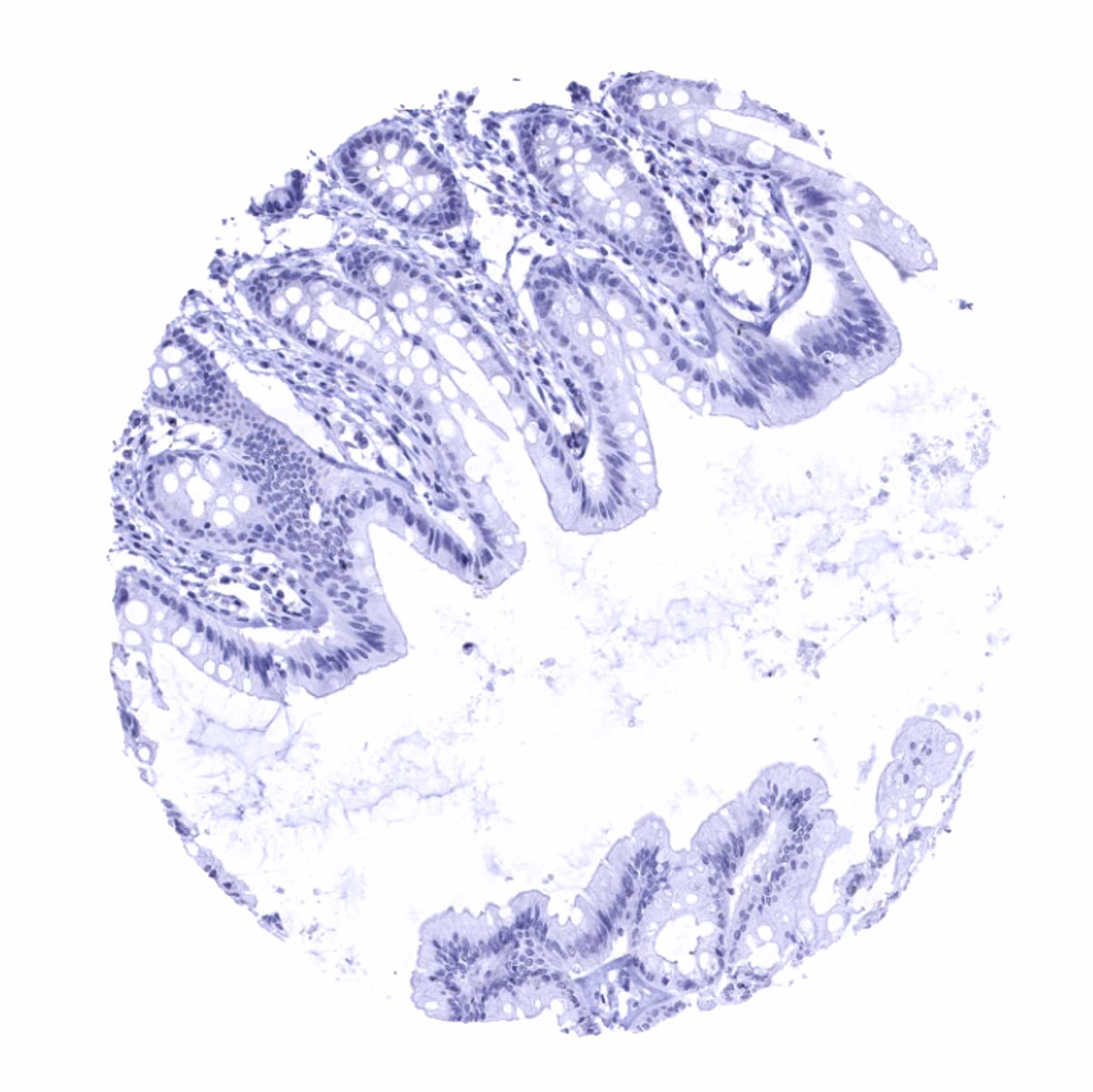
Staining Pattern in Normal Tissues
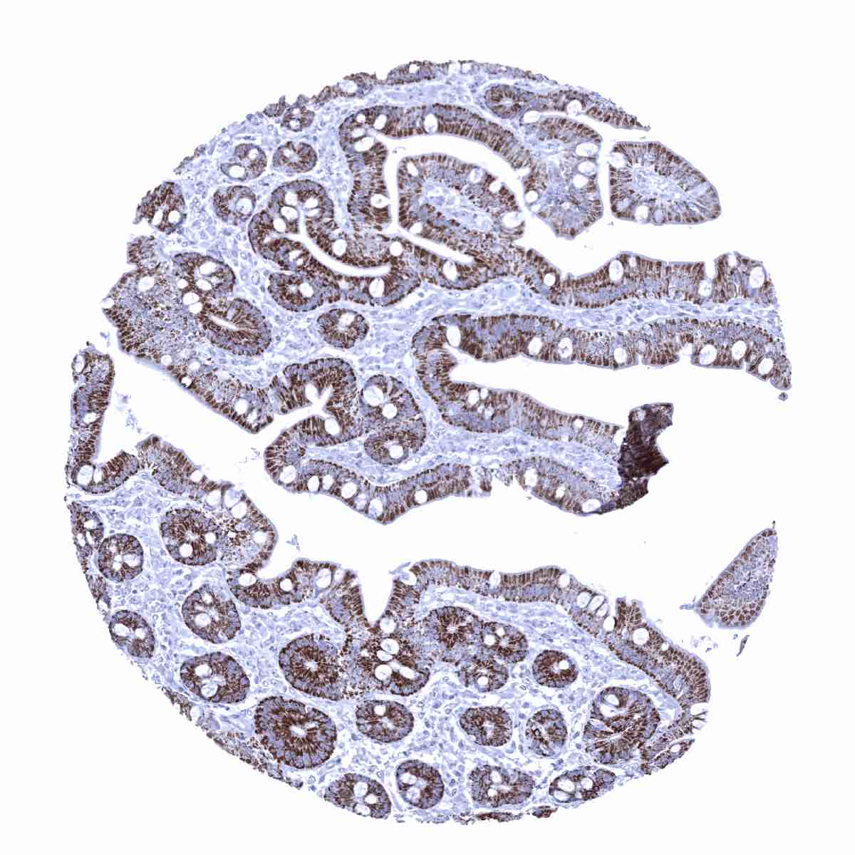
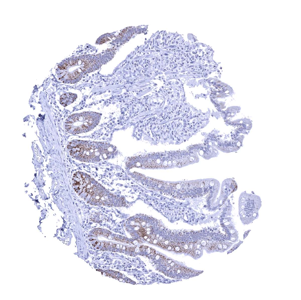
A strong “hepatocyte specific” immunostaining is seen in hepatocytes of the liver and to lower and variable extent in epithelial cells of small intestine (weak to strong). A faint “hepatocyte specific” staining can also occur in syncytiotrophoblast cells of the early placenta.
As the antigen is unknown, normal tissue findings cannot be compared with data from the protein atlas.
Positive control = Liver: Virtually all hepatocytes should show an at least moderate granular cytoplasmic staining.
Negative control = Tonsil: No staining should be seen.
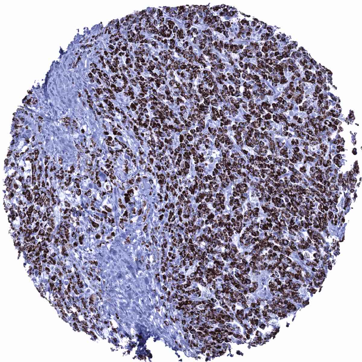
Staining Pattern in Relevant Tumor Types
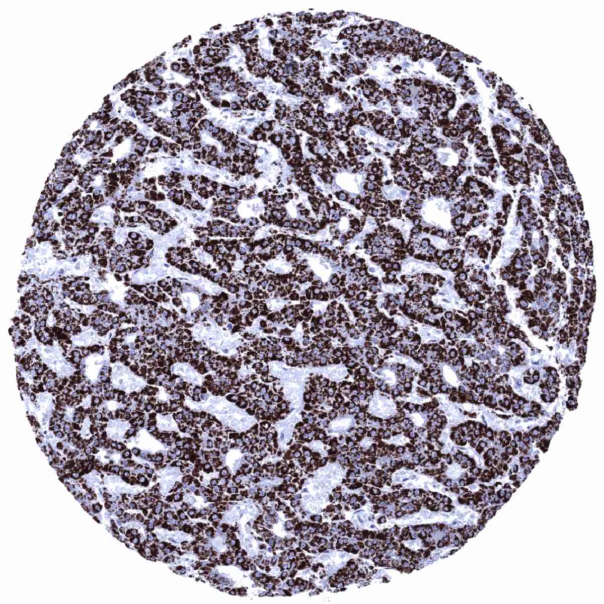
A positive “hepatocyte specific” immunostaining is usually seen in tumors derived from hepatocytes but it can also occur in other neoplasms, especially in case of hepatoid differentiation.
TCGA findings on “hepatocyte specific” RNA expression are not available as the target protein of the antibody is unknown.


Compatibility of Antibodies
No data available at the moment
Protocol Recommendations
IHC users have different preferences on how the stains should look like. Some prefer high staining intensity of the target stain and even accept some background. Others favor absolute specificity and lighter target stains. Factors that invariably lead to more intense staining include higher concentration of the antibody and visualization tools, longer incubation time, higher temperature during incubation, higher temperature and longer duration of the heat induced epitope retrieval (slide pretreatment). The impact of the pH during slide pretreatment has variable effects and depends on the antibody and the target protein.
All images and data shown here and in our image galleries are obtained by the manual protocol described below. Other protocols resulting in equivalent staining are described as well.
Manual protocol
Freshly cut sections should be used (less than 10 days between cutting and staining). Heat-induced antigen retrieval for 5 minutes in an autoclave at 121°C in pH 7,8 Target Retrieval Solution buffer. Apply MSVA-OCE5 at a dilution of 1:150 at 37°C for 60 minutes. Visualization of bound antibody by the EnVision Kit (Dako, Agilent) according to the manufacturer’s directions.

Agilent / Dako – Autostainer Link 48
Pretreatment in PT-Link for 30 minutes at 95°C (pH high); FLEX peroxidase blocking for 5 minutes (room temperature), MSVA-OCE5 1:150 for 20 minutes (room temperature), FLEX+ mouse/rabbit (LINKER) for 15 minutes (room temperature), horseradish peroxidase (HRP) for 20 minutes (room temperature), FLEX DAB+Sub-Chromo for 10 minutes (room temperature), FLEX hematoxylin for 5 minutes (room temperature).

These images reflect stainings by the protocol described above. It is of note that a comparable staining result can also be obtained by different protocols. In general, a longer pretreatment, a longer incubation time of the primary antibody, a higher antibody concentration, and a longer incubation time of FLEX+LINKER result in stronger staining, potentially at the cost of more background staining. Modifications of the protocol with a strengthening effect on staining intensity in combination with changes of other parameters that result in lower staining intensity can result in a comparable result as shown above.
Leica – BOND RX
Dewax at 72°C for 30 seconds; Pretreatment in Bond Epitope Retrieval Solution (ER2 – EDTA pH9) for 20 minutes at 100°C; Peroxidase blocking for 5 minutes (room temperature), MSVA-OCE5 1:225 for 15 minutes (room temperature), Post primary (rabbit anti mouse) for 8 minutes (room temperature), Polymer (goat anti rabbit) for 8 minutes (room temperature), mixed DAB refine for 10 minutes (room temperature), hematoxylin for 5 minutes (room temperature).

These images reflect stainings by the protocol described above. It is of note that a comparable staining result can also be obtained by different protocols. In general, a longer pretreatment, a longer incubation time of the primary antibody, a higher antibody concentration, a higher temperature during incubation, and a longer incubation time of Post primary and or the Polymer result in stronger staining, potentially at the cost of more background staining. Modifications of the protocol with a strengthening effect on staining intensity in combination with changes of other parameters that result in lower staining intensity can result in a comparable result as shown above.
Roche – Ventana Discovery ULTRA
Pretreatment for 64 minutes at 100°C (pH 8,4); CM peroxidase blocking for 12 minutes (room temperature), MSVA-OCE5 1:150 for 20 minutes at 36°C, secondary antibody (anti-mouse HQ) for 12 minutes at 36°C, anti-HQ HRP for 12 minutes at room temperature, DAB at room temperature, hematoxylin II at room temperature for 8 minutes, bluing reagent at room temperature for 4 minutes.
These images depict staining results obtained by the protocol described above. It is of note, that the Ventana machines generally require higher antibody concentrations than other commonly used autostainers because the antibodies are automatically diluted during the procedure. Various other protocols can result in an identical result as shown above. A longer pretreatment, a longer incubation time of the primary antibody, a higher antibody concentration, a higher temperature during incubation, and a longer incubation time of secondary antibody and or the anti-HQ HRP result in stronger staining, potentially at the cost of more background staining.
Potential Research Applications
- A comprehensive study analyzing “hepatocyte specific” in various different tumor entities would be helpful to better assess the diagnostic impact of Hep-Par-1 staining.
