
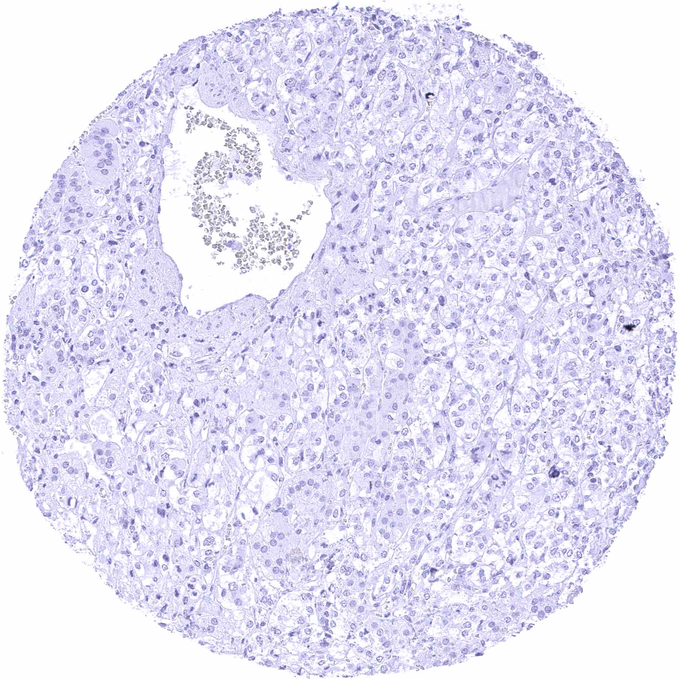
Adrenal gland
Aorta, endothelium
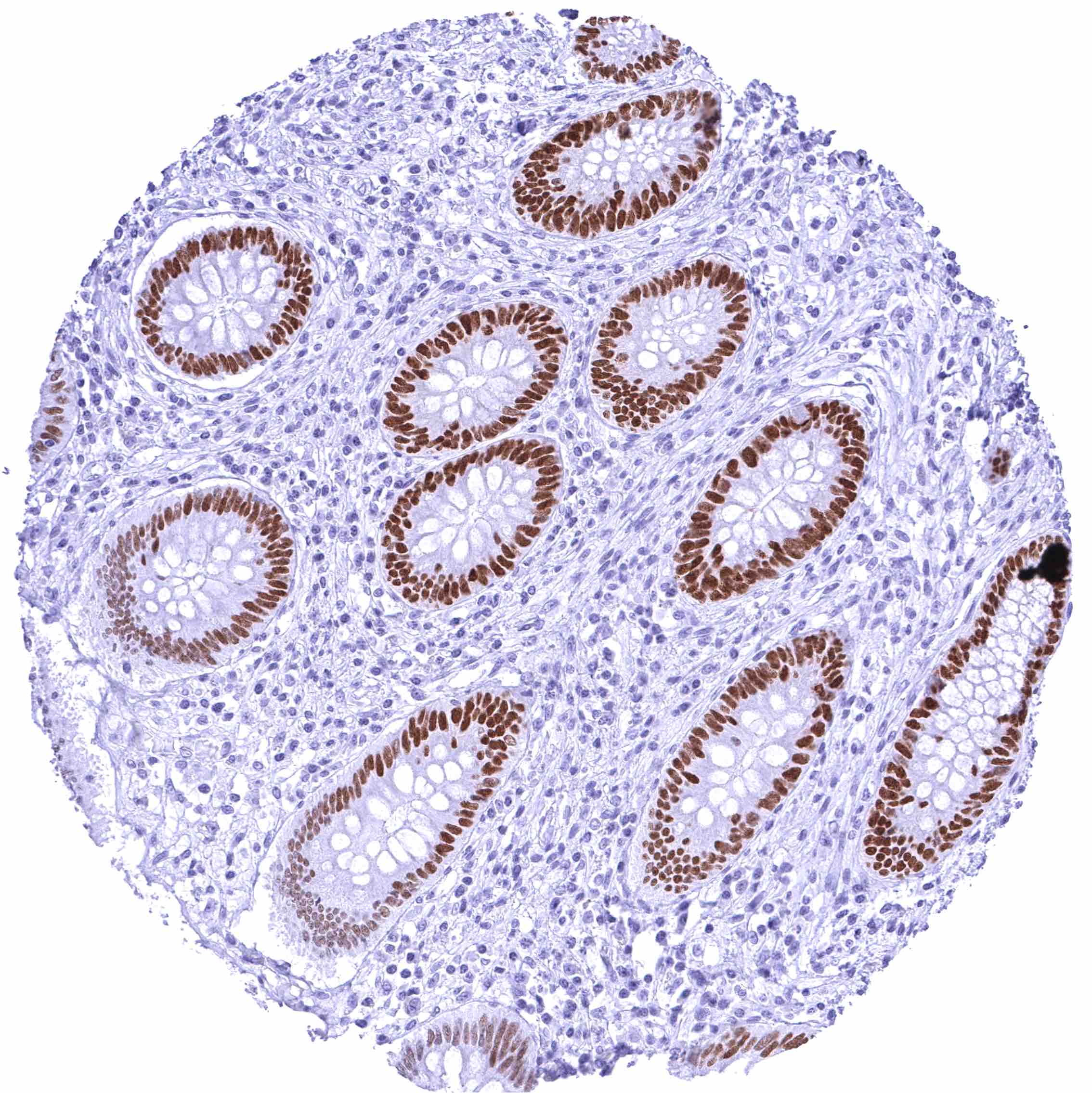
Appendix, mucosa – Strong nuclear FOXA1 staining of epithelial cells in the crypts. The staining intensity gradually decreases towards the surface epithelium.
Appendix, muscular wall
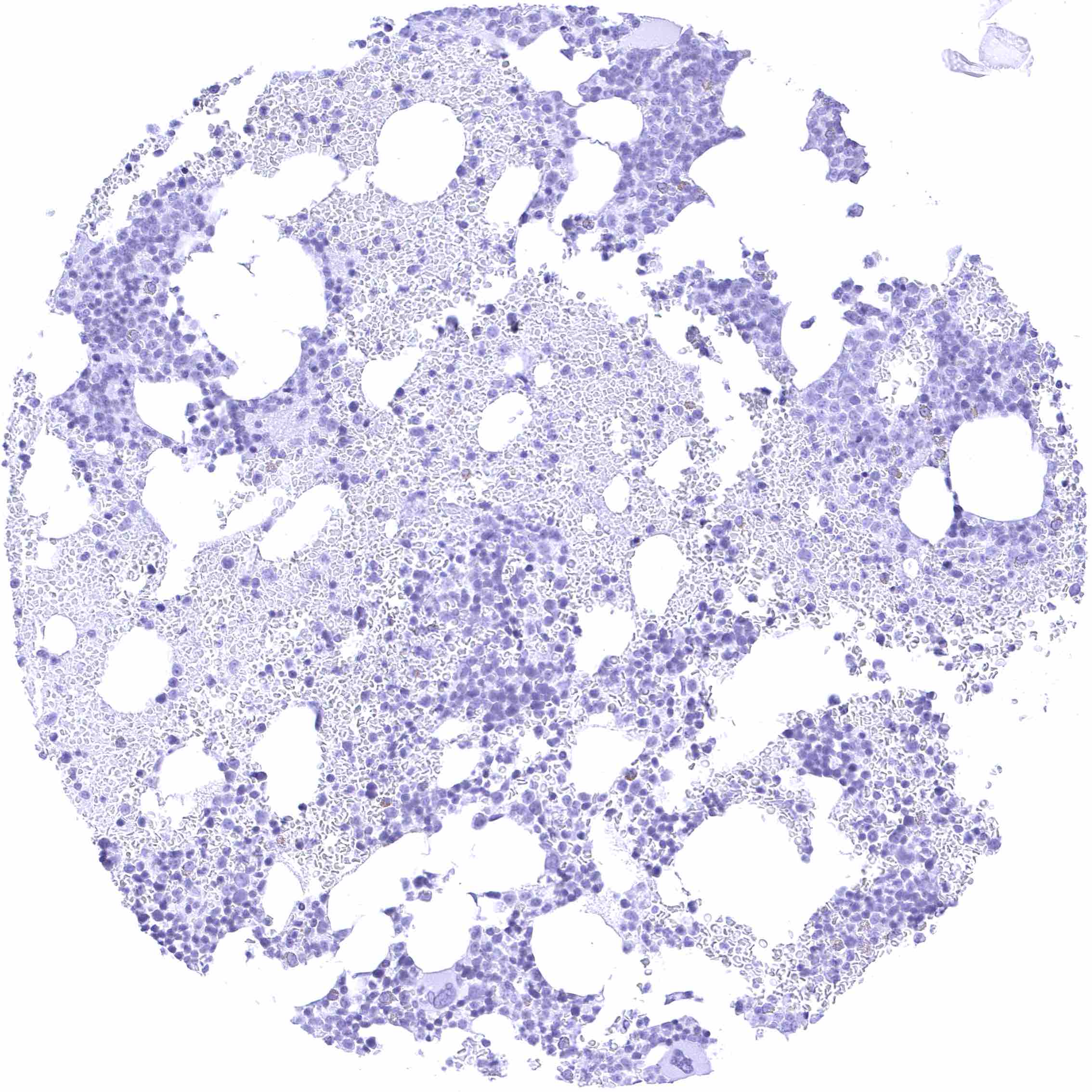
Bone marrow
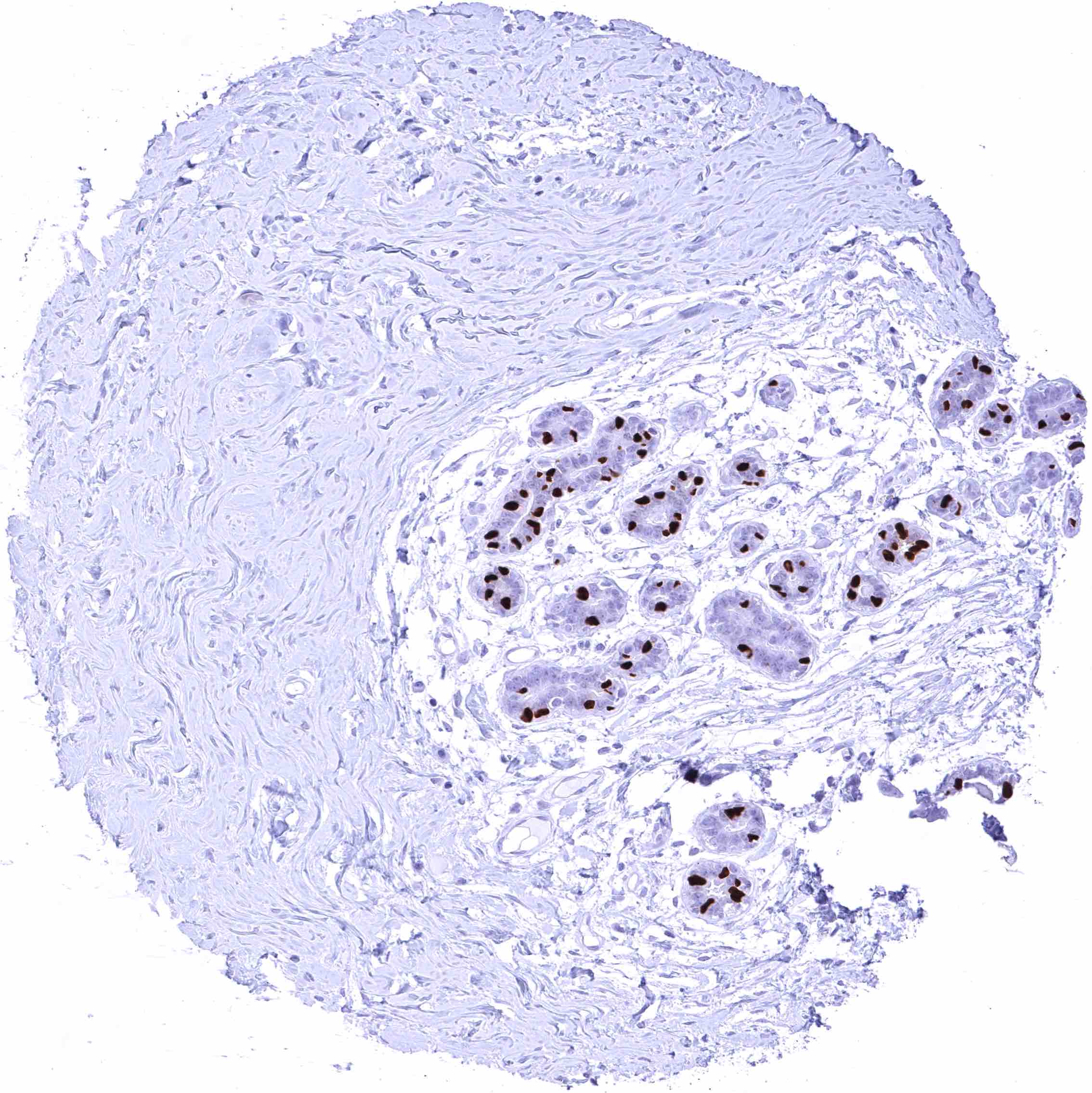
Breast – Intense nuclear FOXA1 staining of a subset of luminal epithelial cells.
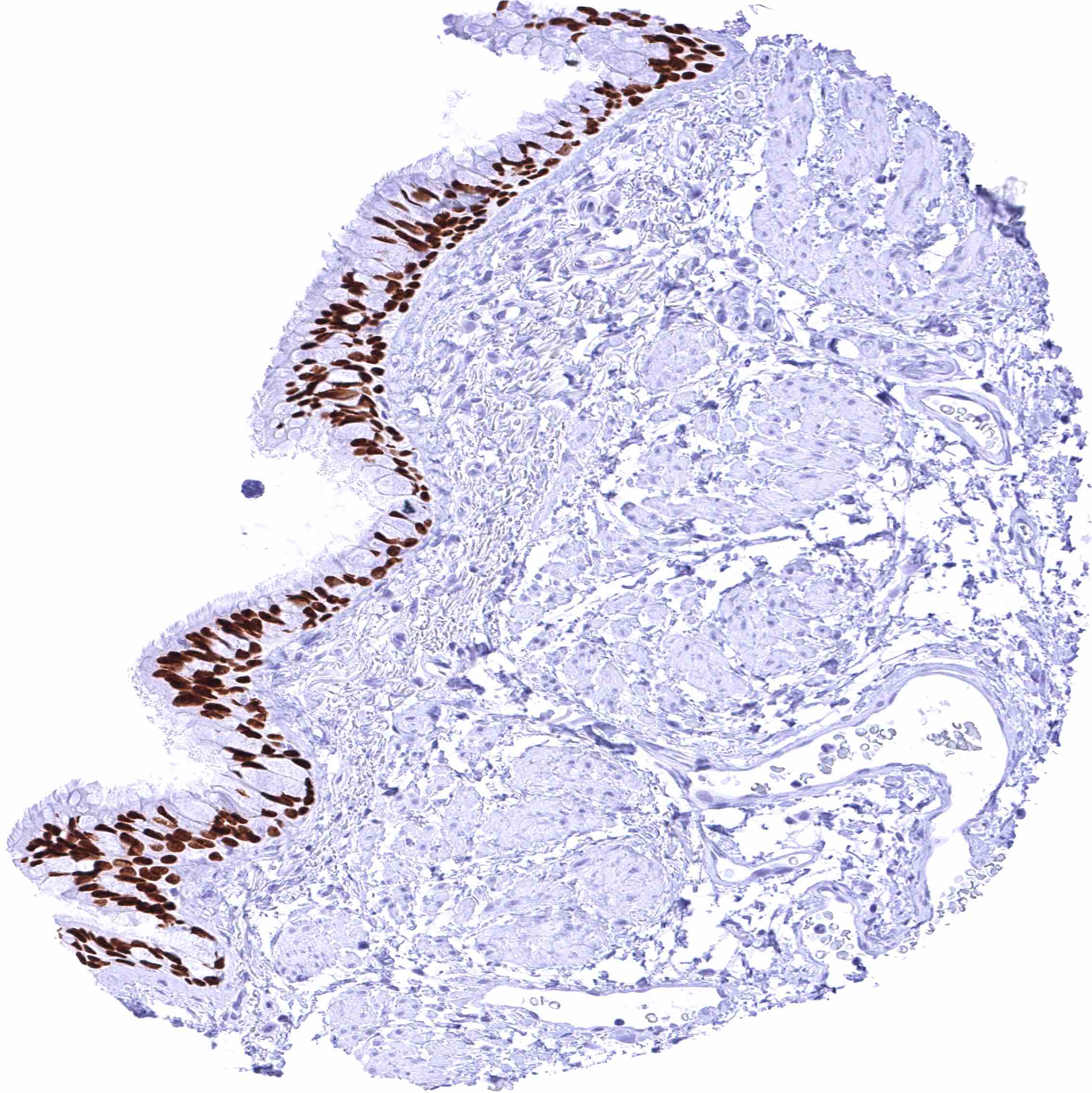
Bronchus, mucosa – Intense nuclear FOXA1 staining of all epithelial cells of the respiratory epithelium.
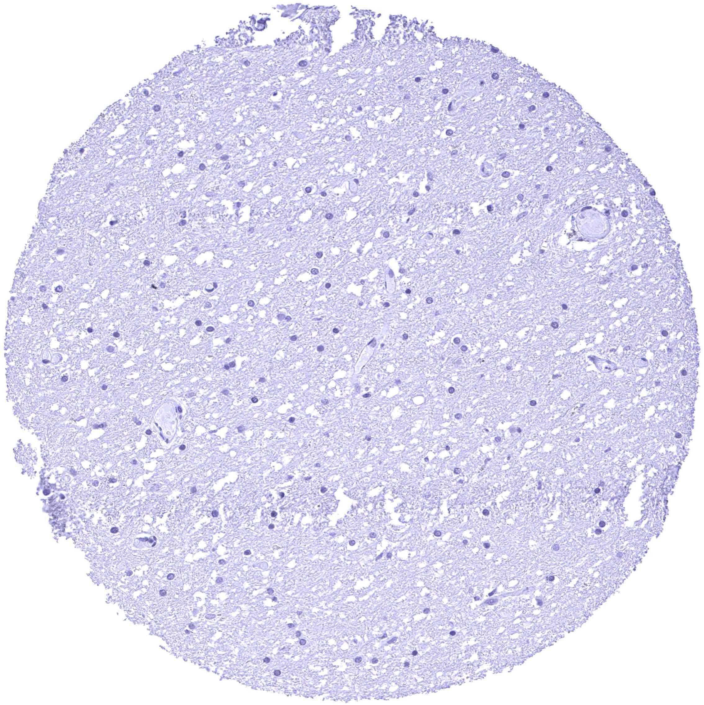
Cerebellum (white matter)
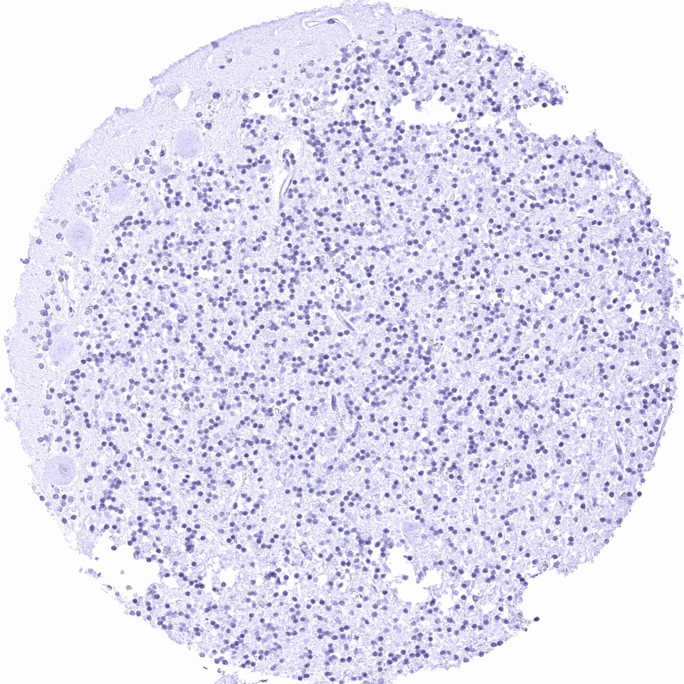
Cerebellum, cortex (grey)
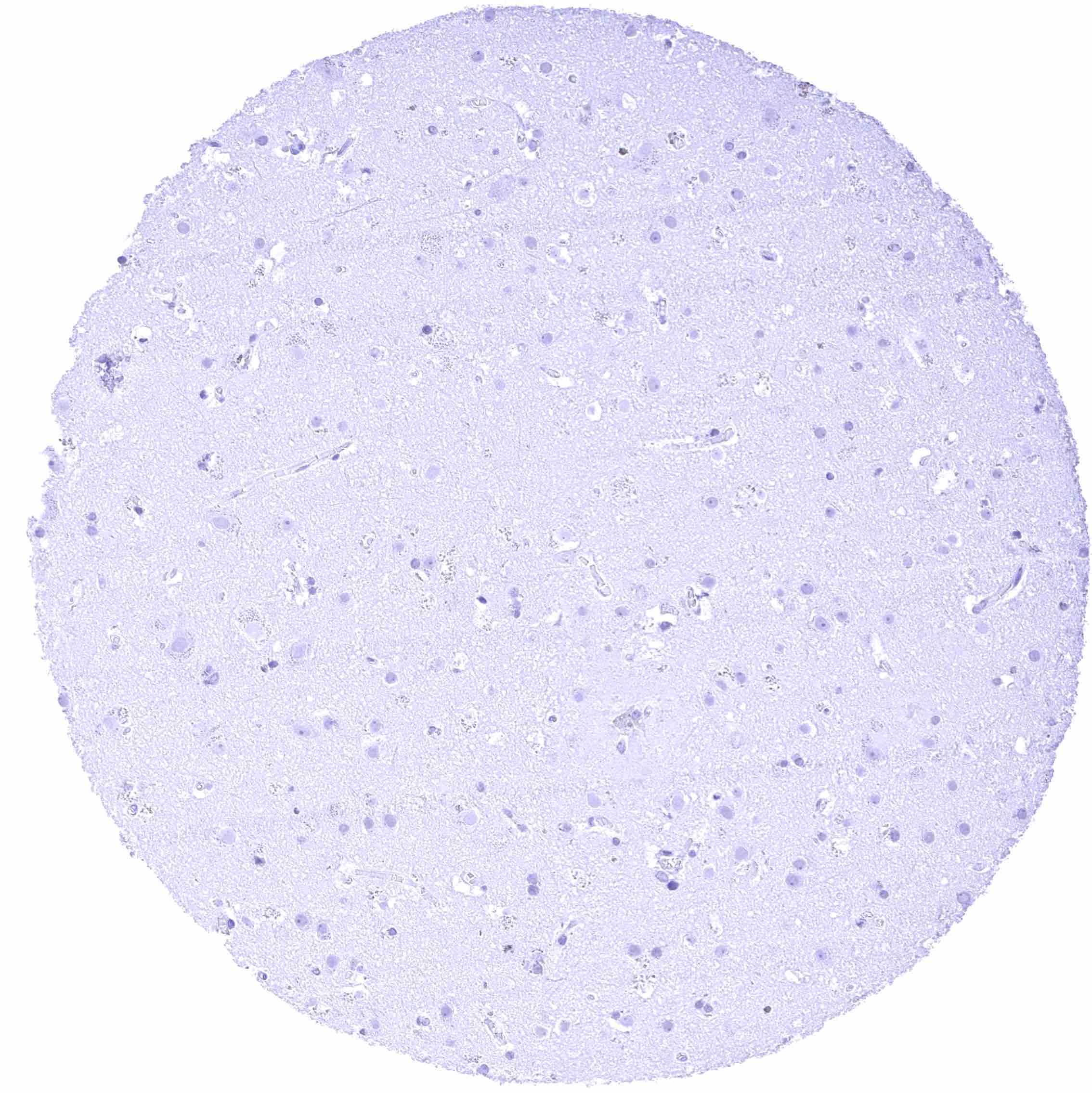
Cerebrum (grey matter)
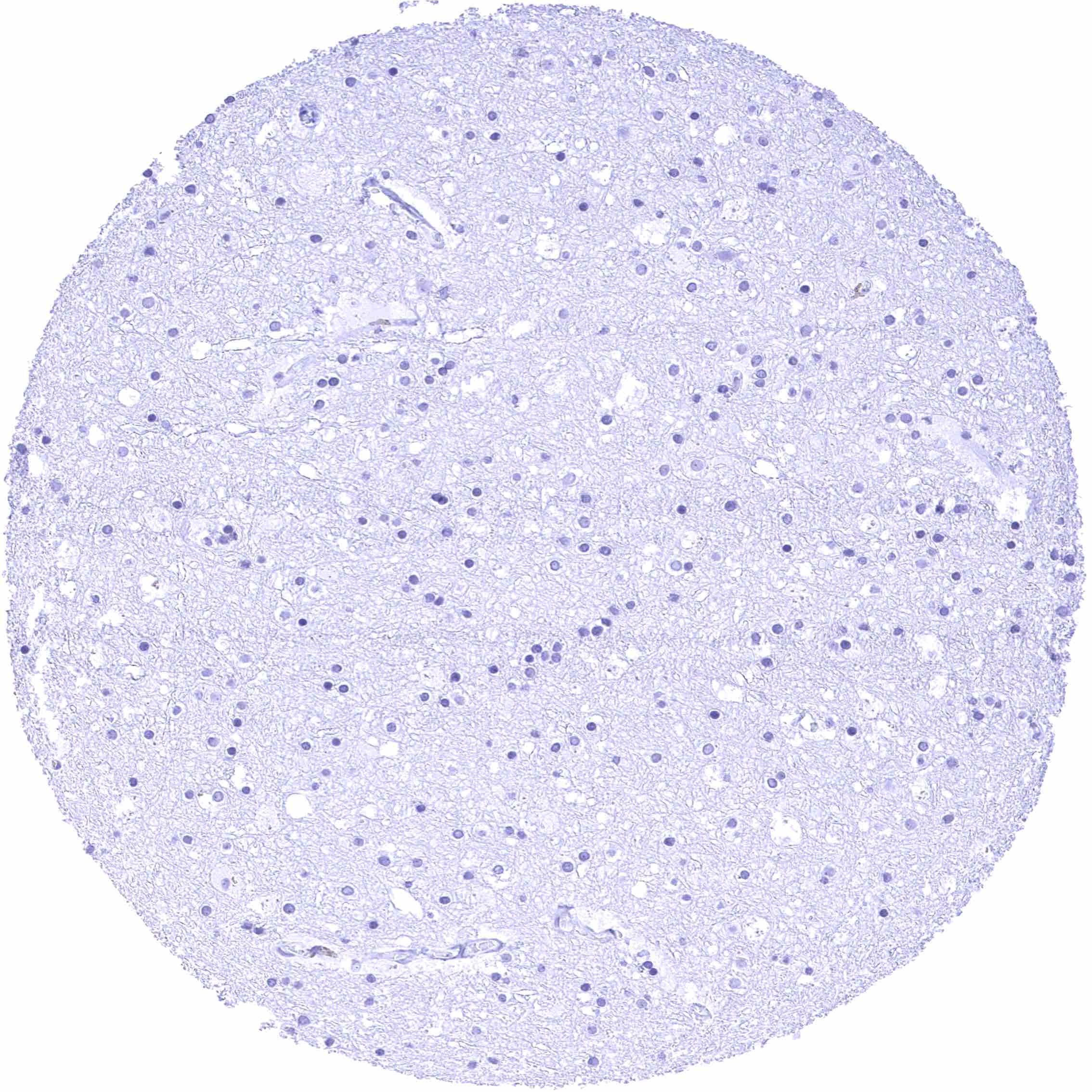
Cerebrum (white matter)
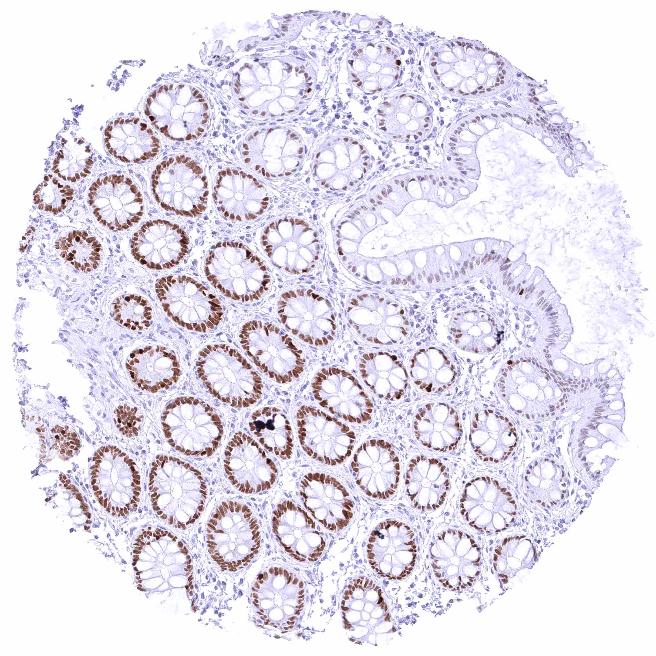
Colon descendens, mucosa
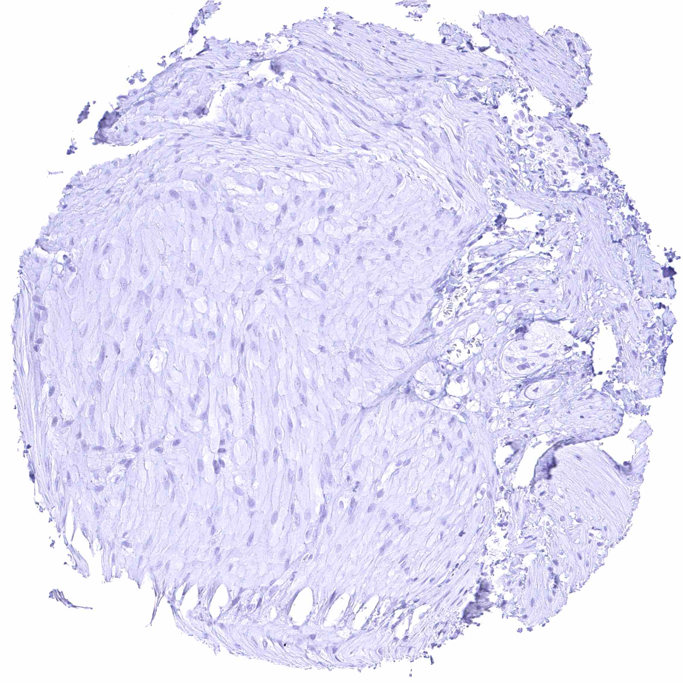
Colon descendens, muscular wall
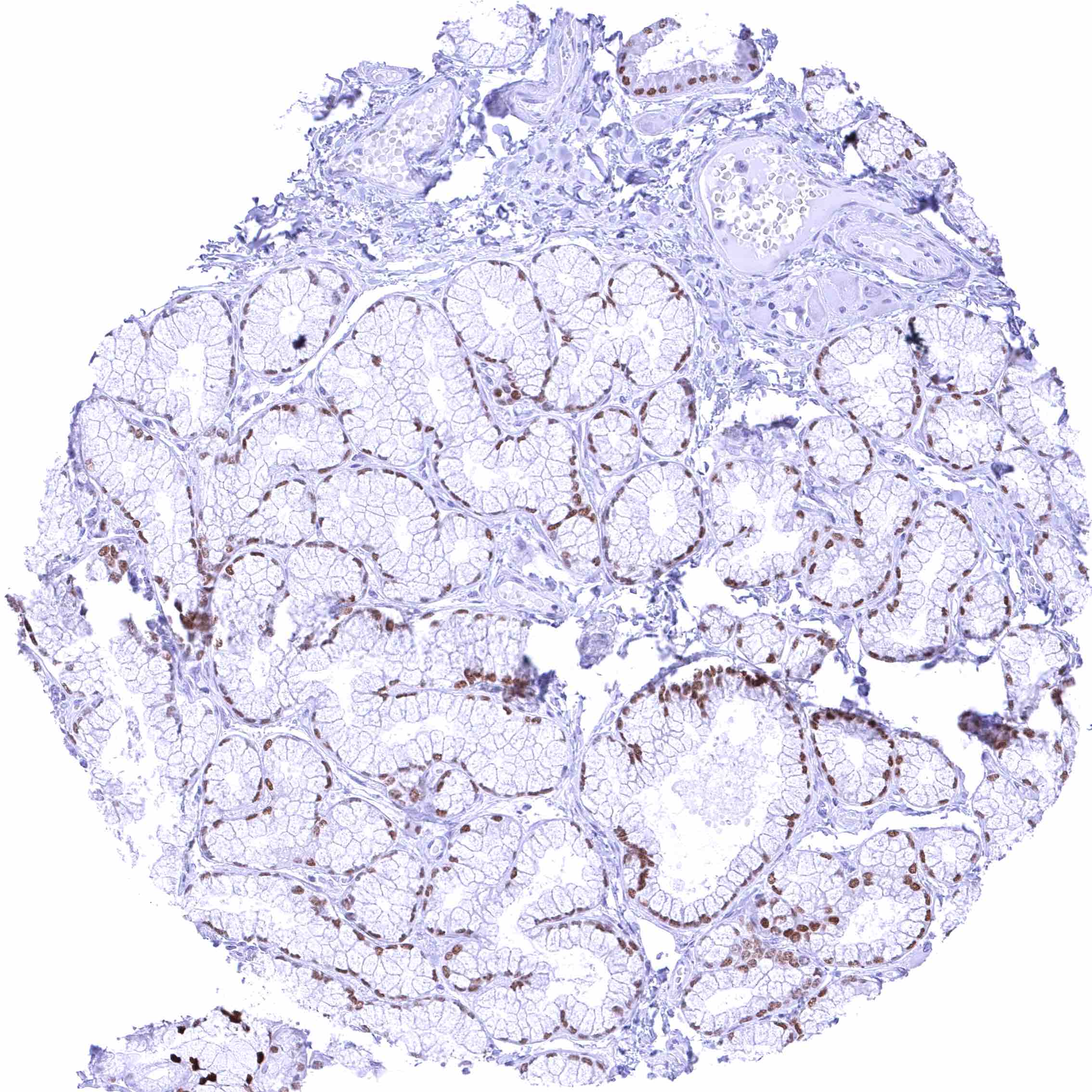
Duodenum, Brunner gland – Distinct nuclear FOXA1 staining of mucinous glandular cells of Brunner glands.
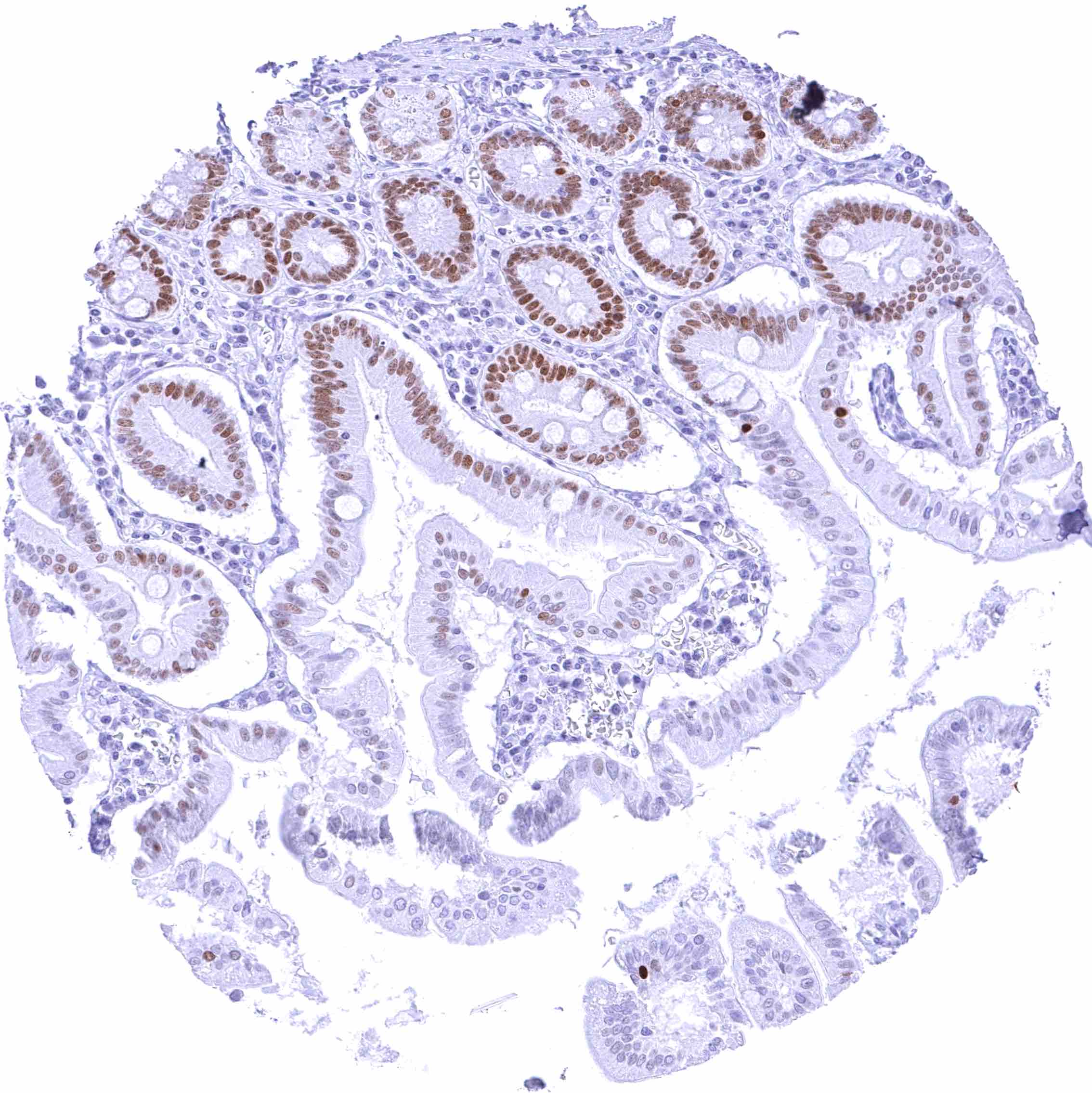
Duodenum, mucosa – Moderate to strong nuclear FOXA1 staining of crypt epithelial cells. The staining intensity gradually decreases towards the surface epithelium and the peaks of the villi
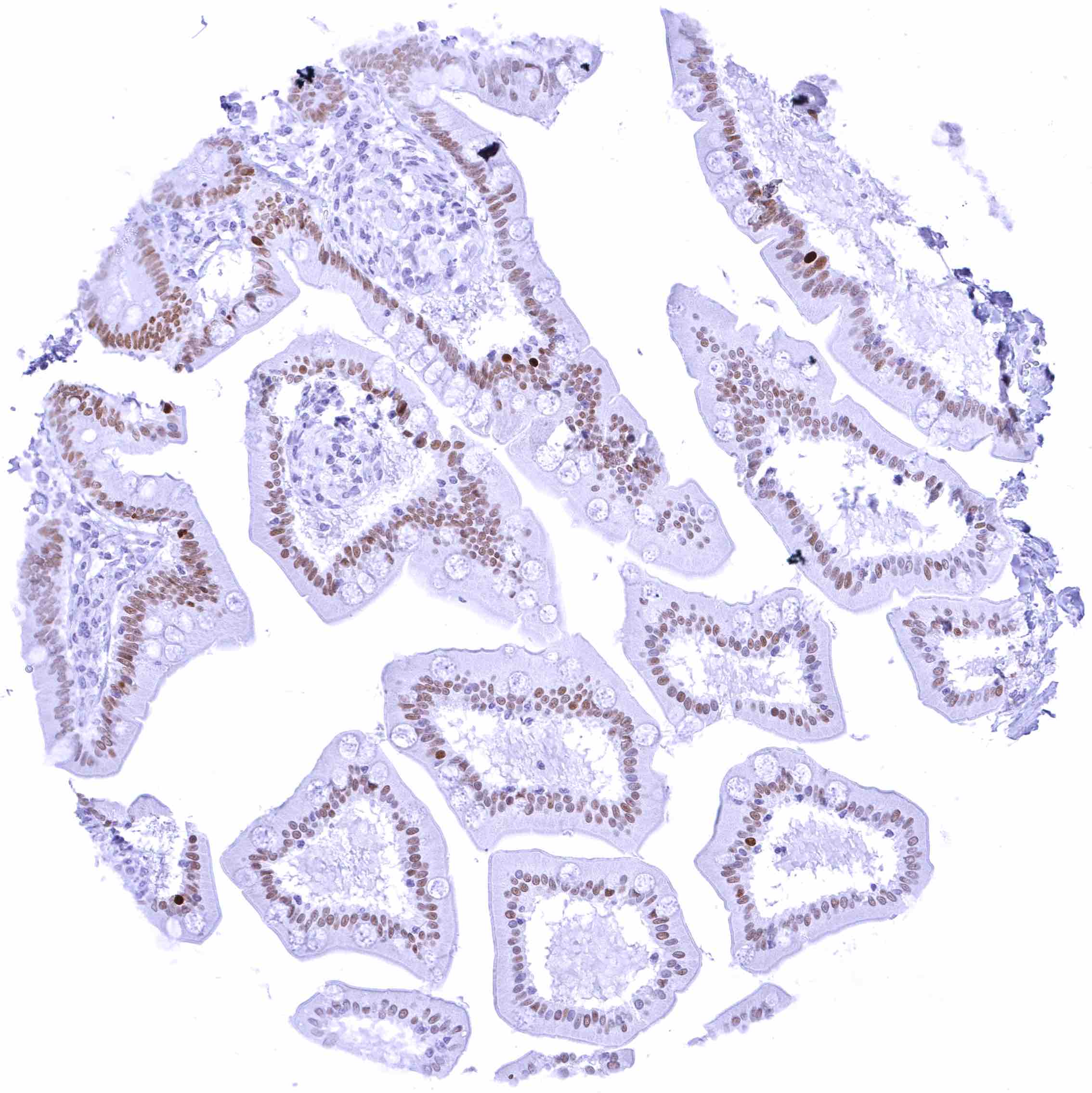
Duodenum, mucosa
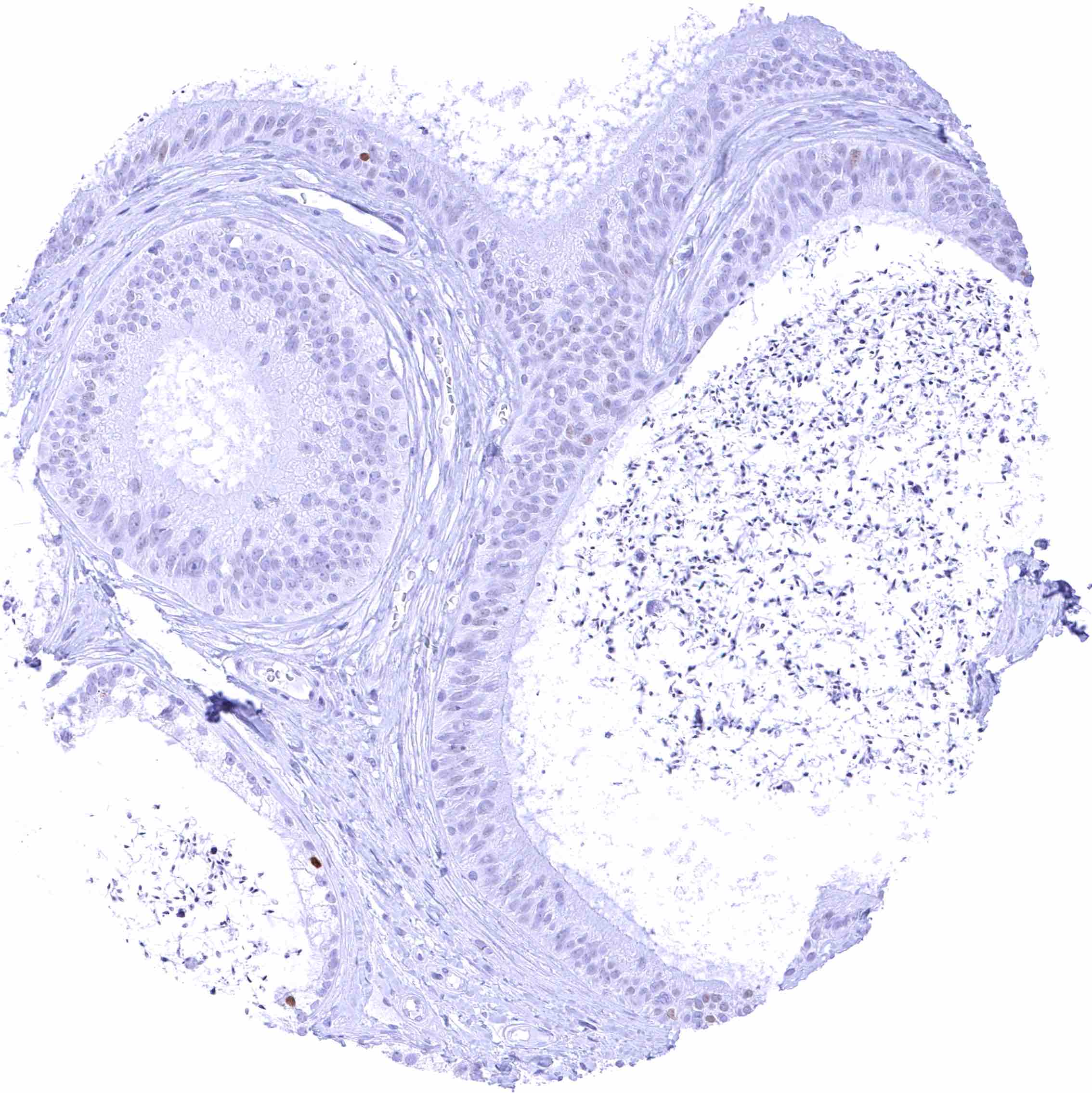
Epididymis (Caput)
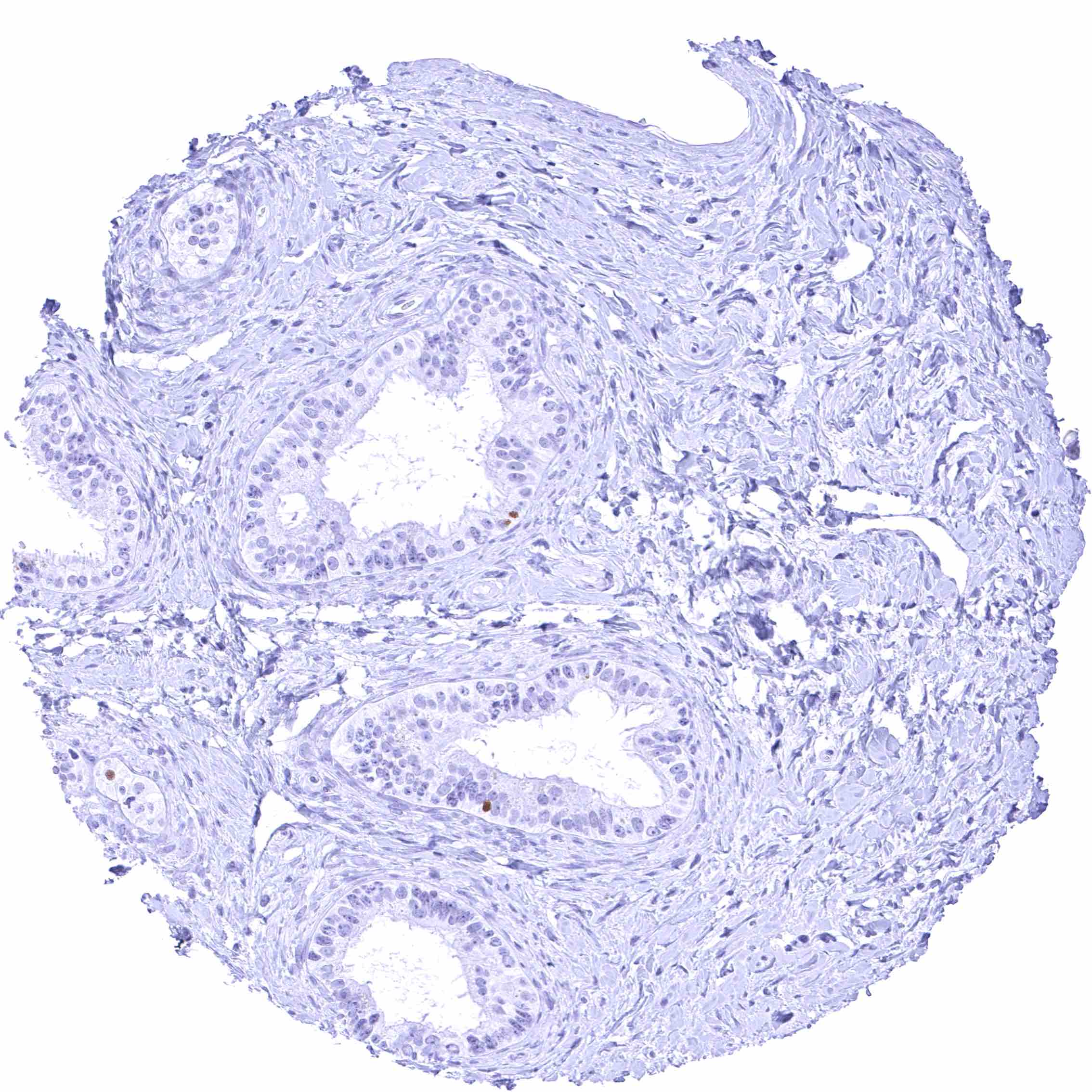
Epididymis (Cauda)
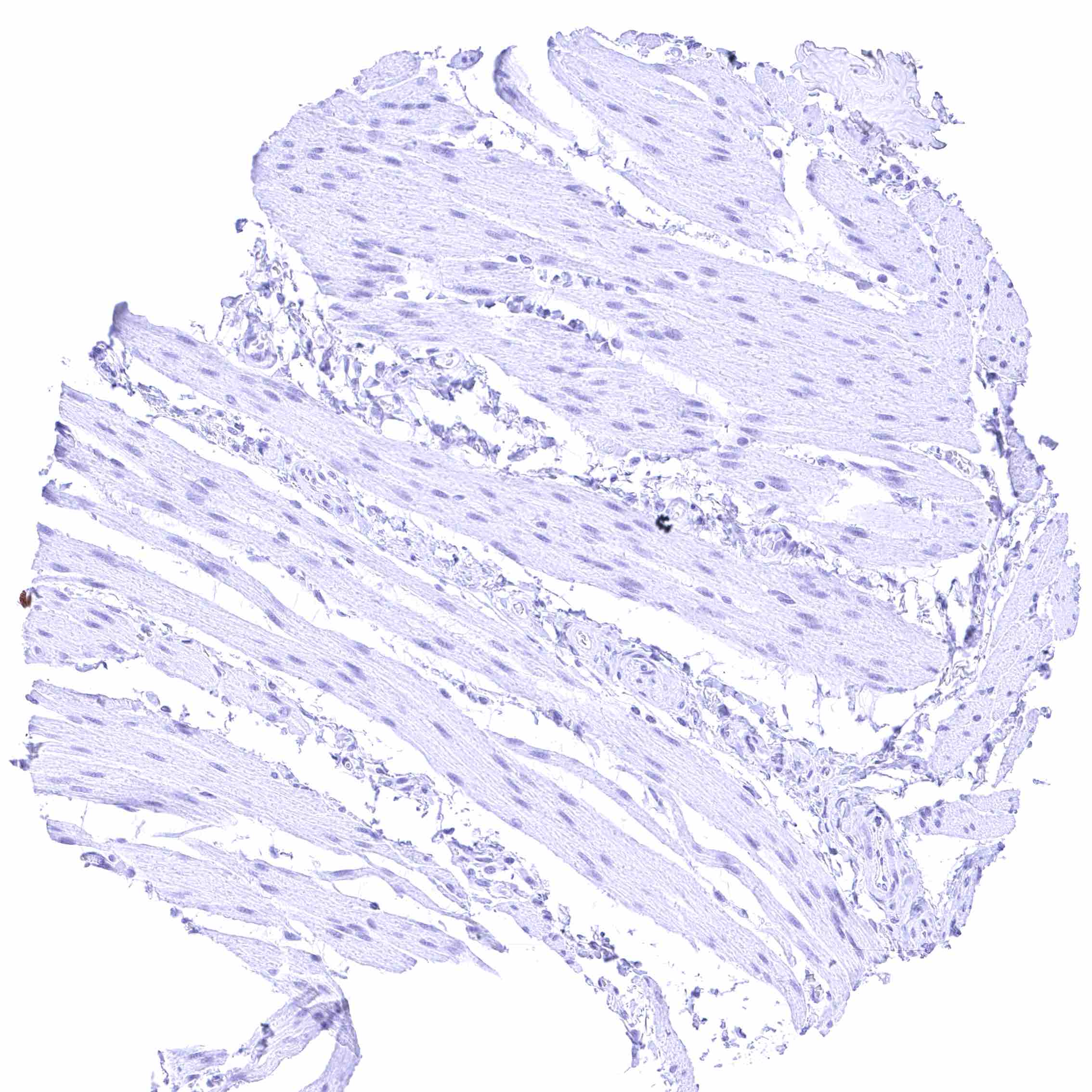
Esophagus, muscular wall
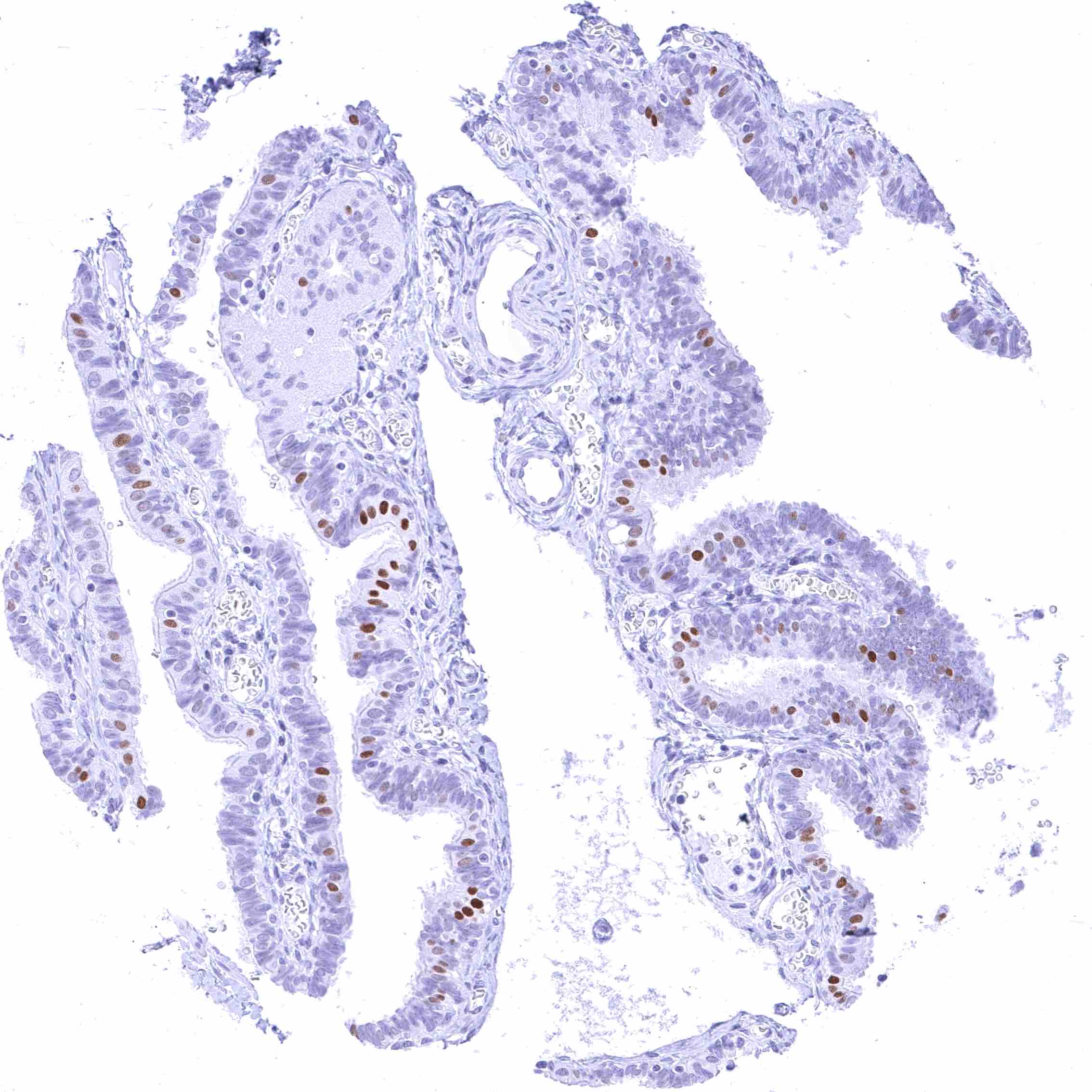
Fallopian tube, mucosa – Distinct nuclear FOXA1 staining of a subset of epithelial cells
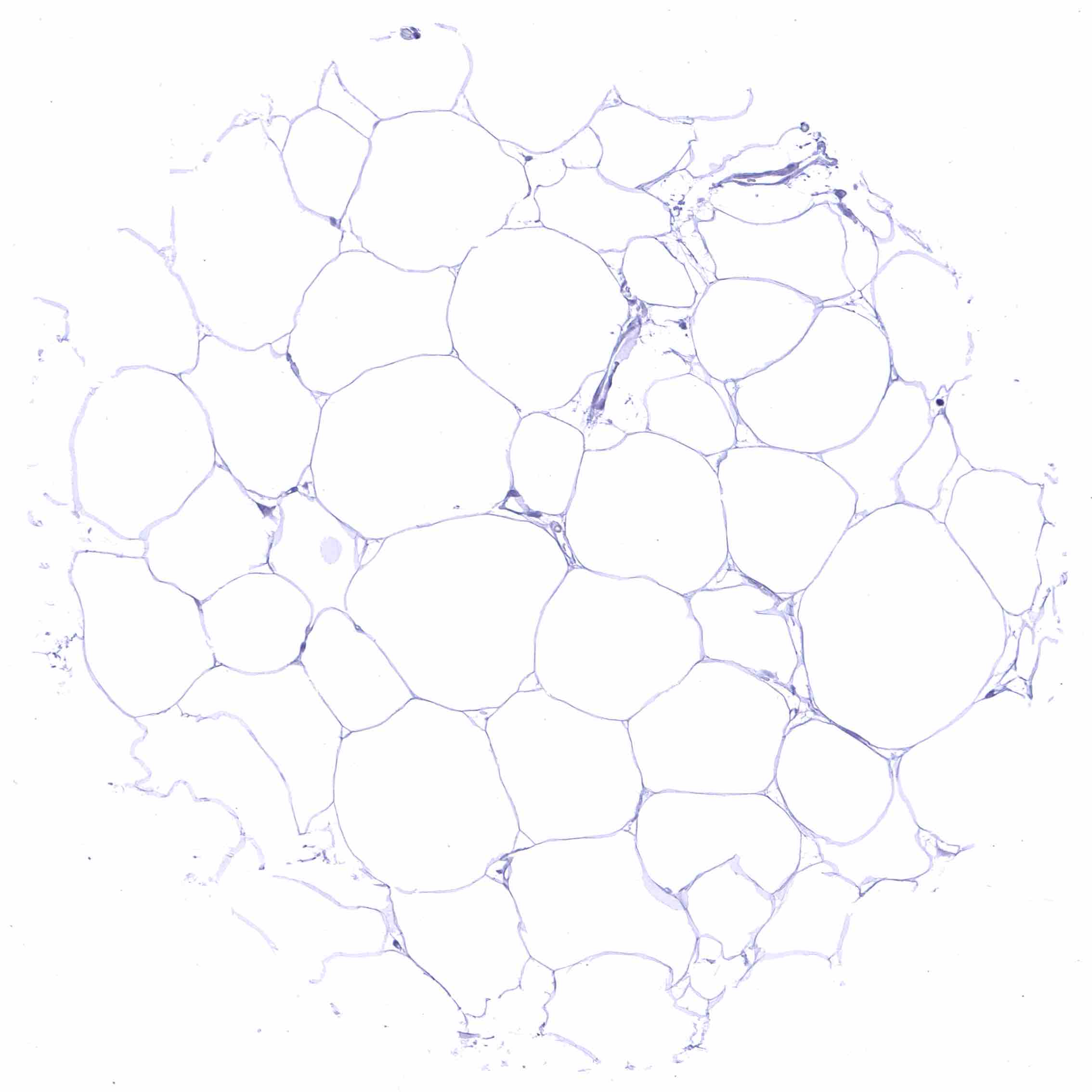
Fat
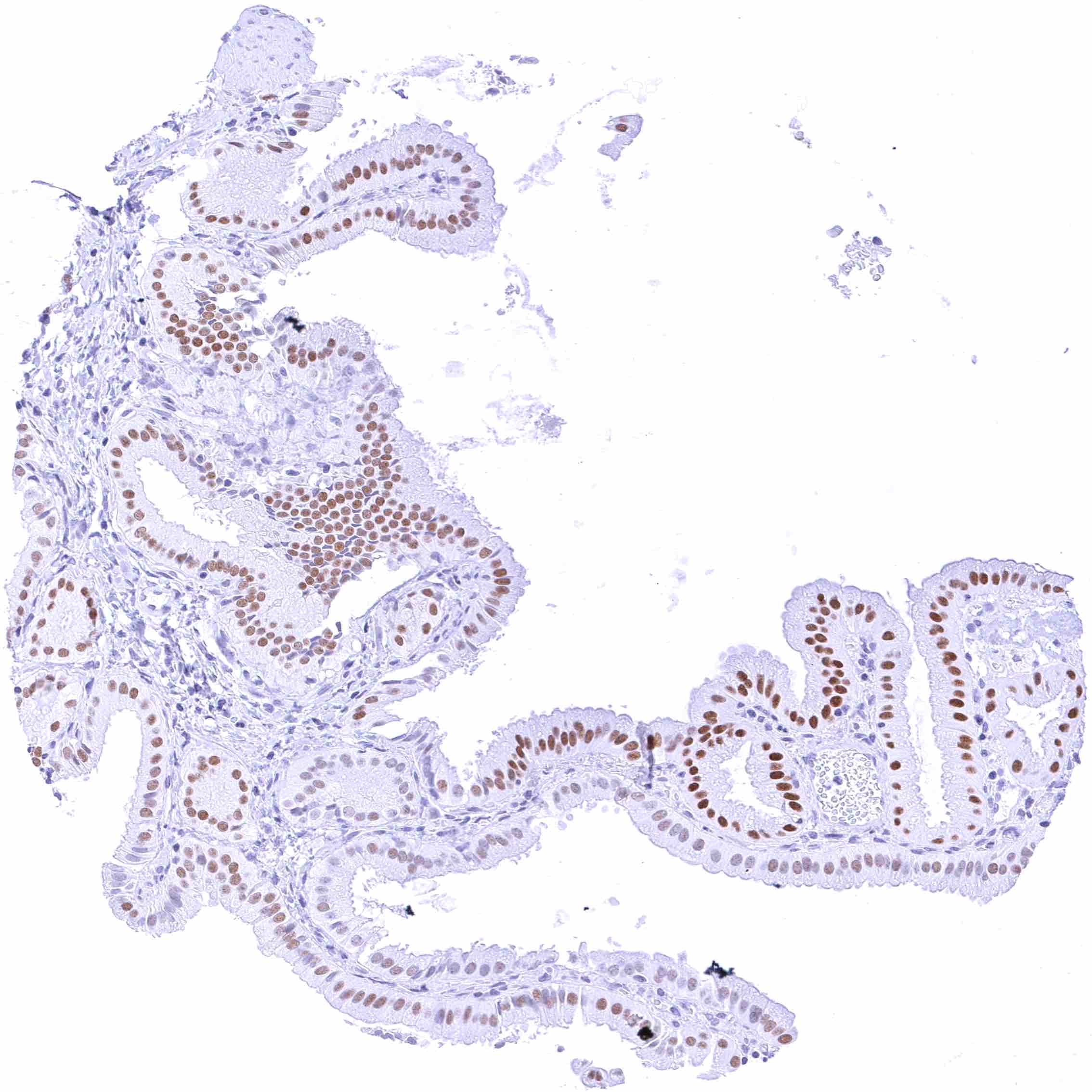
Gallbladder, epithelium – Nuclear FOXA1 staining of variable intensity - ranging from negative to strong - in epithelial cells.
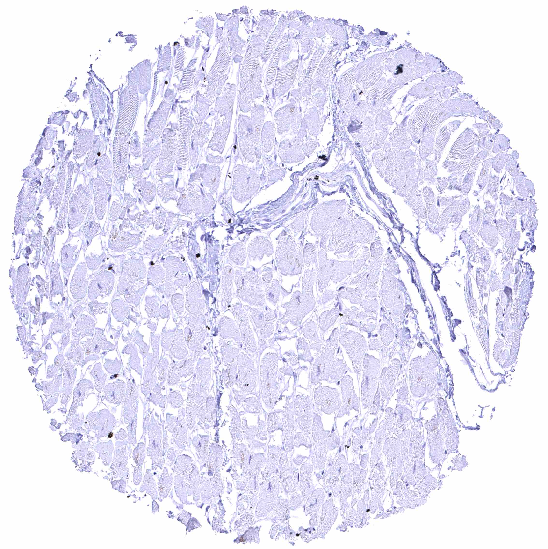
Heart muscle
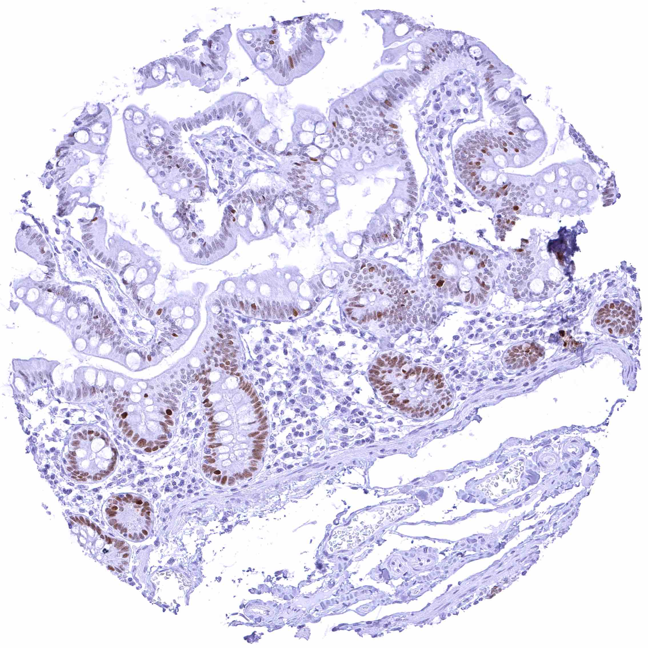
Ileum, mucosa – Moderate to strong nuclear FOXA1 staining of crypt epithelial cells. The staining intensity gradually decreases towards the surface epithelium and the peaks of the villi.
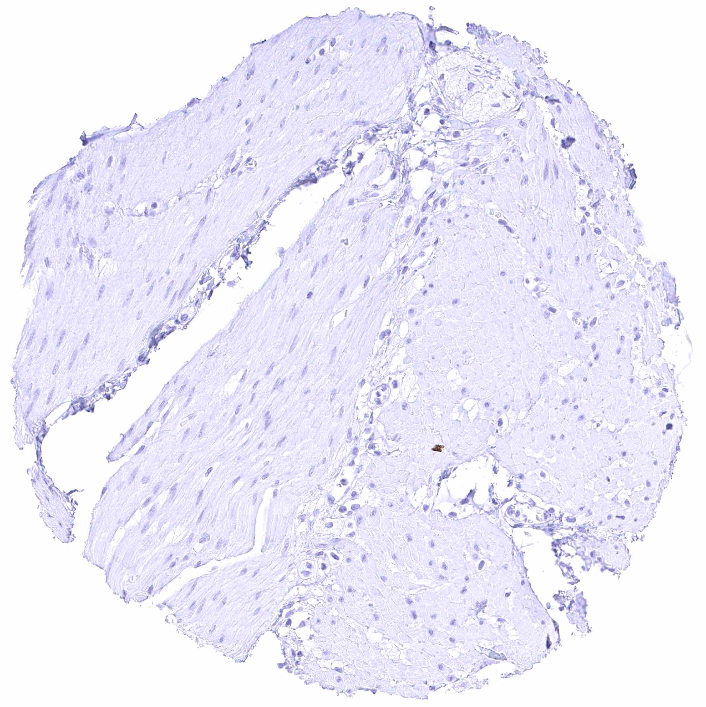
Ileum, muscular wall
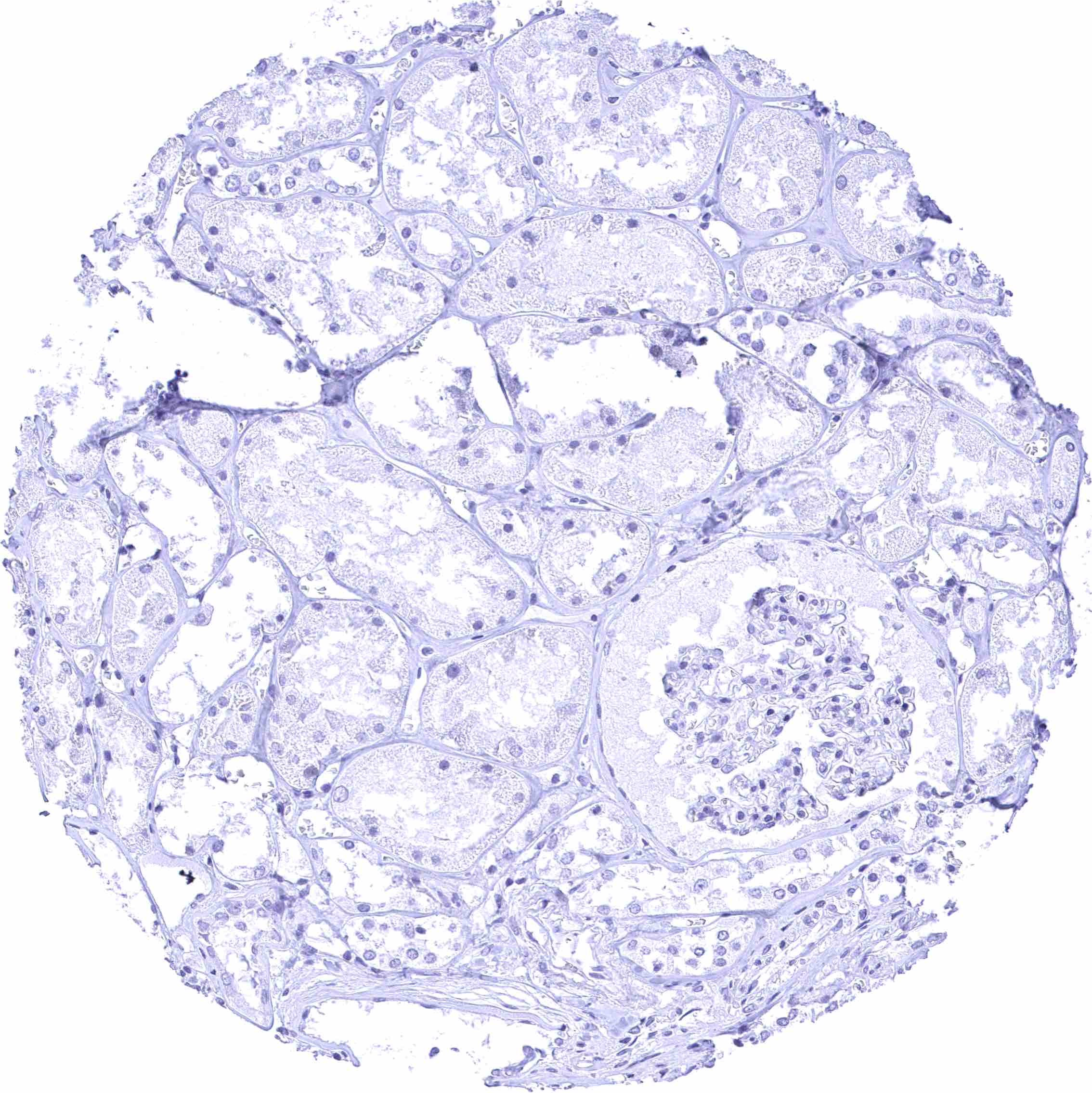
Kidney, cortex
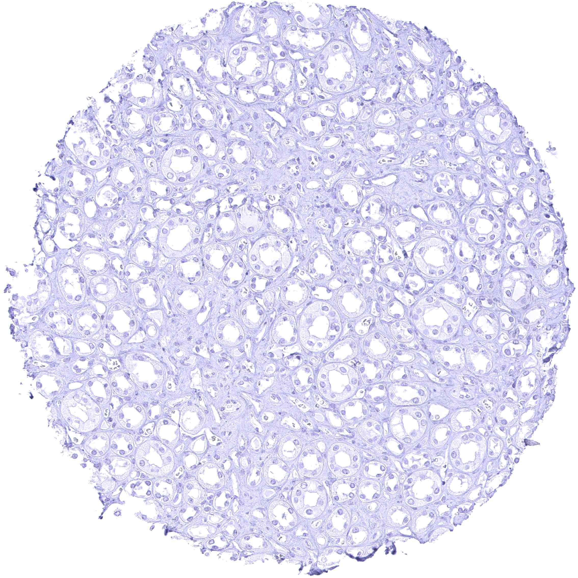
Kidney, medulla
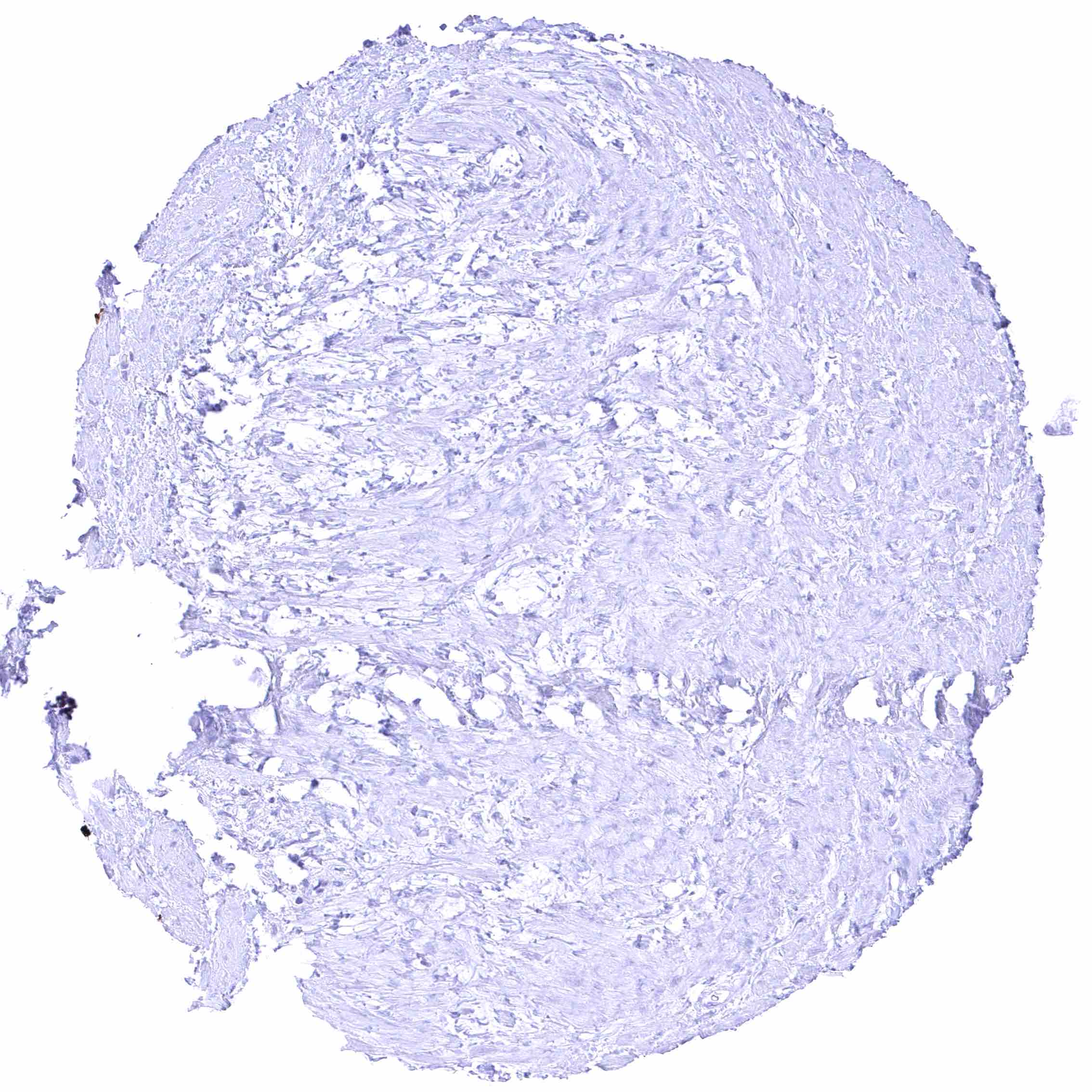
Kidney, pelvis, muscular wall
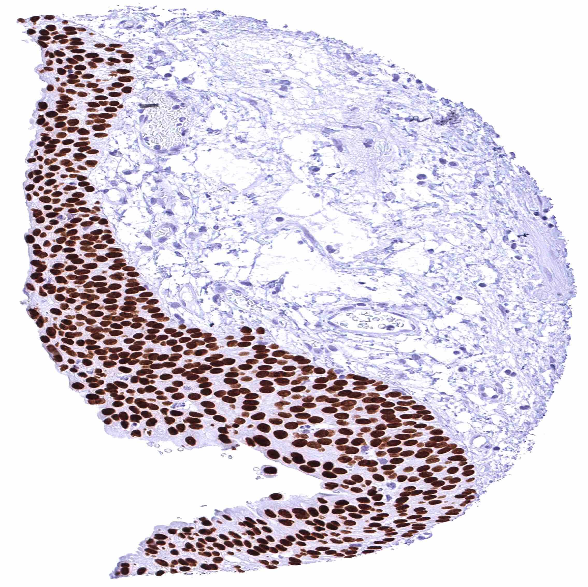
Kidney, pelvis, urothelium – Intense nuclear FOXA1 staining of urothelial cells.
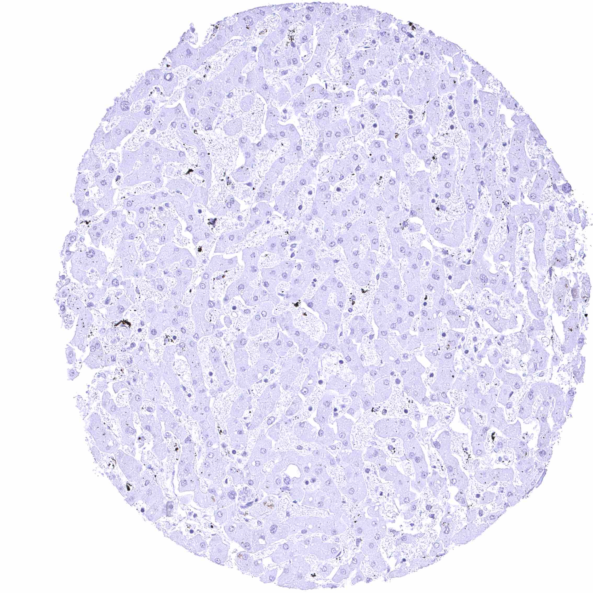
Liver – Absence of nuclear FOXA1 staining of hepatocytes in this sample.
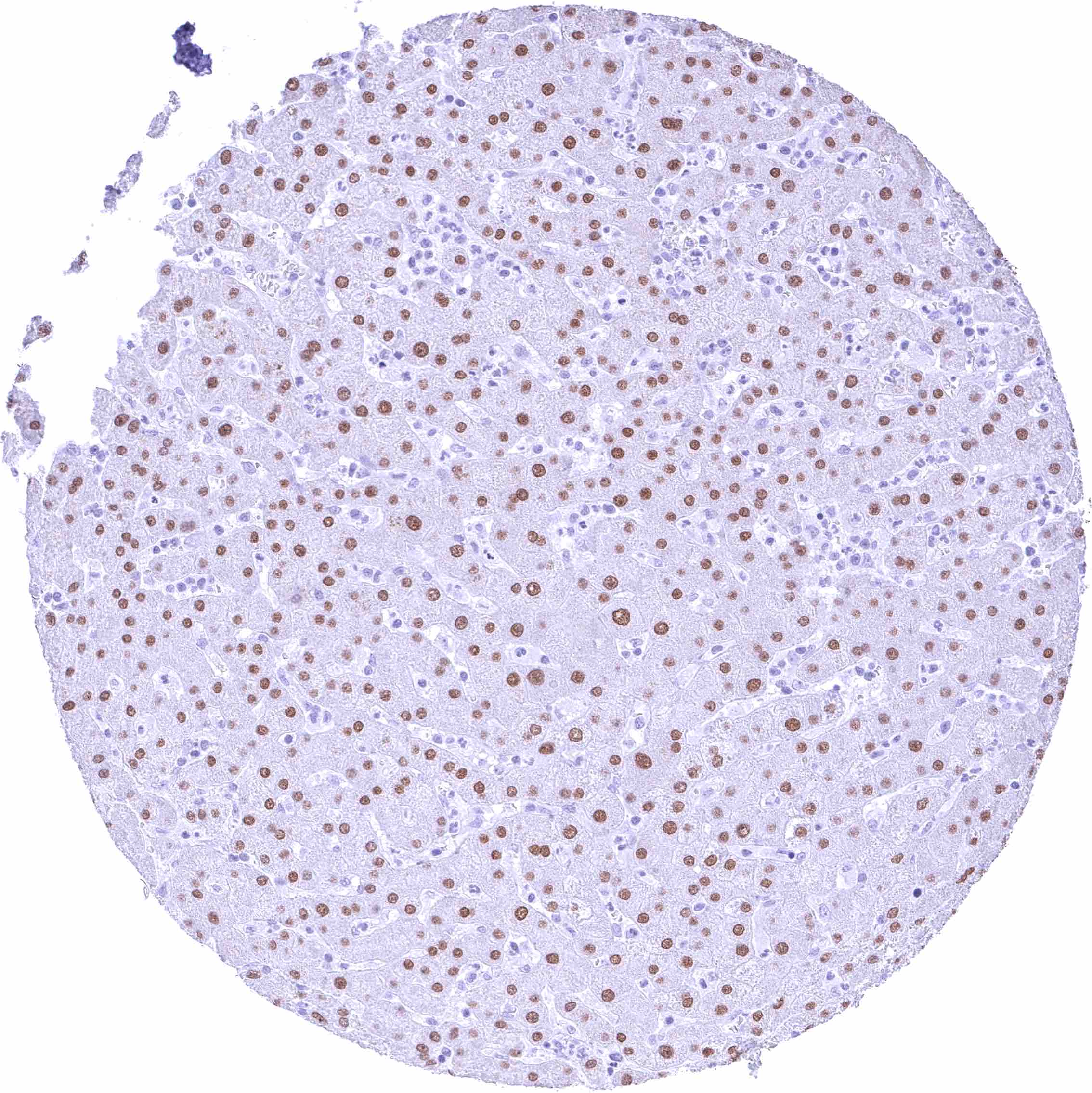
Liver – Moderate nuclear FOXA1 staining of hepatocytes.
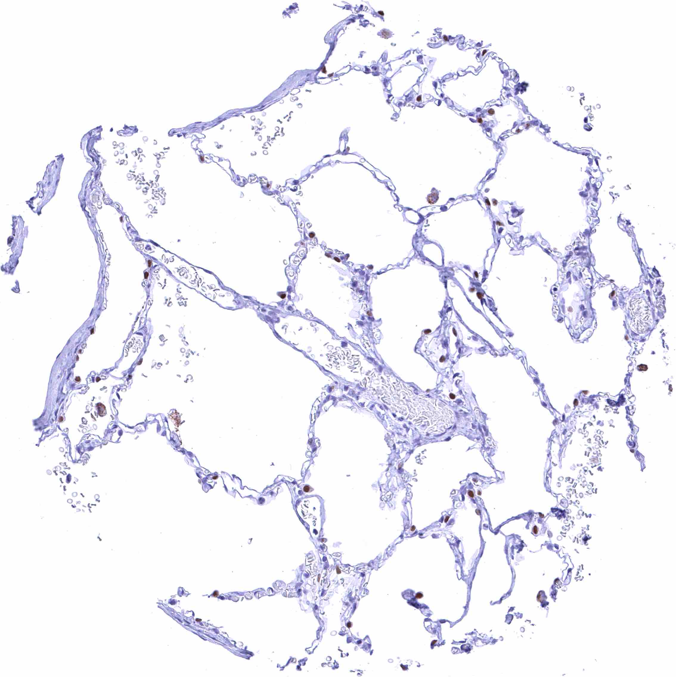
Lung
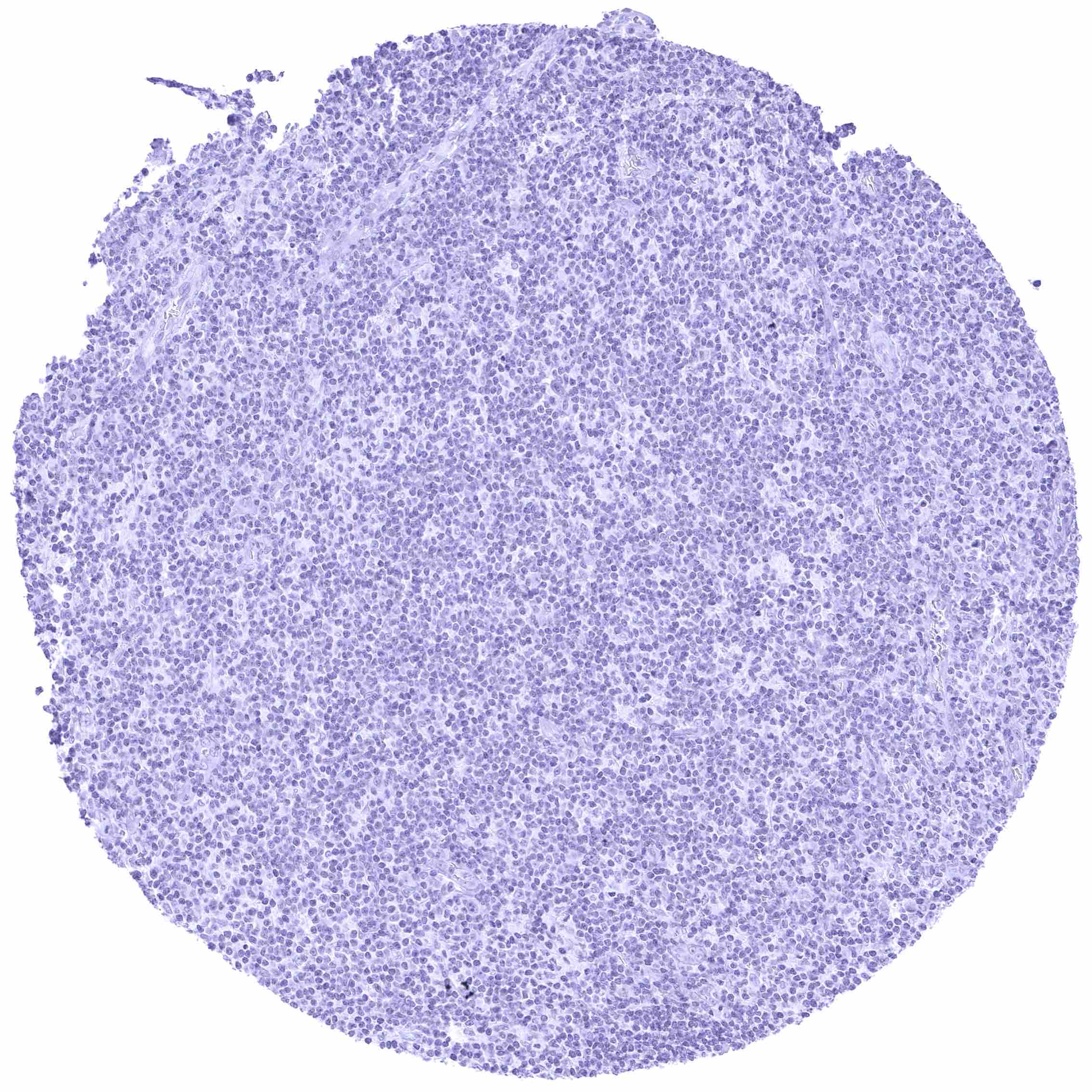
Lymph node
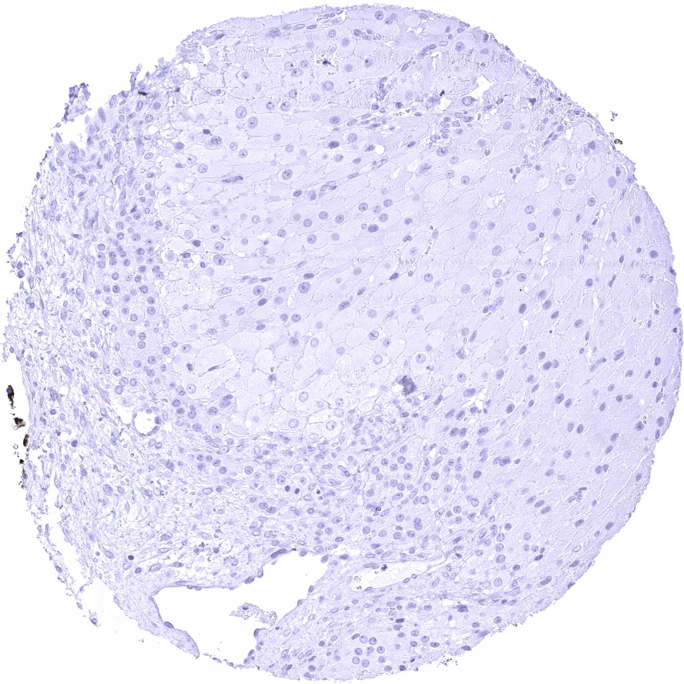
Ovary, corpus luteum
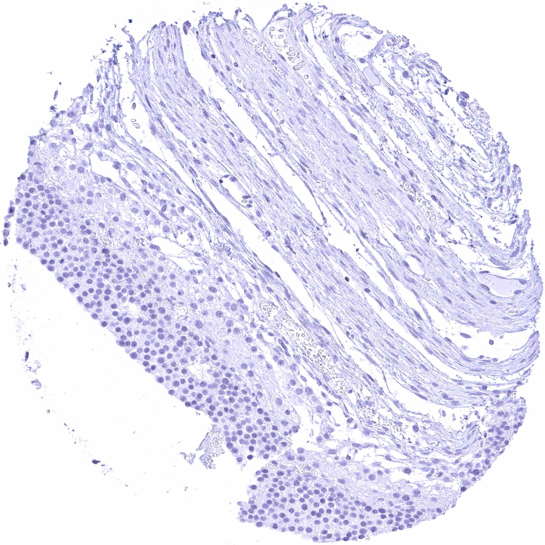
Ovary, follicular cyst
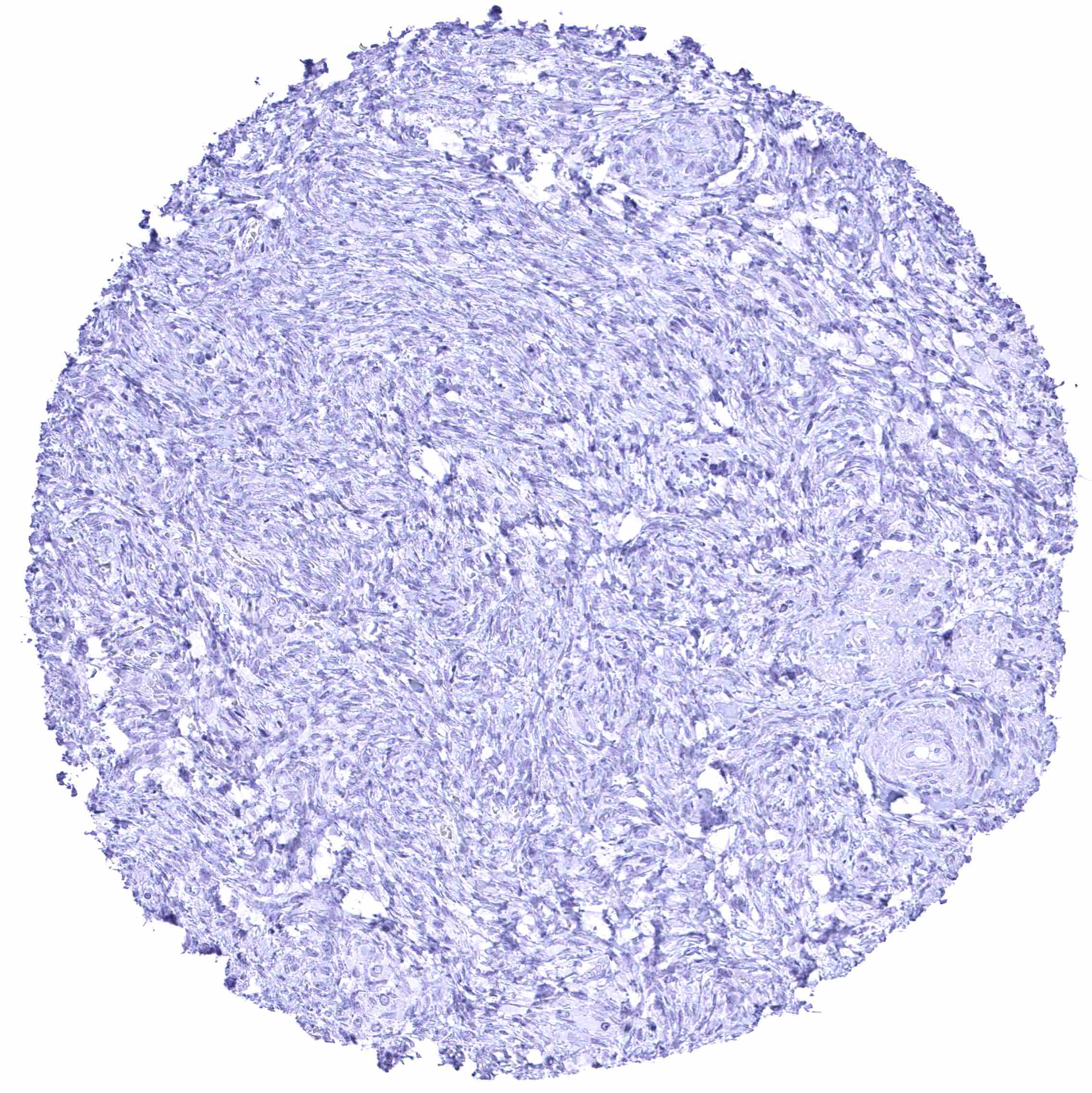
Ovary, stroma
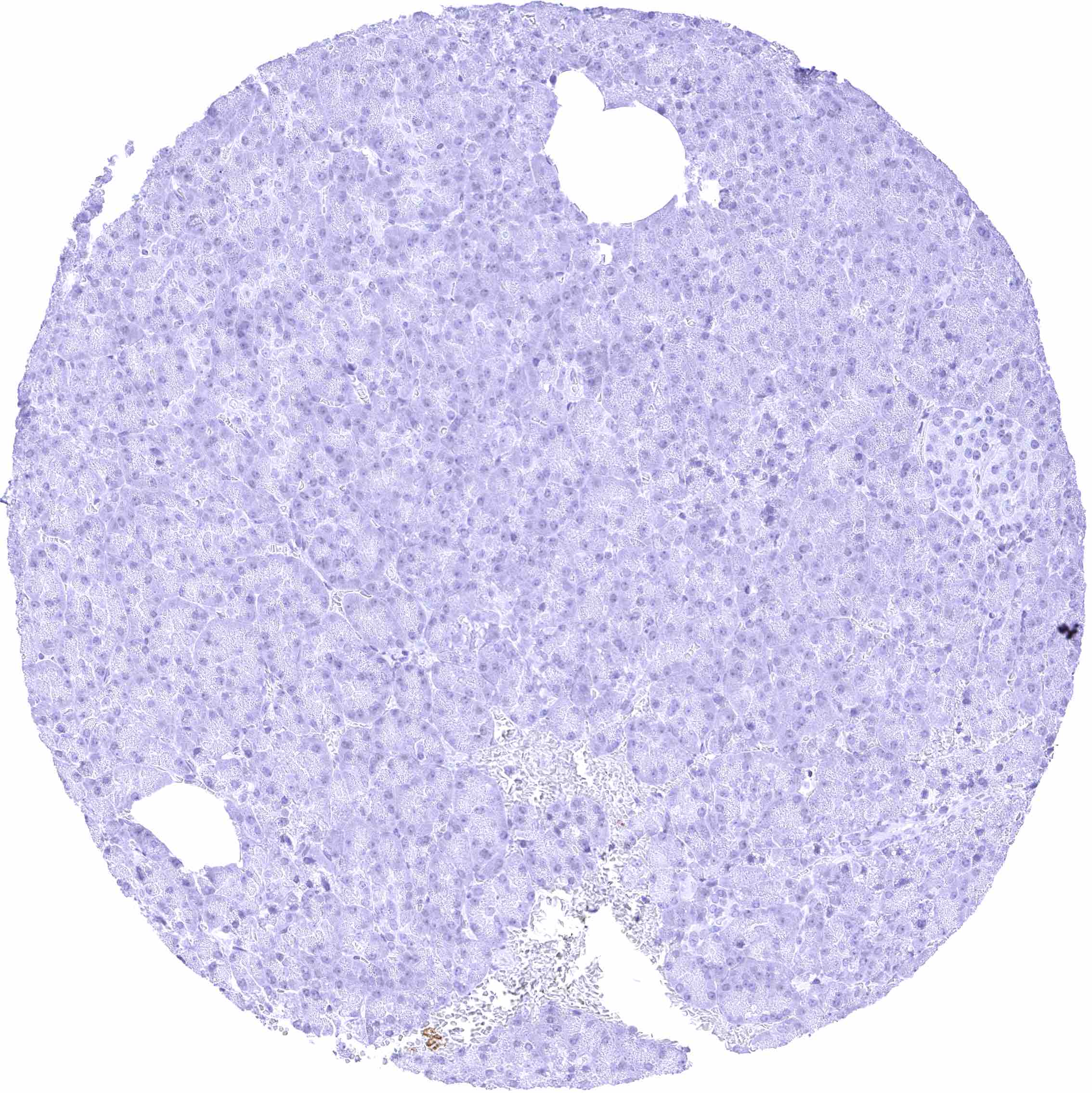
Pancreas
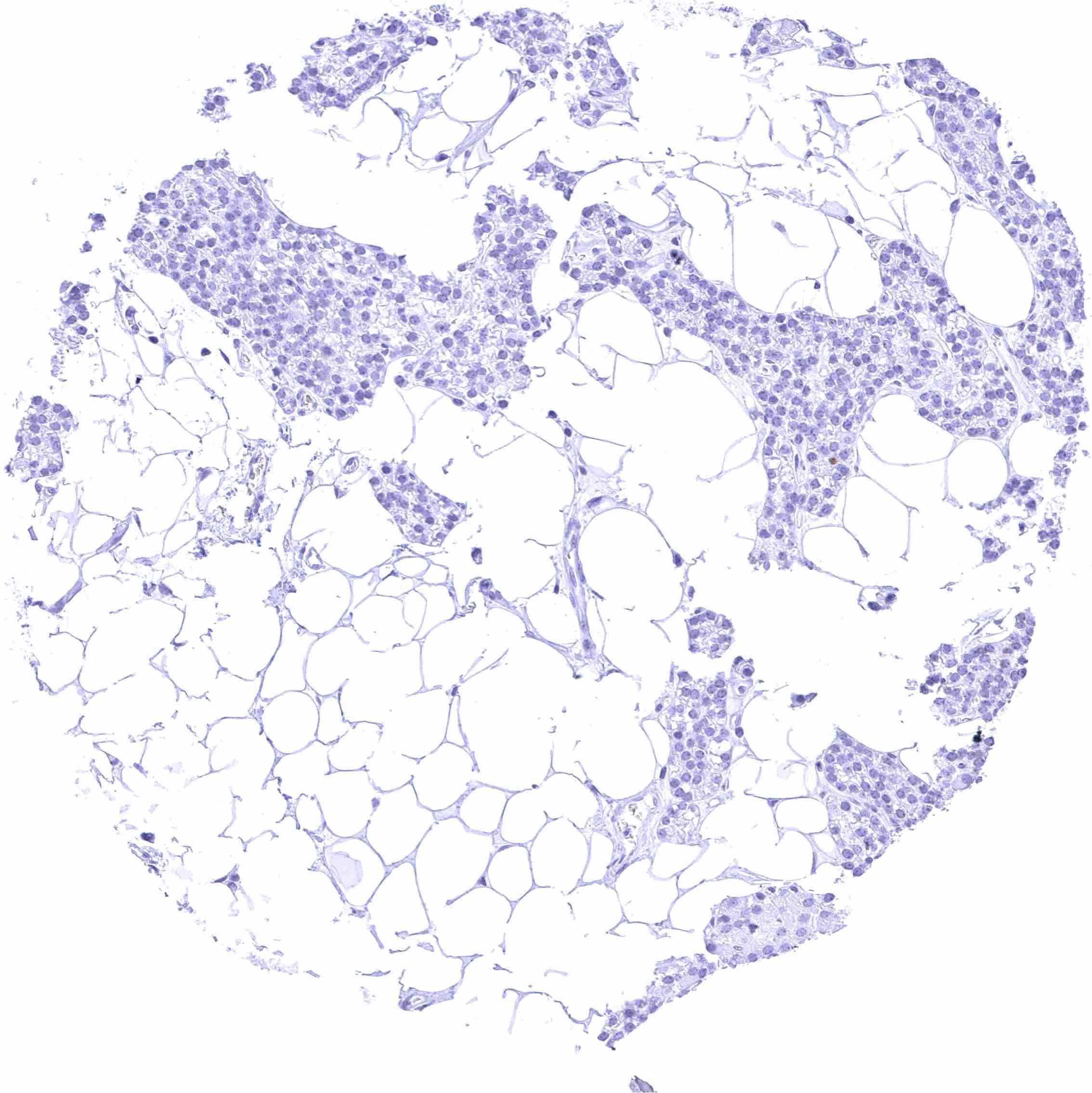
Parathyroid gland
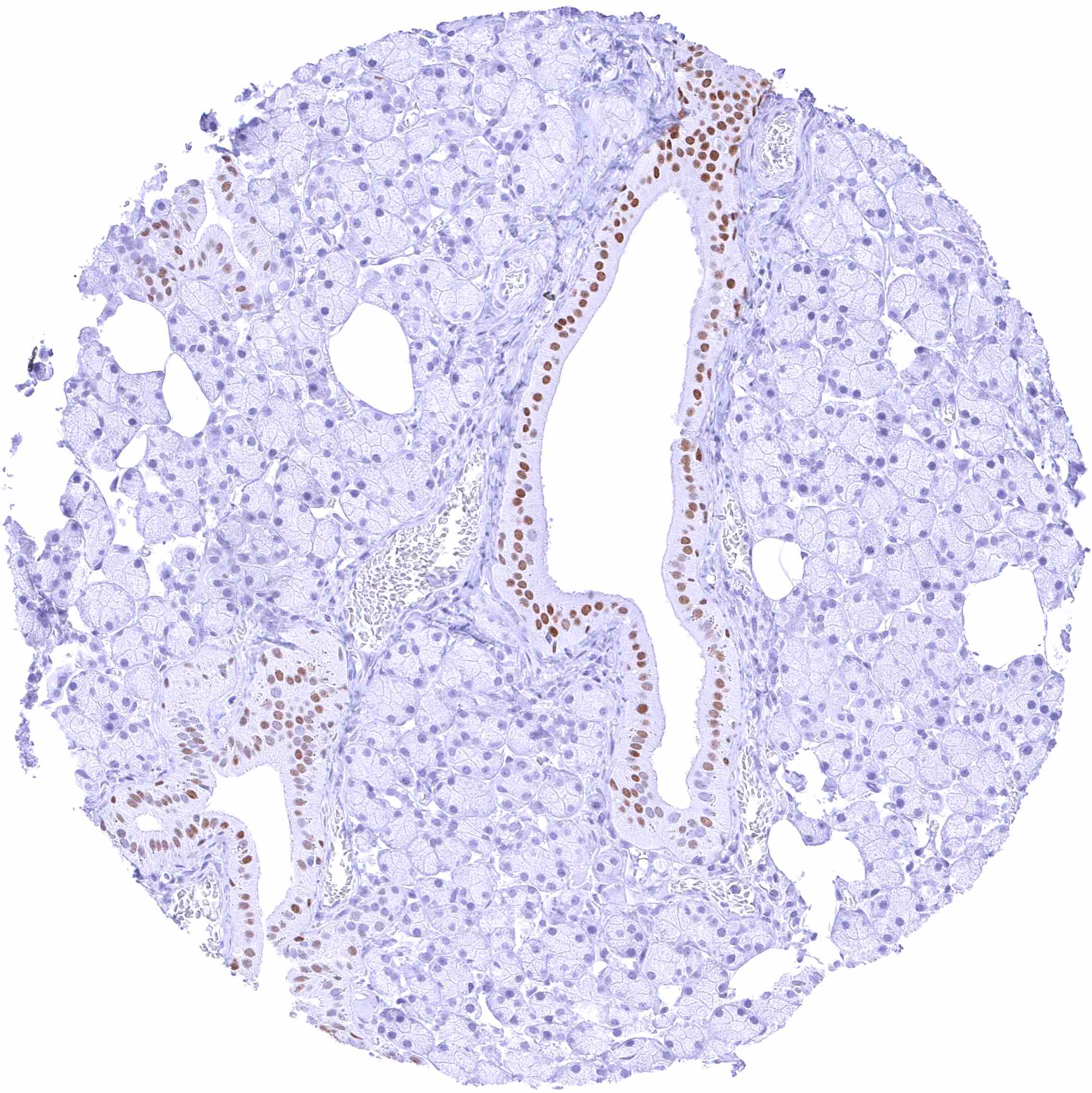
Parotid gland – Strong nuclear FOXA1 staining of excretory ducts while serous glands are only faintly stained or FOXA1 negative.
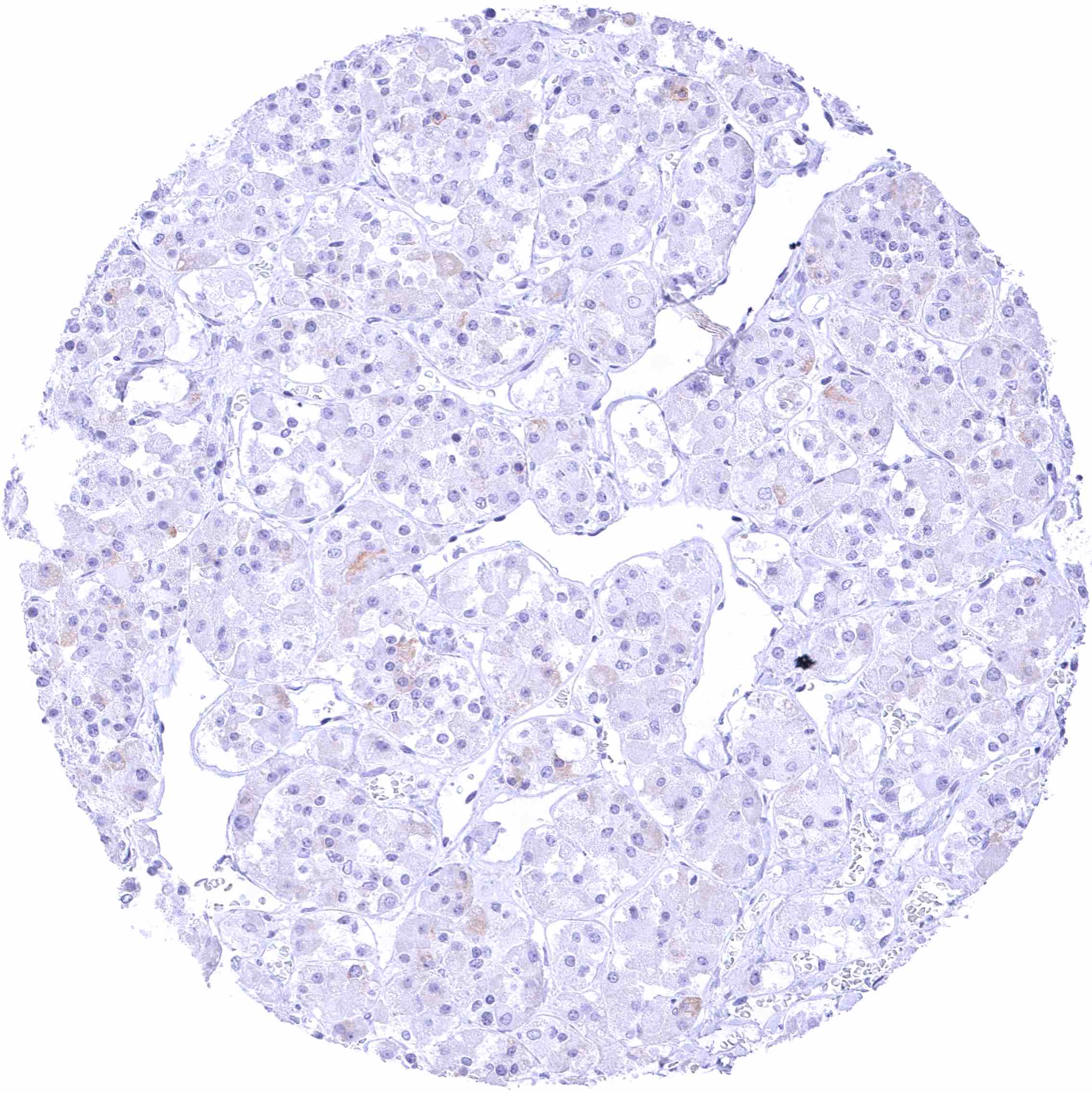
Pituitary gland, anterior lobe
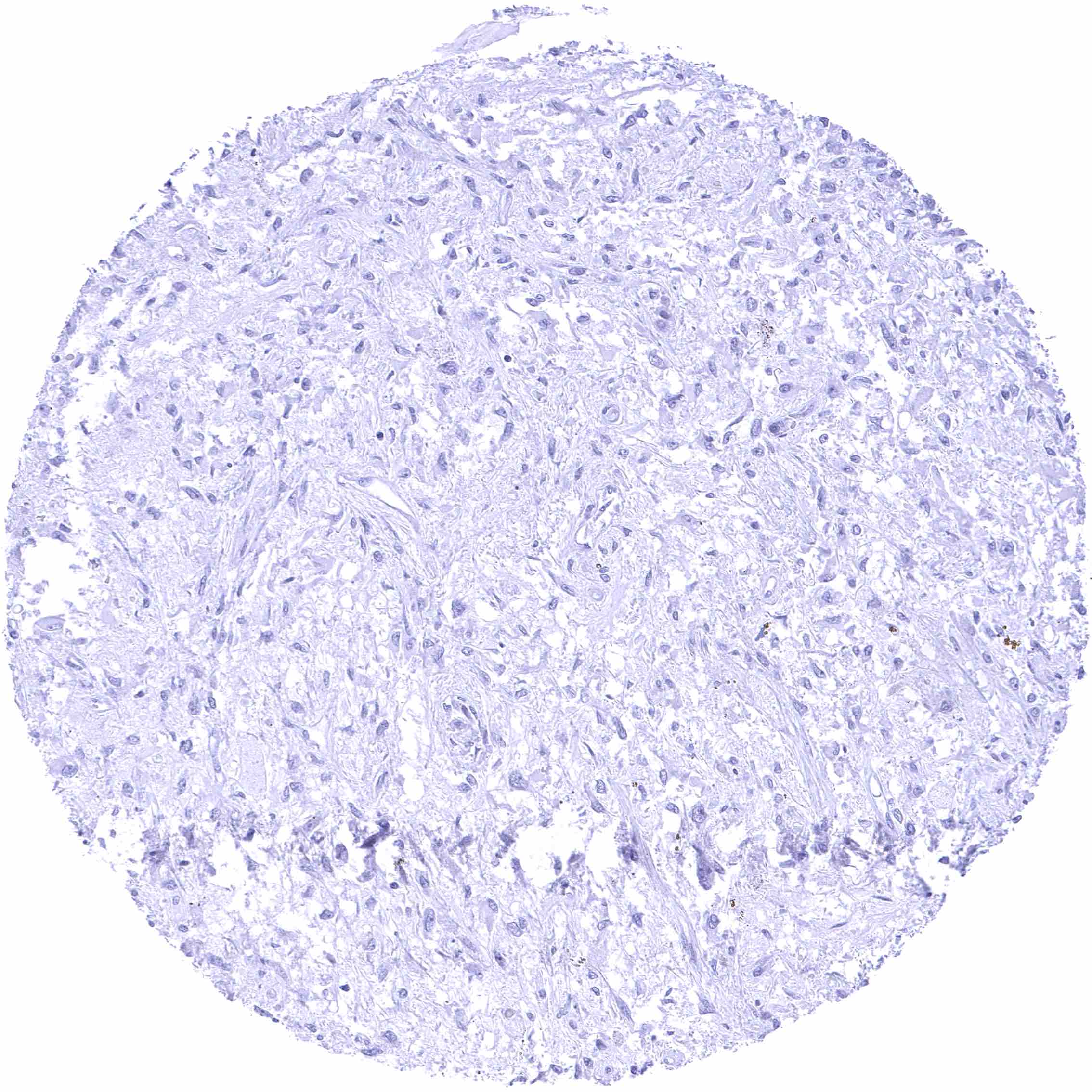
Pituitary gland, posterior lobe
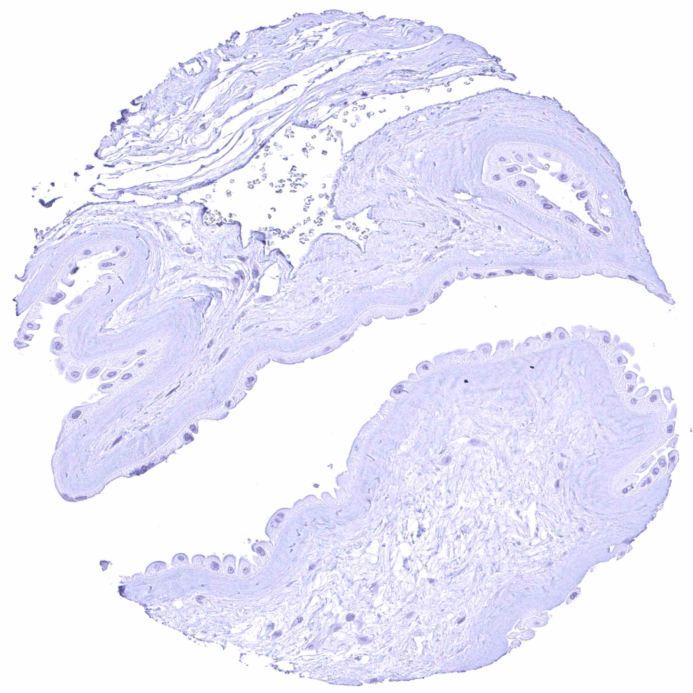
Placenta (amnion)
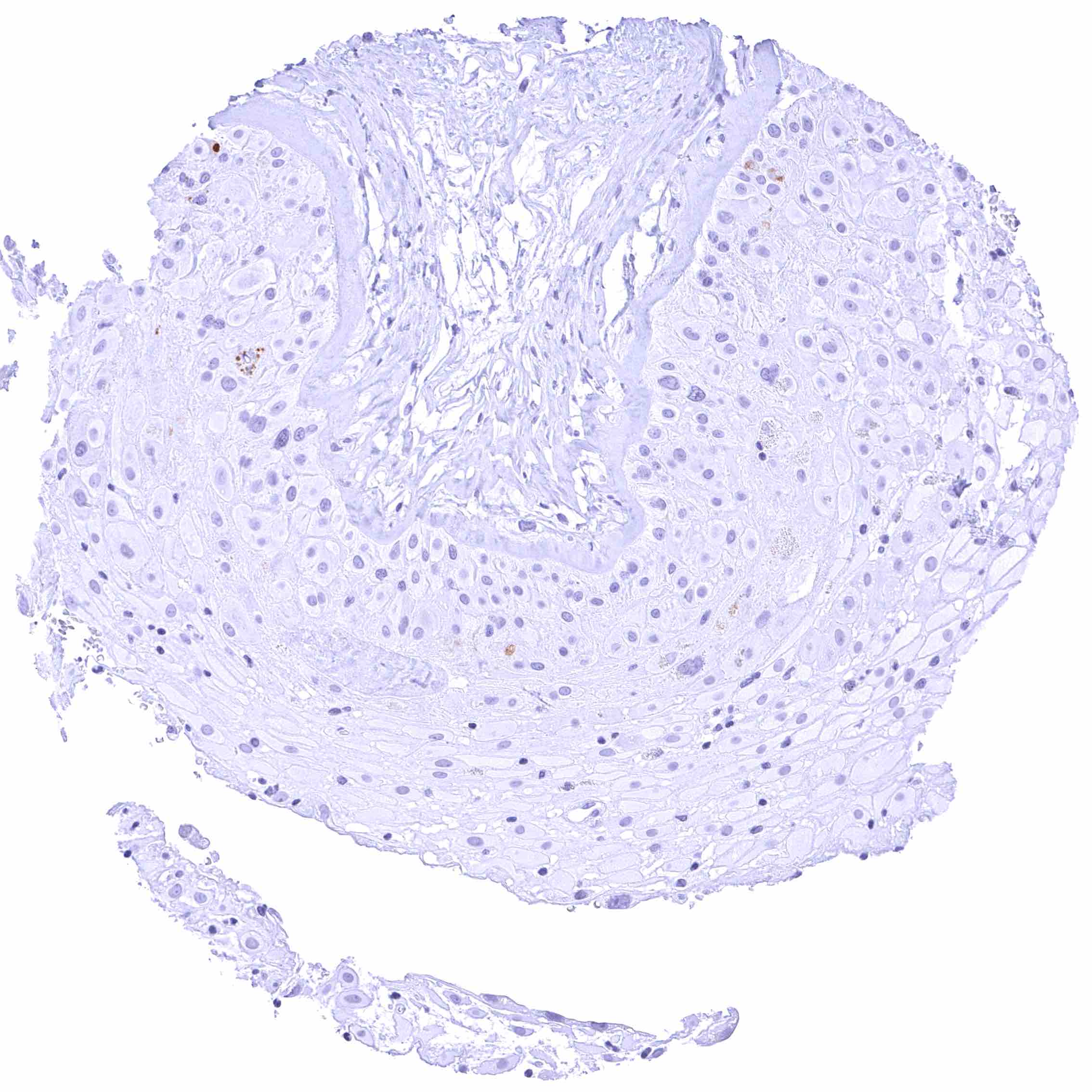
Placenta (chorion)
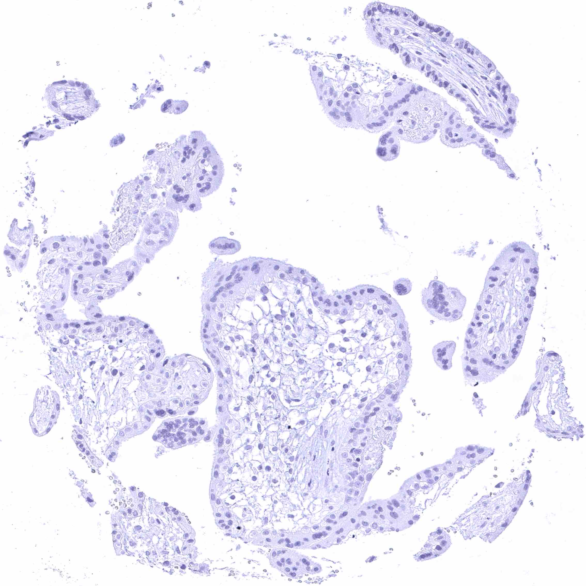
Placenta, early
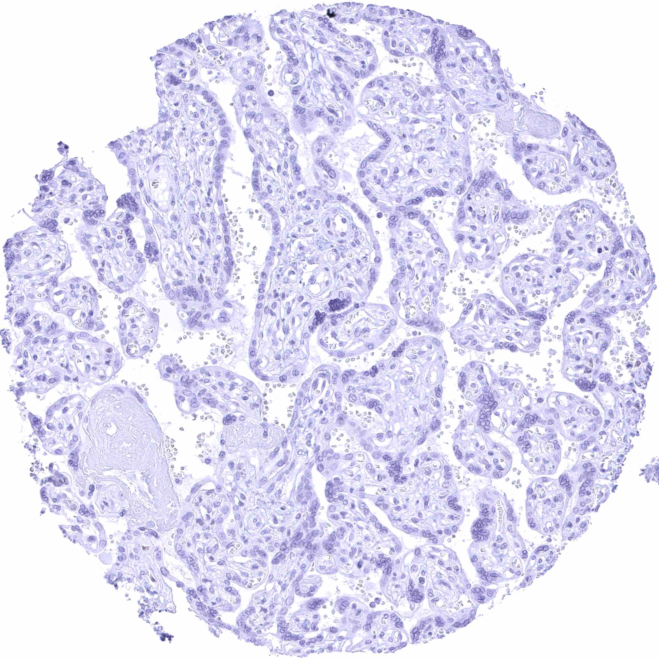
Placenta, mature
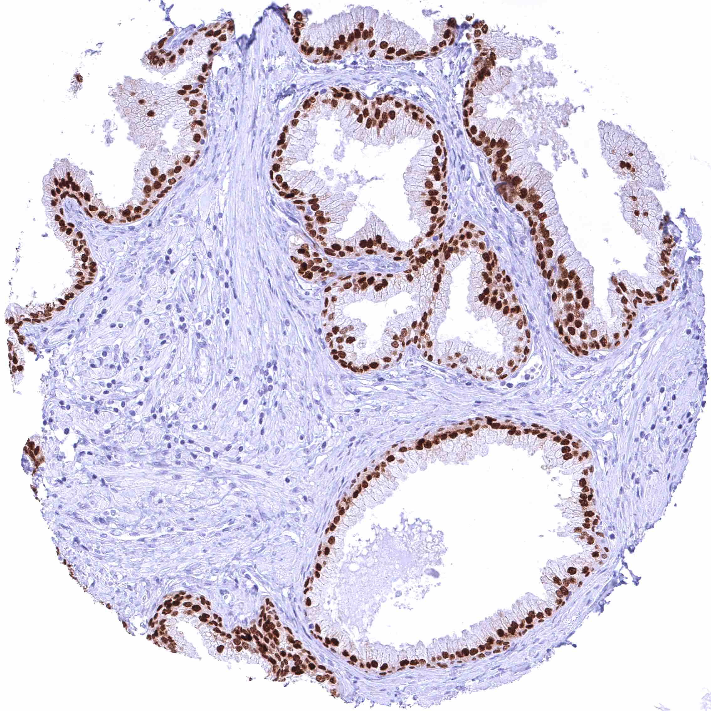
Prostate – Intense nuclear FOXA1 staining of all epithelial cells.
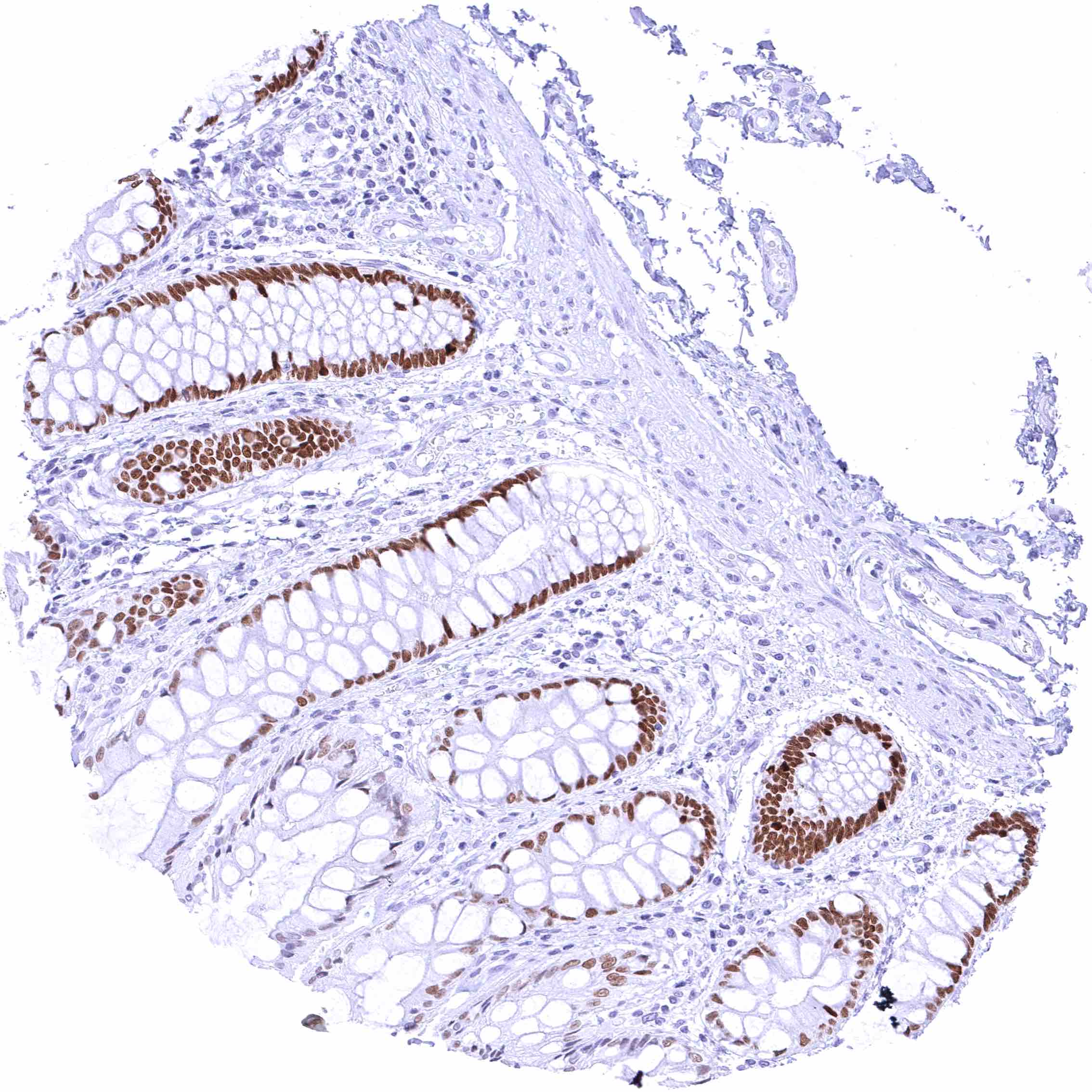
Rectum, mucosa – Strong nuclear FOXA1 staining of epithelial cells in the crypts. The staining intensity gradually decreases towards the surface epithelium.
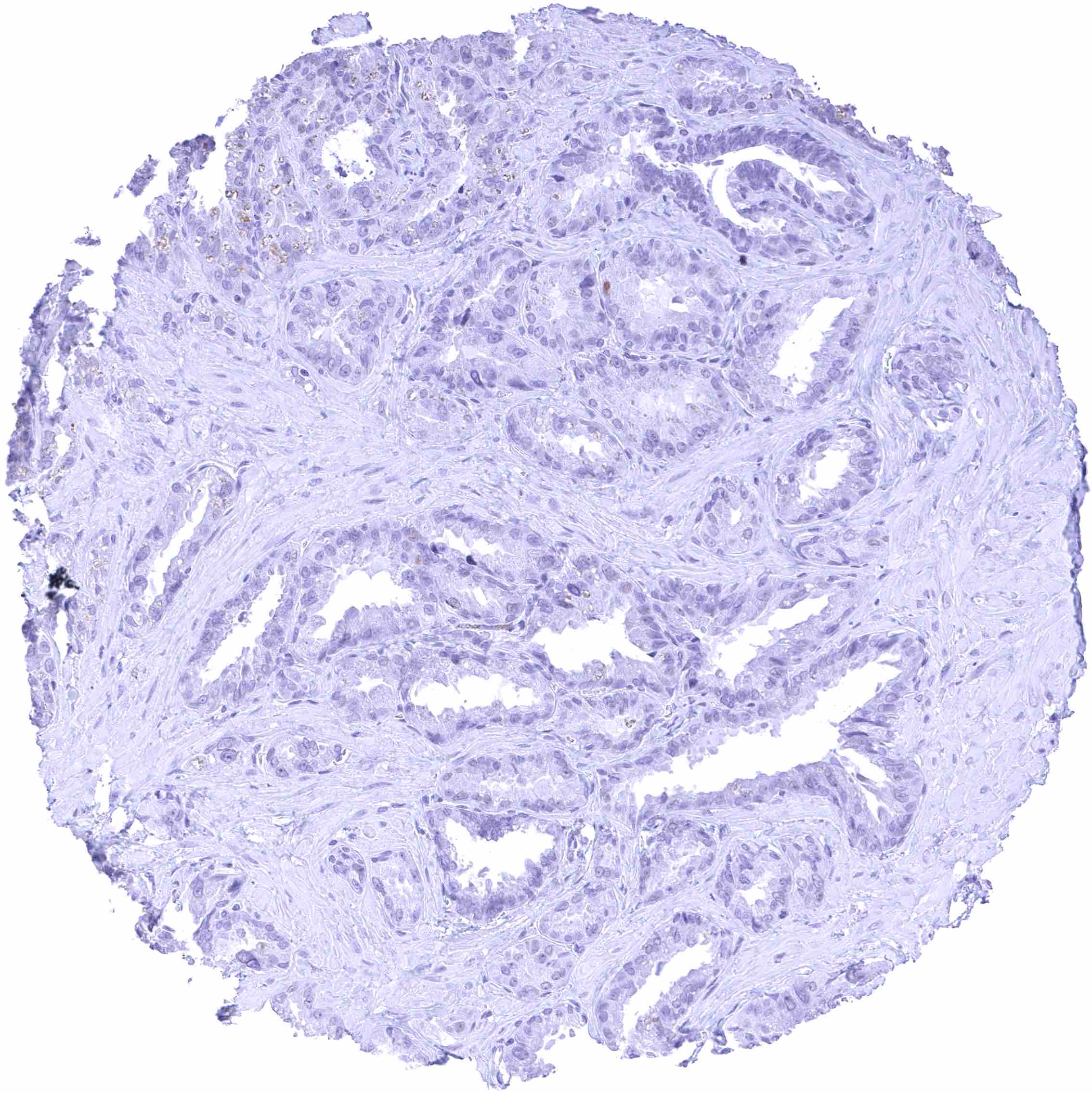
Seminal vesicle
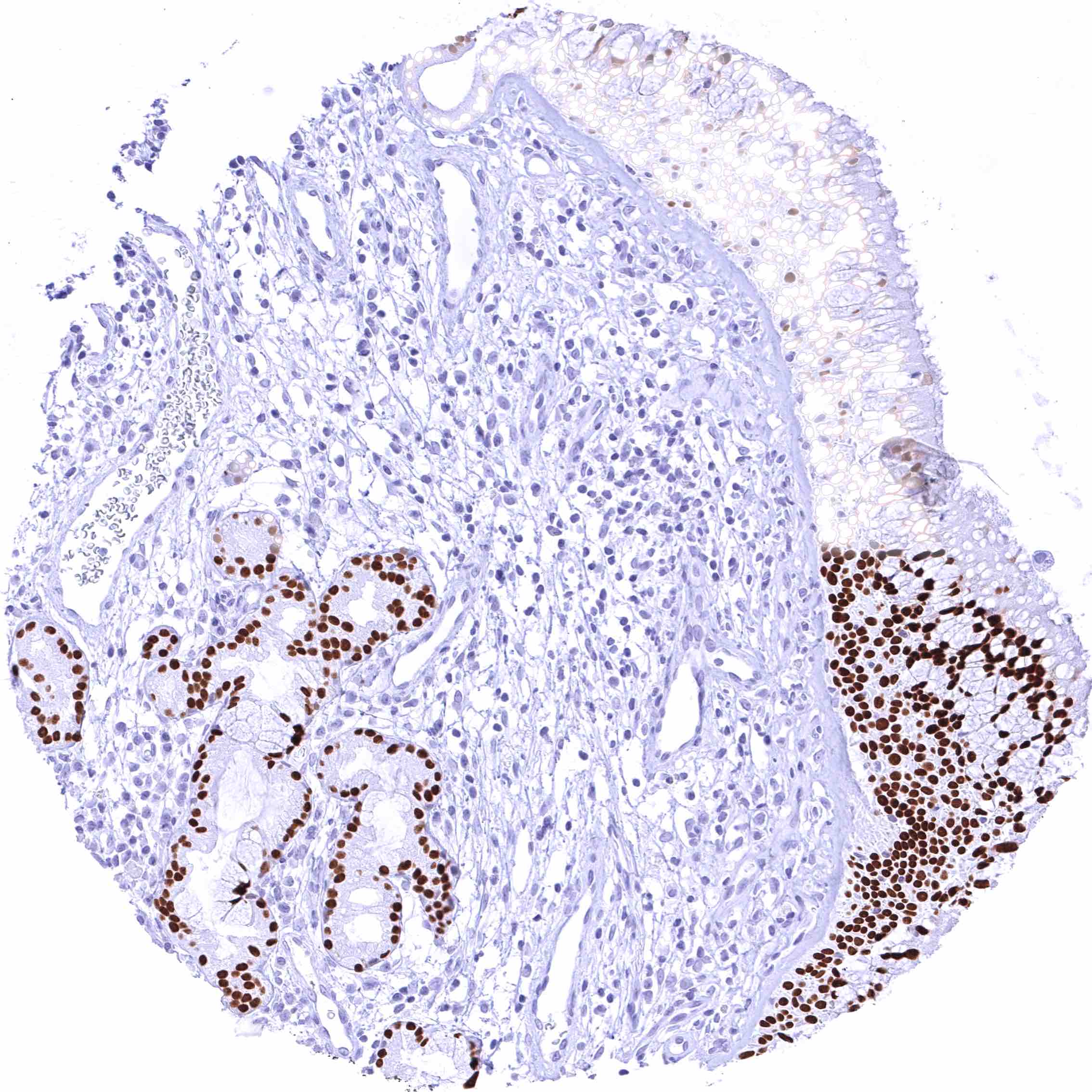
Sinus paranasales – Intense nuclear FOXA1 staining of all epithelial cells of the respiratory epithelium – and of bronchial glands.
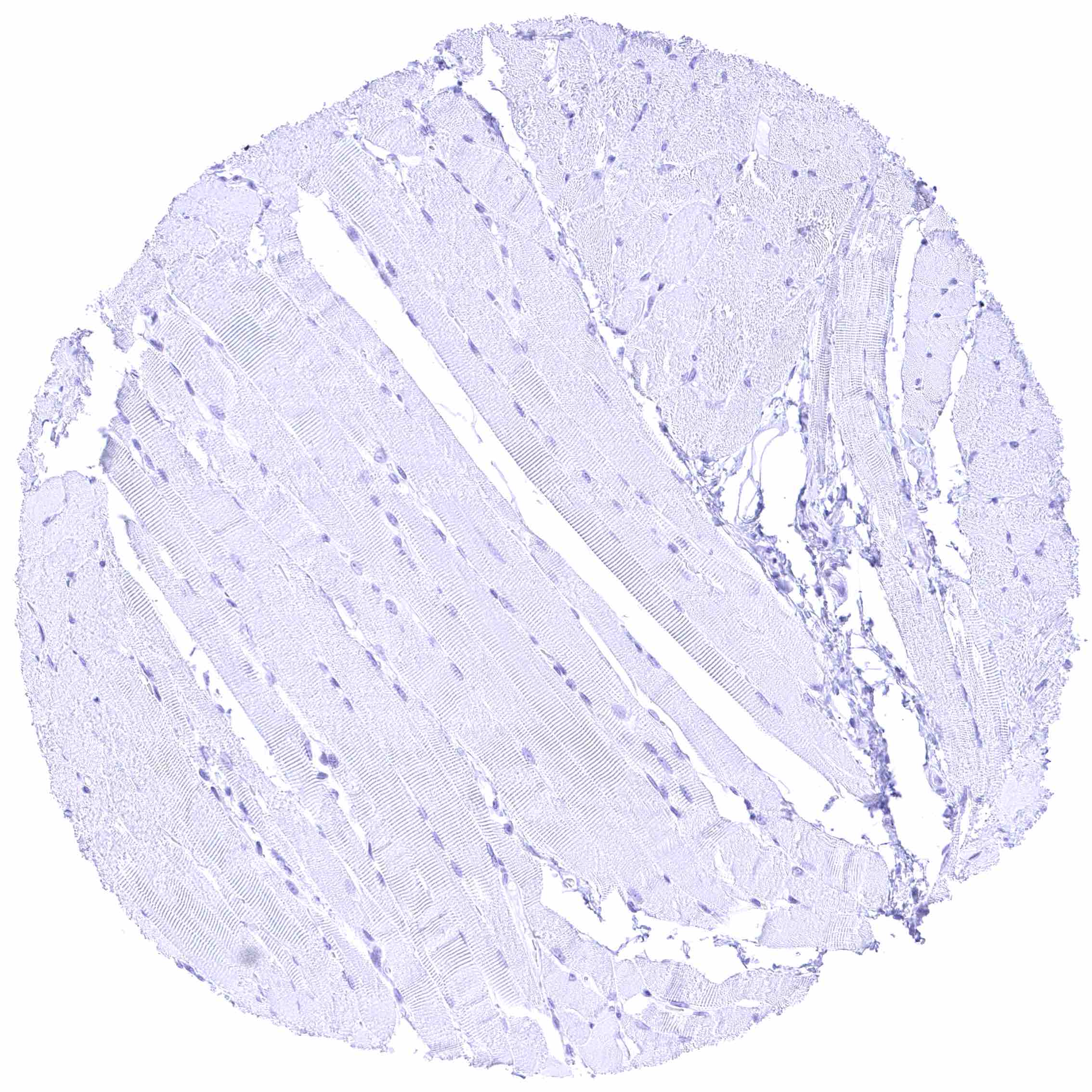
Skeletal muscle
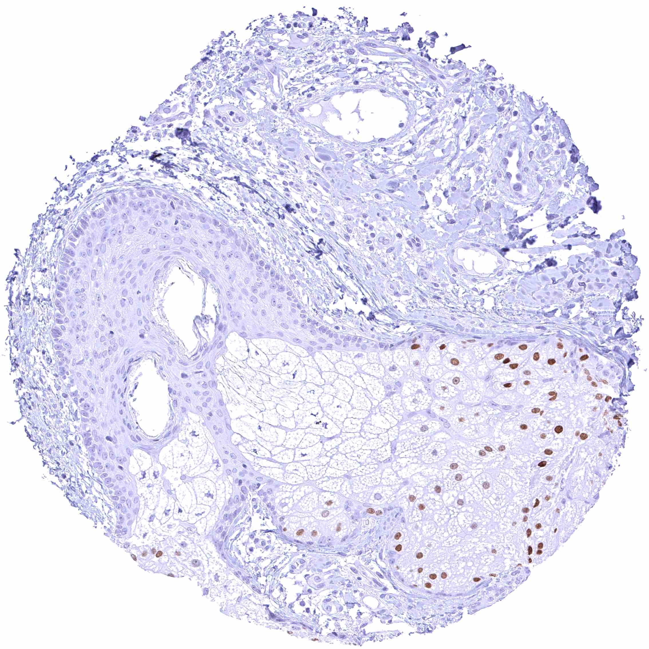
Skin, hairfollicel and sebaceous glands – Distinct nuclear FOXA1 staining of a subset of sebaceous epithelial cells.
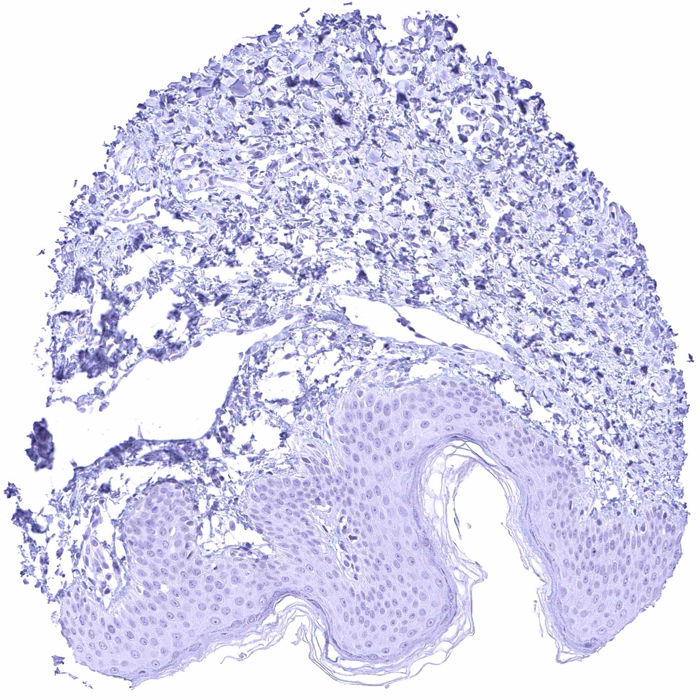
Skin
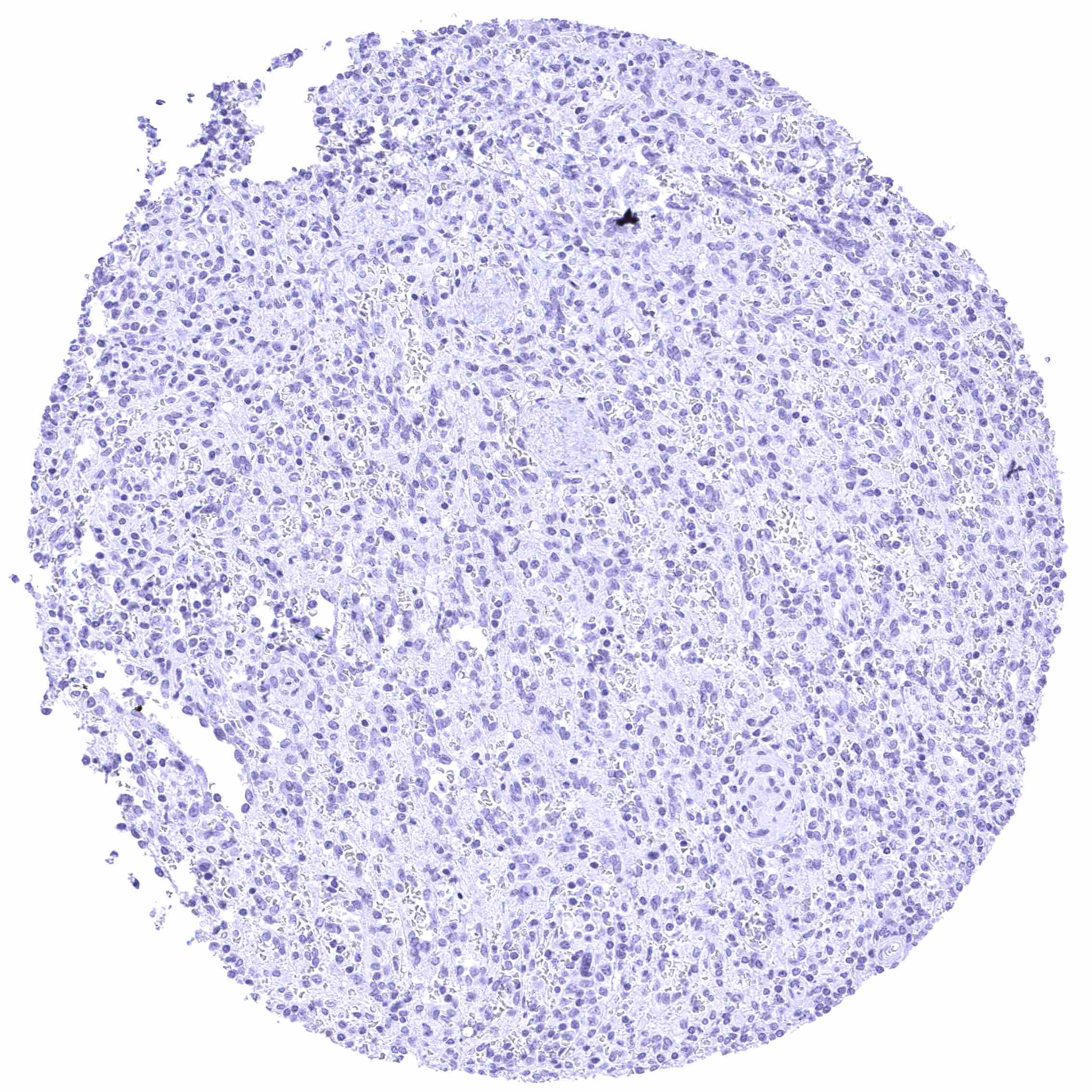
Spleen
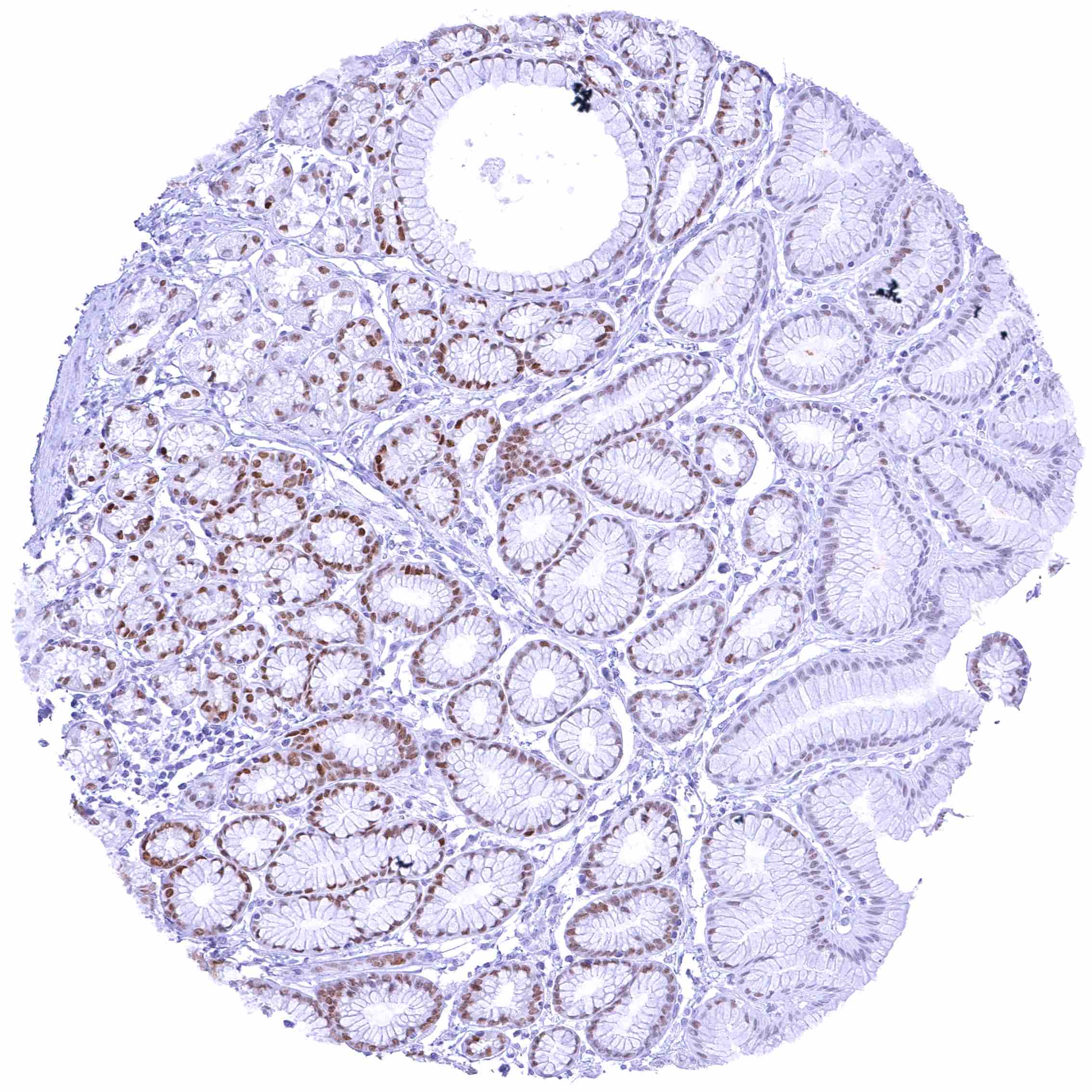
Stomach, antrum – Moderate to strong nuclear FOXA1 staining of glandular cells. The staining intensity gradually decreases towards the surface epithelium.
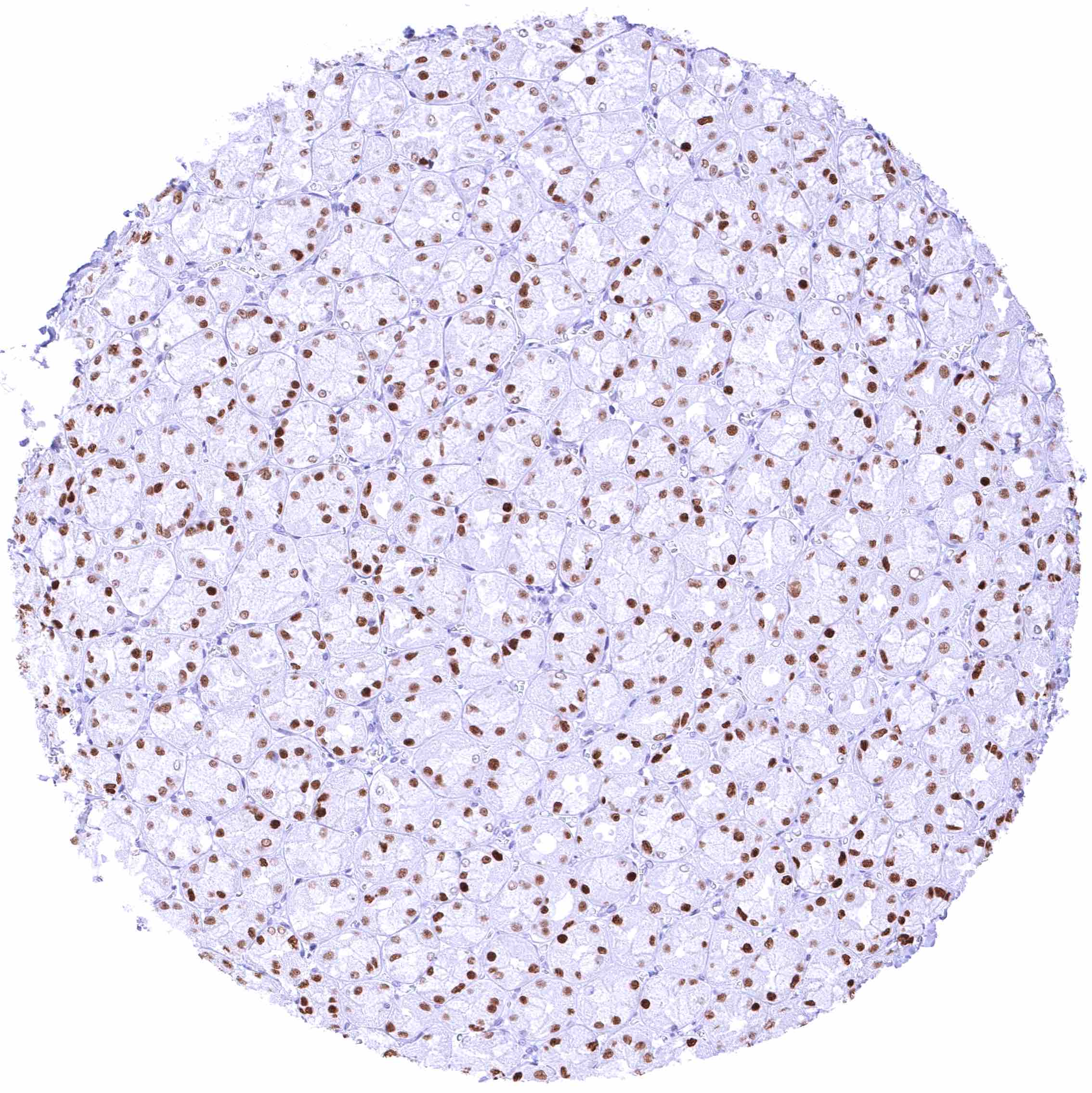
Stomach, corpus – Moderate to strong nuclear FOXA1 staining of glandular cells.
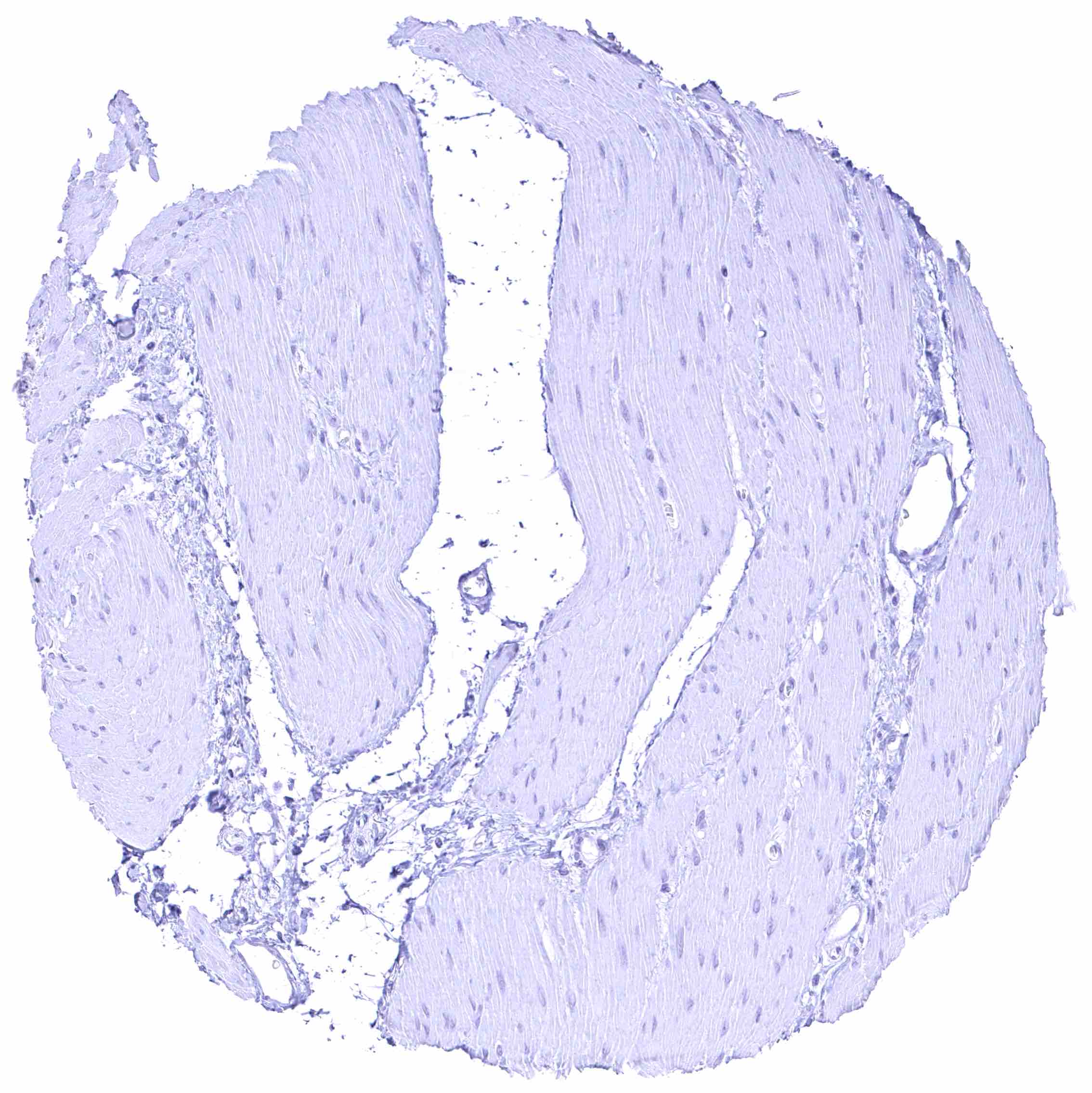
Stomach, muscular wall
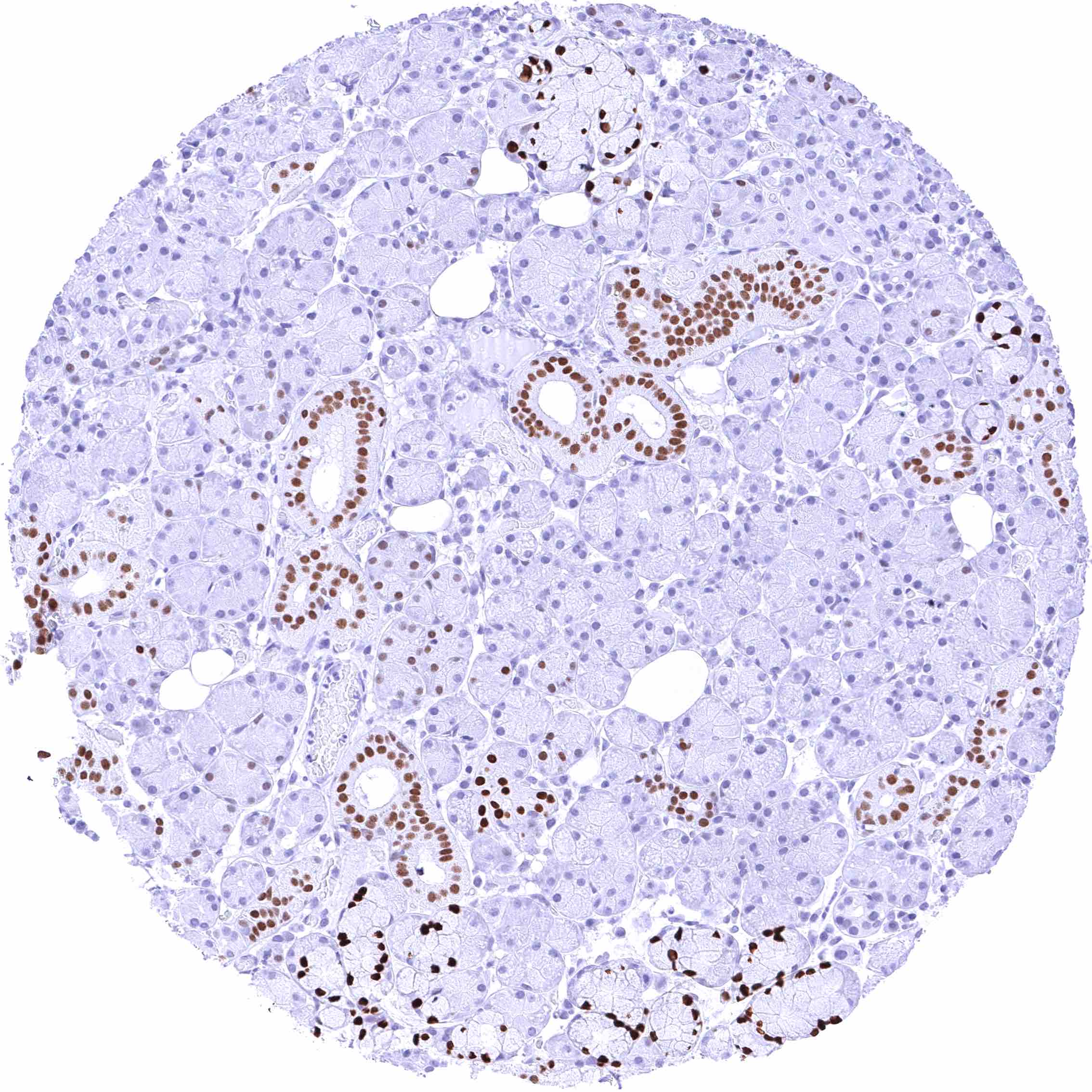
Submandibular gland – Strong nuclear FOXA1 staining of excretory ducts and of mucinous glands cells while serous glands are only faintly stained or FOXA1 negative.
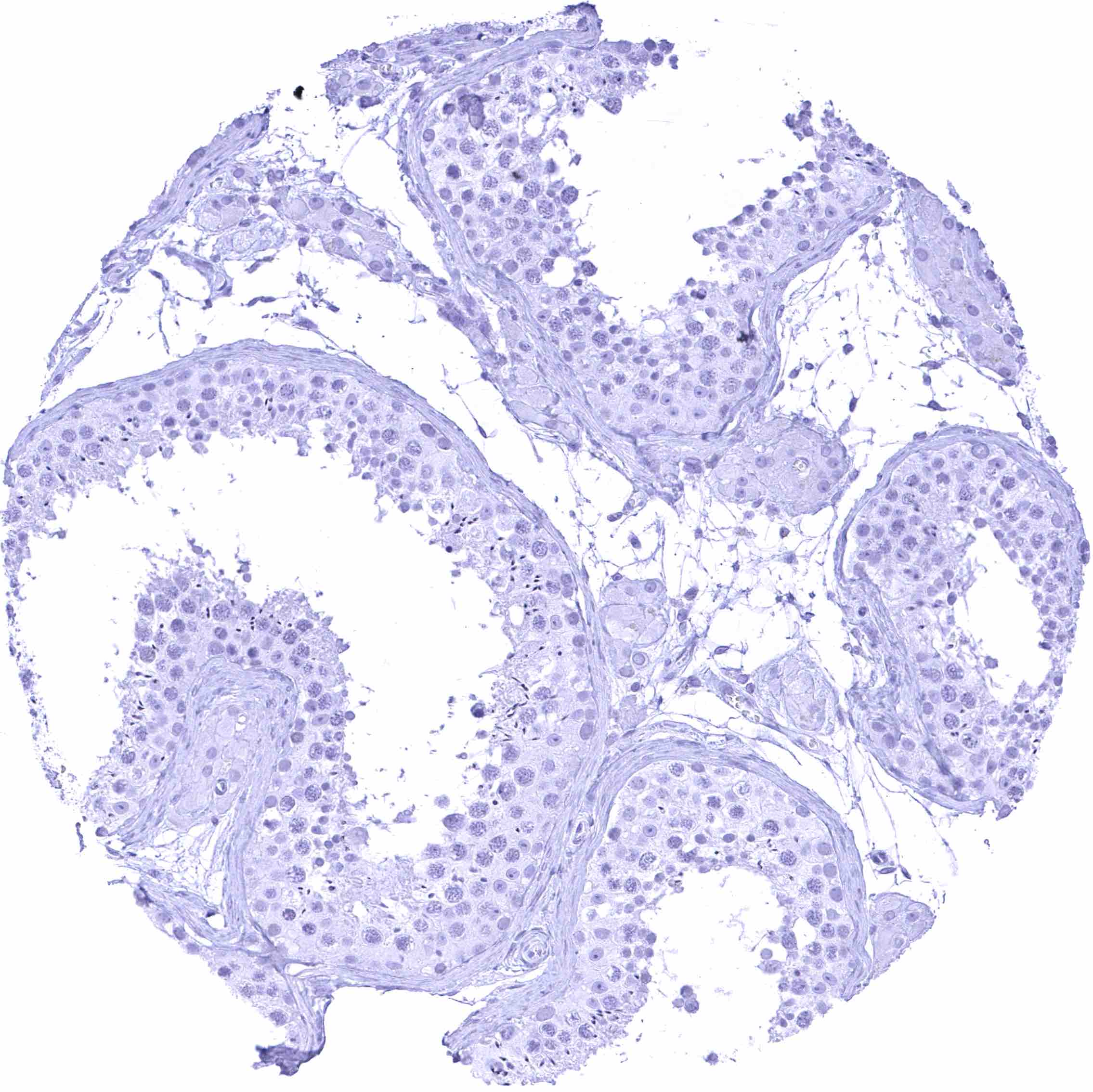
Testis
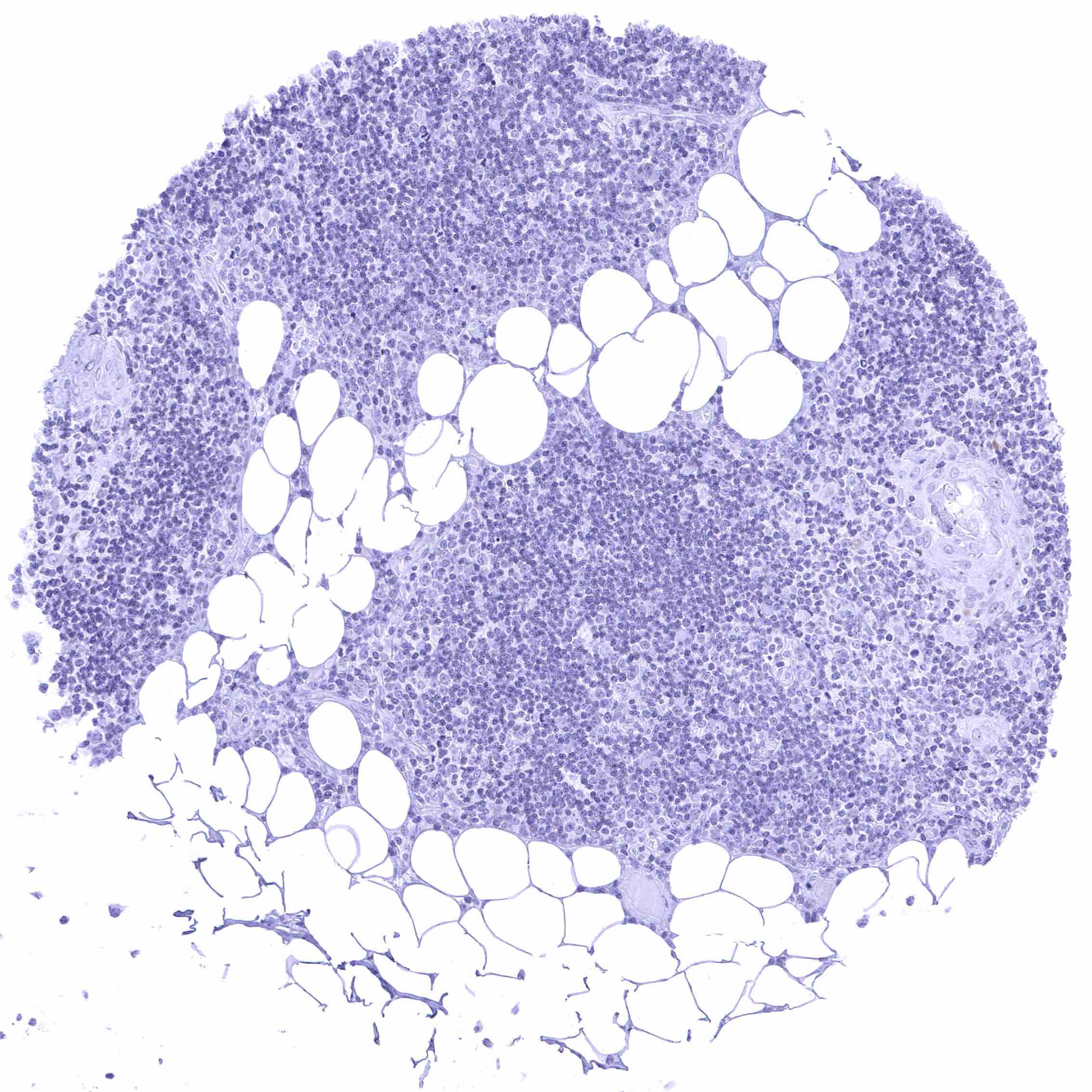
Thymus
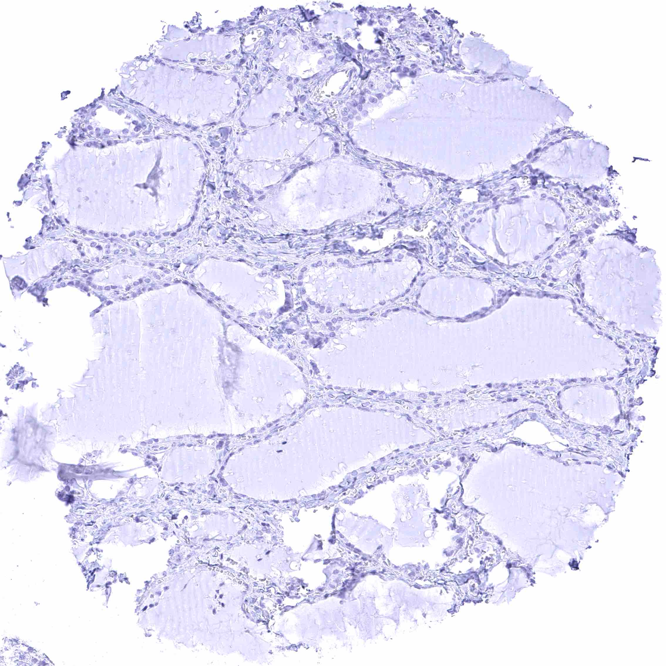
Thyroid gland
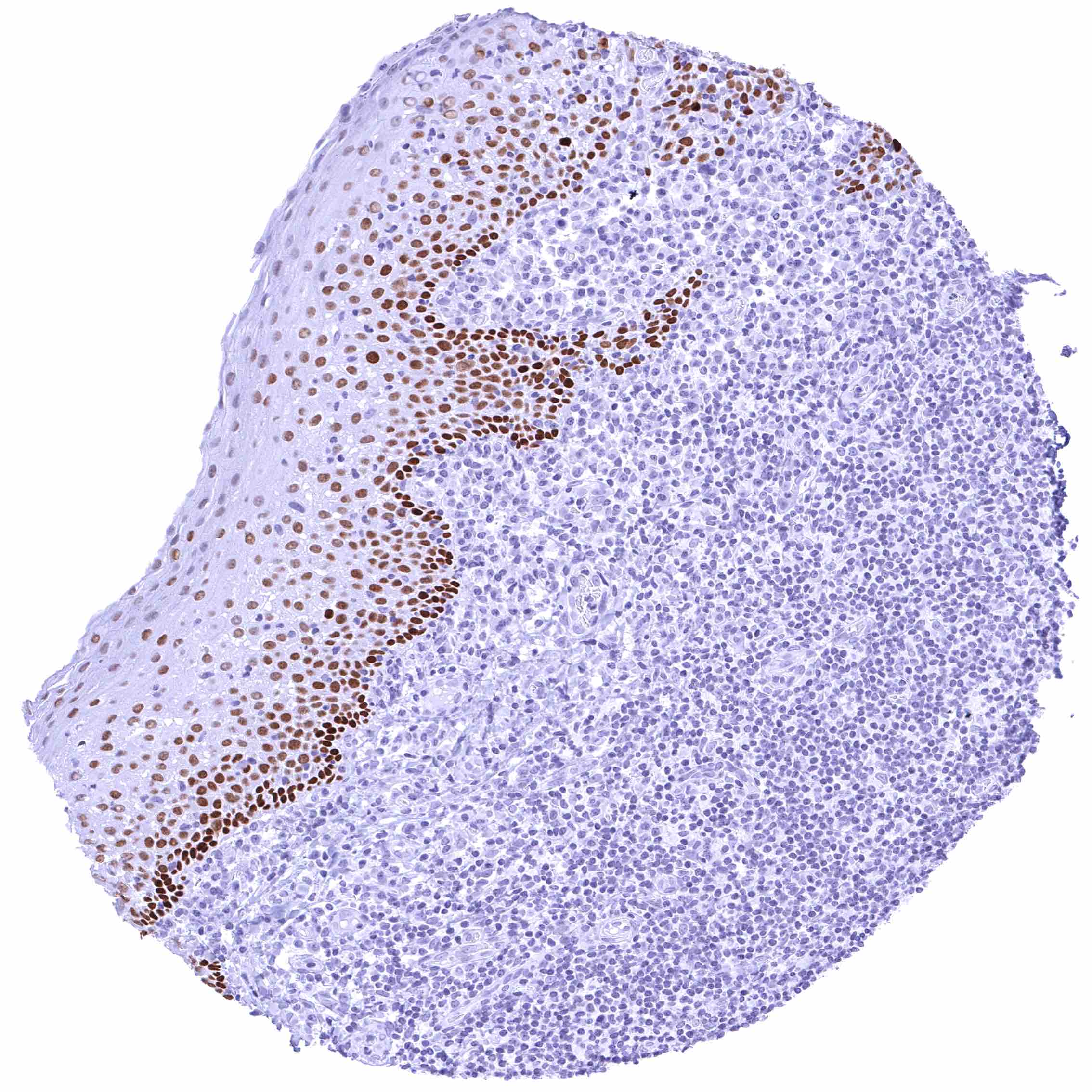
Tonsil, surface epithelium – Distinct nuclear FOXA1 staining of a subset of squamous epithelial cells. The staining intensity gradually decreases towards the surface epithelium.
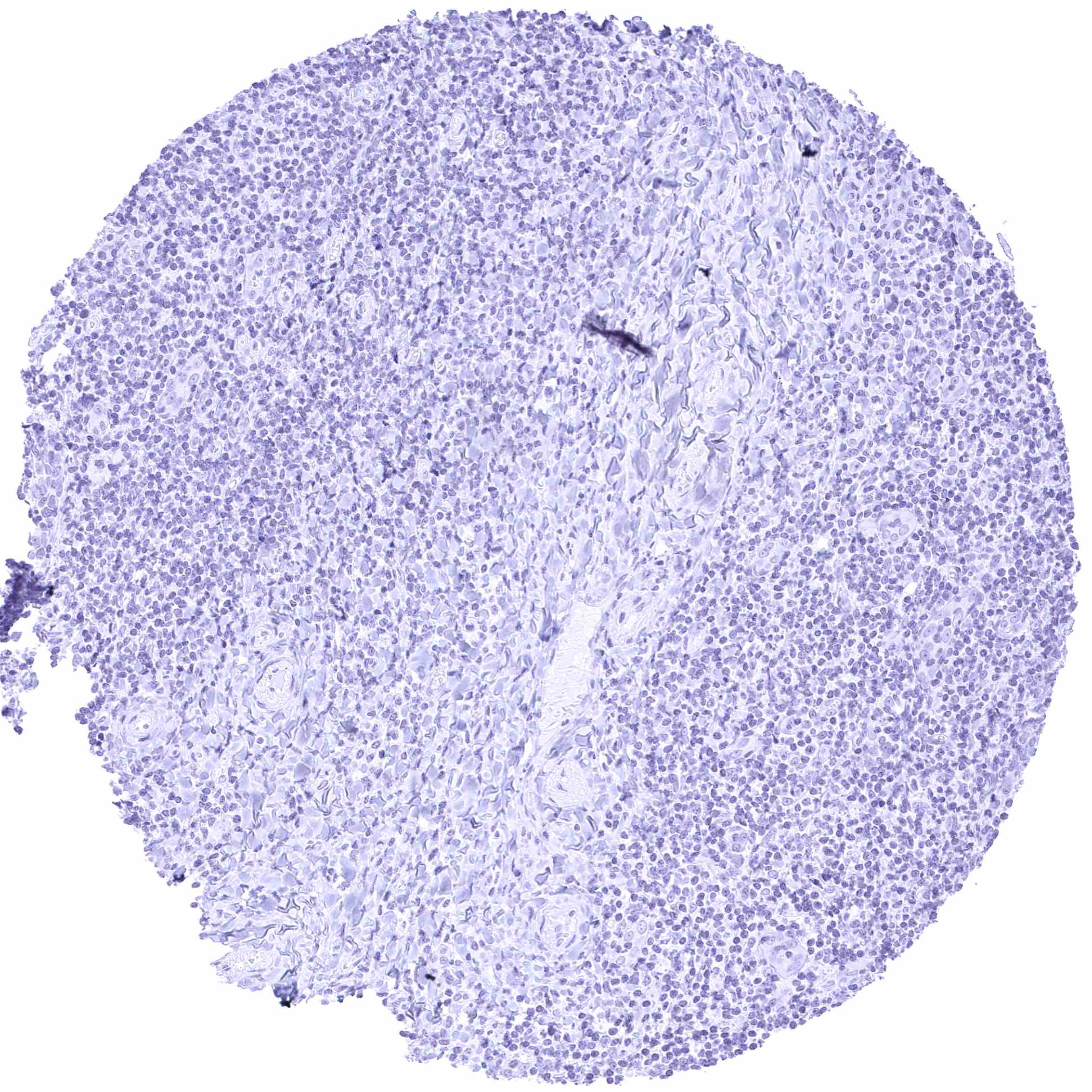
Tonsil
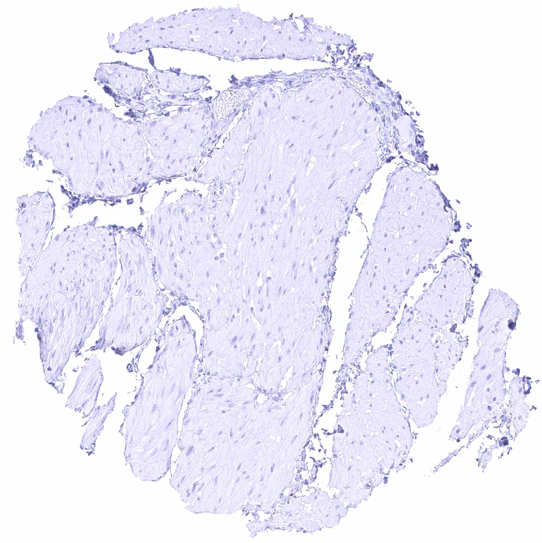
Urinary bladder, muscular wall
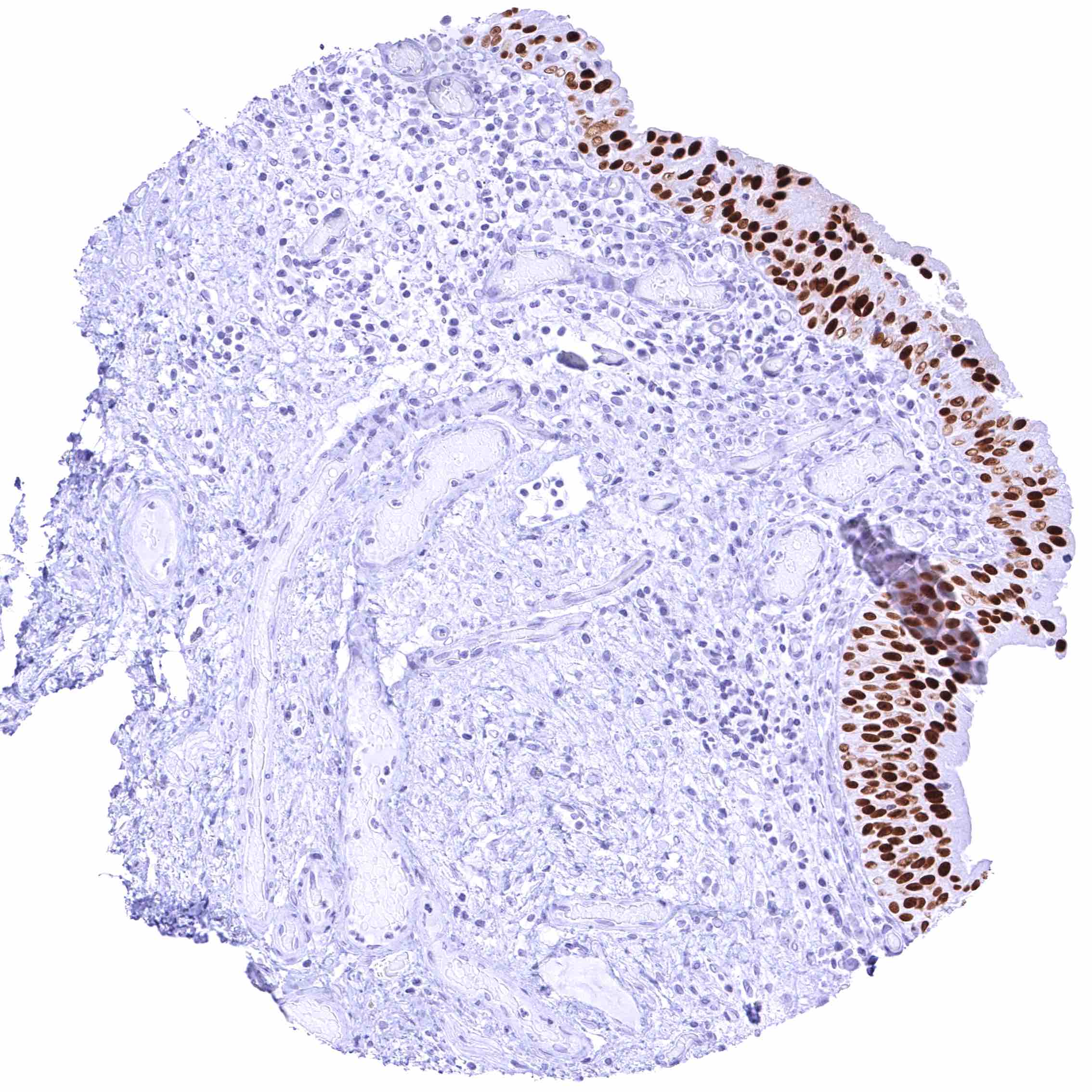
Urinary bladder, urothelium – Intense nuclear FOXA1 staining of urothelial cells.
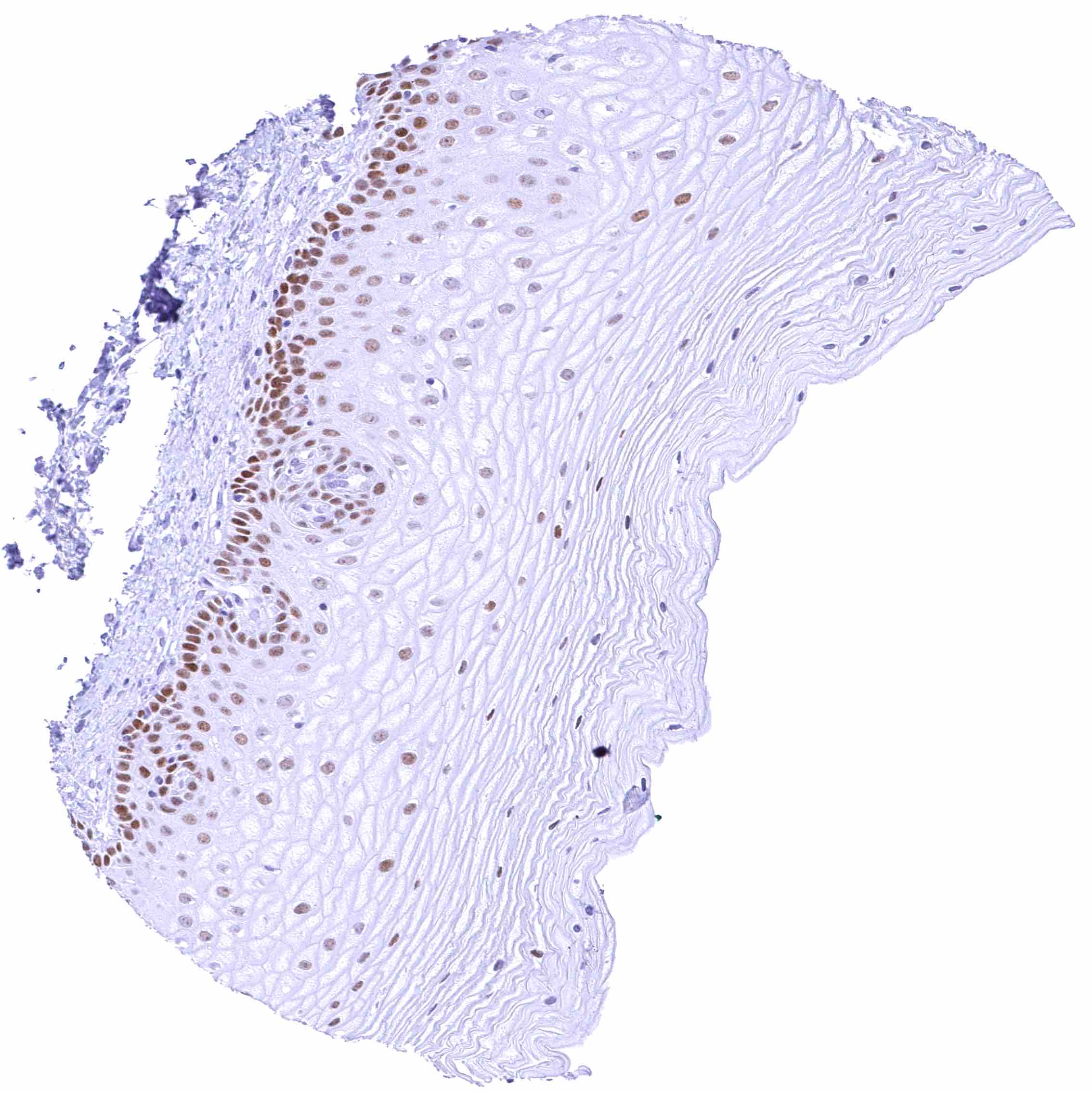
Uterus, ectocervix – Distinct nuclear FOXA1 staining of the basal and suprabasal layers of the squamous epithelium. The staining intensity markedly decreases towards the more superficial cell layers
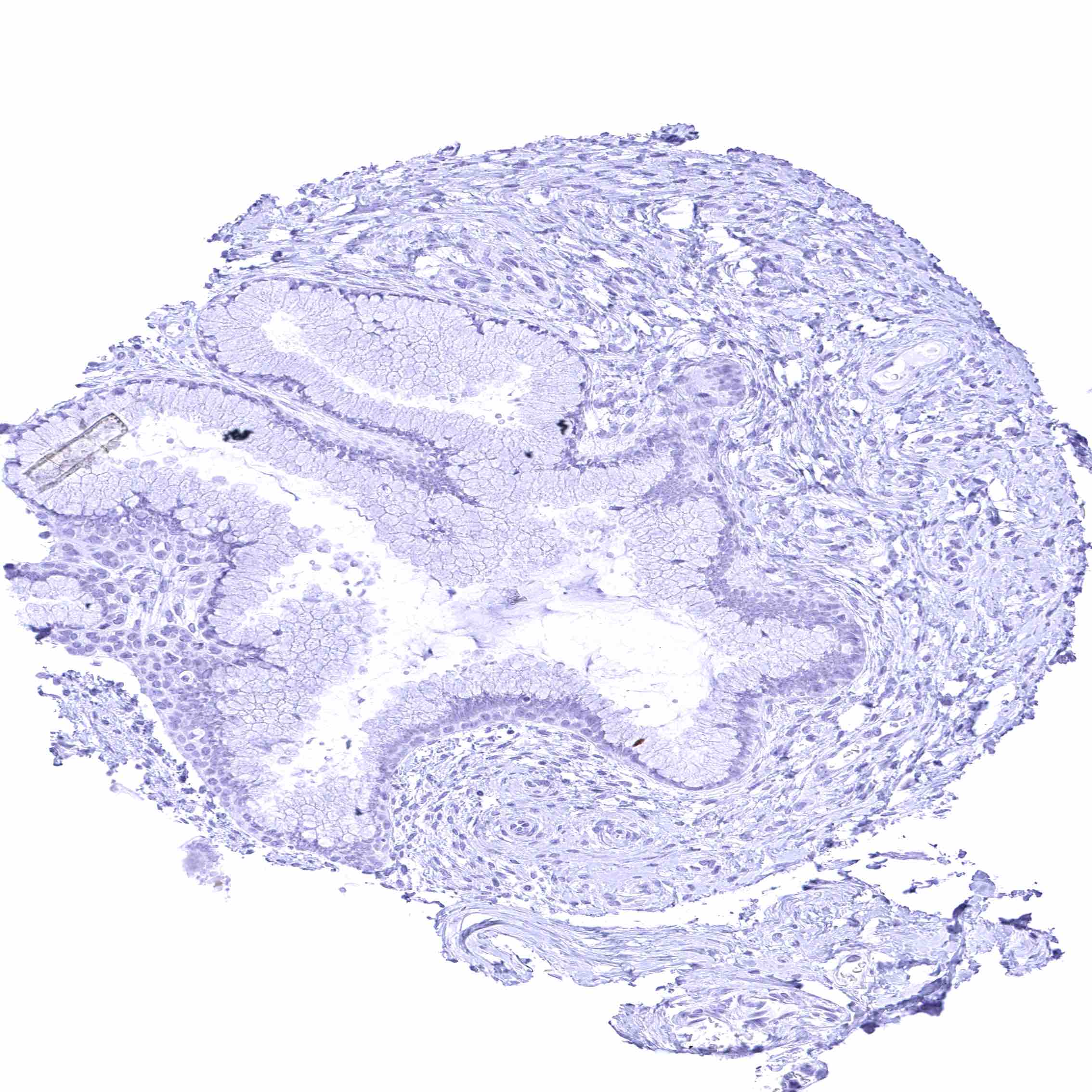
Uterus, endocervix
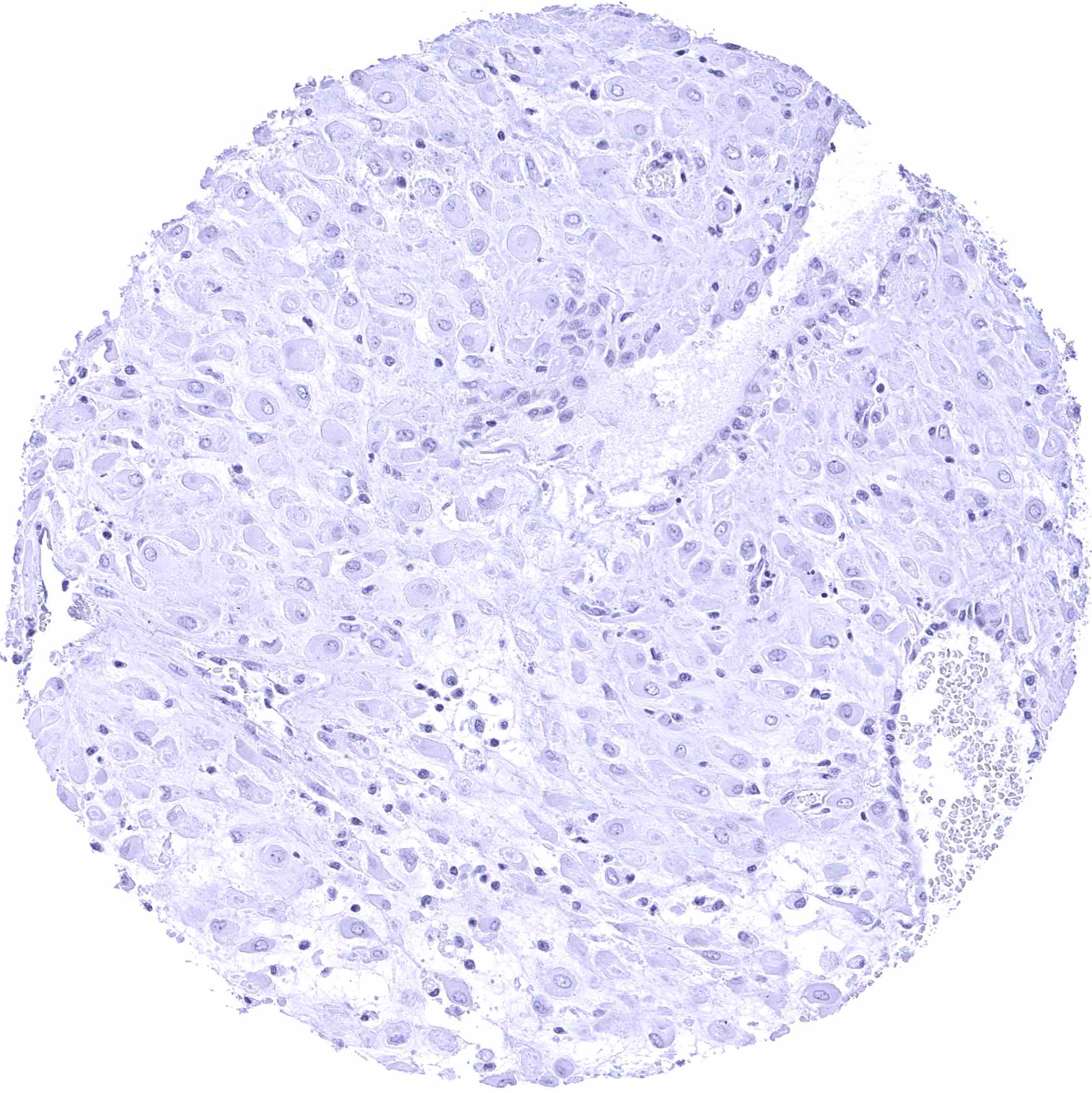
Uterus, endometrium (pregnancy)
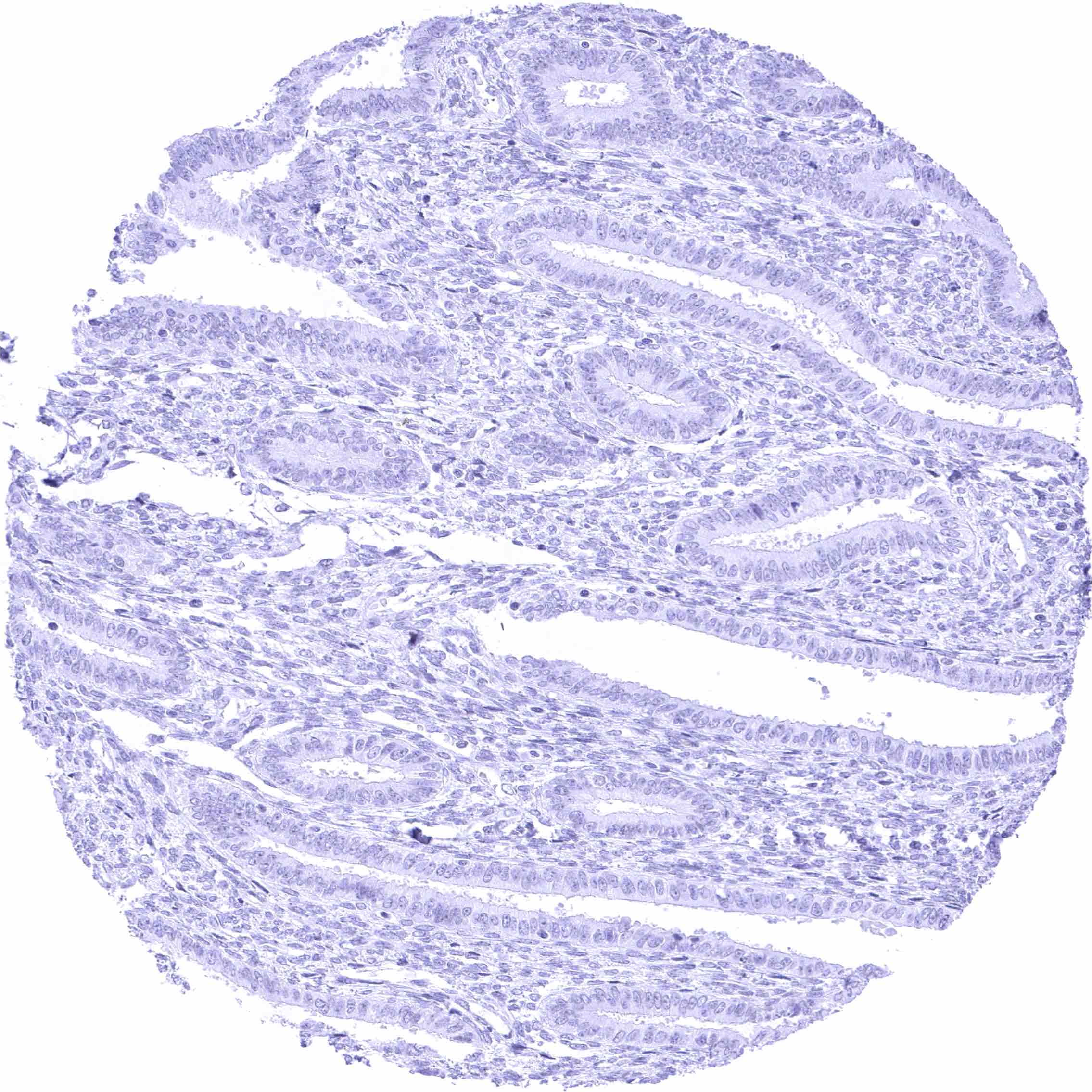
Uterus, endometrium (proliferation)
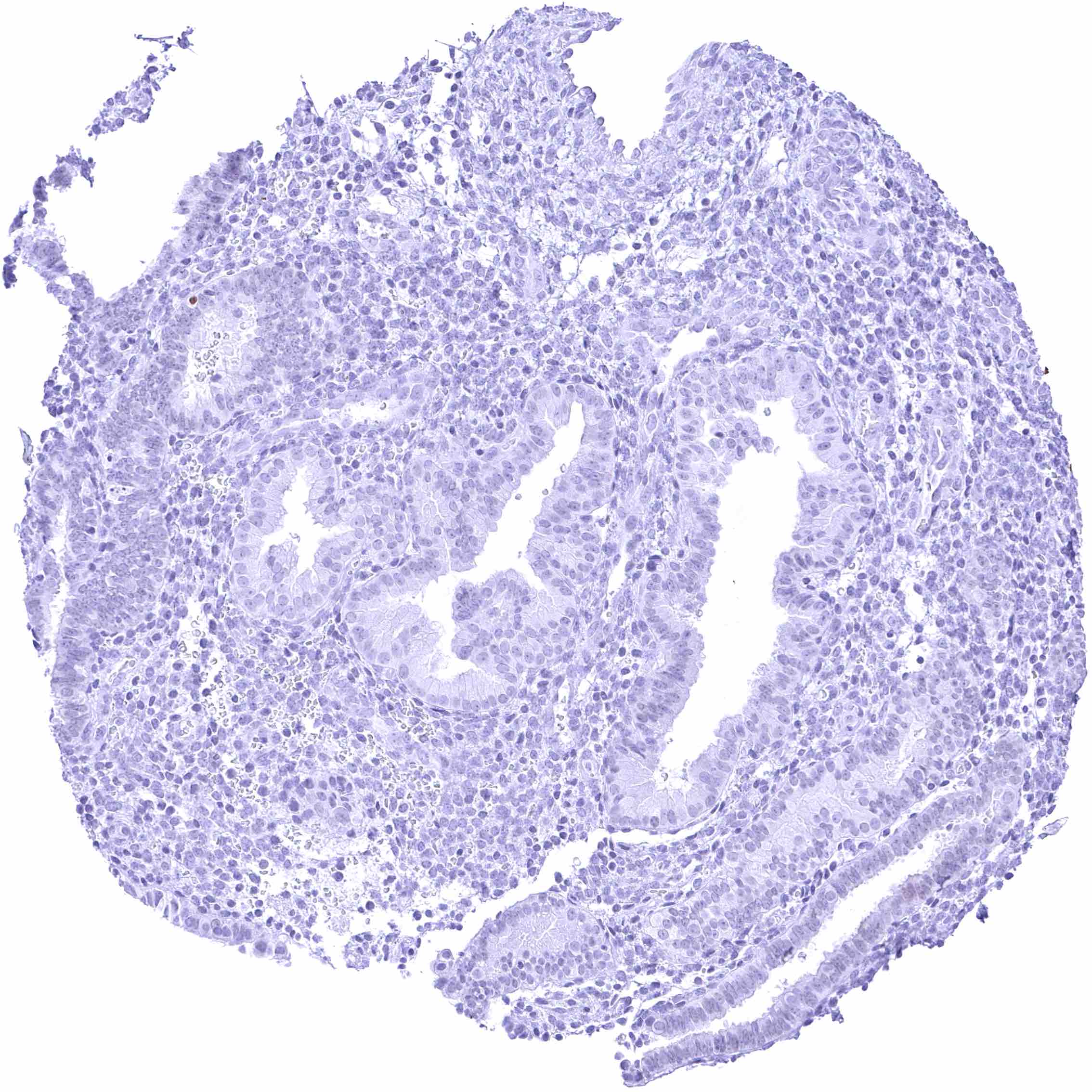
Uterus, endometrium (secretion)
Uterus, myometrium