
295,00 € – 995,00 €
Product details
Synonyms = FABP1, L-FABP
Antibody type = Mouse monoclonal / IgG
Clone = MSVA-501M
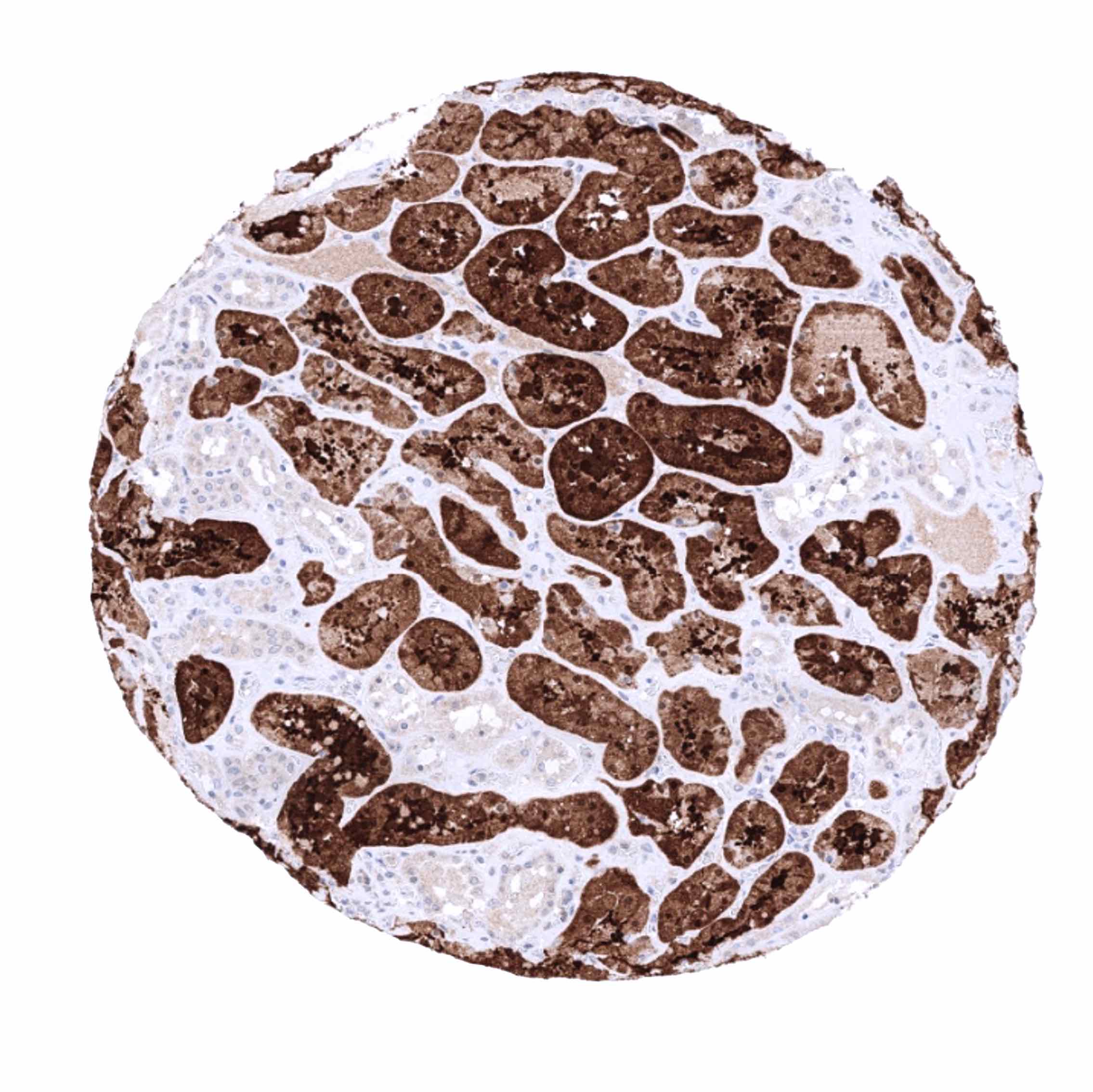
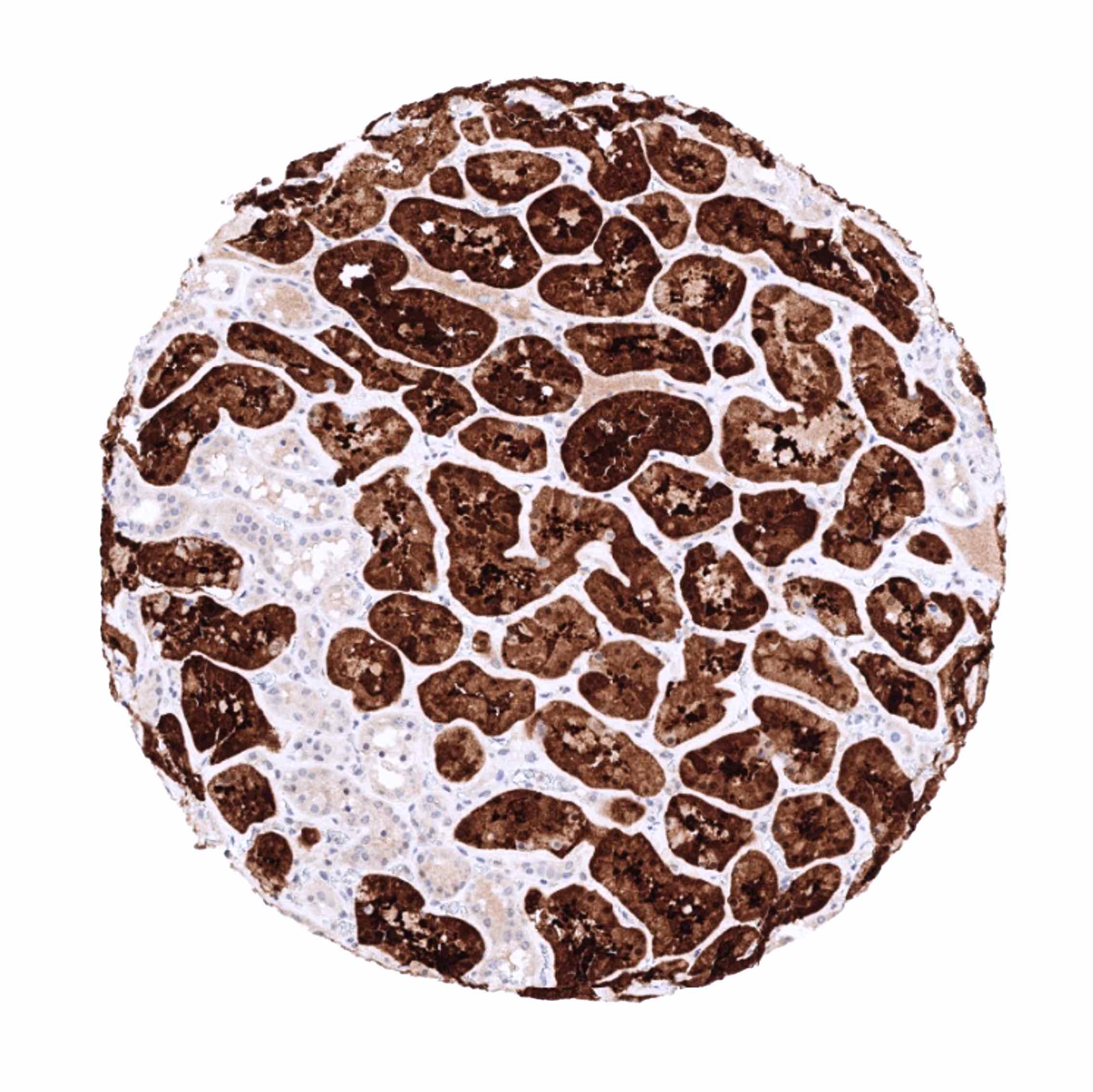
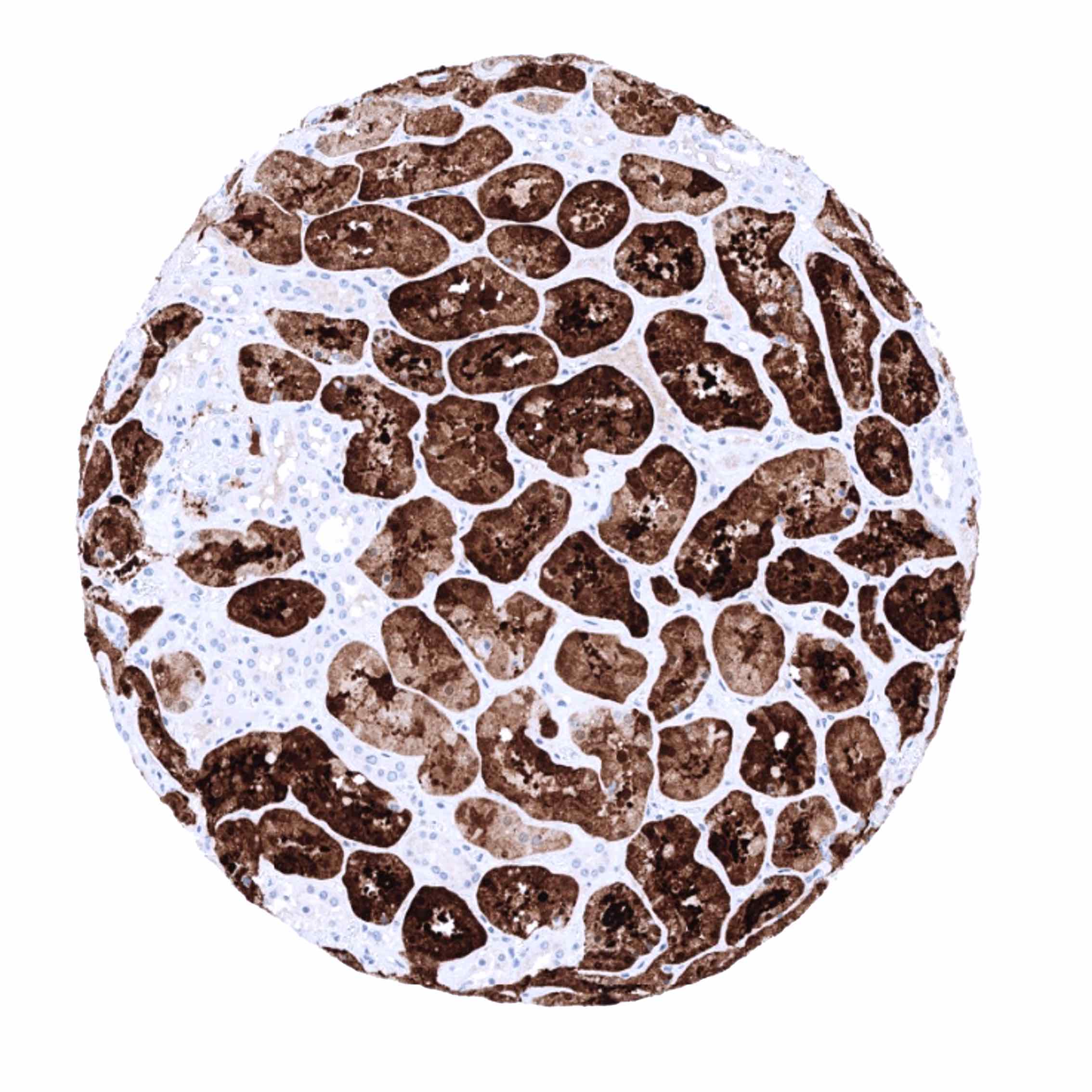
Positive control = Kidney: A strong cytoplasmic FABP1 immunostaining should be seen in cells of the proximal tubule while other cell types remain negative or display only a faint staining.
Negative control = Tonsil: All epithelial and lymphocytic cells must not show any FABP1 immunostaining.
Cellular localization = Cytoplasmic
Reactivity = Human
Application = Immunohistochemistry
Dilution = 1:100-200
Intended Use = Research Use Only
Relevance of Antibody
FABP1 is expressed in hepatocytes, kidney, and gastrointestinal epithelium.
Biology Behind
Fatty acid binding protein 1 (FABP1) is a 14kDa protein coded by the FABP1 gene at 2p11.2. FABP1 is found in the liver where it is expressed at high levels, with the expression level reflecting the rate of fatty acid metabolism. There are 9 different isoforms with tissue specific expression in the liver (L-FABP/FABP1), intestinal (I-FABP/FABP2), heart (H-FABP/FABP3), adipocyte (A-FABP/FABP4/aP2), epidermal (E-FABP/FABP5/mal1), ileal (Il-FABP/FABP6), brain (B-FABP/FABP7), myelin (M-FABP/FABP8), and testis (T-FABP/FABP9). Sequence similarity between different FABP isoforms is only 15–70% but all FABPs share a similar three-dimensional structure which enables a binding to various fatty acids. FABP1 is involved in the uptake, intracellular transport and metabolism of long-chain fatty acids, endocannabinoids, phytocannabinoids and other hydrophobic molecules in hepatocytes. In contrast to other fatty acid binding proteins, the FABP1 protein is characterized by a large hydrophobic pocket that can bind multiple ligands, including bile acids, bilirubin, cholesterol, free fatty acids and their derivates. Diseases associated with polymorphisms in the FABP1 gene include Hepatocellular Adenoma and Type 1 Diabetes Mellitus.
Staining Pattern in Normal Tissues
FABP1 staining pattern in Normal Tissues with antibody MSVA-501M (images are shown in our “Normal Tissue Gallery”)
| Brain | Cerebrum | Negative. |
| Cerebellum | Negative. | |
| Endocrine Tissues | Thyroid | Negative. |
| Parathyroid | Negative. | |
| Adrenal gland | Negative. | |
| Pituitary gland | Negative. | |
| Respiratory system | Respiratory epithelium | Negative. |
| Lung | Negative. | |
| Gastrointestinal Tract | Salivary glands | Negative. |
| Esophagus | Negative. | |
| Stomach | Surface epithelium is usually FABP1 negative, but focal positivity may occur in the antrum. | |
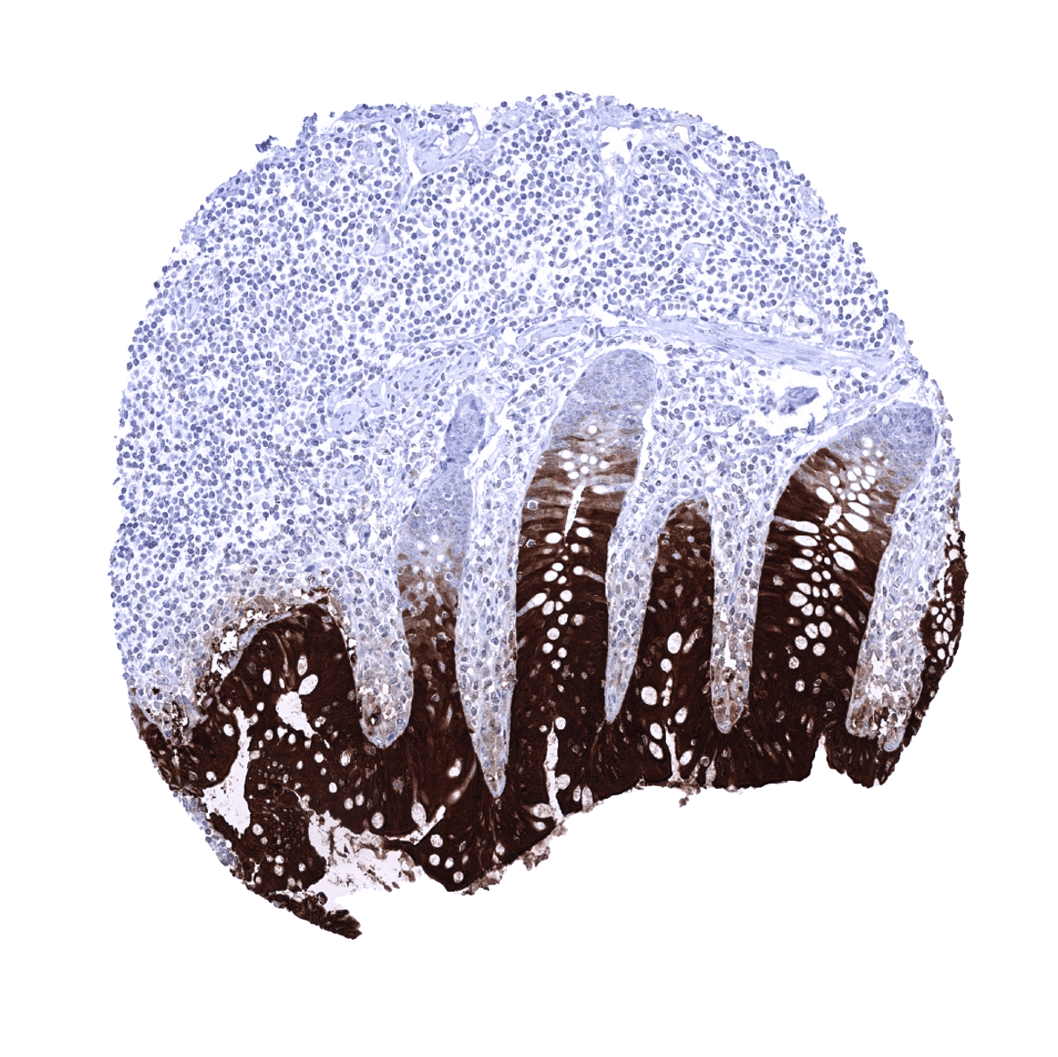
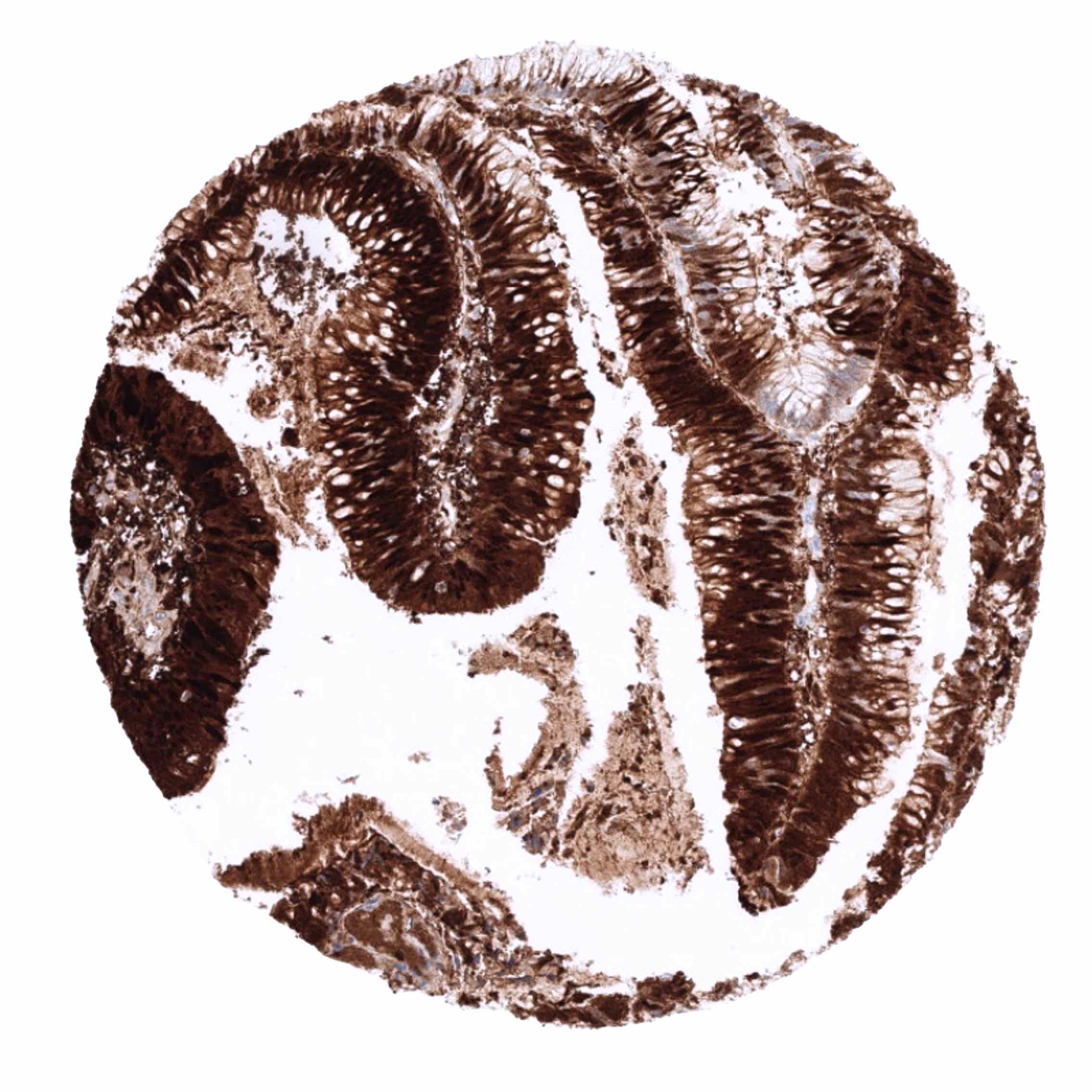
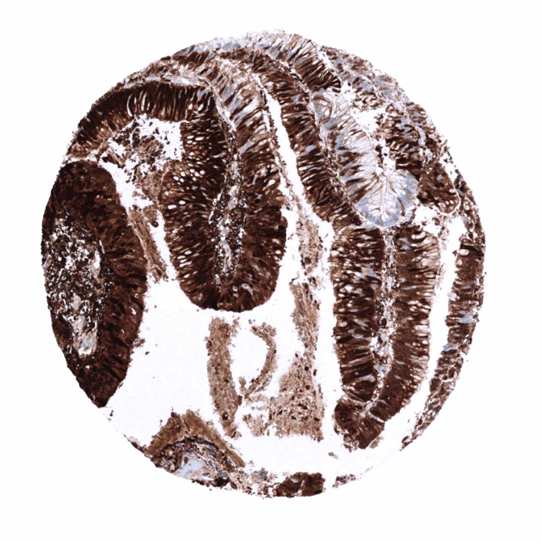
| Duodenum | Strong FABP1 staining of epithelial cells. The FABP1 staining intensity is strongest at the surface epithelium and decreases towards the crypt bases. Crypt bases may be FABP1 negative. | |
| Small intestine | Strong FABP1 staining of epithelial cells. The FABP1 staining intensity is strongest at the surface epithelium and decreases towards the crypt bases. Crypt bases may be FABP1 negative. | |
| Appendix | Strong FABP1 staining of epithelial cells. The FABP1 staining intensity is strongest at the surface epithelium and decreases towards the crypt bases. Crypt bases may be FABP1 negative. | |
| Colon | Strong FABP1 staining of epithelial cells. The FABP1 staining intensity is strongest at the surface epithelium and decreases towards the crypt bases. Crypt bases may be FABP1 negative. | |
| Rectum | Strong FABP1 staining of epithelial cells. The FABP1 staining intensity is strongest at the surface epithelium and decreases towards the crypt bases. Crypt bases may be FABP1 negative. | |
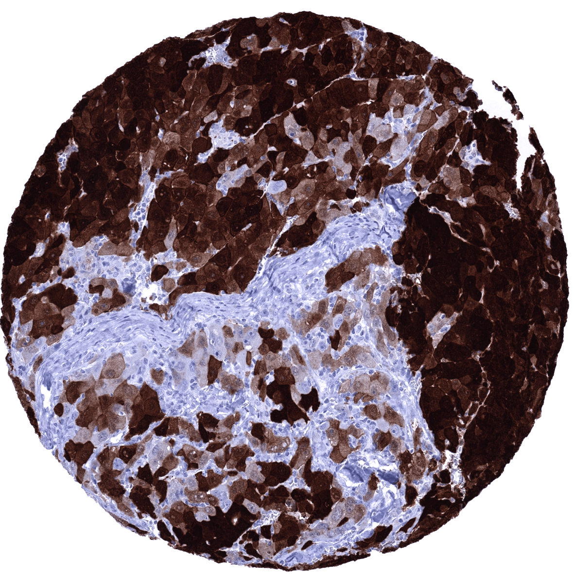
| Liver | Strong FABP1 staining of hepatocytes. | |
| Gallbladder | Negative. | |
| Pancreas | Negative. | |
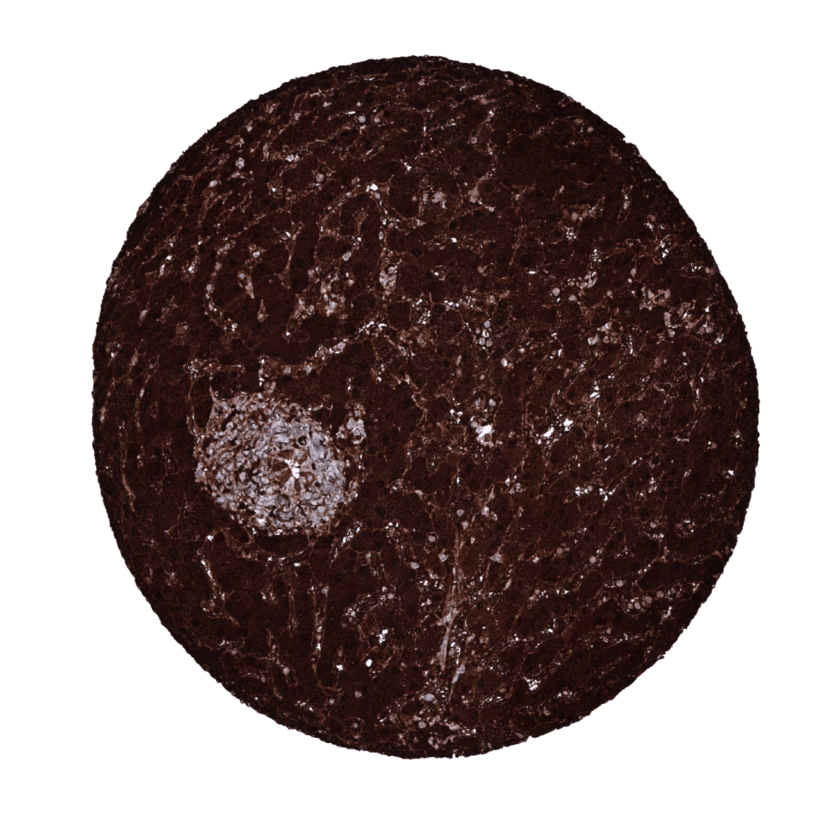
| Genitourinary | Kidney | Strong FABP1 staining of proximal tubuli. |
| Urothelium | Negative. | |
| Male genital | Prostate | Negative. |
| Seminal vesicles | Negative. | |
| Testis | Negative. | |
| Epididymis | Negative. | |
| Female genital | Breast | Negative. |
| Uterus, myometrium | Negative. | |
| Uterus, ectocervix | Negative. | |
| Uterus endocervix | Negative. | |
| Uterus, endometrium | Negative. | |
| Fallopian Tube | Negative. | |
| Ovary | Negative. | |
| Placenta early | Negative. | |
| Placenta mature | Negative. | |
| Amnion | Negative. | |
| Chorion | Negative. | |
| Skin | Epidermis | Negative. |
| Sebaceous glands | Negative. | |
| Muscle/connective tissue | Heart muscle | Negative. |
| Skeletal muscle | Negative. | |
| Smooth muscle | Negative. | |
| Vessel walls | Negative. | |
| Fat | Negative. | |
| Stroma | Negative. | |
| Endothelium | Negative. | |
| Bone marrow/ lymphoid tissue | Bone marrow | Negative. |
| Lymph node | Negative. | |
| Spleen | Negative. | |
| Thymus | Negative. | |
| Tonsil | Negative. | |
| Remarks | Because of the very high FABP1 expression in gastrointestinal or liver cells, adjacent structures may show some FABP1 immunostaining due to a contamination artifact. |
These findings are fully consistent with the RNA and protein data described in the Human Protein Atlas (Tissue expression FABP1). All organs with documented FABP1 RNA expression (duodenum, small intestine, colon, rectum, appendix, kidney, liver) are IHC positive for MSVA-501M.
Positive control: Kidney: A strong cytoplasmic FABP1 immunostaining should be seen in cells of the proximal tubule while other cell types remain negative or display only a faint staining.
Negative control: Tonsil: All epithelial and lymphocytic cells must not show any FABP1 immunostaining.


Staining Pattern in Relevant Tumor Types
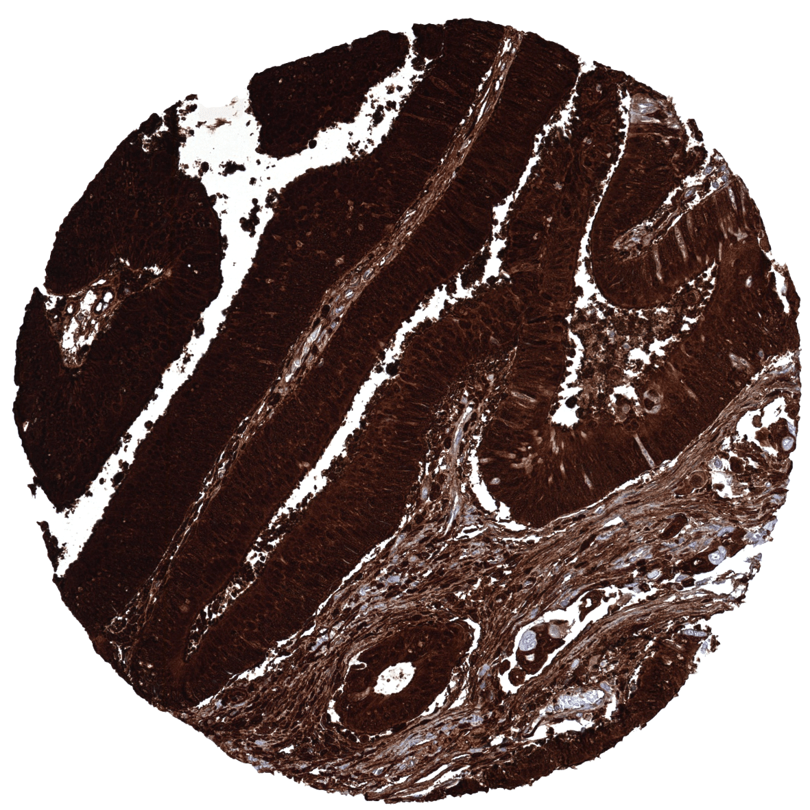
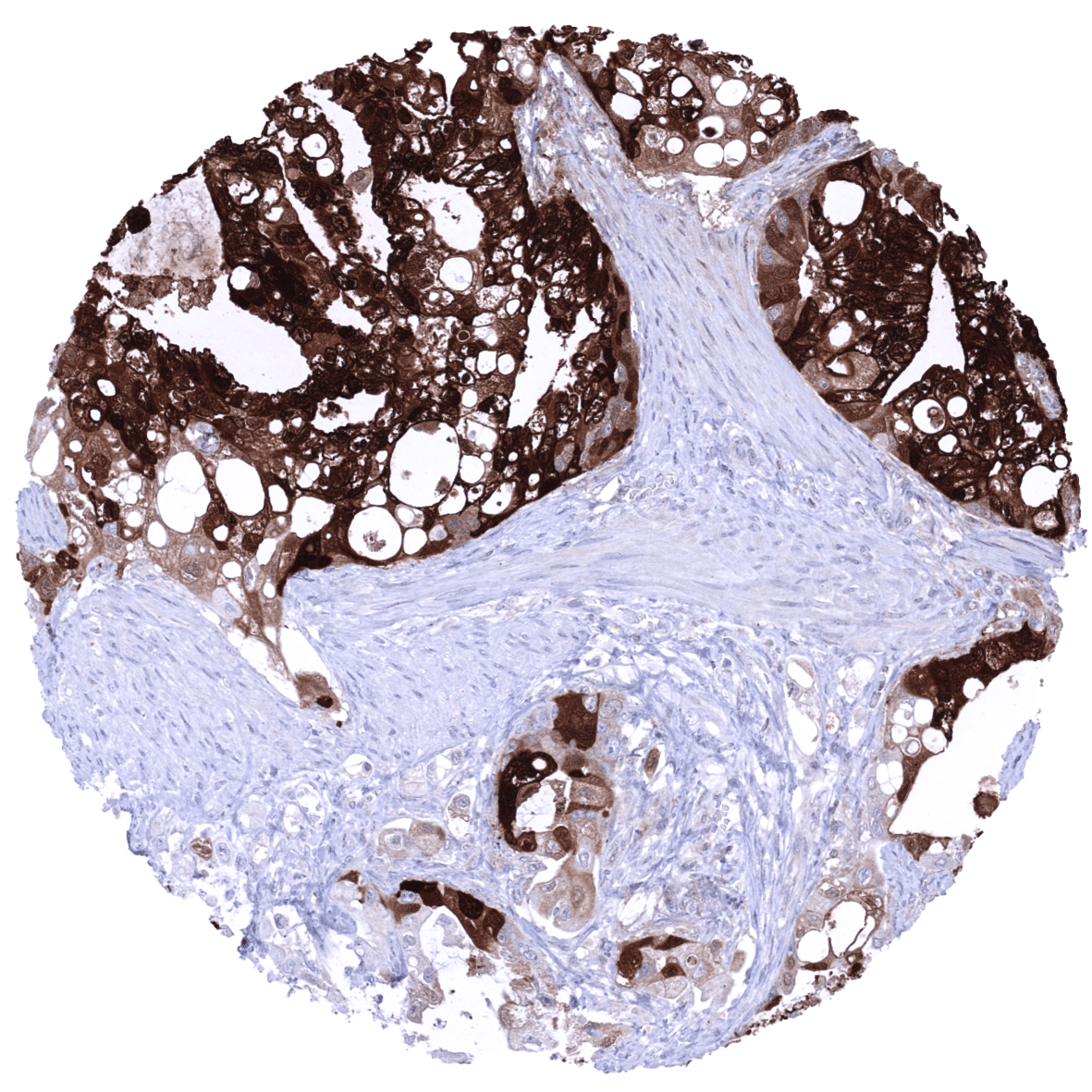
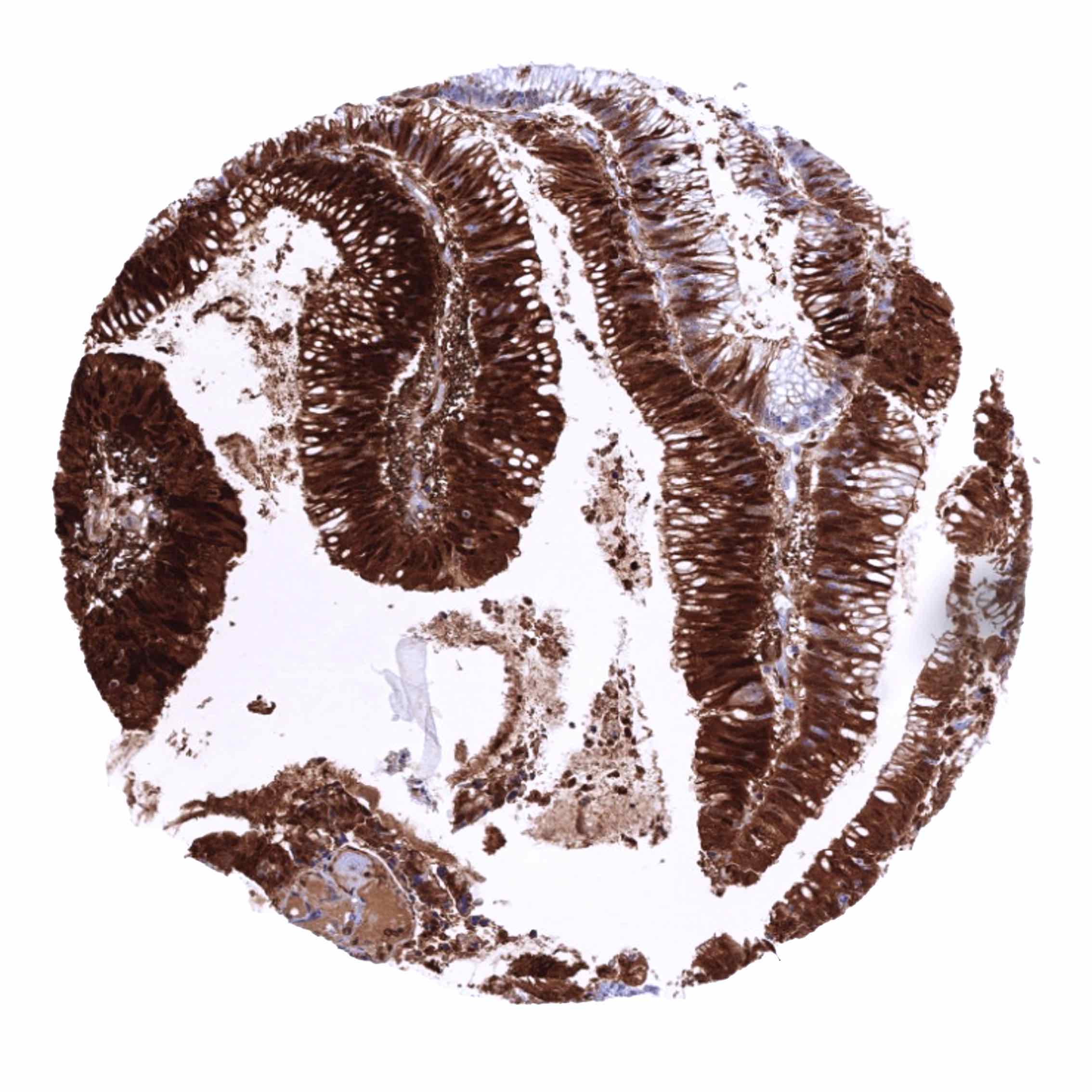
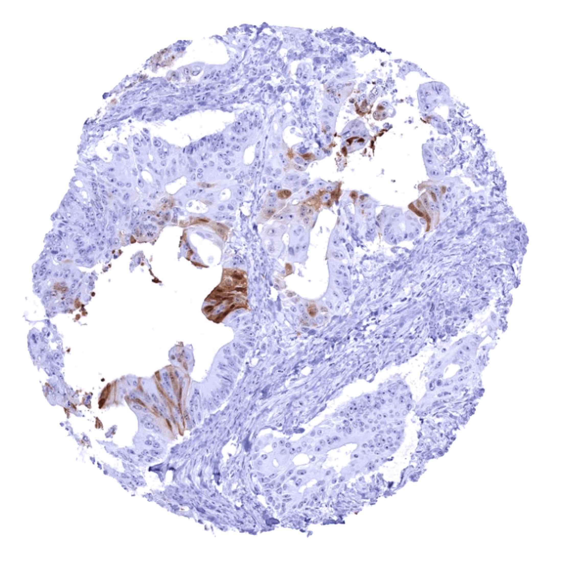
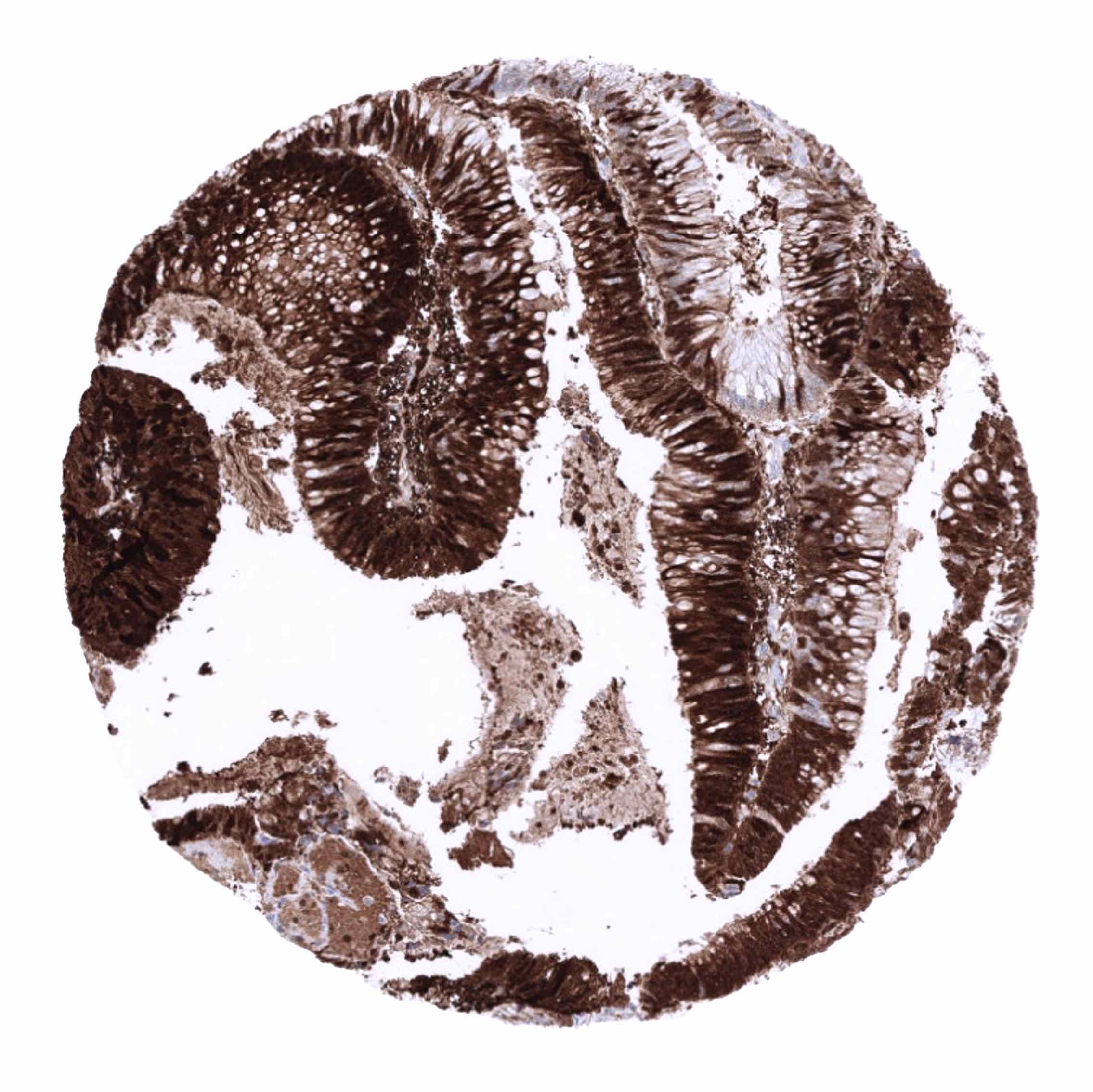
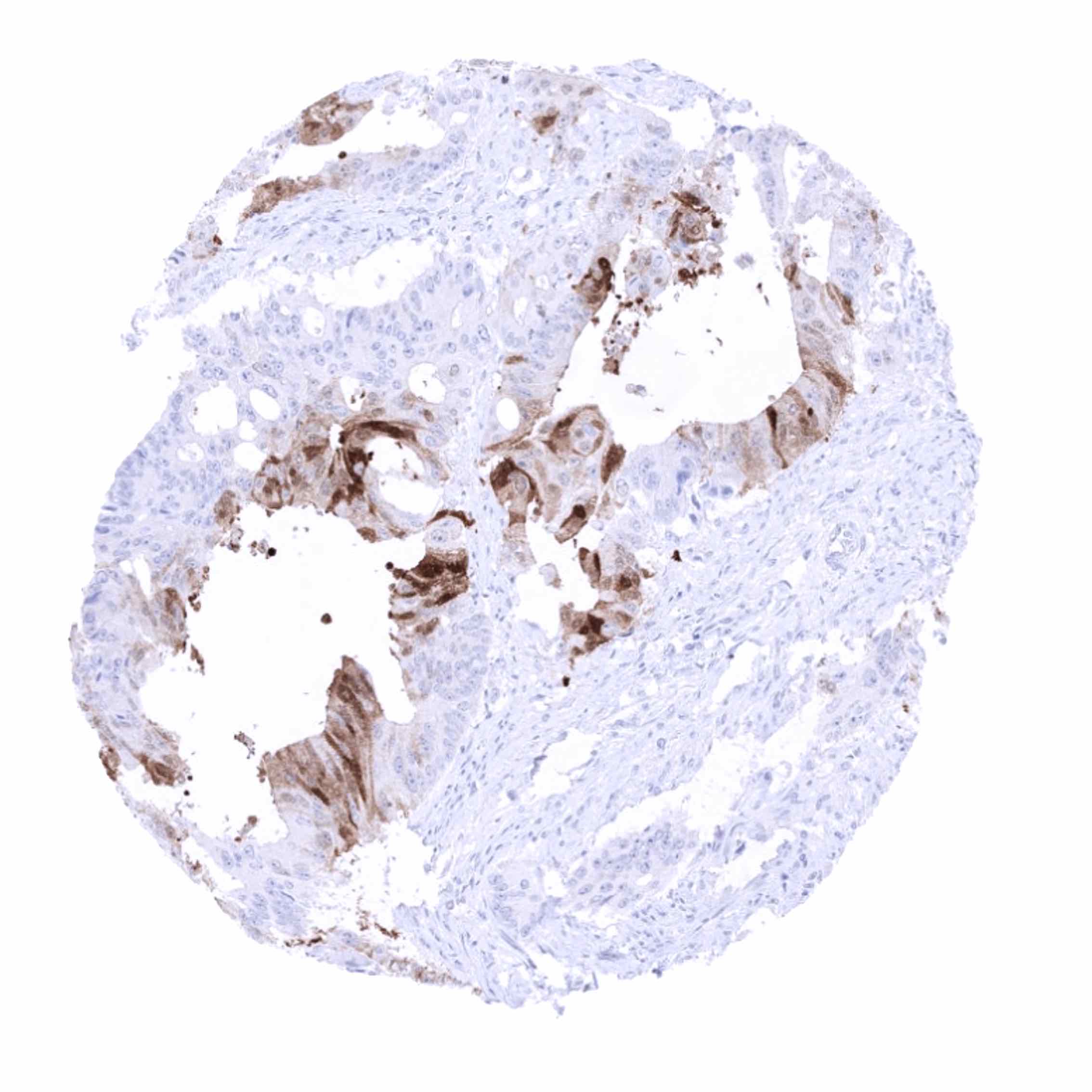
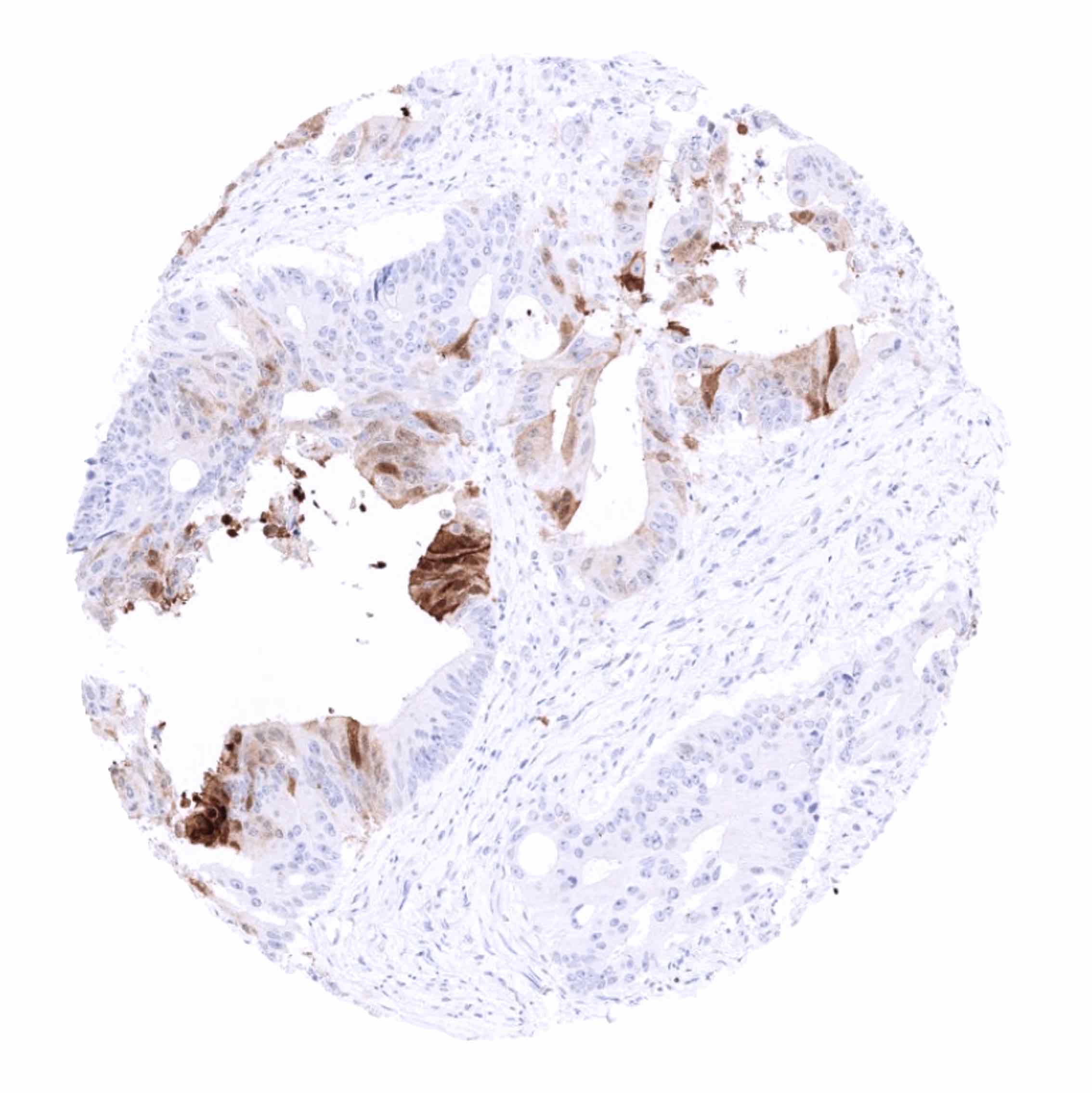
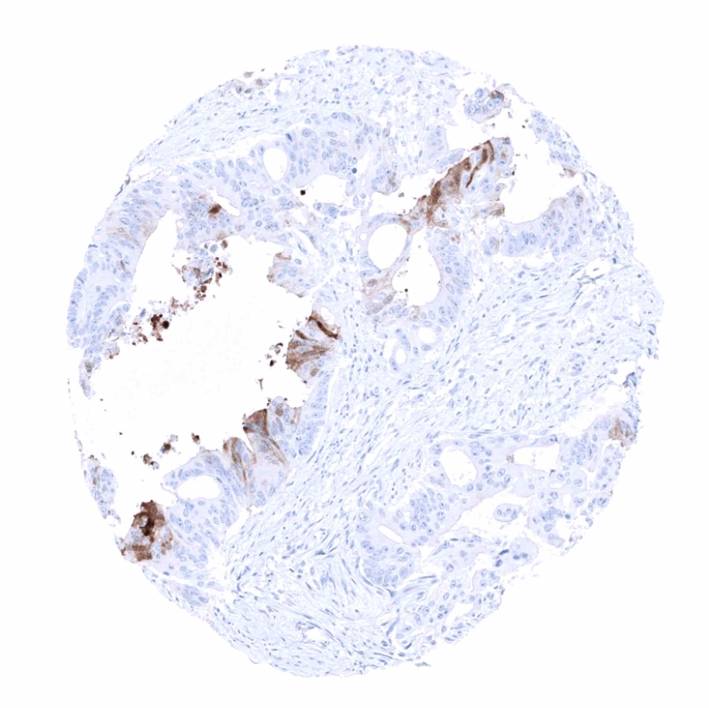
The TCGA database on RNA expression in cancer has described upregulation of FABP1 in most hepatocellular carcinomas and a fraction of colorectal and stomach cancers. Most other important tumor entities are usually negative.
The TCGA findings on FABP1 RNA expression in different tumor categories have been summarized in the Human Protein Atlas.

Compatibility of Antibodies
FABP1 (MSVA-501M) publication summary
Relevant publication: Dum et al.: “FABP1 expression in human tumors: A tissue microarray study on 17,071 tumors.” Published in Virchows Archiv Aug 11. doi: 10.1007/s00428-022-03394-5. Epub ahead of print. PMID: 35951102.
A total of 14’597 tumors from 150 different tumor categories were successfully analyzed by using the following protocol: Heat-induced antigen retrieval for 5 minutes in an autoclave at 121°C in pH 7,8 Target Retrieval Solution buffer. MSVA-501M at a dilution of 1:150 at 37°C for 60 minutes. Visualization of bound antibody by the EnVision Kit (Dako, Agilent). This protocol was also used for all stainings depicted in our tumor and normal tissue galleries.
Overall, 24 (16%) of 150 tumor categories showed detectable FABP1 expression in at least one case and 17 (11%) tumor categories included at least one case with strong FABP1 positivity. By far the highest positivity rates were seen in colorectal adenomas (44-88%), colorectal adenocarcinomas (71%) and in hepatocellular carcinomas (65%), followed by mucinous carcinoma of the ovary (35%), cholangiocarcinoma (22%), and various other adenocarcinomas from the digestive tract (10-23%). Eleven additional entities had positivity rates between 0.2 and 6.5%. The distribution of positive staining results is shown in “organ-systematic” and in “ranking order” figures below (images based on data from Dum et al).
Authors conclusions on diagnostic utility of FABP1 IHC with respect to the distinction of different tumor entities (Dum et al.):
- A positive FABP1 immunostaining in a metastatic tissue of unknown origin pinpoints towards the liver or the gastrointestinal tract as the most likely sites of cancer origin.
- Distinction of pulmonary adenocarcinomas (nearly always FABP1 negative) from metastatic adenocarcinoma from the gastrointestinal tract (FABP1 positive in 50-70%) as part of a panel.
- In case of an adenocarcinoma in the pancreas, FABP1 positivity argues in favor of a carcinoma derived from the ampulla Vaterii (23% positive) and against a ductal adenocarcinoma (1.8% positive).
Authors conclusions on the prognostic role of FABP1 immunostaining results (Dum et al.):
- Absent or low FABP1 expression is strongly linked to MSI and right sided tumor location but unrelated to pT and pN stage in colorectal cancer.
Data from the publication: “FABP1 expression in human tumors: A tissue microarray study on 17,071 tumors.” Published by Dum et al. in Virchows Archiv Aug 11. doi: 10.1007/s00428-022-03394-5. Epub ahead of print. PMID: 35951102.
Summarized in own graphics:
Figure 1. FABP1 staining in cancer (“organ-systematic”; according to Dum et al.)

Figure 2. FABP1 staining in cancer (“ranking list”; according to Dum et al.)

Figure 3. Clinico-pathological associations described by Dum et al. (p-value)

Protocol Recommendations
IHC users have different preferences on how the stains should look like. Some prefer high staining intensity of the target stain and even accept some background. Others favor absolute specificity and lighter target stains. Factors that invariably lead to more intense staining include higher concentration of the antibody and visualization tools, longer incubation time, higher temperature during incubation, higher temperature and longer duration of the heat induced epitope retrieval (slide pretreatment). The impact of the pH during slide pretreatment has variable effects and depends on the antibody and the target protein.
All images and data shown here and in our image galleries are obtained by the manual protocol described below. Other protocols resulting in equivalent staining are described as well.
Manual protocol
Freshly cut sections should be used (less than 10 days between cutting and staining). Heat-induced antigen retrieval for 5 minutes in an autoclave at 121°C in pH 7,8 Target Retrieval Solution buffer. Apply MSVA-501M at a dilution of 1:150 at 37°C for 60 minutes. Visualization of bound antibody by the EnVision Kit (Dako, Agilent) according to the manufacturer’s directions.

Agilent / Dako – Autostainer Link 48
Pretreatment in PT-Link for 30 minutes at 95°C (pH high); FLEX peroxidase blocking for 5 minutes (room temperature), MSVA-501M 1:150 for 20 minutes (room temperature), FLEX+ mouse/rabbit (LINKER) for 15 minutes (room temperature), horseradish peroxidase (HRP) for 20 minutes (room temperature), FLEX DAB+Sub-Chromo for 10 minutes (room temperature), FLEX hematoxylin for 5 minutes (room temperature).
These images reflect stainings by the protocol described above. It is of note that a comparable staining result can also be obtained by different protocols. In general, a longer pretreatment, a longer incubation time of the primary antibody, a higher antibody concentration, and a longer incubation time of FLEX+LINKER result in stronger staining, potentially at the cost of more background staining. Modifications of the protocol with a strengthening effect on staining intensity in combination with changes of other parameters that result in lower staining intensity can result in a comparable result as shown above.
Leica – BOND RX
Dewax at 72°C for 30 seconds; Pretreatment in Bond Epitope Retrieval Solution (ER2 – EDTA pH9) for 20 minutes at 100°C; Peroxidase blocking for 5 minutes (room temperature), MSVA-501M 1:150 for 15 minutes (room temperature), Post primary (rabbit anti mouse) for 8 minutes (room temperature), Polymer (goat anti rabbit) for 8 minutes (room temperature), mixed DAB refine for 10 minutes (room temperature), hematoxylin for 5 minutes (room temperature).
These images reflect stainings by the protocol described above. It is of note that a comparable staining result can also be obtained by different protocols. In general, a longer pretreatment, a longer incubation time of the primary antibody, a higher antibody concentration, a higher temperature during incubation, and a longer incubation time of Post primary and or the Polymer result in stronger staining, potentially at the cost of more background staining. Modifications of the protocol with a strengthening effect on staining intensity in combination with changes of other parameters that result in lower staining intensity can result in a comparable result as shown above.
Roche – Ventana Discovery ULTRA
Pretreatment for 64 minutes at 100°C (pH 8,4); CM peroxidase blocking for 12 minutes (room temperature), MSVA-501M 1:50 for 20 minutes at 36°C, secondary antibody (anti-mouse HQ) for 12 minutes at 36°C, anti-HQ HRP for 12 minutes at room temperature, DAB at room temperature, hematoxylin II at room temperature for 8 minutes, bluing reagent at room temperature for 4 minutes.
These images depict staining results obtained by the protocol described above. It is of note, that the Ventana machines generally require higher antibody concentrations than other commonly used autostainers because the antibodies are automatically diluted during the procedure. Various other protocols can result in an identical result as shown above. A longer pretreatment, a longer incubation time of the primary antibody, a higher antibody concentration, a higher temperature during incubation, and a longer incubation time of secondary antibody and or the anti-HQ HRP result in stronger staining, potentially at the cost of more background staining.
Potential Research Applications
- The diagnostic utility of FABP1 IHC should be investigated in a large cohort of tumors from different entities.
- Clinical and prognostic significance of FABP1 expression levels in gastrointestinal cancers is unknown.
- The clinical significance of FABP1 gene variants is under investigation.
