295,00 € – 895,00 €
Product details
Synonyms = Cadherin family member 4; CDHF4; Desmoglein-1; Desmosomal glycoprotein 1; DG1; DSG1; EPKHE; EPKHIA; Pemphigus foliaceus antigen; PPKS1; SPPK1
Antibody type = Mouse monoclonal / IgG
Clone = MSVA-544M
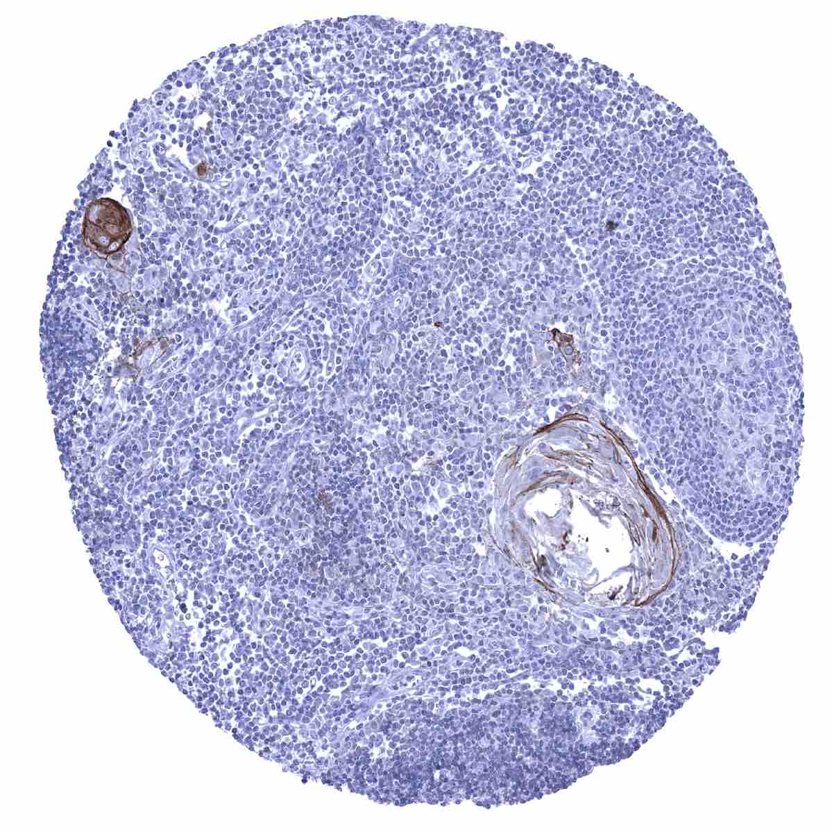
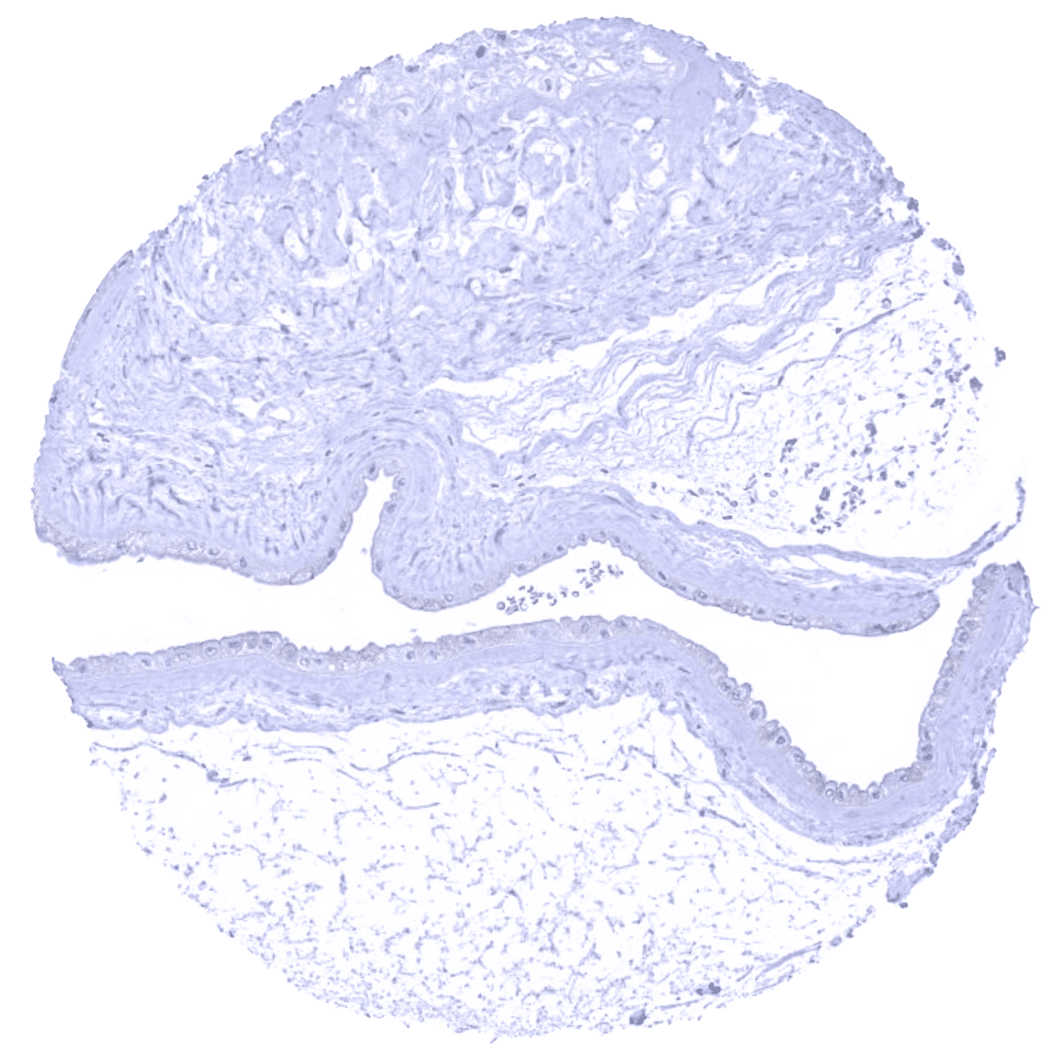
Positive control = Tonsil: A moderate to strong desmoglein 1 immunostaining should be seen in a fraction of squamous epithelial cells.
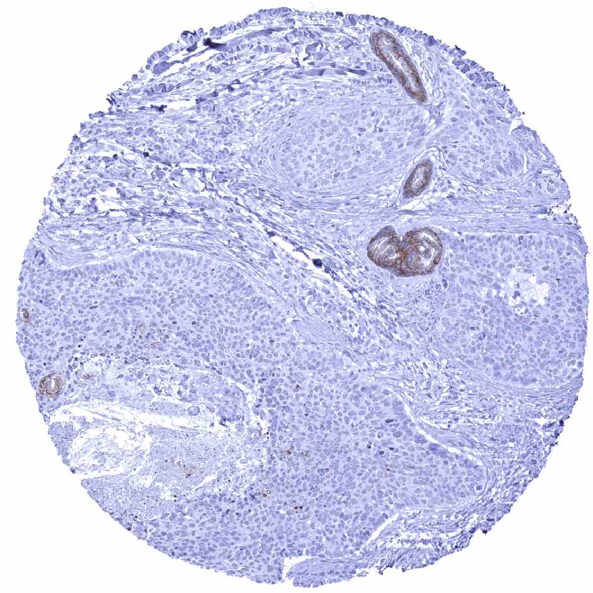
Negative control = Desmoglein 1 immunostaining should be absent in all non-squamous epithelial cell types.
Cellular localization = Cell surface
Reactivity = Human
Application = Immunohistochemistry
Dilution = 1:100
Intended Use = Research Use Only
Relevance of Antibody
Desmoglein 1 is expressed in squamous epithelium and cells undergoing squamous differentiation.
Biology Behind
Desmoglein-1 (Dsg1) is a 114 kDa calcium-binding transmembrane glycoprotein which is coded by the DSG1 gene at chromosome 18q12.1. Dsg1 is a component of desmosomes. Dsg1 and three other desmogleins belong to the cadherin cell adhesion molecule superfamily. Dsg1 is relevant for cell-cell adhesion but also has important functional roles for organization and maintenance of squamous epithelial tissues. When squamous cells exit the proliferating basal layer, Dsg1 attenuates MAPK/ERK signaling to enable further cell differentiation. In response to UV exposure, Dsg1 expression is downregulated which results in a re-activation of the MAPK/ERK pathway and increased epidermal inflammation. In the skin, Dsg1 modulates pigment production and secretion and other functions of melanocytes through regulation of the production of cytokines, chemokines, and other secreted factors. Deficiency or decreased expression of the Dsg1 protein results in an increased expression of several allergy-associated cytokines. Loss-of-function mutations in Dsg1 lead to severe dermatitis, multiple allergies, and metabolic wasting (SAM) syndrome.
Staining Pattern in Normal Tissues
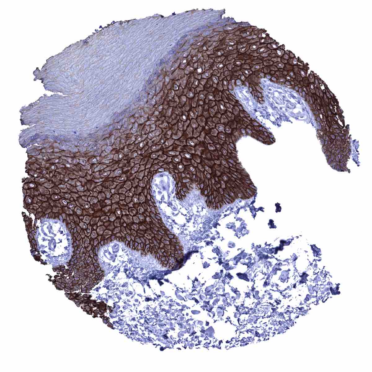
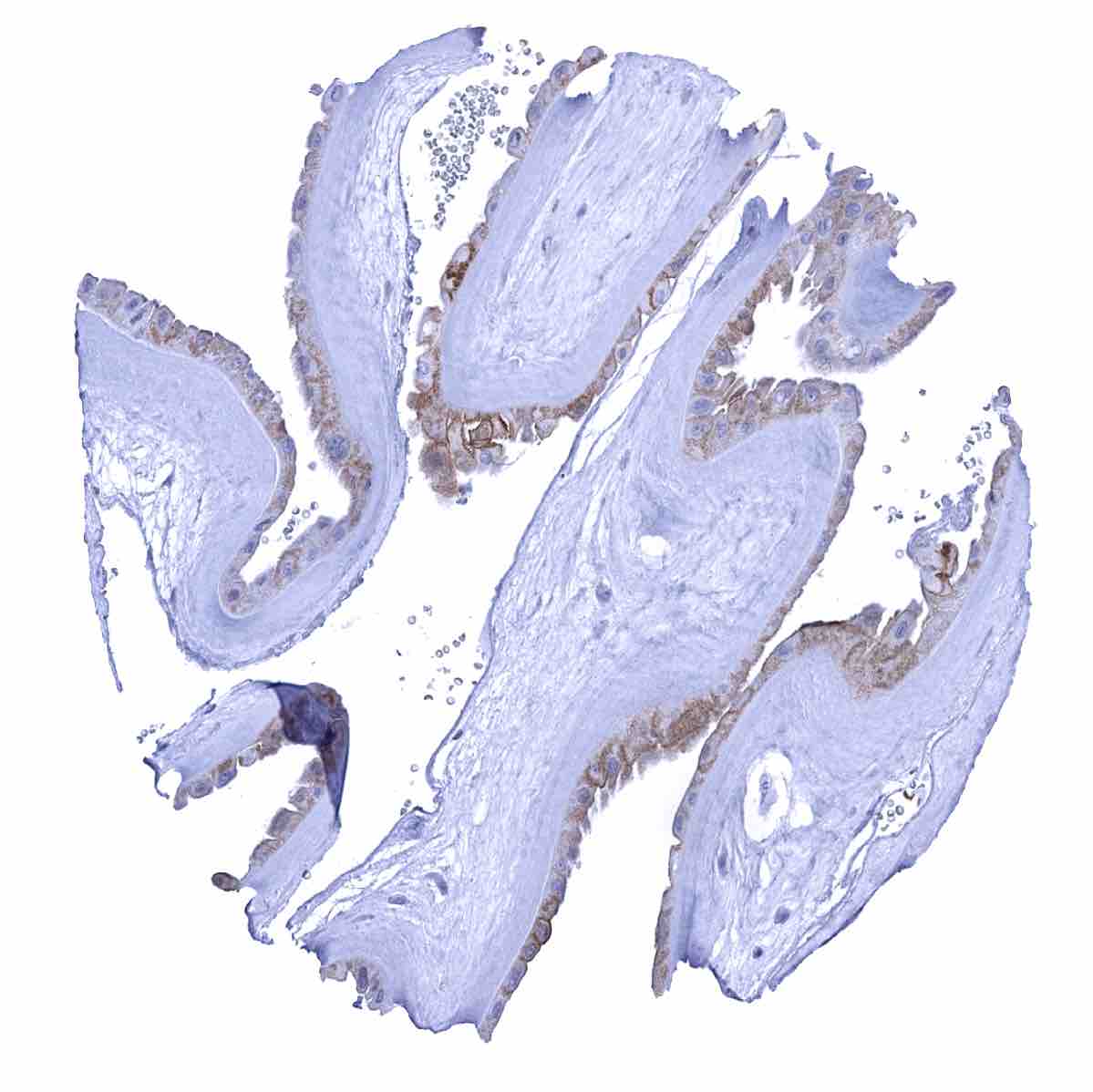
Desmoglein 1 immunostaining occurs in keratinizing and non-keratinizing squamous epithelia where a predominantly membranous staining pattern can be observed. The extent of staining is variable and ranges from a staining of the entire squamous epithelium to a predominant staining of the suprabasal 2/3 of the epithelium with weak or absent staining of the basal cells and decreasing staining intensity towards the surface of the epithelium. Squamous cell staining also includes corpuscles of Hassall’s and a fraction of (upper half) cells in tonsil crypt epithelium as well as in sebaceous glands and hair follicles of the skin. A weak to moderate immunostaining is also seen in amnion cells of the placenta. Desmoglein 1 immunostaining is not seen in the gastrointestinal tract, pancreas, liver, gallbladder, salivary glands, Brunner glands, kidney, prostate, seminal vesicles, testis, thyroid gland, parathyroid gland, adrenal gland, respiratory epithelium, bronchial glands, lung, breast gland, endometrium, endocervix, fallopian tube, ovary (including follicular cysts and corpora lutea), urothelium, mesenchymal tissues, lymph node, spleen, bone marrow, pituitary gland, and the brain. A non-specific binding of the antibody to pigments can occur in the brain, liver, kidney, adrenal gland, and heart muscle.
These findings are largely consistent with the RNA and protein data described in the Human Protein Atlas (Tissue expression Desmoglein 1)
Positive control = Tonsil: A moderate to strong desmoglein 1 immunostaining should be seen in a fraction of squamous epithelial cells.
Negative control = Desmoglein 1 immunostaining should be absent in all non-squamous epithelial cell types.

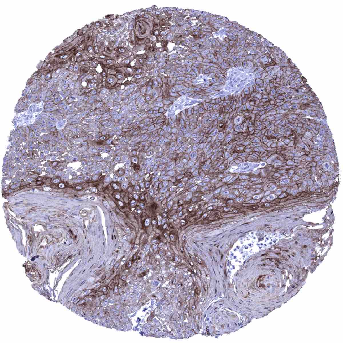
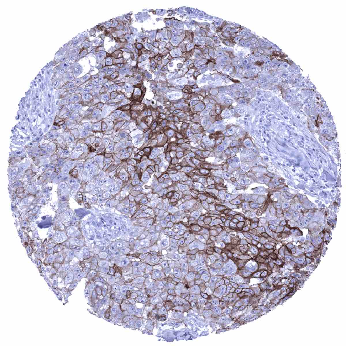
Staining Pattern in Relevant Tumor Types
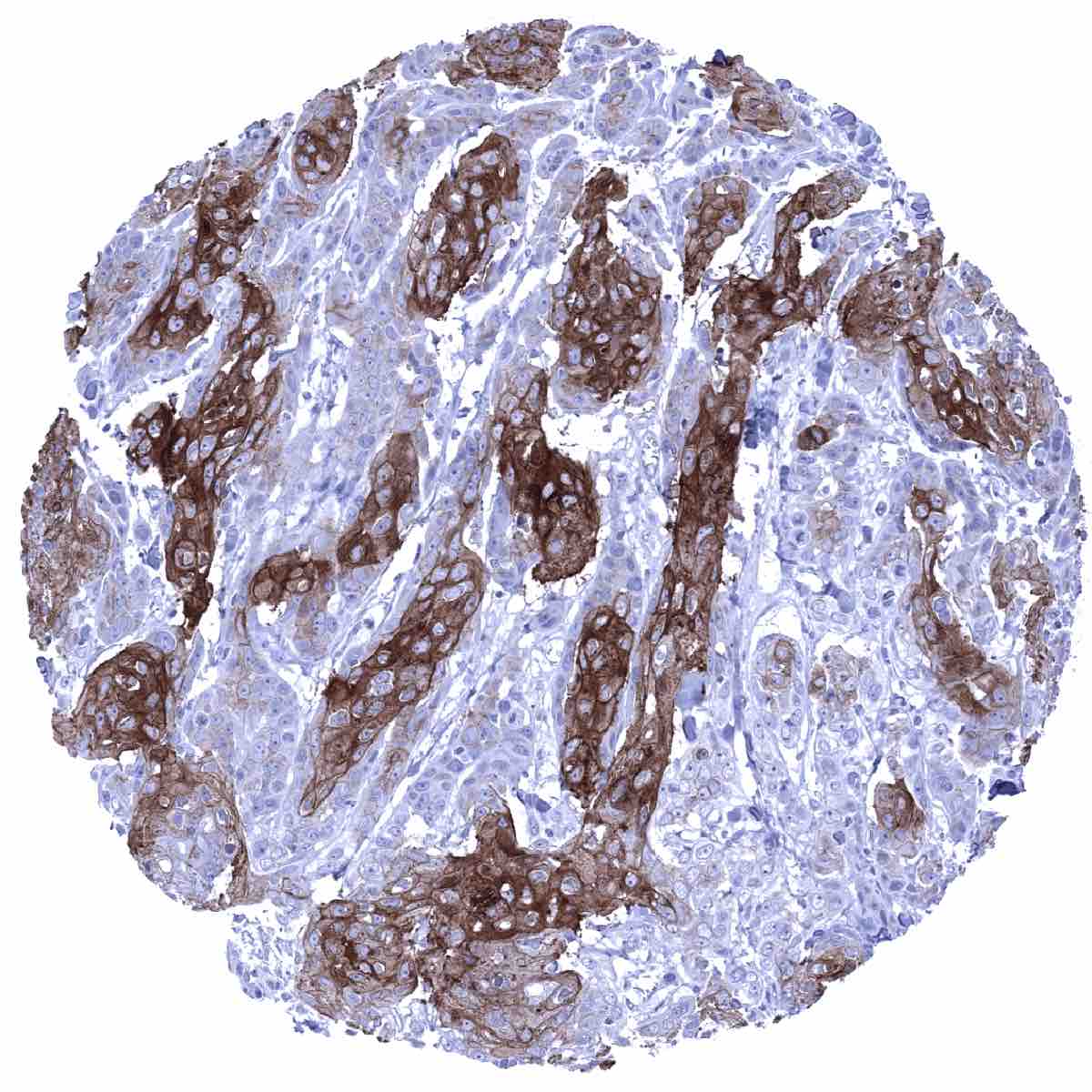
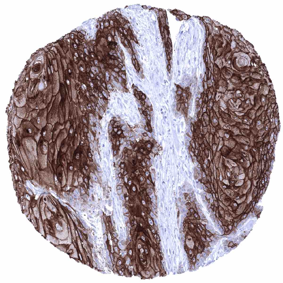
A positive desmoglein 1 immunostaining is usually seen in squamous cell carcinomas irrespective of their site of origin and in tumor areas exhibiting squamous differentiation. Squamous differentiation can occur in many different tumor entities including urothelial cancer, gastric cancer, pancreatic adenocarcinoma, endometrioid carcinoma and others.
The TCGA findings on Desmoglein 1 RNA expression in different tumor categories have been summarized in the Human Protein Atlas.
Compatibility of Antibodies
No data available at the moment
Protocol Recommendations
IHC users have different preferences on how the stains should look like. Some prefer high staining intensity of the target stain and even accept some background. Others favor absolute specificity and lighter target stains. Factors that invariably lead to more intense staining include higher concentration of the antibody and visualization tools, longer incubation time, higher temperature during incubation, higher temperature and longer duration of the heat induced epitope retrieval (slide pretreatment). The impact of the pH during slide pretreatment has variable effects and depends on the antibody and the target protein.
All images and data shown here and in our image galleries are obtained by the manual protocol described below. Other protocols resulting in equivalent staining are described as well.
-Manual protocol
Freshly cut sections should be used (less than 10 days between cutting and staining). Heat-induced antigen retrieval for 5 minutes in an autoclave at 121°C in pH 9 Target Retrieval Solution buffer. Apply MSVA-544M at a dilution of 1:100 at 37°C for 60 minutes. Visualization of bound antibody by the EnVision Kit (Dako, Agilent) according to the manufacturer’s directions.
Potential Research Applications
- The diagnostic utility of desmoglein 1 IHC should be investigated in a large cohort of tumors from different entities.
- Desmoglein 1 has been suggested as a prognostic parameter in several different cancer types. This subject deserves further investigation.
Evidence for Antibody Specificity in IHC
There are two ways how the specificity of antibodies can be documented for immunohistochemistry on formalin fixed tissues. These are: 1. Comparison with a second independent method for target expression measurement across a large number of different tissue types (orthogonal strategy), and 2. Comparison with one or several independent antibodies for the same target and showing that all positive staining results are also seen with other antibodies for the same target (independent antibody strategy).
Orthogonal validation: Data derived from three independent RNA screening studies, including the Human Protein Atlas (HPA) RNA-seq tissue dataset, the FANTOM5 project, and the Genotype-Tissue Expression (GTEx) project, which are all summarized in the Human Protein Atlas (Tissue expression Desmoglein 1) suggested that desmoglein 1 expression only occurs in tissues that are covered by squamous epithelium. This fits very well with protein expression data obtained by MSVA-544M. In addition, desmoglein 1 immunostaining was seen in amnion cells of the placenta. Given the paucity of this cell type in the entire placenta it is very likely, that this desmoglein 1 expression will be missed in RNA analyses of placenta tissue. Because desmoglein 1 and desmoglein 3 are often co-expressed, true expression of desmoglein 1 in amnion cells of the placenta is indirectly suggested by the observation of desmoglein 3 expression in amnion cells (data shown here)
