295,00 € – 995,00 €
Product details
Synonyms = BLAST-2; C-type lectin domain family 4, member J; Fc fragment of IgE low affinity II receptor; Fc-epsilon-RII; FCER2; FCER2A; FceRII; IgE-binding factor (IGEBF); Immunoglobulin E receptor, low affinity II; Lymphocyte IgE receptor
Antibody type = Mouse monoclonal / IgG
Clone = MSVA-023M
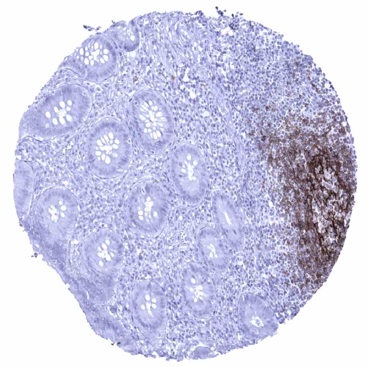
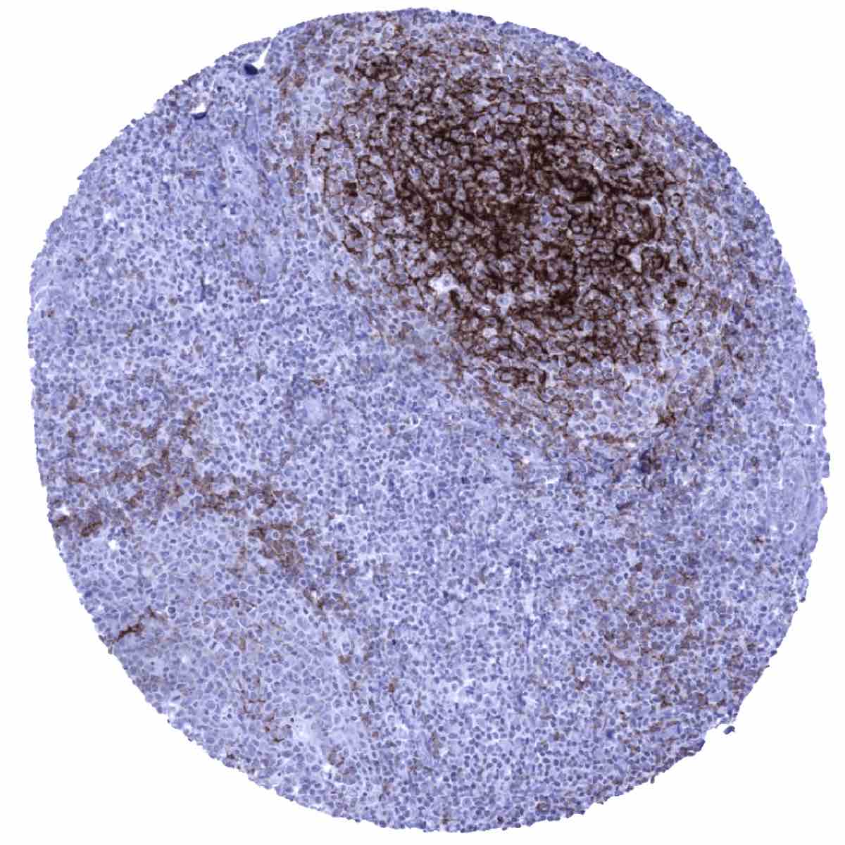
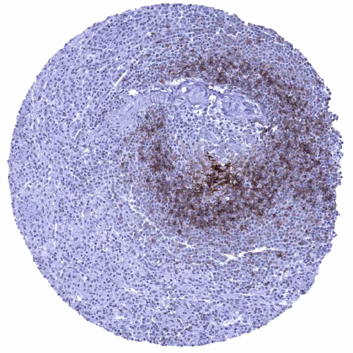
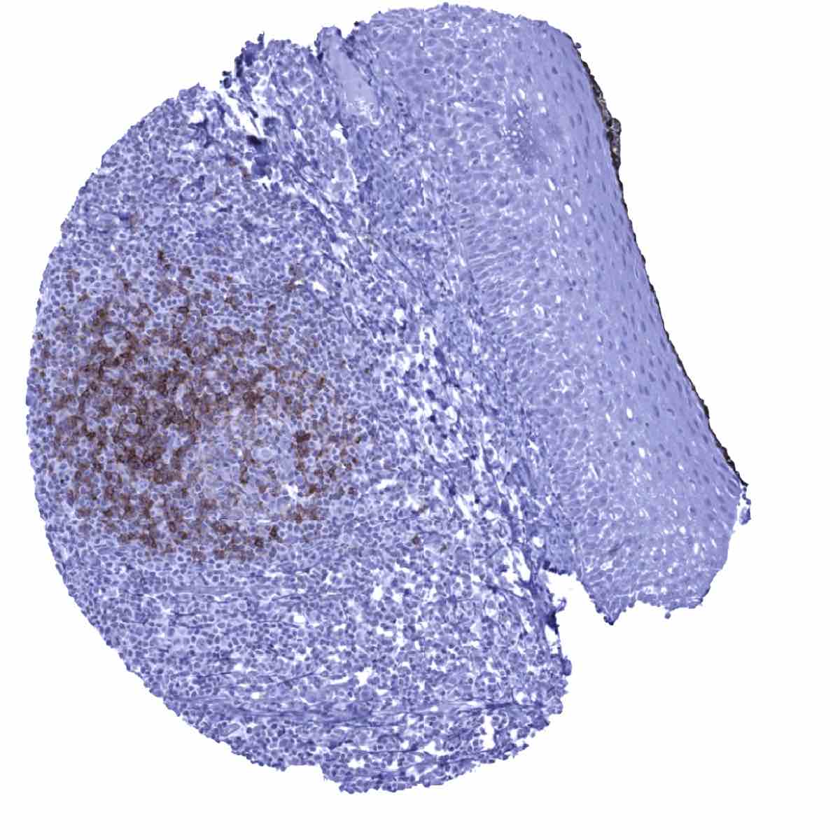
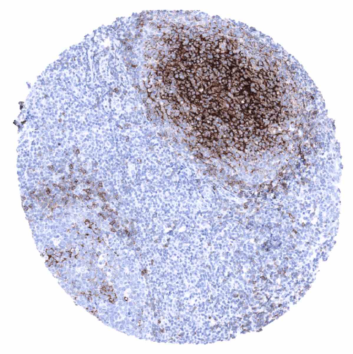
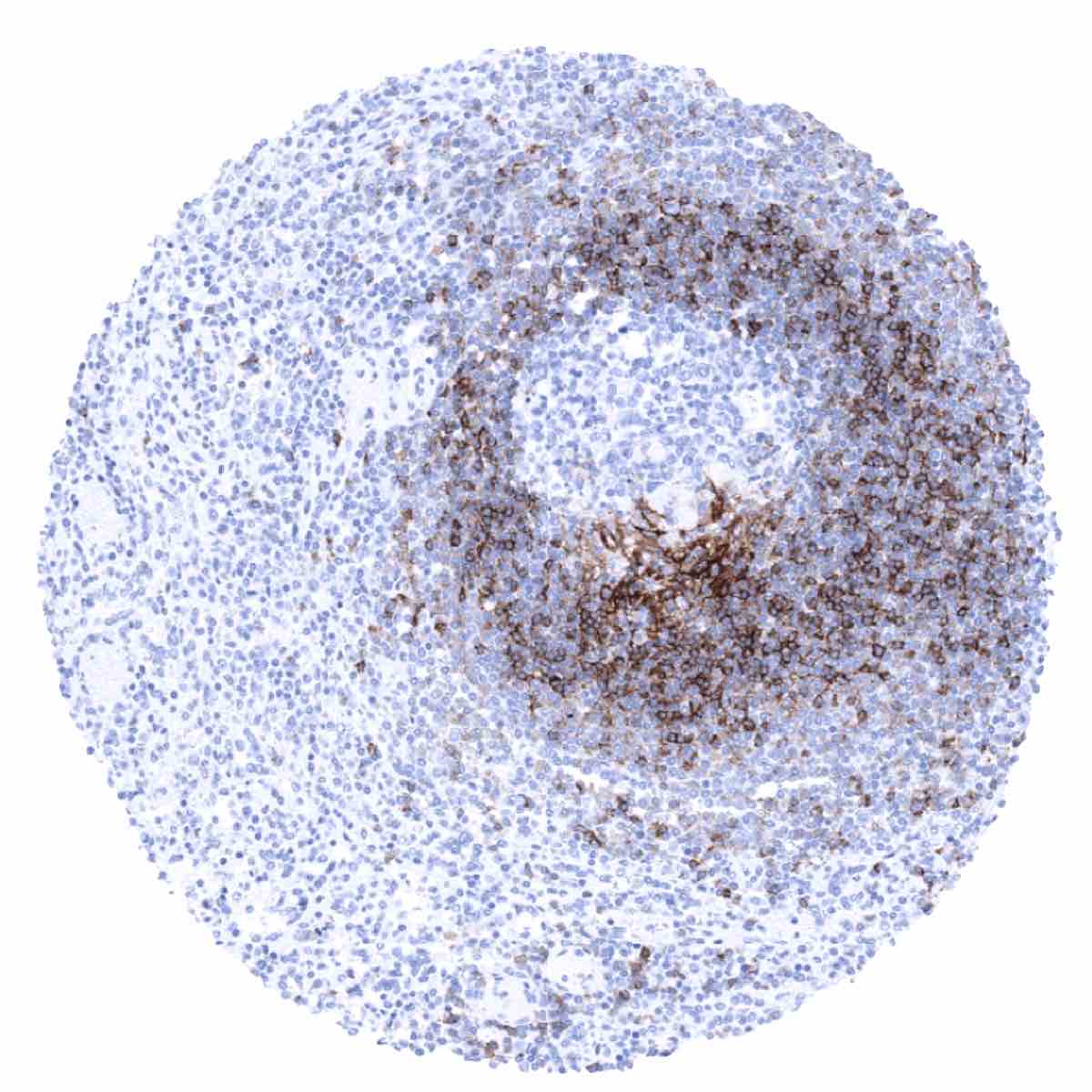
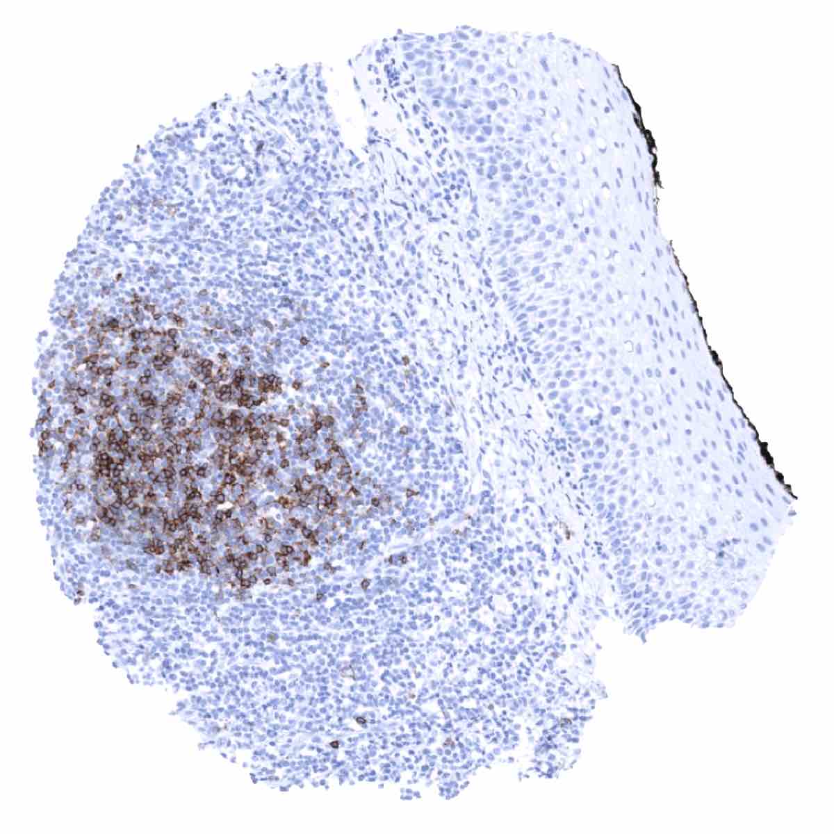
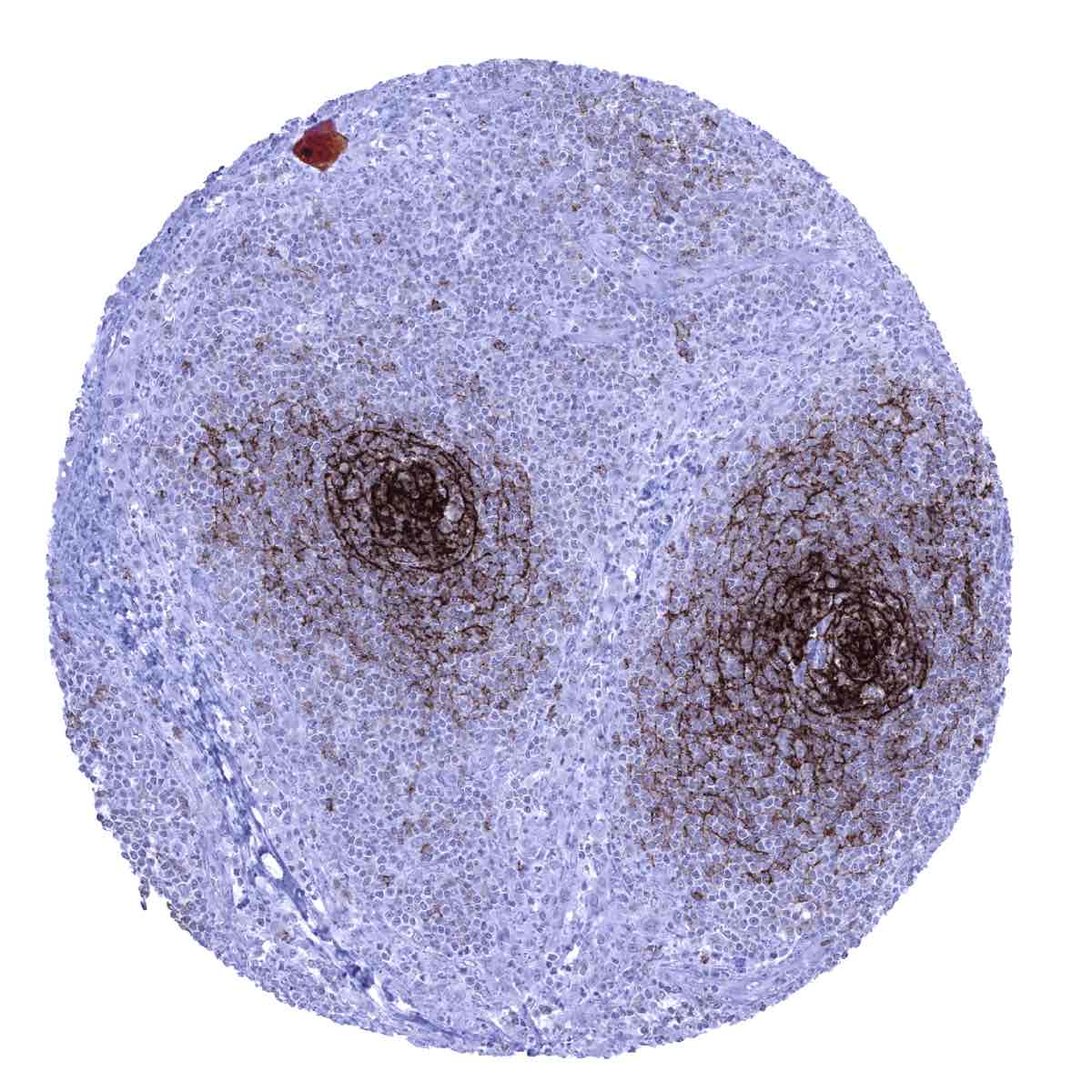
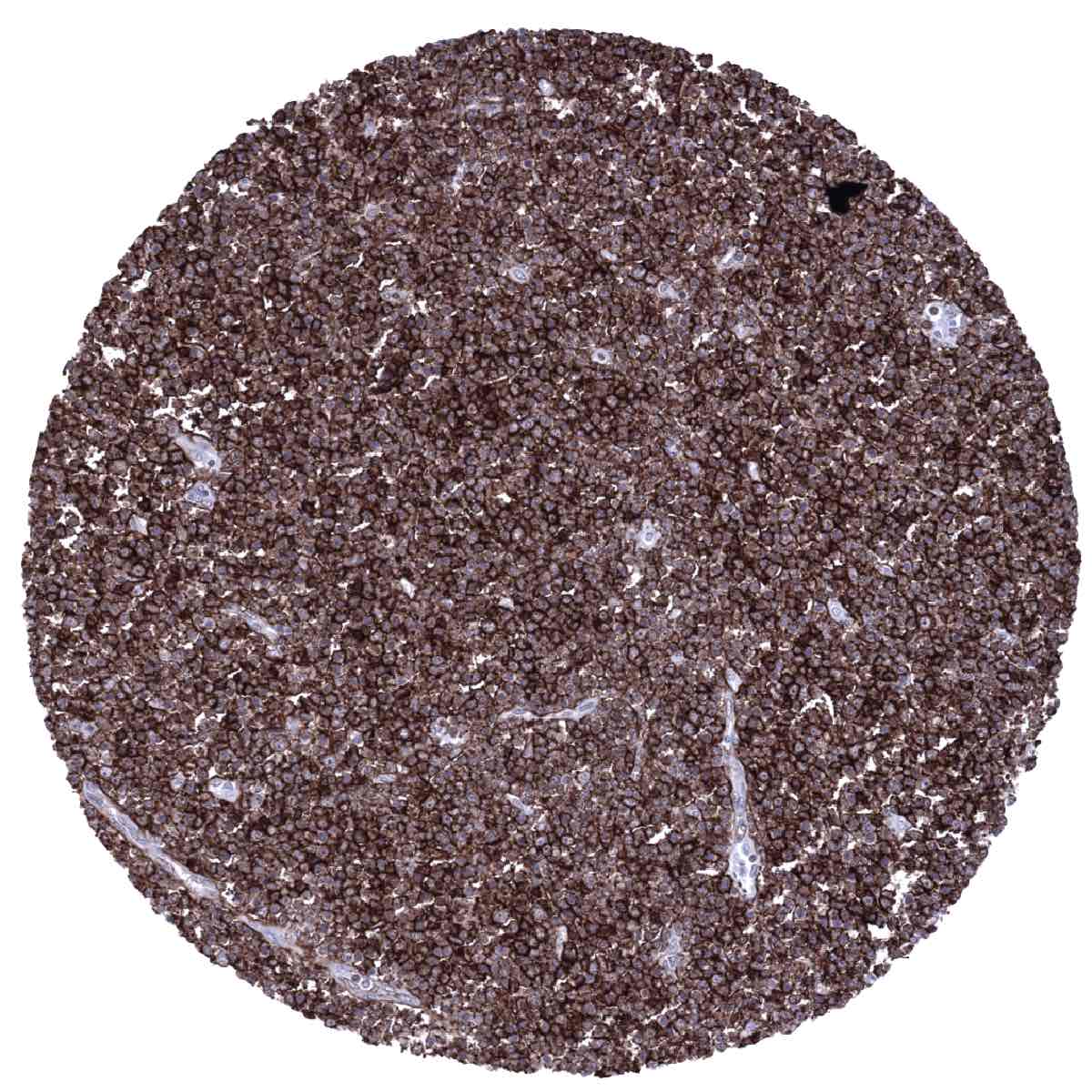
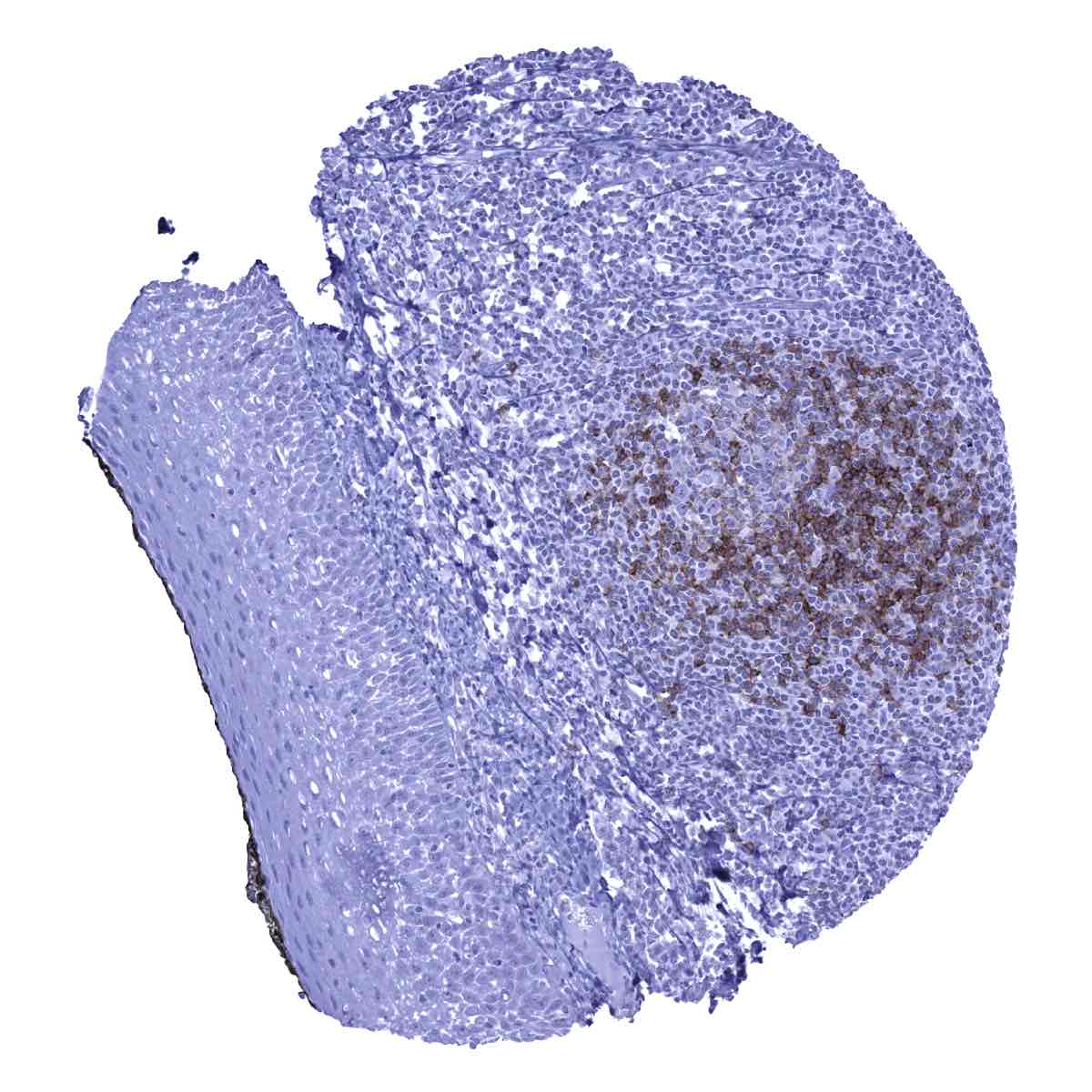
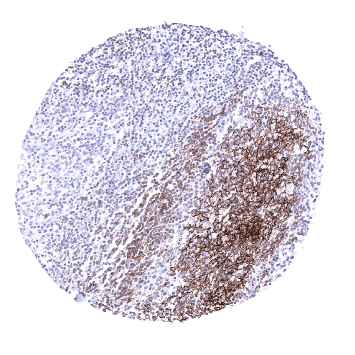
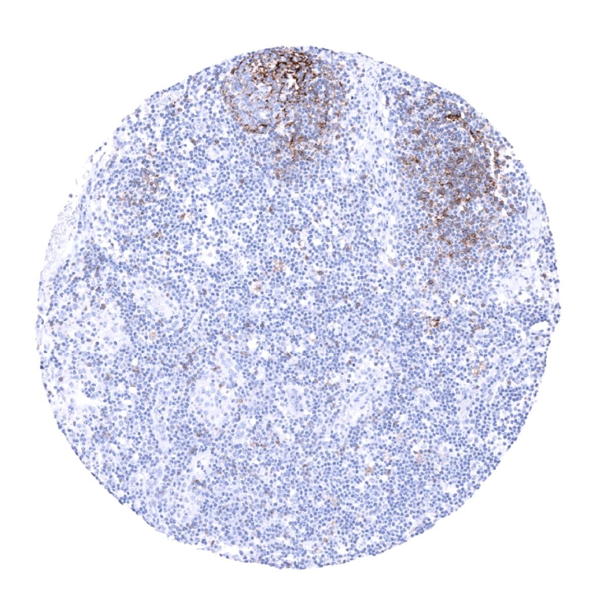
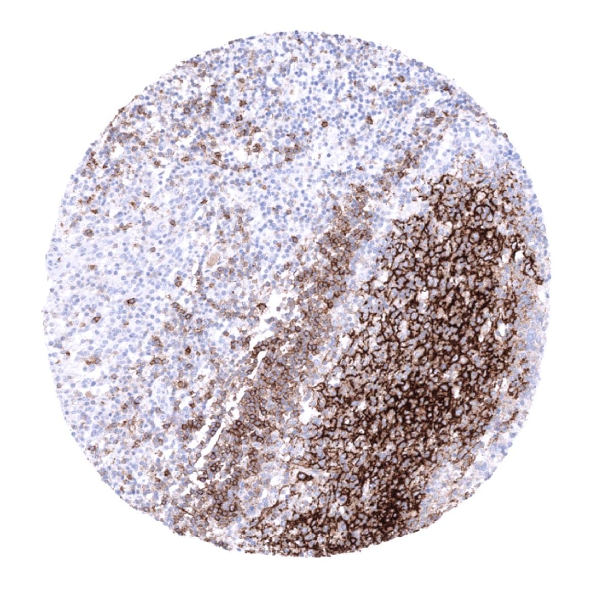
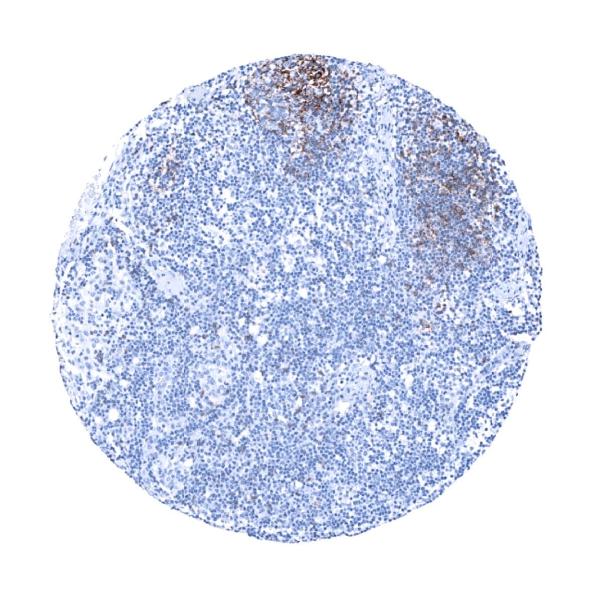
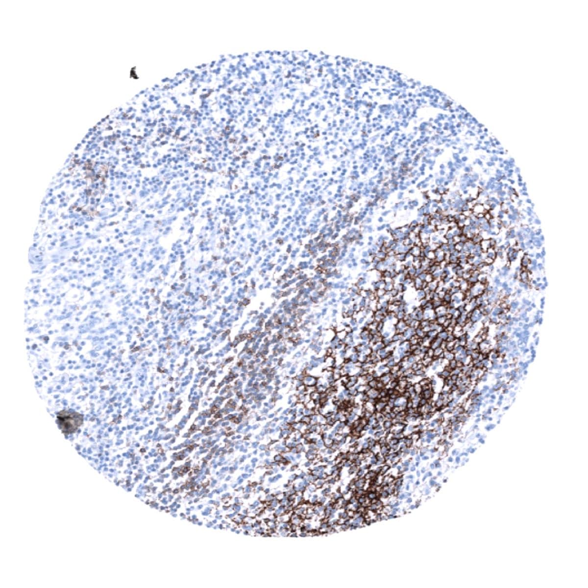
Positive control = Tonsil: The follicular dendritic cells of the germinal centres should show a strong CD23 immunostaining while the majority of lymphocytes in the mantle zone of the follicles should show an at least weak staining.
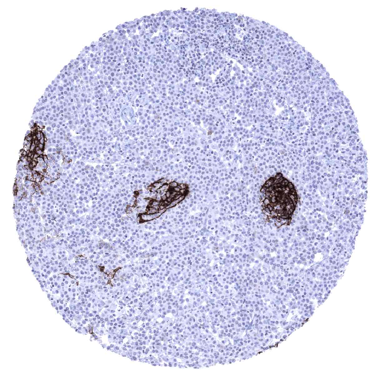
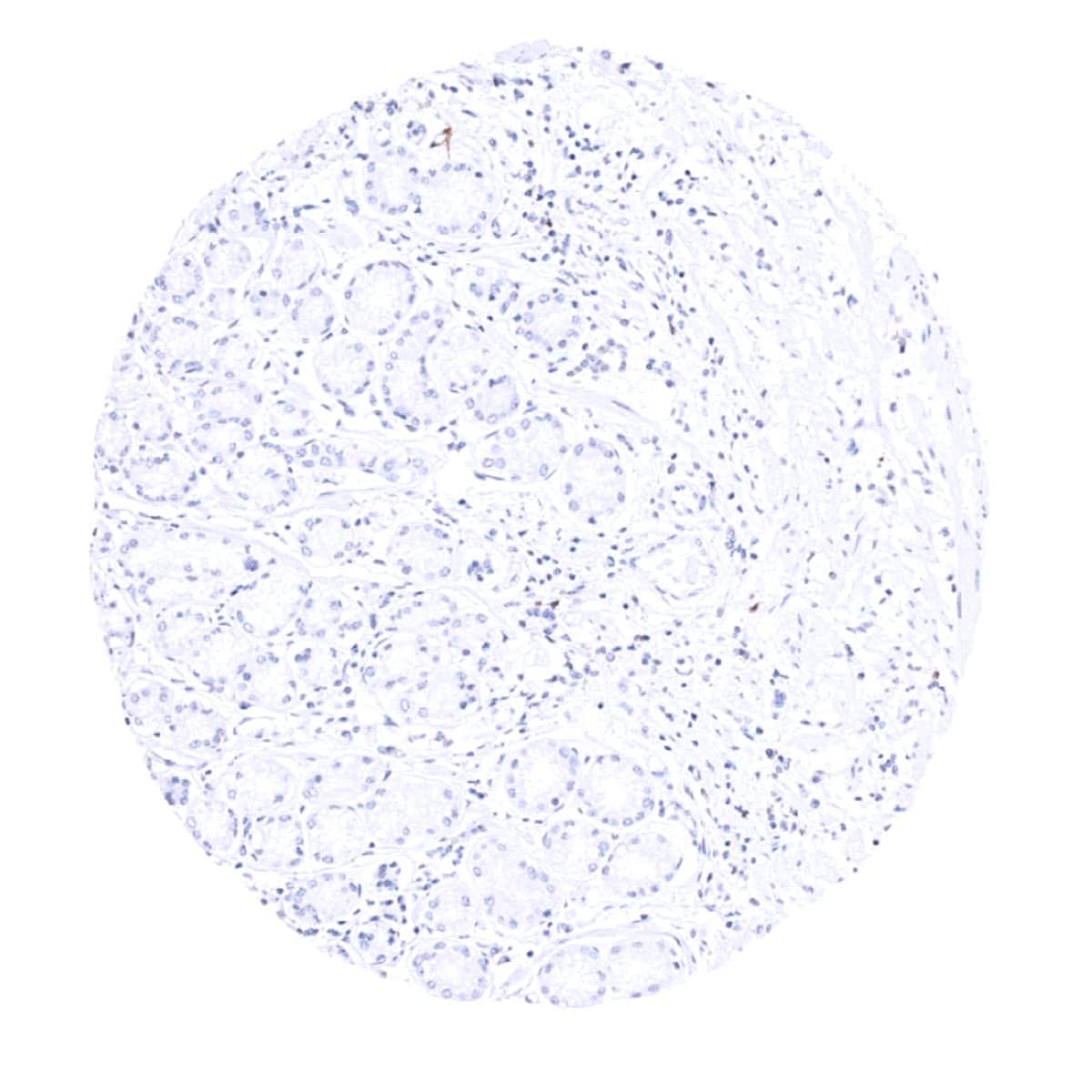
Negative control = Tonsil: The squamous epithelial cells and T-cells in the interfollicular T-zones should not show any CD23 immunostaining.
Cellular localization = Cell surface
Reactivity = Human
Application = Immunohistochemistry
Dilution = 1:100 – 1:200
Intended Use = Research Use Only
Relevance of Antibody
CD23 is expressed on follicular dendritic cells and mature B-lymphocytes.
Biology Behind
CD23 is a 45 kDa receptor protein coded by the FCER2 (Fc epsilon receptor II) gene on chromosome 19p. CD23 is found on follicular dendritic cells and on mature B cells. It binds the Fc domain of IgE and several other ligands including CD21 and major histocompatibility locus class II receptors. For IgE it acts as a “low-affinity” receptor which plays a role in cellular implementation of antibody feedback and regulates IgE synthesis. CD23 is a soluble protein which can also be shedded into the blood plasma where it may support the recruitment of non-sensitized B-cells.
Staining Pattern in Normal Tissues
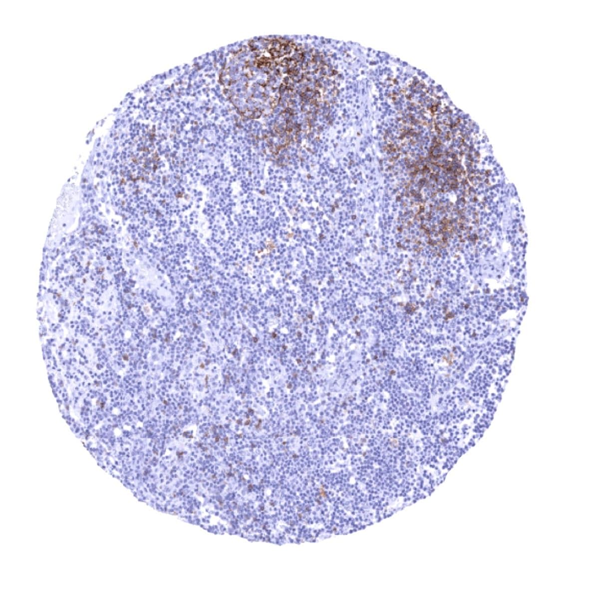
A strong CD23 immunostaining is seen in follicular dendritic cells (FDCs) predominantly in the apical light zone of the germinal center. CD23 staining is moderate to strong in B-lymphocytes (mostly follicular/perifollicular).
These findings are largely consistent with the RNA and protein data described in the Human Protein Atlas (Tissue expression CD23)
Positive control = Tonsil: The follicular dendritic cells of the germinal centres should show a strong CD23 immunostaining while the majority of lymphocytes in the mantle zone of the follicles should show an at least weak staining.
Negative control = Tonsil: The squamous epithelial cells and T-cells in the interfollicular T-zones should not show any CD23 immunostaining.
Staining Pattern in Relevant Tumor Types
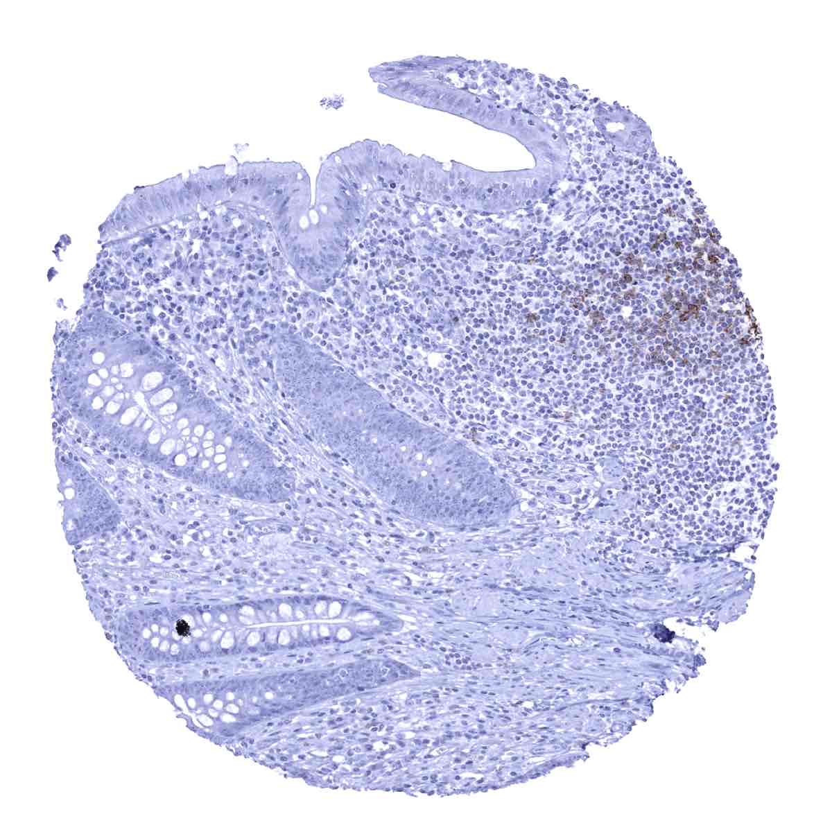
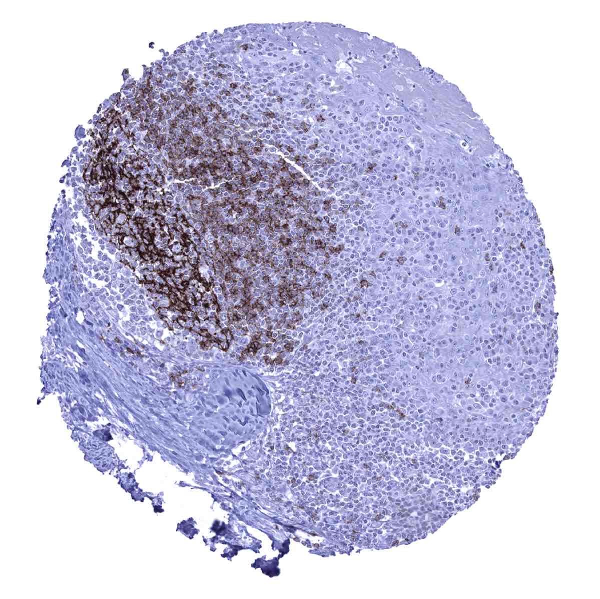
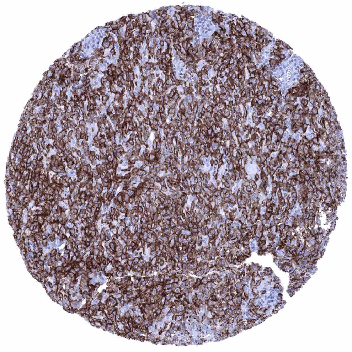
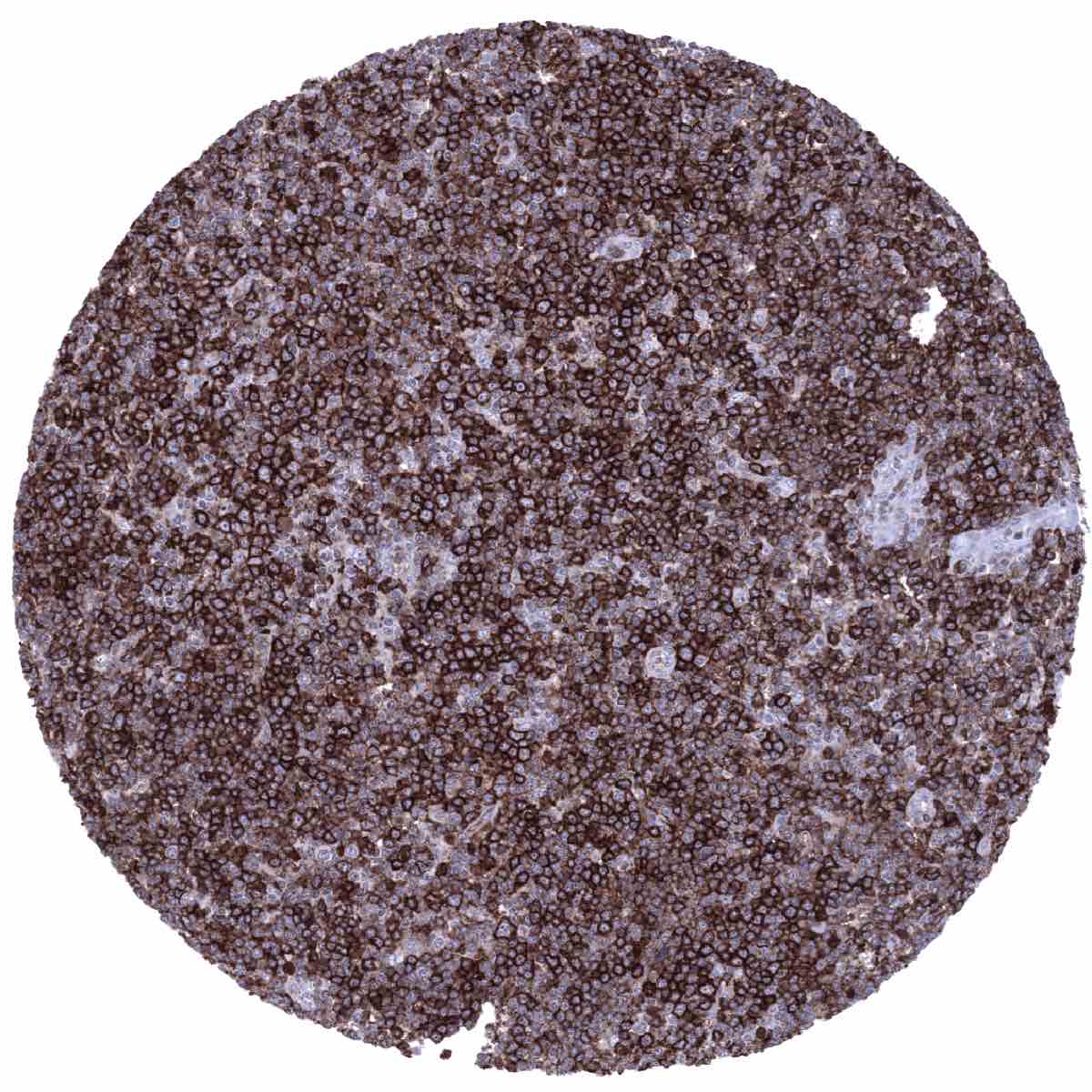
CD23 is typically expressed (85%) in chronic lymphocytic leukemia (CLL) where the immunostaining is highest in proliferation centers. CD23 immunostaining can also occur in other B-cell lymphomas such as follicular lymphomas but it is rare in marginal zone (<10%) and lymphoplasmacytic lymphoma and it hardly ever occurs in mantle cell lymphoma (2%). CD23 expression is also rare in extranodal marginal zone lymphomas such as MALT lymphoma (<10%). In diffuse large B-cell lymphoma CD23 positivity was suggested to be associated with good prognosis. CD23 immunostaining is also seen in the rare tumors derived from follicular dendritic cells.
The TCGA findings on CD23 RNA expression in different tumor categories have been summarized in the Human Protein Atlas.

Compatibility of Antibodies
No data available at the moment
Protocol Recommendations
IHC users have different preferences on how the stains should look like. Some prefer high staining intensity of the target stain and even accept some background. Others favor absolute specificity and lighter target stains. Factors that invariably lead to more intense staining include higher concentration of the antibody and visualization tools, longer incubation time, higher temperature during incubation, higher temperature and longer duration of the heat induced epitope retrieval (slide pretreatment). The impact of the pH during slide pretreatment has variable effects and depends on the antibody and the target protein.
All images and data shown here and in our image galleries are obtained by the manual protocol described below. Other protocols resulting in equivalent staining are described as well.
Manual protocol
Freshly cut sections should be used (less than 10 days between cutting and staining). Heat-induced antigen retrieval for 5 minutes in an autoclave at 121°C in pH 9 Target Retrieval Solution buffer. Apply MSVA-023M at a dilution of 1:150 at 37°C for 60 minutes. Visualization of bound antibody by the EnVision Kit (Dako, Agilent) according to the manufacturer’s directions.

Agilent / Dako – Autostainer Link 48
Pretreatment in PT-Link for 30 minutes at 95°C (pH high); FLEX peroxidase blocking for 5 minutes (room temperature), MSVA-023M 1:150 for 20 minutes (room temperature), FLEX+ mouse/rabbit (LINKER) for 15 minutes (room temperature), horseradish peroxidase (HRP) for 20 minutes (room temperature), FLEX DAB+Sub-Chromo for 10 minutes (room temperature), FLEX hematoxylin for 5 minutes (room temperature).


These images reflect stainings by the protocol described above. It is of note that a comparable staining result can also be obtained by different protocols. In general, a longer pretreatment, a longer incubation time of the primary antibody, a higher antibody concentration, and a longer incubation time of FLEX+LINKER result in stronger staining, potentially at the cost of more background staining. Modifications of the protocol with a strengthening effect on staining intensity in combination with changes of other parameters that result in lower staining intensity can result in a comparable result as shown above.
Leica – BOND RX
Dewax at 72°C for 30 seconds; Pretreatment in Bond Epitope Retrieval Solution (ER2 – EDTA pH9) for 20 minutes at 100°C; Peroxidase blocking for 5 minutes (room temperature), MSVA-023M 1:150 for 15 minutes (room temperature), Post primary (rabbit anti mouse) for 8 minutes (room temperature), Polymer (goat anti rabbit) for 8 minutes (room temperature), mixed DAB refine for 10 minutes (room temperature), hematoxylin for 5 minutes (room temperature).

These images reflect stainings by the protocol described above. It is of note that a comparable staining result can also be obtained by different protocols. In general, a longer pretreatment, a longer incubation time of the primary antibody, a higher antibody concentration, a higher temperature during incubation, and a longer incubation time of Post primary and or the Polymer result in stronger staining, potentially at the cost of more background staining. Modifications of the protocol with a strengthening effect on staining intensity in combination with changes of other parameters that result in lower staining intensity can result in a comparable result as shown above.
Roche – Ventana Discovery ULTRA
Pretreatment for 64 minutes at 100°C (pH 8,4); CM peroxidase blocking for 12 minutes (room temperature), MSVA-023M 1:75 for 20 minutes at 36°C, secondary antibody (anti-mouse HQ) for 12 minutes at 36°C, anti-HQ HRP for 12 minutes at room temperature, DAB at room temperature, hematoxylin II at room temperature for 8 minutes, bluing reagent at room temperature for 4 minutes.

These images depict staining results obtained by the protocol described above. It is of note, that the Ventana machines generally require higher antibody concentrations than other commonly used autostainers because the antibodies are automatically diluted during the procedure. Various other protocols can result in an identical result as shown above. A longer pretreatment, a longer incubation time of the primary antibody, a higher antibody concentration, a higher temperature during incubation, and a longer incubation time of secondary antibody and or the anti-HQ HRP result in stronger staining, potentially at the cost of more background staining.
Potential Research Applications
- CD23 is a suitable component of multicolor assays analyzing the role of lymphocyte subsets in cancer and other diseases.
- The prevalence of CD23 expression in hematological and non-hematological neoplasms should be further investigated.
- Some studies have suggested that CD23 may – rarely – be expressed on epithelial tumor cells. The significance of this finding is unknown.
Evidence for Antibody Specificity in IHC
There are two ways, how the specificity of antibodies can be documented for immunohistochemistry on formalin fixed tissues. These are: 1. Comparison with a second independent method for measuring target expression across a large number of different tissue types (orthogonal strategy), and 2. Comparison with one or several independent antibodies for the same target and showing that all positive staining results are also seen with other antibodies for the same target (independent antibody strategy).
As a standard orthogonal validation process for MSVA antibodies, RNA data summarized in the Human Protein Atlas (Tissue expression CD23) are used for comparison. However, this procedure has limited validity for proteins that are expressed in significant subsets of lymphocytes because these cells occur in virtually all organs. The validation of MSVA-023M is therefore based on a comparison with a second independent, commercially available CD23 antibody (termed “Validation-antibody”). This comparison of antibodies revealed identical staining patterns for MSVA-023M and the “Validation-antibody” in lymphatic tissues with identical subsets of lymphocytes staining. It is also of note, that the staining pattern of MSVA-023M matches the expectations as the staining is limited to follicular dendritic cells and lymphocytes that are located in areas consistent with a B-cell origin (follicular/parafollicular).
Antibody Comparison: MSVA-023M vs another commercially available CD23 antibody called “Validation Antibody”