295,00 € – 895,00 €
Product details
Synonyms = Arresten; BSVD; COL4A1; COL4A1 NC1 domain; COL4A2; COL4A3; COL4A4; COL4A5; collagen alpha-1(IV) chain; Collagen IV Alpha 1 Polypeptide; Collagen IV Alpha 2 Polypeptide; Collagen Type IV Alpha 2; Collagen Type IV Alpha 3; Collagen Type IV Alpha 4; Collagen Type IV Alpha 5; RATOR
Antibody type = Recombinant Rabbit monoclonal / IgG
Clone = MSVA-704R
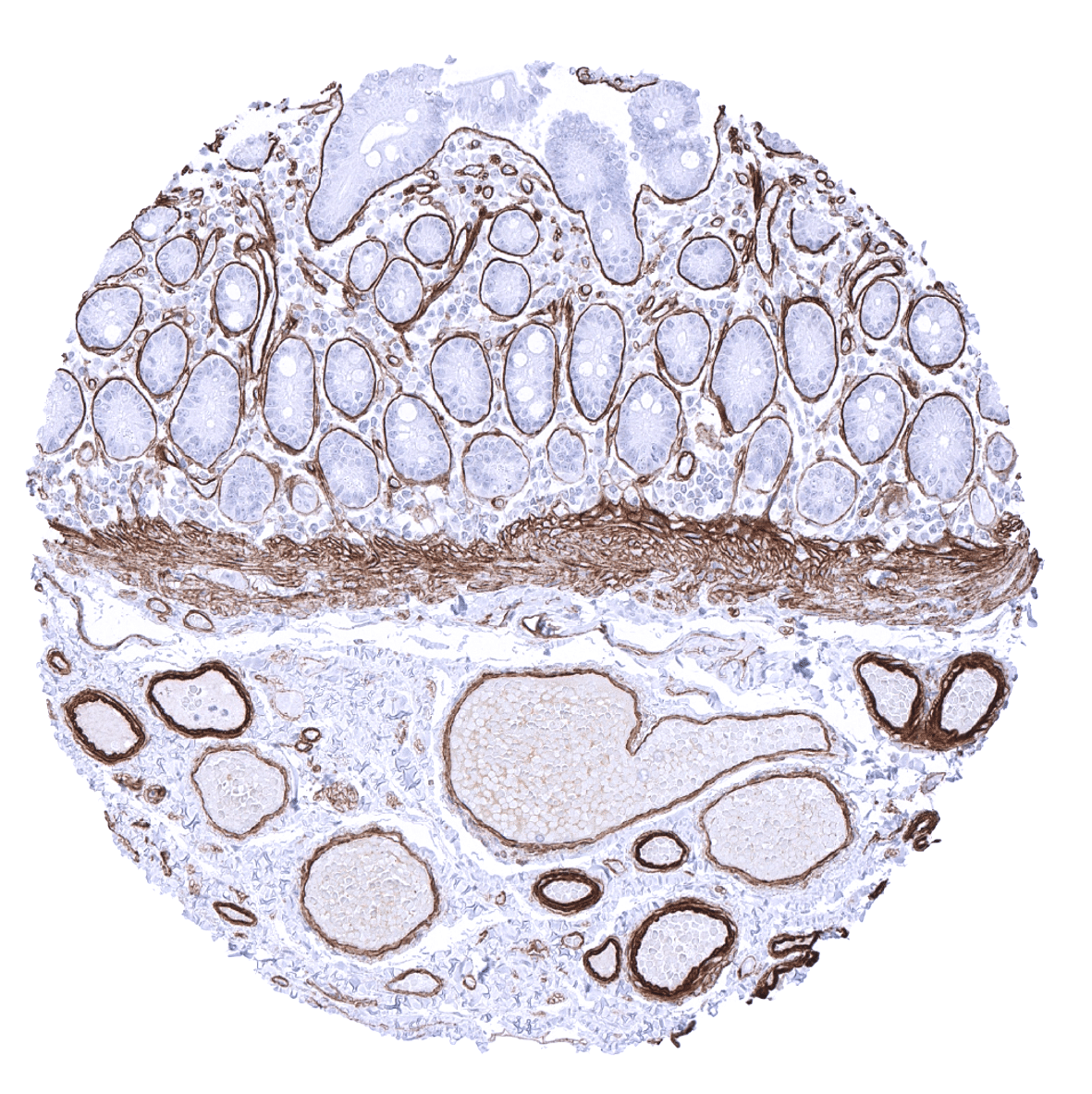
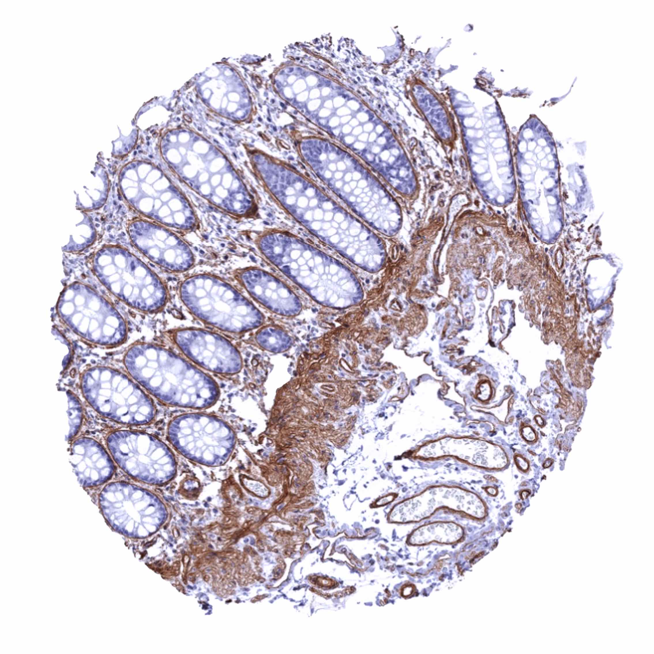
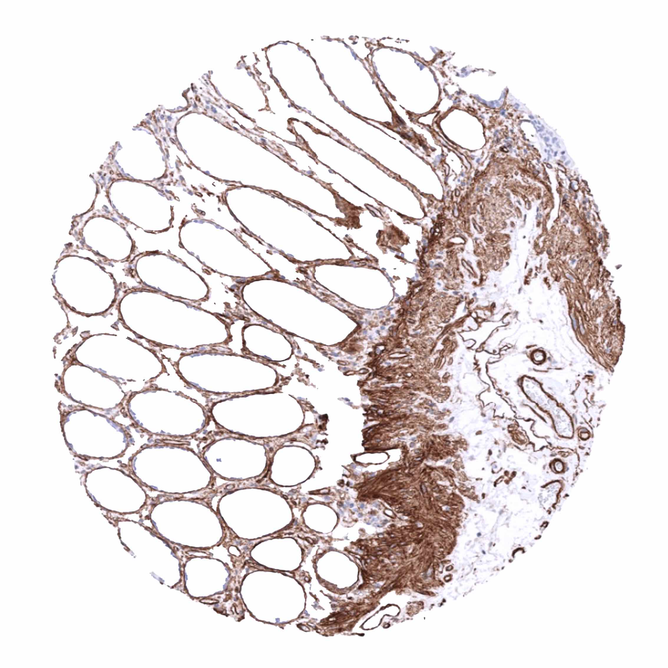
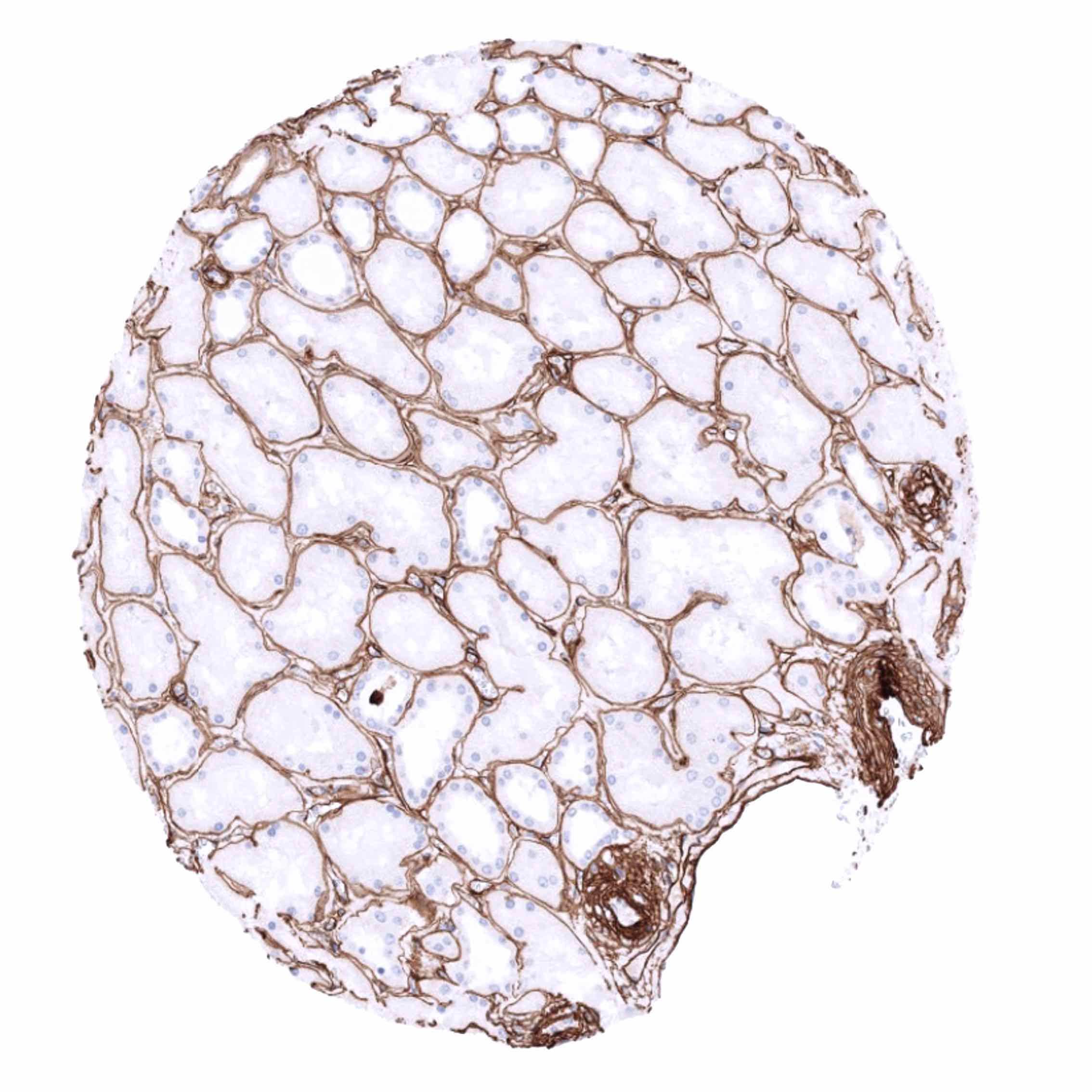
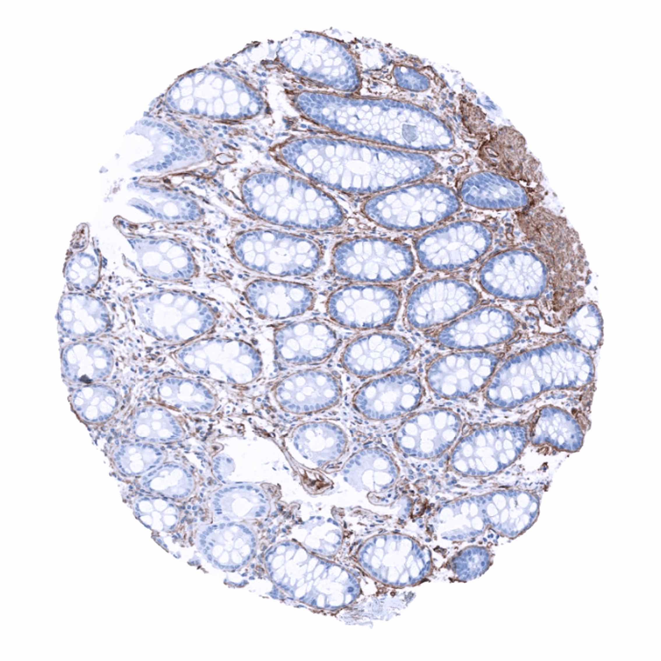
Positive control = Colon: An at least moderate collagen IV immunostaining of basement membranes and a strong staining of vessels and the tunica muscularis mucosa should be seen.
Negative control = Colon: Collagen IV immunostaining must be absent in epithelial cells.
Cellular localization = Cytoplasmic and extracellular.
Reactivity = Human
Application = Immunohistochemistry
Dilution = 1:100 – 1:200
Intended Use = Research Use Only
Relevance of Antibody
Collagen IV is a marker for basement membranes
Biology Behind
Collagen type IV is a 160-190 kDa protein composed of multiple subunits. The protein is a major component of the basement membrane, which is the specialized sheet-like extracellular matrix that occurs between connective tissues and epithelial cells, endothelial cells, or Schwann cells and surrounds various cell types such as heart muscle cells, skeletal muscle cells, smooth muscle cells, and adipocytes. Collagen type IV functions as a barrier between tissue compartments and has many binding partners. Because subdomains, such as tumstatin, are released when the protein is degraded, Collagen type IV also has a signaling role. Mutations of Collagen type IV cause Alport’s syndrome, a chronic kidney disease. Goodpasture syndrome, an autoimmune disease affecting the lungs and the kidneys, is caused by antibodies to the α3 chain of Collagen type IV.
Staining Pattern in Normal Tissues
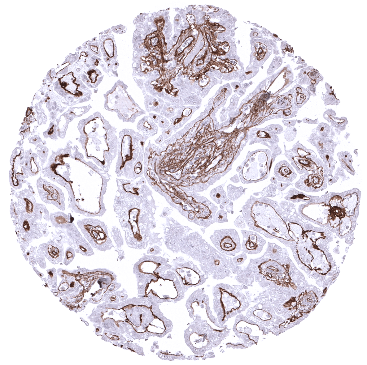
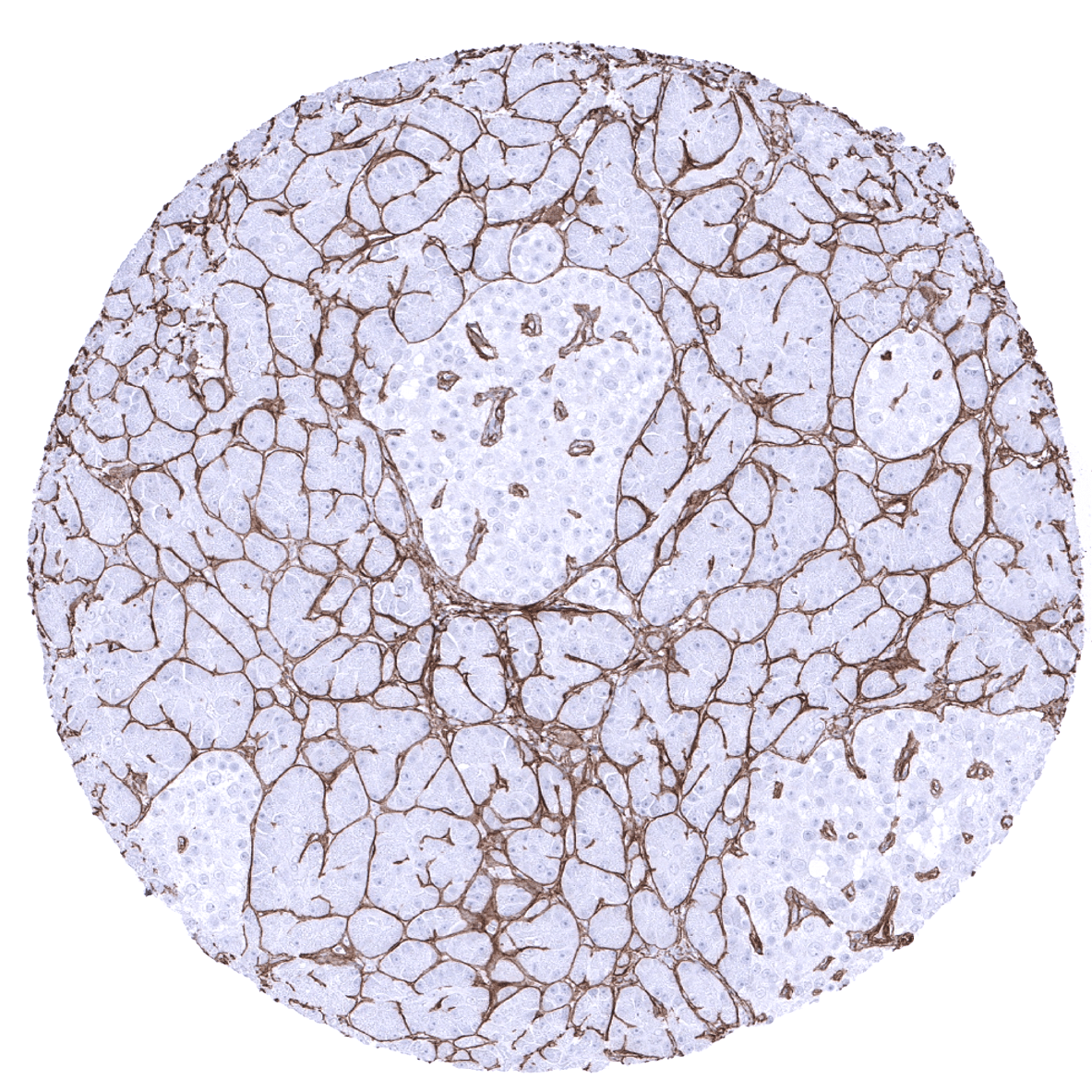
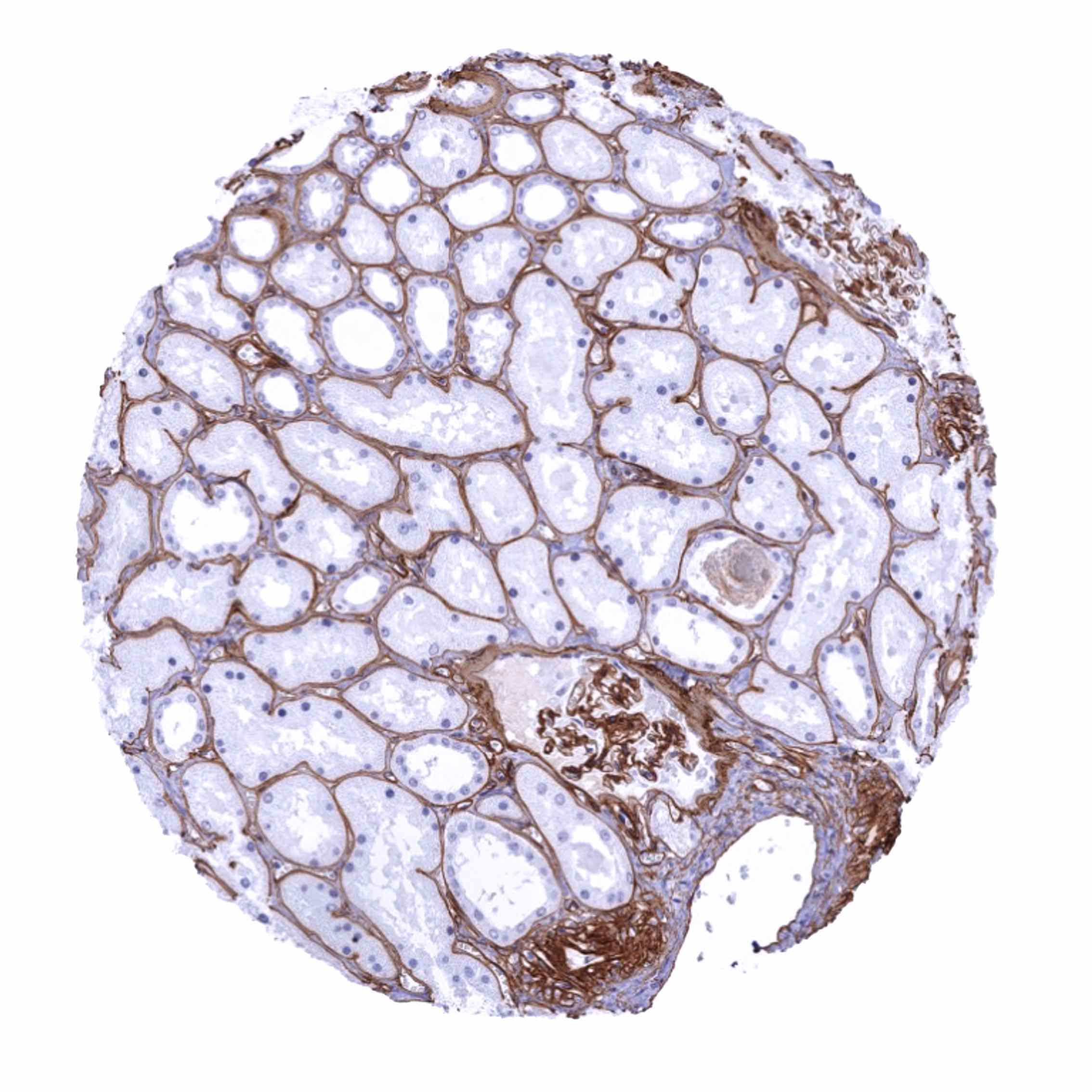
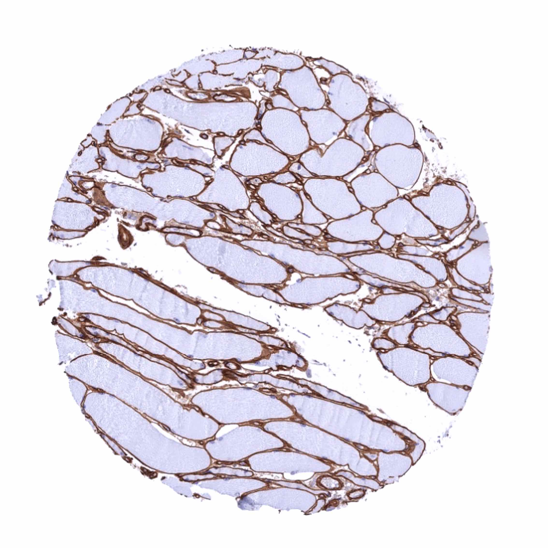
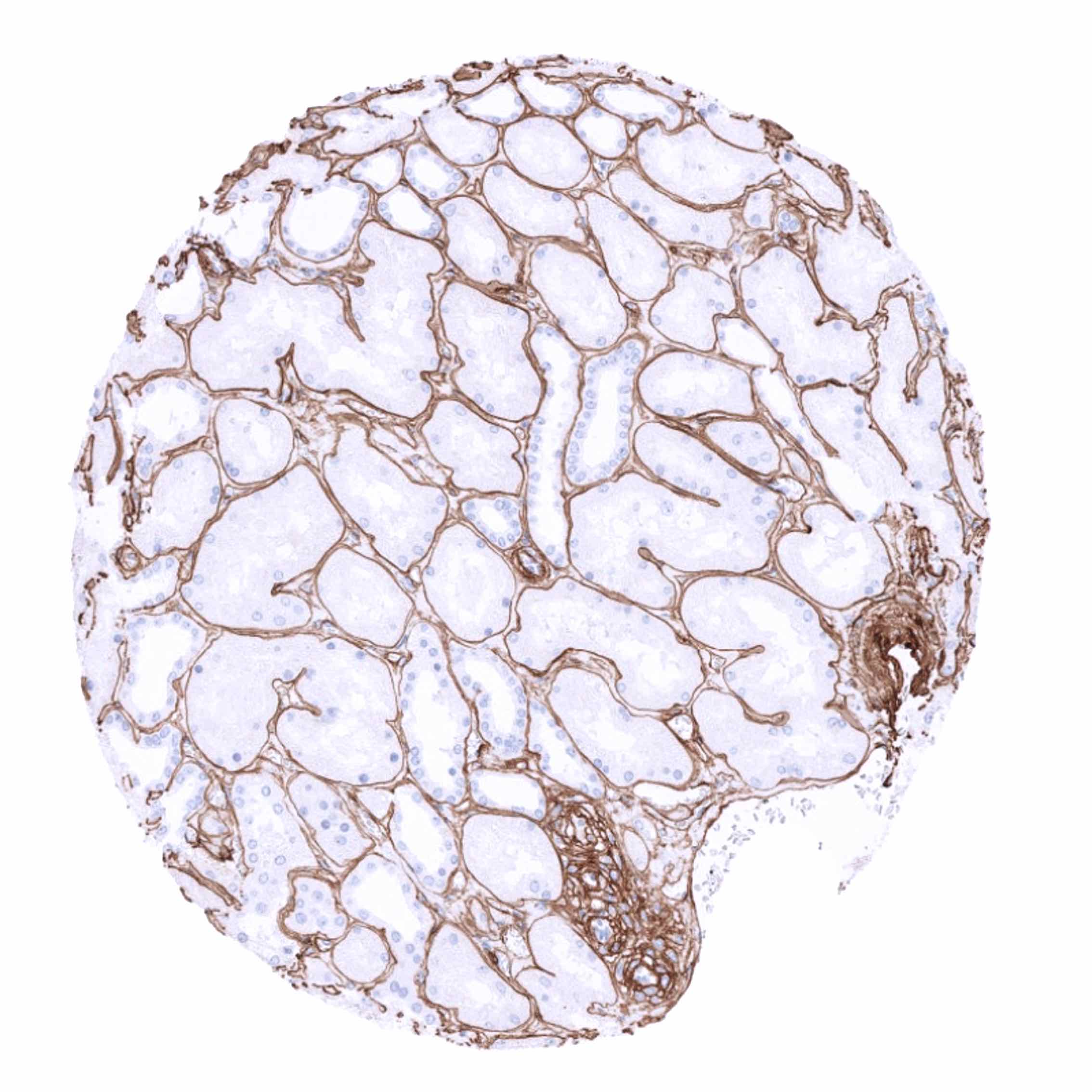
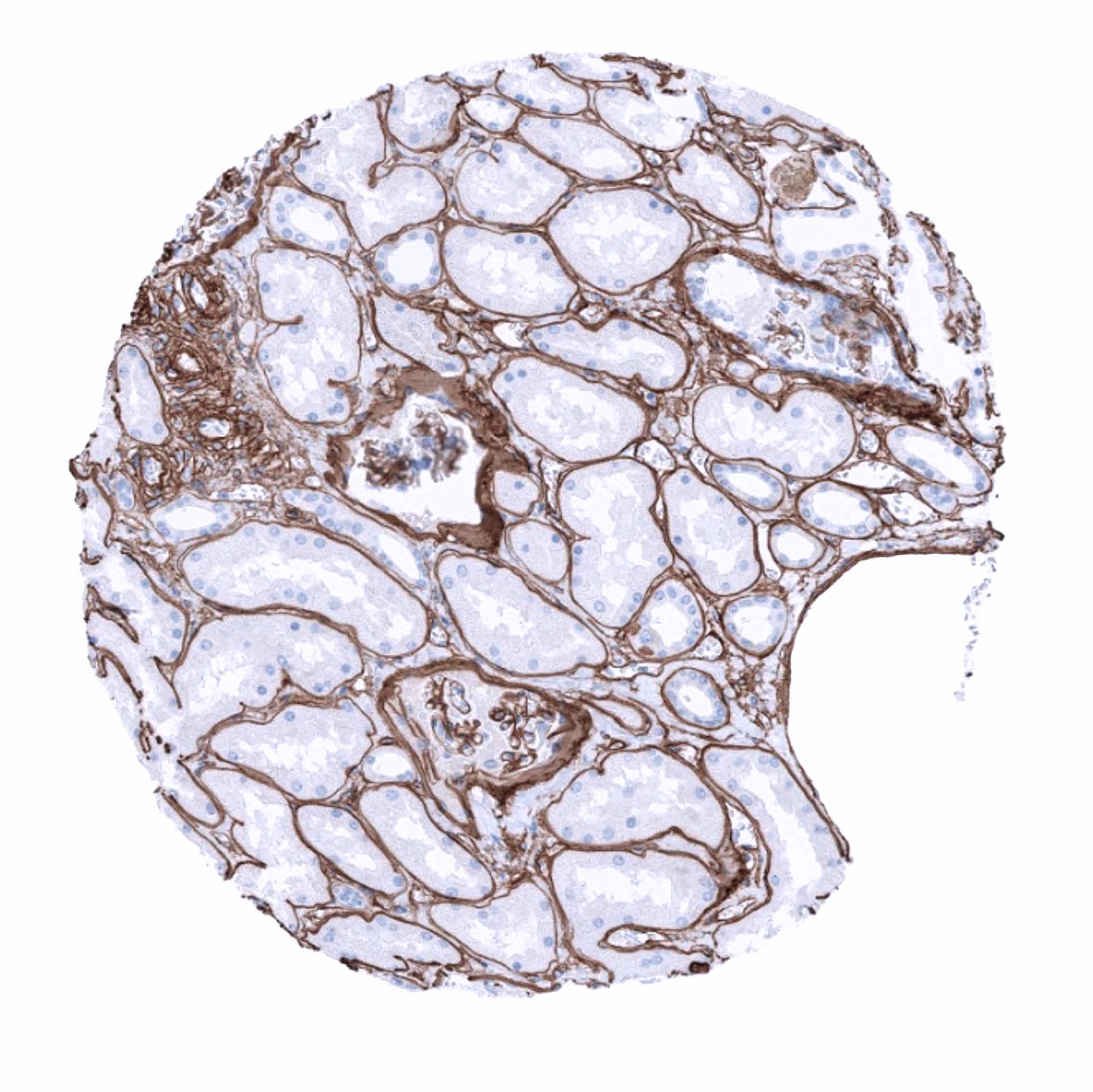
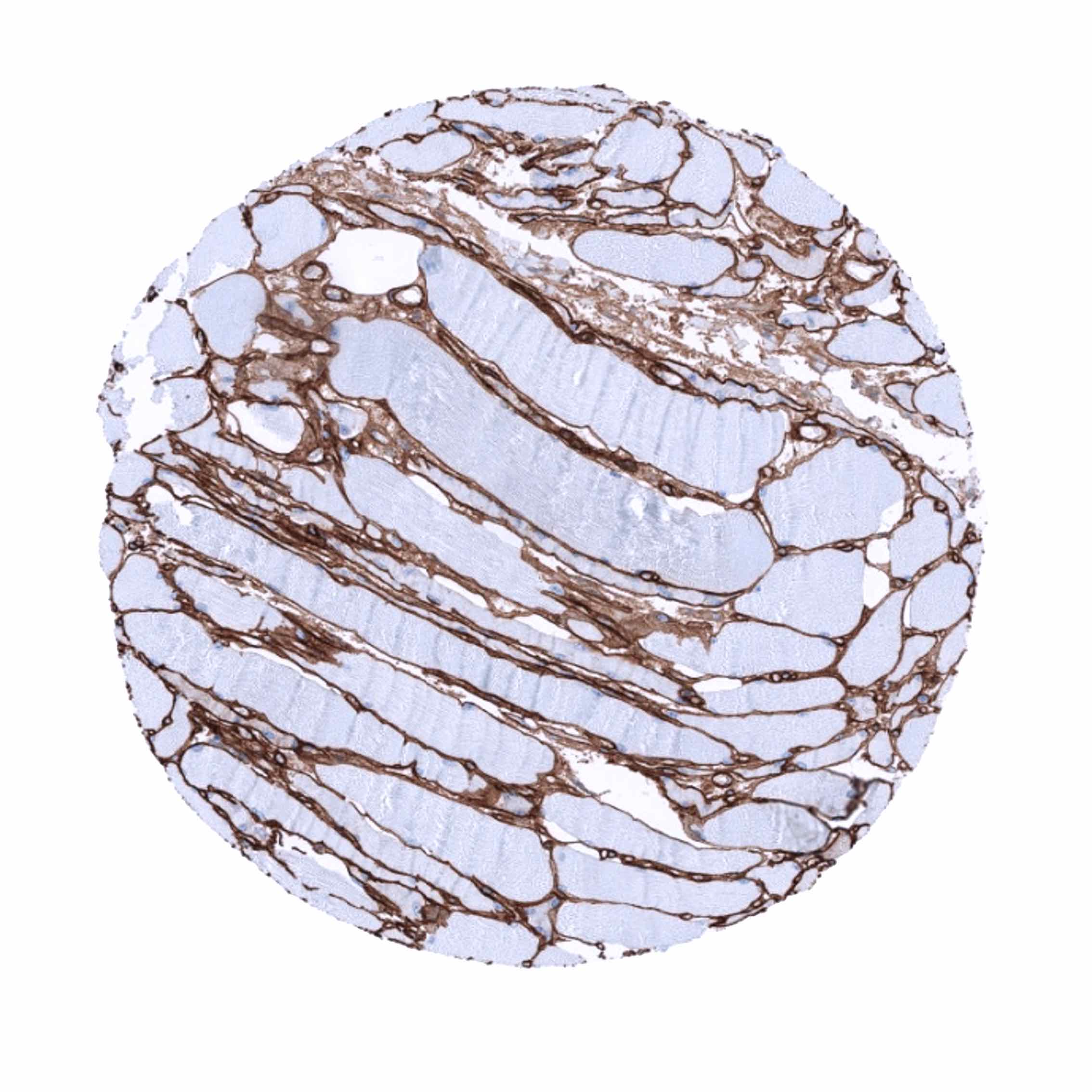
Collagen type IV immunostaining is seen in all basement membranes separating epithelial cells from neighboring tissues. Collagen IV also surrounds individual smooth muscle, heart muscle and skeletal muscle cells. Accordingly all muscular tissues und vessels show significant collagen IV immunostaining. Collagen IV also surrounds decidua cells.
These findings are largely consistent with the RNA and protein data described in the Human Protein Atlas (Tissue expression Collagen IV)
Positive control: Colon: An at least moderate collagen IV immunostaining of basement membranes and a strong staining of vessels and the tunica muscularis mucosa should be seen.
Negative control: Colon: Collagen IV immunostaining must be absent in epithelial cells.


Staining Pattern in Relevant Tumor Types
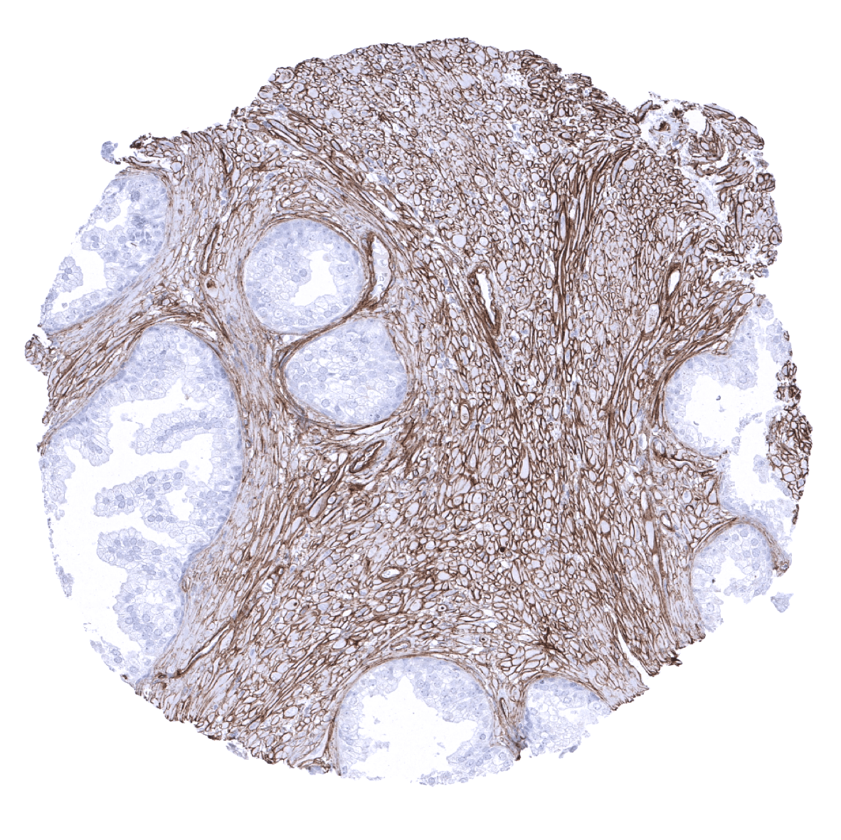
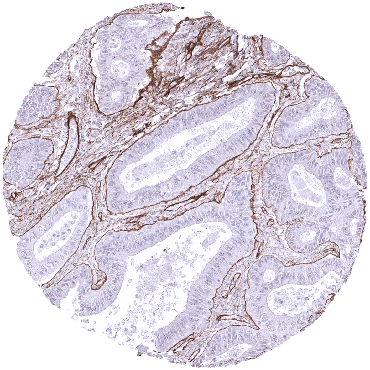
A positive collagen IV immunostaining is usually seen in tumors derived from muscle or fat cells. In other tumor entities a variable quantity of collagen IV immunostaining is usually seen in the tumor stroma (especially in vessels) and structures invaded by a tumor (muscle, fat). Collagen IV positive membranes are regularly seen around tumor cell nests, irrespective of whether or not these are invasive.
The TCGA findings on Collagen IV RNA expression in different tumor categories have been summarized in the Human Protein Atlas.


Compatibility of Antibodies
No data available at the moment
Protocol Recommendations
IHC users have different preferences on how the stains should look like. Some prefer high staining intensity of the target stain and even accept some background. Others favor absolute specificity and lighter target stains. Factors that invariably lead to more intense staining include higher concentration of the antibody and visualization tools, longer incubation time, higher temperature during incubation, higher temperature and longer duration of the heat induced epitope retrieval (slide pretreatment). The impact of the pH during slide pretreatment has variable effects and depends on the antibody and the target protein.
All images and data shown here and in our image galleries are obtained by the manual protocol described below. Other protocols resulting in equivalent staining are described as well.
Manual protocol
Freshly cut sections should be used (less than 10 days between cutting and staining). Heat-induced antigen retrieval for 5 minutes in an autoclave at 121°C in pH 7,8 Target Retrieval Solution buffer. Apply MSVA-704R at a dilution of 1:150 at 37°C for 60 minutes. Visualization of bound antibody by the EnVision Kit (Dako, Agilent) according to the manufacturer’s directions.
Agilent / Dako – Autostainer Link 48
Pretreatment in PT-Link for 30 minutes at 95°C (pH high); FLEX peroxidase blocking for 5 minutes (room temperature), MSVA-704R 1:150 for 20 minutes (room temperature), FLEX+ mouse/rabbit (LINKER) for 15 minutes (room temperature), horseradish peroxidase (HRP) for 20 minutes (room temperature), FLEX DAB+Sub-Chromo for 10 minutes (room temperature), FLEX hematoxylin for 5 minutes (room temperature).


These images reflect stainings by the protocol described above. It is of note that a comparable staining result can also be obtained by different protocols. In general, a longer pretreatment, a longer incubation time of the primary antibody, a higher antibody concentration, and a longer incubation time of FLEX+LINKER result in stronger staining, potentially at the cost of more background staining. Modifications of the protocol with a strengthening effect on staining intensity in combination with changes of other parameters that result in lower staining intensity can result in a comparable result as shown above.
Leica – BOND RX
Dewax at 72°C for 30 seconds; Pretreatment in Bond Epitope Retrieval Solution (ER2 – EDTA pH9) for 20 minutes at 100°C; Peroxidase blocking for 5 minutes (room temperature), MSVA-704R 1:150 for 15 minutes (room temperature), Post primary (rabbit anti mouse) for 8 minutes (room temperature), Polymer (goat anti rabbit) for 8 minutes (room temperature), mixed DAB refine for 10 minutes (room temperature), hematoxylin for 5 minutes (room temperature).

These images reflect stainings by the protocol described above. It is of note that a comparable staining result can also be obtained by different protocols. In general, a longer pretreatment, a longer incubation time of the primary antibody, a higher antibody concentration, a higher temperature during incubation, and a longer incubation time of Post primary and or the Polymer result in stronger staining, potentially at the cost of more background staining. Modifications of the protocol with a strengthening effect on staining intensity in combination with changes of other parameters that result in lower staining intensity can result in a comparable result as shown above.
Roche – Ventana Discovery ULTRA
Pretreatment for 64 minutes at 100°C (pH 8,4); CM peroxidase blocking for 12 minutes (room temperature), MSVA-704R 1:150 for 20 minutes at 36°C, secondary antibody (anti-rabbit HQ) for 12 minutes at 36°C, anti-HQ HRP for 12 minutes at room temperature, DAB at room temperature, hematoxylin II at room temperature for 8 minutes, bluing reagent at room temperature for 4 minutes.
These images depict staining results obtained by the protocol described above. It is of note, that the Ventana machines generally require higher antibody concentrations than other commonly used autostainers because the antibodies are automatically diluted during the procedure. Various other protocols can result in an identical result as shown above. A longer pretreatment, a longer incubation time of the primary antibody, a higher antibody concentration, a higher temperature during incubation, and a longer incubation time of secondary antibody and or the anti-HQ HRP result in stronger staining, potentially at the cost of more background staining.
Potential Research Applications
- Collagen IV is a useful component of multicolor immunofluorescence if different tissue compartments require a separate automated analysis.
Evidence for Antibody Specificity in IHC
Evidence of antibody specificity comes from complete concordance of the staining data with RNA and protein expression data provided in the Human Protein Atlas (Tissue expression Collagen IV) in combination with the complete absence of collagen IV staining seen in epithelial, hematopoietic, lymphoid, and brain tissues.