295,00 € – 995,00 €
Product details
Synonyms = L3T4; Leu3; Ly-4; Lymphocyte antigen CD4; p55; T cell antigen T4/LEU3; T cell differentiation antigen L3T4; T-cell surface antigen T4/Leu-3; T-cell surface glycoprotein CD4
Antibody type = Recombinant Rabbit monoclonal / IgG
Clone = MSVA-004R
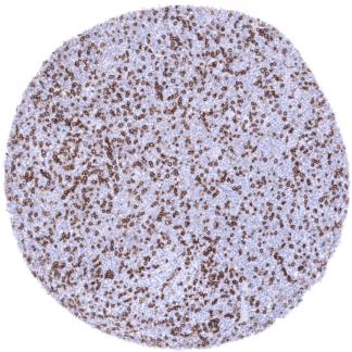
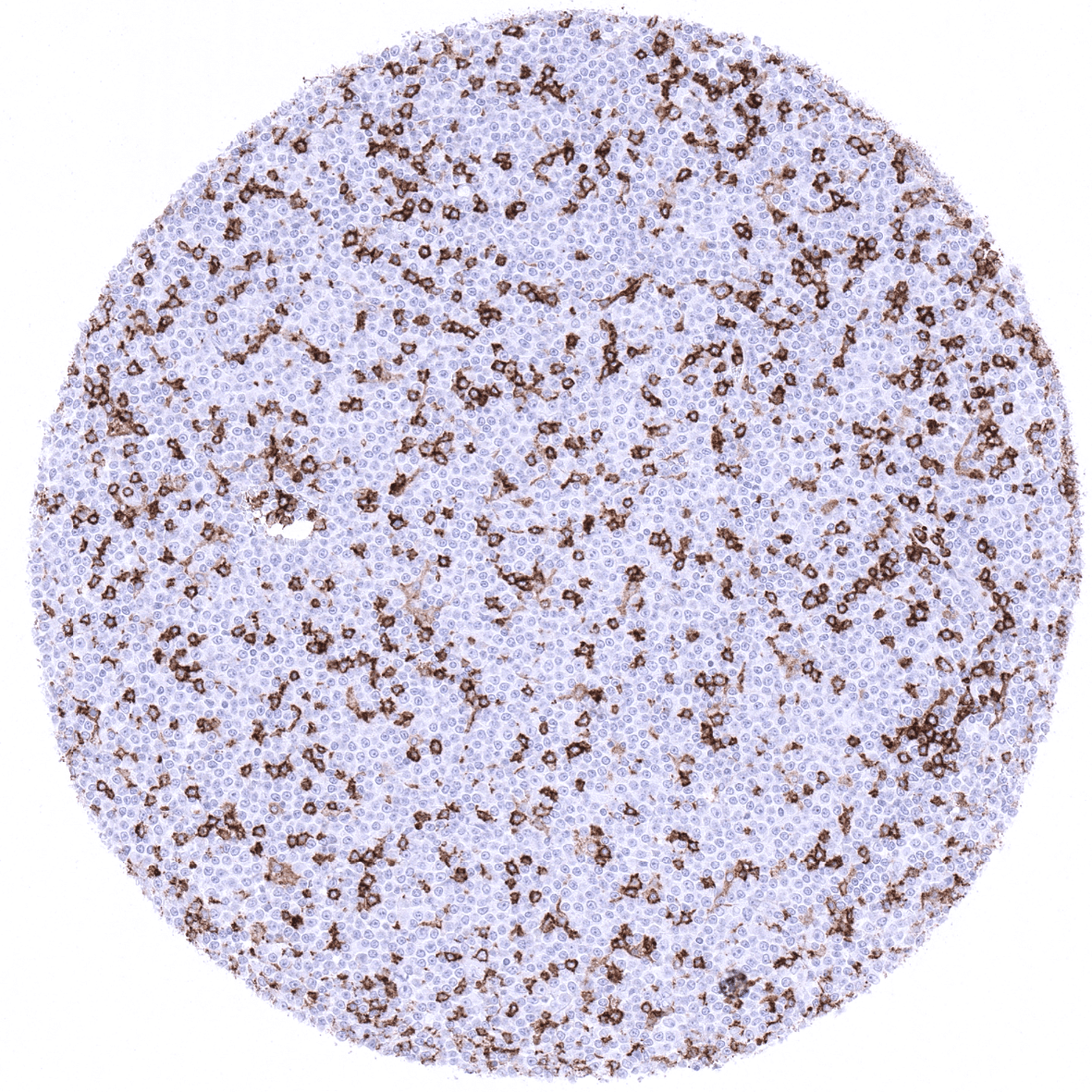
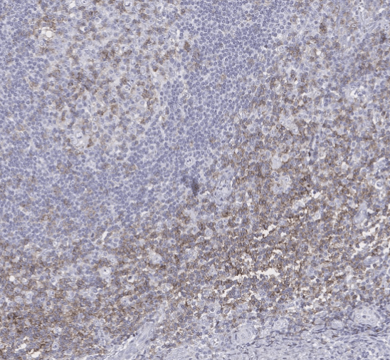
Positive control = Tonsil: A moderate to strong membranous CD4 immunostaining should be seen in a subset of T-lymphocytes which is predominantly located in the interfollicular area. Macrophages in the germinal centres should show a moderate CD4 positivity. Liver: Kupffer cells and endothelial cells in the liver sinusoids must show at least a moderate CD4 staining.
Negative control = Tonsil: B-cells and epithelial cells must not show any CD4 staining. Liver: Hepatocytes must not show any CD4 staining.
Cellular localization = Cell surface
Reactivity = Human
Application = Immunohistochemistry
Dilution = 1:100 – 1:200
Intended Use = Research Use Only
Relevance of Antibody
CD4 is expressed on T helper lymphocytes.
Biology Behind
CD4 (cluster of differentiation 4; leu-3) is a glycoprotein belonging to the immunoglobulin superfamily which is present on the surface of T helper cells, monocytes, macrophages, and dendritic cells. CD4+ T helper cells are an essential part of the human immune system. One of their main roles is to send signals to other types of immune cells, including CD8 killer cells, which then destroy the infectious particle. CD4 has four immunoglobulin domains (D1 to D4) that are exposed on the extracellular surface of the cell: D1 and D3 resemble immunoglobulin variable (IgV) domains. D2 and D4 resemble immunoglobulin constant (IgC) domains. CD4 interacts through its D1 domain with the β2-domain of MHC class II molecules. T helper cells are thus specific for antigens presented by MHC II and not by MHC class I (MHC class II-restricted). CD4 is a co-receptor of the T cell receptor (TCR) and assists the latter in communicating with antigen-presenting cells. The TCR complex and CD4 bind to distinct regions of the antigen-presenting MHC class II molecule. The extracellular D1 domain of CD4 binds to the β2 region of MHC class II. The resulting close proximity of TCR complex and CD4 allows results in a signaling cascade that eventually leads to an activation of various transcription factors, including NF-κB, NFAT, AP-1, to promote T cell activation. CD4 Serves as HIV receptor on T cells and macrophages. If CD4 cells become depleted in untreated HIV infection or following iatrogenic immune suppression, the body is left vulnerable to a wide range of infections that it would otherwise have been able to fight.
Staining Pattern in Normal Tissues
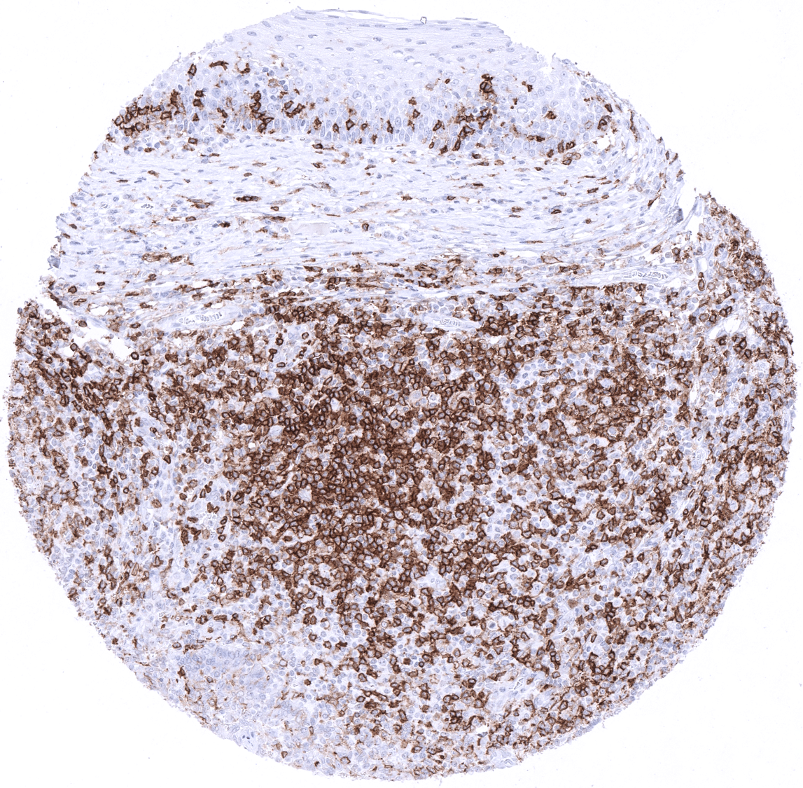
CD4 immunostaining is seen in >90% of thymocytes and in a large subset of extrathymic T-lymphocytes which – at variable numbers – can be found in almost every organ/tissue. At a lower level CD4 expression is also seen in monocytes and macrophages. A moderate to strong staining is seen in Langerhans cells of the skin and non-keratinizing squamous epithelium of various sites. Endothelial CD4 positivity is seen at moderate intensity in liver sinusoids and at a lower level also in some small vessels of the adrenal gland and the adenohypophysis. A moderate to strong membranous CD4 positivity is seen in all epithelial cells of the parathyroid gland. A weak CD4 staining can be seen in some littoral cells of the spleen.
These findings are largely consistent with RNA and protein data summarized in the Human Protein Atlas (Tissue expression CD4).
Suggested positive tissue control: Tonsil: A moderate to strong membranous CD4 immunostaining should be seen in a subset of T-lymphocytes which is predominantly located in the interfollicular area. Macrophages in the germinal centres should show a moderate CD4 positivity. Liver: Kupffer cells and endothelial cells in the liver sinusoids must show at least a moderate CD4 staining.
Suggested negative tissue control: Tonsil: B-cells and epithelial cells must not show any CD4 staining. Liver: Hepatocytes must not show any CD4 staining.

Staining Pattern in Relevant Tumor Types
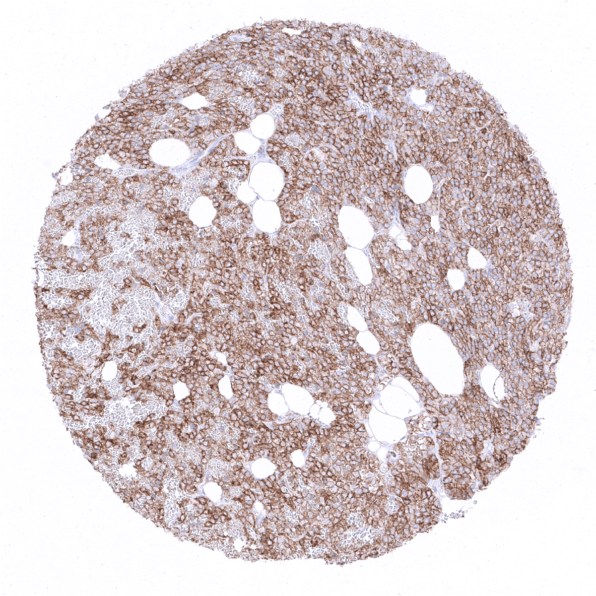
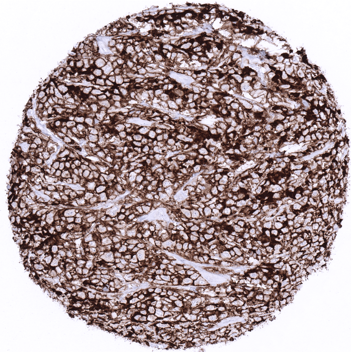
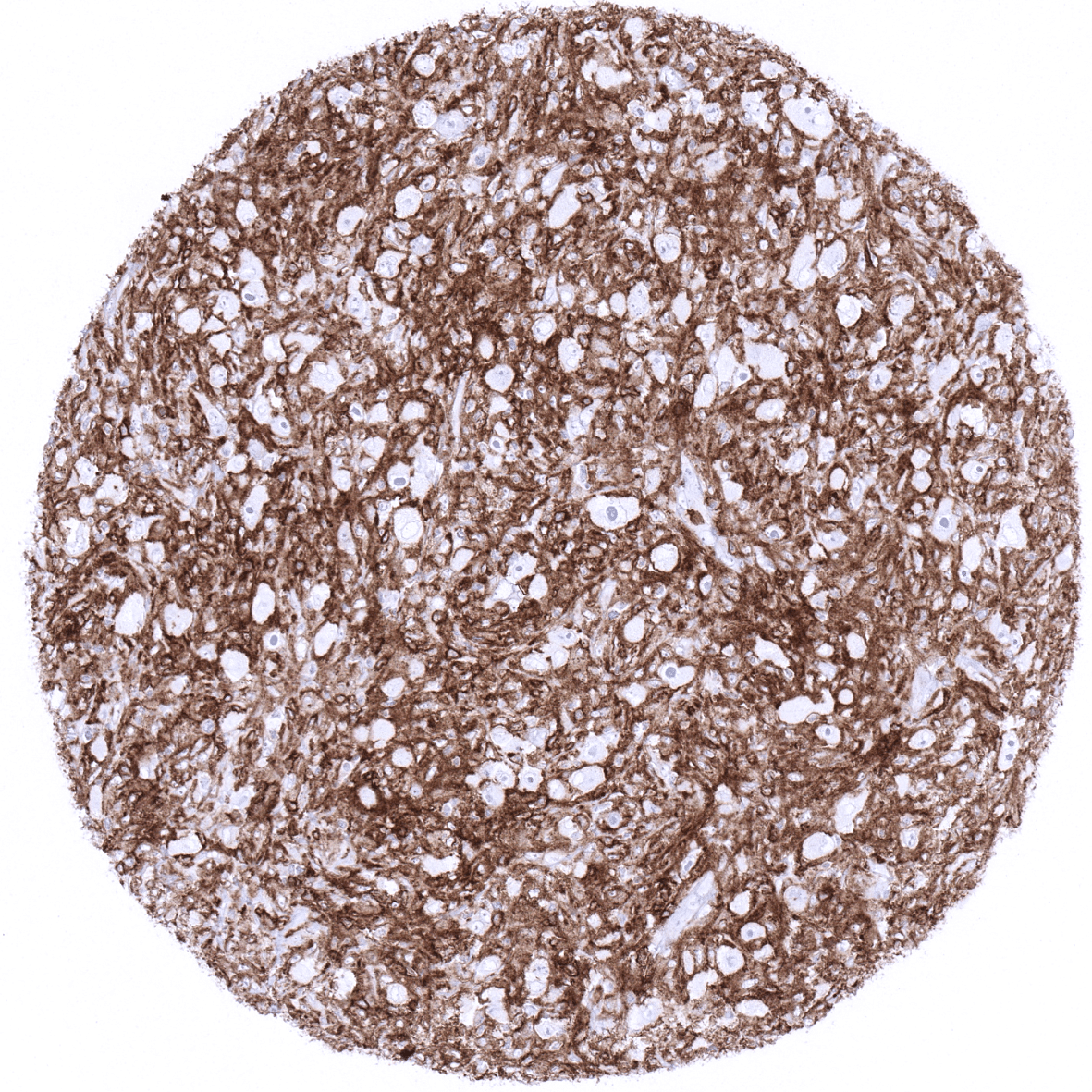
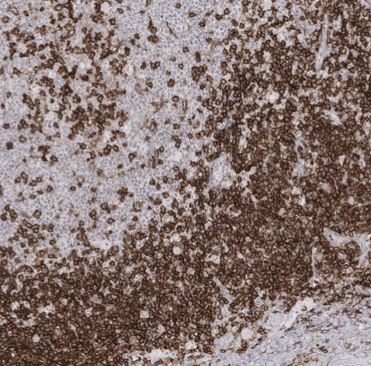
Most mature T-cell lymphomas are CD4 positive, while precursor T-lymphoblastic lymphomas show variable CD4 expression. CD4 is also expressed in histiocytic sarcomas and Langerhans cell histiocytosis. Several rare T-cell lymphomas are CD4 negative such as extranodal NK/T-cell lymphomas, NK-cell leukaemia, hepatosplenic T-cell lymphoma, subcutaneous panniculitis-like T-cell lymphoma, and enteropathy-type T-cell lymphoma. Non-neoplastic CD4 positive t-cells and macrophages are commonly seen in variable numbers in tumors of all kinds.
The TCGA findings on CD4 RNA expression in different tumor categories have been summarized in the Human Protein Atlas.
Compatibility of Antibodies
No data available at the moment
Protocol Recommendations
IHC users have different preferences on how the stains should look like. Some prefer high staining intensity of the target stain and even accept some background. Others favor absolute specificity and lighter target stains. Factors that invariably lead to more intense staining include higher concentration of the antibody and visualization tools, longer incubation time, higher temperature during incubation, higher temperature and longer duration of the heat induced epitope retrieval (slide pretreatment). The impact of the pH during slide pretreatment has variable effects and depends on the antibody and the target protein. Accordingly, multiple different protocols can generate identical staining results.
All images and data shown here and in our image galleries are obtained by the manual protocol described below. Other protocols resulting in equivalent staining are described as well.
-Manual protocol
Freshly cut sections should be used (less than 10 days between cutting and staining). Heat-induced antigen retrieval for 5 minutes in an autoclave at 121°C in pH9 Target Retrieveal Solution buffer. Apply MSVA-004R at a dilution of 1:200 at 37°C for 60 minutes. Visualization of bound antibody by the EnVision Kit (Dako, Agilent) according to the manufacturer’s directions.
Impact of pH:

Potential Research Applications
- CD4 positive cells play a major but not yet fully understood role in inflammation and cancer.
- CD4 is a key component of multicolor assays analyzing the role of lymphocyte subsets in cancers and other diseases.
- The prevalence of a positive CD3 immunostaining in hematological and non-hematological neoplasms should be further evaluated.
Evidence for Antibody Specificity in IHC
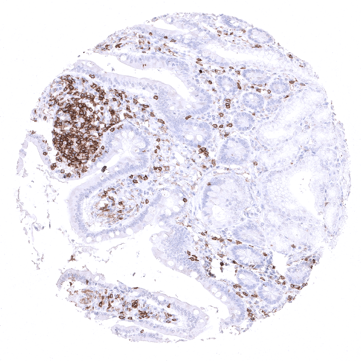
Specificity of MSVA-004R is documented by strong positive staining in cell types that are well documented to express CD4 such as T-lymphocytes, macrophages and liver sinusoids in combination with absence of staining in all tissues known to not express CD4 such as all epithelial tissues including those being notorious for non-specific IHC background (kidney, colonic mucosa, stomach glands).