295,00 € – 895,00 €
Product details
Synonyms = Hematopoietic Progenitor Cell Antigen, HPCA1, Mucosialin
Antibody type = Recombinant Rabbit monoclonal / IgG
Clone = MSVA-034R
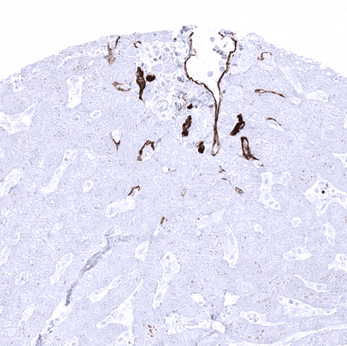
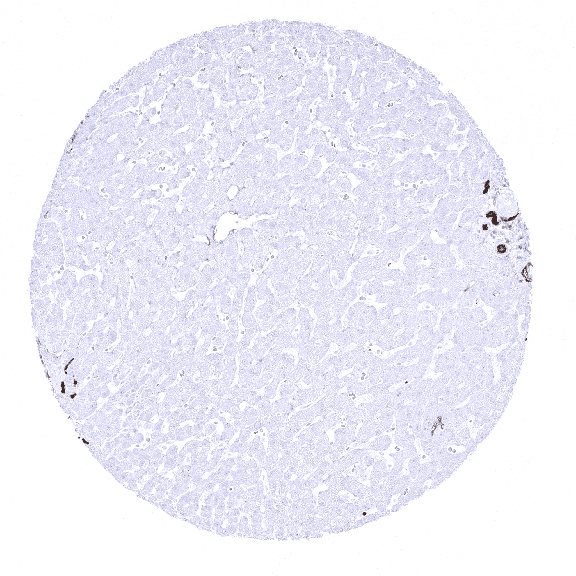
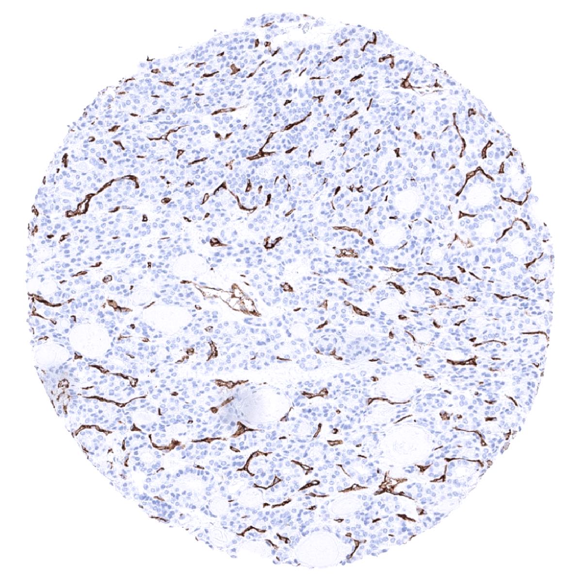
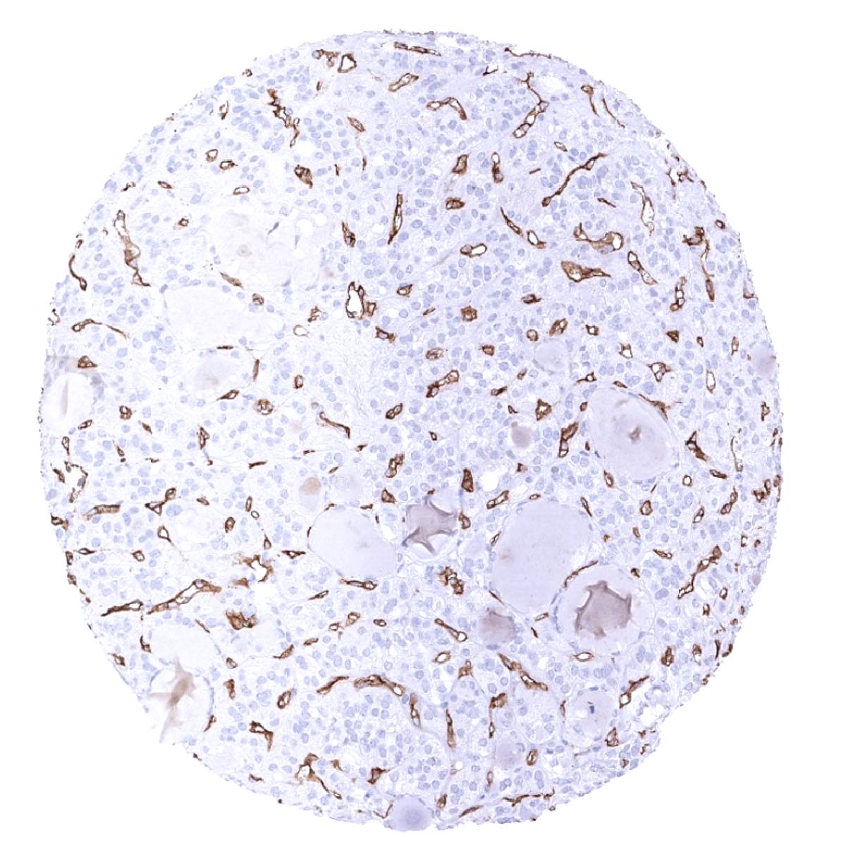
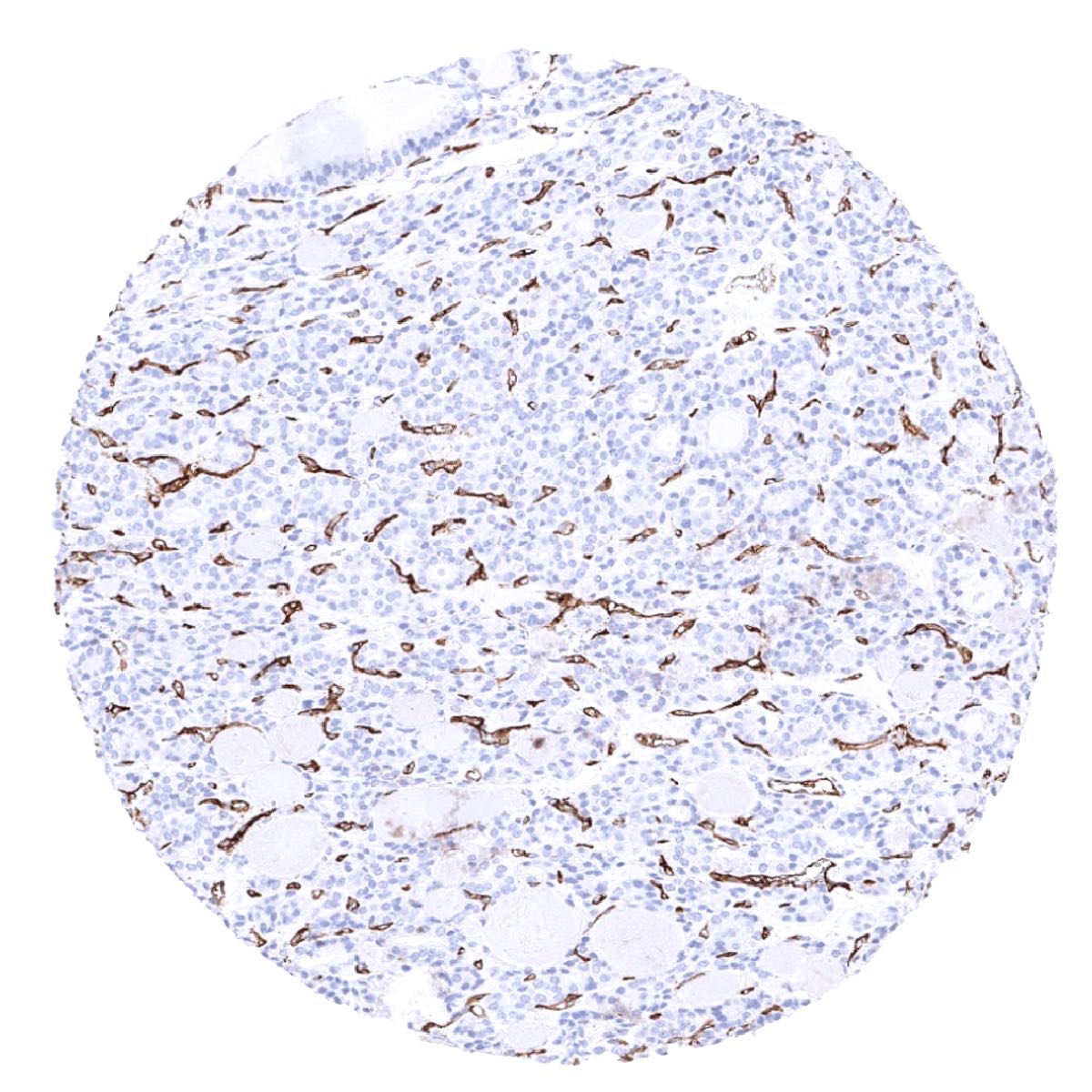
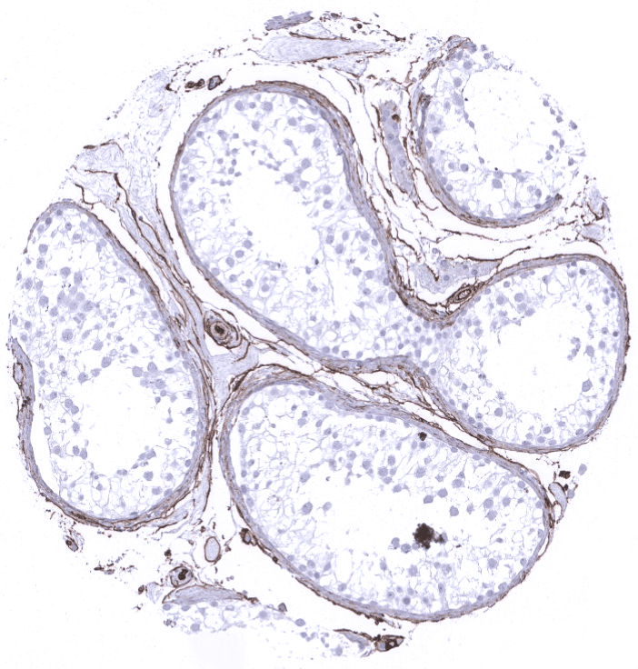
Positive control = Liver: A moderate to strong, predominantly membranous staining of endothelial cells should be seen in portal vessels and periportal sinusoidal endothelial cells. Endothelial cells located more remotely from the portal field show a rapidly decreasing CD34 expression level.
Negative control = Liver: Hepatocytes should always stain CD34 negative. Sinusoidal cells remote from the portal fields must also be CD34 negative.
Cellular localization = Cell Surface and Cytoplasmic
Application = Immunohistochemistry
Dilution = 1:100 – 1:200
Intended Use = Research Use Only
Relevance of Antibody
CD34 is expressed on endothelial cells and on interstitial cells of Cajal (GIST).
Biology Behind
CD34 is a commonly used marker of hematopoietic progenitor cells and endothelial cells. It was first described as a cell surface protein on hematopoietic stem cells. CD34 is a heavily glycosylated 110 kDa protein. The CD34 gene at 1q32.2 codes for two forms of the protein that result from alternative splicing. The CD34 function is not entirely clear. It may function as a cell-cell adhesion factor, facilitate cell migration, and mediate the attachment of hematopoietic stem cells to bone marrow extracellular matrix or stromal cells. CD34 is required for T cells to enter lymph nodes. CD34 is thus expressed on lymph node endothelial and T cells can bind to it. CD34 expression occurs in hematopoietic and mesenchymal stem cells, endothelial progenitor cells, endothelial cells of blood vessels (but much less in lymphatics), mast cells, a sub-population of dendritic cells (termed dendritic interstitial cells), as well as cells in various hematopoietic and soft tissue tumors. All hematological cell types loose CD34 during maturation, only megakaryocytes retain a variable CD34 expression.
Staining Pattern in Normal Tissues
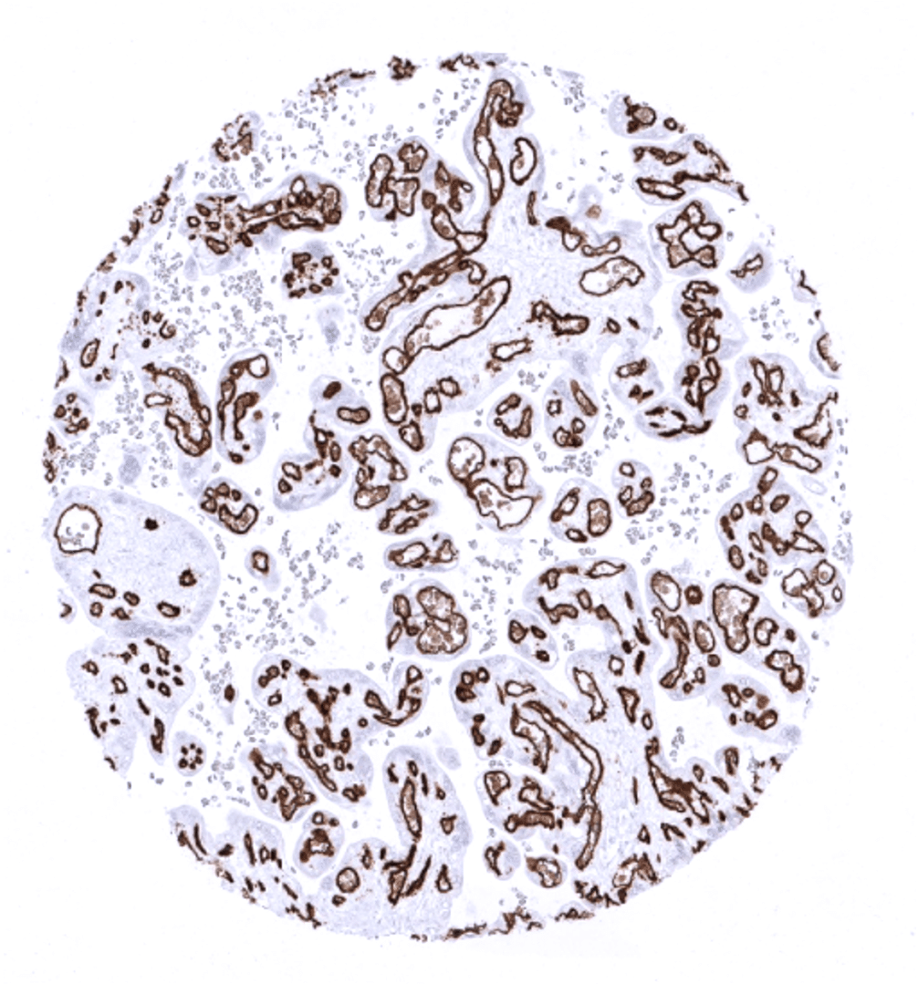
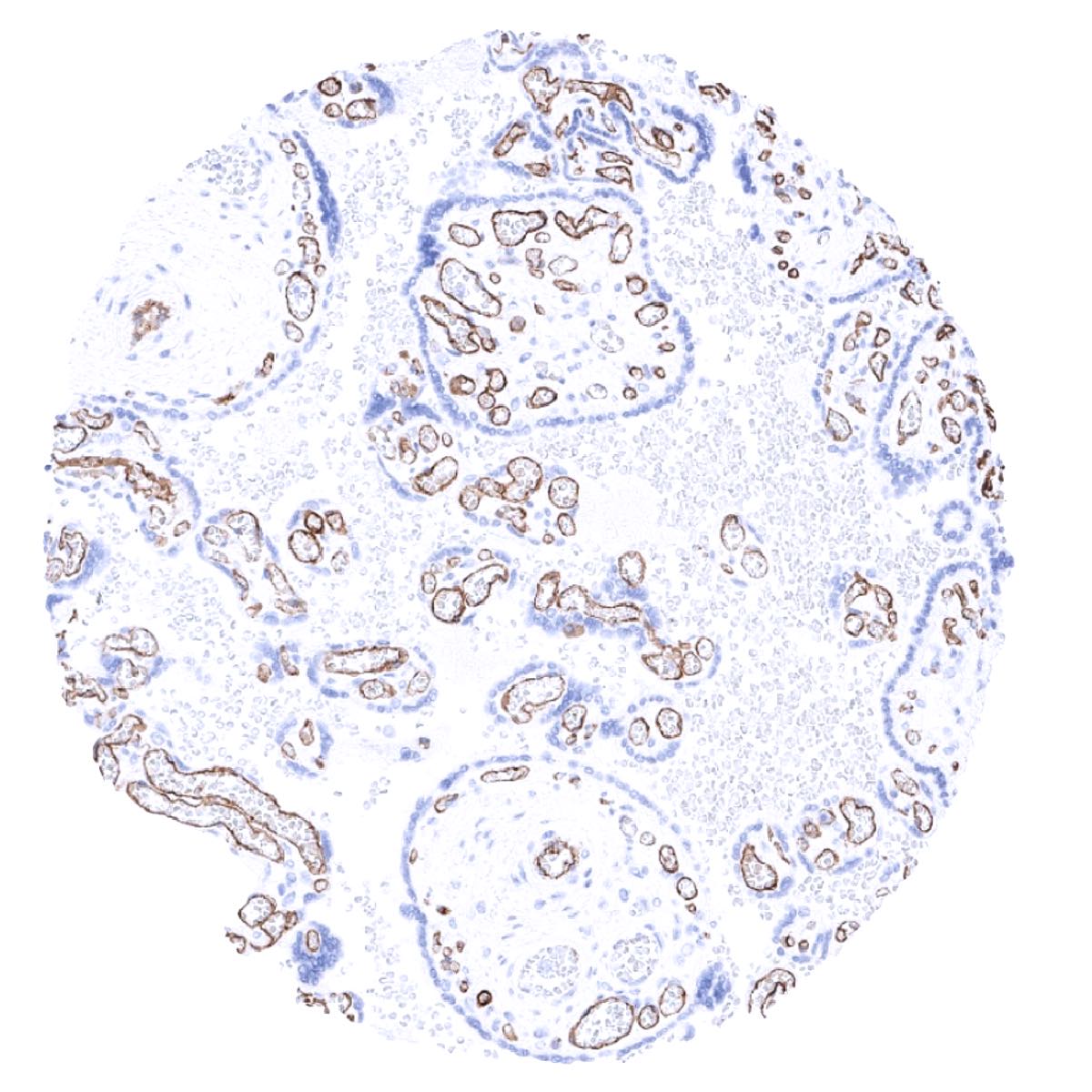
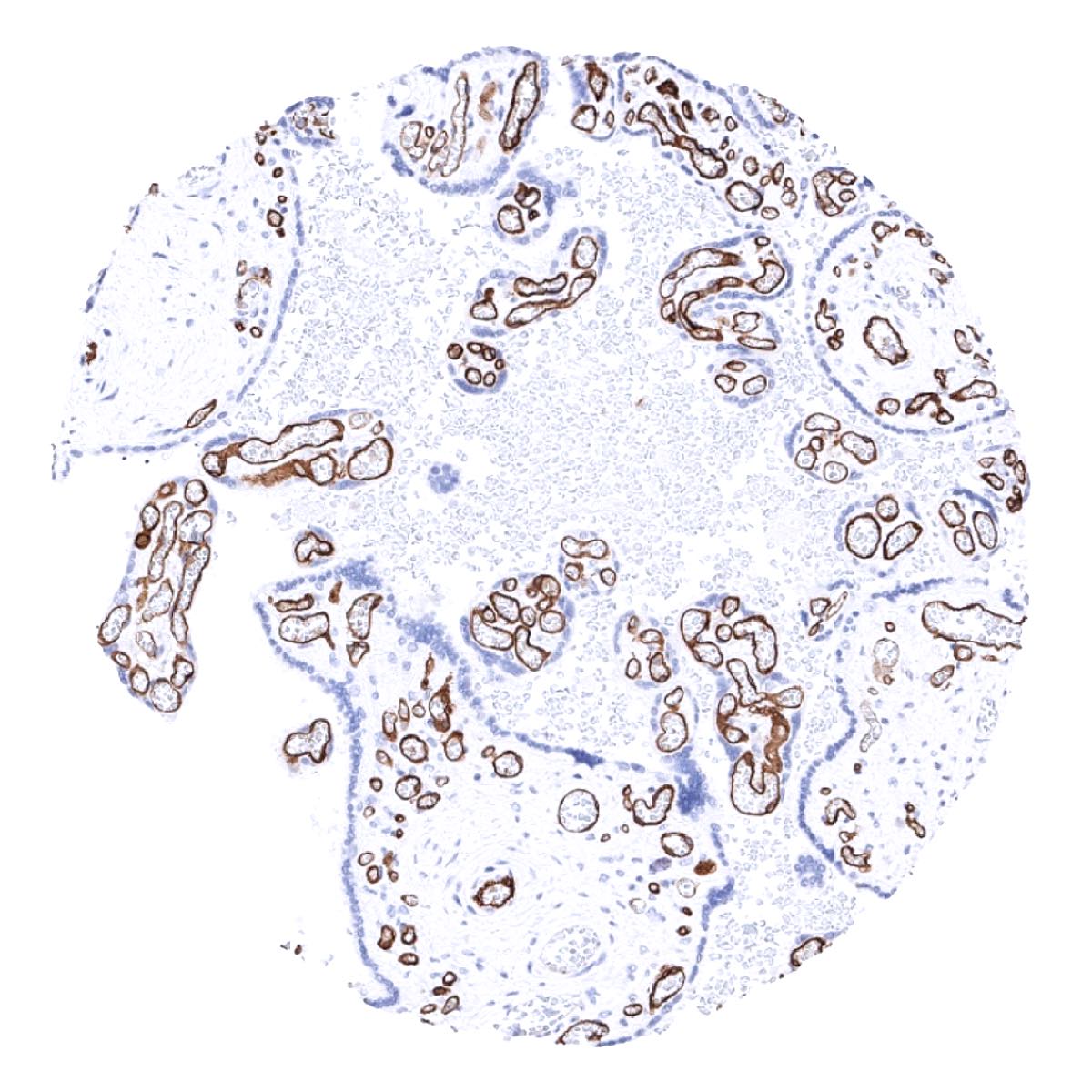
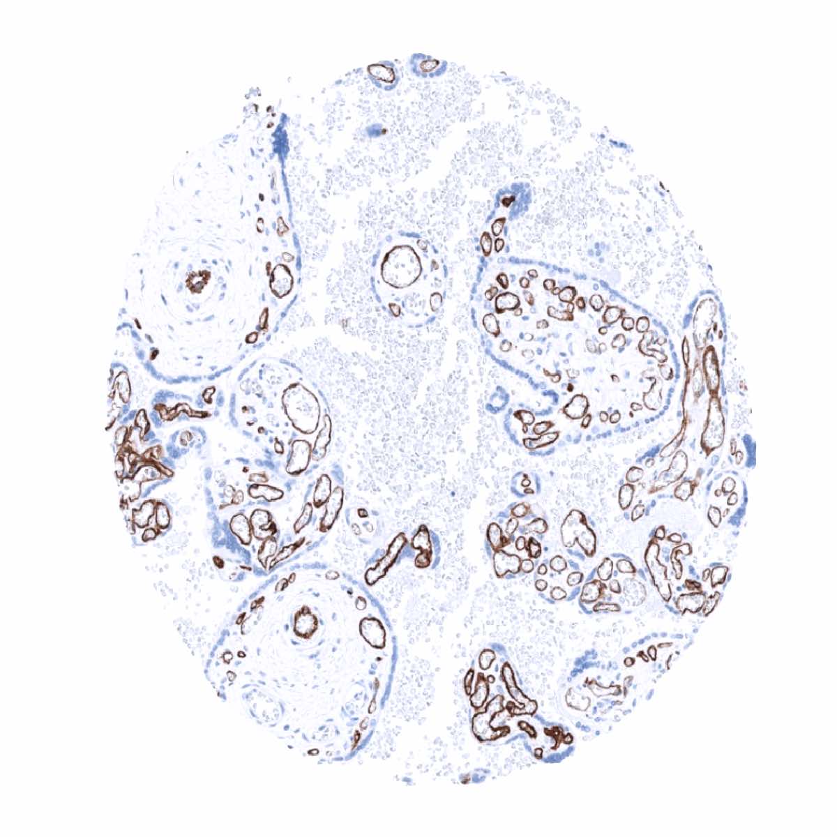
CD34 is found in most endothelium cells of blood vessels but is absent from sinuses in the placenta, and spleen, and may be less intensely expressed or absent in endothelia of large veins and arteries. CD34 is expressed at low level in endothelial cells of lymphatics. In the liver, CD34 is seen in portal blood vessels and periportal sinusoids but expression is lost in sinus that are more remote from the portal fields. CD34 is also expressed in stroma cells of endometrium and cervix uteri, fibroblast-like dendritic cells, as well as in some fibroblasts, fibrocytes, interstitial cells of Cajal, mast cells, megakaryocytes, and – at variable levels – in smooth muscle cells. According to this broad spectrum of cell types that can express CD34, a positive CD34 immunostaining is regularly seen in the stroma of thyroid, fallopian tube, testis, epididymis, seminal vesicle, bronchial mucosa, prostate, Brunner gland, and salivary glands.
These findings are largely comparable to the RNA described and IHC findings depicted in the Human Protein Atlas (Tissue expression CD34) even though CD34 expression in some scattered epithelial cells of the gastrointestinal tract distal of the stomach is not described in the protein atlas.
Suggested positive tissue control: Liver: A moderate to strong, predominantly membranous staining of endothelial cells should be seen in portal vessels and periportal sinusoidal endothelial cells. Endothelial cells located more remotely from the portal field show a rapidly decreasing CD34 expression level.
Suggested negative tissue control: Liver: Hepatocytes should always stain CD34 negative. Sinusoidal cells remote from the portal fields must also be CD34 negative.

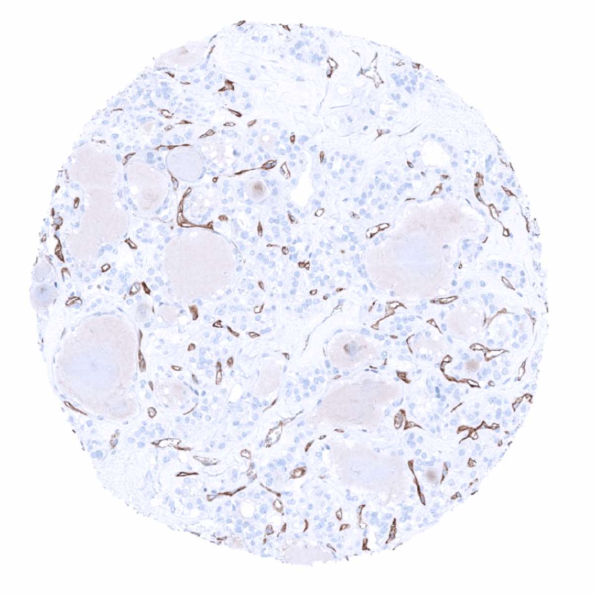
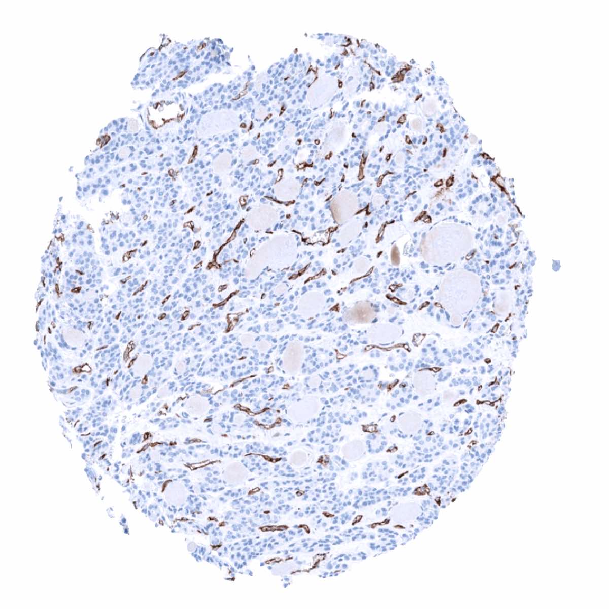
Staining Pattern in Relevant Tumor Types
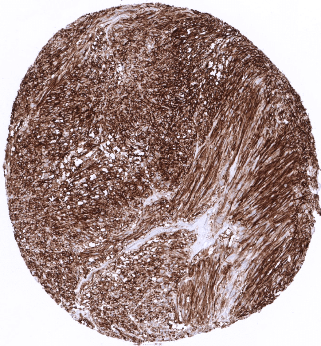
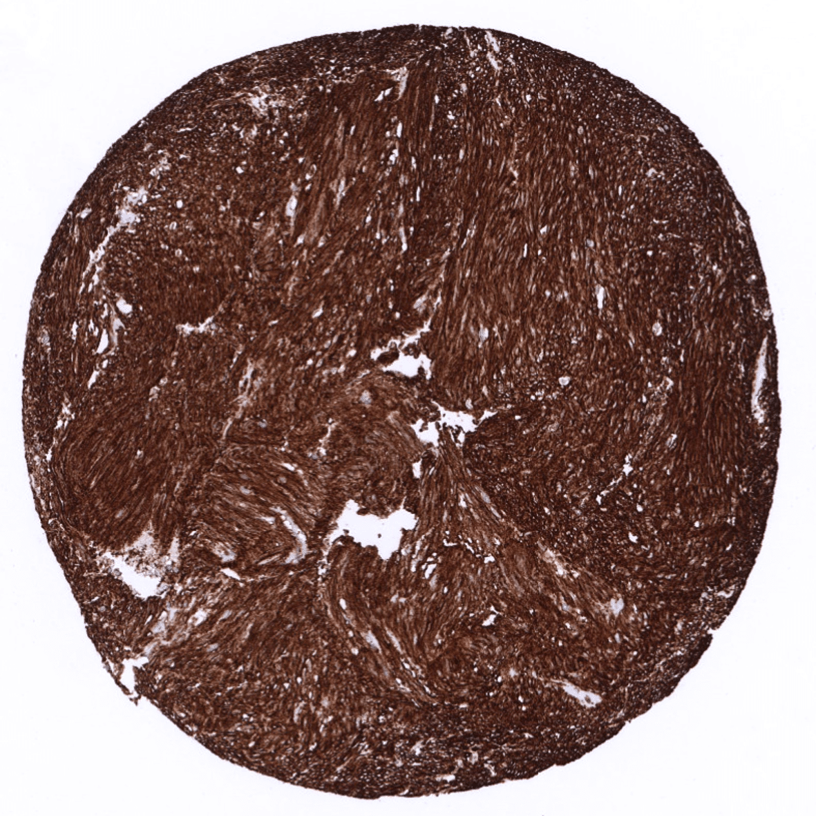
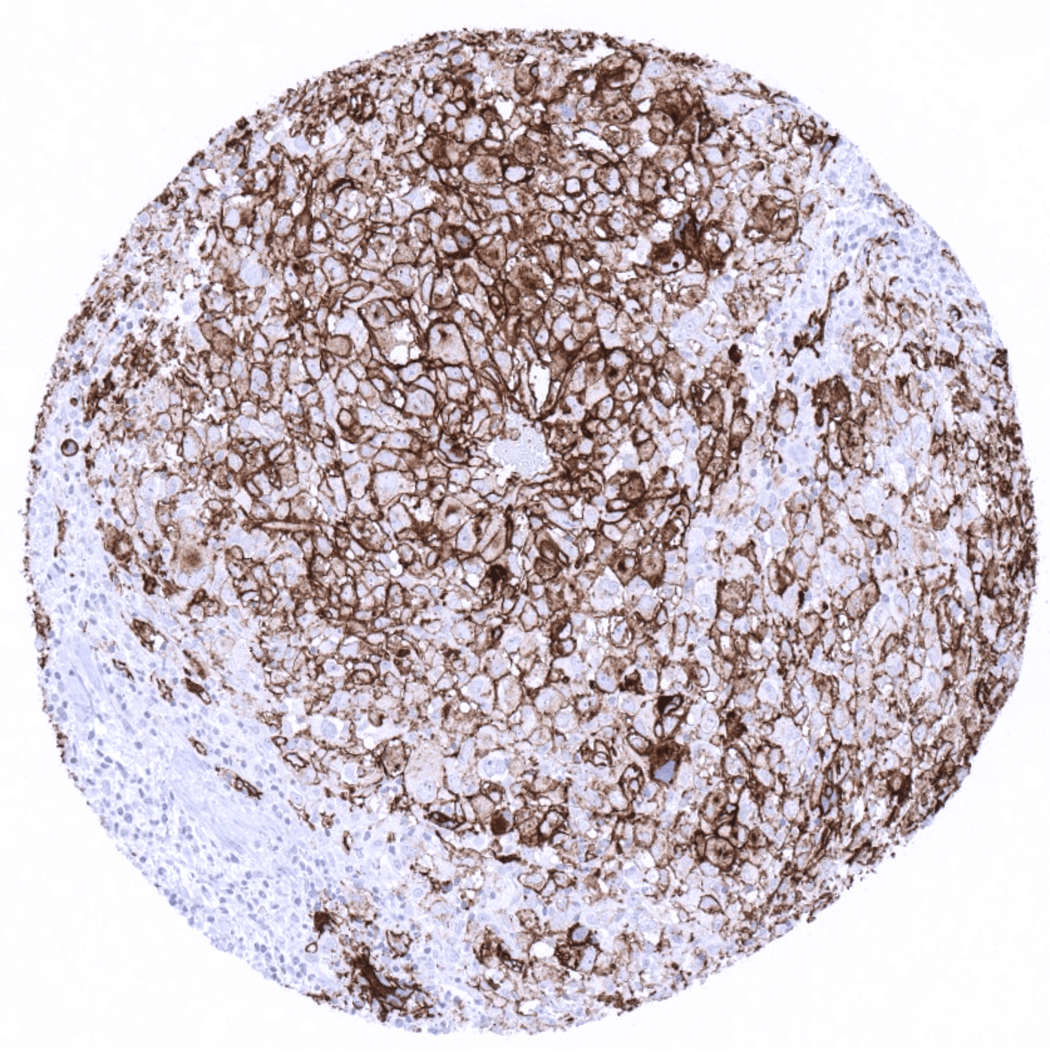
CD34 is found in myeloid blasts in myelodysplastic syndrome and acute myeloid leukemia in most cases as well as lymphoblasts in most cases of B-acute lymphoblastic leukemia but mature B- and T-cell lymphomas and leukemias are CD34 negative. CD34 is detectable in most vascular tumors, including hemangiosarcoma and Kaposi sarcoma but only in about 30% of lymphangiomas. Strong CD34 positivity is seen in most cases of dermatofibrosarcoma protuberans, most gastrointestinal stromal tumors and solitary fibrous tumors. A variable CD34 staining, sometimes at high intensity, can however be seen in a wide variety of soft tissue tumors such as lipoma (particularly spindle cell lipoma), liposarcoma, meningioma, sarcoma NOS, myxofibrosarcoma. A mostly weak to moderate CD34 expression can also be observed in leiomyoma, epitheloid sarcomas, synovial sarcomas, neurofibroma, neurofibrosarcoma, myofibroblastoma, schwannoma (preferably in Antoni B areas) and others.
The TCGA findings on CD34 RNA expression in different tumor categories have been summarized in the Human Protein Atlas.

Compatibility of Antibodies
No data available at the moment
Protocol Recommendations
IHC users have different preferences on how the stains should look like. Some prefer high staining intensity of the target stain and even accept some background. Others favor absolute specificity and lighter target stains. Factors that invariably lead to more intense staining include higher concentration of the antibody and visualization tools, longer incubation time, higher temperature during incubation, higher temperature and longer duration of the heat induced epitope retrieval (slide pretreatment). The impact of the pH during slide pretreatment has variable effects and depends on the antibody and the target protein. Accordingly, multiple different protocols can generate identical staining results.
All images and data shown here and in our image galleries are obtained by the manual protocol described below. Other protocols resulting in equivalent staining are described as well.
Manual protocol
Freshly cut sections should be used (less than 10 days between cutting and staining). Heat-induced antigen retrieval for 5 minutes in an autoclave at 121°C in pH 7.8 Target Retrieveal Solution buffer. Apply MSVA-034R at a dilution of 1:150 at 37°C for 60 minutes. Visualization of bound antibody by the EnVision Kit (Dako, Agilent) according to the manufacturer’s directions.


Agilent / Dako – Autostainer Link 48
Pretreatment in PT-Link for 30 minutes at 95°C (pH high); FLEX peroxidase blocking for 5 minutes (room temperature), MSVA-034R 1:150 for 20 minutes (room temperature), FLEX+ mouse/rabbit (LINKER) for 15 minutes (room temperature), horseradish peroxidase (HRP) for 20 minutes (room temperature), FLEX DAB+Sub-Chromo for 10 minutes (room temperature), FLEX hematoxylin for 5 minutes (room temperature).

These images reflect stainings by the protocol described above. It is of note that a comparable staining result can also be obtained by different protocols. In general, a longer pretreatment, a longer incubation time of the primary antibody, a higher antibody concentration, and a longer incubation time of FLEX+LINKER result in stronger staining, potentially at the cost of more background staining. Modifications of the protocol with a strengthening effect on staining intensity in combination with changes of other parameters that result in lower staining intensity can result in a comparable result as shown above.
Leica – BOND RX
Dewax at 72°C for 30 seconds; Pretreatment in Bond Epitope Retrieval Solution (ER2 – EDTA pH9) for 20 minutes at 100°C; Peroxidase blocking for 5 minutes (room temperature), MSVA-034R 1:150 for 15 minutes (room temperature), Post primary (rabbit anti mouse) for 8 minutes (room temperature), Polymer (goat anti rabbit) for 8 minutes (room temperature), mixed DAB refine for 10 minutes (room temperature), hematoxylin for 5 minutes (room temperature).
These images reflect stainings by the protocol described above. It is of note that a comparable staining result can also be obtained by different protocols. In general, a longer pretreatment, a longer incubation time of the primary antibody, a higher antibody concentration, a higher temperature during incubation, and a longer incubation time of Post primary and or the Polymer result in stronger staining, potentially at the cost of more background staining. Modifications of the protocol with a strengthening effect on staining intensity in combination with changes of other parameters that result in lower staining intensity can result in a comparable result as shown above.
Roche – Ventana Discovery ULTRA
Pretreatment for 64 minutes at 100°C (pH 8,4); CM peroxidase blocking for 12 minutes (room temperature), MSVA-034R 1:150 for 20 minutes at 36°C, secondary antibody (anti-rabbit HQ) for 12 minutes at 36°C, anti-HQ HRP for 12 minutes at room temperature, DAB at room temperature, hematoxylin II at room temperature for 8 minutes, bluing reagent at room temperature for 4 minutes.
These images depict staining results obtained by the protocol described above. It is of note, that the Ventana machines generally require higher antibody concentrations than other commonly used autostainers because the antibodies are automatically diluted during the procedure. Various other protocols can result in an identical result as shown above. A longer pretreatment, a longer incubation time of the primary antibody, a higher antibody concentration, a higher temperature during incubation, and a longer incubation time of secondary antibody and or the anti-HQ HRP result in stronger staining, potentially at the cost of more background staining.
Impact of pH


The impact of pH is only mild for MSVA-034R.
Potential Research Applications
- The role of CD34 positive peritumoral stroma is not clarified. The extent of CD34 positivity of tumor stroma varies markedly between tumor types and individual tumors.
- Identification of cells with stem cell properties.
- The function of CD34 is still largely unknown
Evidence for Antibody Specificity in IHC
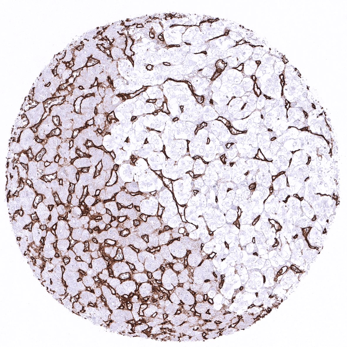
Specificity of MSVA-034R is documented by strong positive staining in cell types that are well documented to express CD34 such as endothelial cells and absence of staining in all tissues known to not express CD34 such as hepatocytes and tissues notorious for non-specific IHC background such as kidney and colonic mucosa.
