295,00 € – 995,00 €
Product details
Synonyms = CD3 epsilon; CD3 TCR complex; T cell antigen receptor complex epsilon subunit of T3; T-cell surface antigen T3/Leu-4 epsilon chain; T-cell surface glycoprotein CD3 epsilon chain; T3E; TCRE; TiT3 complex
Antibody type = Recombinant Rabbit monoclonal / IgG
Clone = MSVA-003R
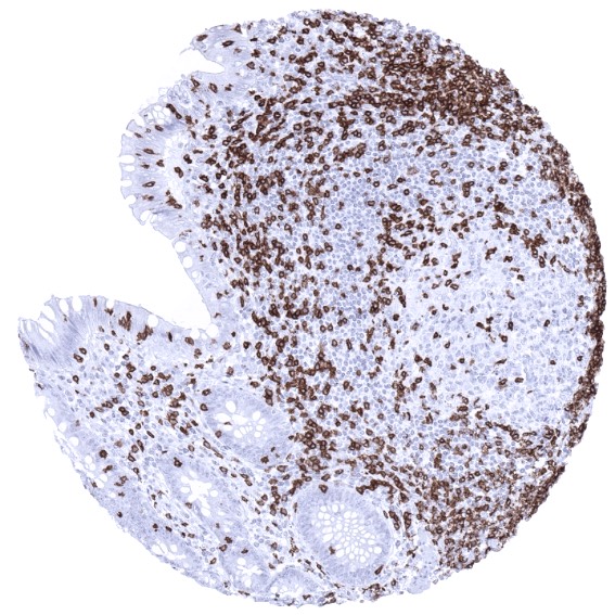
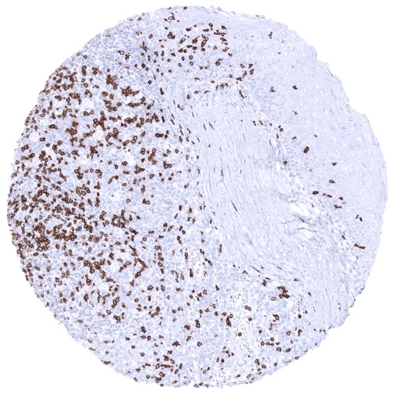
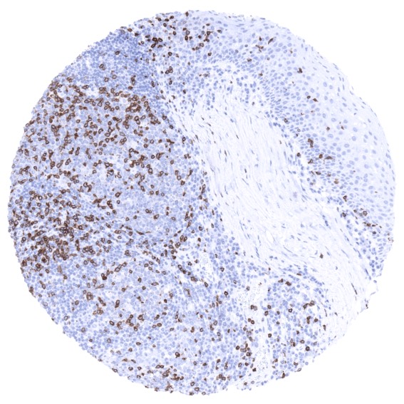
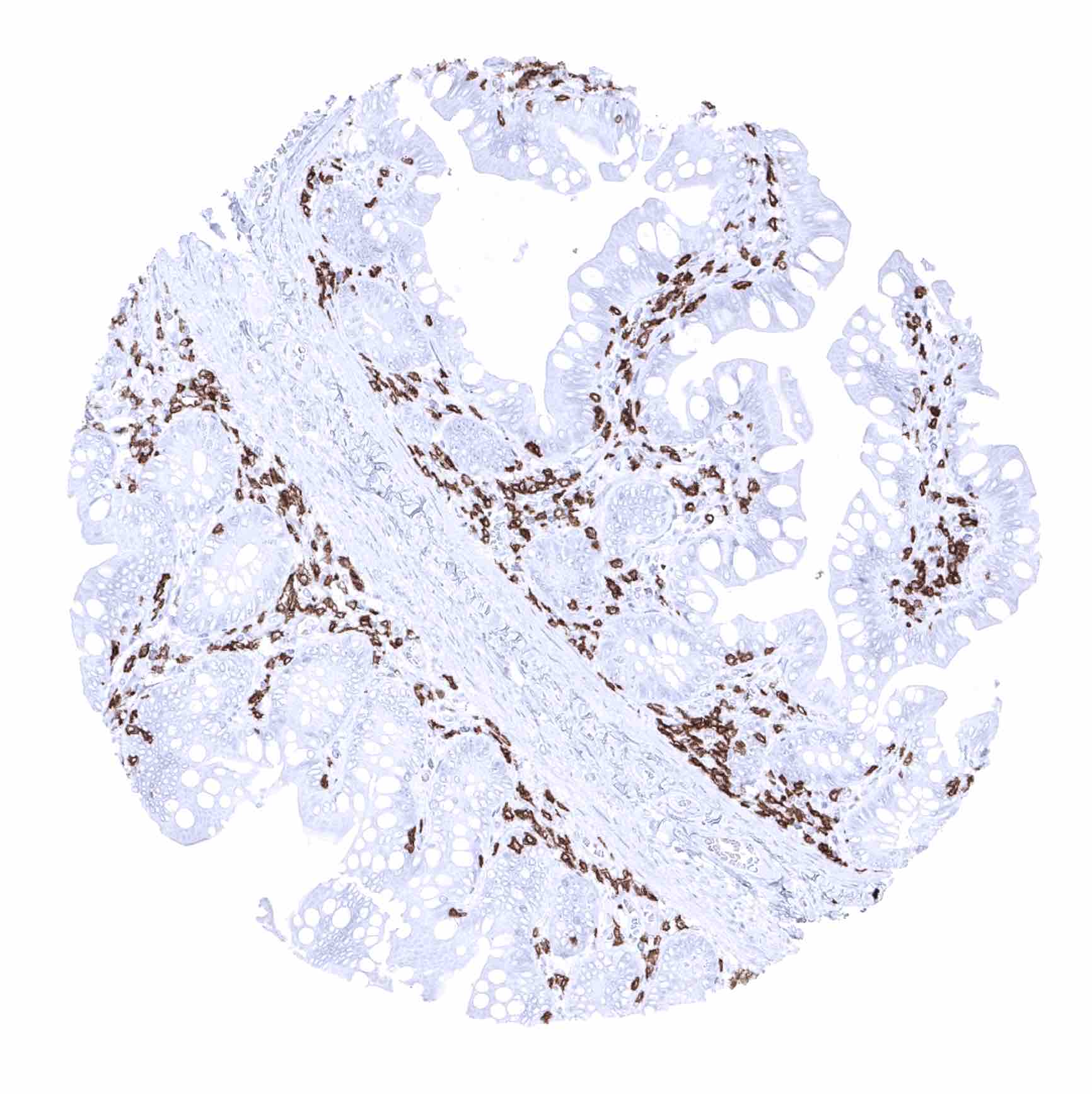
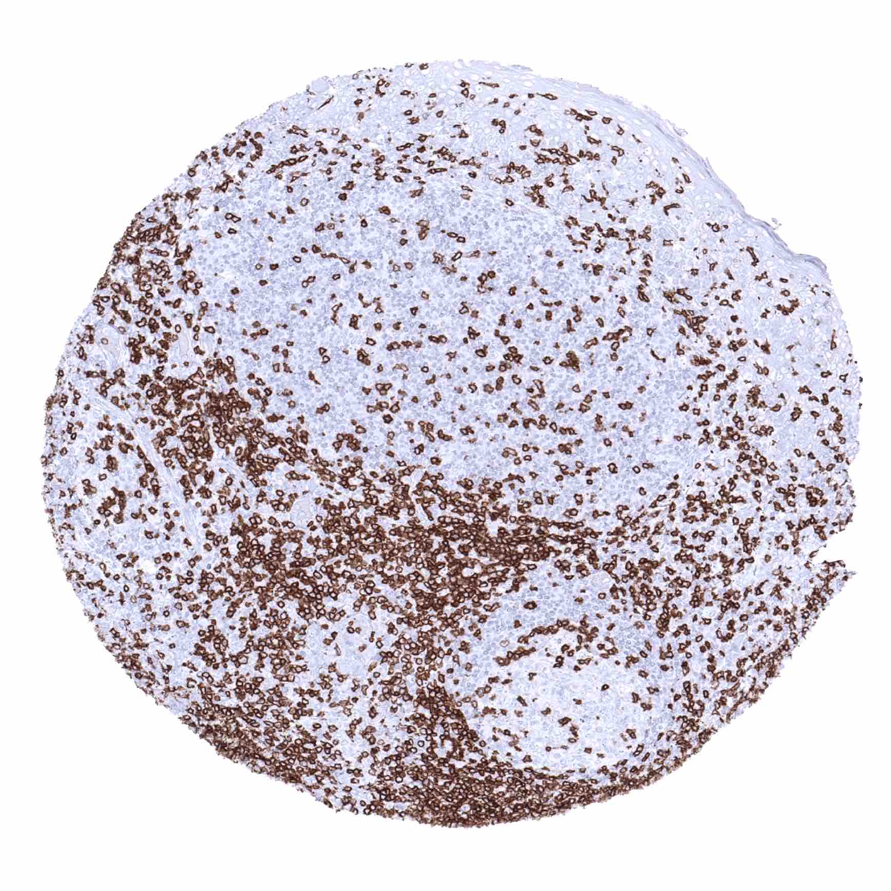
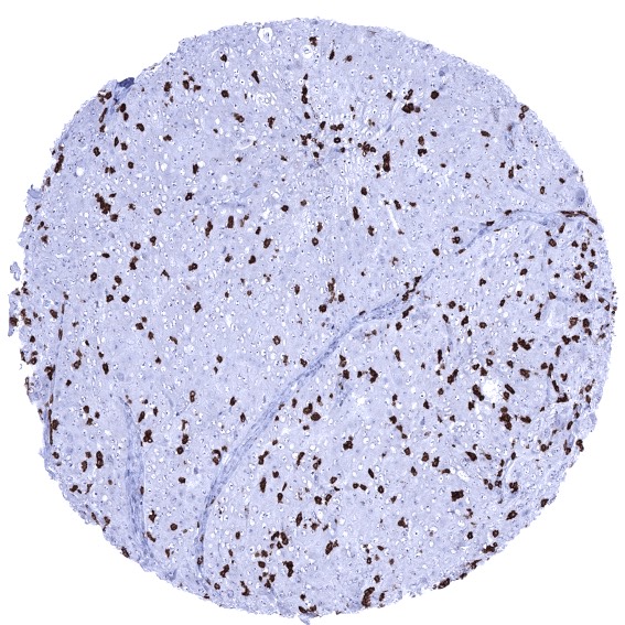
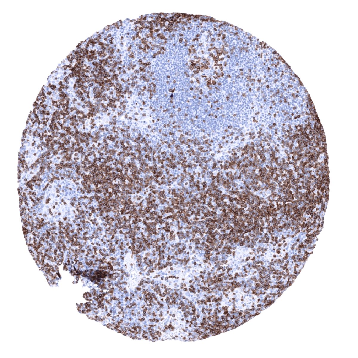
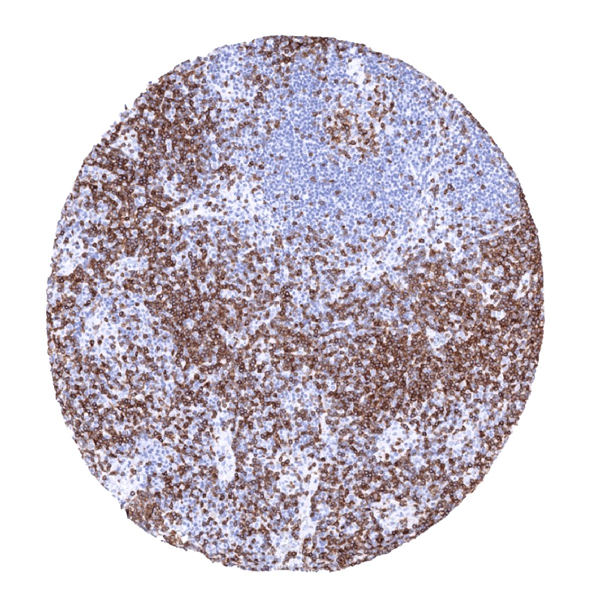
Positive control = Tonsil: A moderate to strong membranous staining should be seen in all T-cells in the interfollicular T-zones and in the germinal centres (few cells).
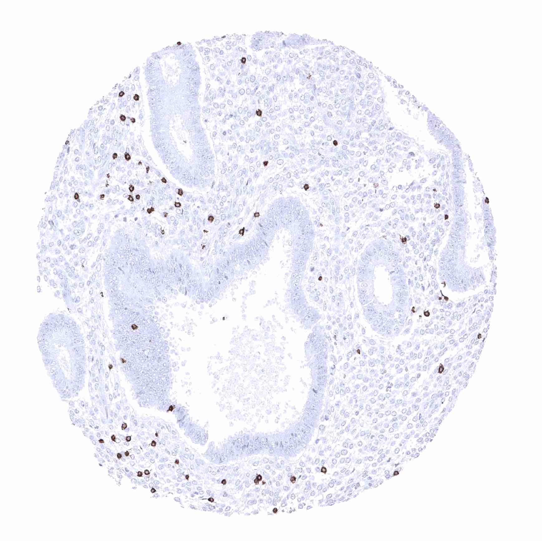
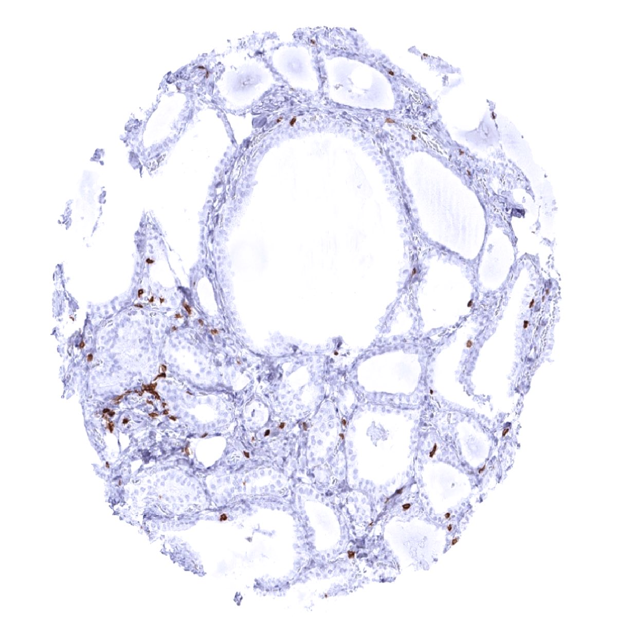
Negative control = Tonsil: All epithelial structures and B-cells should not show any CD3 immunostaining.
Cellular localization = Cell surface. Cytoplasm.
Reactivity = Human
Application = Immunohistochemistry
Dilution = 1:100 – 1:200
Intended Use = Research Use Only
Relevance of Antibody
CD3 is expressed on T-lymphocytes.
Biology Behind
CD3 (cluster of differentiation 3; OKT3) is a protein complex containing four distinct chains that jointly act as a T cell co-receptor. It is involved in activating both cytotoxic T cells (CD8+ naive T cells) and T helper cells (CD4+ naive T cells). The CD3 protein complex associates with the T-cell receptor (TCR) and the zeta-chain to form the TCR complex which generates an activation signal in T lymphocytes. CD3 is initially expressed in the cytoplasm of pro-thymocytes, the stem cells from which T-lymphocytes arise in the thymus. When the pro-thymocytes differentiate into common thymocytes, and subsequently into medullary thymocytes, the CD3 antigen begins to migrate to the cell membrane. CD3 is then found at the membranes of all mature T-cells. CD3 is a drug target for antibody-based treatment of acute graft rejection after transplantation. For cancer therapy, bispecific antibodies using anti-CD3 in combination with cancer surface proteins such as B7-H3 are being explored.
Staining Pattern in Normal Tissues
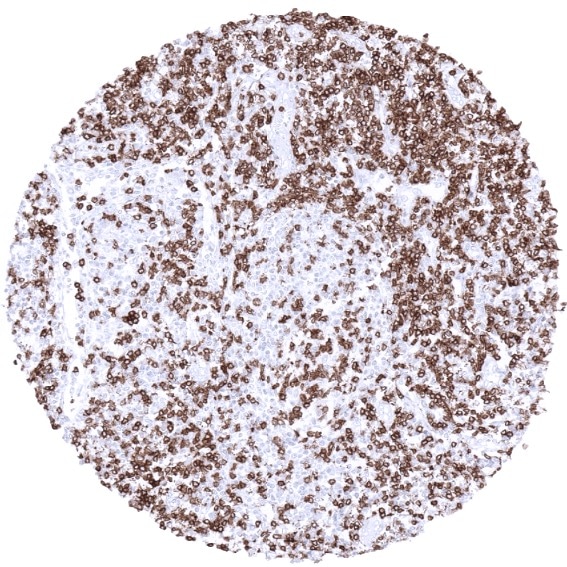
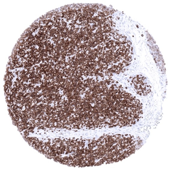
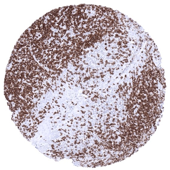
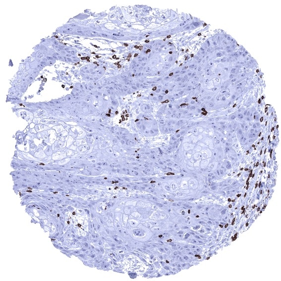
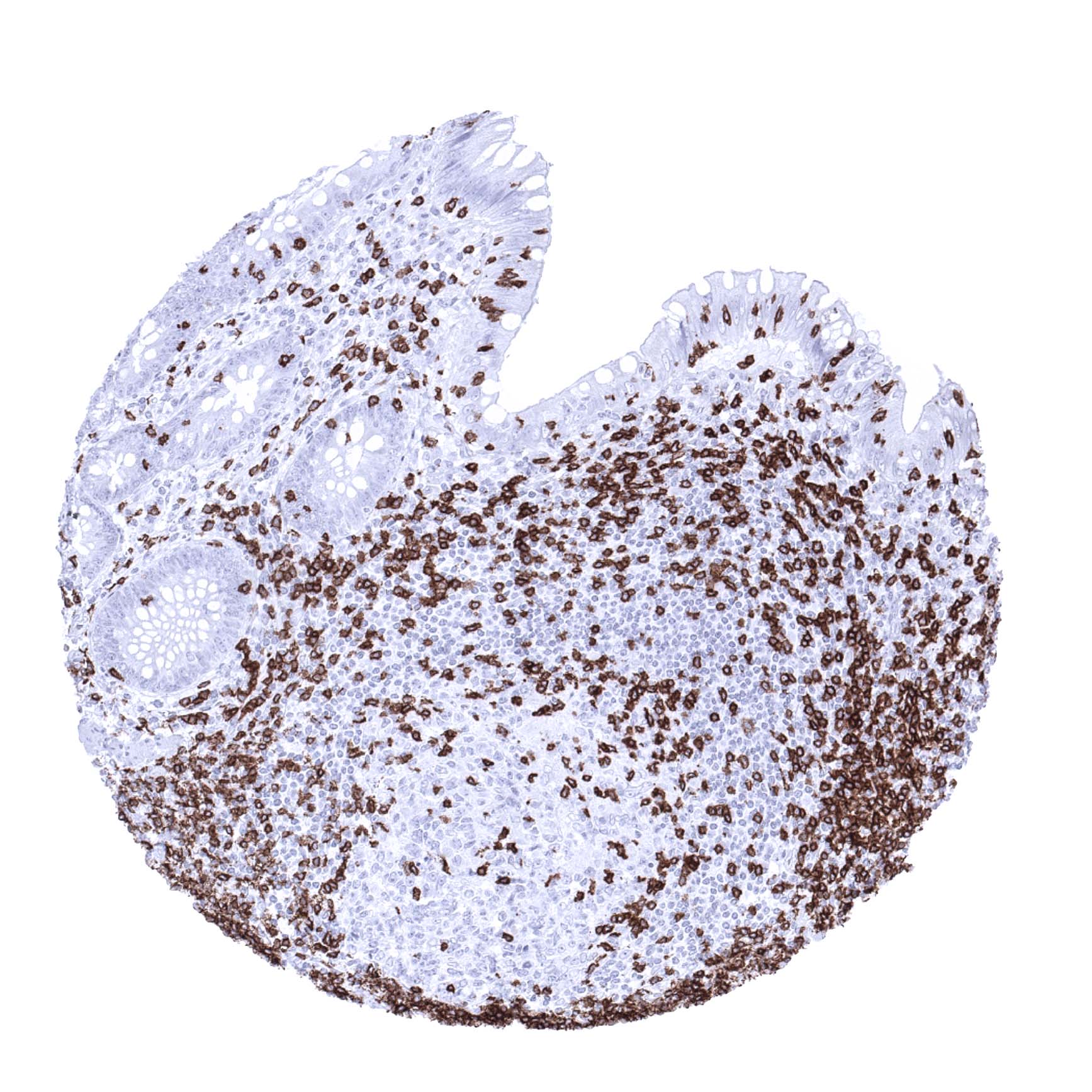
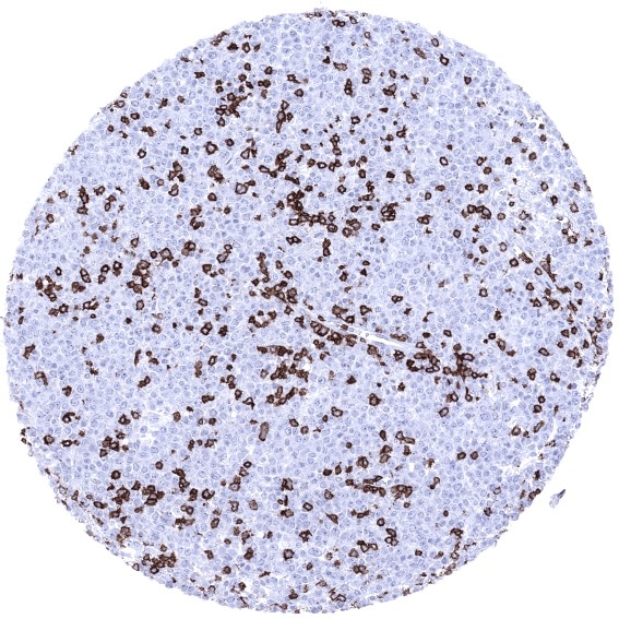
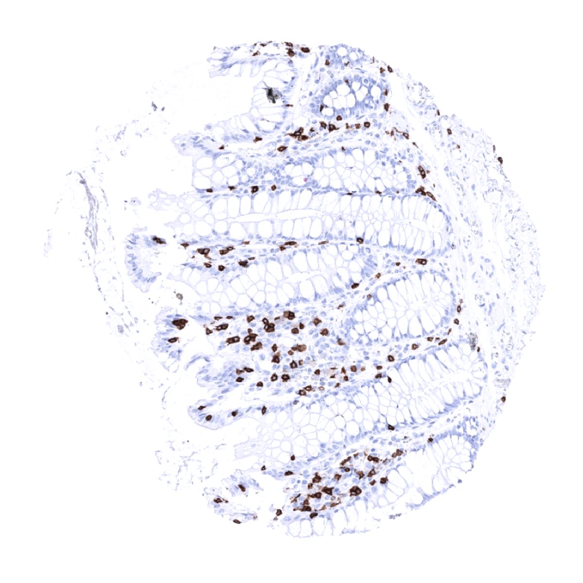
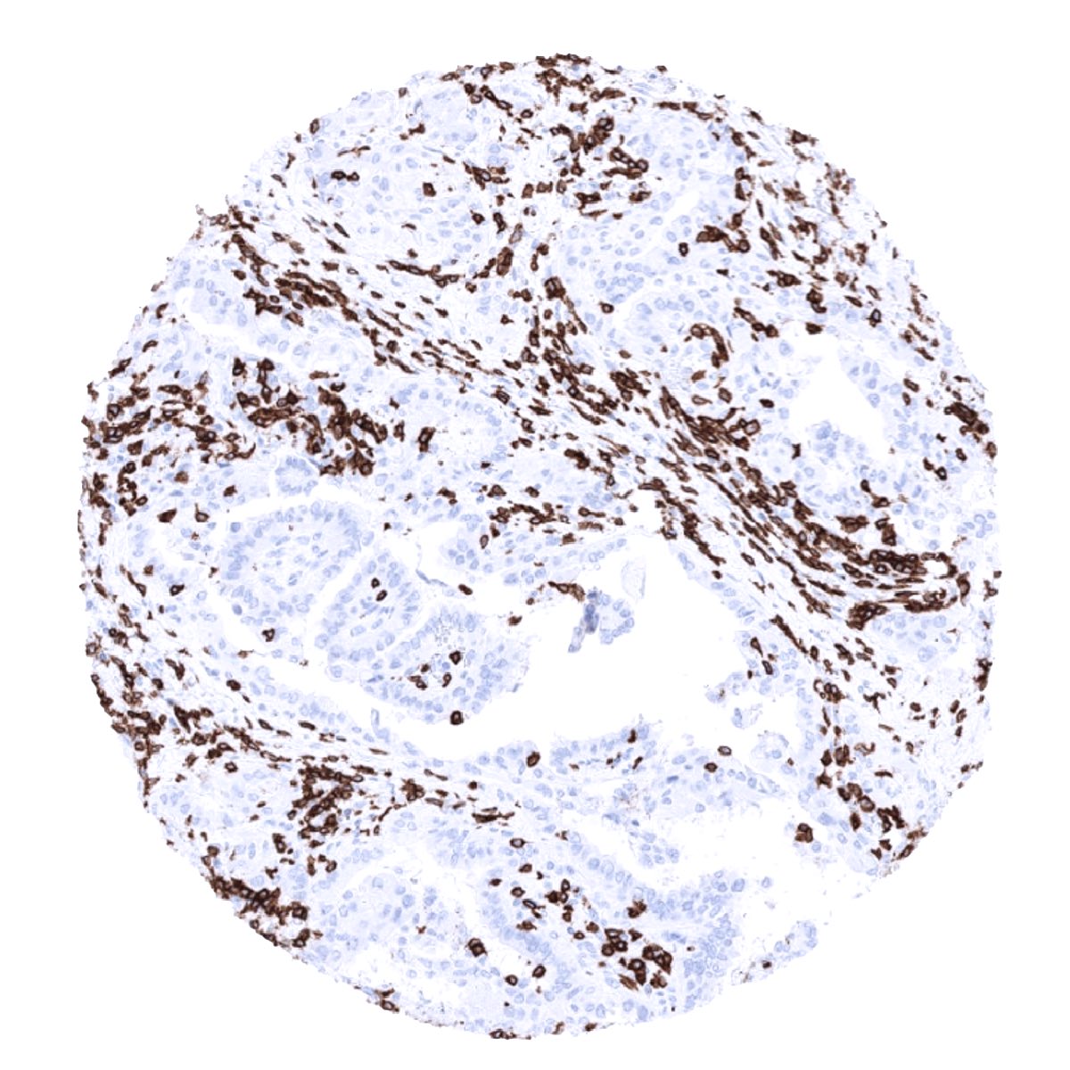
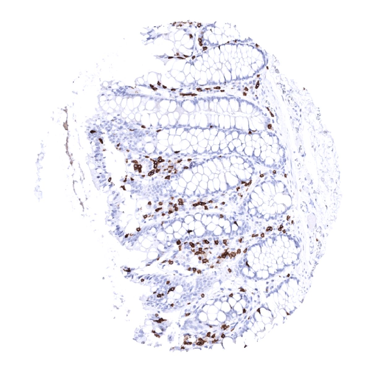
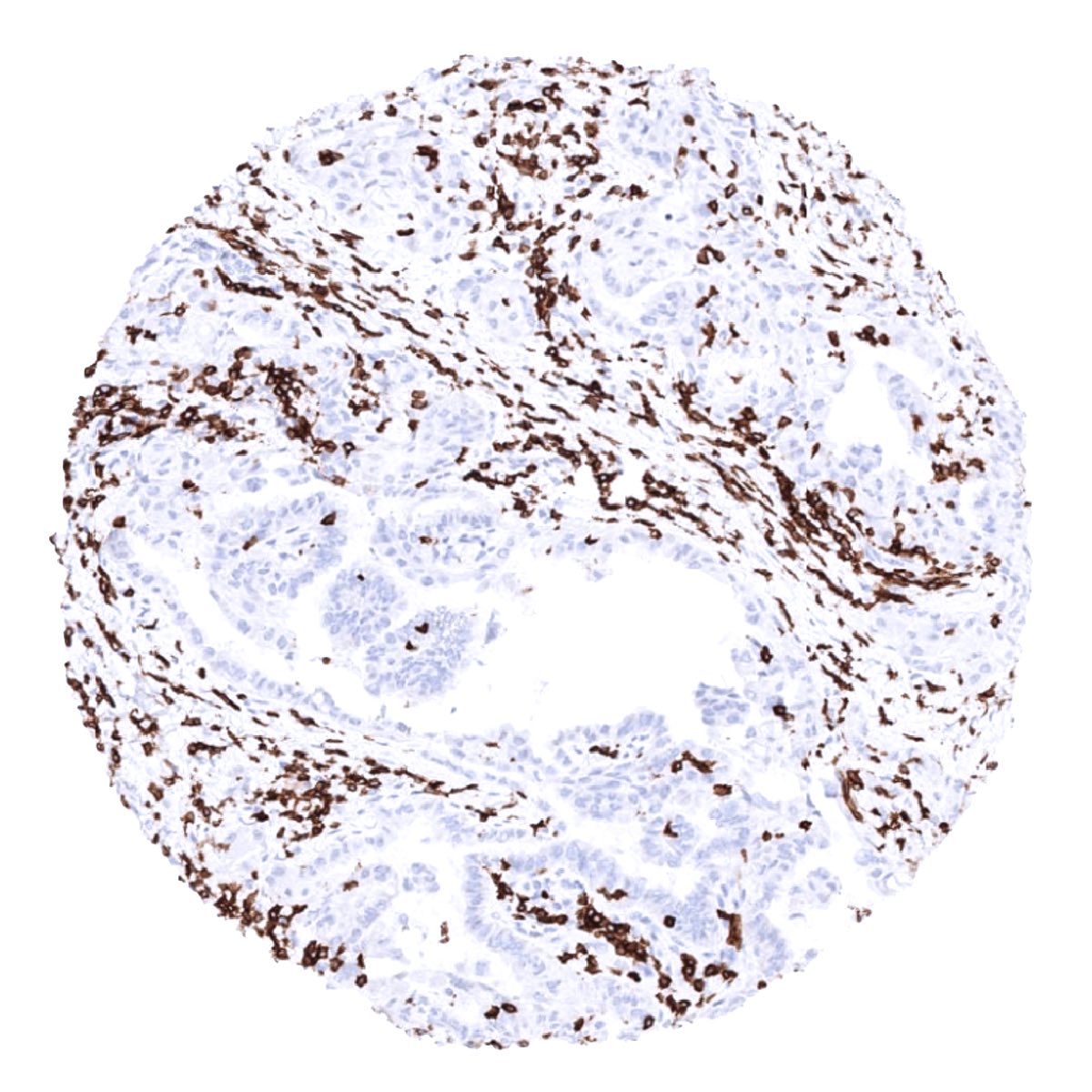
CD3 immunostaining is only seen in T-cells. These predominantly occur in lymphatic organs such as thymus, lymph nodes and tonsils but are regularly also seen – at lower density – in many other tissue types.
These findings are largely consistent with the RNA and protein data described in the Human Protein Atlas (Tissue expression CD3)
Positive control = Tonsil: A moderate to strong membranous staining should be seen in all T-cells in the interfollicular T-zones and in the germinal centres (few cells).
Negative control = Tonsil: All epithelial structures and B-cells should not show any CD3 immunostaining.
Staining Pattern in Relevant Tumor Types
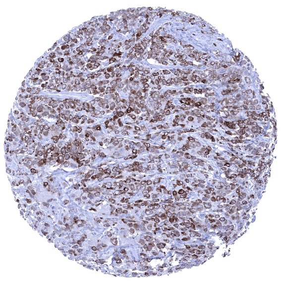
CD3 is retained in almost all T-cell lymphomas and leukemias. A variable number of CD3 positive tumor infiltrating lymphocytes regularly occur in virtually all other neoplasms. Of note, CD3 can rarely be also expressed in B-cell lymphoma.
The TCGA findings on CD3 RNA expression in different tumor categories have been summarized in the Human Protein Atlas.

Compatibility of Antibodies
No data available at the moment
Protocol Recommendations
IHC users have different preferences on how the stains should look like. Some prefer high staining intensity of the target stain and even accept some background. Others favor absolute specificity and lighter target stains. Factors that invariably lead to more intense staining include higher concentration of the antibody and visualization tools, longer incubation time, higher temperature during incubation, higher temperature and longer duration of the heat induced epitope retrieval (slide pretreatment). The impact of the pH during slide pretreatment has variable effects and depends on the antibody and the target protein.
All images and data shown here and in our image galleries are obtained by the manual protocol described below. Other protocols resulting in equivalent staining are described as well.
Manual protocol
Freshly cut sections should be used (less than 10 days between cutting and staining). Heat-induced antigen retrieval for 5 minutes in an autoclave at 121°C in pH 9 Target Retrieval Solution buffer. Apply MSVA-003R at a dilution of 1:150 at 37°C for 60 minutes. Visualization of bound antibody by the EnVision Kit (Dako, Agilent) according to the manufacturer’s directions.


Agilent / Dako – Autostainer Link 48
Pretreatment in PT-Link for 30 minutes at 95°C (pH high); FLEX peroxidase blocking for 5 minutes (room temperature), MSVA-003R 1:150 for 20 minutes (room temperature), FLEX+ mouse/rabbit (LINKER) for 15 minutes (room temperature), horseradish peroxidase (HRP) for 20 minutes (room temperature), FLEX DAB+Sub-Chromo for 10 minutes (room temperature), FLEX hematoxylin for 5 minutes (room temperature).
These images reflect stainings by the protocol described above. It is of note that a comparable staining result can also be obtained by different protocols. In general, a longer pretreatment, a longer incubation time of the primary antibody, a higher antibody concentration, and a longer incubation time of FLEX+LINKER result in stronger staining, potentially at the cost of more background staining. Modifications of the protocol with a strengthening effect on staining intensity in combination with changes of other parameters that result in lower staining intensity can result in a comparable result as shown above.
Leica – BOND RX
Dewax at 72°C for 30 seconds; Pretreatment in Bond Epitope Retrieval Solution (ER2 – EDTA pH9) for 40 minutes at 100°C; Peroxidase blocking for 5 minutes (room temperature), MSVA-003R 1:150 for 30 minutes (room temperature), Post primary (rabbit anti mouse) for 8 minutes (room temperature), Polymer (goat anti rabbit) for 8 minutes (room temperature), mixed DAB refine for 10 minutes (room temperature), hematoxylin for 5 minutes (room temperature).
These images reflect stainings by the protocol described above. It is of note that a comparable staining result can also be obtained by different protocols. In general, a longer pretreatment, a longer incubation time of the primary antibody, a higher antibody concentration, a higher temperature during incubation, and a longer incubation time of Post primary and or the Polymer result in stronger staining, potentially at the cost of more background staining. Modifications of the protocol with a strengthening effect on staining intensity in combination with changes of other parameters that result in lower staining intensity can result in a comparable result as shown above.
Potential Research Applications
- The clinical significance of the number of intratumoral CD3 positive lymphocytes is under intensive research.
- CD3 is a key component of multicolor assays analyzing the role of lymphocyte subsets in cancers and other diseases.
- The prevalence of a positive CD3 immunostaining in hematological and non-hematological neoplasms should be further evaluated.
Evidence for Antibody Specificity in IHC
There are two ways, how the specificity of antibodies can be documented for immunohistochemistry on formalin fixed tissues.
These are: 1. Comparison with a second independent method for measuring target expression across a large number of different tissue types (orthogonal strategy), and 2. Comparison with one or several independent antibodies for the same target and showing that all positive staining results are also seen with other antibodies for the same target (independent antibody strategy).
As a standard orthogonal validation process for MSVA antibodies, RNA data summarized in the Human Protein Atlas (Tissue expression CD3) are used for comparison. However, this procedure has limited validity for proteins that are expressed in significant subsets of lymphocytes because these cells occur in virtually all organs.
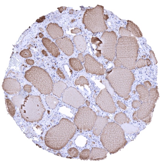
The preferential CD3 RNA expression in the thymus and other lymphatic tissues such as the tonsil and lymph nodes fits with the staining pattern observed by MSVA-003R in normal tissues. The validation of MSVA-003R is, however, preferentially based on a comparison with a second independent, commercially available CD3 antibody (termed “Validation-antibody”). This comparison of antibodies revealed identical staining patterns for MSVA-003R and the “Validation-antibody” in lymphatic tissues with identical subsets of lymphocytes staining. The independence of the validation antibody is demonstrated by its significant staining of thyroidal colloid which represents an antibody specific cross-reactivity. It is also of note, that the staining pattern of MSVA-003R very much matches the expectations as lymphocytes are preferable stained that occur at locations where T-lymphocytes are known to predominate such as the thymus, interfollicular areas of lymph node and the tonsil, and in mucosa associated tissues.
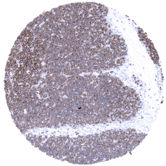
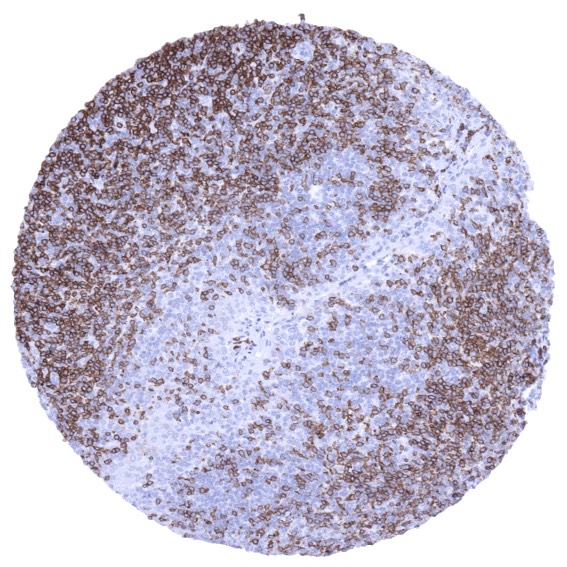
Antibody Comparison: MSVA-003R vs another commercially available CD3 antibody called “Validation Antibody”