295,00 € – 995,00 €
Product details
Synonyms = CAD; CALD1; Caldesmon 1 Isoform 1; Caldesmon 1 Isoform 2; Caldesmon 1 Isoform 3; Caldesmon 1 Isoform 4; Caldesmon 1 Isoform 5; CDM; HCAD; LCAD; NAG22
Antibody type = Recombinant Rabbit monoclonal / Rabbit IgG
Clone = MSVA-538R
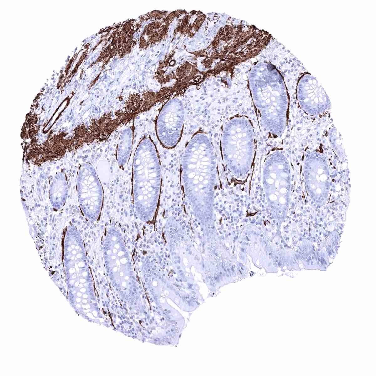
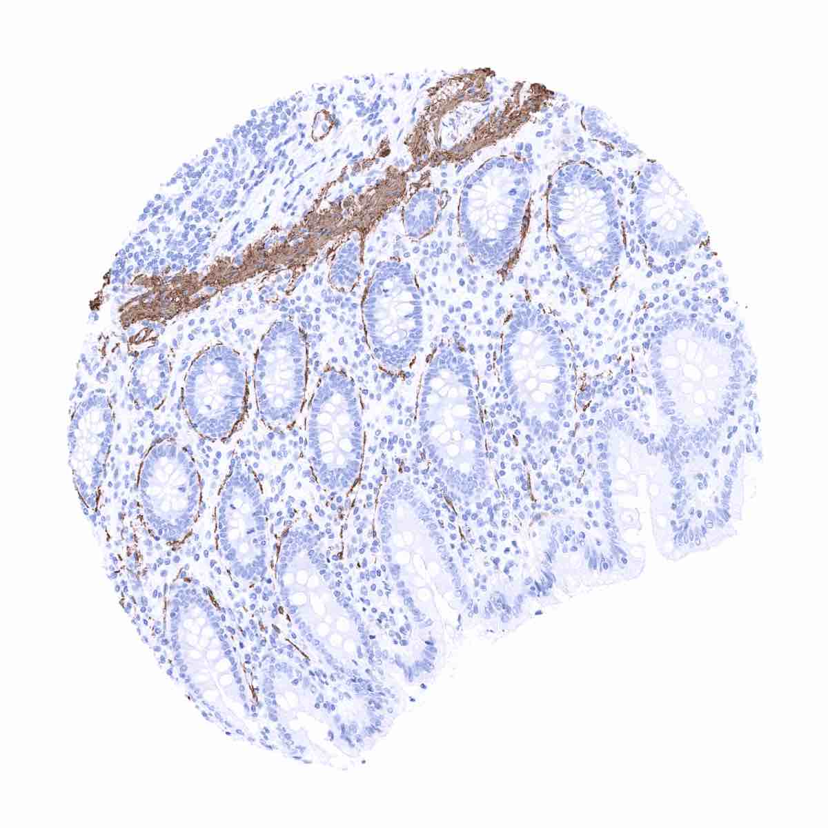
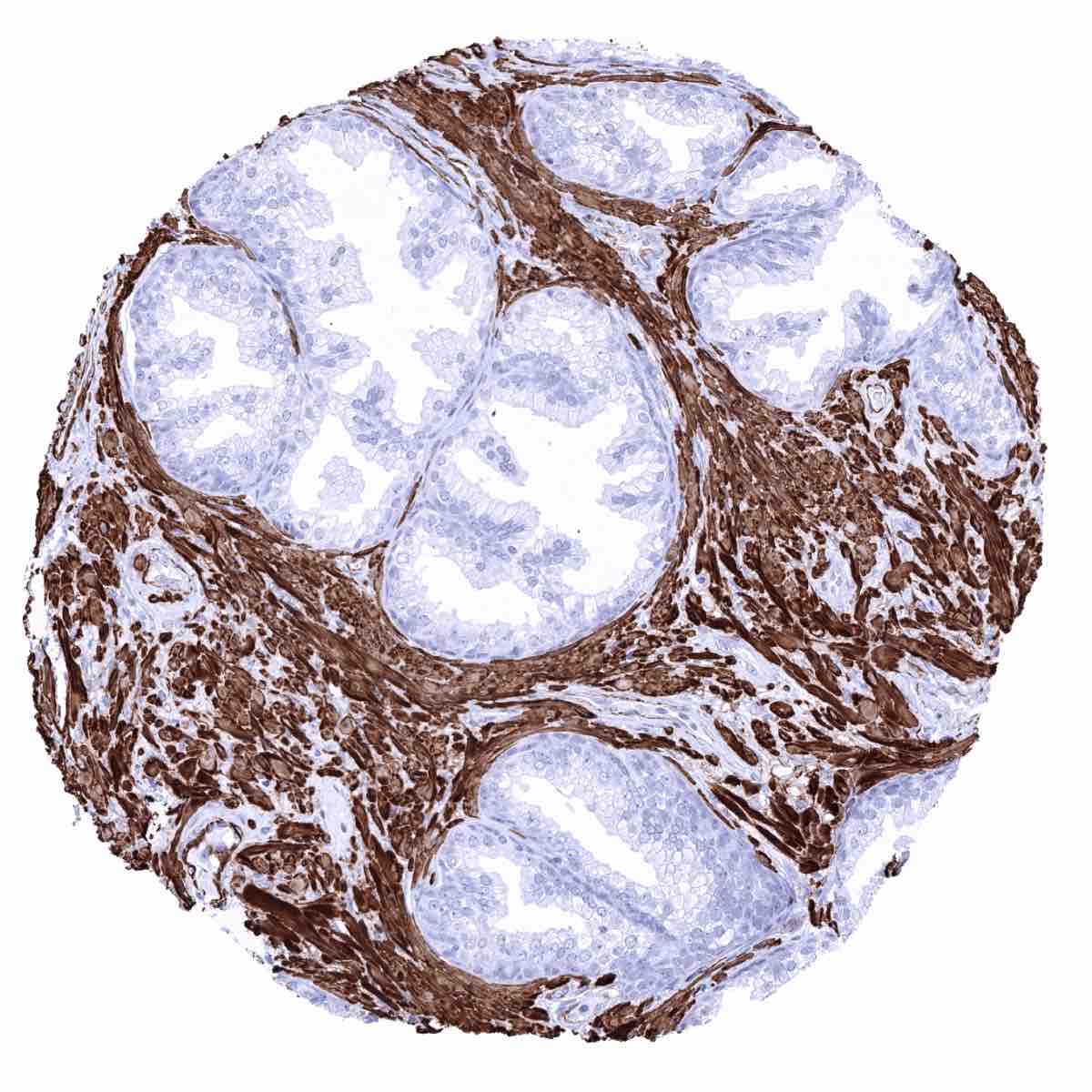
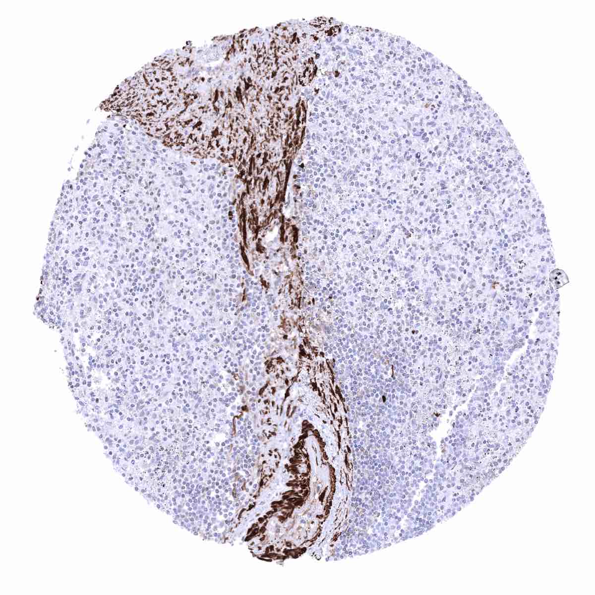
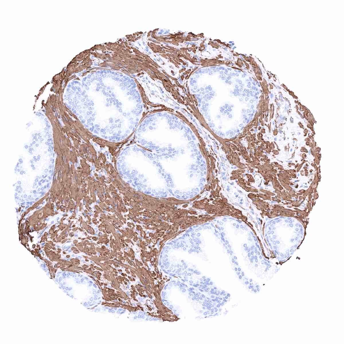
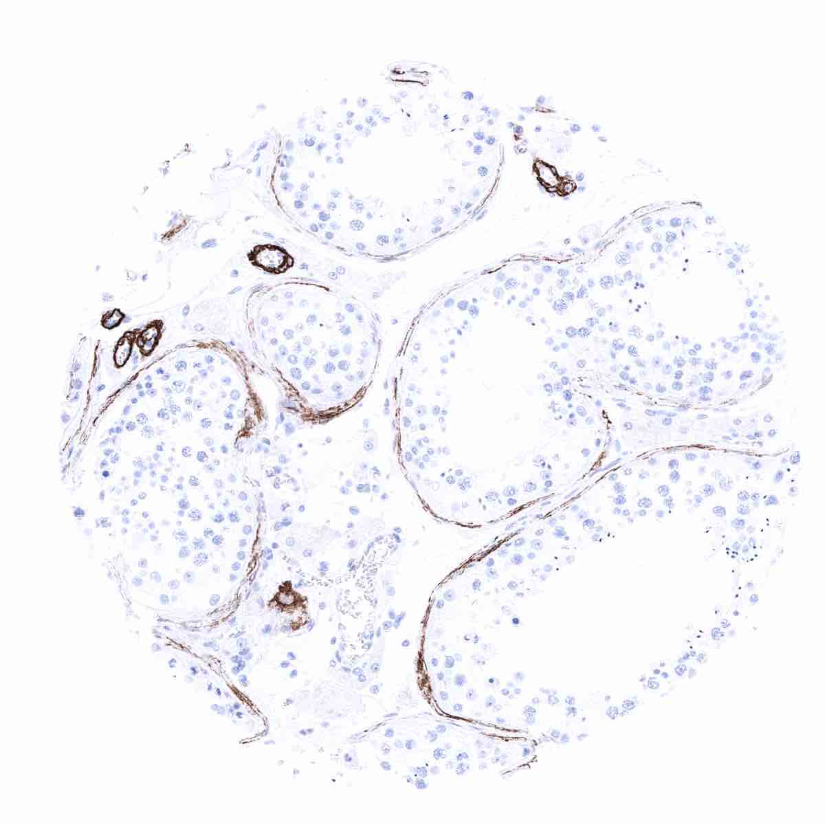
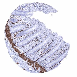
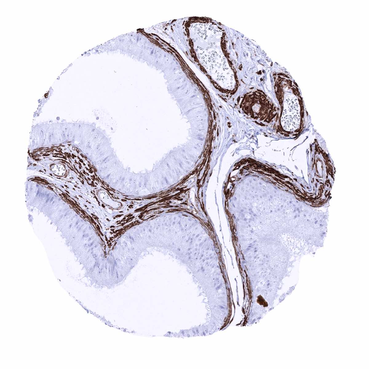
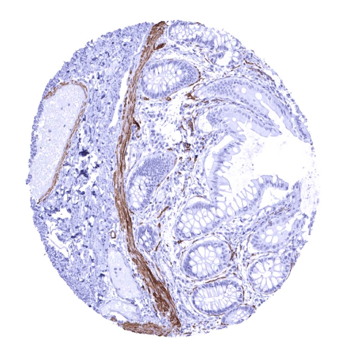
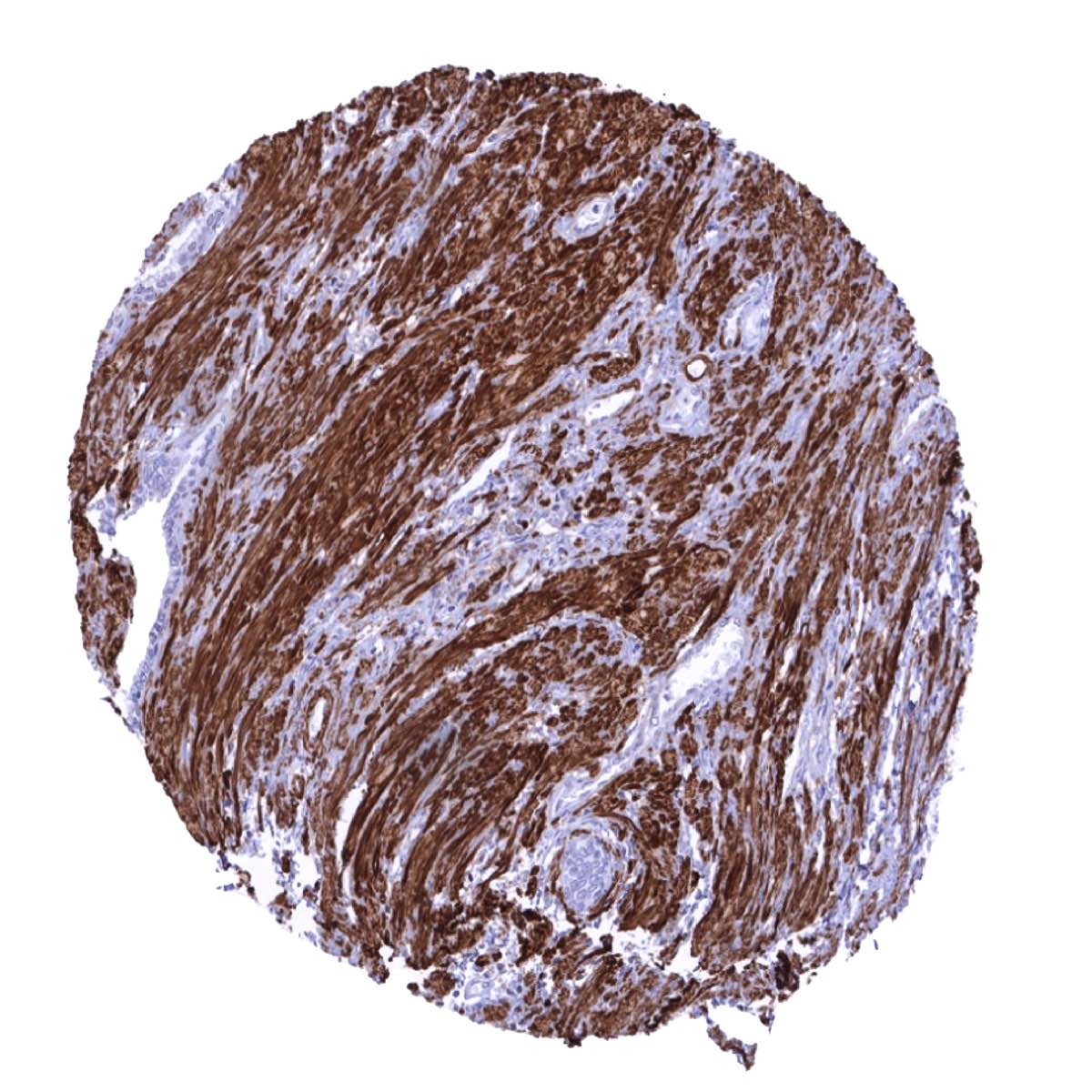
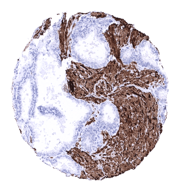
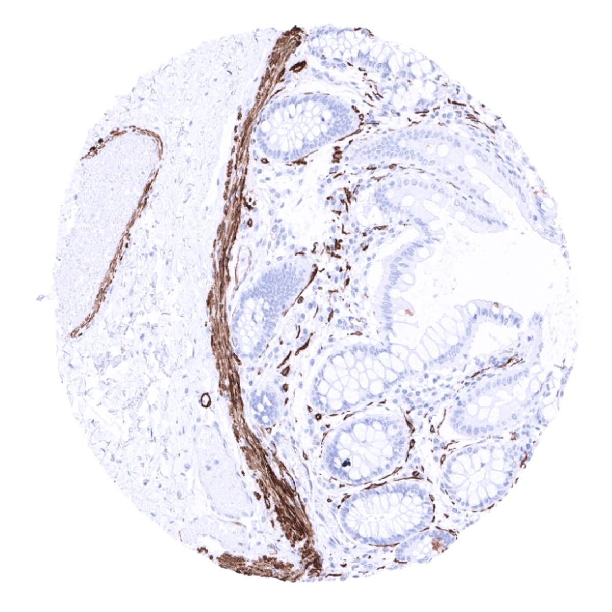
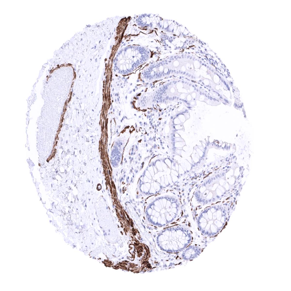
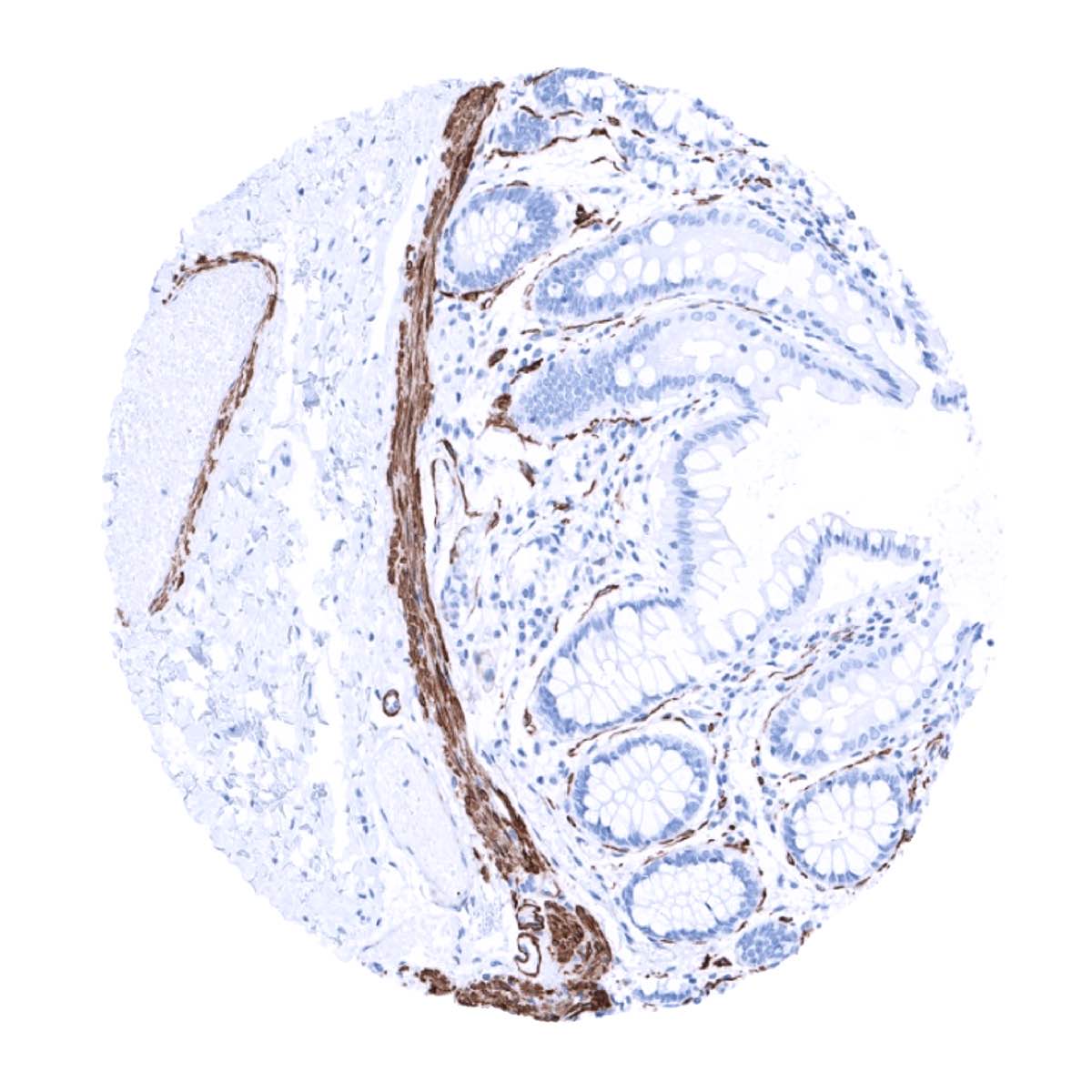
Positive control = Colon: A strong caldesmon staining should be seen of the muscularis mucosa and of a membrane-like layer along the basement membranes of crypts (intestinal subepithelial myofibroblasts).
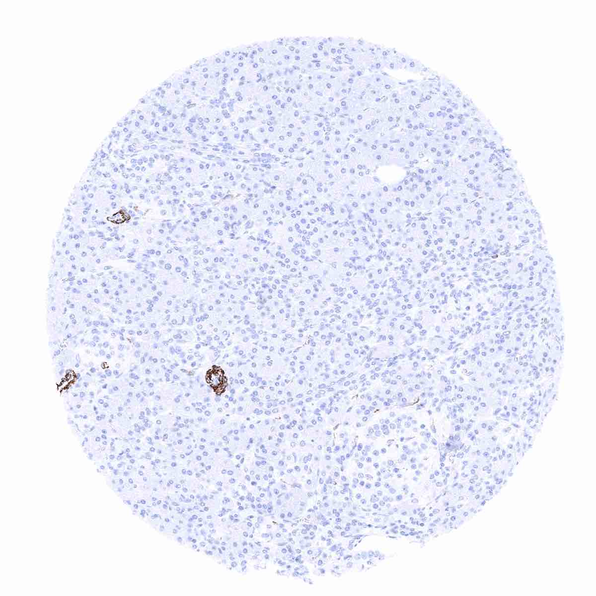
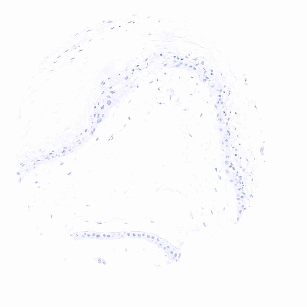
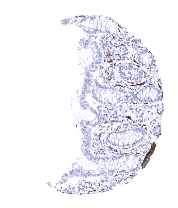
Negative control = Colon: Normal epithelial cells must not show caldesmon staining.
Cellular localization = Cytoplasmic
Reactivity = Human
Application = Immunohistochemistry
Dilution = 1:100 – 1:200
Intended Use = Research Use Only
Relevance of Antibody
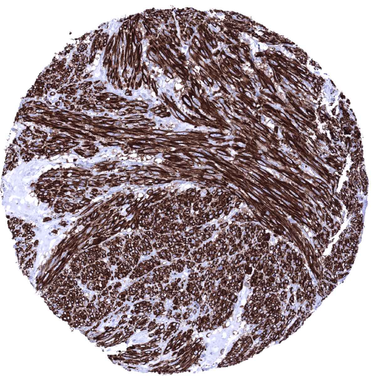
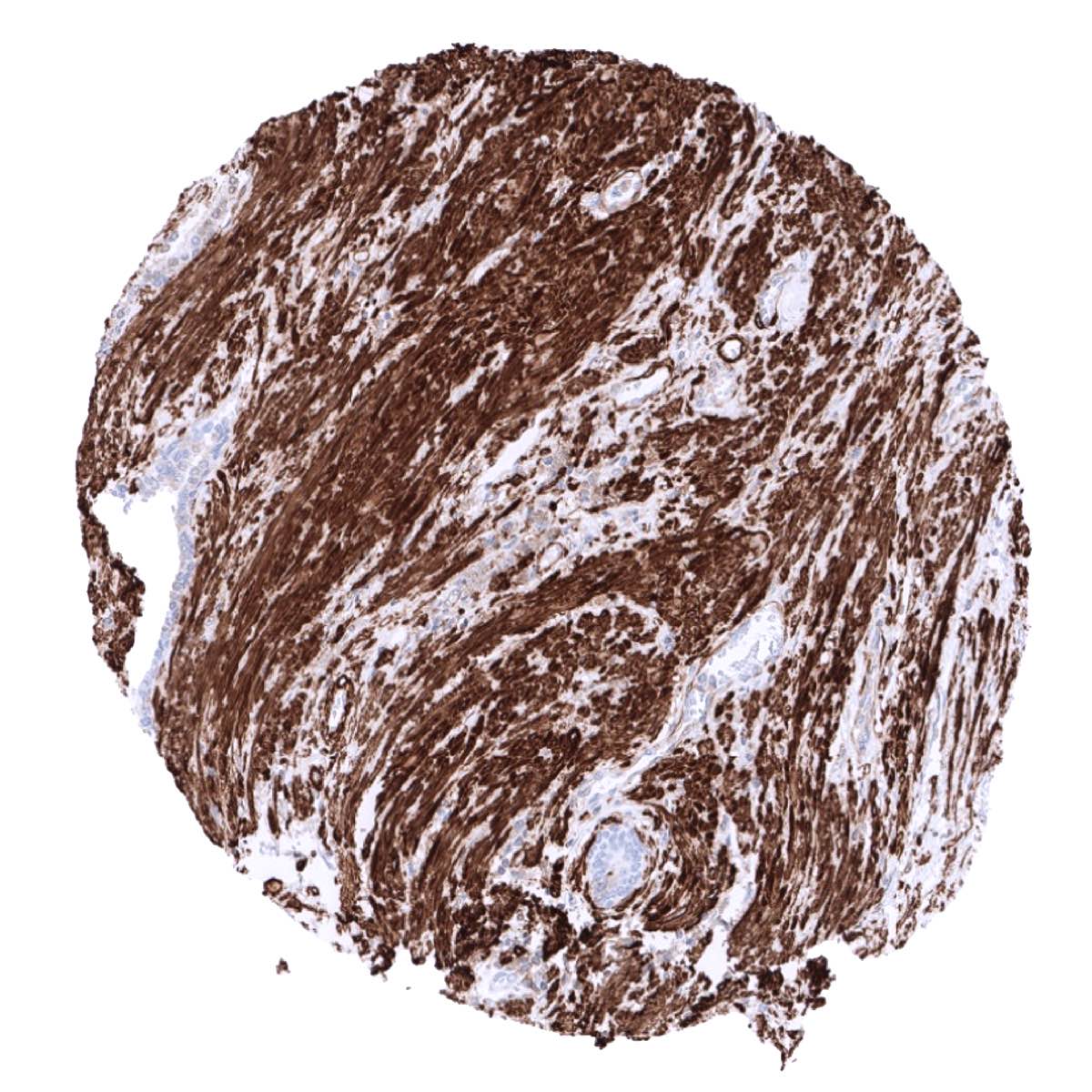
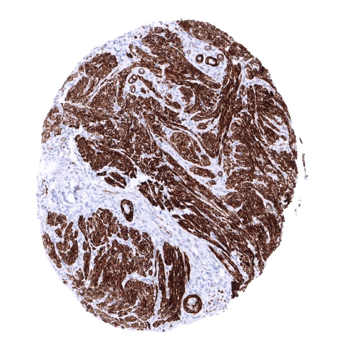
Caldesmon is a sensitive and specific marker for smooth muscle cells.
Biology Behind
Caldesmon is a particularly long (75 nm) and flexible molecule that is coded by the CALD1 gene located at chromosome 7q33. Together with tropomyosin, it regulates the binding of myosin to actin. In relaxed smooth muscle the actin binding site on myosin is blocked by the caldesmon-tropomyosin complex. Stimulation of smooth muscle results in ERK-dependent phosphorylation of caldesmon and subsequent removal of the caldesmon-tropomyosin complex from the myosin binding site on actin and consequently in muscle contraction. Two alternatively spliced isoforms of caldesmon occur. The heavy isoform (h-Caldesmon) is largely restricted to differentiated contractile smooth muscle cells but the light isoform (L-Caldesmon) also exists in other cell types. The antibody MSVA-538R recognizes h-caldesmon.
Staining Pattern in Normal Tissues
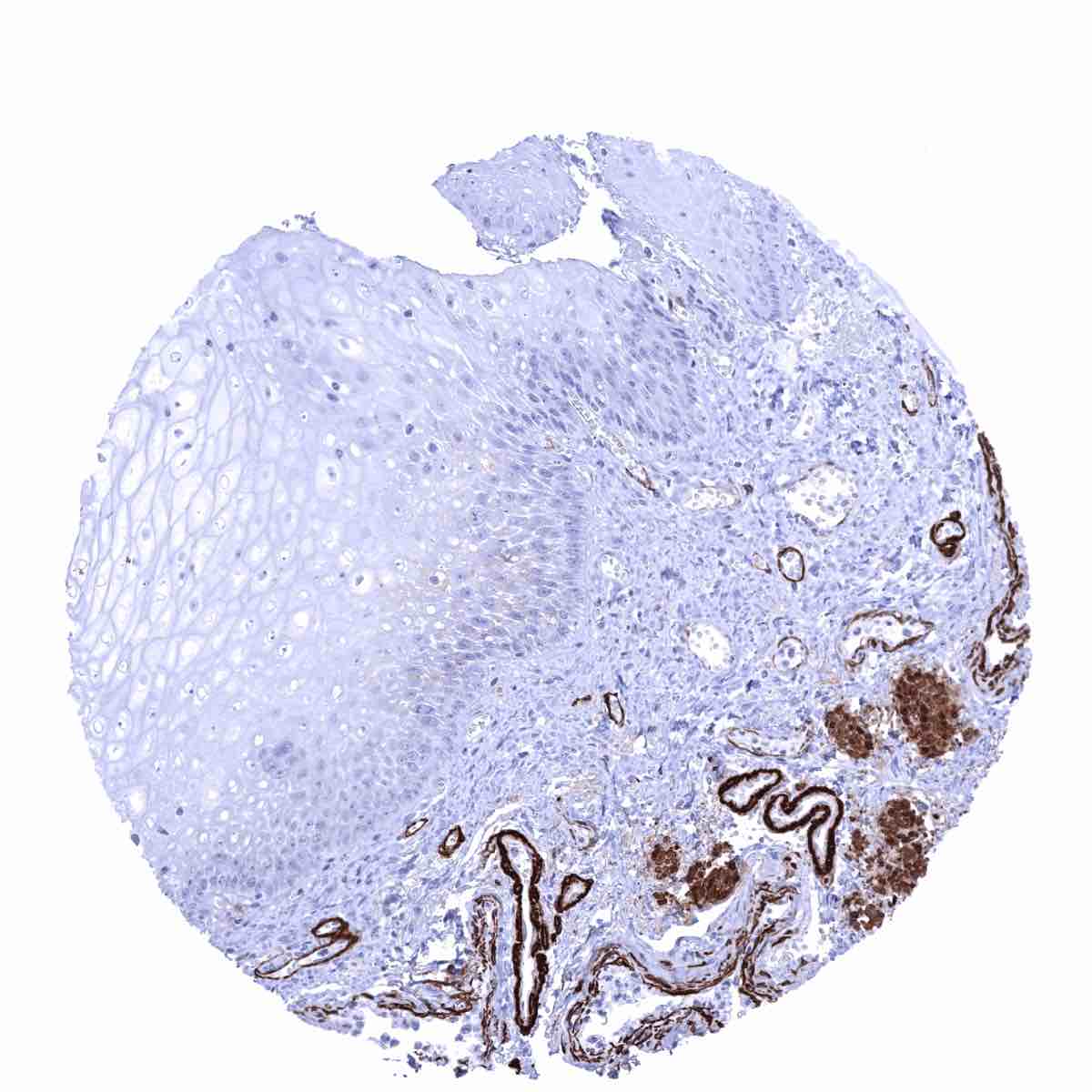
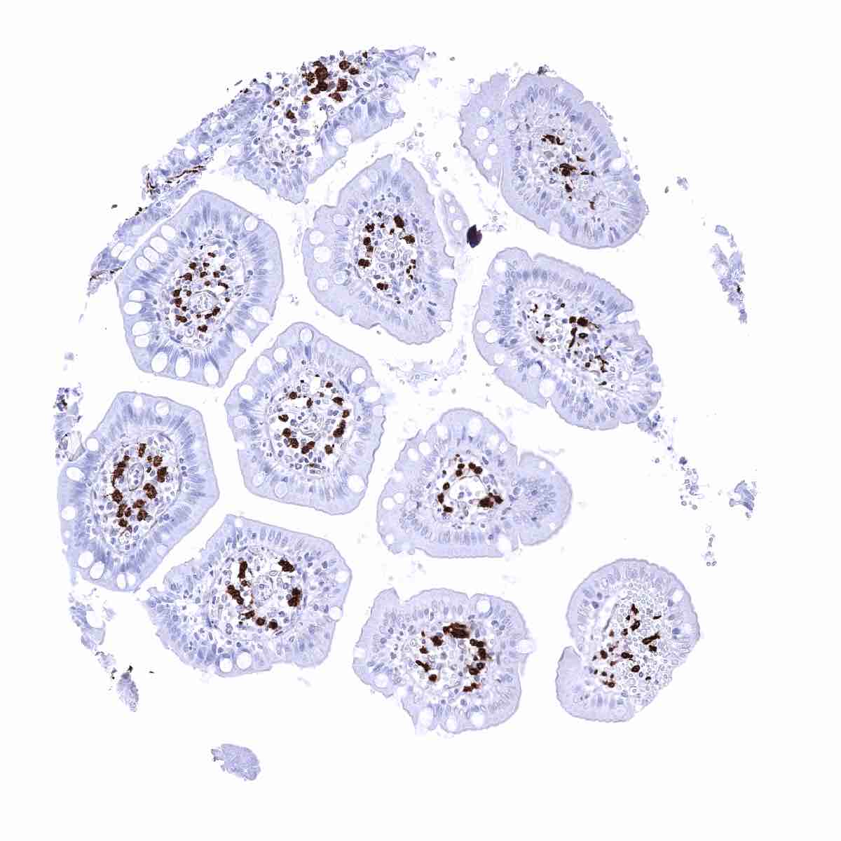
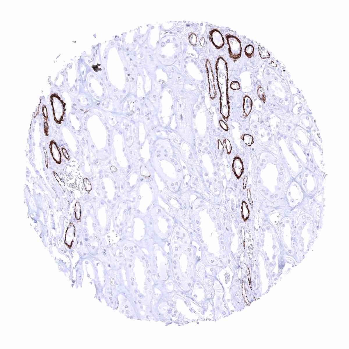
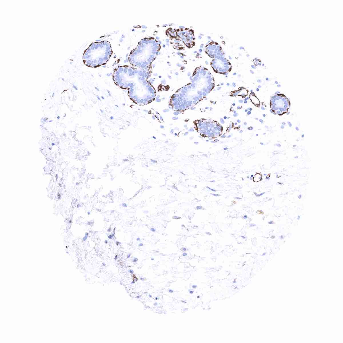
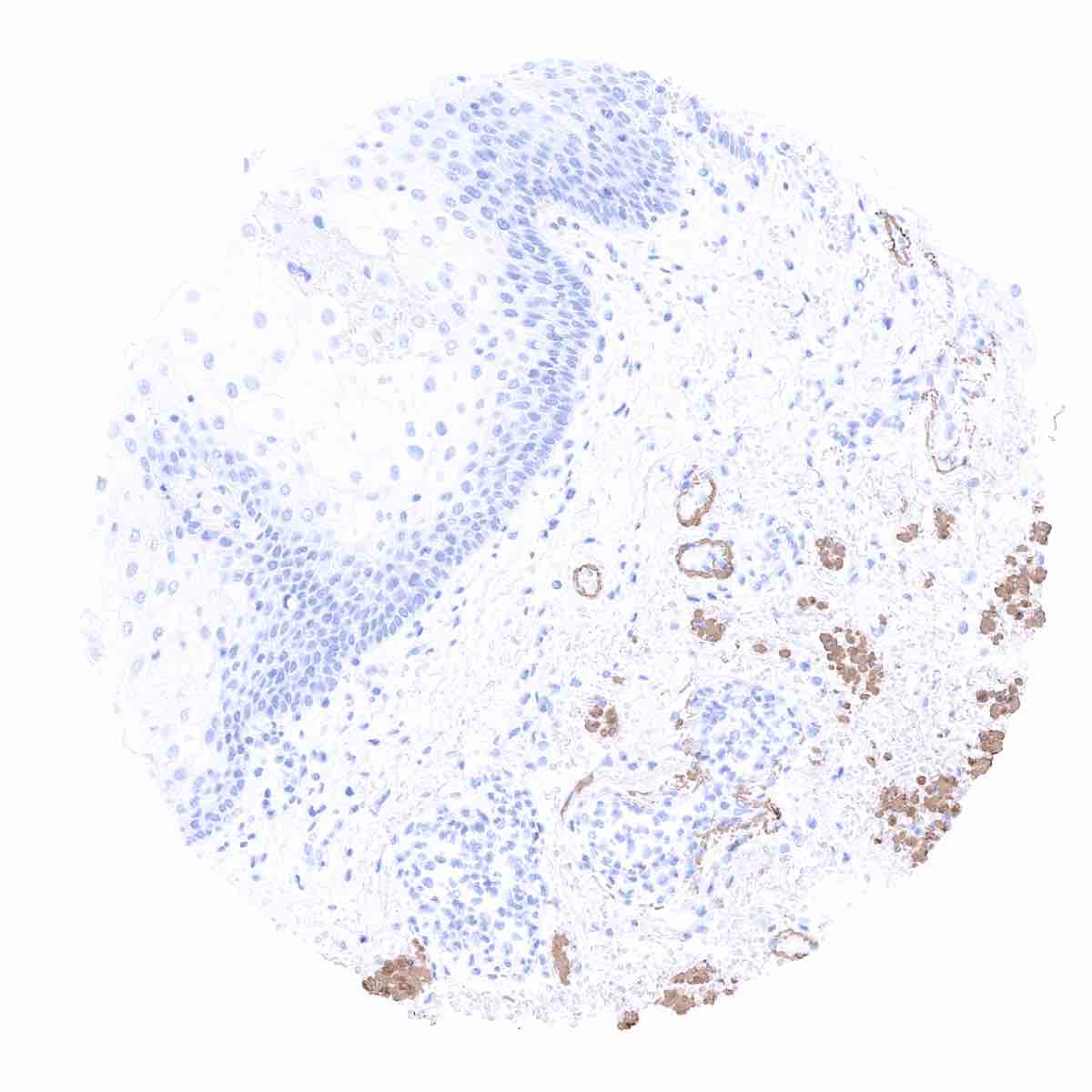
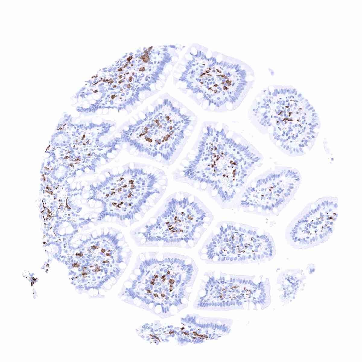
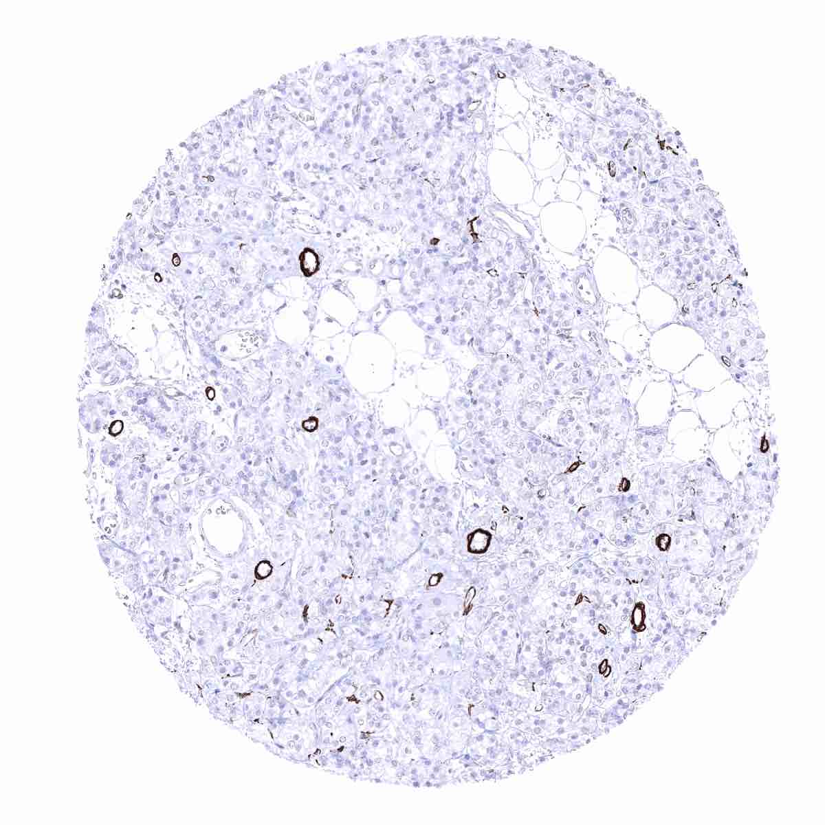
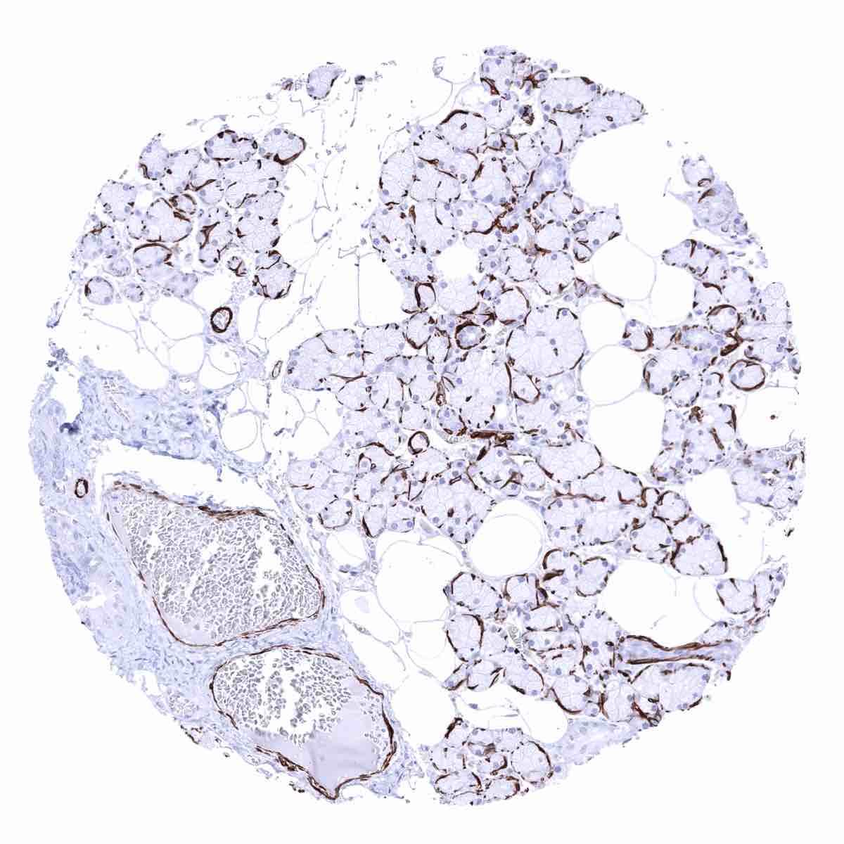
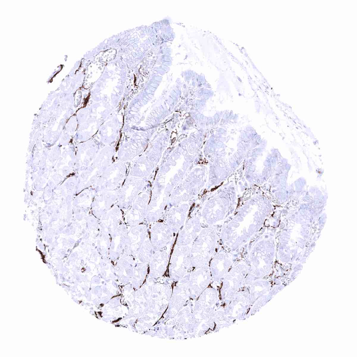
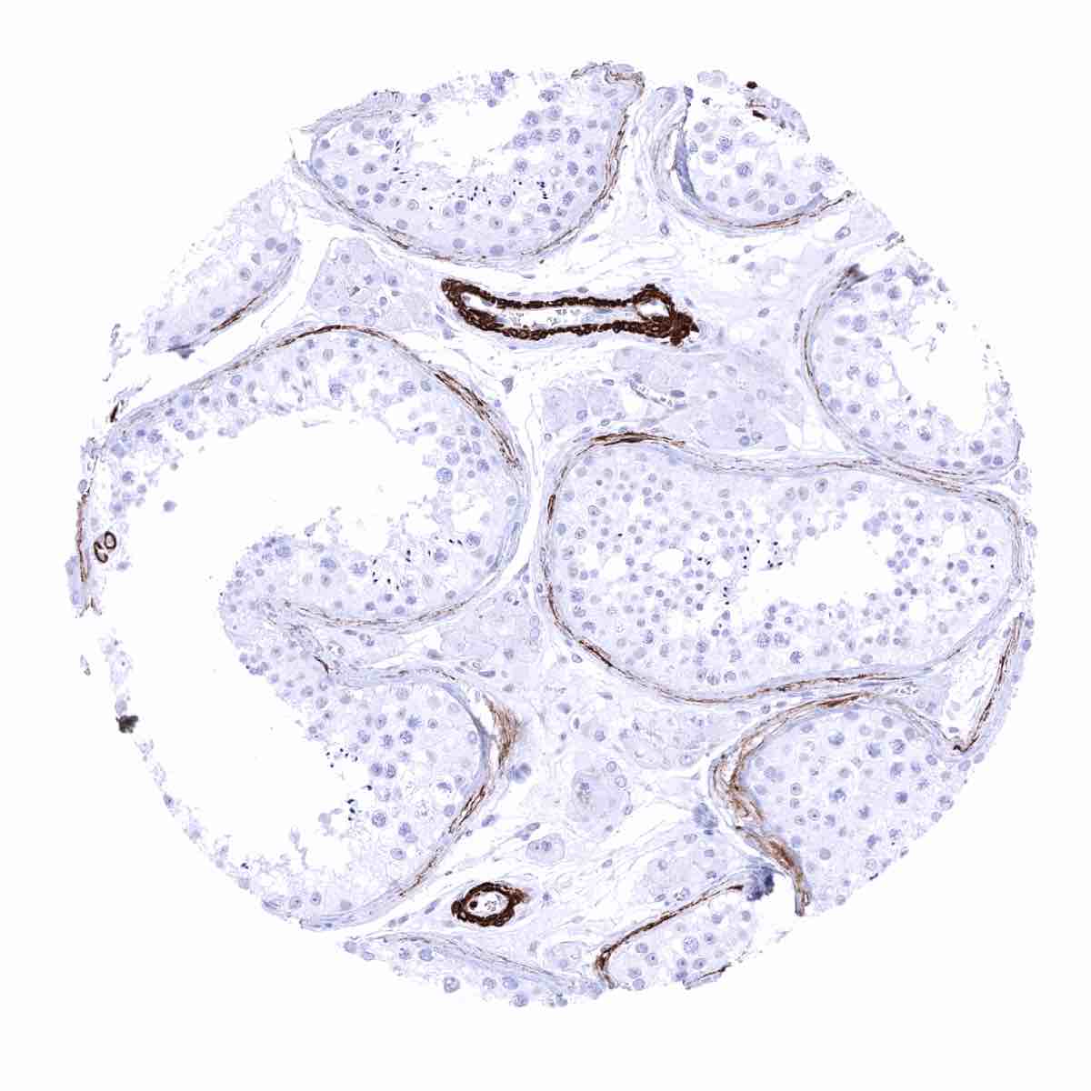
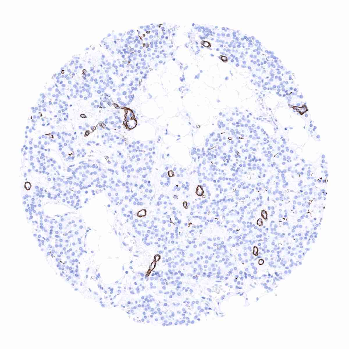
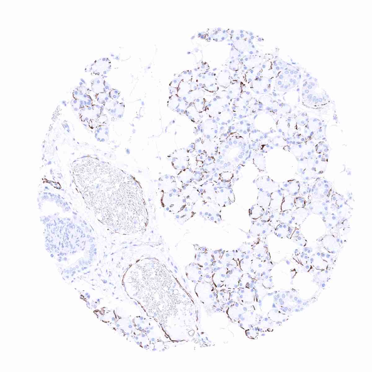
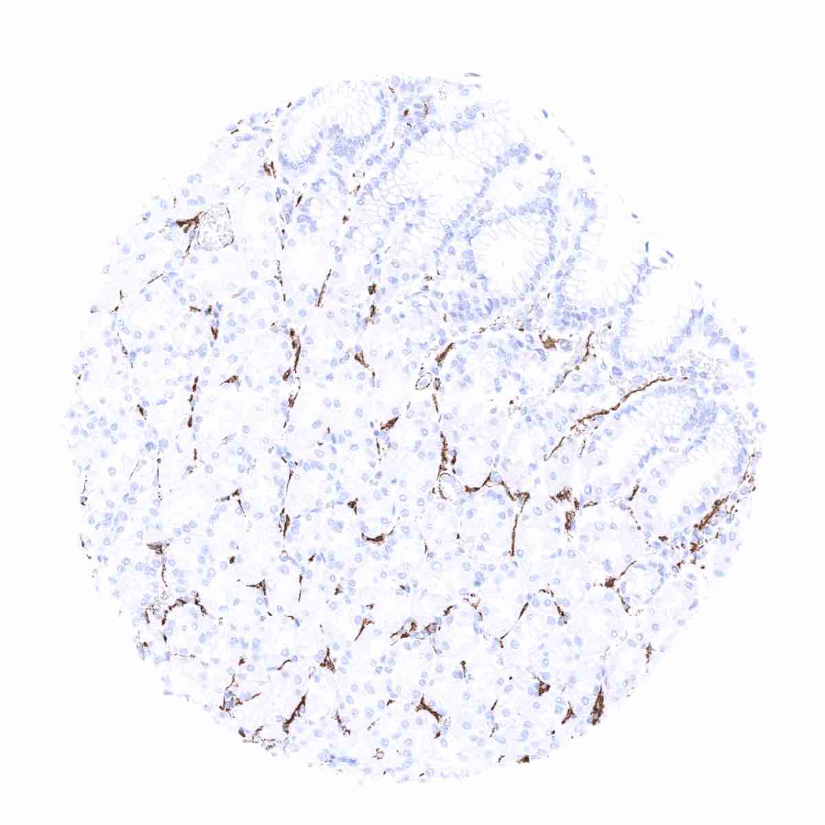
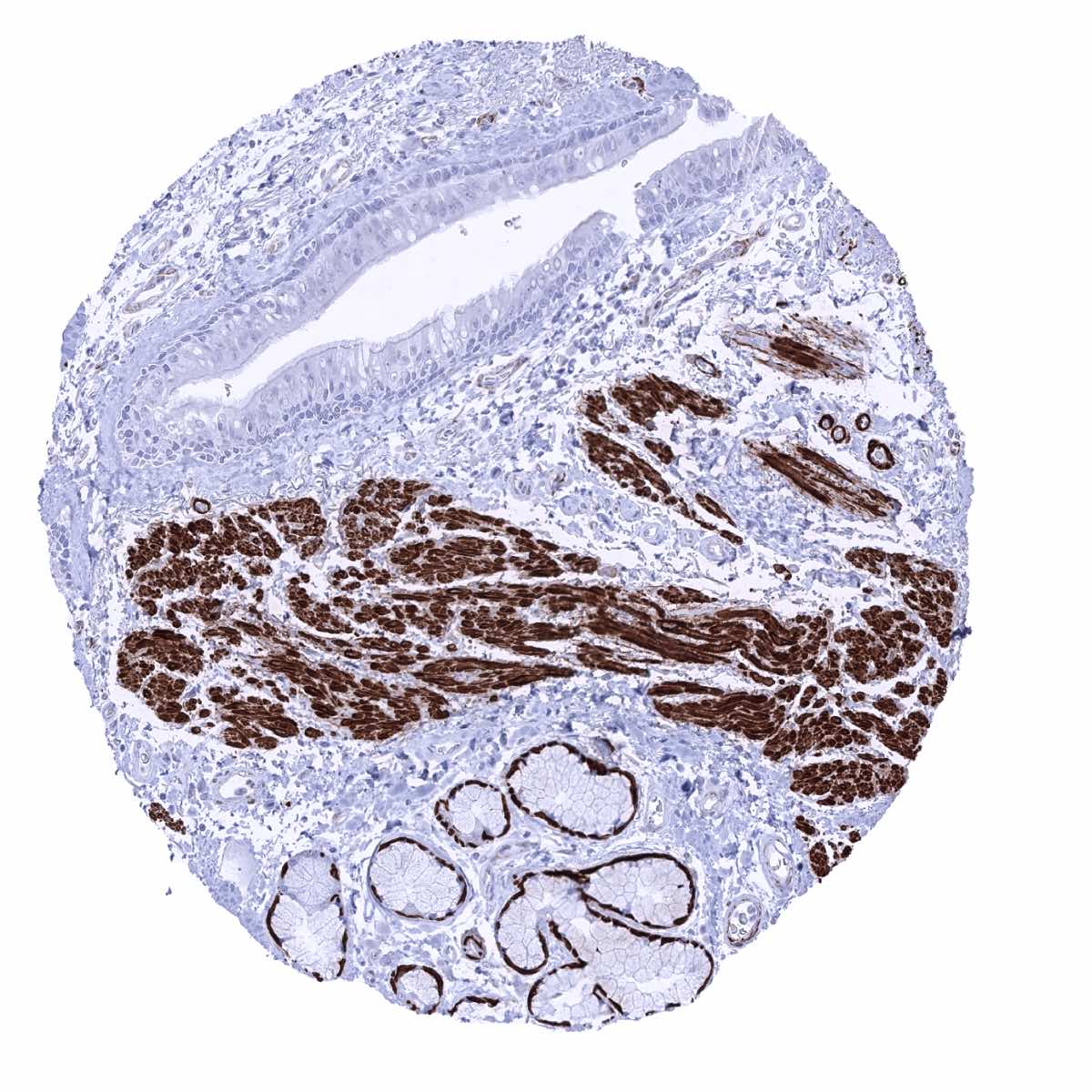
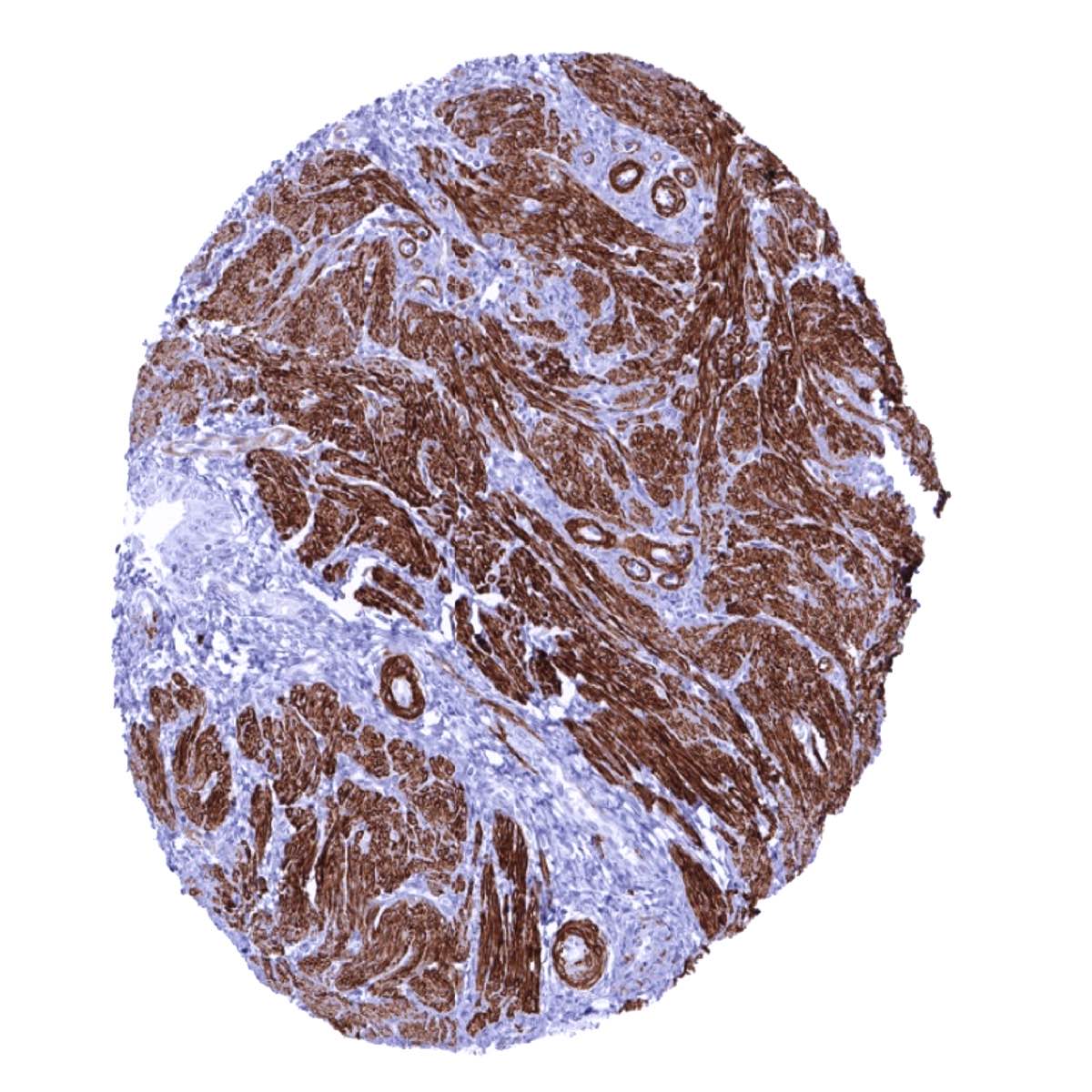
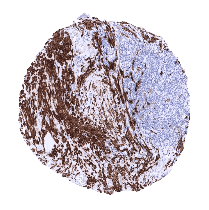
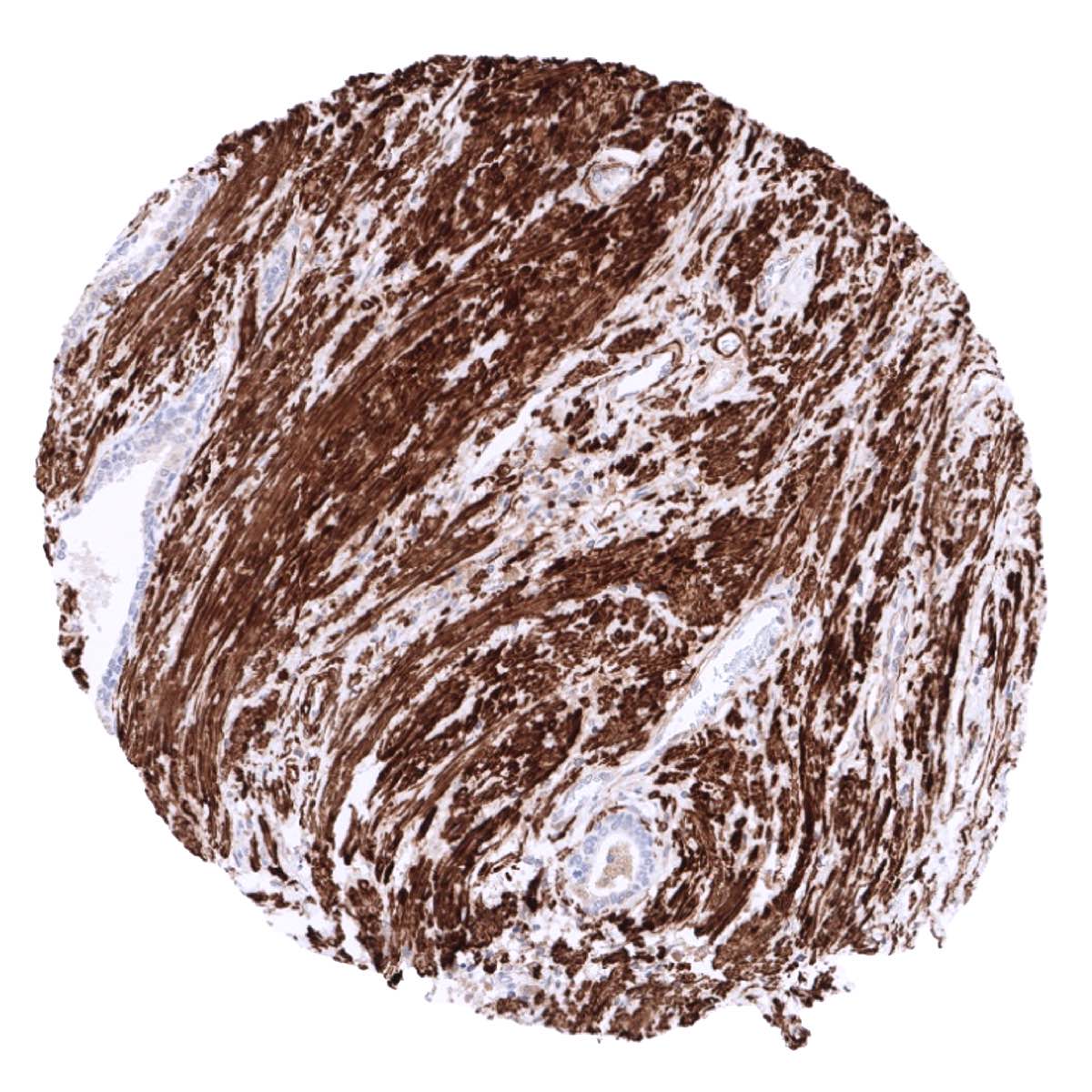
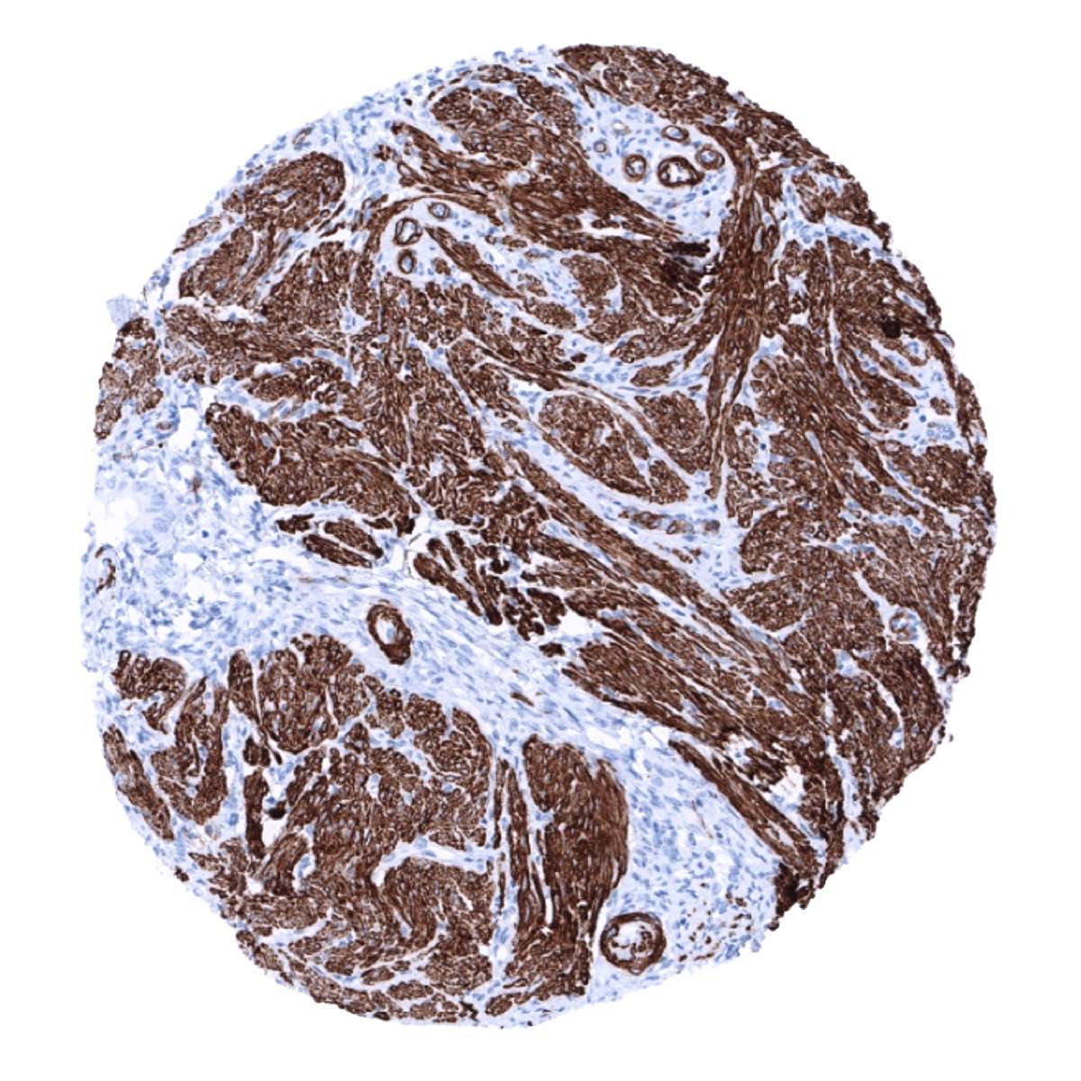
Caldesmon-h staining is seen in the cytoplasm and cell membrane of smooth muscles in various organs including the walls of small, medium sized, and large vessels, the aortic media and smooth muscle elements of the ovarian stroma. Myoepithelial cells and basal cells of excretory ducts in salivary glands as well as myoepithelial cells of the breast are also caldesmon-h positive. In the colorectum, caldesmon-h stains intestinal subepithelial myofibroblasts (ISEMFs) which form a thin membrane-like layer around the colon crypts. A weak but distinct membranous caldesmon-h staining also occurs at the basal membrane of amnion cells.
The findings described above are this consistent with the RNA data described in the Human Protein Atlas (Tissue expression Caldesmon)
Positive control = Colon: A strong caldesmon staining should be seen of the muscularis mucosa and of a membrane-like layer along the basement membranes of crypts (intestinal subepithelial myofibroblasts).
Negative control = Colon: Normal epithelial cells must not show caldesmon staining.

Staining Pattern in Relevant Tumor Types
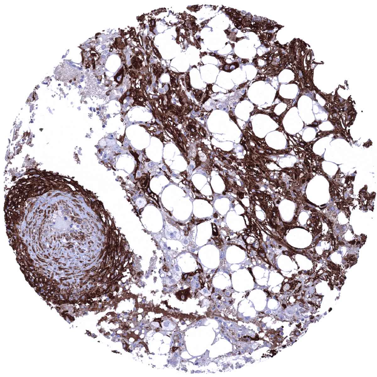
A positive caldesmon immunostaining can be seen in the vast majority of leiomyomas and leiomyosarcomas as well as in other more rare smooth muscle tumors such as myopericytomas or Glomus tumors.
The TCGA findings on Caldesmon RNA expression in different tumor categories have been summarized in the Human Protein Atlas.
Of note, these data are likely to not reflect h-caldesmon expression of tumor cells. They may rather be driven by the quantity of non-neoplastic smooth muscle invaded by the tumor cells and possibly also include L-caldesmon RNA.


Compatibility of Antibodies
No data available at the moment
Protocol Recommendations
IHC users have different preferences on how the stains should look like. Some prefer high staining intensity of the target stain and even accept some background. Others favor absolute specificity and lighter target stains. Factors that invariably lead to more intense staining include higher concentration of the antibody and visualization tools, longer incubation time, higher temperature during incubation, higher temperature and longer duration of the heat induced epitope retrieval (slide pretreatment). The impact of the pH during slide pretreatment has variable effects and depends on the antibody and the target protein.
All images and data shown here and in our image galleries are obtained by the manual protocol described below. Other protocols resulting in equivalent staining are described as well.
Manual protocol
Freshly cut sections should be used (less than 10 days between cutting and staining). Heat-induced antigen retrieval for 5 minutes in an autoclave at 121°C in pH 7,8 Target Retrieval Solution buffer. Apply MSVA-538R at a dilution of 1:150 at 37°C for 60 minutes. Visualization of bound antibody by the EnVision Kit (Dako, Agilent) according to the manufacturer’s directions.
Agilent / Dako – Omnis
Unmasking IHC EnV FLEX TRS High pH 97°C ; FLEX peroxidase blocking for 3 minutes (32°C), MSVA-538R 1:150 for 30 minutes at 32°C, FLEX+ mouse/rabbit (LINKER) for 10 minutes at 32°C, horseradish peroxidase (HRP) for 30 minutes (32°C), FLEX DAB+Sub-Chromo for 5 minutes (32°C), FLEX hematoxylin for 5 minutes (room temperature).
These images reflect stainings by the protocol described above. It is of note that a comparable staining result can also be obtained by different protocols. In general, a longer pretreatment, a longer incubation time of the primary antibody, a higher antibody concentration, and a longer incubation time of FLEX+LINKER result in stronger staining, potentially at the cost of more background staining. Modifications of the protocol with a strengthening effect on staining intensity in combination with changes of other parameters that result in lower staining intensity can result in a comparable result as shown above.
Agilent / Dako – Autostainer Link 48
Pretreatment in PT-Link for 30 minutes at 95°C (pH high); FLEX peroxidase blocking for 5 minutes (room temperature), MSVA-538R 1:150 for 20 minutes (room temperature), FLEX+ mouse/rabbit (LINKER) for 15 minutes (room temperature), horseradish peroxidase (HRP) for 20 minutes (room temperature), FLEX DAB+Sub-Chromo for 10 minutes (room temperature), FLEX hematoxylin for 5 minutes (room temperature).

These images reflect stainings by the protocol described above. It is of note that a comparable staining result can also be obtained by different protocols. In general, a longer pretreatment, a longer incubation time of the primary antibody, a higher antibody concentration, and a longer incubation time of FLEX+LINKER result in stronger staining, potentially at the cost of more background staining. Modifications of the protocol with a strengthening effect on staining intensity in combination with changes of other parameters that result in lower staining intensity can result in a comparable result as shown above.
Leica – BOND RX
Dewax at 72°C for 30 seconds; Pretreatment in Bond Epitope Retrieval Solution (ER2 – EDTA pH9) for 20 minutes at 100°C; Peroxidase blocking for 5 minutes (room temperature), MSVA-538R 1:150 for 15 minutes (room temperature), Post primary (rabbit anti mouse) for 8 minutes (room temperature), Polymer (goat anti rabbit) for 8 minutes (room temperature), mixed DAB refine for 10 minutes (room temperature), hematoxylin for 5 minutes (room temperature).
These images reflect stainings by the protocol described above. It is of note that a comparable staining result can also be obtained by different protocols. In general, a longer pretreatment, a longer incubation time of the primary antibody, a higher antibody concentration, a higher temperature during incubation, and a longer incubation time of Post primary and or the Polymer result in stronger staining, potentially at the cost of more background staining. Modifications of the protocol with a strengthening effect on staining intensity in combination with changes of other parameters that result in lower staining intensity can result in a comparable result as shown above.
Roche – Ventana Discovery ULTRA
Pretreatment for 64 minutes at 100°C (pH 8,4); CM peroxidase blocking for 12 minutes (room temperature), MSVA-538R 1:150 for 20 minutes at 36°C, secondary antibody (anti-rabbit HQ) for 12 minutes at 36°C, anti-HQ HRP for 12 minutes at room temperature, DAB at room temperature, hematoxylin II at room temperature for 8 minutes, bluing reagent at room temperature for 4 minutes.

These images depict staining results obtained by the protocol described above. It is of note, that the Ventana machines generally require higher antibody concentrations than other commonly used autostainers because the antibodies are automatically diluted during the procedure. Various other protocols can result in an identical result as shown above. A longer pretreatment, a longer incubation time of the primary antibody, a higher antibody concentration, a higher temperature during incubation, and a longer incubation time of secondary antibody and or the anti-HQ HRP result in stronger staining, potentially at the cost of more background staining.
Potential Research Applications
- h-Caldesmon expression has been suggested to occur in non-smooth muscle tumors such as mesotheliomas, gastrointestinal stromal tumors, or granulosa cell tumors. A study analyzing caldesmon expression in a broad spectrum of tumors is desirable.
Evidence for Antibody Specificity in IHC
There are two ways how the specificity of antibodies can be documented for immunohistochemistry on formalin fixed tissues. These are: 1. Comparison with a second independent method for target expression measurement across a large number of different tissue types (orthogonal strategy), and 2. Comparison with one or several independent antibodies for the same target and showing that all positive staining results are also seen with other antibodies for the same target (independent antibody strategy).
Orthogonal validation: Is not applicable for caldesmon-h antibodies because the protein occurs in blood vessels and is therefore ubiquitously detectable in all organs. The particularly high rate of RNA expression described in smooth muscle and organs that contain high quantities of smooth muscle such as gastrointestinal organs or the urinary bladder are consistent, however, with caldesmon-h immunostaining detected by MSVA-538R Human Protein Atlas (Tissue expression Caldesmon).
Comparison of antibodies: True expression of h-caldesmon in smooth muscle, walls of blood vessels, ovarian stroma and myoepithelial cells is corroborated by comparison with a commercially available independent second antibody (termed “validation antibody”). Complete concordance of MSVA-538R staining with the staining by “validation antibody” fully confirms the specificity of MSVA-538R.