
Adrenal gland
Aorta, media - Intense caldesmon-h staining of smooth muscle cells (Caldesmon-h immunohistochemistry)
Appendix, mucosa - Distinct caldesmon-h staining of pericryptal intestinal subepithelial myofibroblasts (ISEMFs) and of smooth muscle cells of the muscularis mucosae (Caldesmon-h immunohistochemistry)
Appendix, mucosa - Intense caldesmon-h positivity of intestinal subepithelial myofibroblasts (ISEMFs) and of the muscularis mucosae (Caldesmon-h immunohistochemistry)
Appendix, muscular wall - Intense caldesmon-h staining of smooth muscle cells (Caldesmon-h immunohistochemistry)
Bone marrow
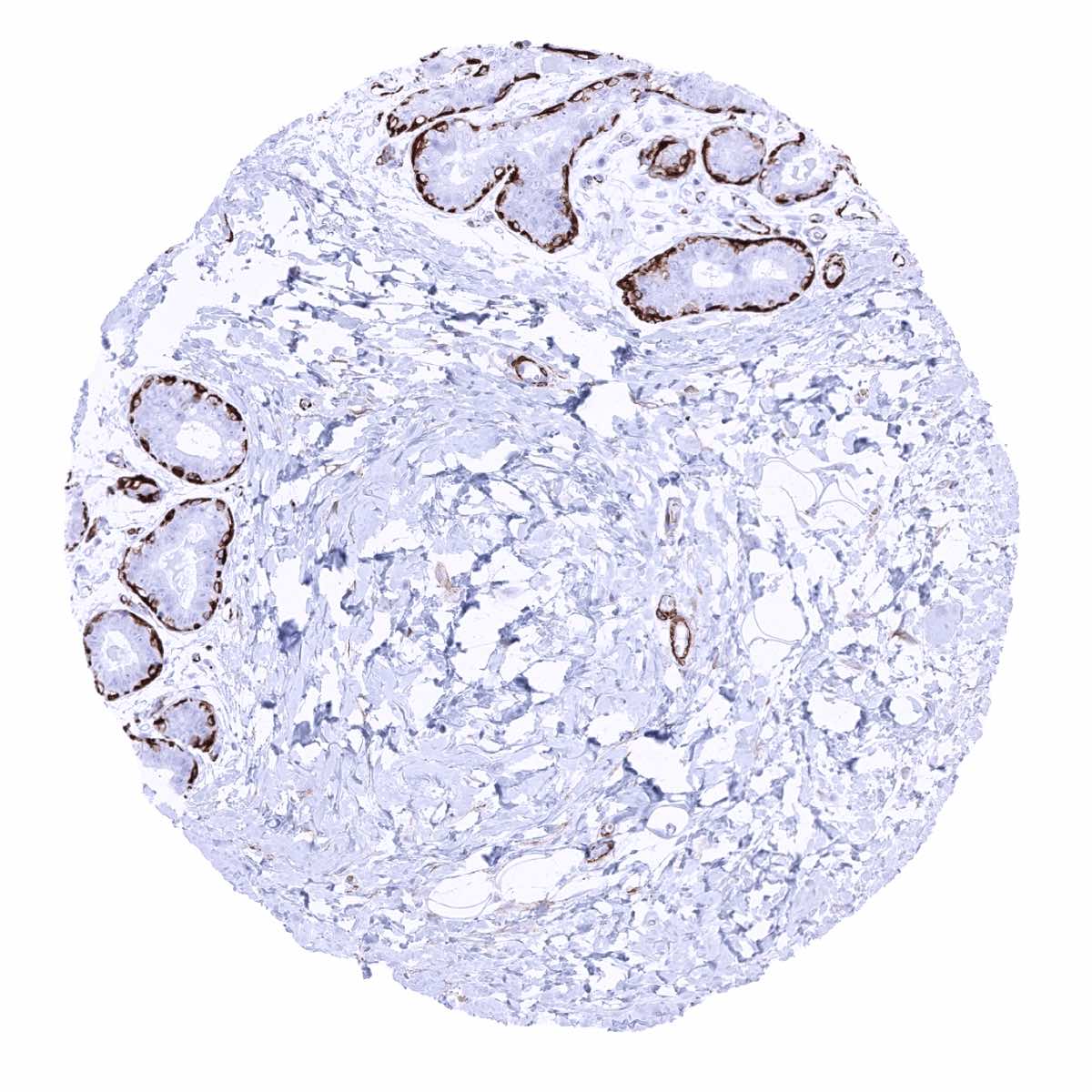
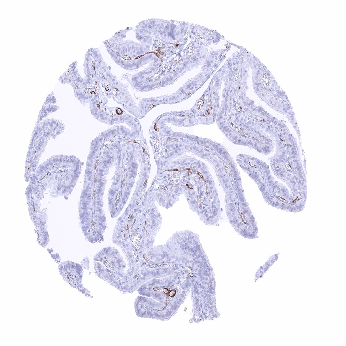
Breast - Caldesmon-h staining of myoepithelial cells (Caldesmon-h immunohistochemistry)
Bronchus, mucosa - Caldesmon-h staining of smooth muscle and of myoepithelial cells (Caldesmon-h immunohistochemistry)

Cerebellum (molecular layer, Purkinje cell layer, granule cell layer, white matter)
Cerebellum (white matter)
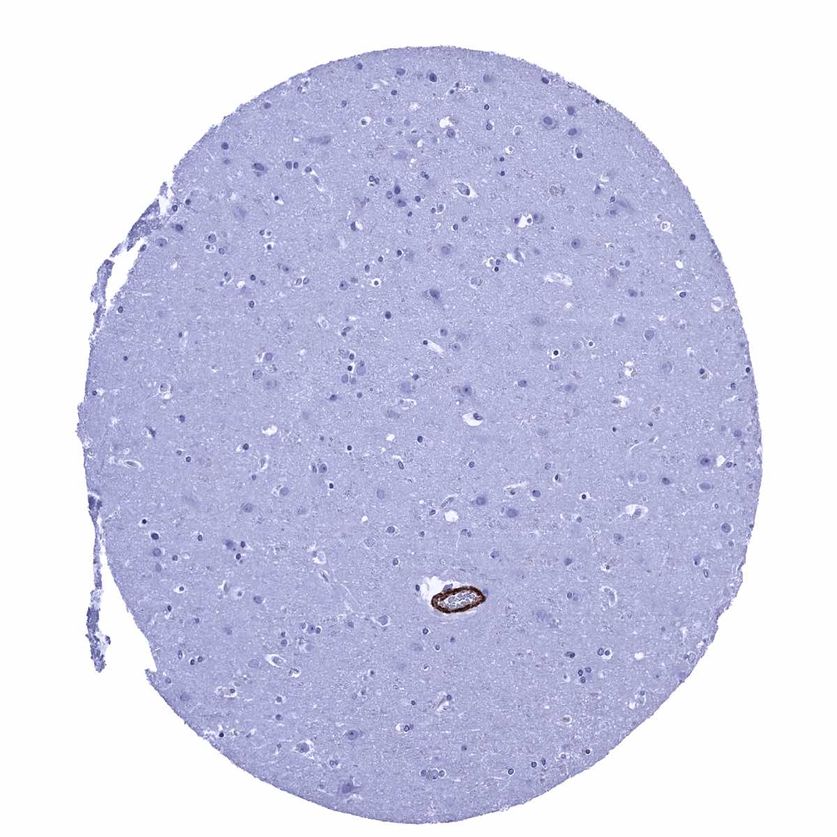
Cerebrum, grey matter
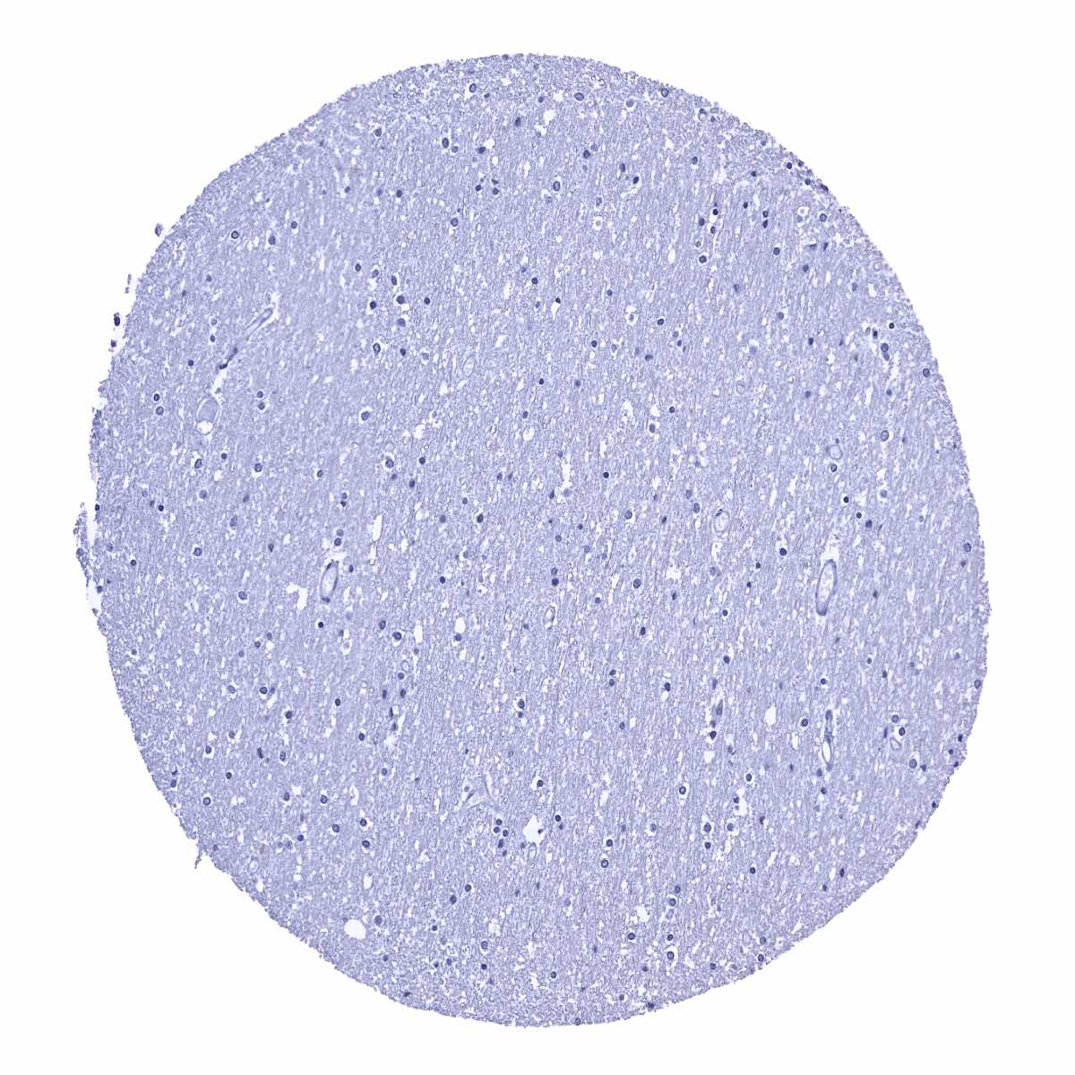
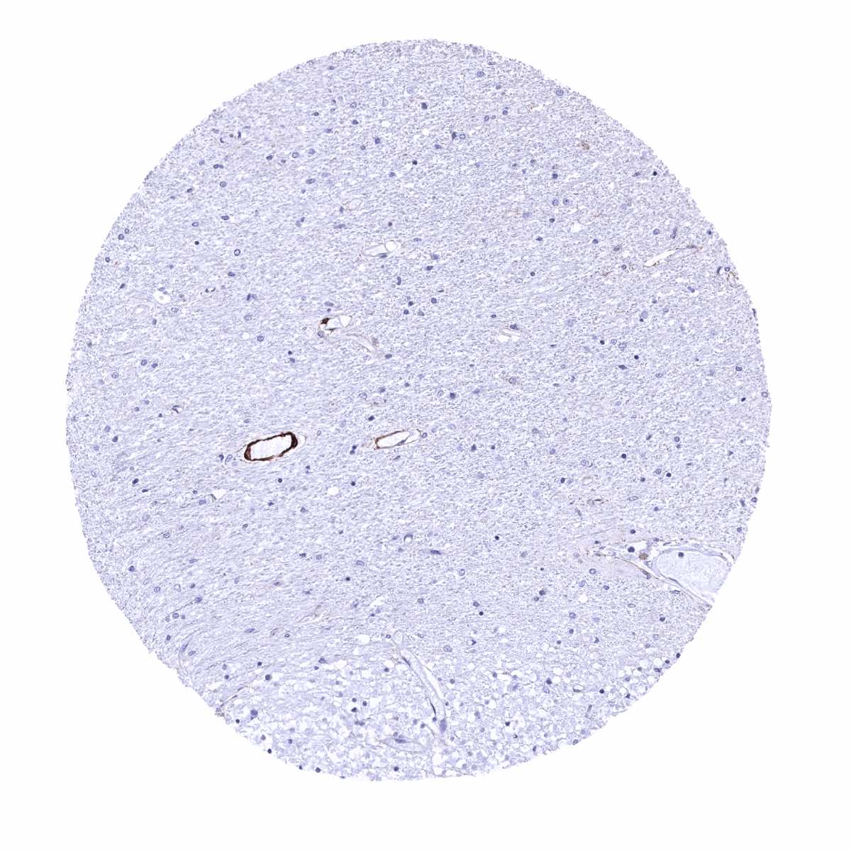
Cerebrum, white matter
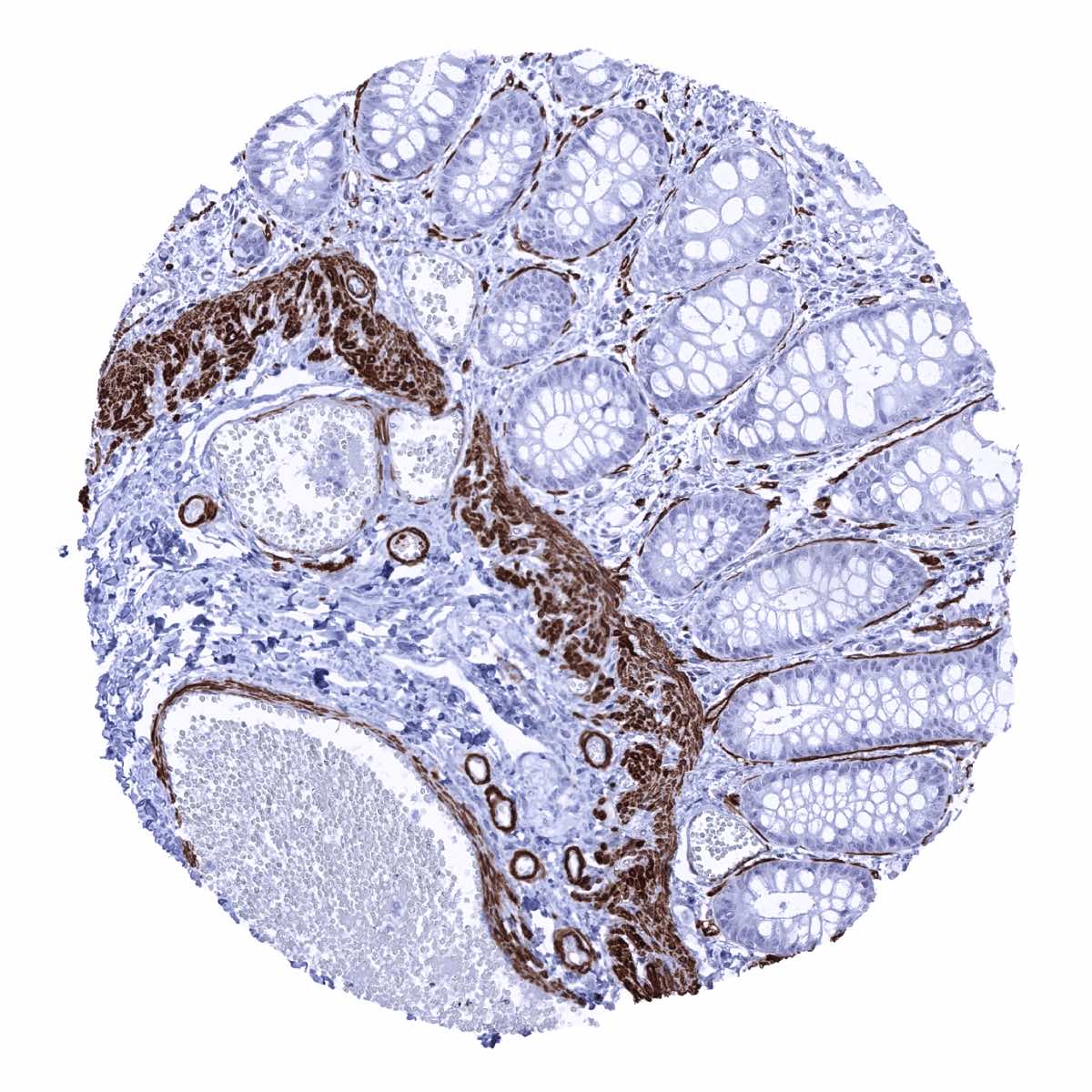
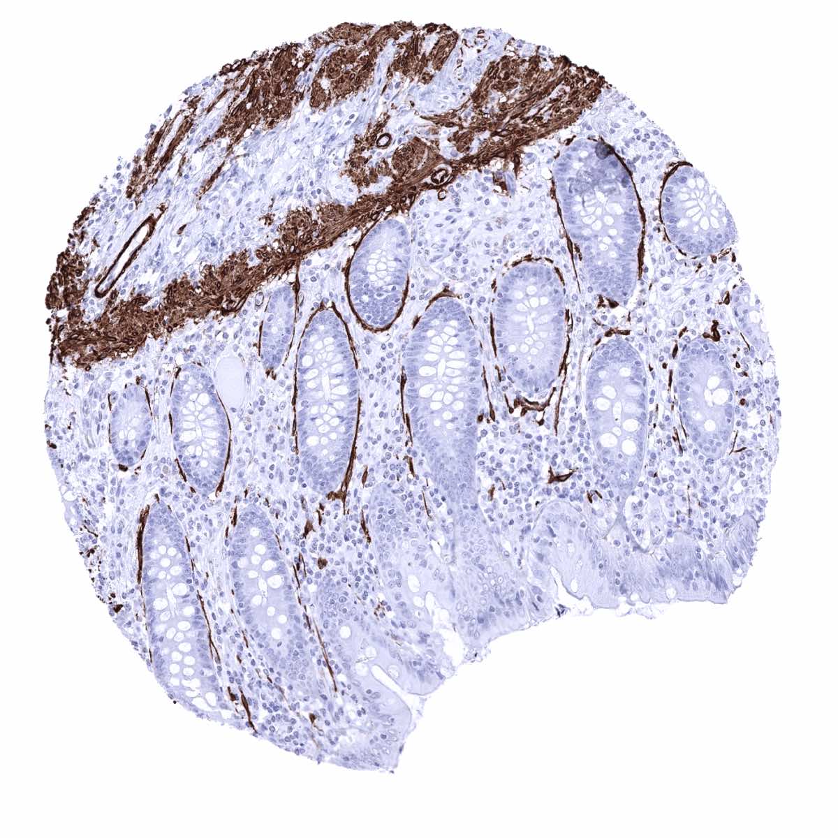
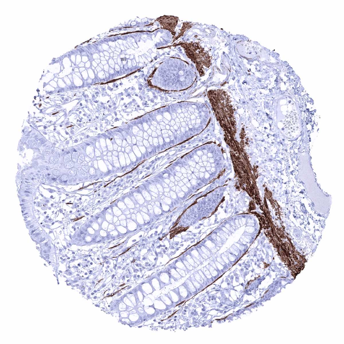
Colon descendens, mucosa - Caldesmon-h staining of intestinal subepithelial myofibroblasts (ISEMFs), muscular vessels, and of the muscularis mucosae (Caldesmon-h immunohistochemistry)

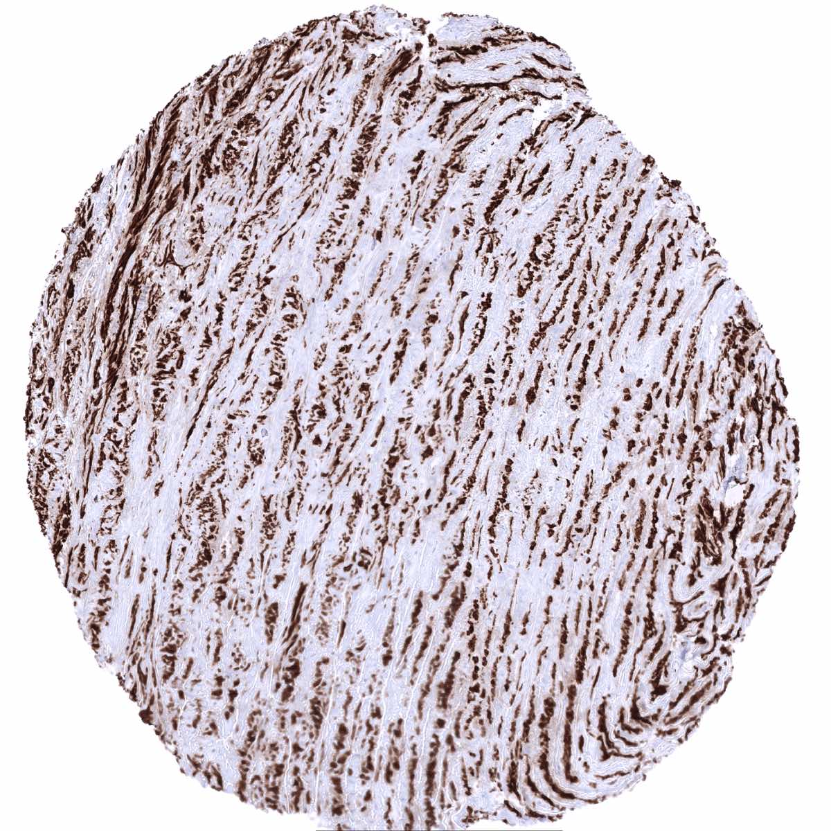
Colon descendens, muscular wall - Intense caldesmon-h staining of smooth muscle cells (Caldesmon-h immunohistochemistry)
Duodenum, Brunner gland

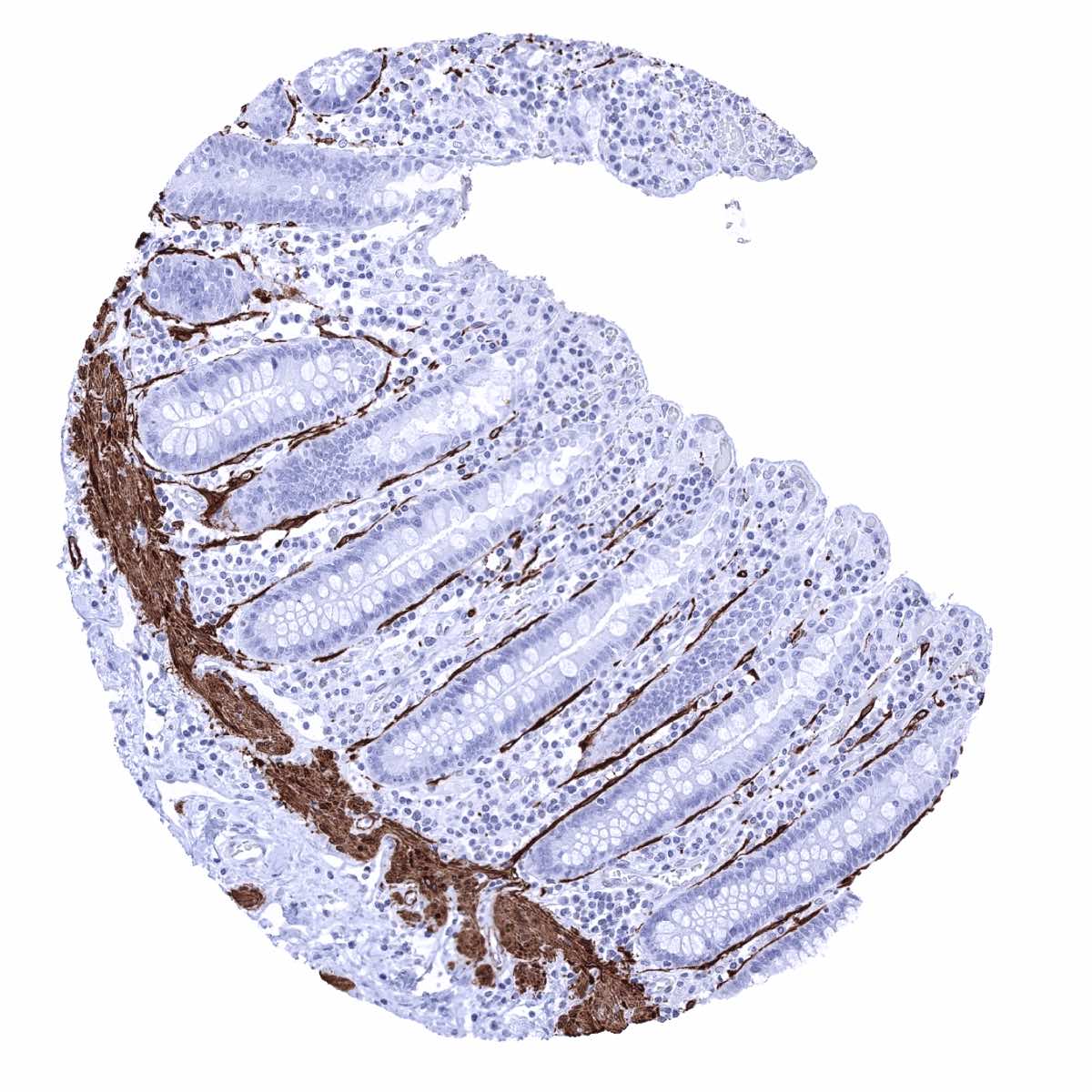
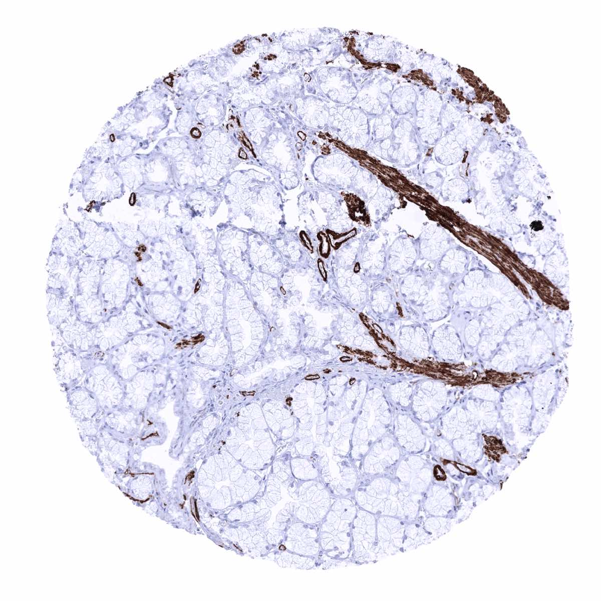
Duodenum, mucosa - Caldesmon-h staining of smooth muscle cells in small vessels, pericryptal, and of the muscularis mucosae (Caldesmon-h immunohistochemistry)
Epididymis - Caldesmon-h positivity of vessel walls and of pericanalicular smooth muscle cells (Caldesmon-h immunohistochemistry)
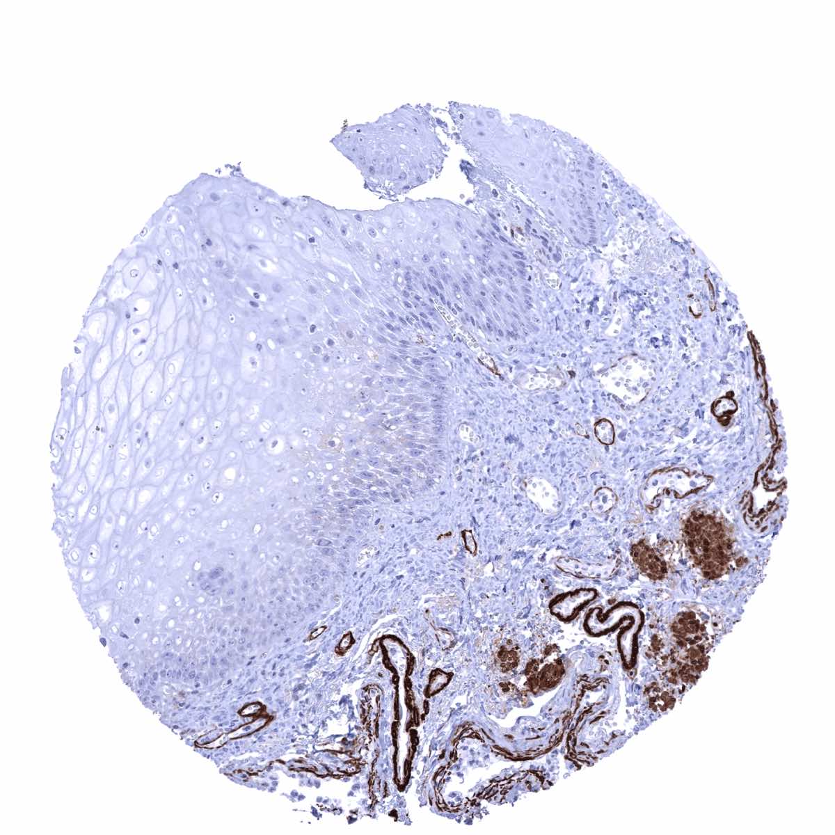
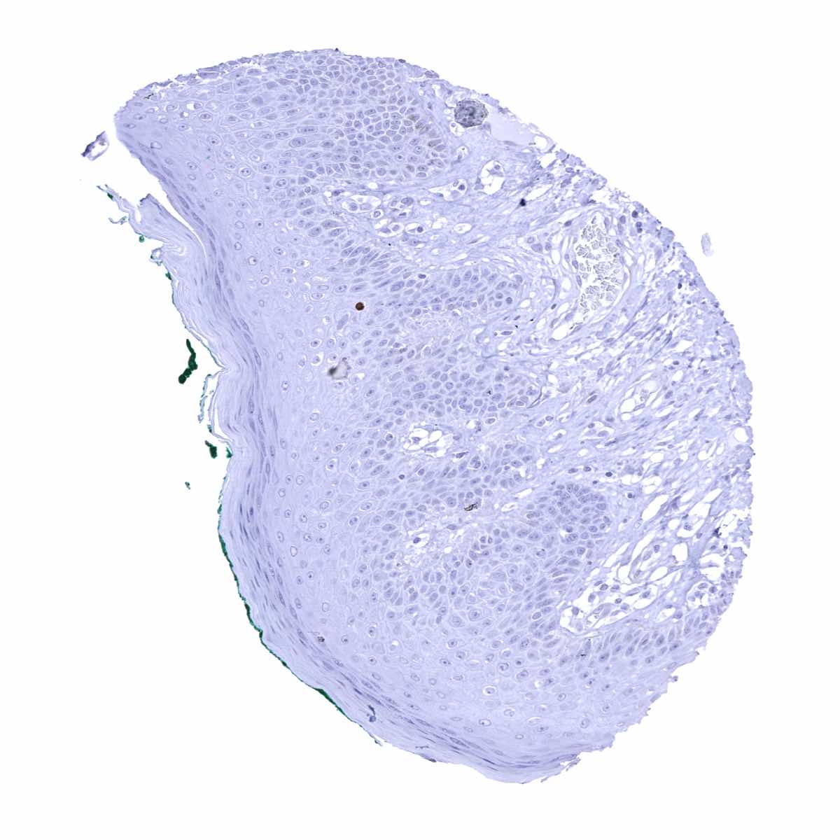
Esophagus, squamous epithelium
Fallopian tube, mucosa

Fat
Gallbladder, epithelium
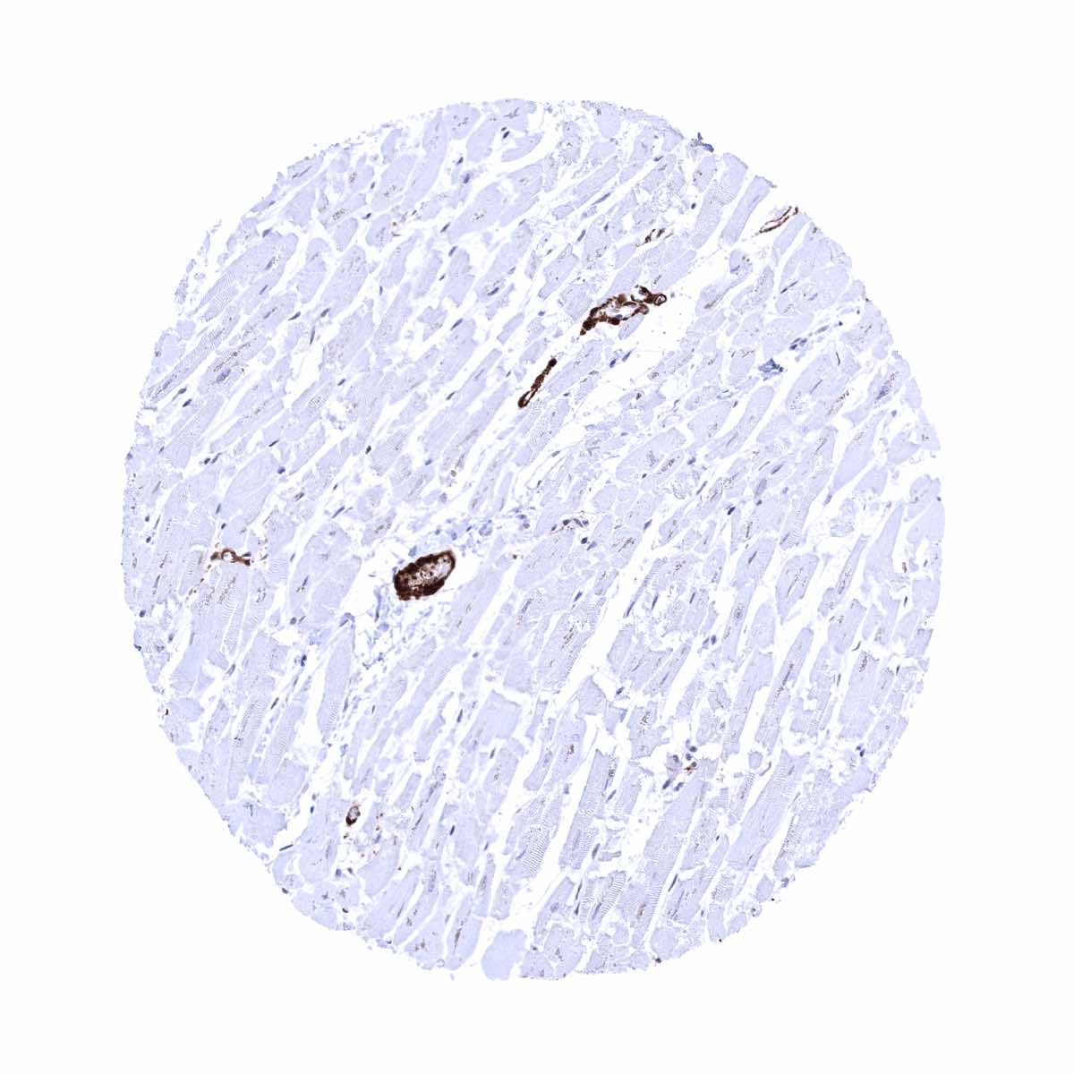
Heart muscle - Caldesmon-h staining of vascular smooth muscle cells (Caldesmon-h immunohistochemistry)
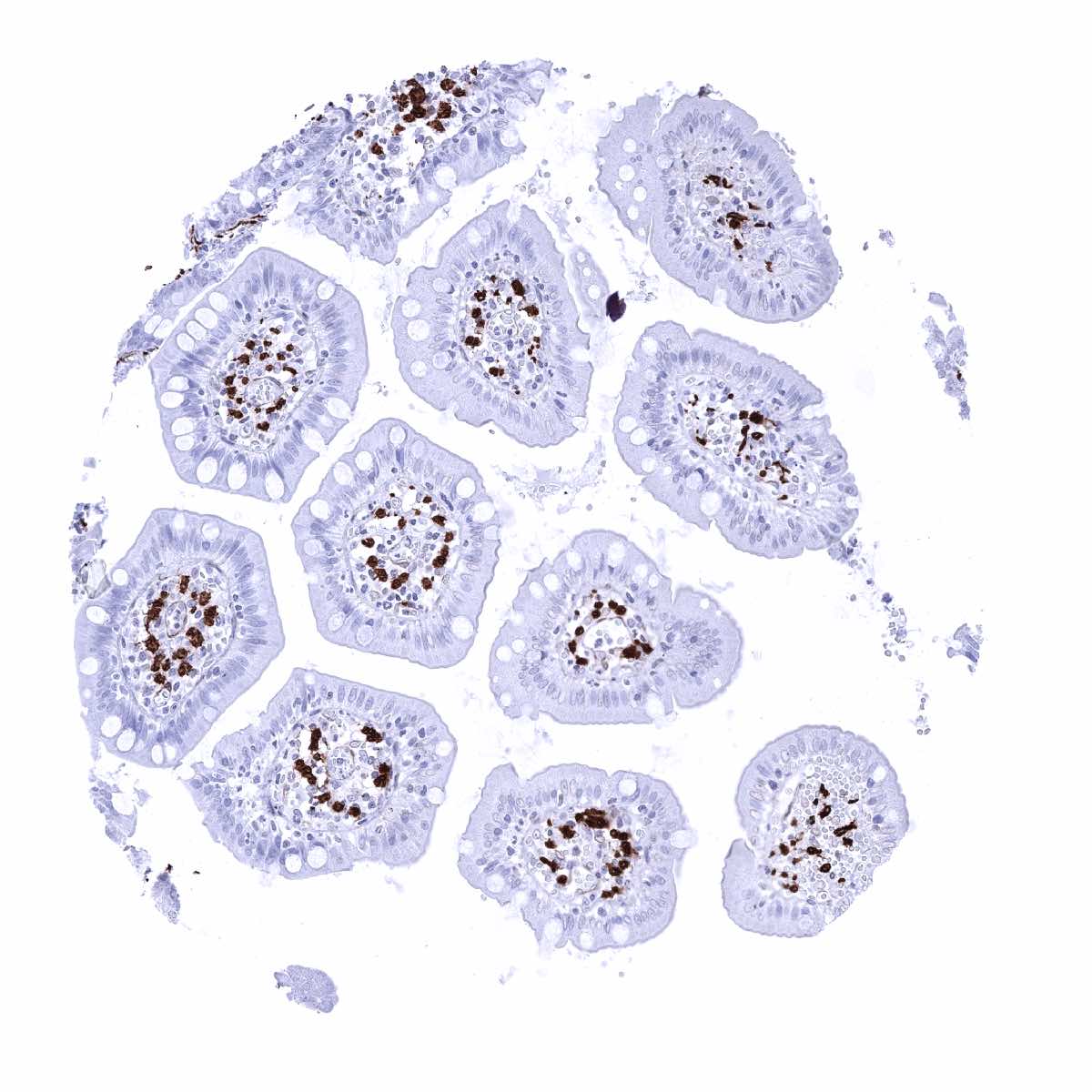
Ileum, mucosa - Caldesmon-h staining of vascular smooth muscle cells (Caldesmon-h immunohistochemistry)

Kidney, cortex
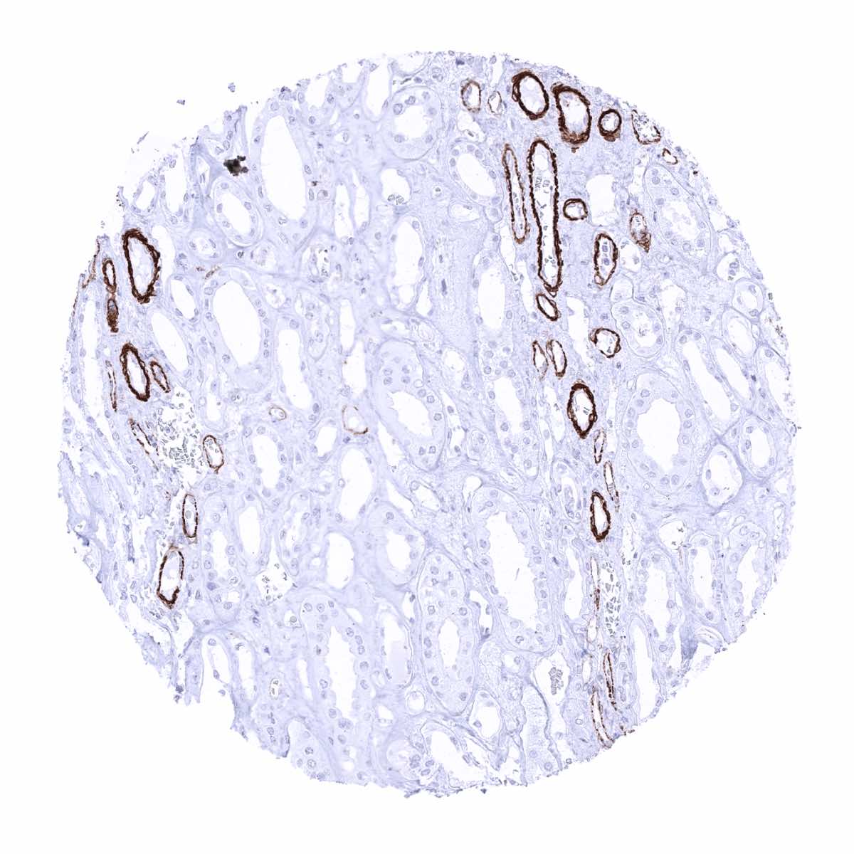
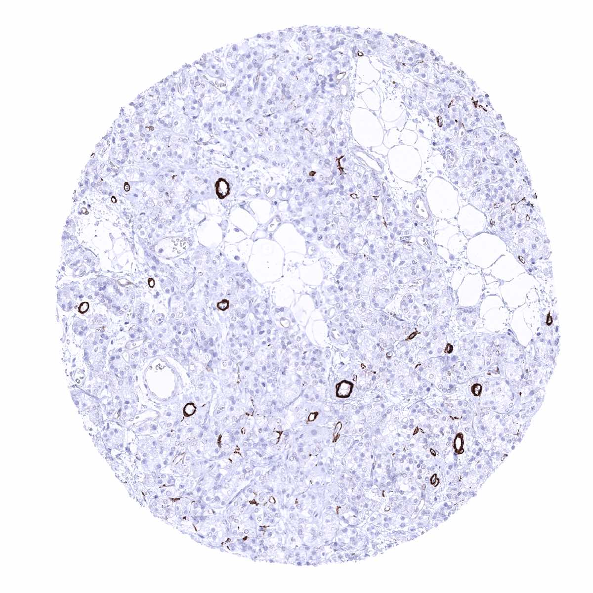
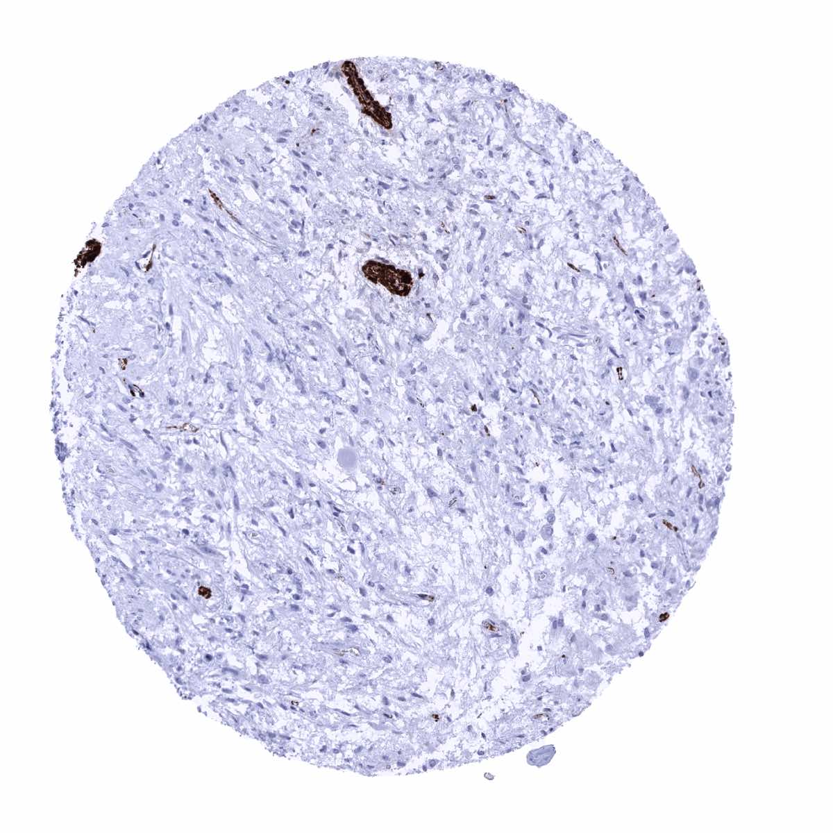
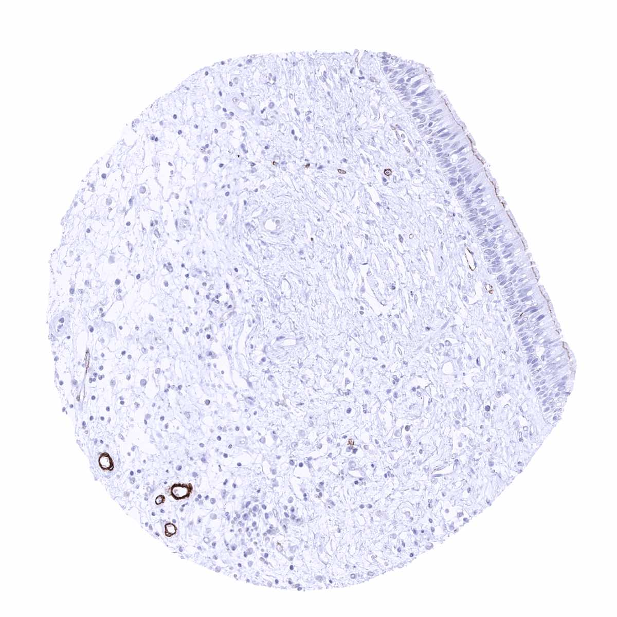
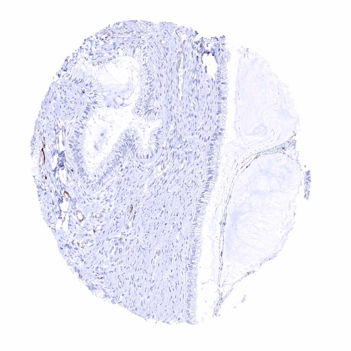
Kidney, medulla - Caldesmon-h staining is restricted to the wall of muscular vessels (Caldesmon-h immunohistochemistry)
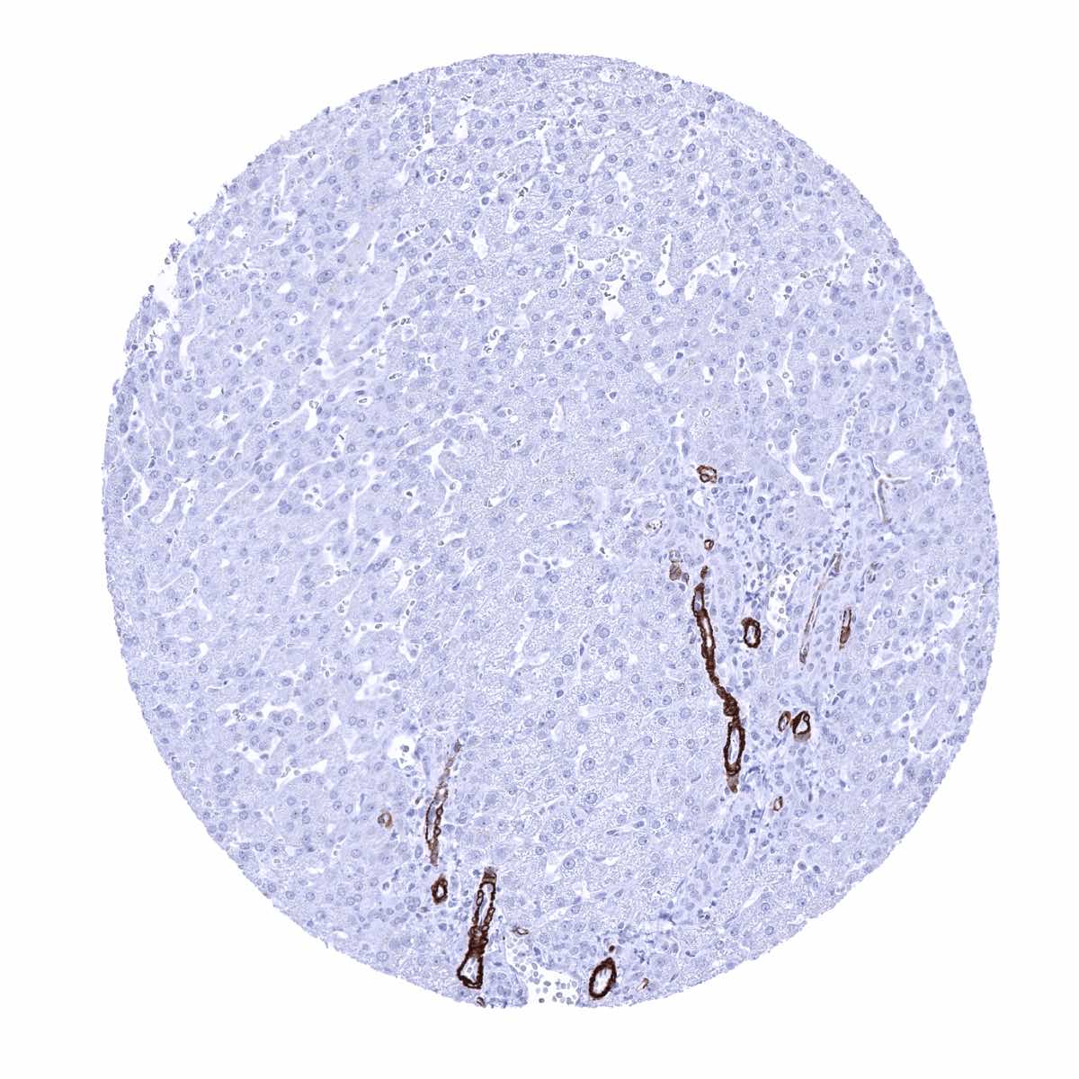
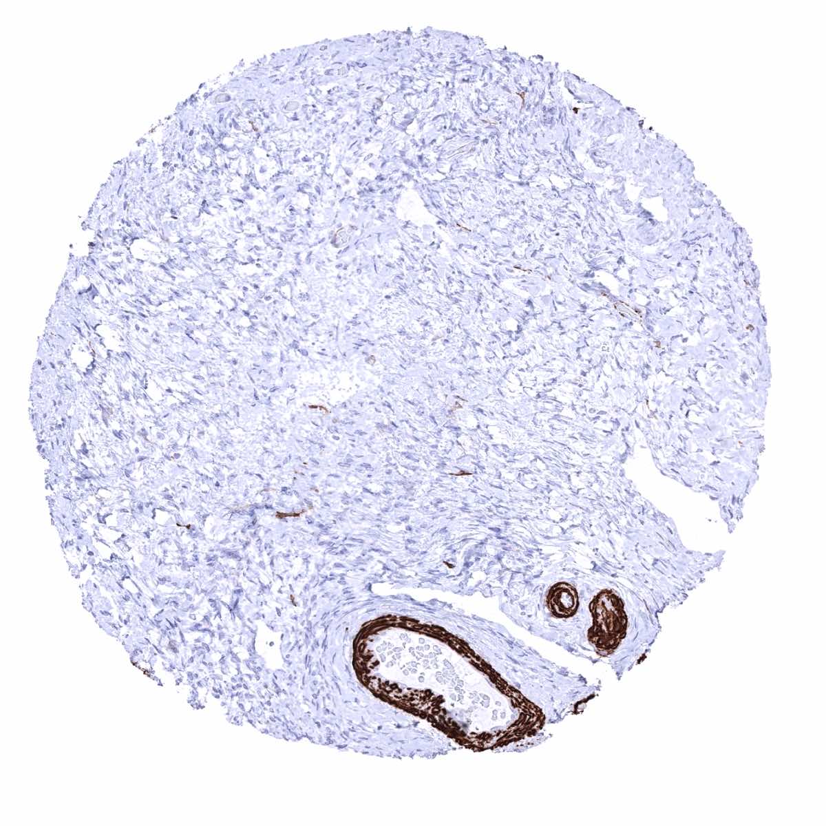
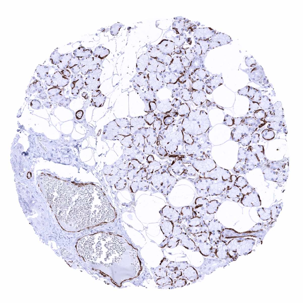
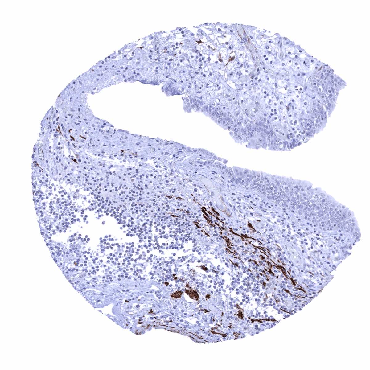
Liver - Caldesmon-h staining is limited to the wall of muscular vessels (Caldesmon-h immunohistochemistry)

Lung - Caldesmon-h staining is limited to vessel walls (Caldesmon-h immunohistochemistry)
Lymph node
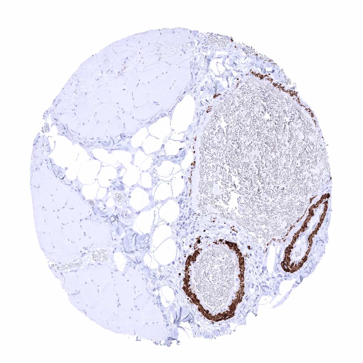
Ovary, stroma - Caldesmon-h staining of vascular smooth muscle cells (Caldesmon-h immunohistochemistry)

Ovary, stroma - The walls of small vessels and interspersed smooth muscle cells show caldesmon-h staining (Caldesmon-h immunohistochemistry)
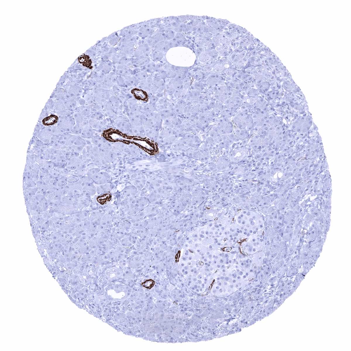
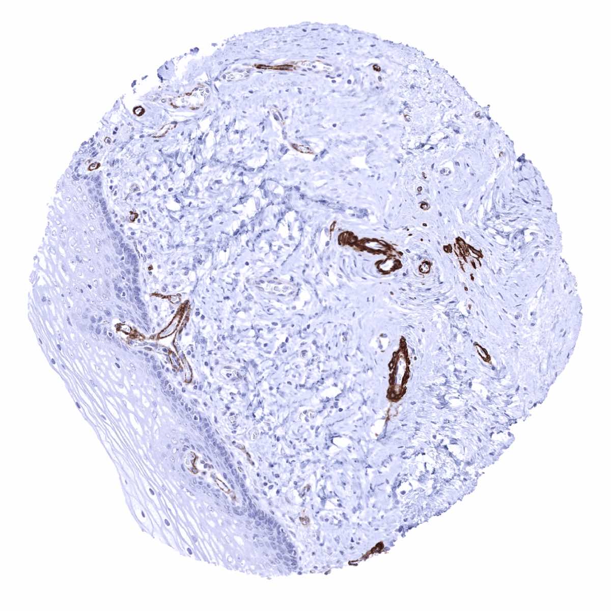
Pancreas - Restriction of caldesmon-h staining to the wall of muscular vessels (Caldesmon-h immunohistochemistry)
Parathyroid gland
Parotid gland - Caldesmon-h staining of myoepithelial cells (Caldesmon-h immunohistochemistry)
Pituitary gland, anterior lobe
Pituitary gland, posterior lobe
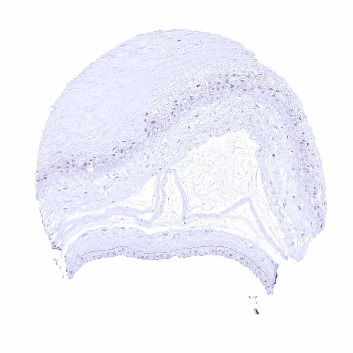
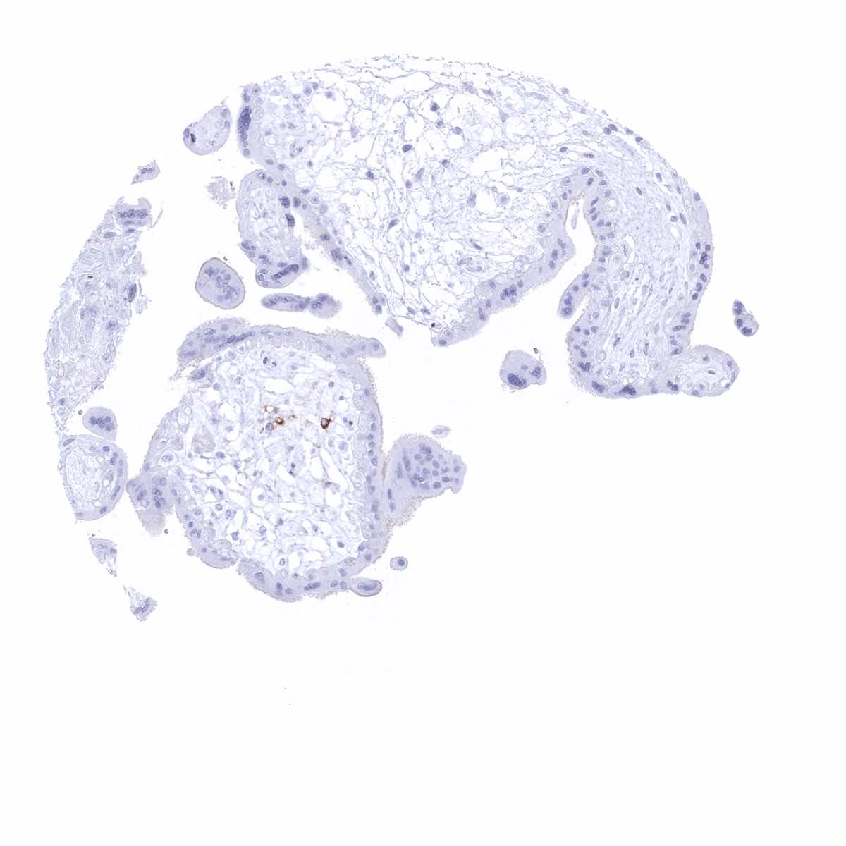
Placenta (amnion and chorion) - Weak but distinct membranous caldesmon-h staining along the basal cell membrane of amnion cells (Caldesmon-h immunohistochemistry)
Placenta, early
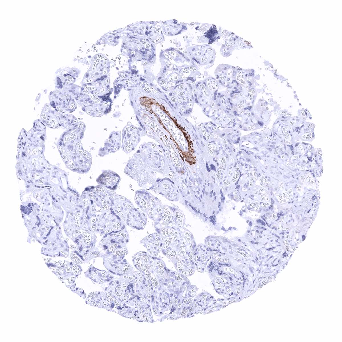
Placenta, mature
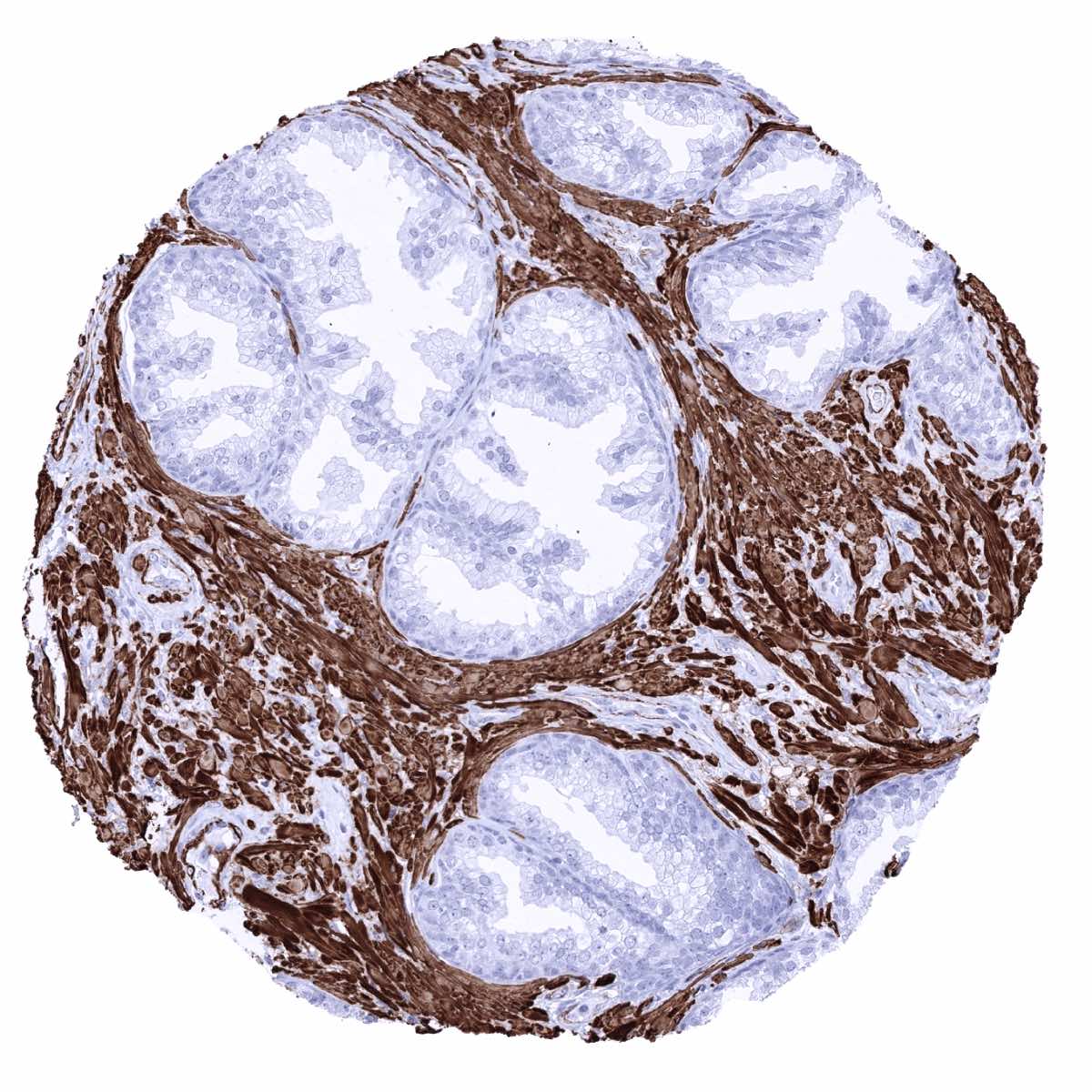
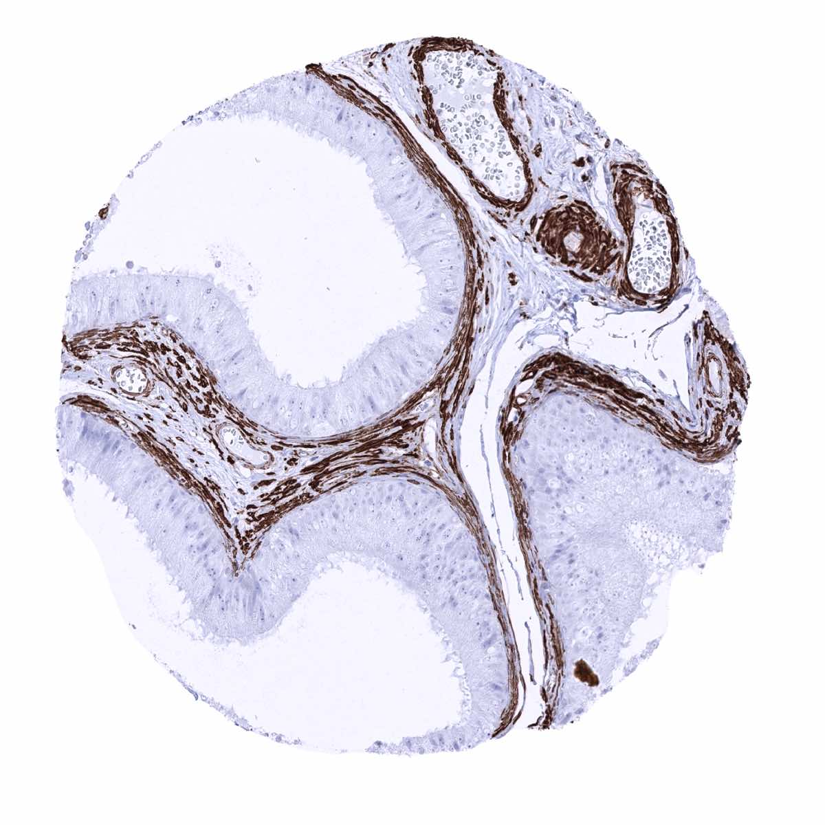
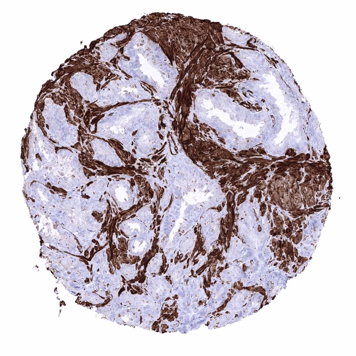
Prostate - Intense caldesmon-h positivity of smooth muscle cells of the prostatic stroma (Caldesmon-h immunohistochemistry)
Rectum, mucosa
Seminal vesicle - Intense caldesmon-h positivity of stromal smooth muscle cells (Caldesmon-h immunohistochemistry)
Sinus paranasales
Skeletal muscle - Caldesmon-h staining of vascular smooth muscle cells (Caldesmon-h immunohistochemistry)
Skin
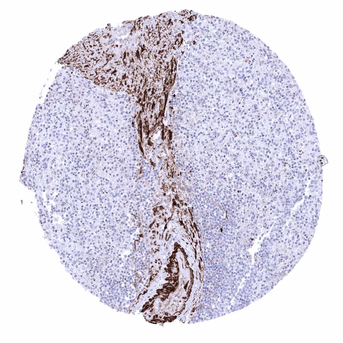
Spleen - Caldesmon-h positivity of smooth muscle cells in and around central arteries (Caldesmon-h immunohistochemistry)
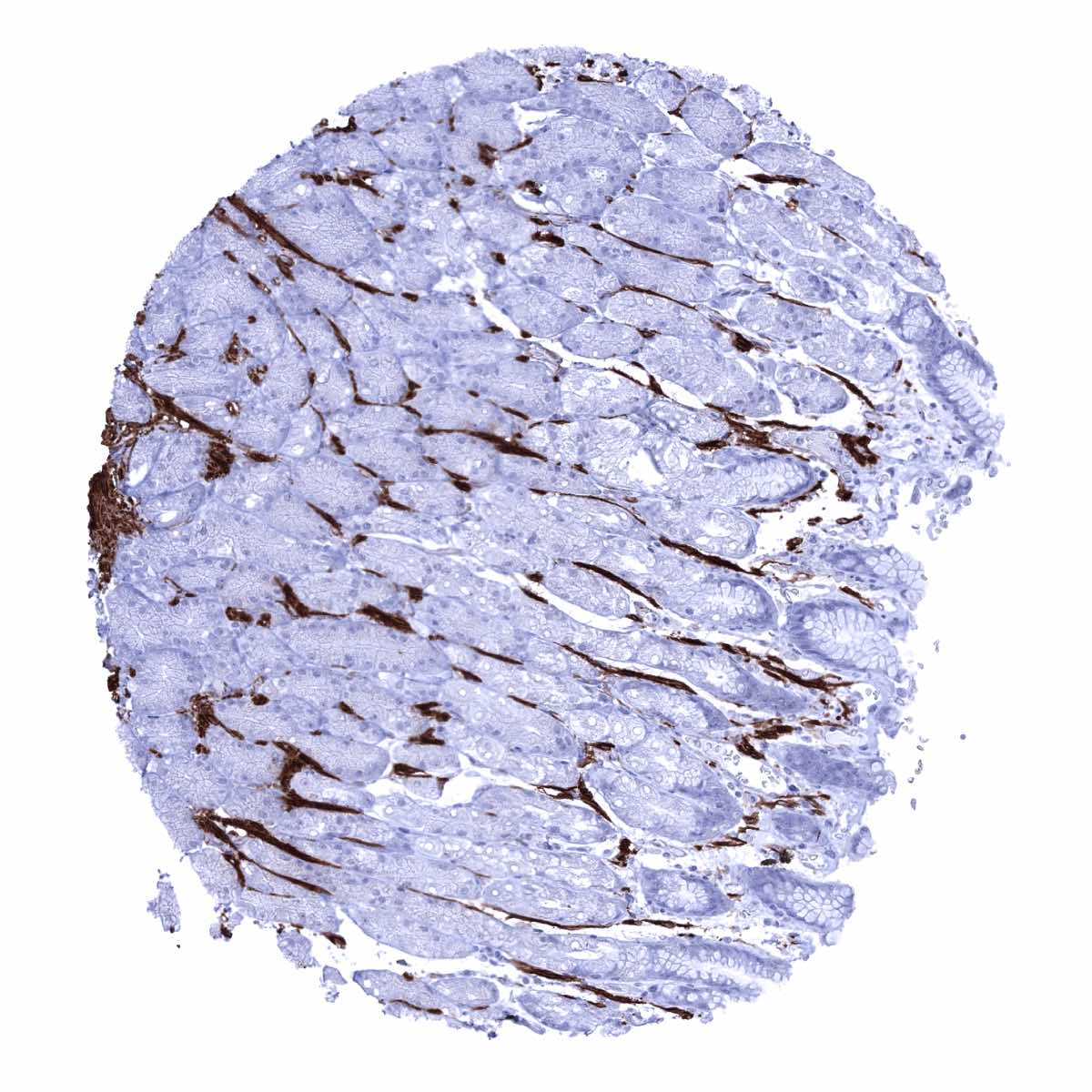
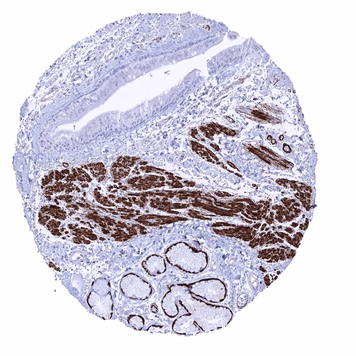
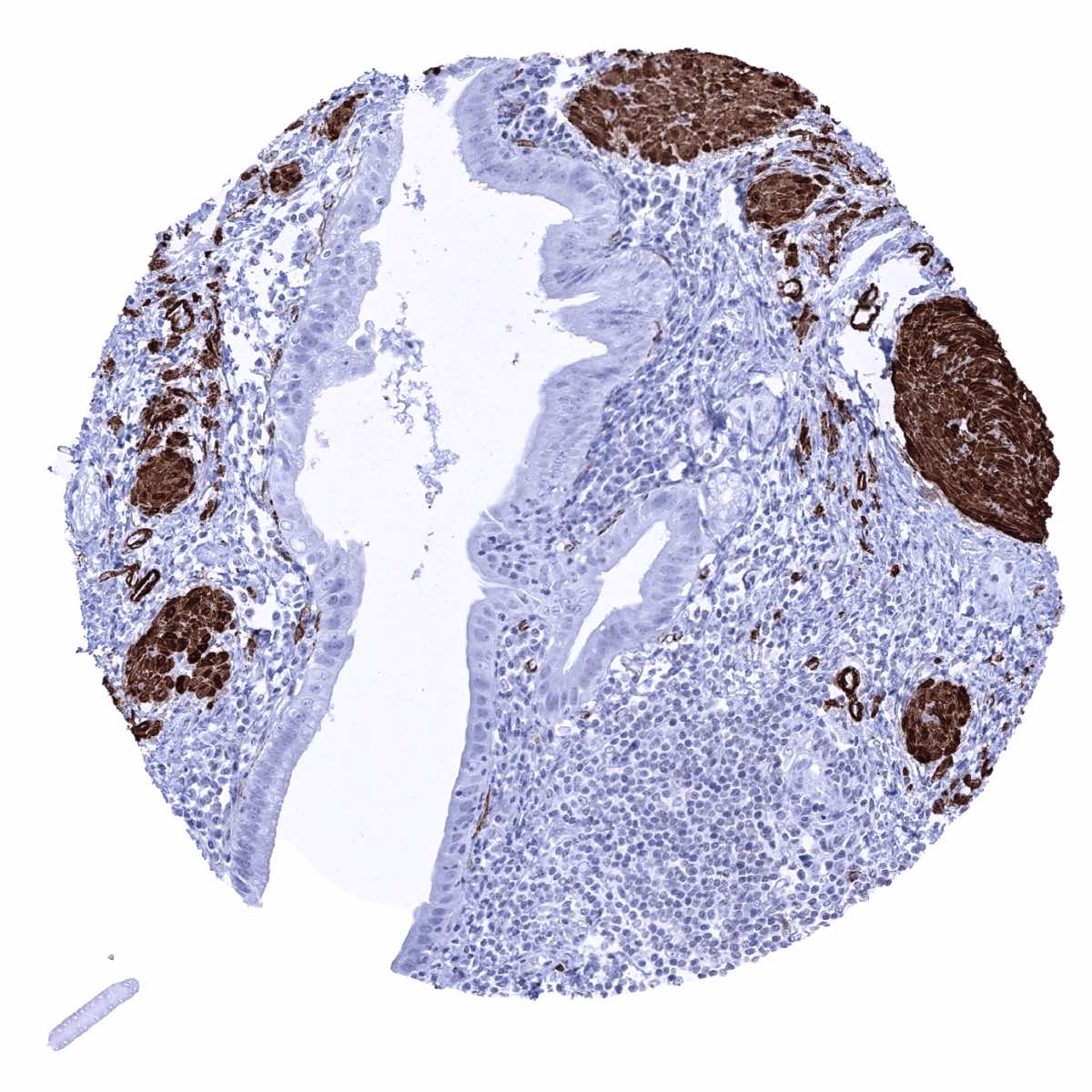
Stomach, antrum - Caldesmon-h positivity of smooth muscles around stomach glands (Caldesmon-h immunohistochemistry)
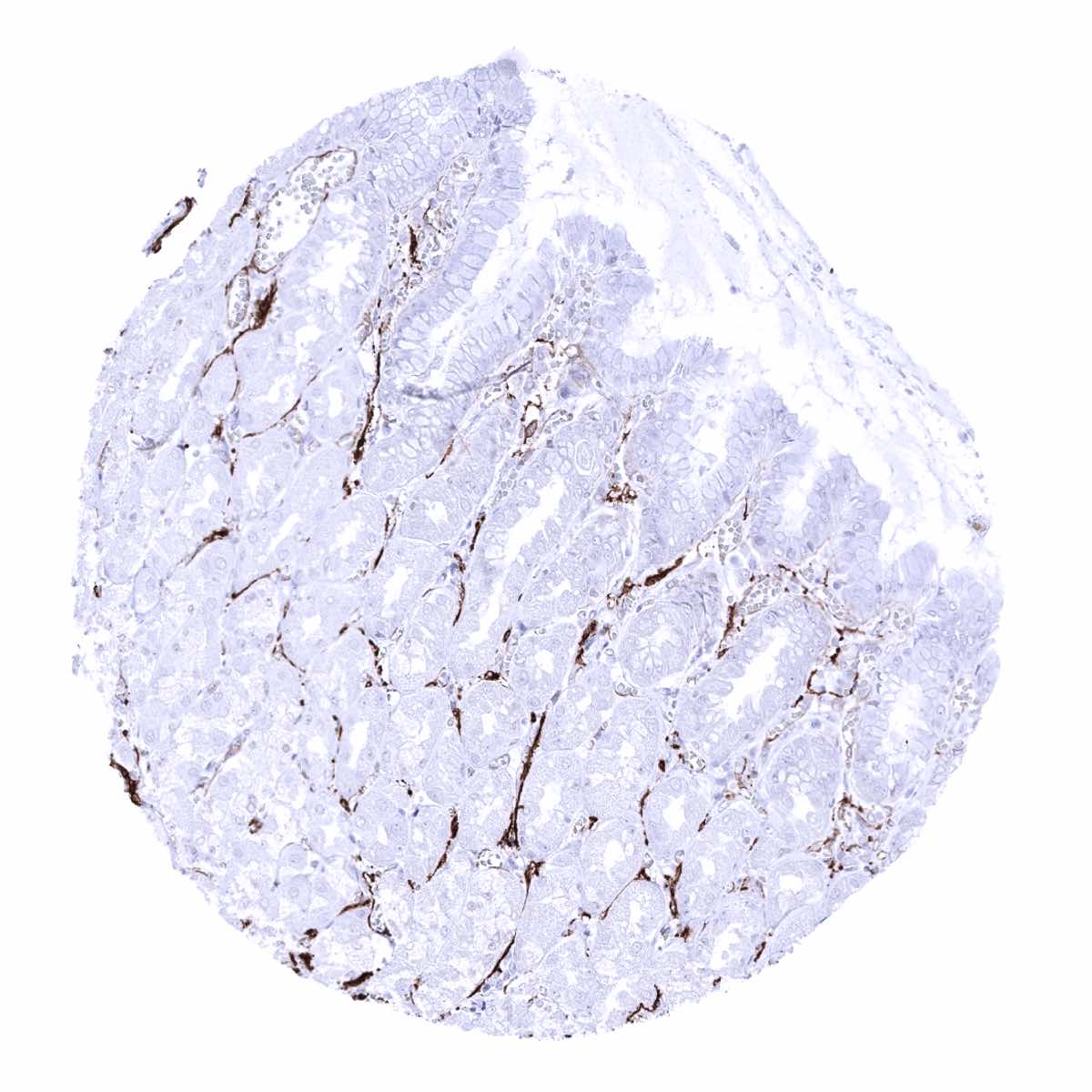
Stomach, corpus - Caldesmon-h positivity of smooth muscles around stomach glands (Caldesmon-h immunohistochemistry)
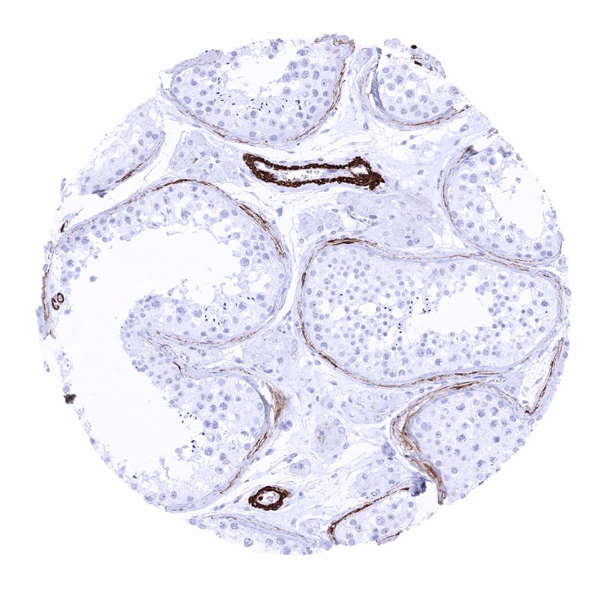
Testis - Caldesmon-h positivity of vessel walls and of peritubular smooth muscle cells (Caldesmon-h immunohistochemistry)
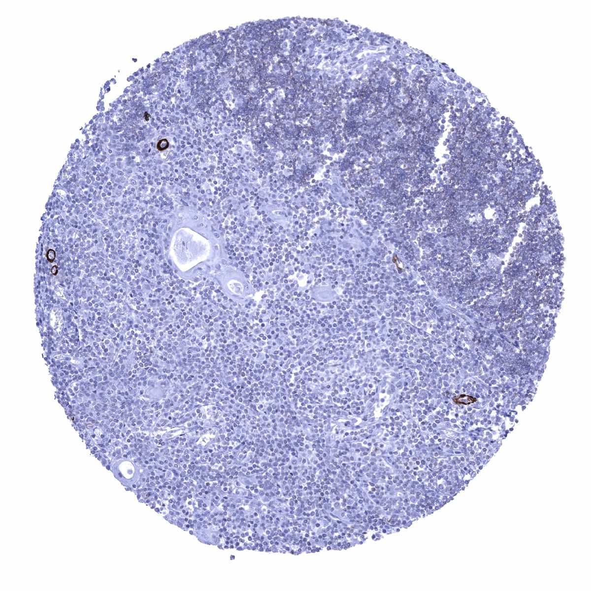
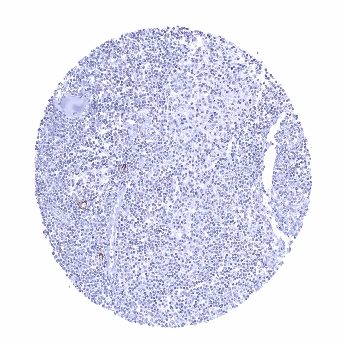
Thymus - Caldesmon-h positivity of smooth muscle cells in few small vessels (Caldesmon-h immunohistochemistry)
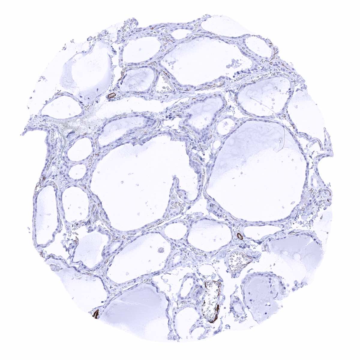
Thyroid gland
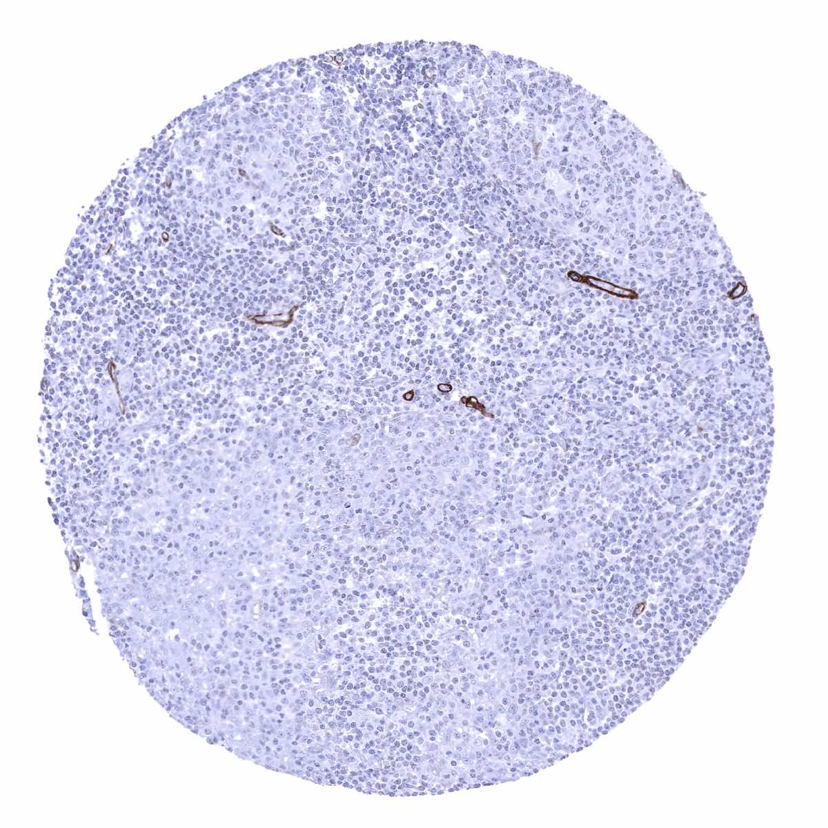
Tonsil - Caldesmon-h staining of smooth muscle cells in few small vessels (Caldesmon-h immunohistochemistry)
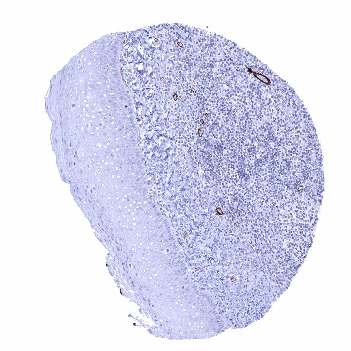
Tonsil, surface epithelium

Urinary bladder, muscular wall - Intense caldesmon-h staining of smooth muscle cells (Caldesmon-h immunohistochemistry)
Urinary bladder, urothelium
Uterus, ectocervix - Caldesmon-h positivity of smooth muscle cells in vascular walls (Caldesmon-h immunohistochemistry)_
Uterus, endocervix
Uterus, endometrium (pregnancy)
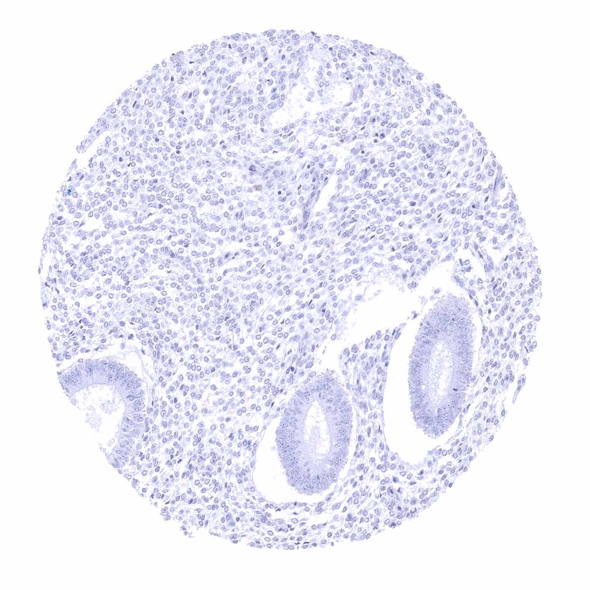
Uterus, endometrium (proliferation)
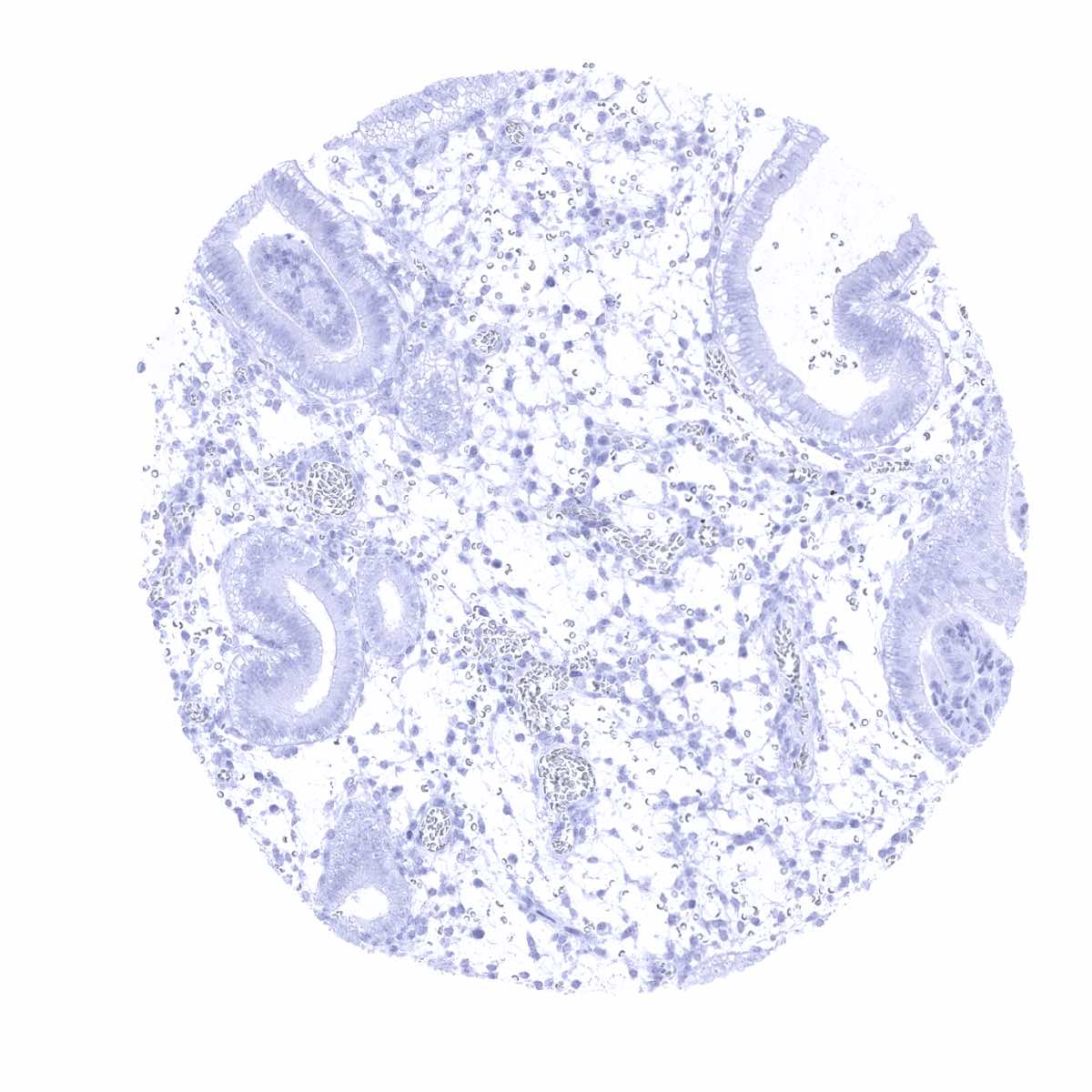
Uterus, endometrium (secretion)
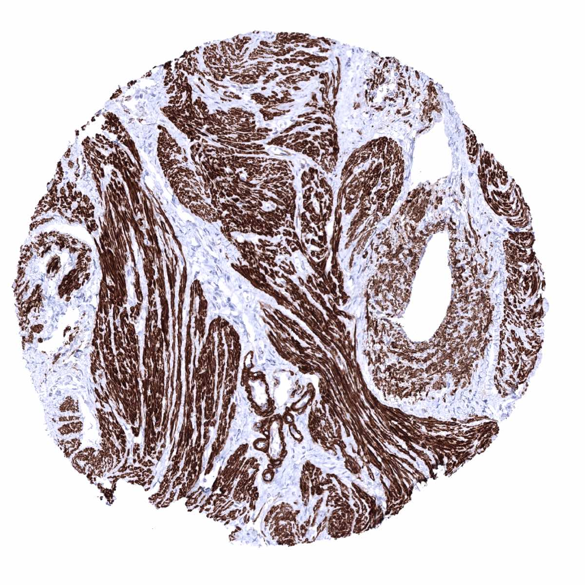
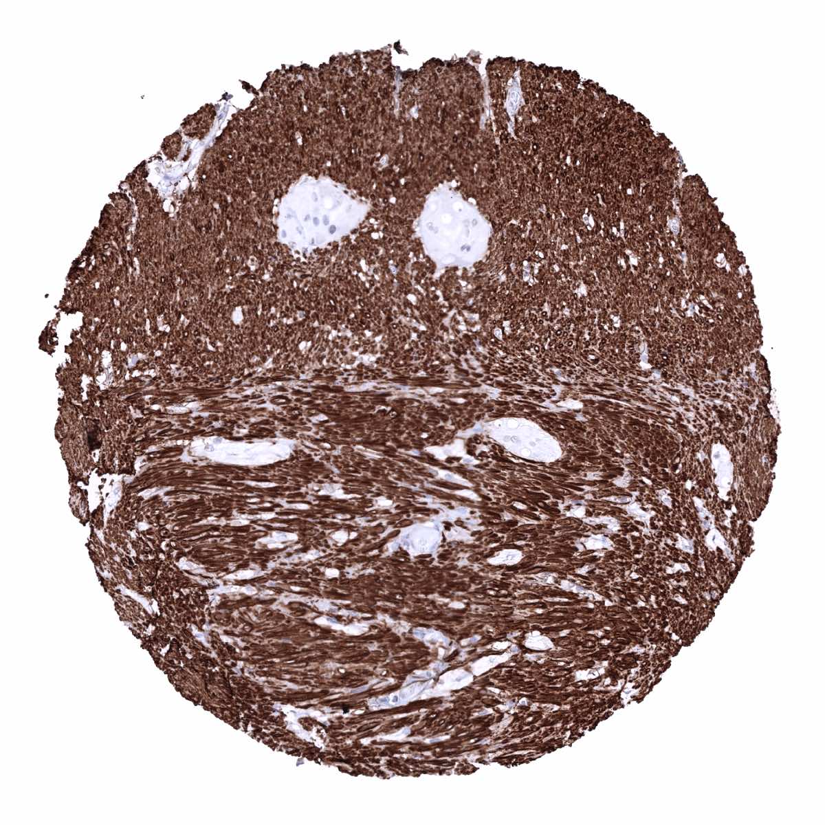
Uterus, myometrium - Strong caldesmon-h positivity of all smooth muscle cells (Caldesmon-h immunohistochemistry)_