
Login / Register
MS Validated Antibodies - Logo
  • About us
  • Products
    • Primary antibodies
    • Gallery
  • Distributors
  • Validation
    • Tissue Microarrays
  • Publications
    • Large Scale Studies
    • Further Publications
    • Events
    • Quiz
  • Contact
  • About us
  • Products
    • Primary antibodies
    • Gallery
  • Distributors
  • Validation
    • Tissue Microarrays
  • Publications
    • Large Scale Studies
    • Further Publications
    • Events
    • Quiz
  • Contact
×

Normal Tissue Gallery BSEP

Path Created with Sketch.
Go to BSEP Cancer Tissue GalleryGo back to Product Page
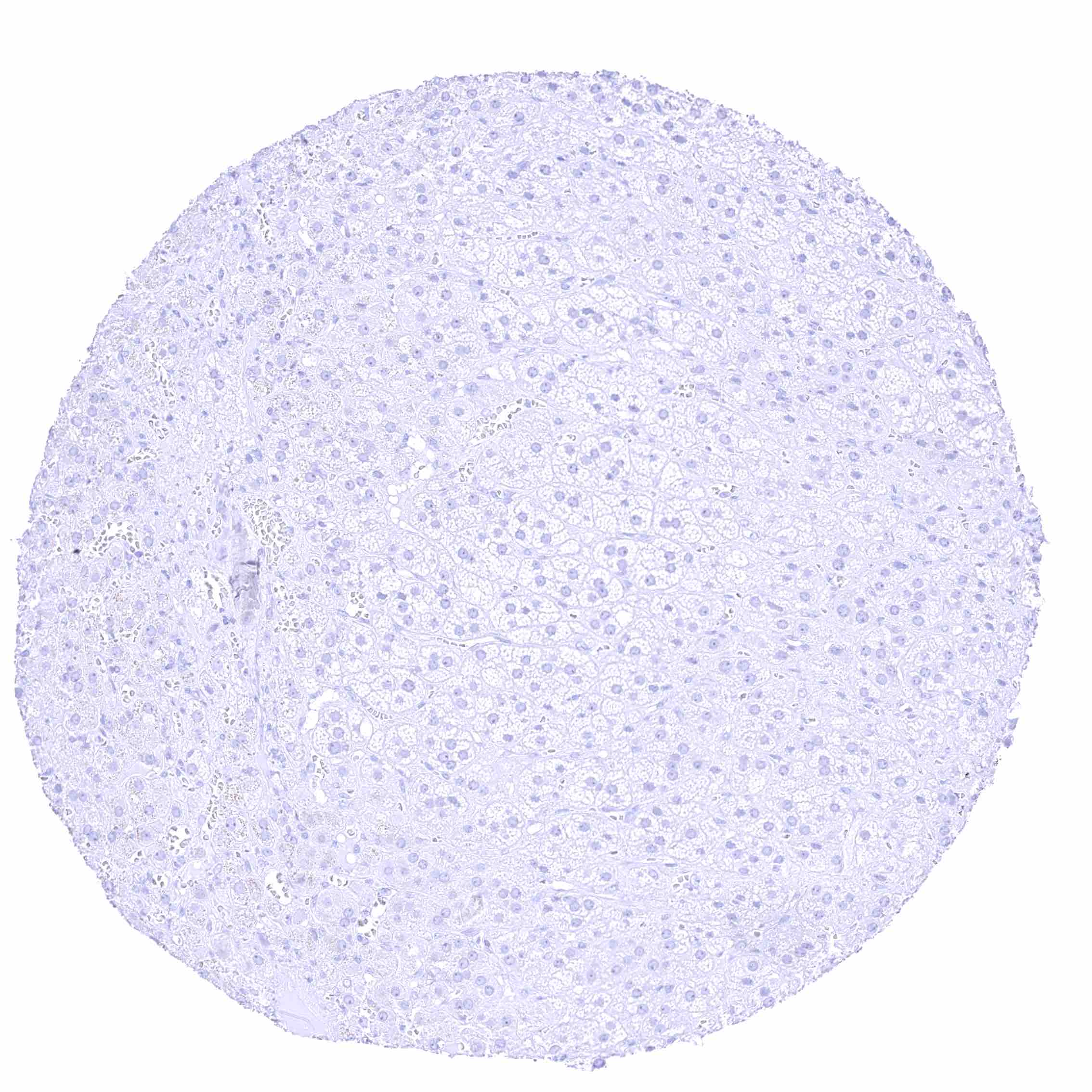
Adrenal gland
Adrenal gland
Aorta
Aorta
Appendix, mucosa
Appendix, mucosa
Appendix, muscular wall
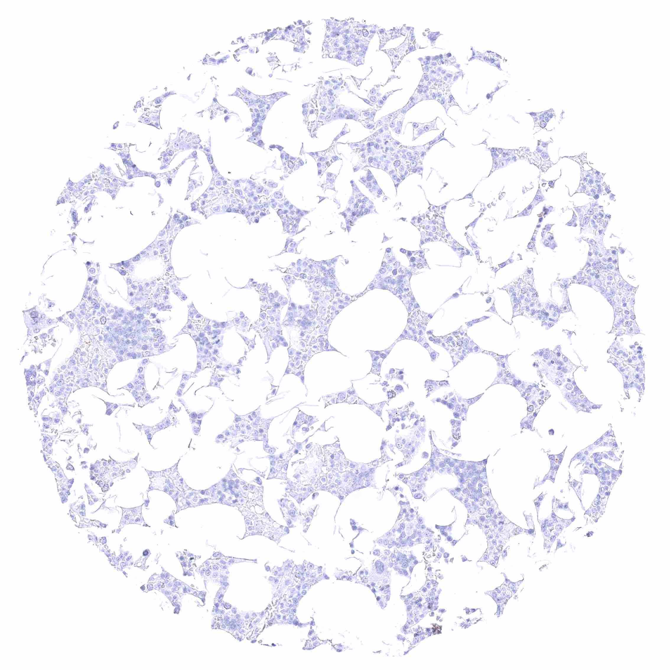
Bone marrow
Bone marrow
Breast
Breast
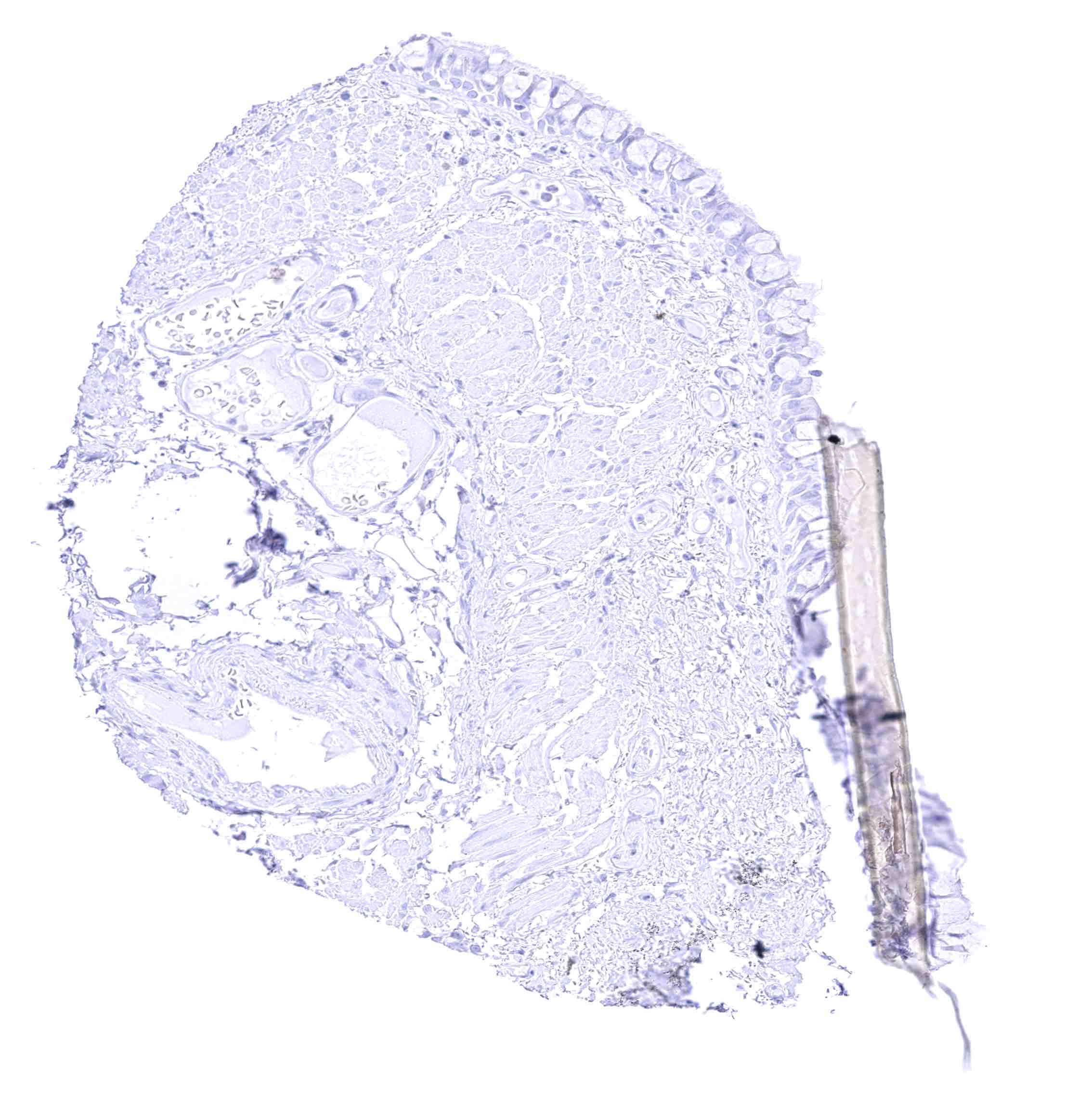
Bronchus, mucosa
Bronchus, mucosa
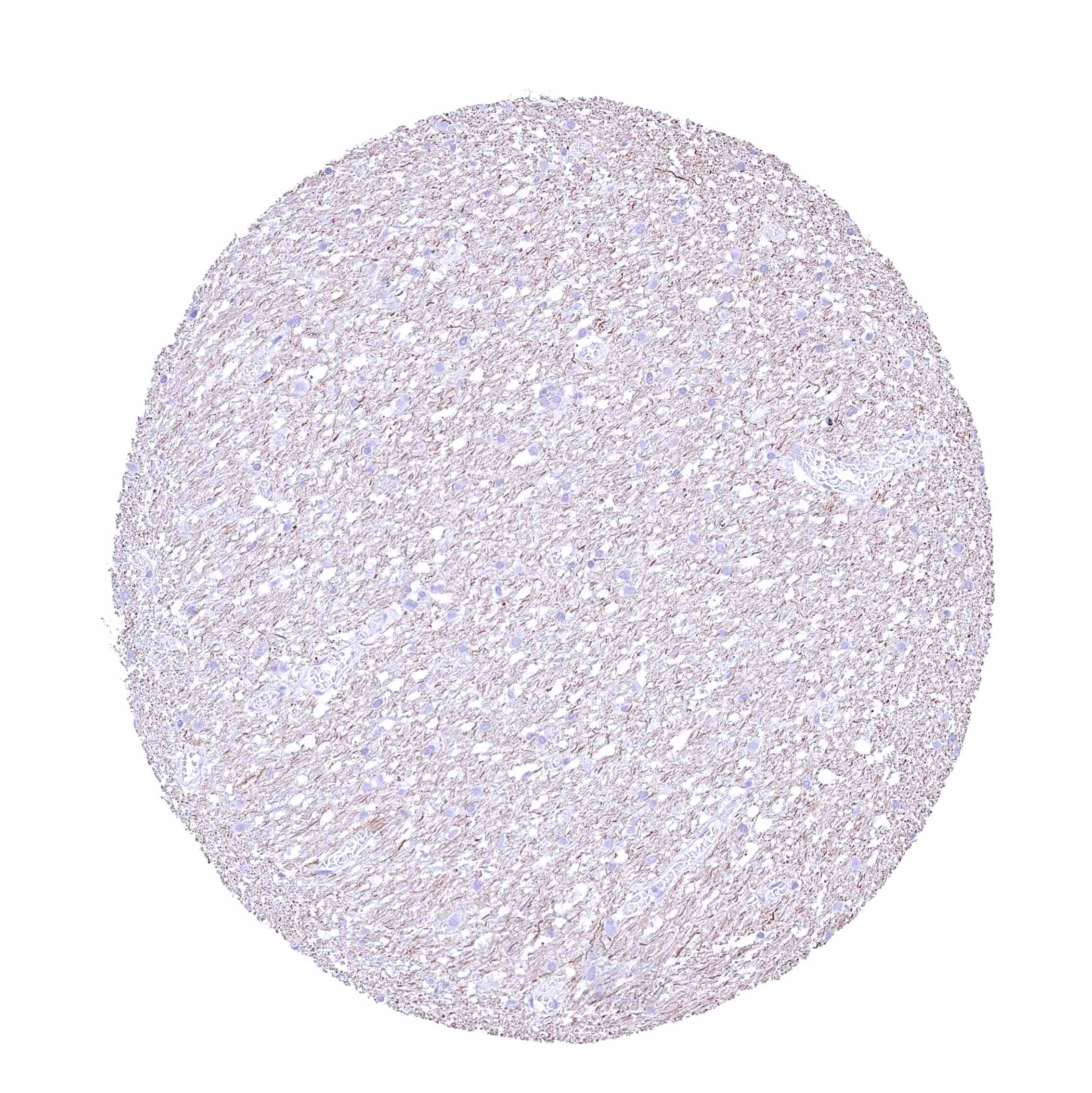
Cerebellum (white matter)
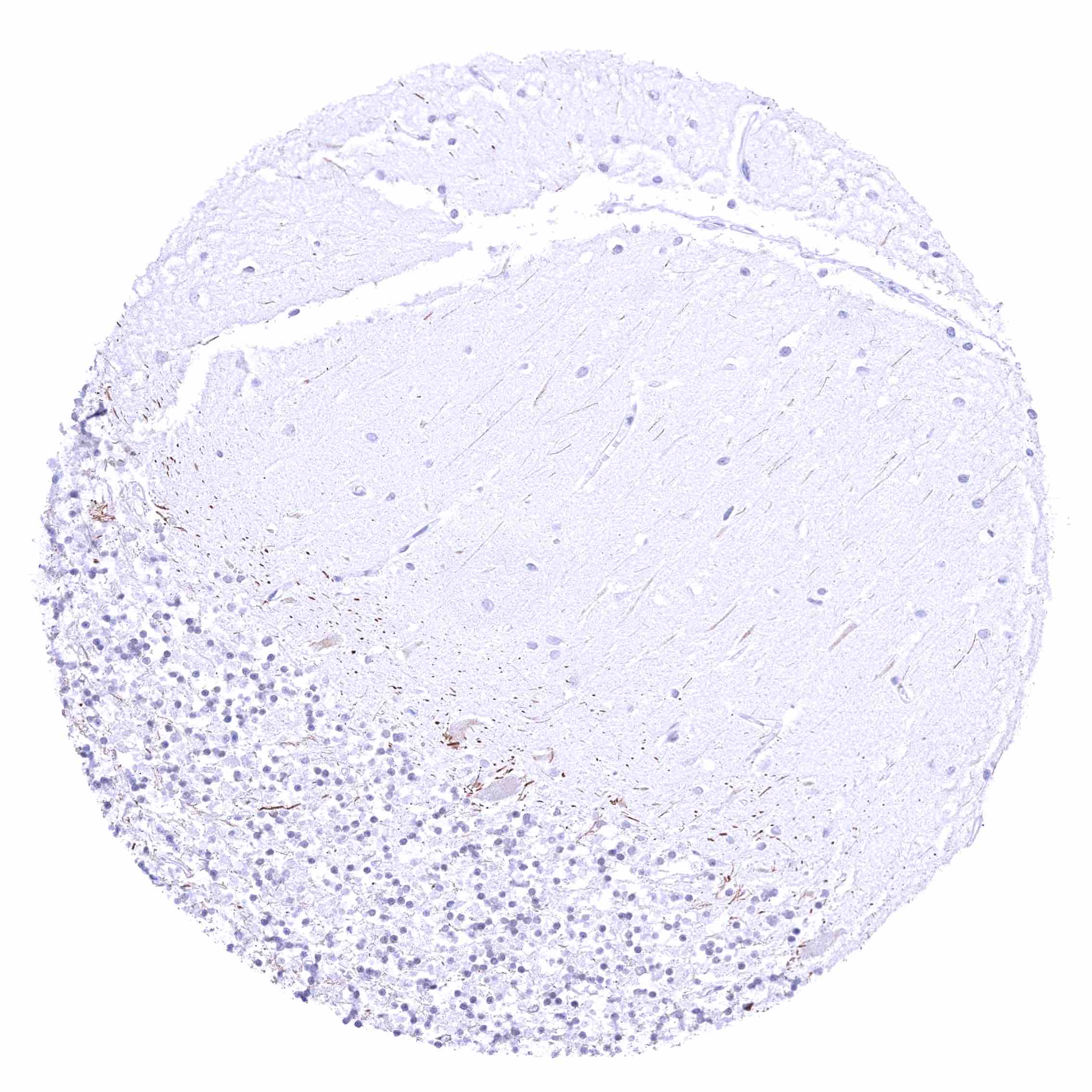
Cerebellum (molecular layer, Purkinje cell layer, granule cell layerA)
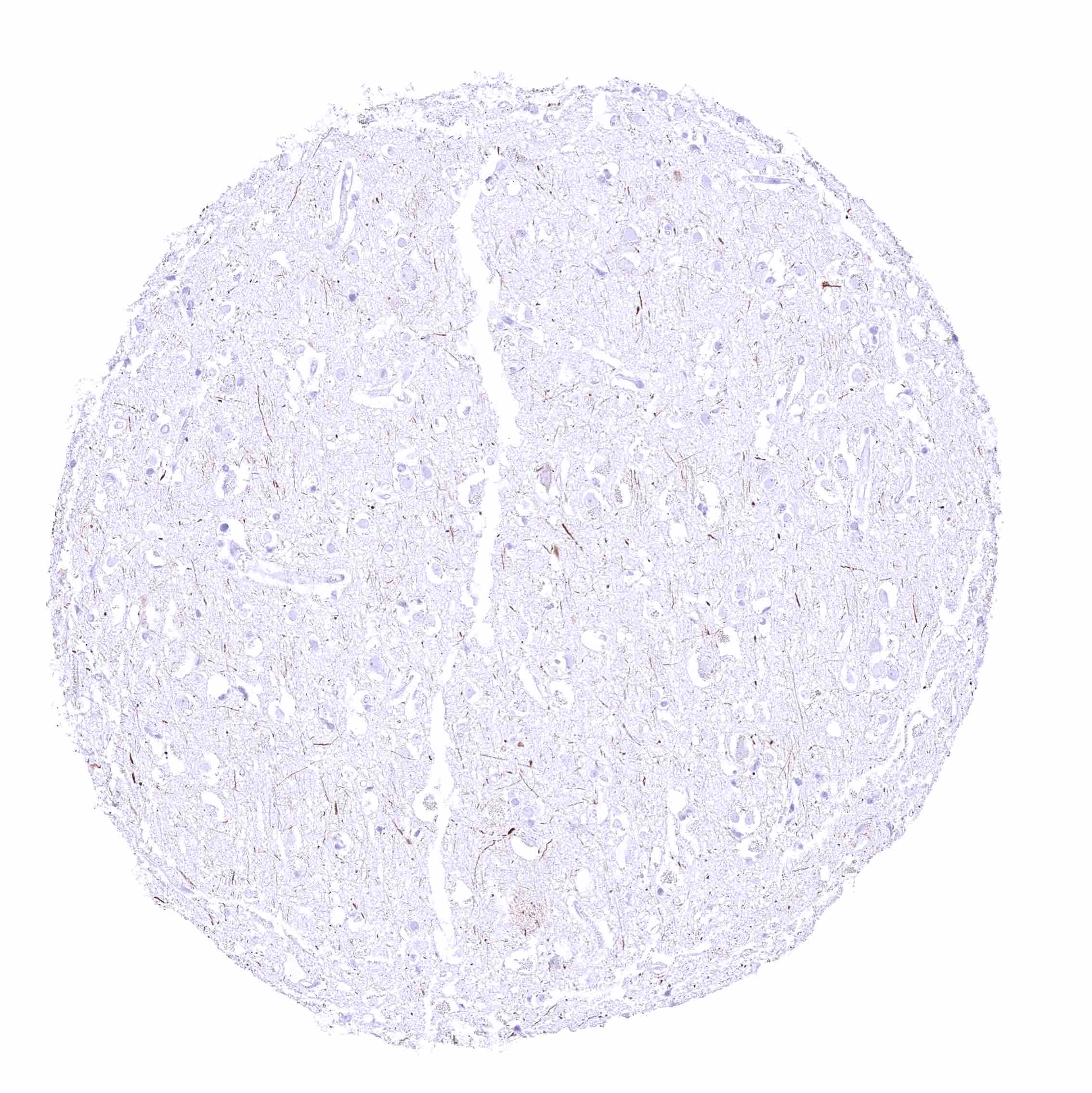
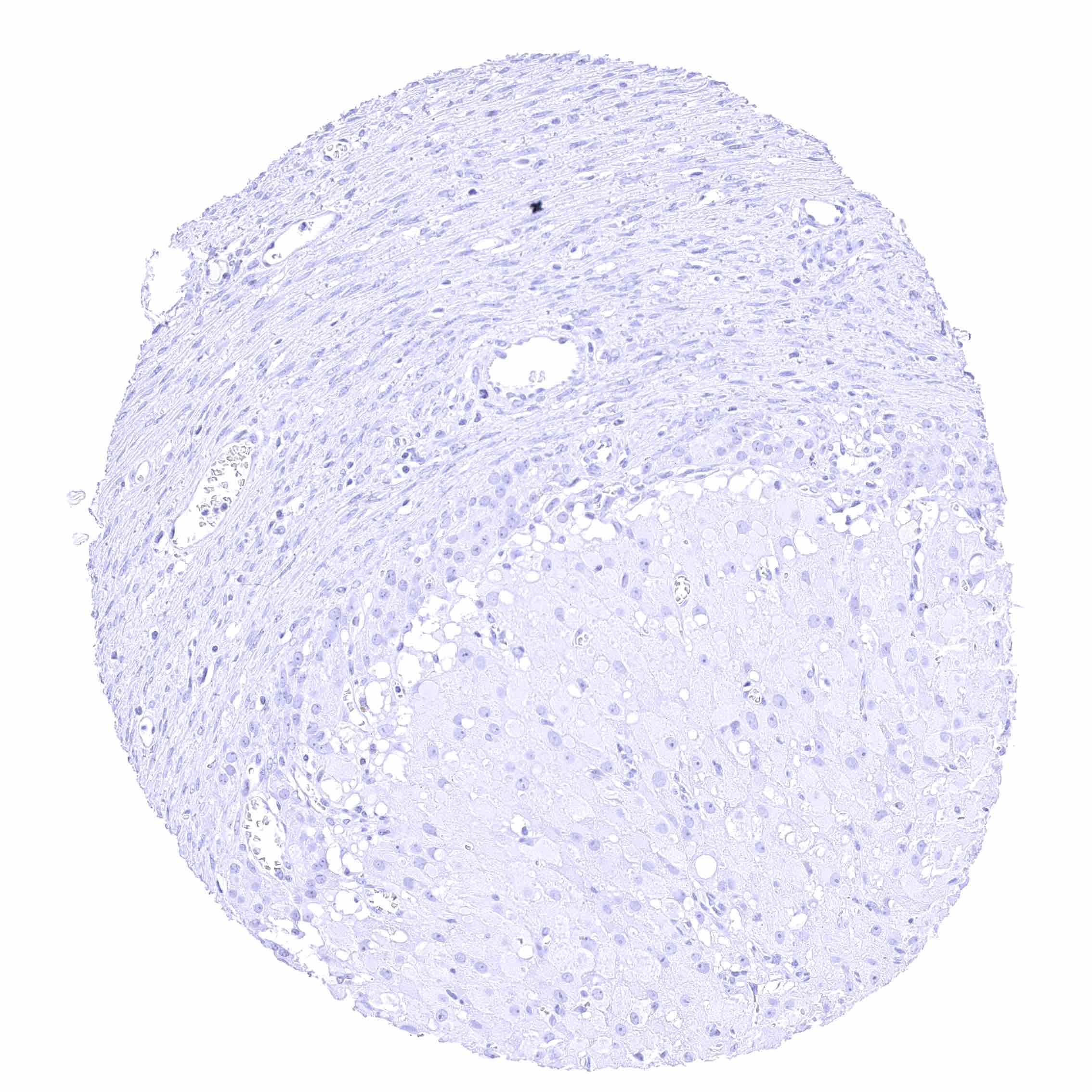
Cerebrum (grey matter)
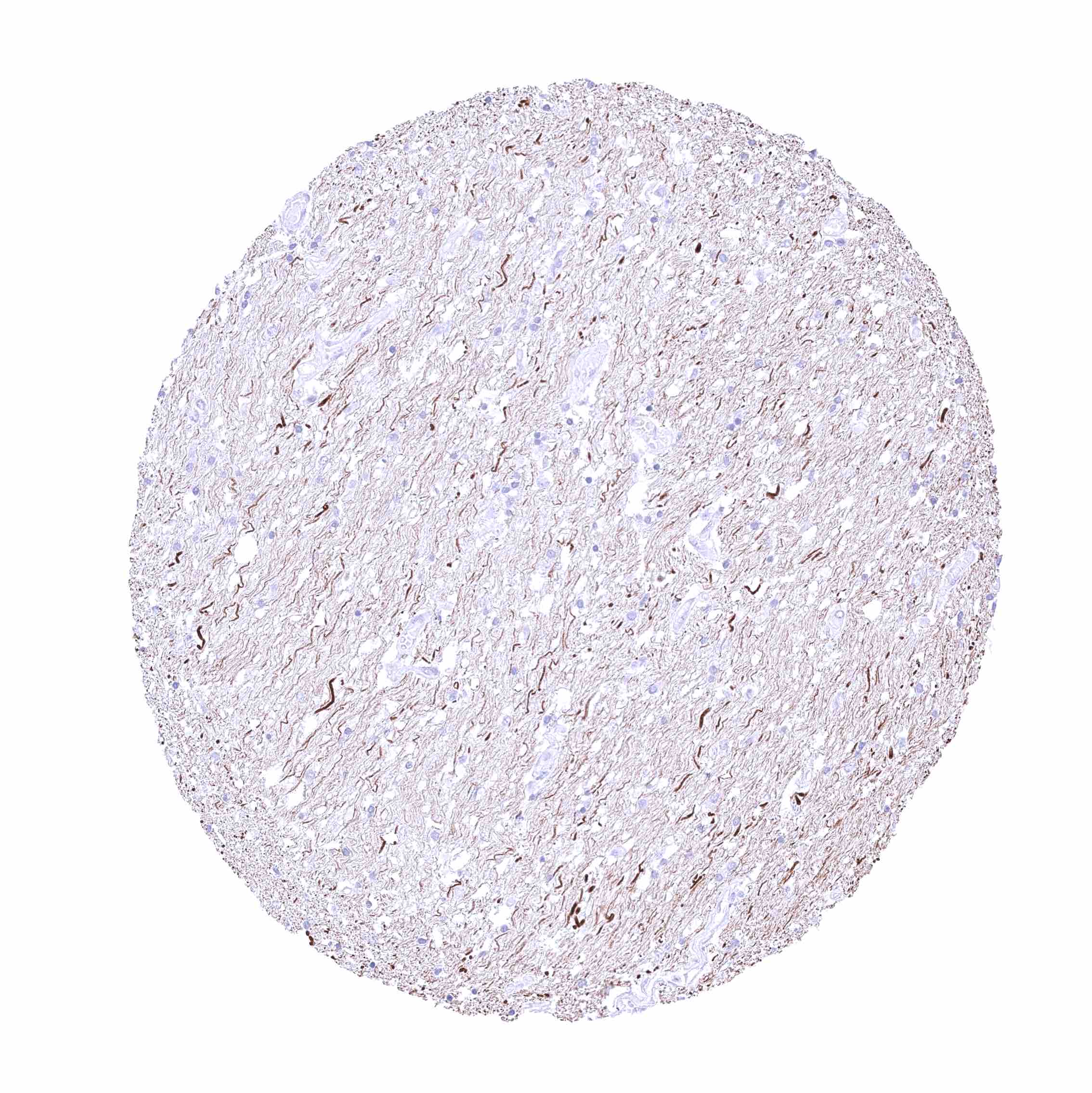
Cerebrum (white matter)
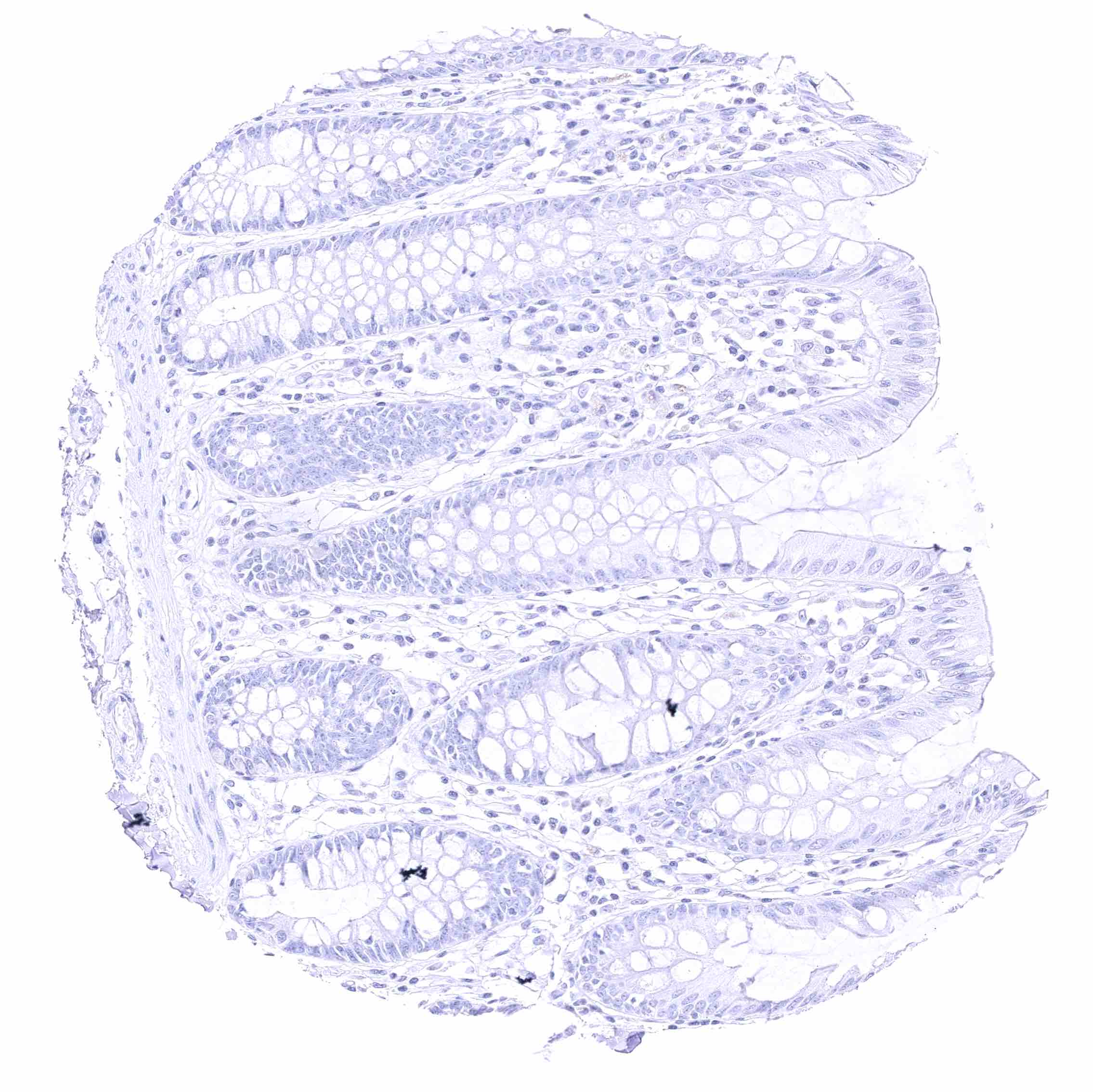
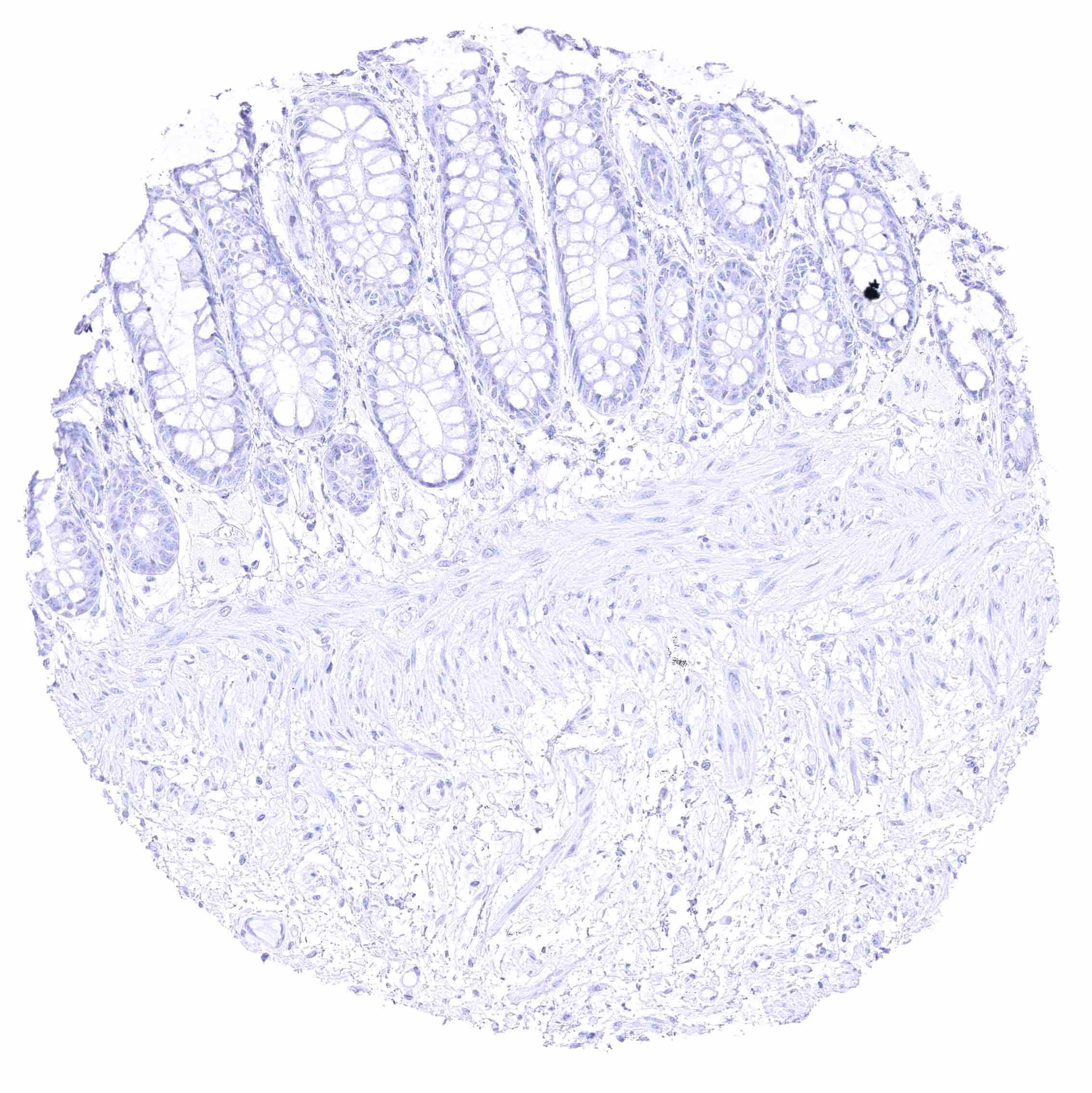
Colon descendens, mucosa
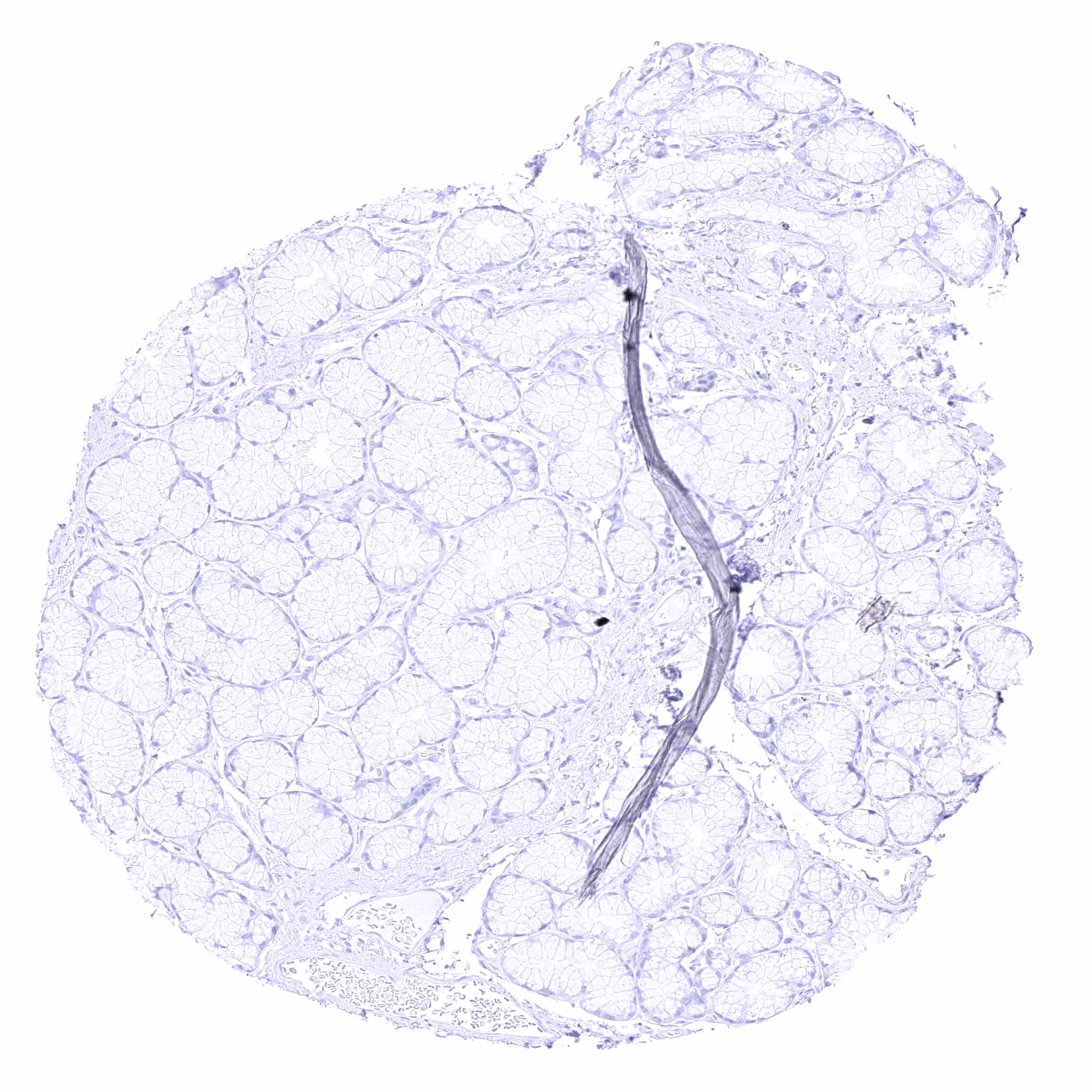
Duodenum, Brunner gland
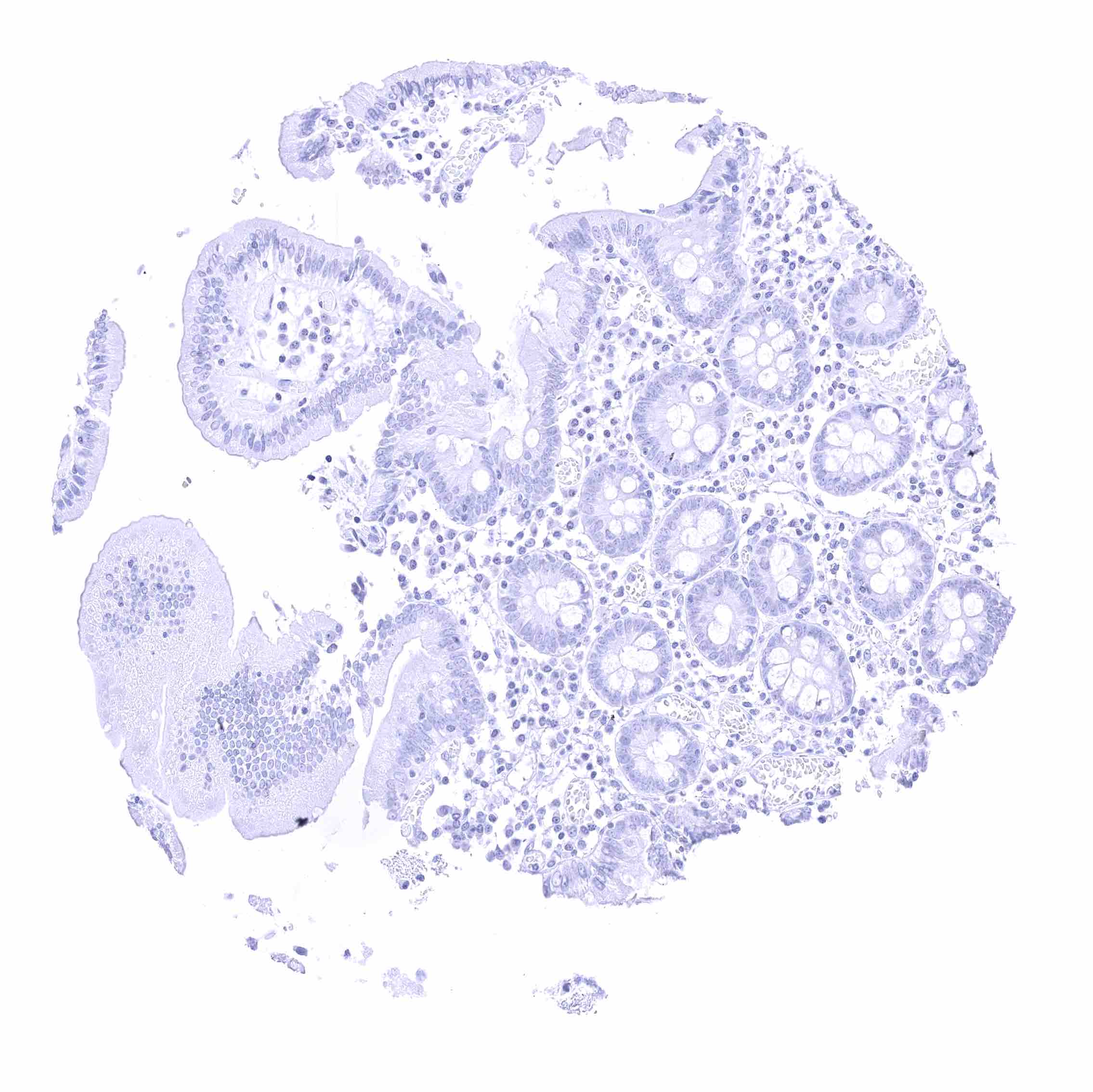
Duodenum, mucosa
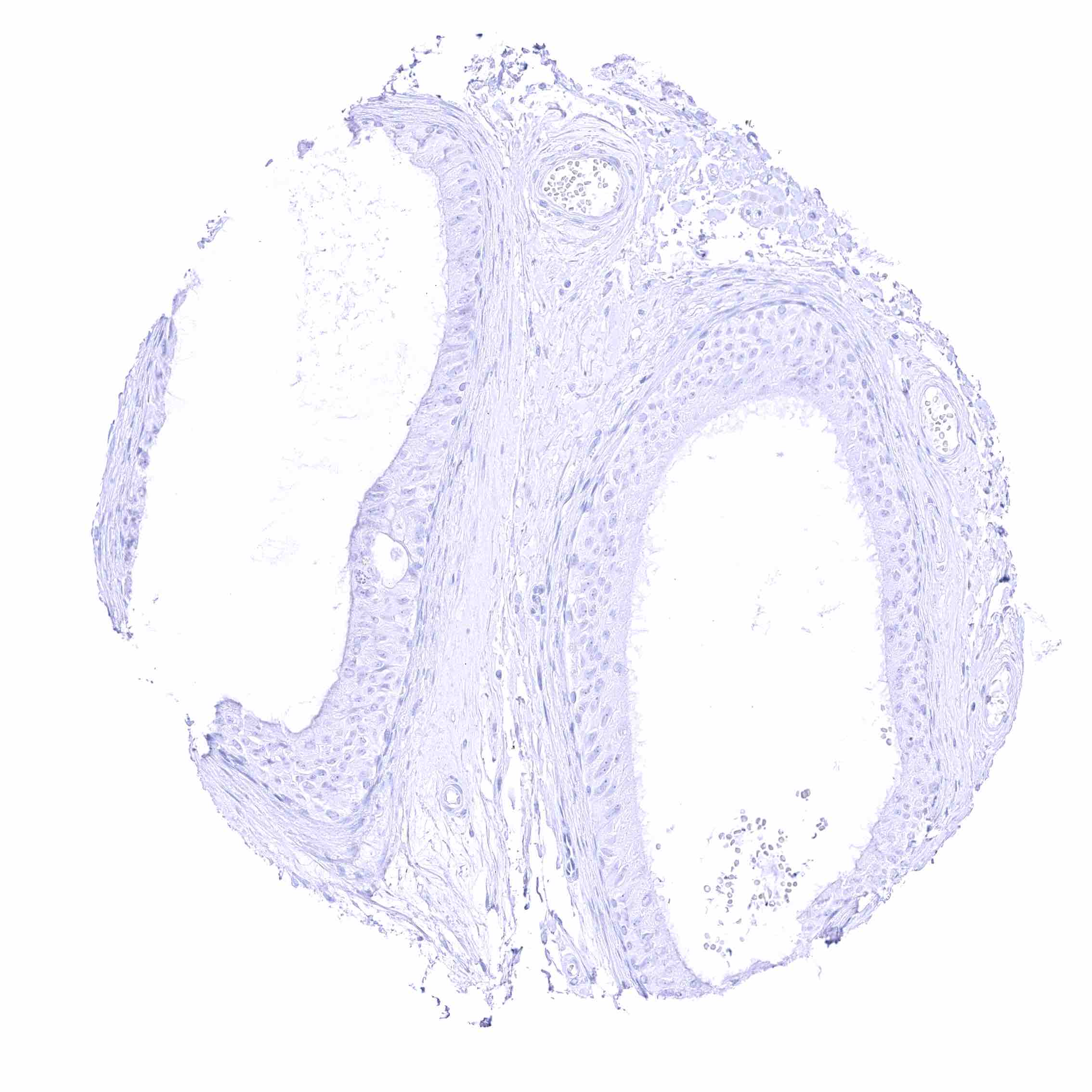
Duodenum, mucosa
Epididymis (Caput)
Epididymis (Caput)
Epididymis (Cauda)
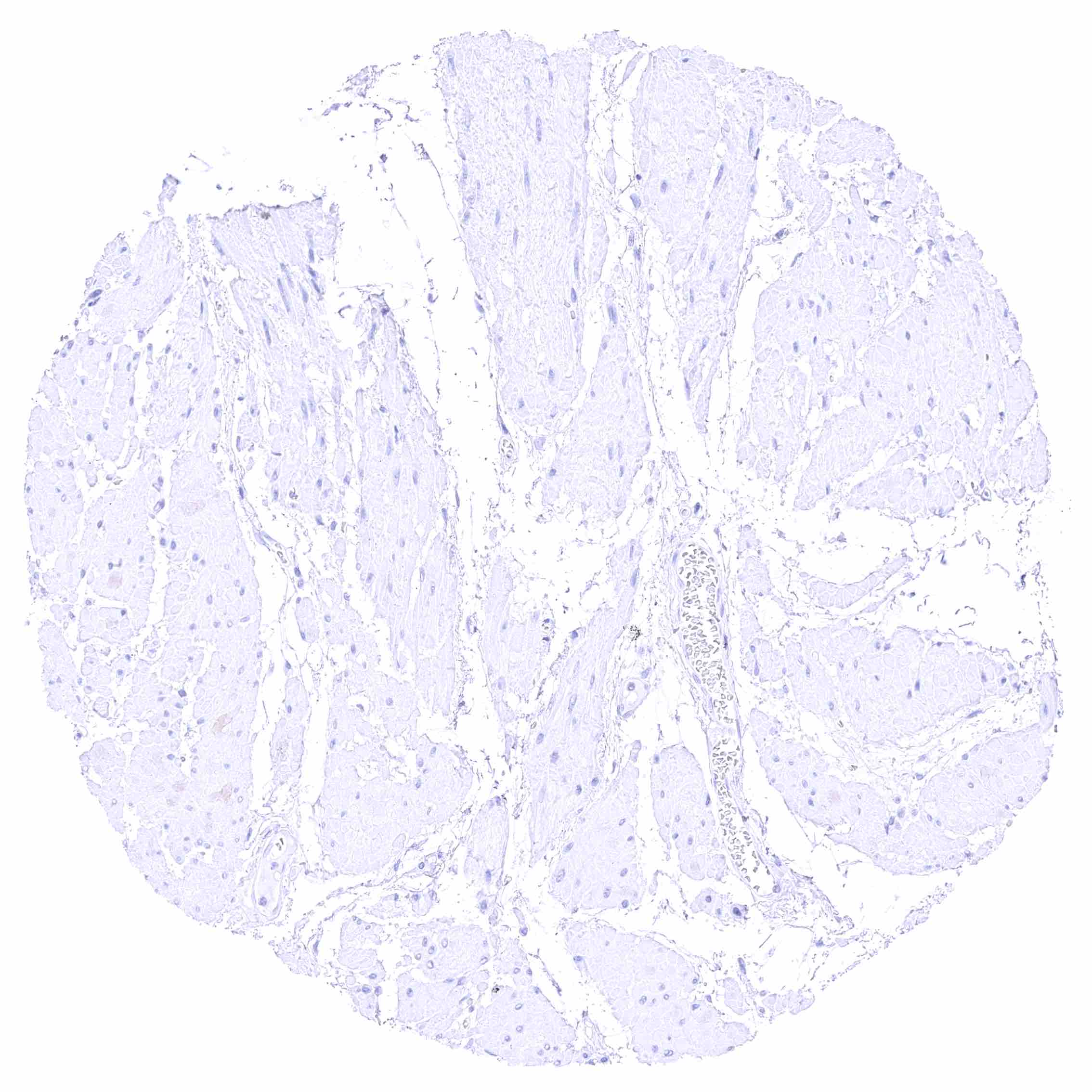
Epididymis (Cauda)
Esophagus, muscular wall
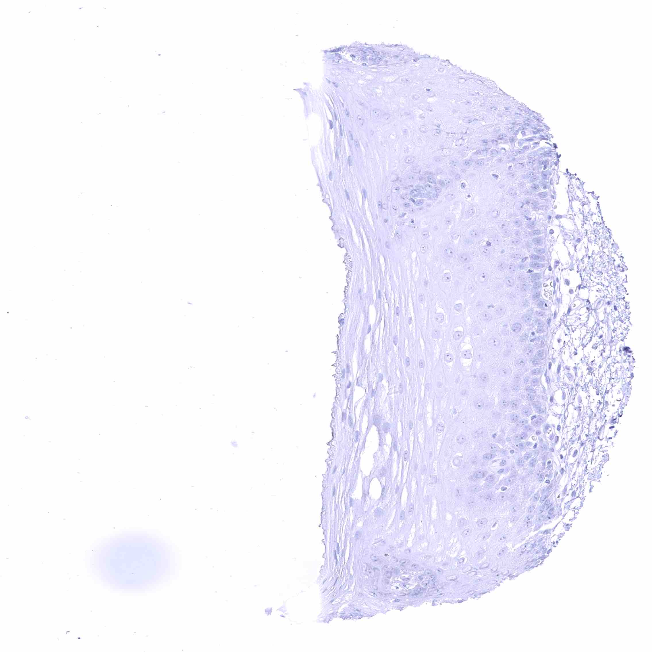
Esophagus, squamous epithelium
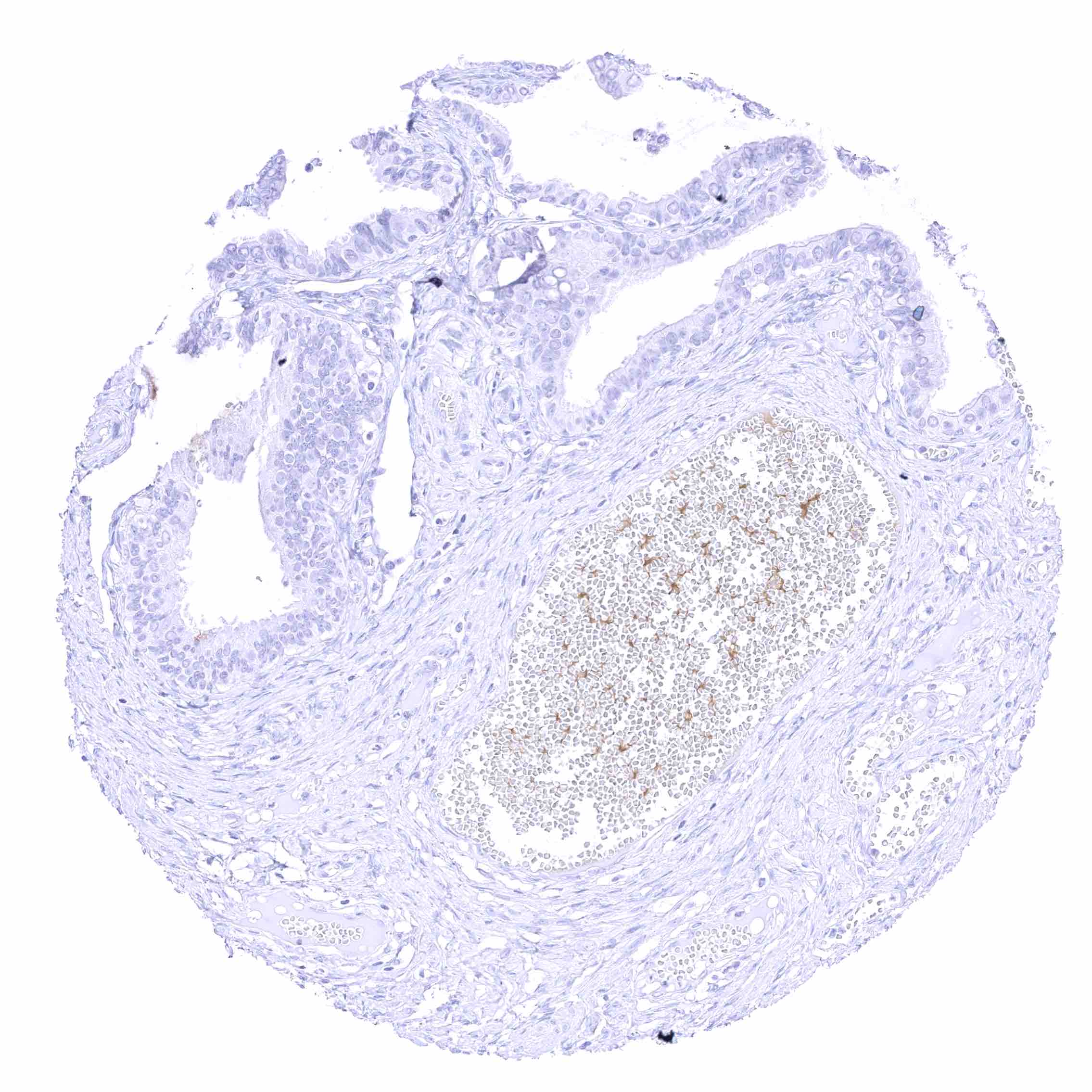
Fallopian tube, mucosa
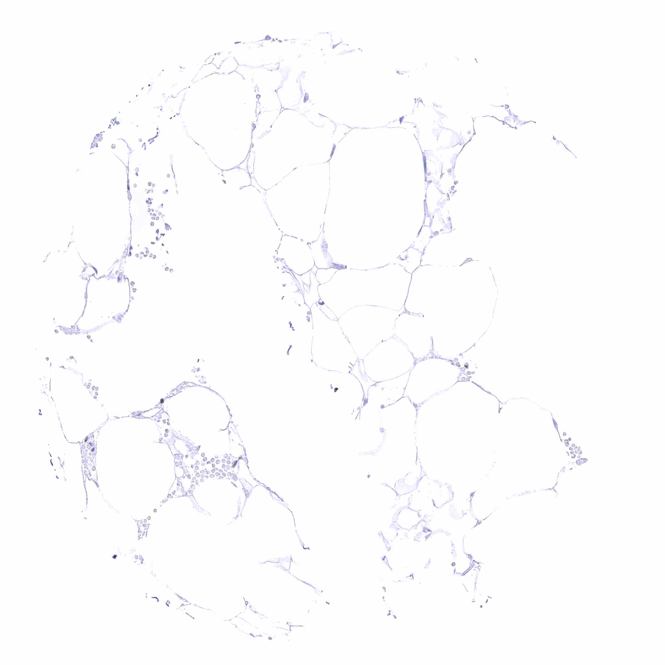
Fat
Fat
Gallbladder, epithelium
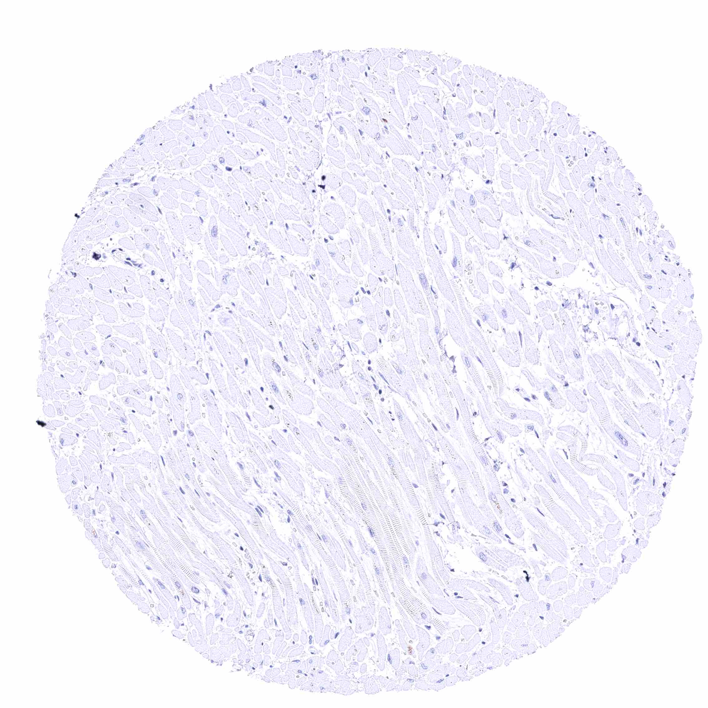
Heart muscle
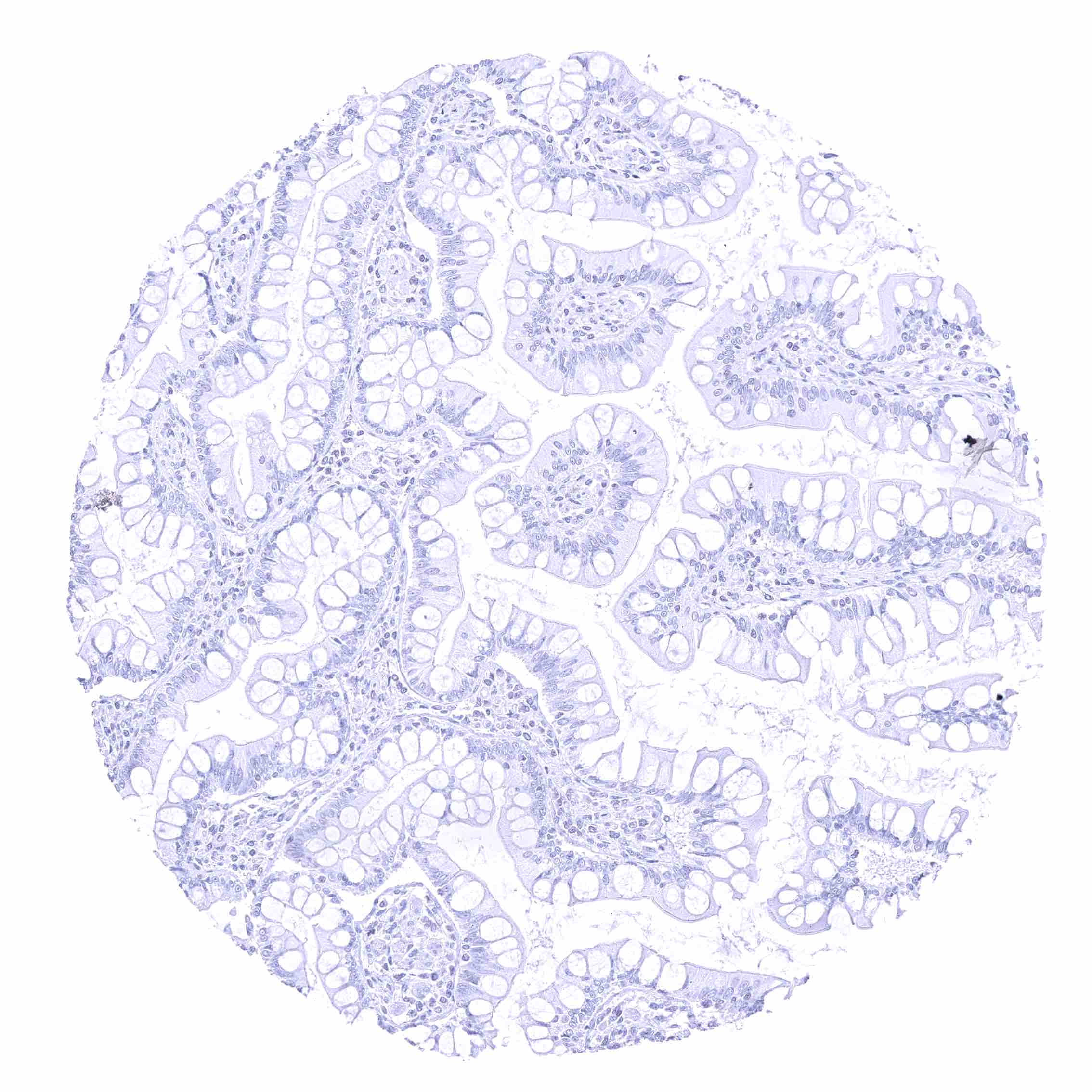
Heart muscle
Ileum, mucosa
Ileum, mucosa
Ileum, muscularis
Ileum, muscularis
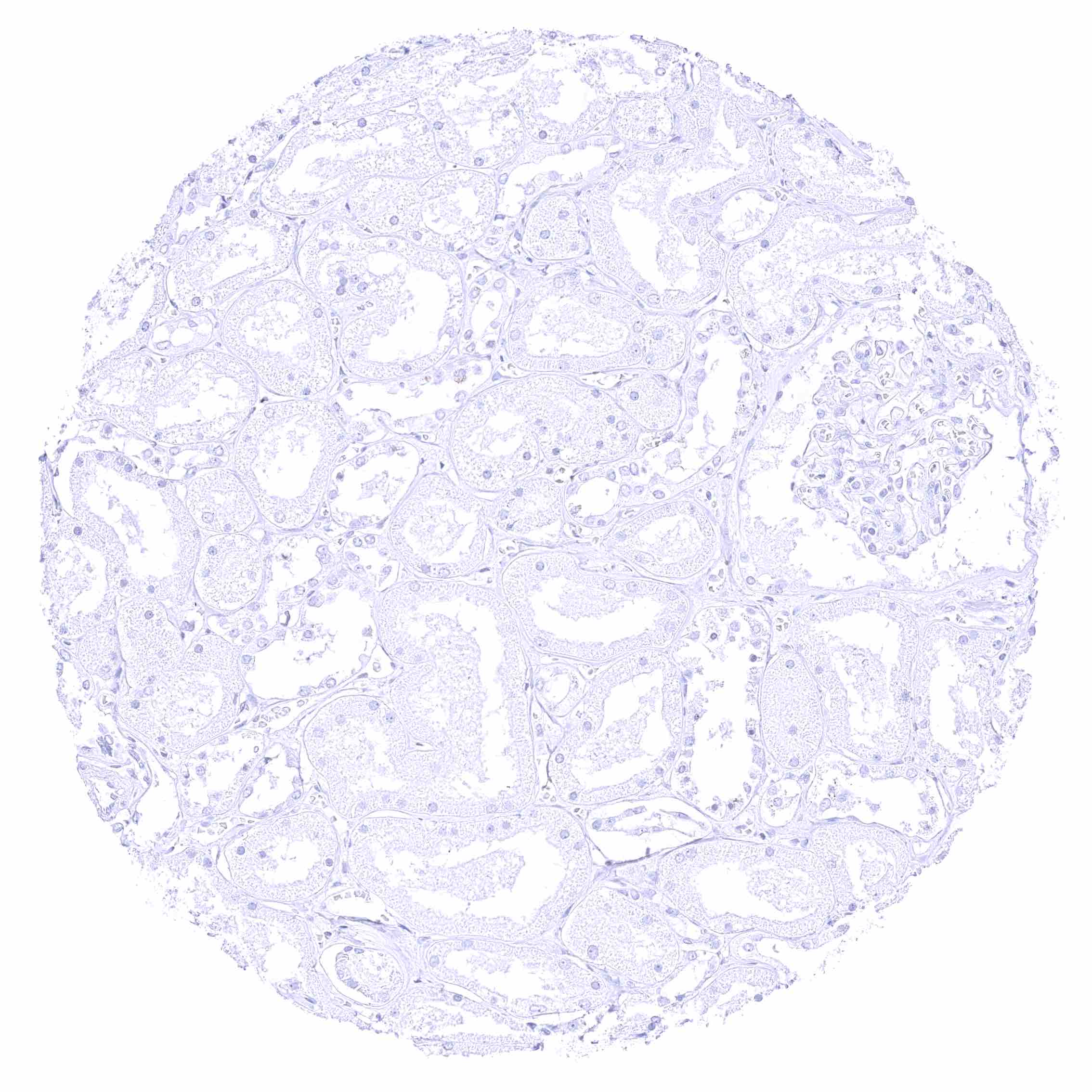
Kidney, cortex
Kidney, cortex
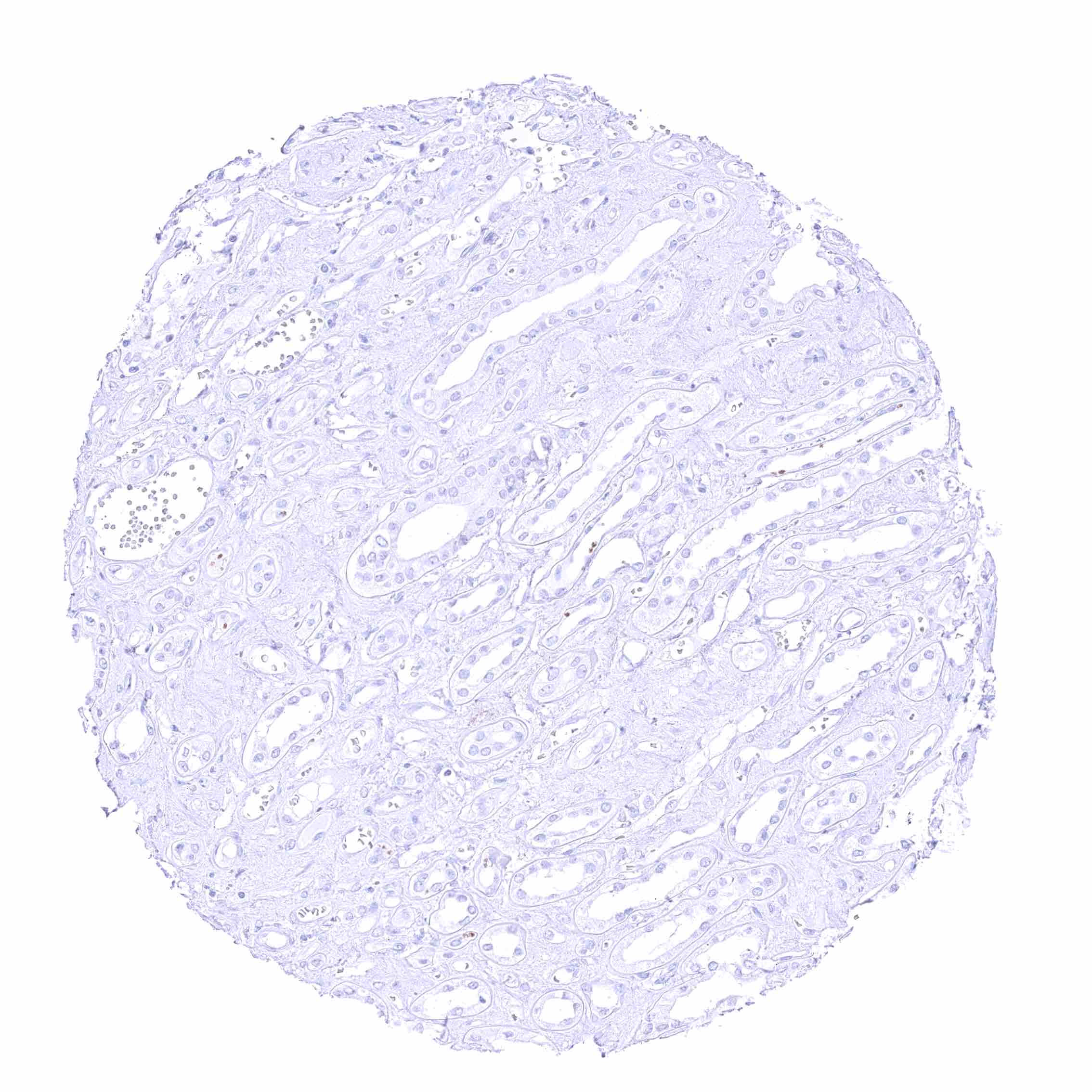
Kidney, medulla
Kidney, medulla
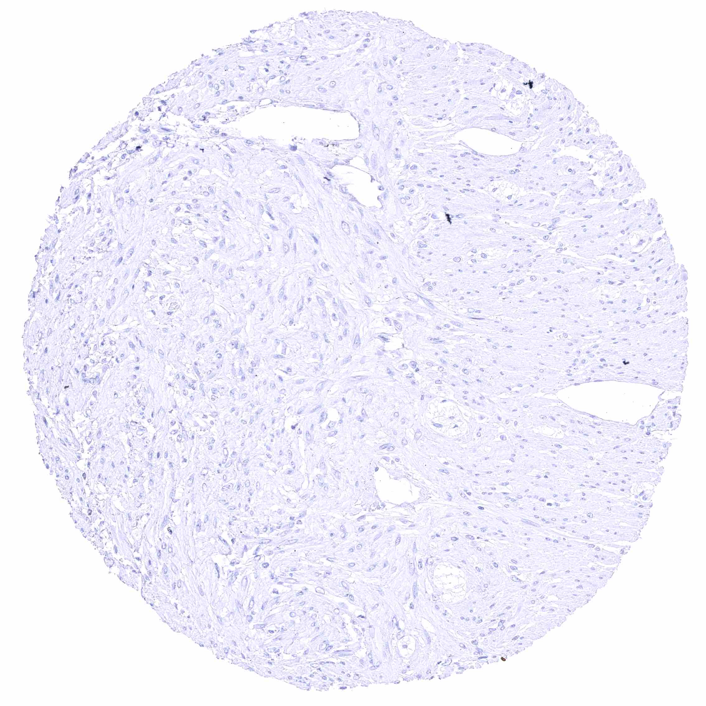
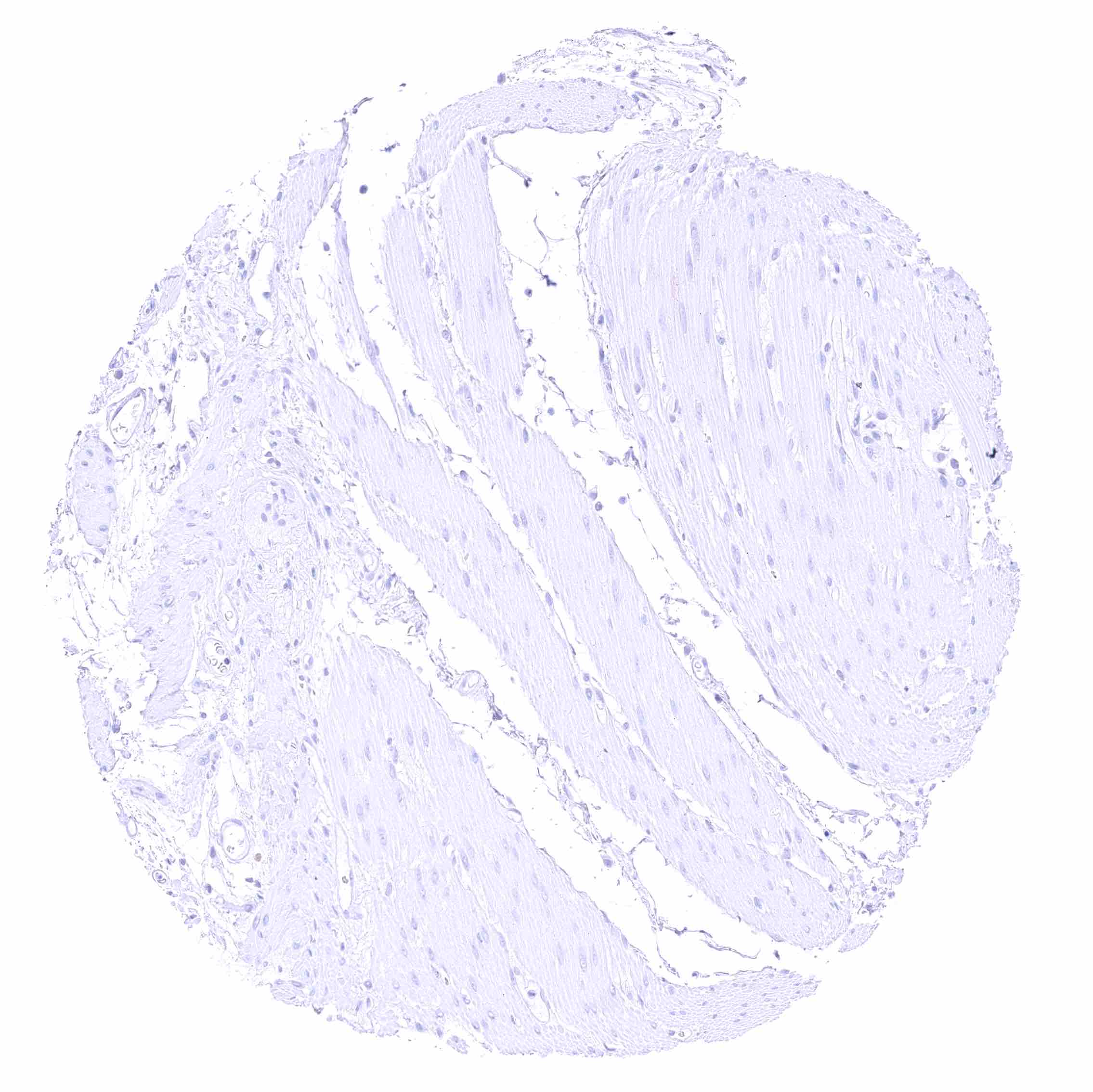
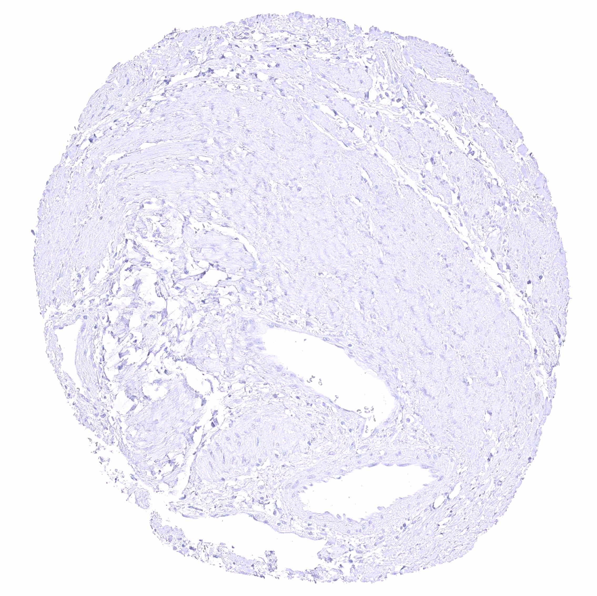
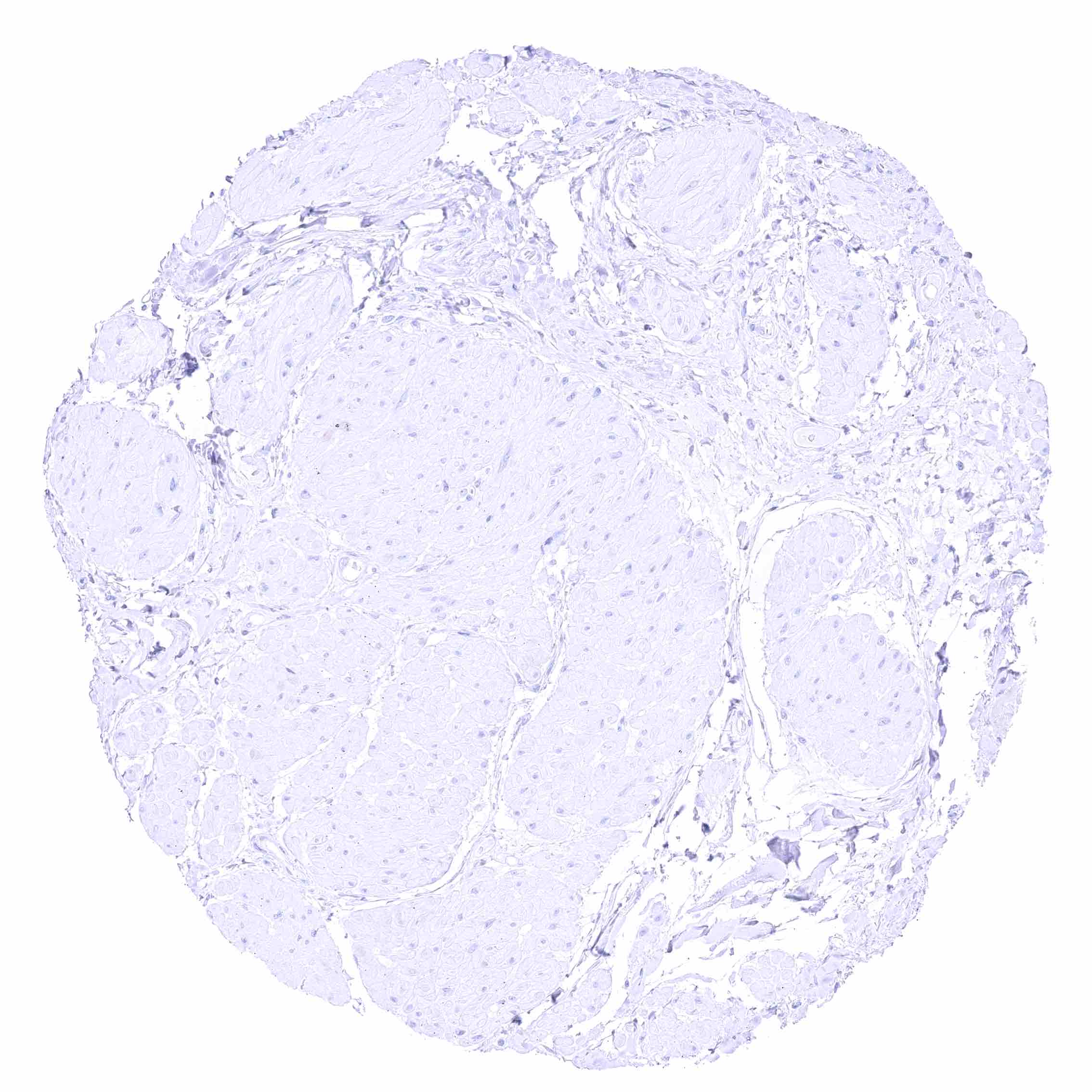
Kidney, pelvis, muscular wall
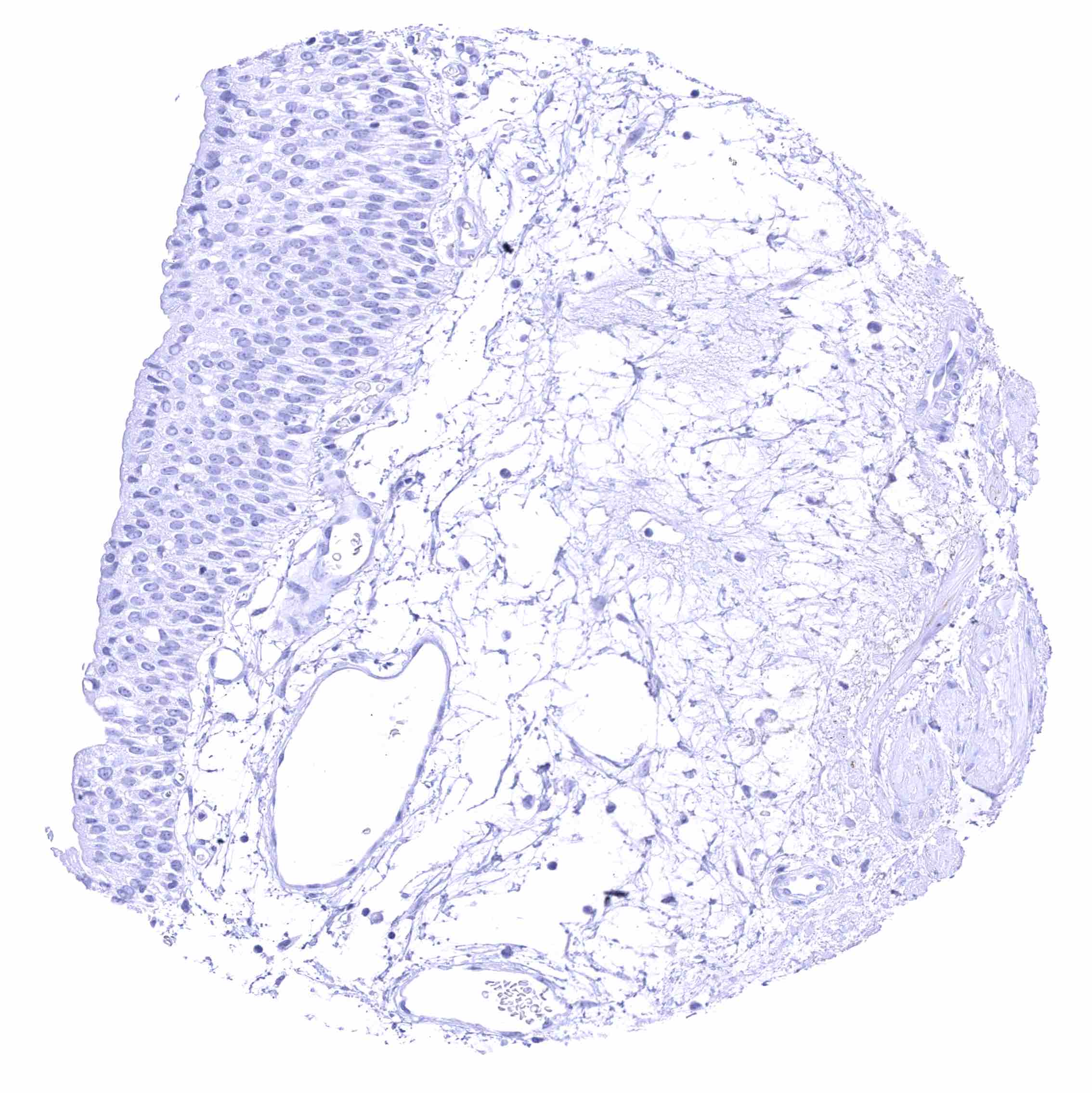
Kidney, pelvis, urothelium
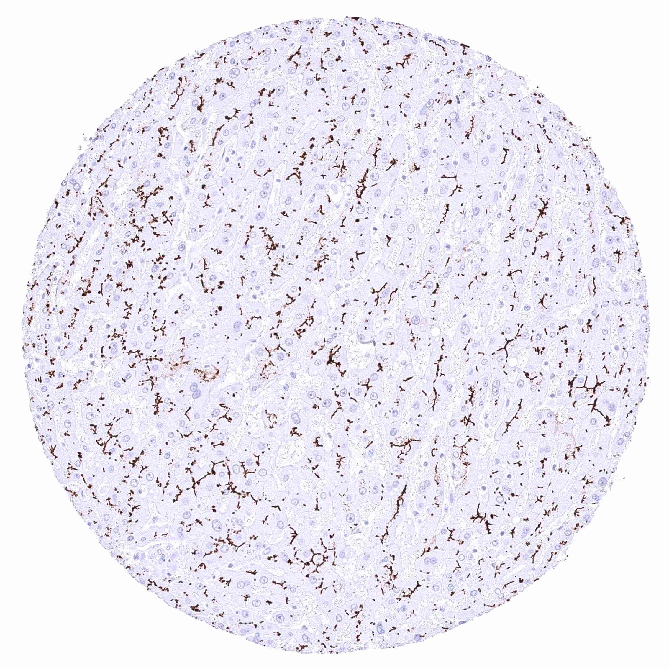
Liver – Intense membranous BSEP staining at the apical („bile-secreting“) pole of hepatocytes.
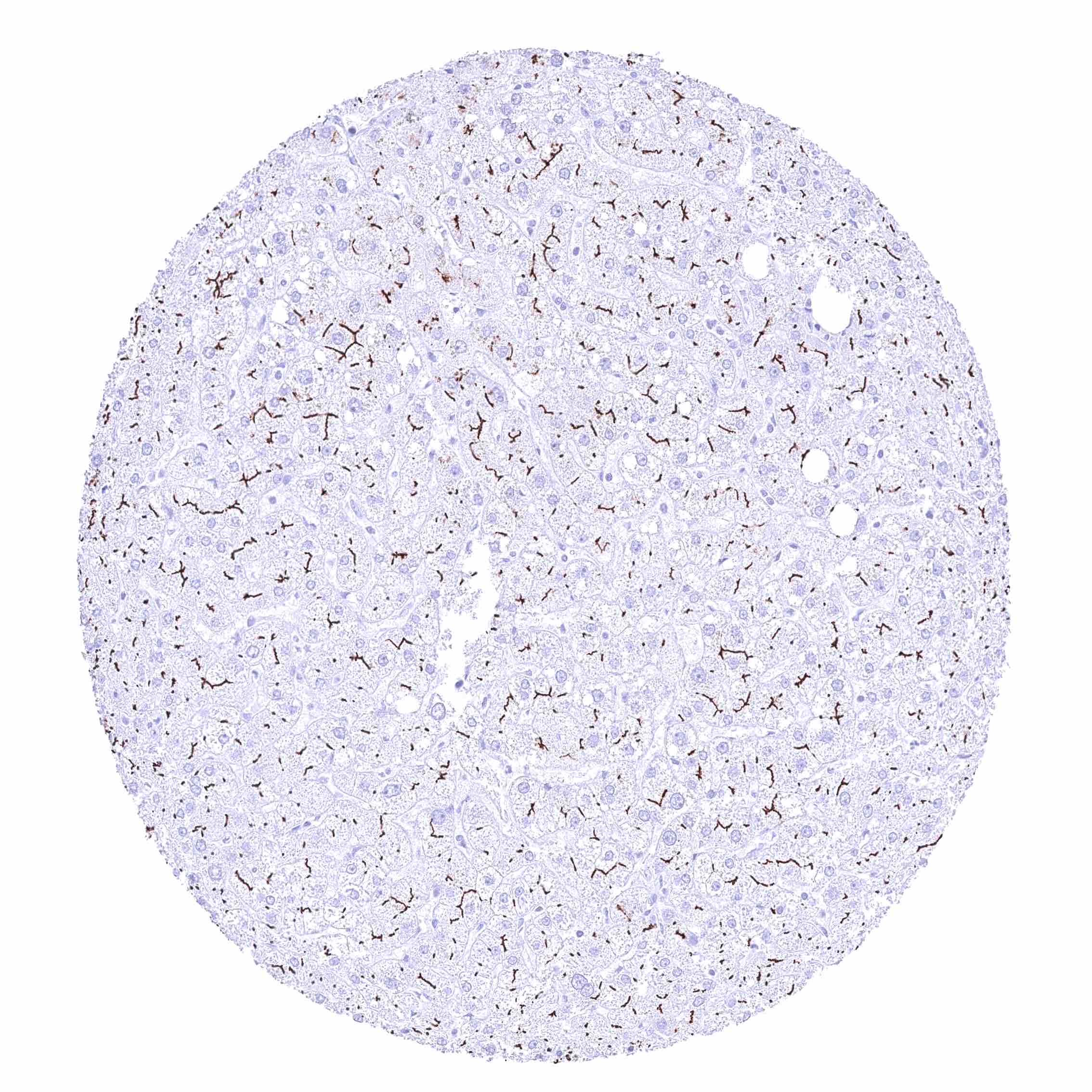
Liver – Strong membranous BSEP staining at the apical („bile-secreting“) pole of hepatocytes.
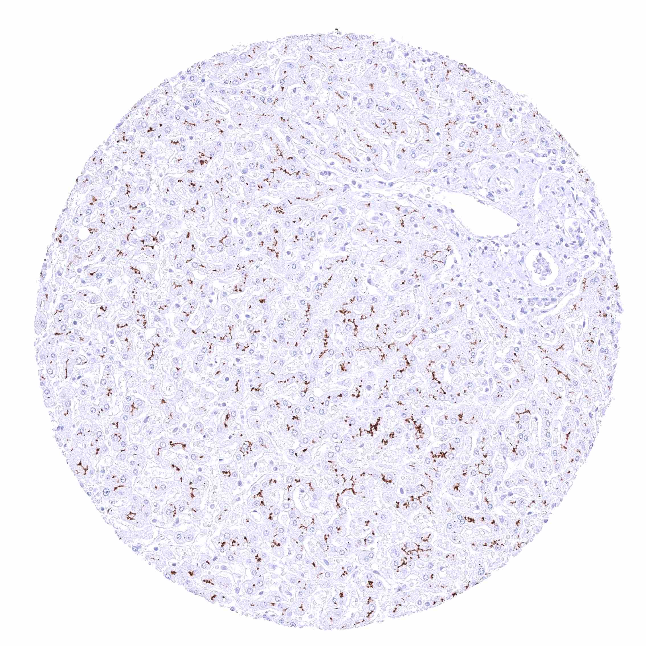
Liver – Strong membranous BSEP staining at the apical („bile-secreting“) pole of hepatocytes. There is a slight zonal variability of the staining intensity.
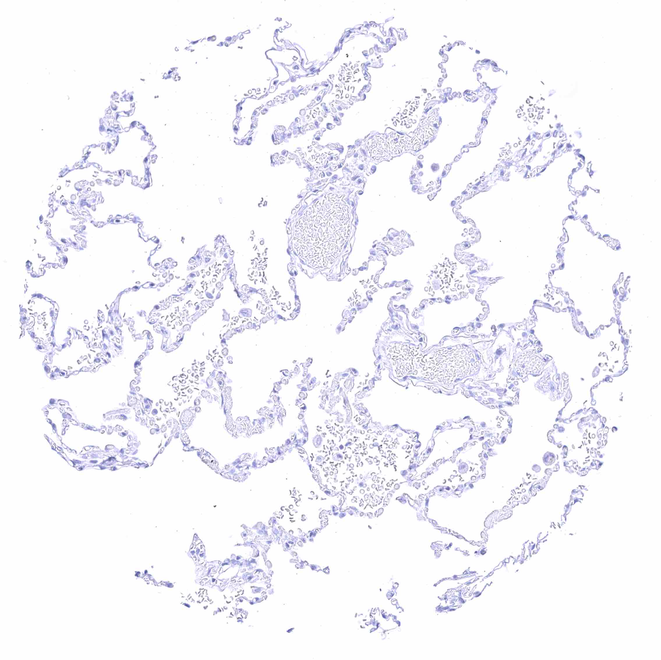
Lung
Lung
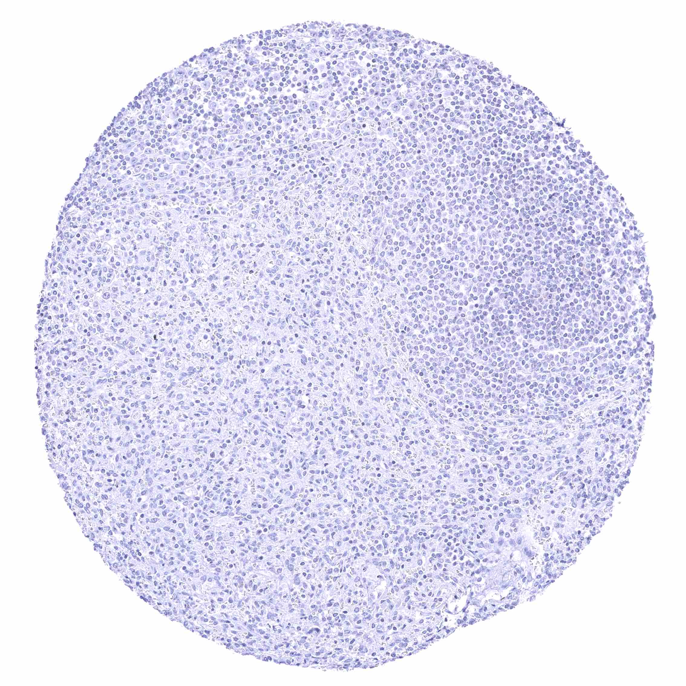
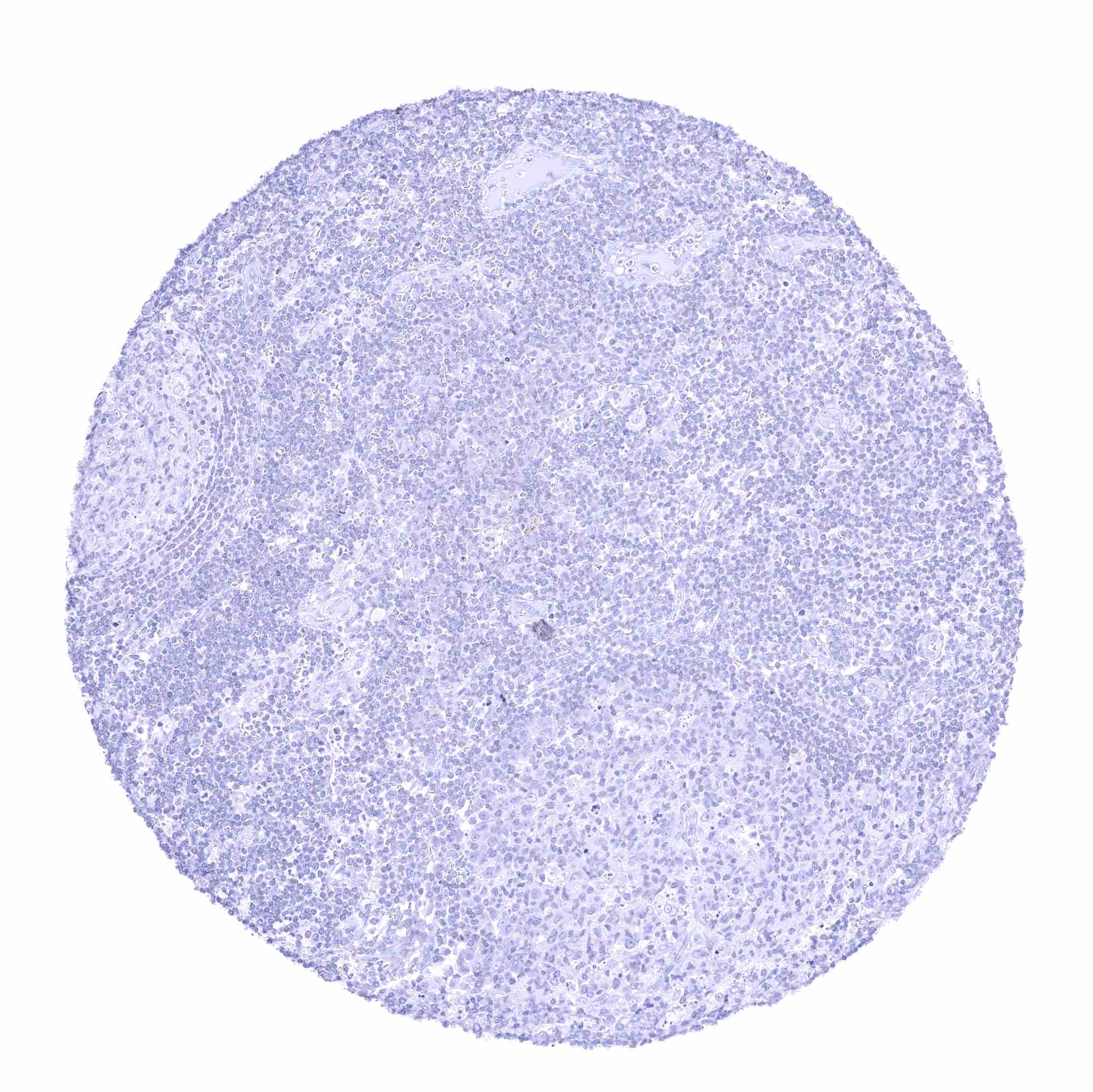
Lymph node
Lymph node
Ovary, corpus luteum
Ovary, stroma
Ovary, stroma
Pancreas – Acinar cells and excretory duct cells completely lack BSEP staining.
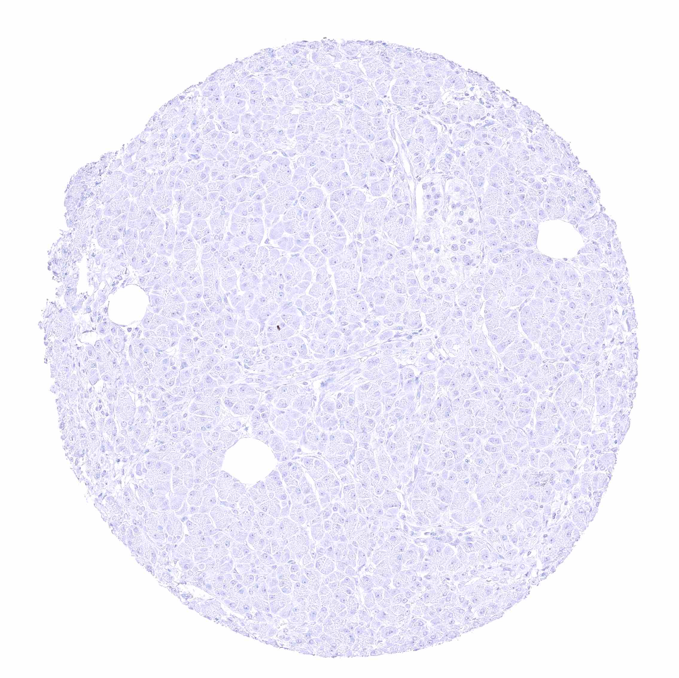
Pancreas – Acinar cells and Islet cells completely lack BSEP staining.
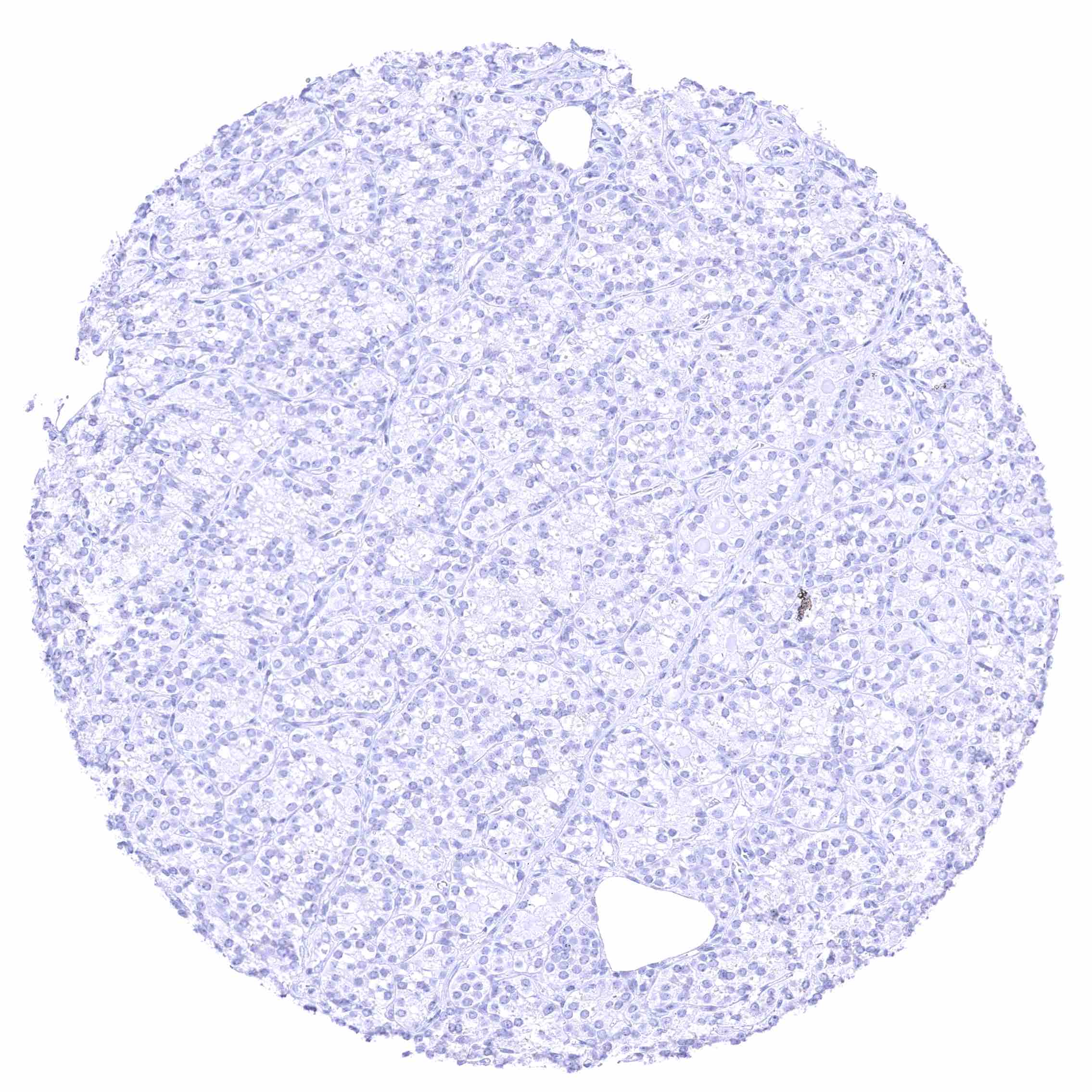
Parathyroid gland
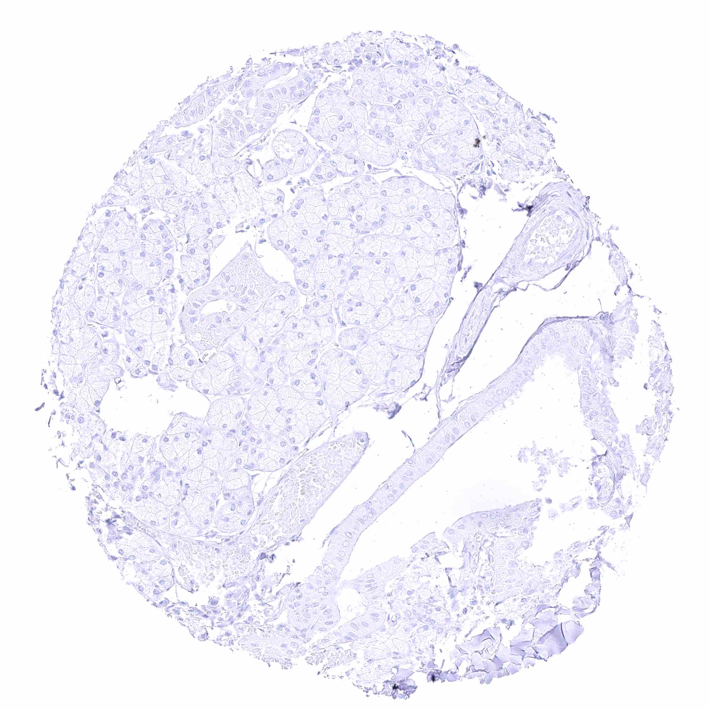
Parathyroid gland
Parotid gland
Parotid gland
Pituitary gland, anterior lobe
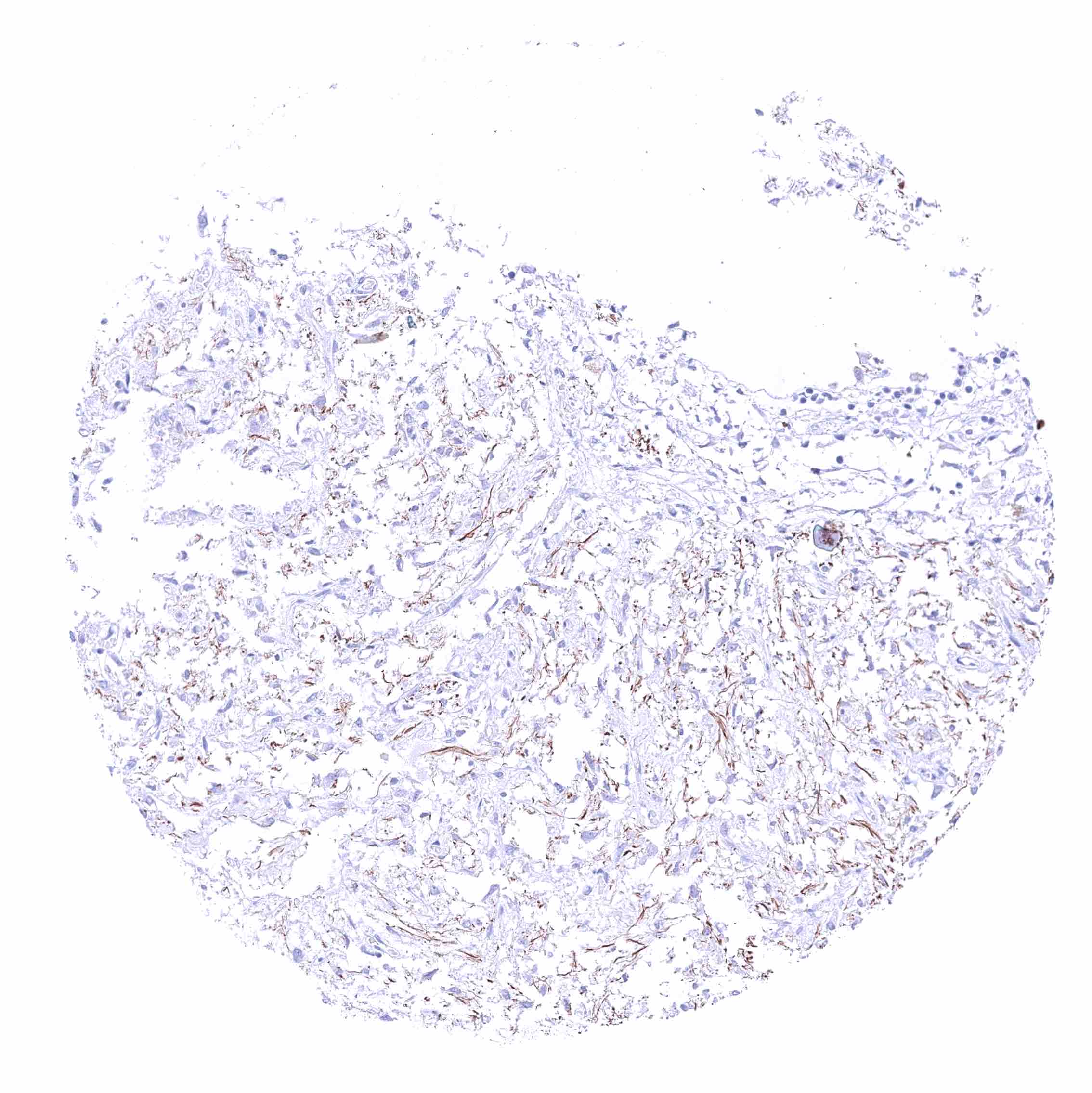
Pituitary gland, posterior lobe
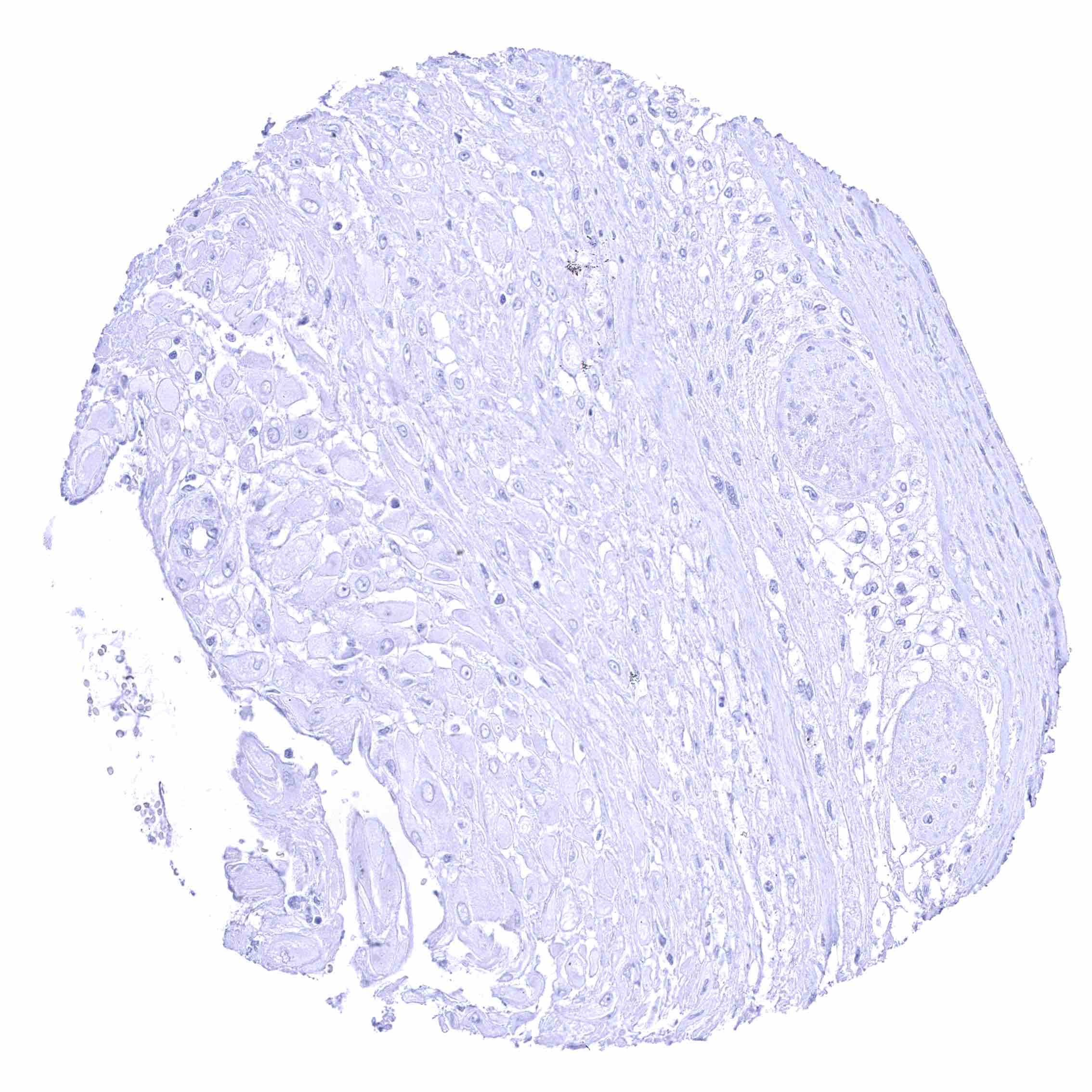
Placenta (chorion)
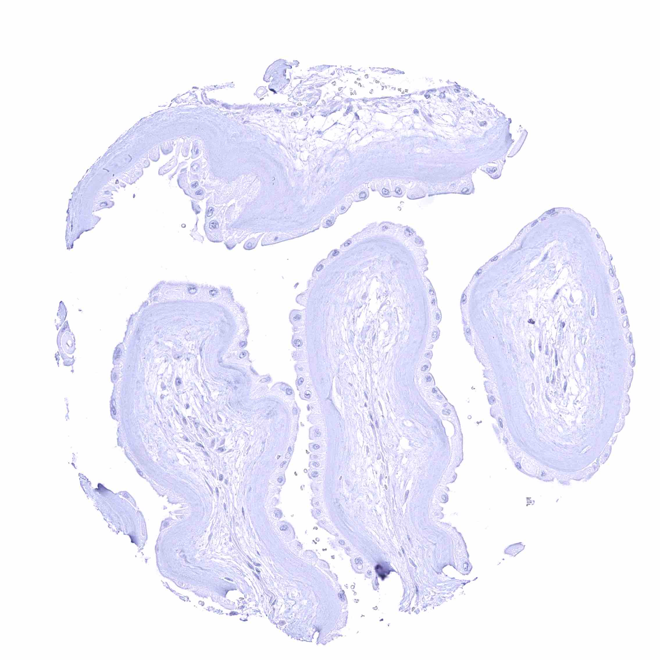
Placenta (chorion)
Placenta (amnion)
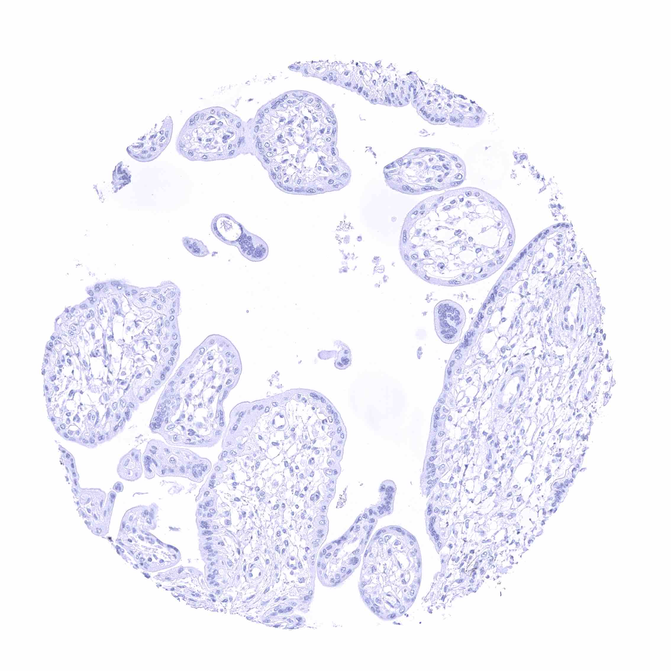
Placenta (amnion)
Placenta, early
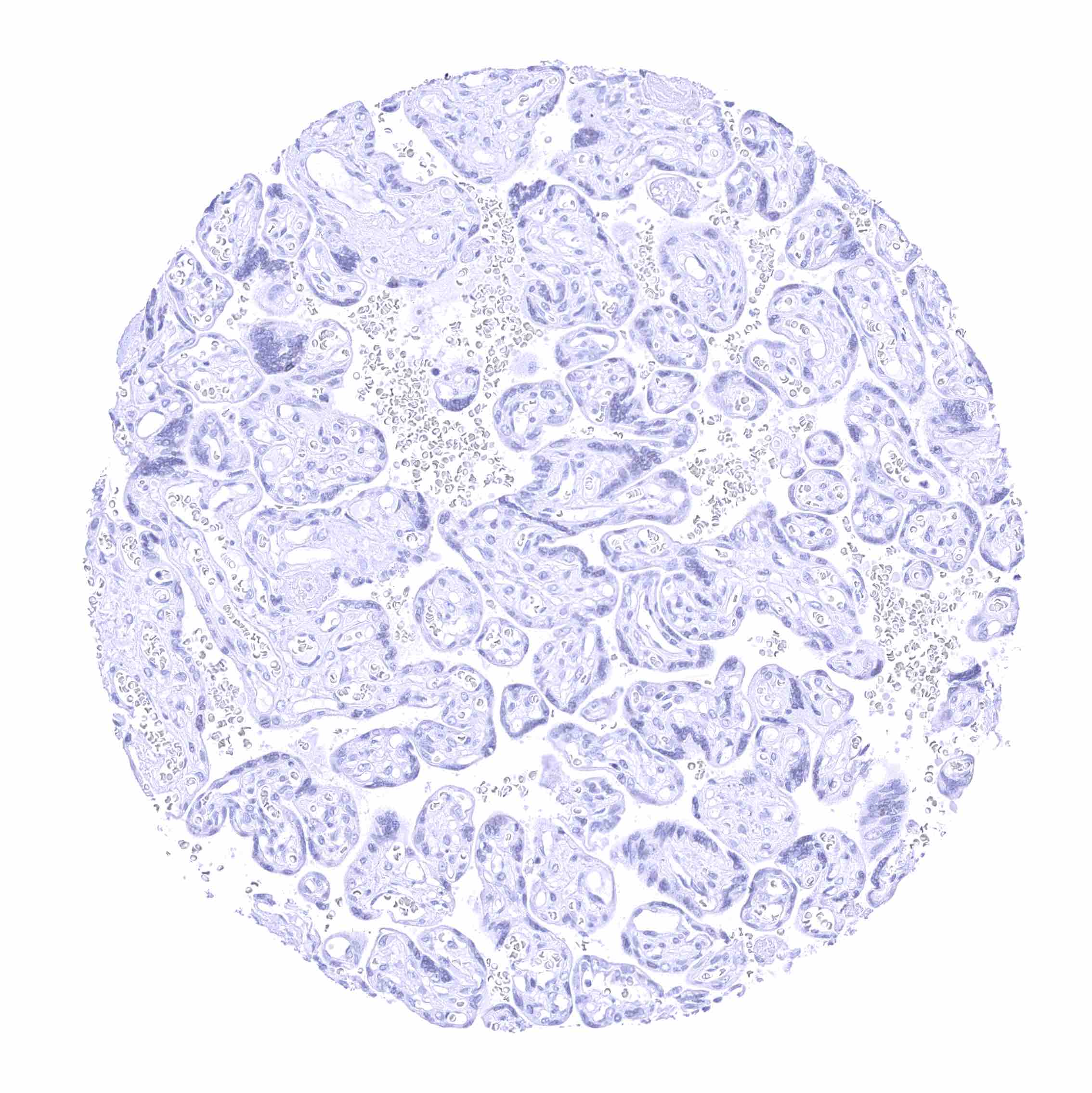
Placenta, early
Placenta, mature
Placenta, mature
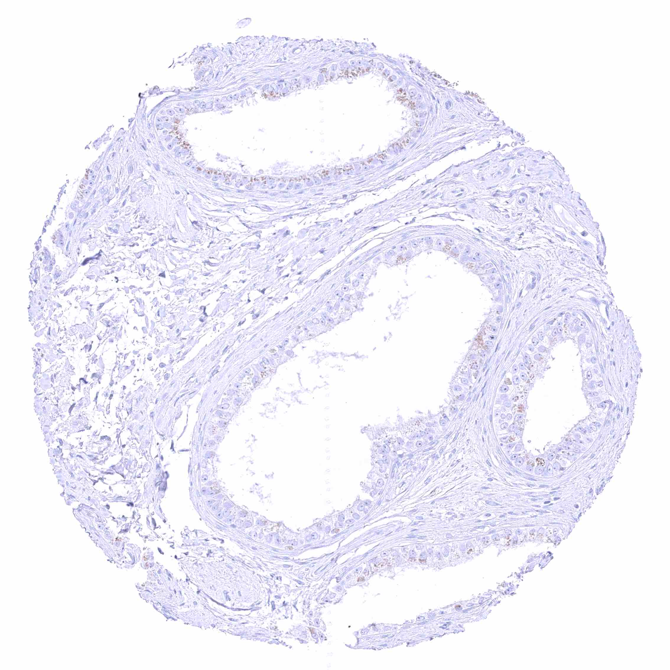
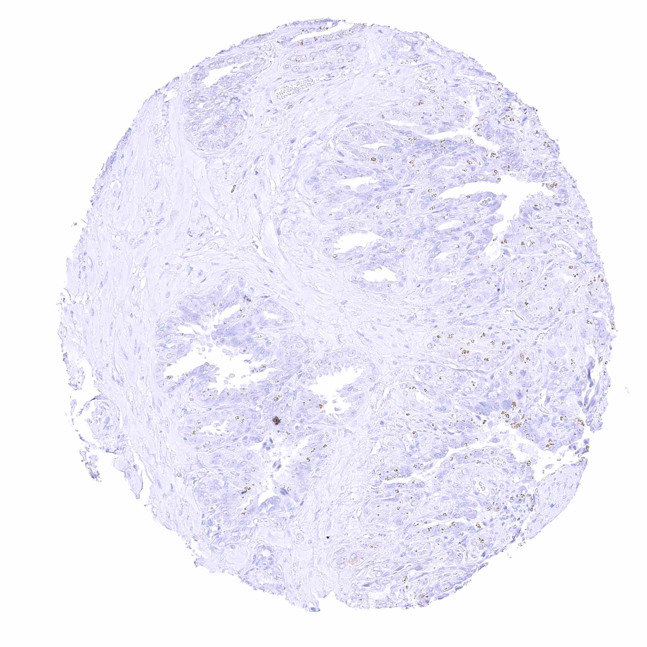
Prostate
Prostate
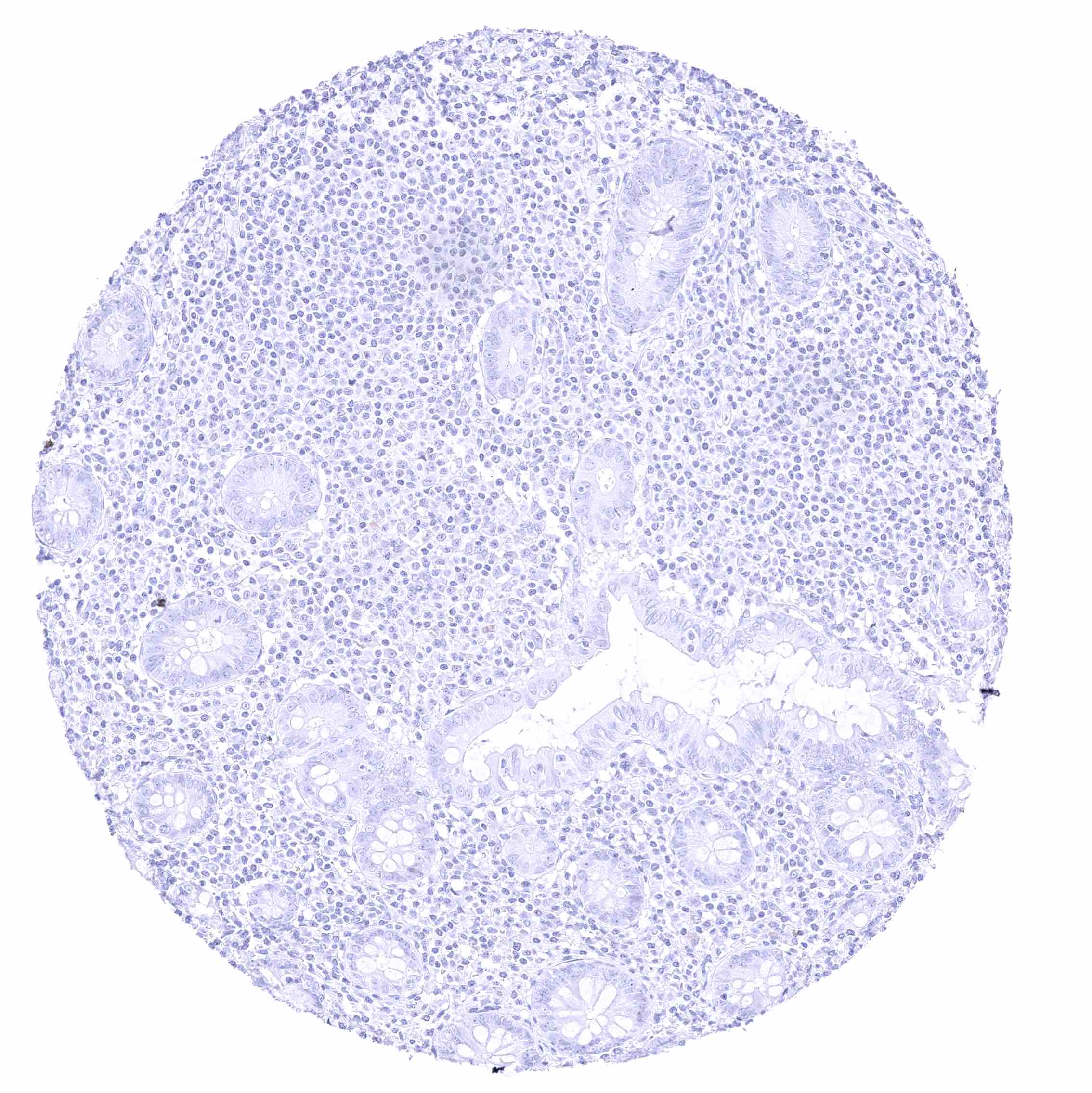
Rectum, mucosa
Rectum, mucosa
Seminal vesicle
Seminal vesicle
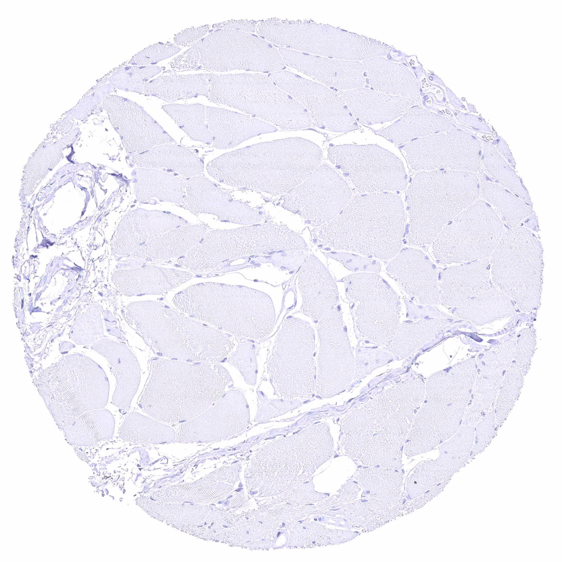
Skeletal muscle
Skeletal muscle
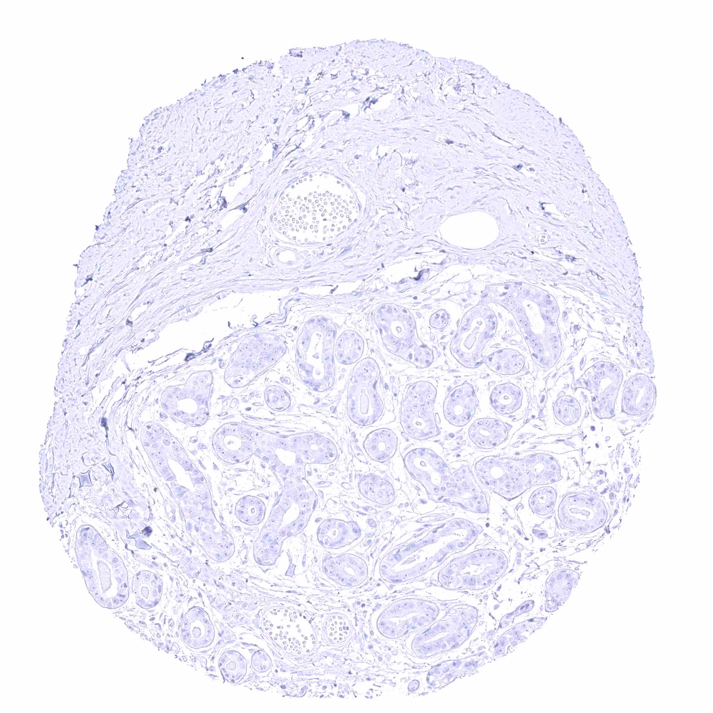
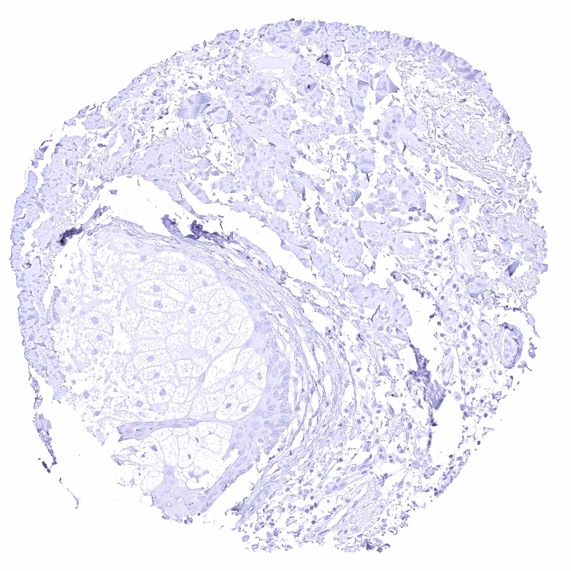
Skin, sebaceous glands
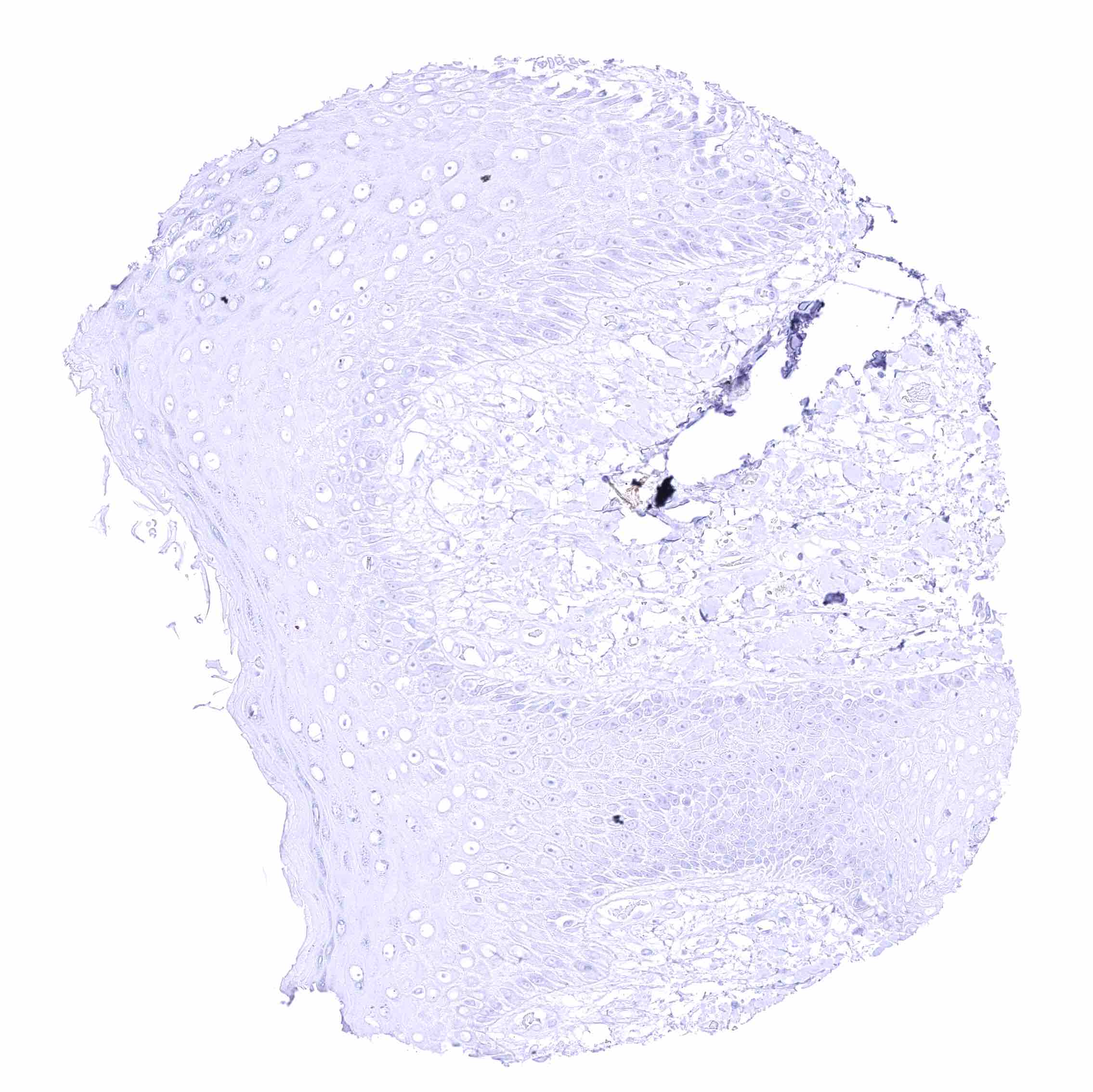
Skin
Skin
Spleen
Spleen
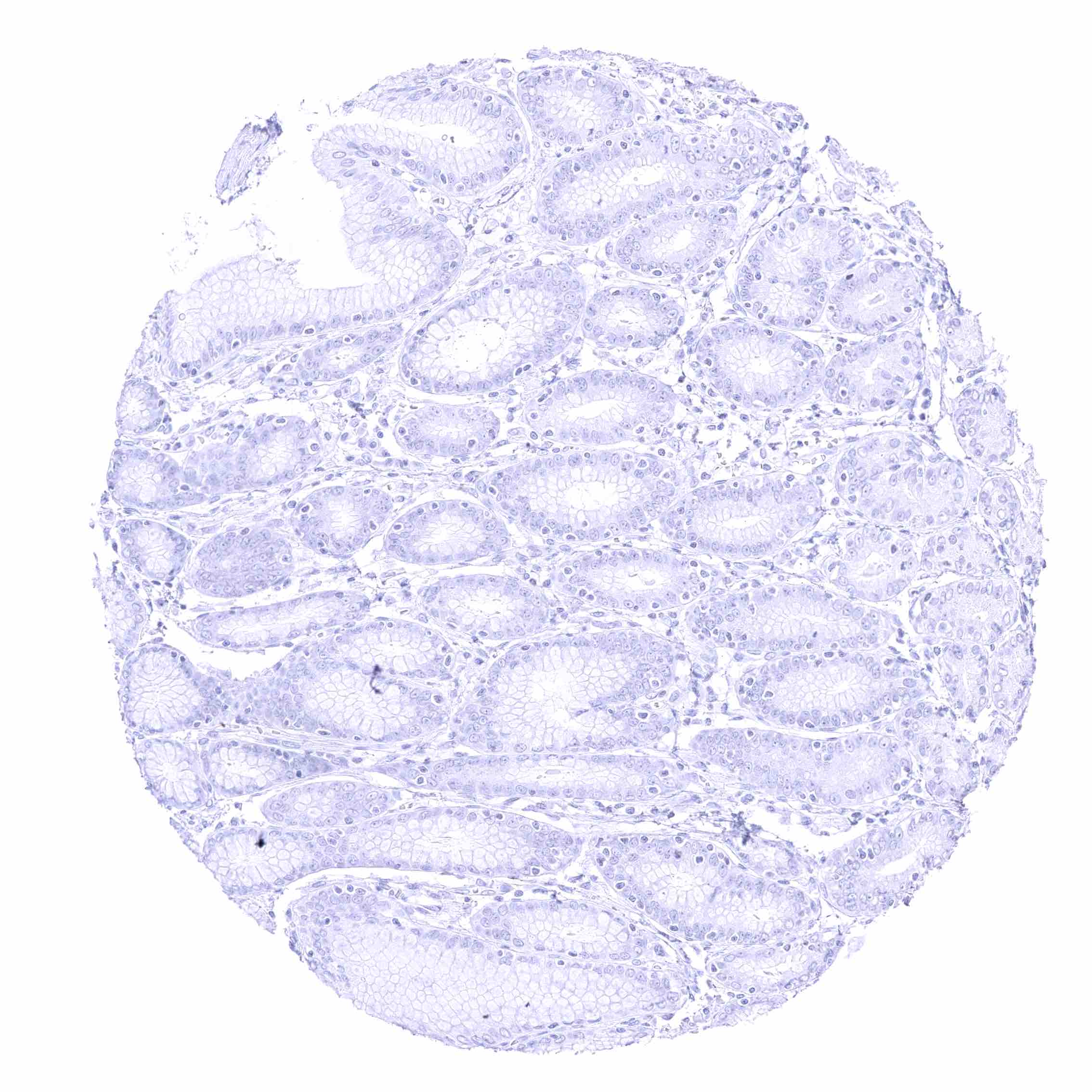
Stomach, antrum – Absence of BSEP staining at the surface epithelium.
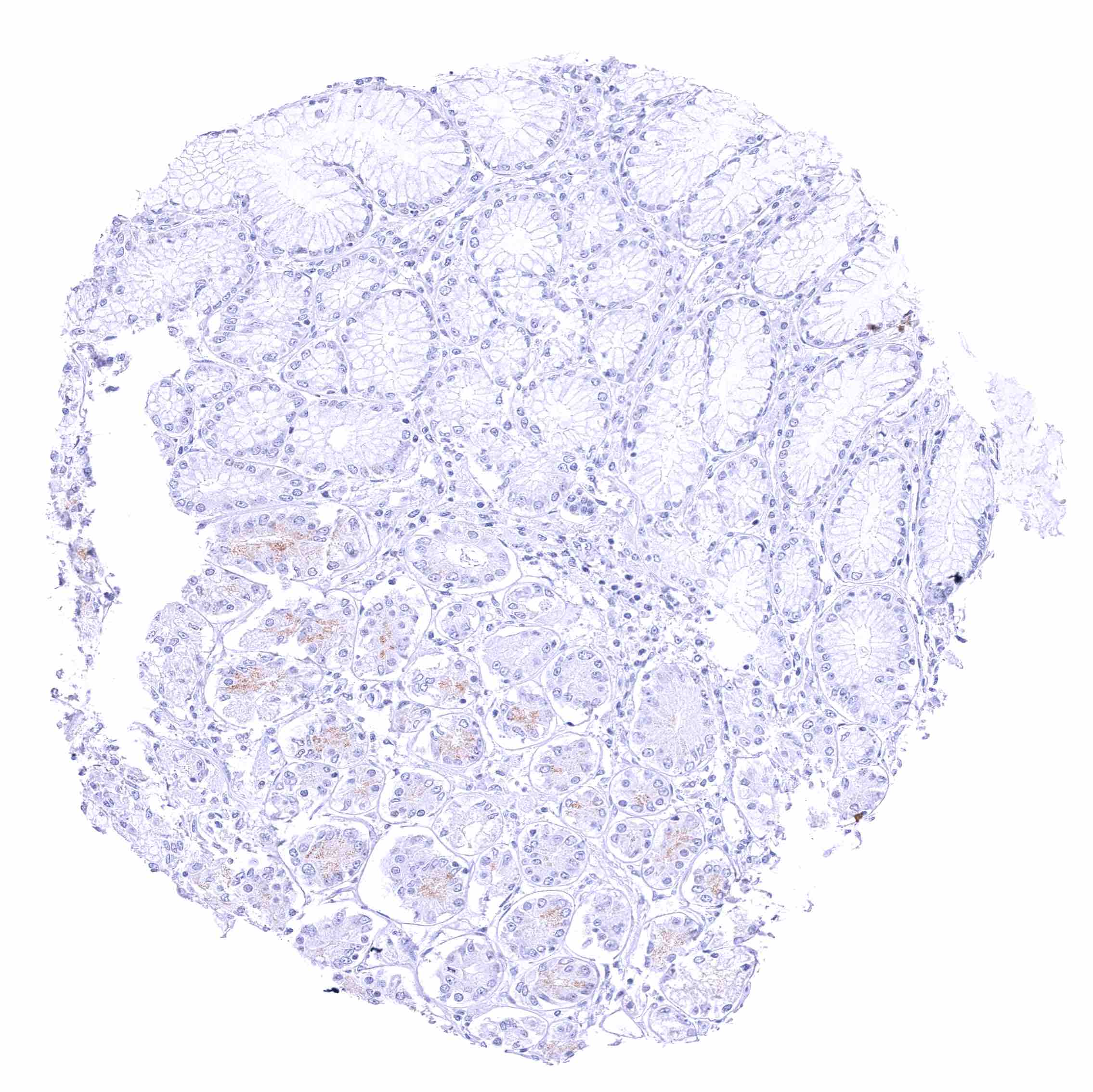
Stomach, antrum – Such a mild gastric gland staining was obtained by multiple independent antibodies and might be due to a binding to secondary antibodies.
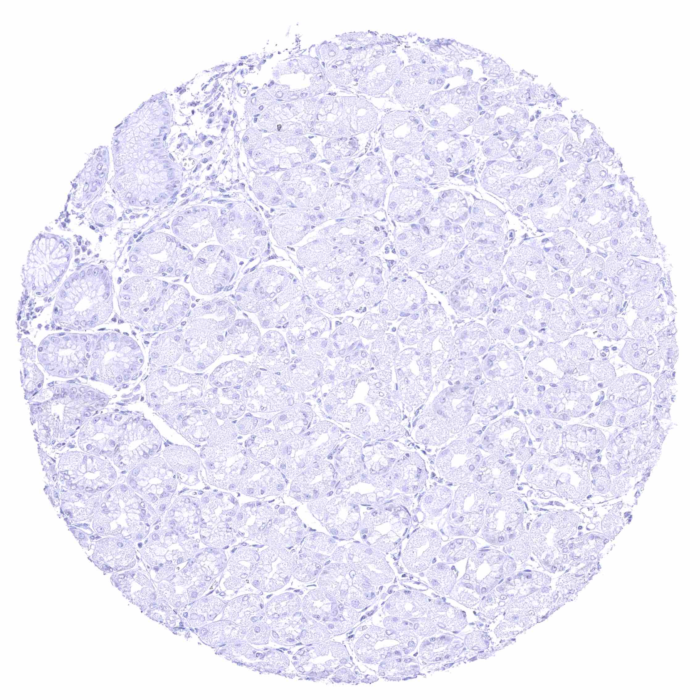
Stomach, corpus – Complete lack of gastric gland staining in this sample.
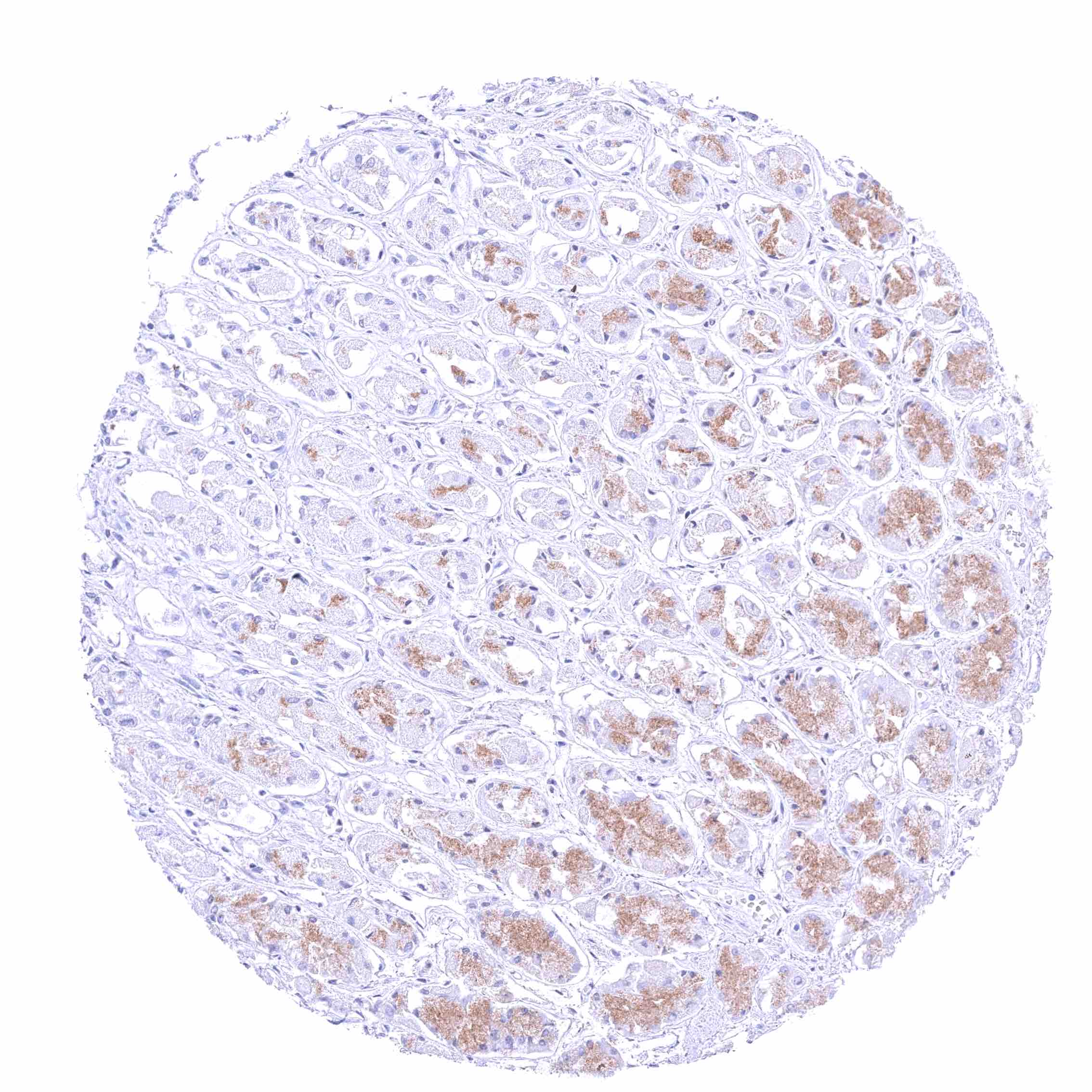
Stomach, corpus – Such a mild to moderate gastric gland staining was obtained by multiple independent antibodies and might be due to a binding to secondary antibodies
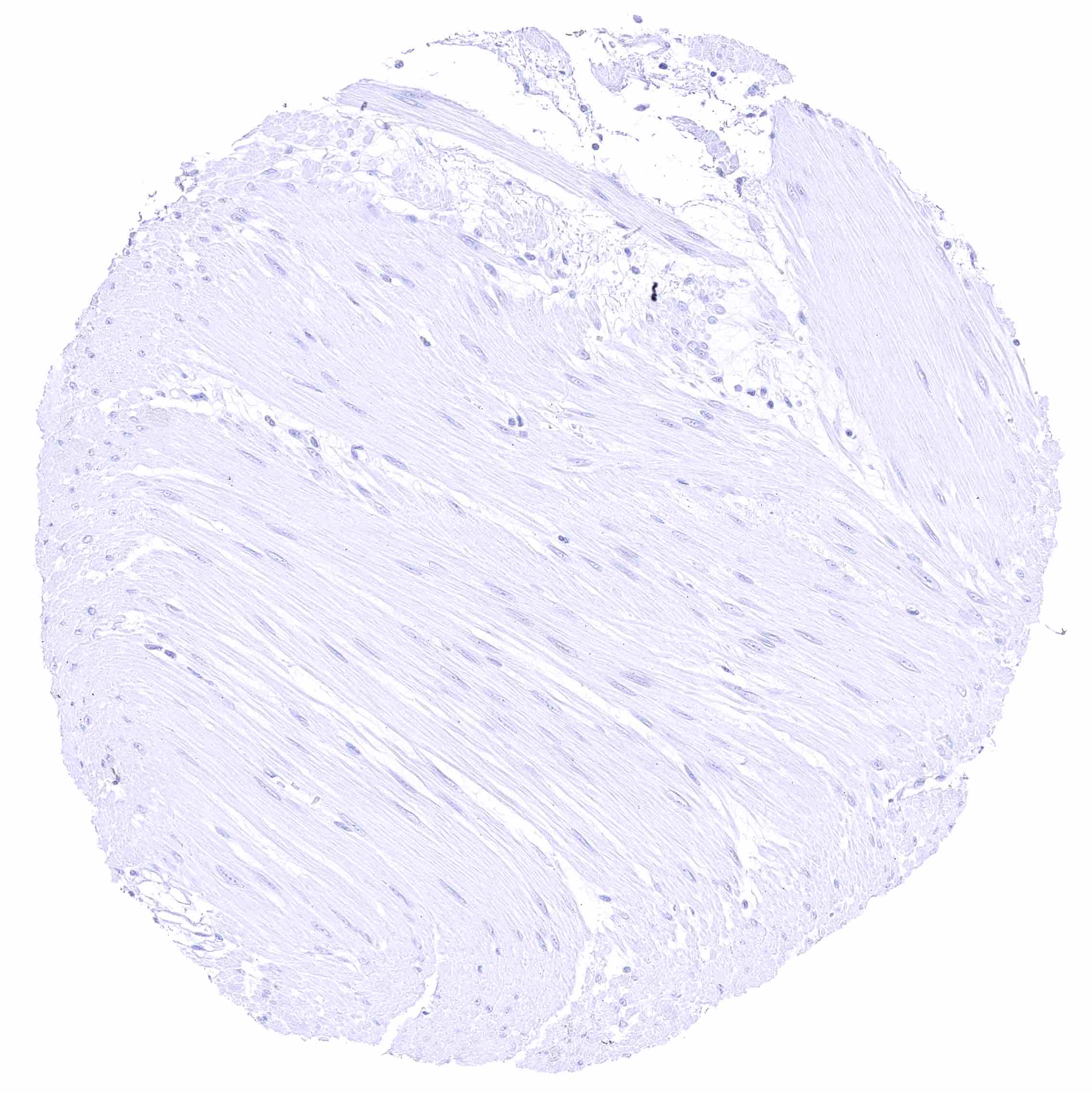
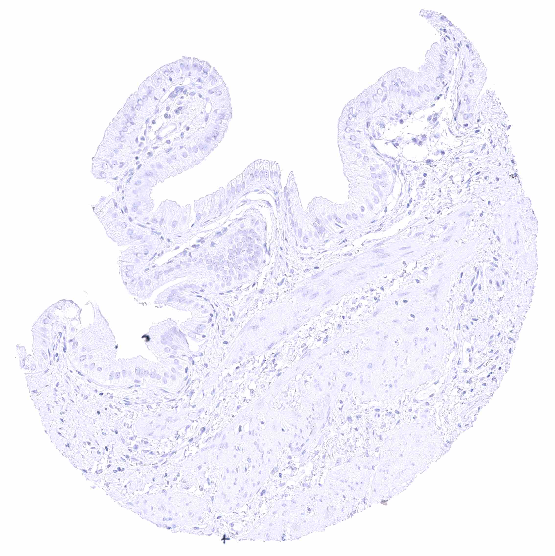
Stomach, muscular wall
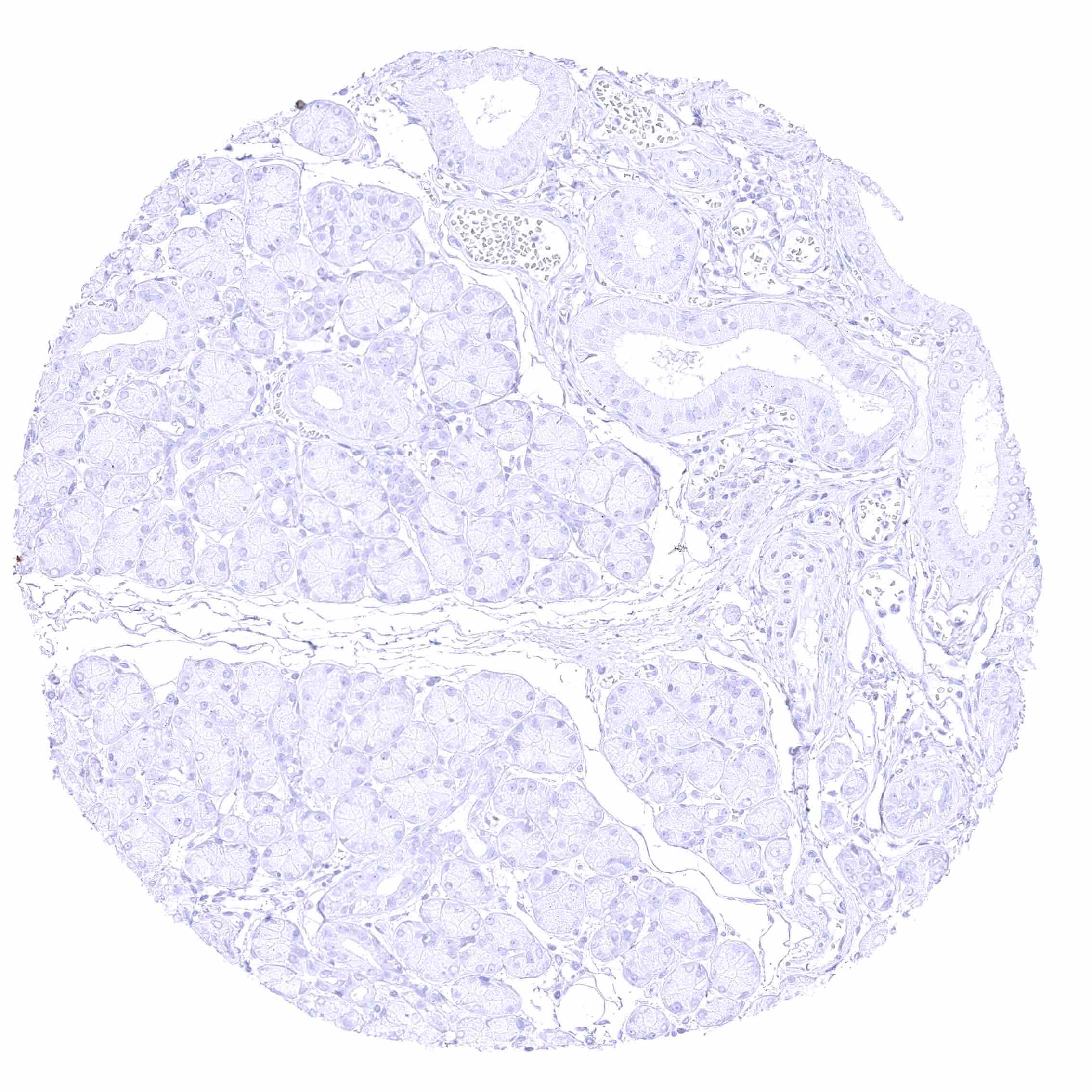
Submandibular gland
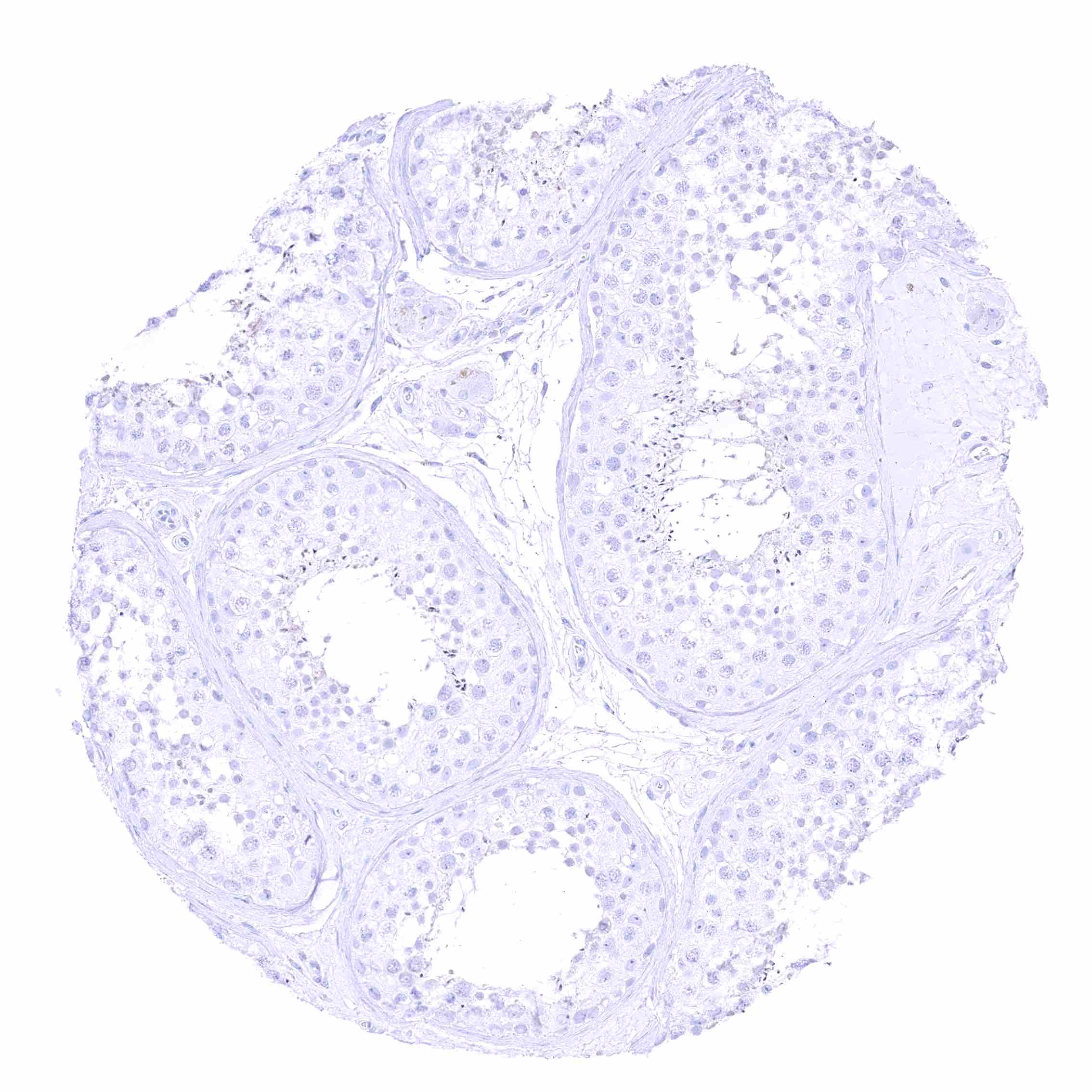
Submandibular gland
Testis
Testis
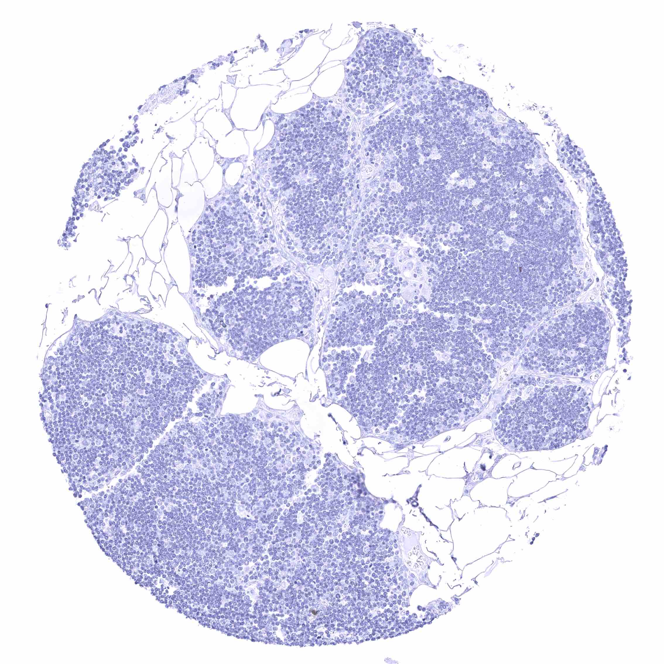
Thymus
Thymus
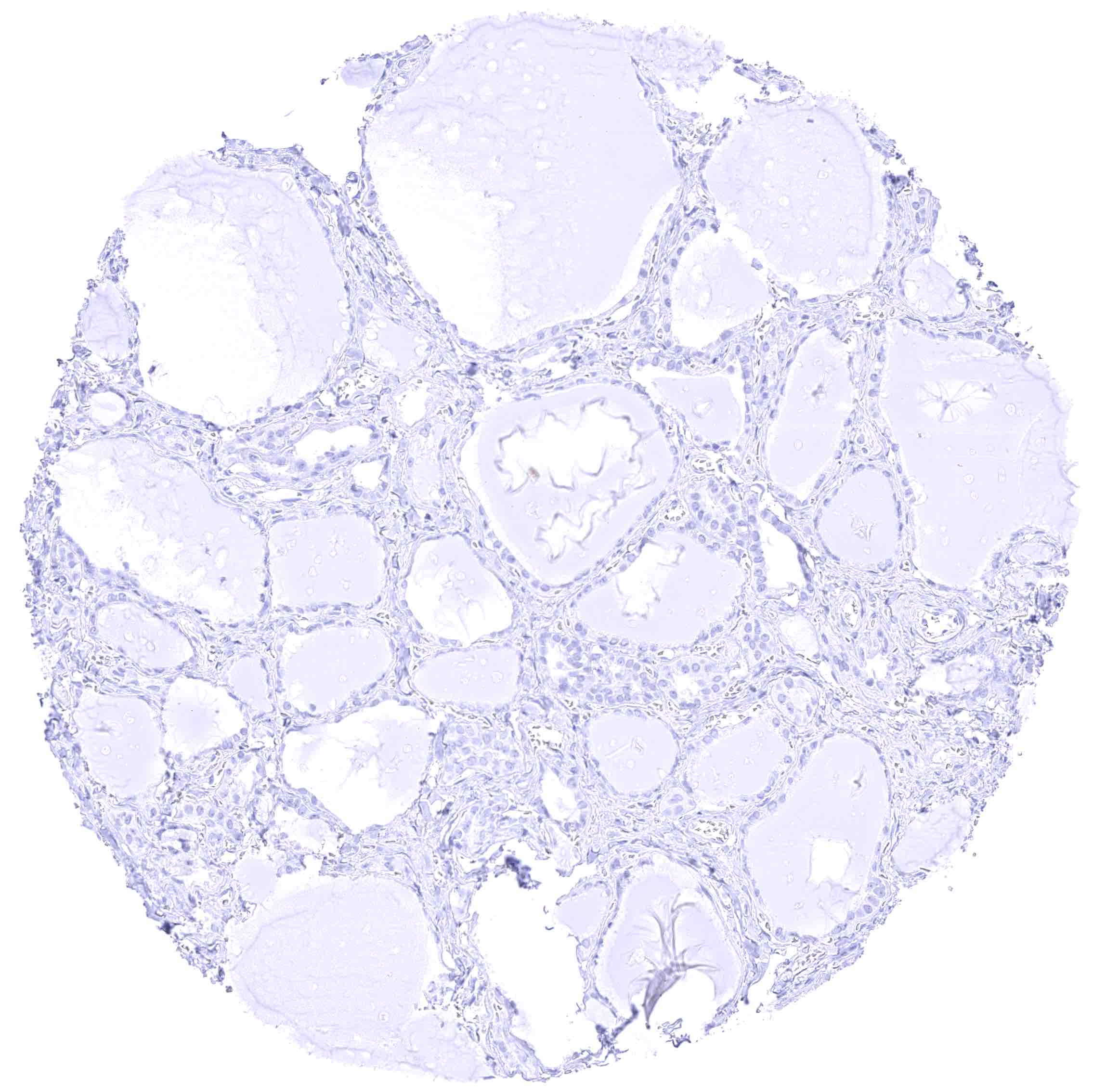
Thyroid gland
Thyroid gland
Tonsil, surface epithelium
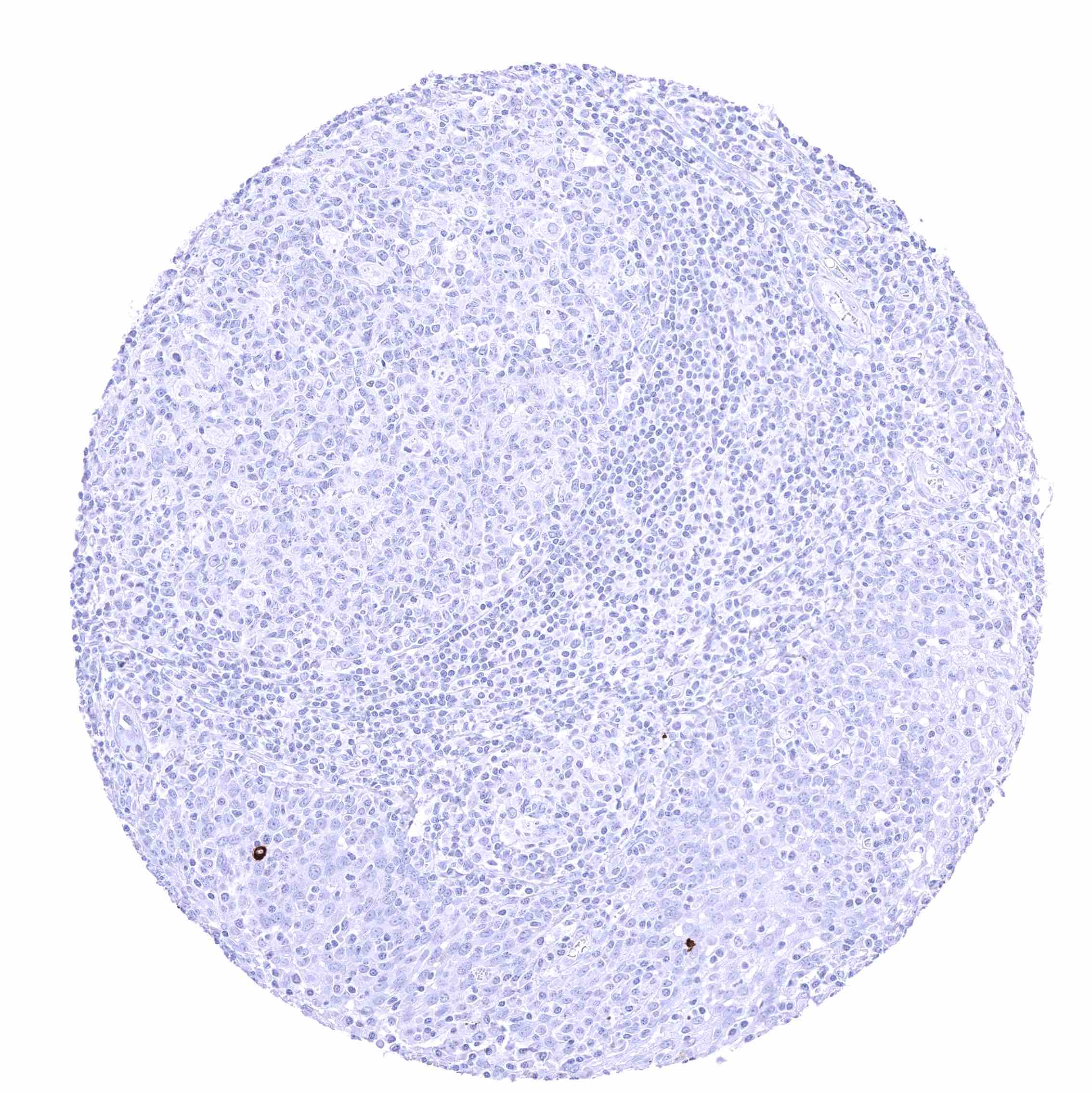
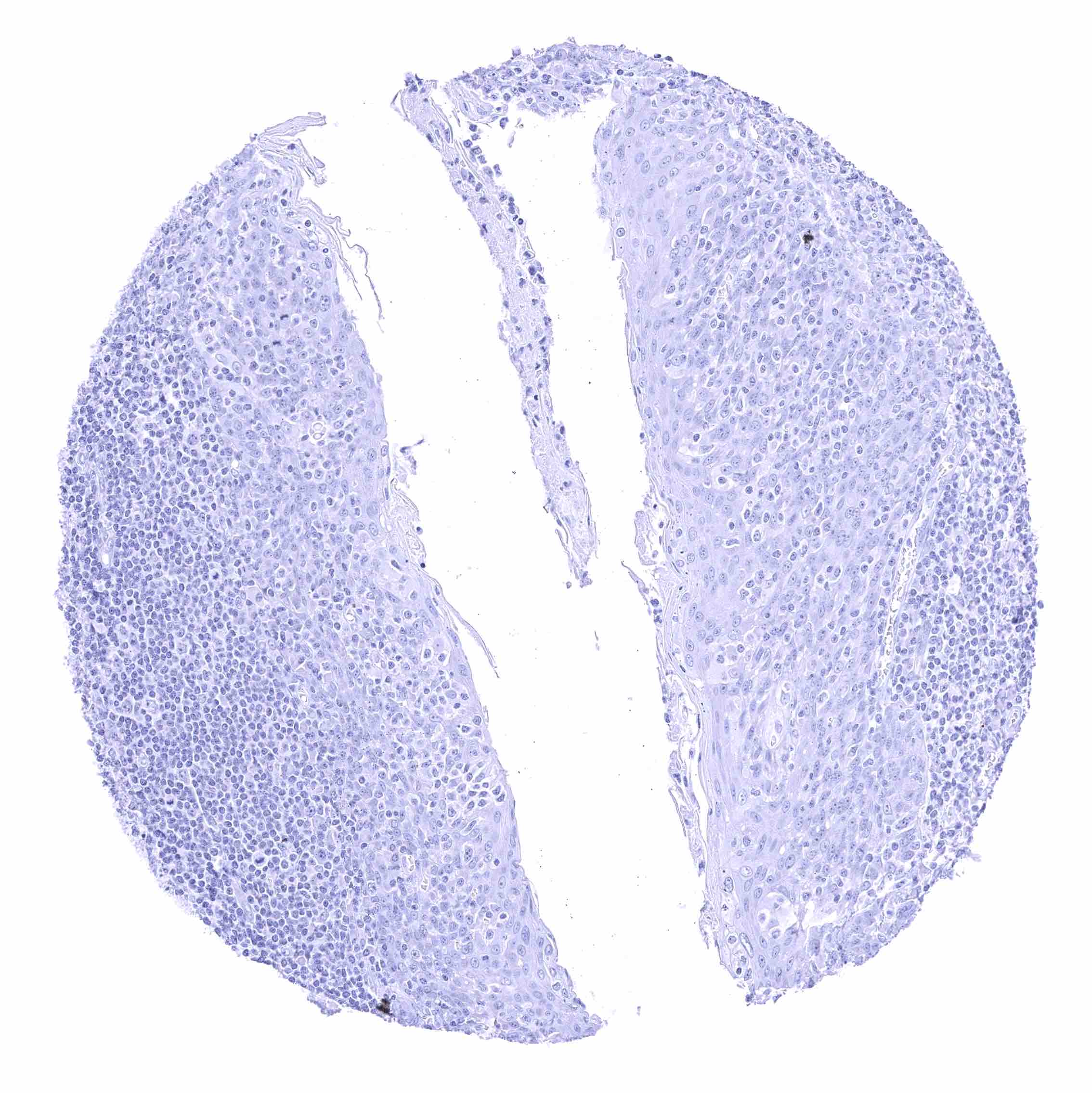
Tonsil
Tonsil
Urinary bladder, muscular wall
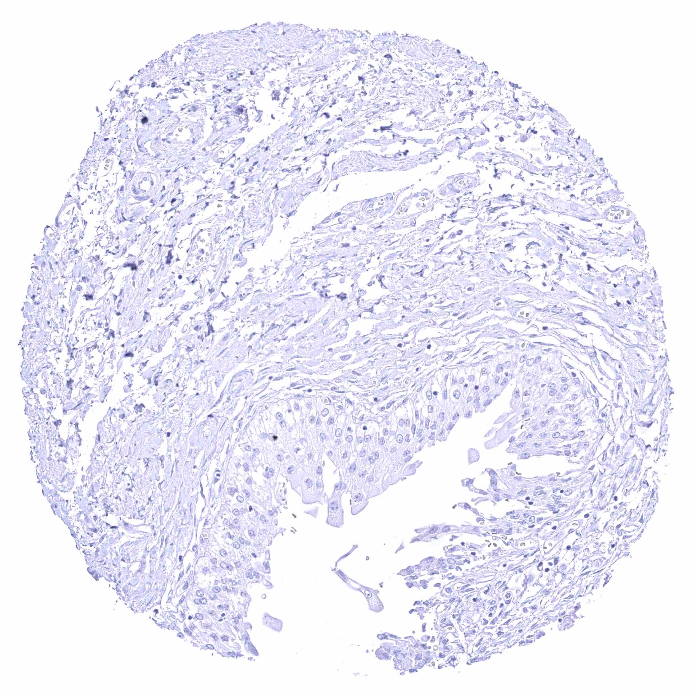
Urinary bladder, urothelium
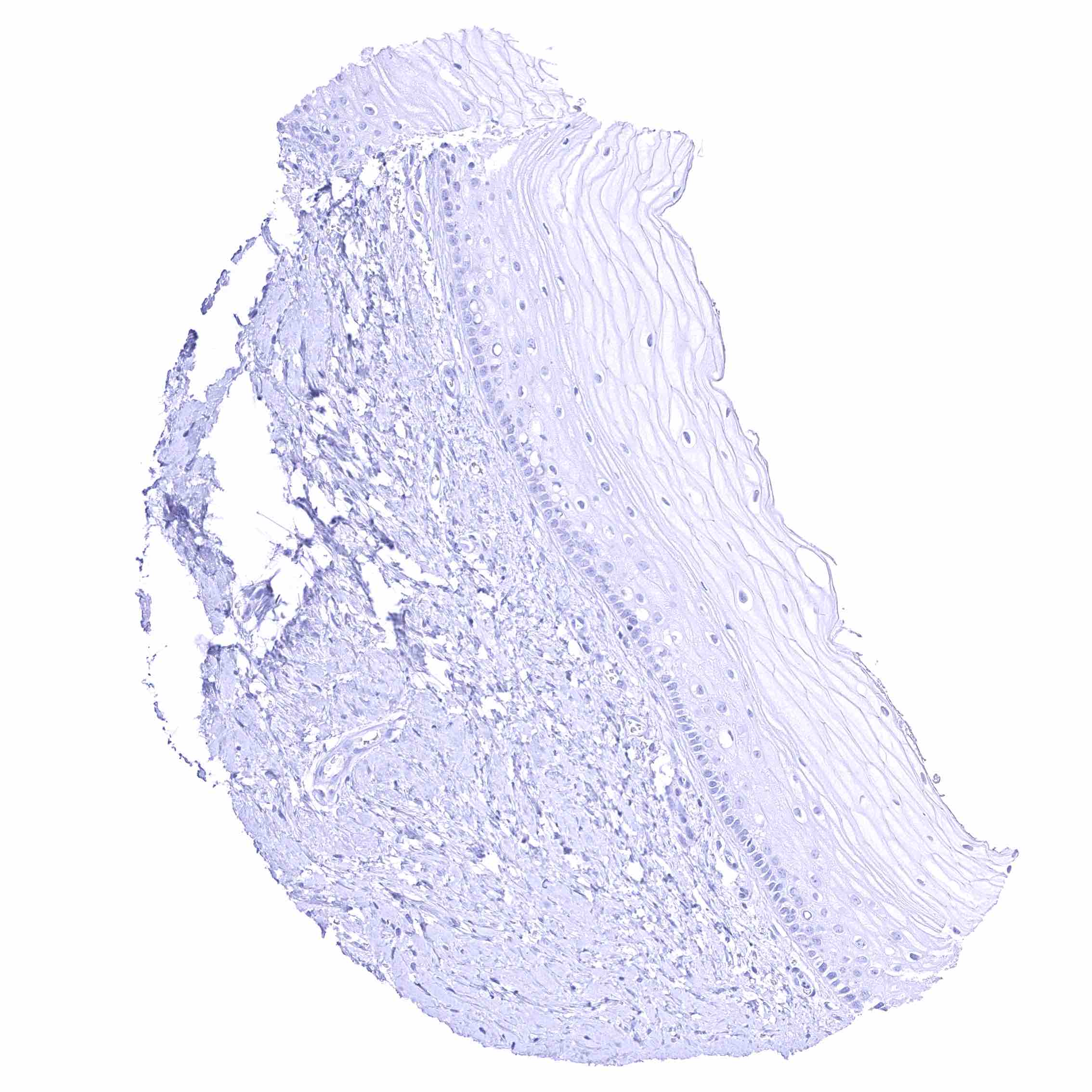
Uterus, ectocervix
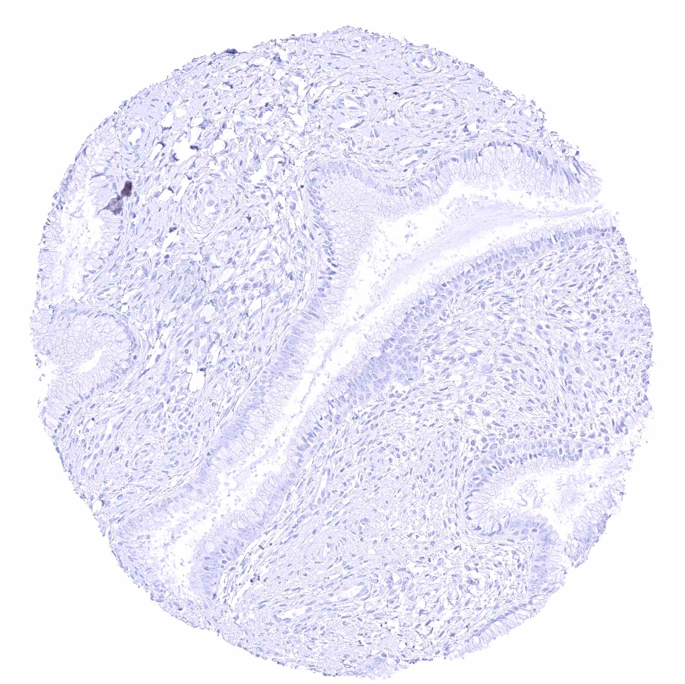
Uterus, ectocervix
Uterus, endocervix
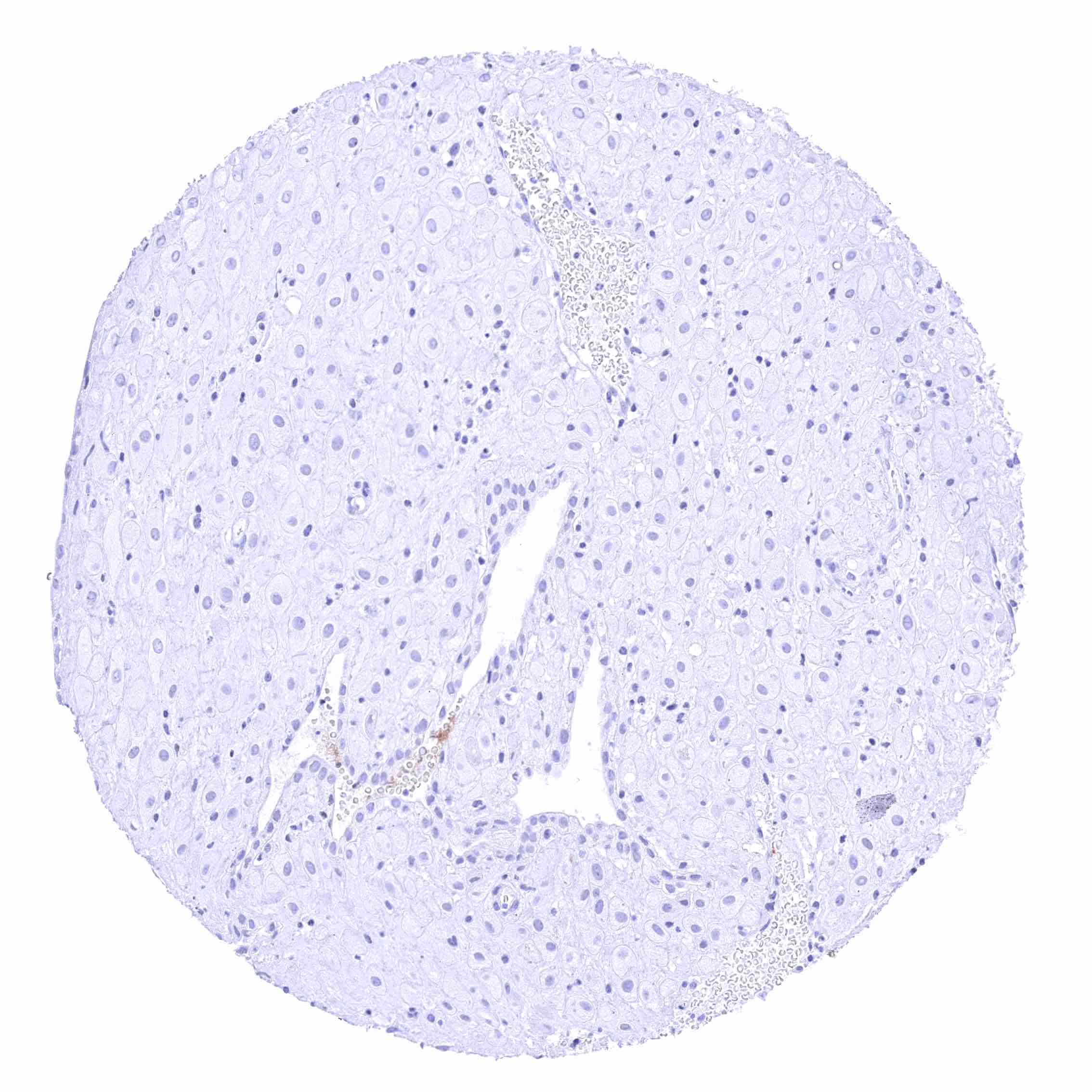
Uterus, endocervix
Uterus, endometrium (pregnancy)
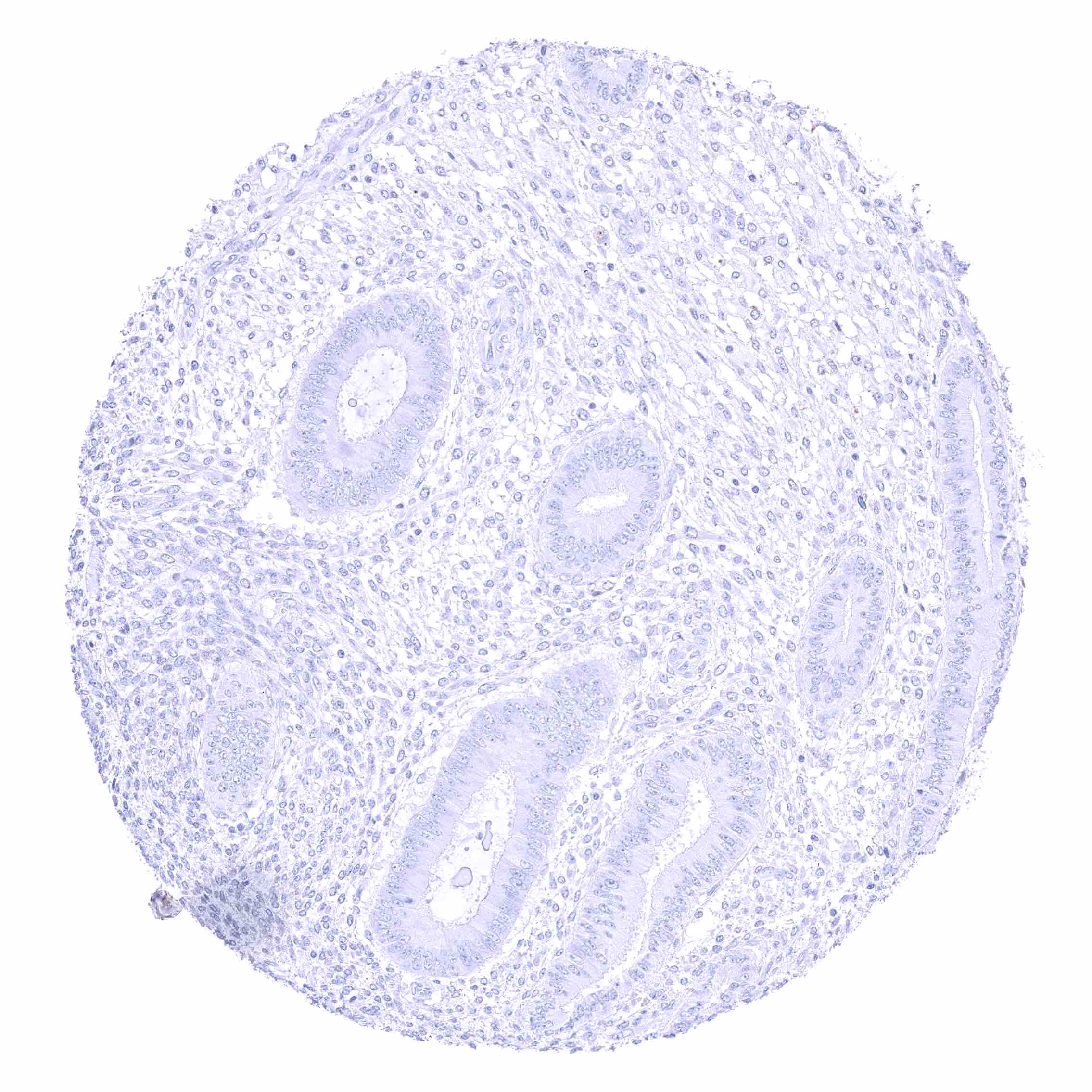
Uterus, endometrium (proliferation)
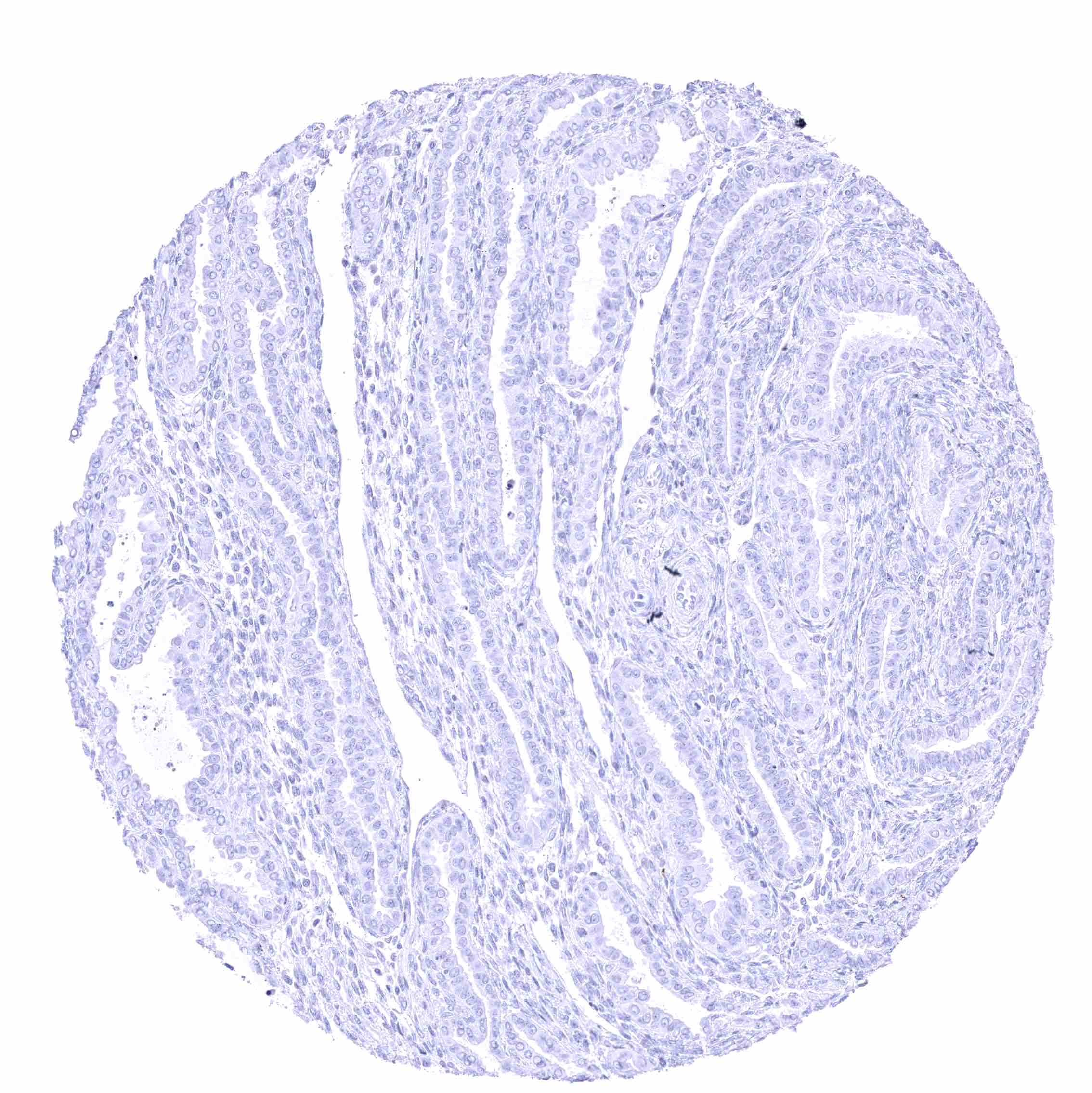
Uterus, endometrium (secretion)
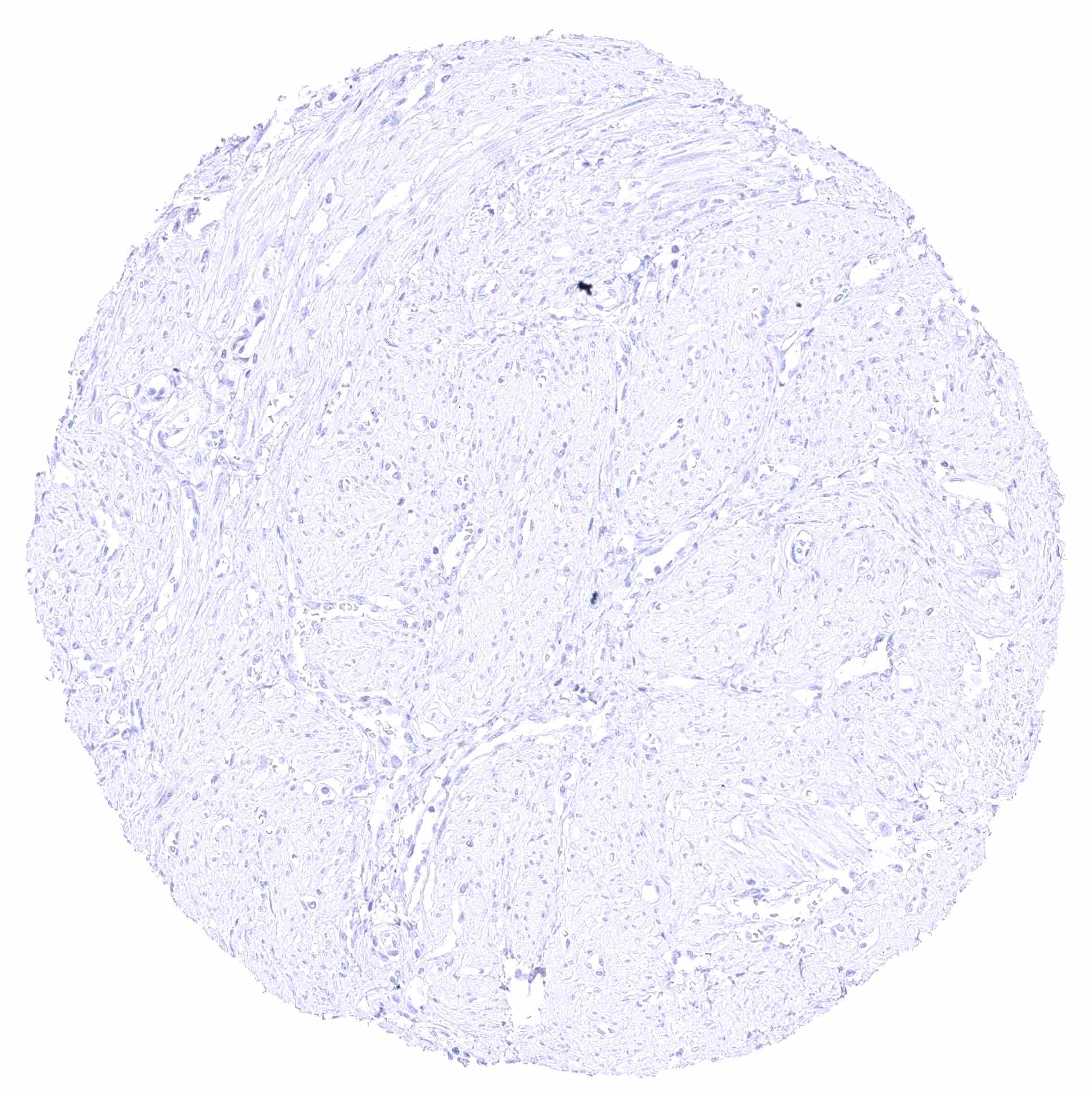
Uterus, myometrium
Uterus, myometrium
  • Linkedin
  • My Account
  • Search
    ×
  • Cart 0
© 2026 MS Validated Antibodies
  • Imprint
  • Privacy Policy
  • Distributors