145,00 € – 995,00 €
Product details
Synonyms = EndoCAM; PECA1; Platelet Endothelial Cell Adhesion Molecule 1; GPIIA
Antibody type = Mouse monoclonal / IgG
Clone = MSVA-031M
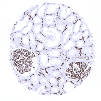
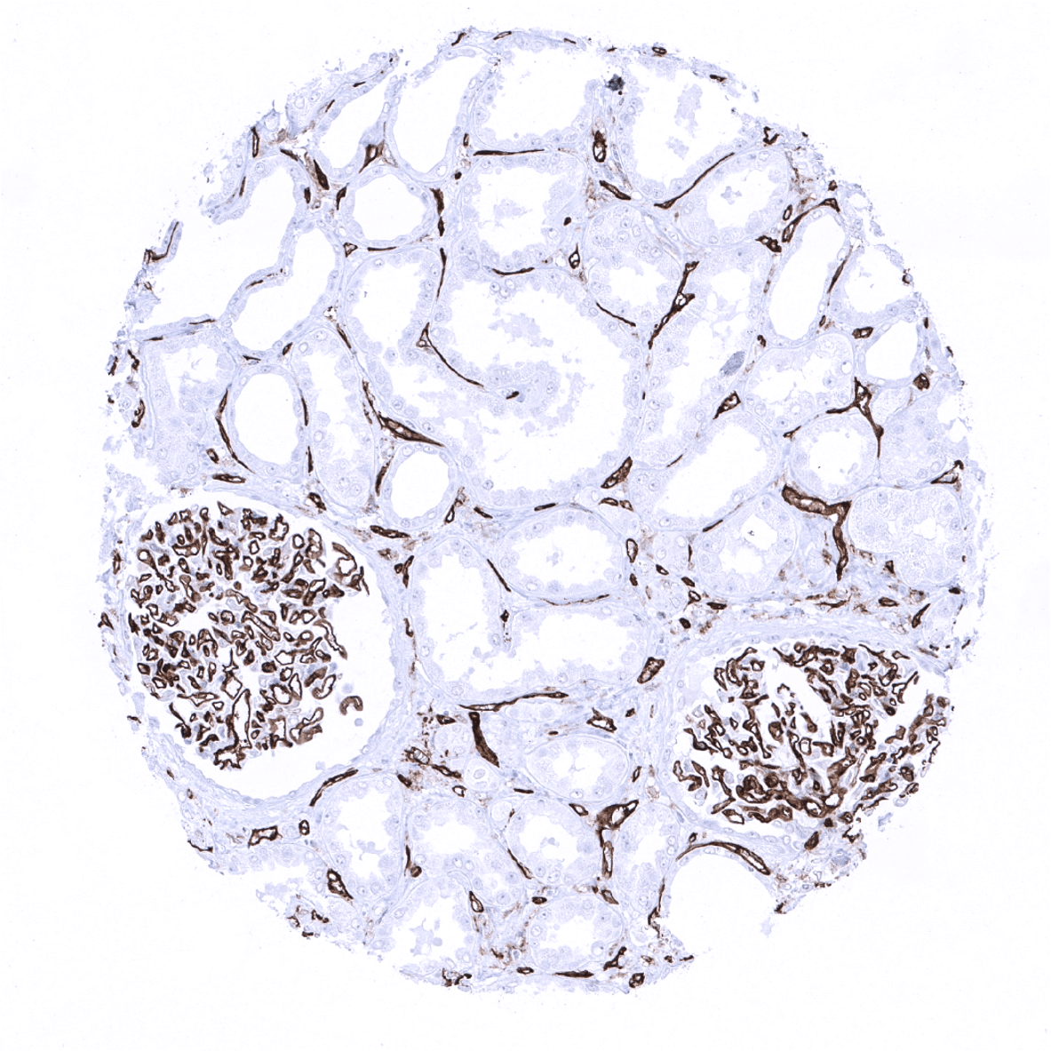
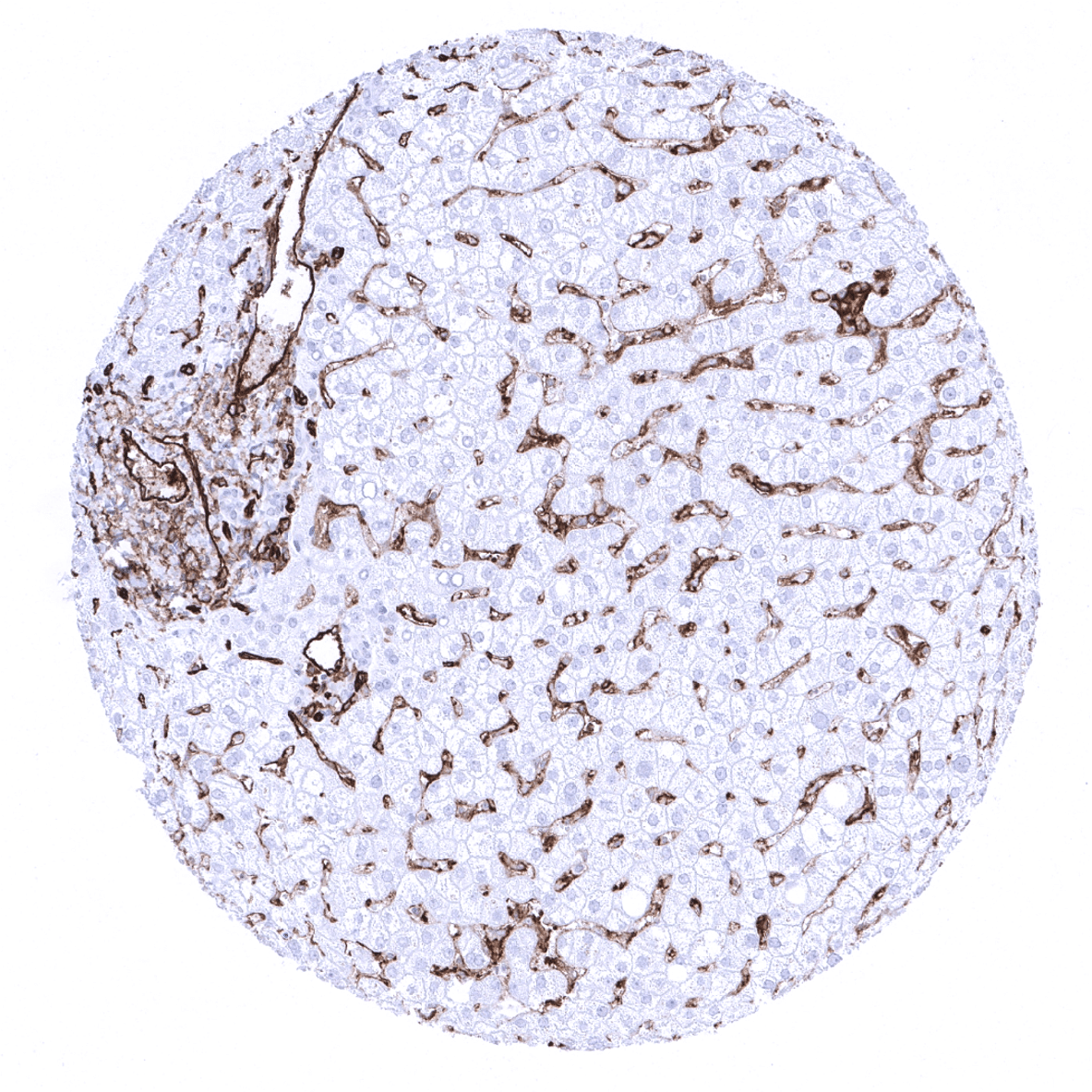
Positive control = Liver: At least a weak to moderate staining of the majority of the hepatic sinusoidal endothelial cells should be seen.
Negative control = Liver: Hepatocytes should not show any CD31 staining.
Cellular localization = Cell surface and cytoplasm
Reactivity = Human
Application = Immunohistochemistry
Dilution = 1:100 – 1:200
Intended Use = Research Use Only
Relevance of Antibody
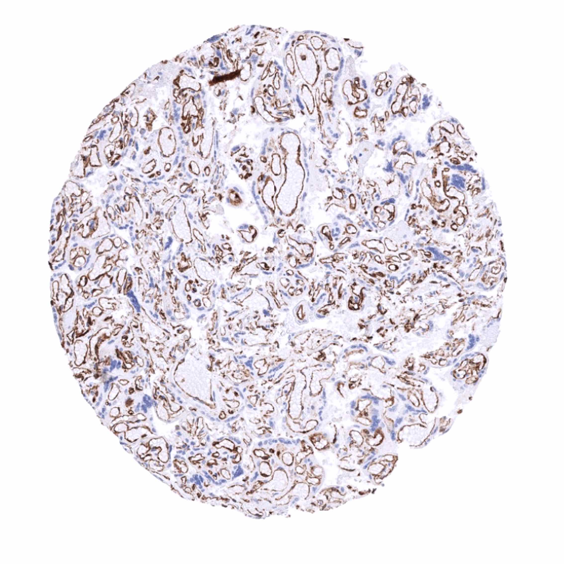
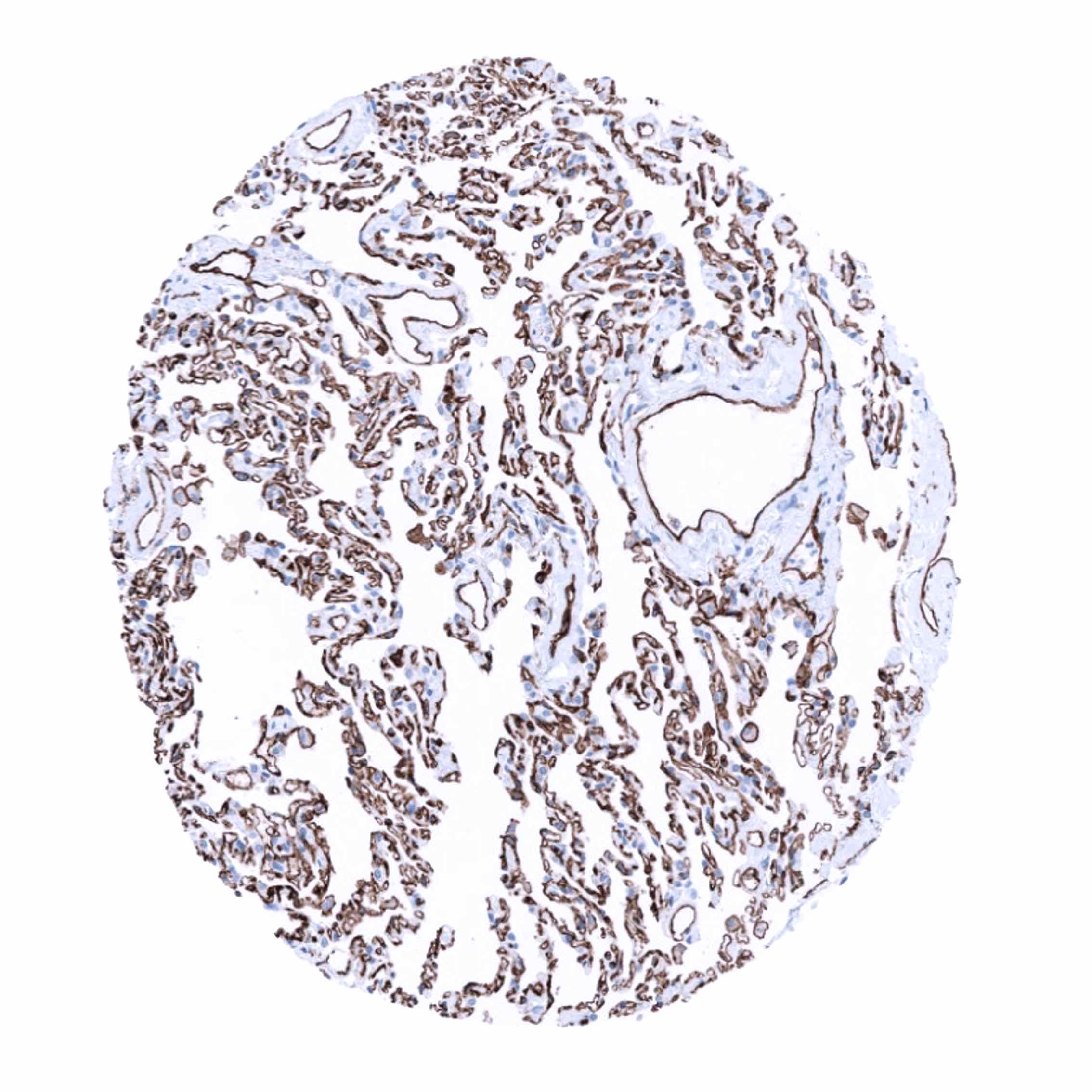
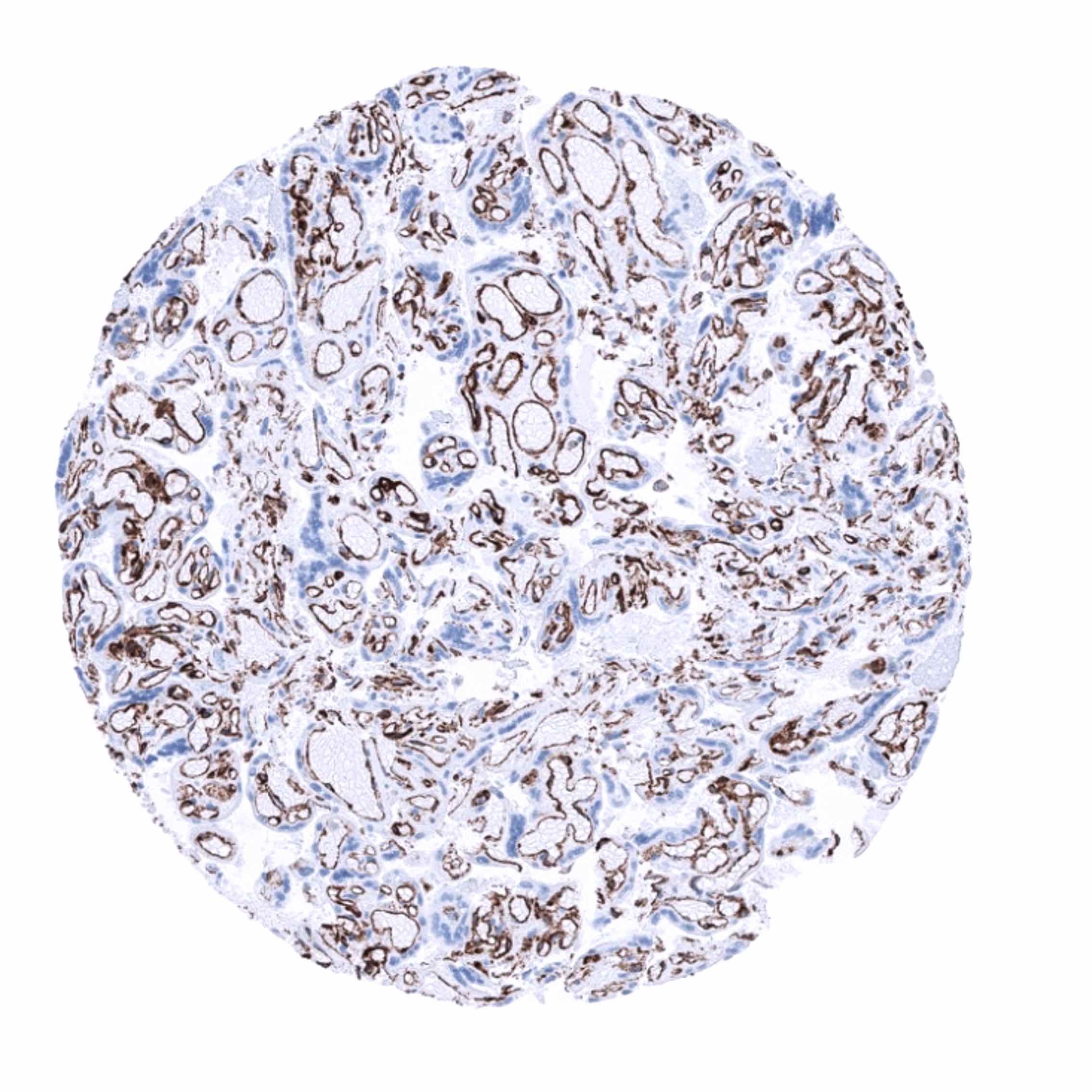
CD31 is expressed on endothelial cells.
Biology Behind
CD31 is a 130-140 kDa transmembrane glycoprotein, belonging to the immunoglobulin super family. The protein, also termed platelet-endothelial cell adhesion molecule 1 (PECAM-1), plays a key role in angiogenesis, removing aged neutrophils from the body, and in thrombosis. CD31 represents a large portion of endothelial cell intercellular junctions where it plays a functional role in leukocyte transmigration.
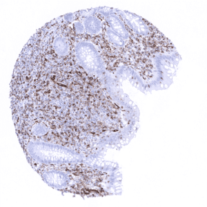
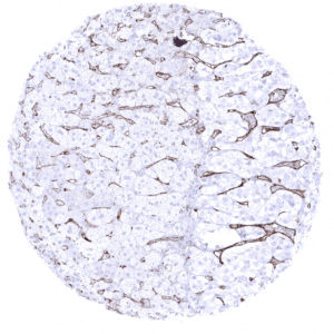
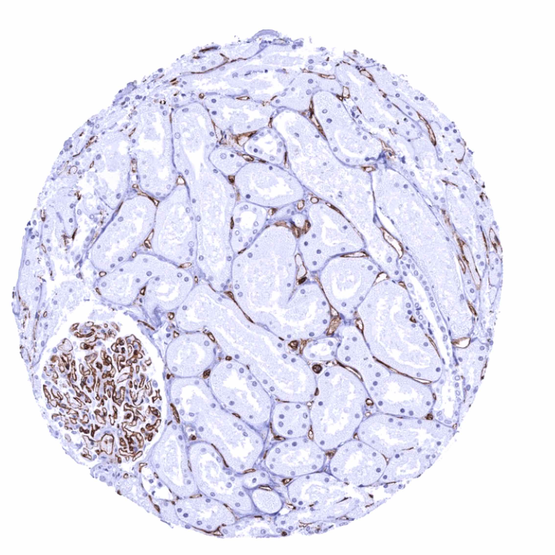
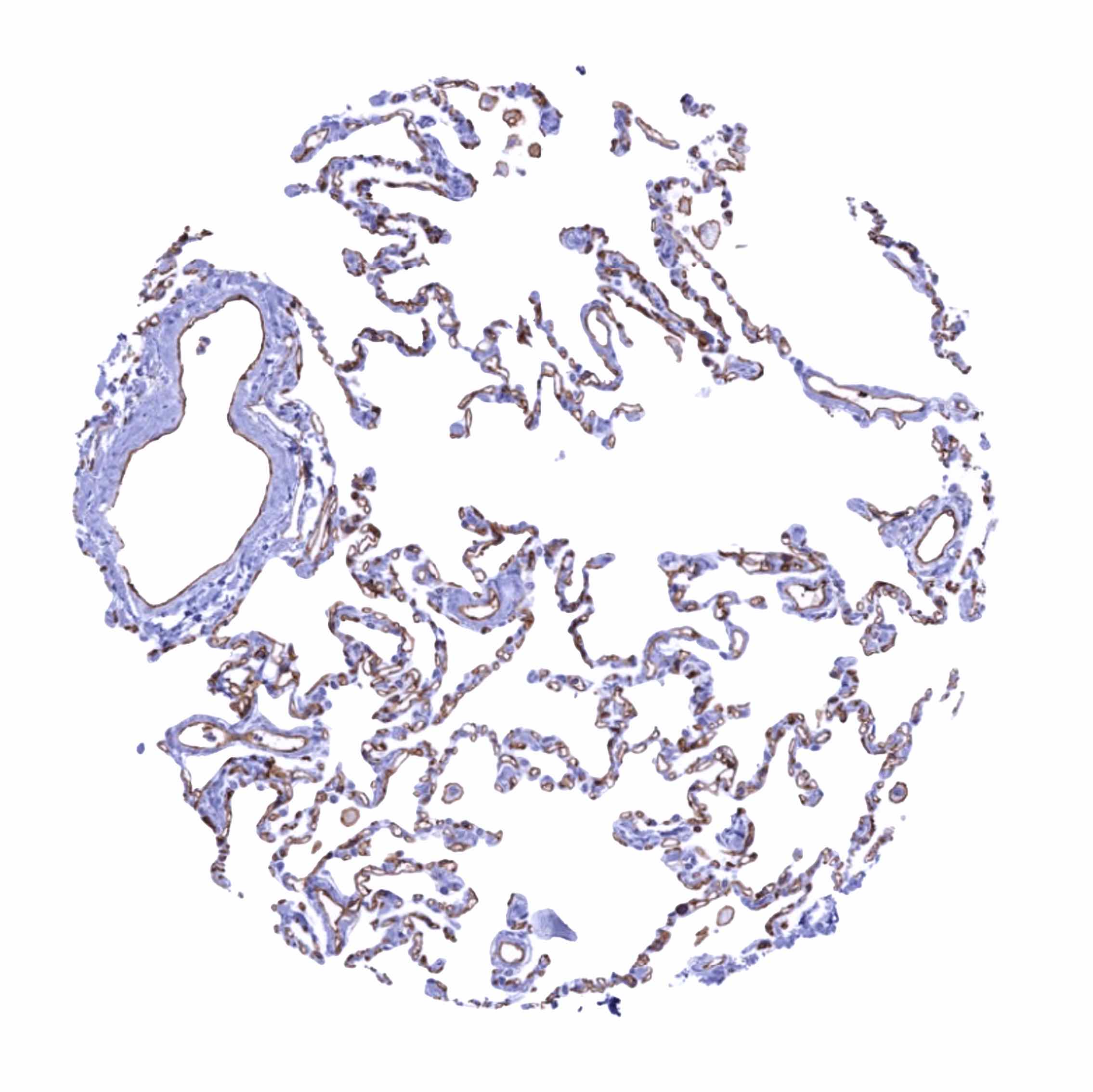
Staining Pattern in Normal Tissues
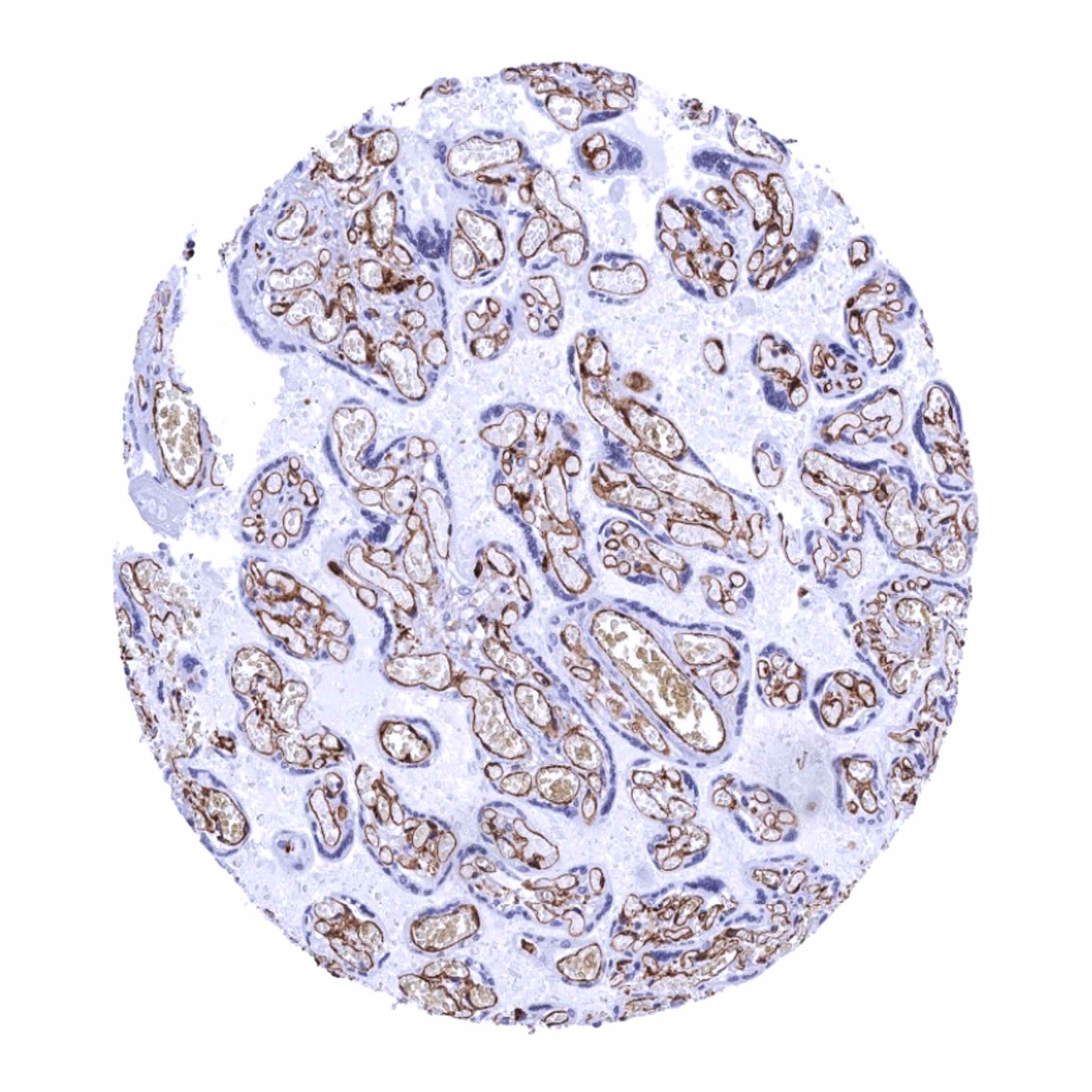
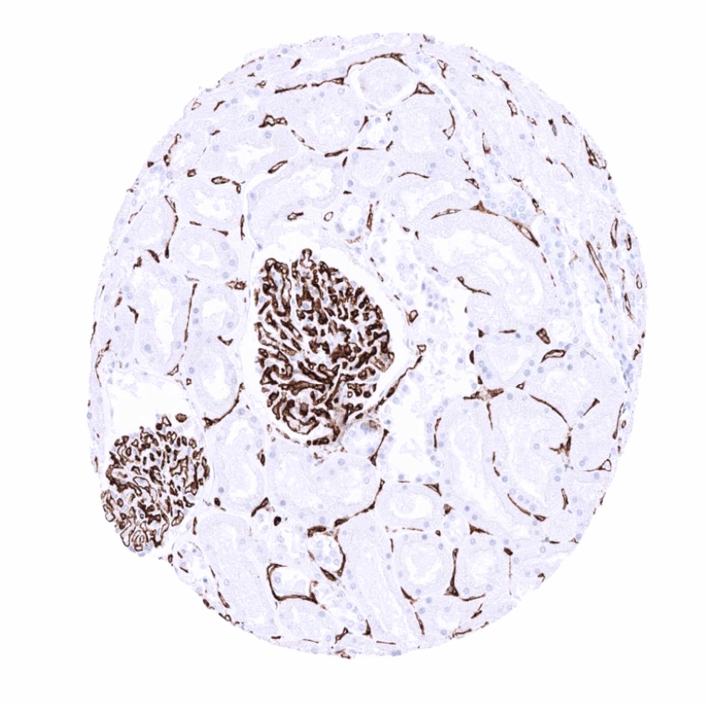
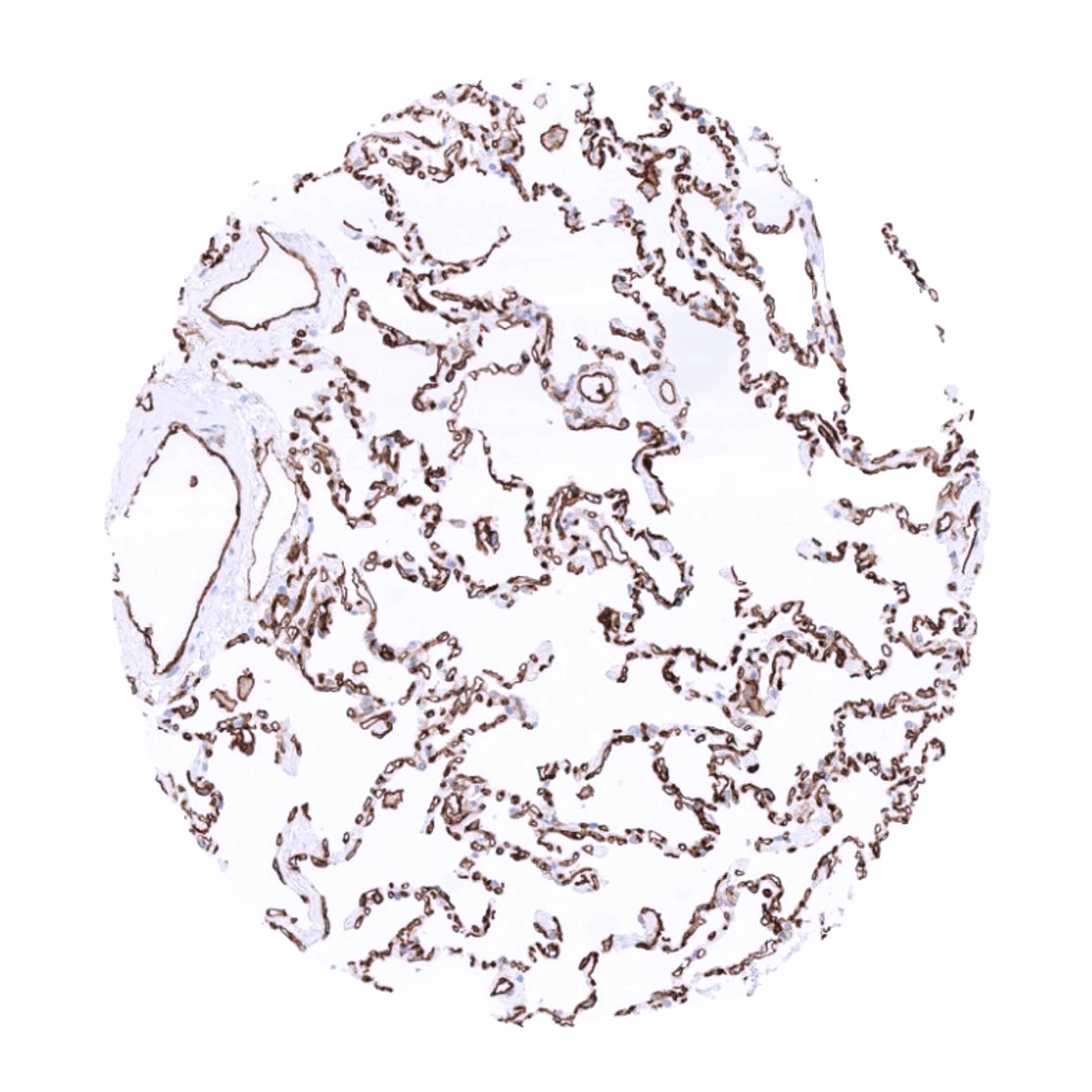
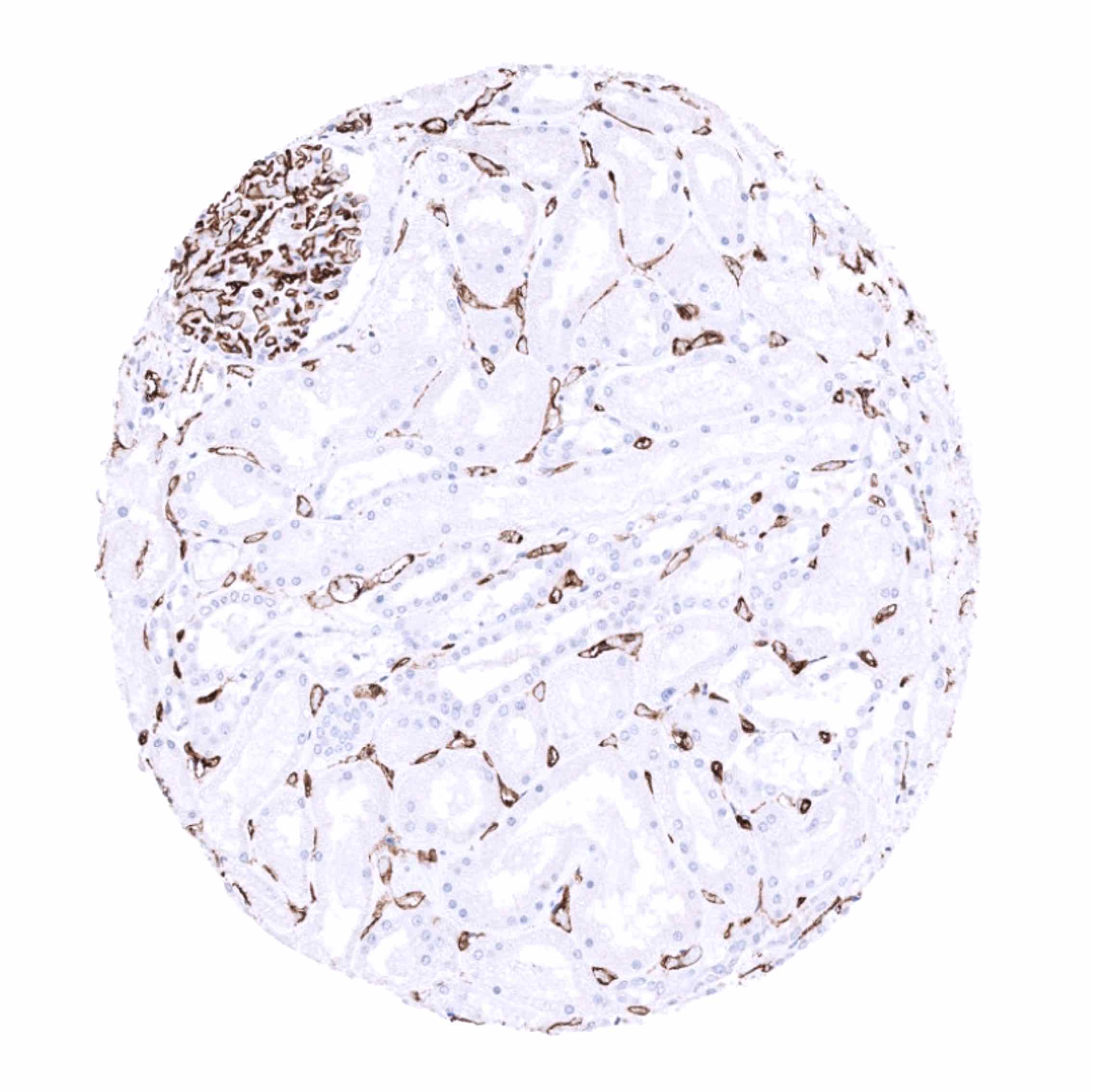
CD31 is strongly expressed in endothelial cells of all organs, including alveolar wall capillaries, glomeruli, lymphatics, or spleen. CD31 is moderately expressed in sinusoids of the liver, and weakly expressed in megakaryocytes, platelets, and neutrophils. Rarely, CD31 can be focally expressed in epithelial cells such as in the endocervix. In B lymphocytes, CD31 expression changes during the differentiation process. CD31 is high in pre-B-I cells, low in pre-B-II precursors, intermediate in the mature B-cell subpopulations, absent in most follicular centre cells, and present in pre- and post- germinal centre cells. CD31 also occurs in a fraction of T-cells. Angiogenic T cells (Tang) – positive for CD3, CD31 and CXCR4 – are assumed to promote the formation of new blood vessels and enhance the repair of damaged endothelium. Macrophages show a variable degree of CD31 positivity and high intensity staining is particularly seen in cancers.
These findings are – in principle – comparable to the data described in the Human Protein Atlas (Tissue expression CD31) even though the immunostainings depicted there are generally too weak to demonstrate low and moderate levels of CD31 expression which are for example seen in lymphocytes and the liver sinusoids.
Suggested positive tissue control: Liver: At least a weak to moderate staining of the majority of the hepatic sinusoidal endothelial cells should be seen.
Suggested negative tissue control: Liver: Hepatocytes should not show any CD31 staining.
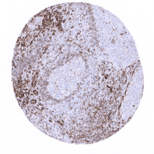
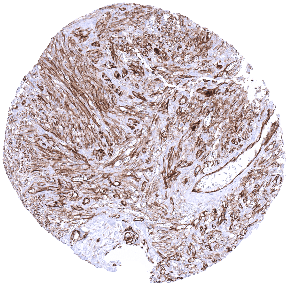
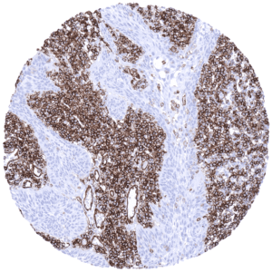
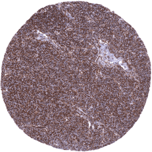
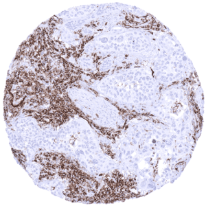
Staining Pattern in Relevant Tumor Types
CD31 is consistently expressed in vascular tumors and B-cell lymphomas. B-cell non Hodgkin lymphoma show a heterogeneous pattern of CD31 expression depending on their histogenetic derivation. CD31 expression is usually high in small lymphocytic lymphoma, intermediate in marginal zone and diffuse large cell lymphomas, and low in follicular lymphoma. CD31 expression has so far not been systematically analyzed in large numbers of different non-angiogenic tumor types. Individual reports have, however, indicated that CD31 can be aberrantly expressed in non-vascular tumors. CD31 positivity has for example been reported for individual cases of peripheral neuroectodermal tumors (PNET), mucoepidermoid carcinoma, papillary thyroid carcinoma, sweat gland tumours, metaplastic breast carcinoma with spindle cells, and malignant mesothelioma.
The TCGA findings on CD31 RNA expression in different tumor categories have been summarized in the Human Protein Atlas.
Compatibility of Antibodies
No data available at the moment
Protocol Recommendations
IHC users have different preferences on how the stains should look like. Some prefer high staining intensity of the target stain and even accept some background. Others favor absolute specificity and lighter target stains. Factors that invariably lead to more intense staining include higher concentration of the antibody and visualization tools, longer incubation time, higher temperature during incubation, higher temperature and longer duration of the heat induced epitope retrieval (slide pretreatment). The impact of the pH during slide pretreatment has variable effects and depends on the antibody and the target protein.
All images and data shown here and on in image galleries are obtained by the manual protocol described below. Other protocols resulting in equivalent staining are described as well.
Manual protocol
Freshly cut TMA sections should be used. Heat-induced antigen retrieval for 5 minutes in an autoclave at 121°C in pH 7.8. MSVA-031M antibody specific against CD31 protein is applied at 37°C for 60 minutes at a dilution of 1:150. Visualization of bound antibody by the EnVision Kit (Dako, Agilent) according to the manufacturer’s directions.
Agilent / Dako – Autostainer Link 48
Pretreatment in PT-Link for 30 minutes at 95°C (pH high); FLEX peroxidase blocking for 5 minutes (room temperature), MSVA-031M 1:150 for 20 minutes (room temperature), FLEX+ mouse/rabbit (LINKER) for 15 minutes (room temperature), horseradish peroxidase (HRP) for 20 minutes (room temperature), FLEX DAB+Sub-Chromo for 10 minutes (room temperature), FLEX hematoxylin for 5 minutes (room temperature).

These images reflect stainings by the protocol described above. It is of note that a comparable staining result can also be obtained by different protocols. In general, a longer pretreatment, a longer incubation time of the primary antibody, a higher antibody concentration, and a longer incubation time of FLEX+LINKER result in stronger staining, potentially at the cost of more background staining. Modifications of the protocol with a strengthening effect on staining intensity in combination with changes of other parameters that result in lower staining intensity can result in a comparable result as shown above.
Leica – BOND RX
Dewax at 72°C for 30 seconds; Pretreatment in Bond Epitope Retrieval Solution (ER2 – EDTA pH9) for 20 minutes at 100°C; Peroxidase blocking for 5 minutes (room temperature), MSVA-031M 1:300 for 15 minutes (room temperature), Post primary (rabbit anti mouse) for 8 minutes (room temperature), Polymer (goat anti rabbit) for 8 minutes (room temperature), mixed DAB refine for 10 minutes (room temperature), hematoxylin for 5 minutes (room temperature).

These images reflect stainings by the protocol described above. It is of note that a comparable staining result can also be obtained by different protocols. In general, a longer pretreatment, a longer incubation time of the primary antibody, a higher antibody concentration, a higher temperature during incubation, and a longer incubation time of Post primary and or the Polymer result in stronger staining, potentially at the cost of more background staining. Modifications of the protocol with a strengthening effect on staining intensity in combination with changes of other parameters that result in lower staining intensity can result in a comparable result as shown above.
Roche – Ventana Discovery ULTRA
Pretreatment for 64 minutes at 100°C (pH 8,4); CM peroxidase blocking for 12 minutes (room temperature), MSVA-031M 1:75 for 20 minutes at 36°C, secondary antibody (anti-mouse HQ) for 12 minutes at 36°C, anti-HQ HRP for 12 minutes at room temperature, DAB at room temperature, hematoxylin II at room temperature for 8 minutes, bluing reagent at room temperature for 4 minutes.

These images depict staining results obtained by the protocol described above. It is of note, that the Ventana machines generally require higher antibody concentrations than other commonly used autostainers because the antibodies are automatically diluted during the procedure. Various other protocols can result in an identical result as shown above. A longer pretreatment, a longer incubation time of the primary antibody, a higher antibody concentration, a higher temperature during incubation, and a longer incubation time of secondary antibody and or the anti-HQ HRP result in stronger staining, potentially at the cost of more background staining.
Potential Research Applications
- The microvessel density is considered a prognostic feature in several cancer types. The relevance of the tumor vessel density needs to be further evaluated, also in combination with a quantitation of other elements (inflammatory cell types) of the tumor microenvironnement.
- CD31 can also be expressed in non-vascular neoplasias. The tumor-biologic role of this phenomenon needs to be evaluated.
- The role of angiogenetic T-cells – positive for CD3, CD31 and CXCR4 – is still insufficiently understood.
Evidence for Antibody Specificity in IHC
Specificity of MSVA-031M is documented by strong positive staining in cell types that are well documented to express CD31 such as endothelial cells of all organs and cells of the hematopoietic system. The complete absence of CD31 immunostaining in all other analyzed cell types documents absence of cross-reactivity and low propensity to generate non-specific background staining.