
295,00 € – 895,00 €
Product details
Synonyms = Deoxynucleotidyltransferase terminal; DNTT; Nucleosidetriphosphate DNA Deoxynucleotidylexotransferase; TDT; Terminal addition enzyme; Terminal deoxynucleotidyltransferase; Terminal deoxyribonucleotidyltransferase
Antibody type = Recombinant Rabbit monoclonal / IgG
Clone = MSVA-453R
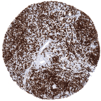
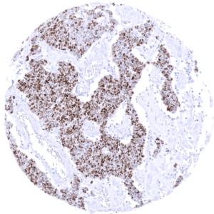
Positive control = Thymus: At least a moderate nuclear immunostaining should be seen in virtually all cortical lymphocytes of the normal thymus.
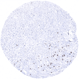
Negative control = Thymus: The vast majority of the medullary lymphocytes of the normal thymus should be negative.
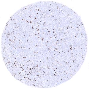
Tonsil: The vast majority of lymphoid cells and all epithelial cells should be negative.
Cellular localization = Nucleus
Reactivity = Human
Application = Immunohistochemistry
Dilution = 1:100
Intended Use = Research Use Only
Relevance of Antibody
Biology Behind
Terminal deoxynucleotidyl transferase (TdT), also known as DNA nucleotidylexotransferase (DNTT) or terminal transferase, is a 58 kDa protein coded by the DNTT gene on 10q23-q24. It is a highly specialized template-independent DNA polymerase which is only expressed in immature, pre-B, pre-T lymphoid cells. In these cells TdT catalyzes the addition of N-nucleotides to the V, D, and J exons of the T-cell receptor (TCR) and B-cell receptor (TCR) genes which creates junctional diversity during antibody gene recombination. A high diversity of TCR and BCR is important for the diversity to antibody recognition sites and thus for the function of the immune system.
Staining Pattern in Normal Tissues
Using antibody MSVA-453R, a moderate to strong nuclear staining is seen in the vast majority of cortical lymphocytes of the normal thymus while only scattered positive cells are present in the medulla of the thymus (especially in areas adjacent to the cortex). Few scattered lymphoid cells with strong TdT expression can be seen in the interfollicular area of lymph nodes and tonsils (especially in non-adult patients). Few scattered positive cells (hematogones) can also be seen in the bone marrow.
These findings are largely consistent with the RNA data described in the Human Protein Atlas (Tissue expression TdT). The data from the protein atlas are complemented by IHC images documenting TdT protein expression in tissues.
Suggested positive tissue control: Thymus: At least a moderate nuclear immunostaining should be seen in virtually all cortical lymphocytes of the normal thymus.
Suggested negative tissue control: Thymus: The vast majority of the medullary lymphocytes of the normal thymus should be negative.
Tonsil: The vast majority of lymphoid cells and all epithelial cells should be negative. y, colonic mucosa, and epidermis.

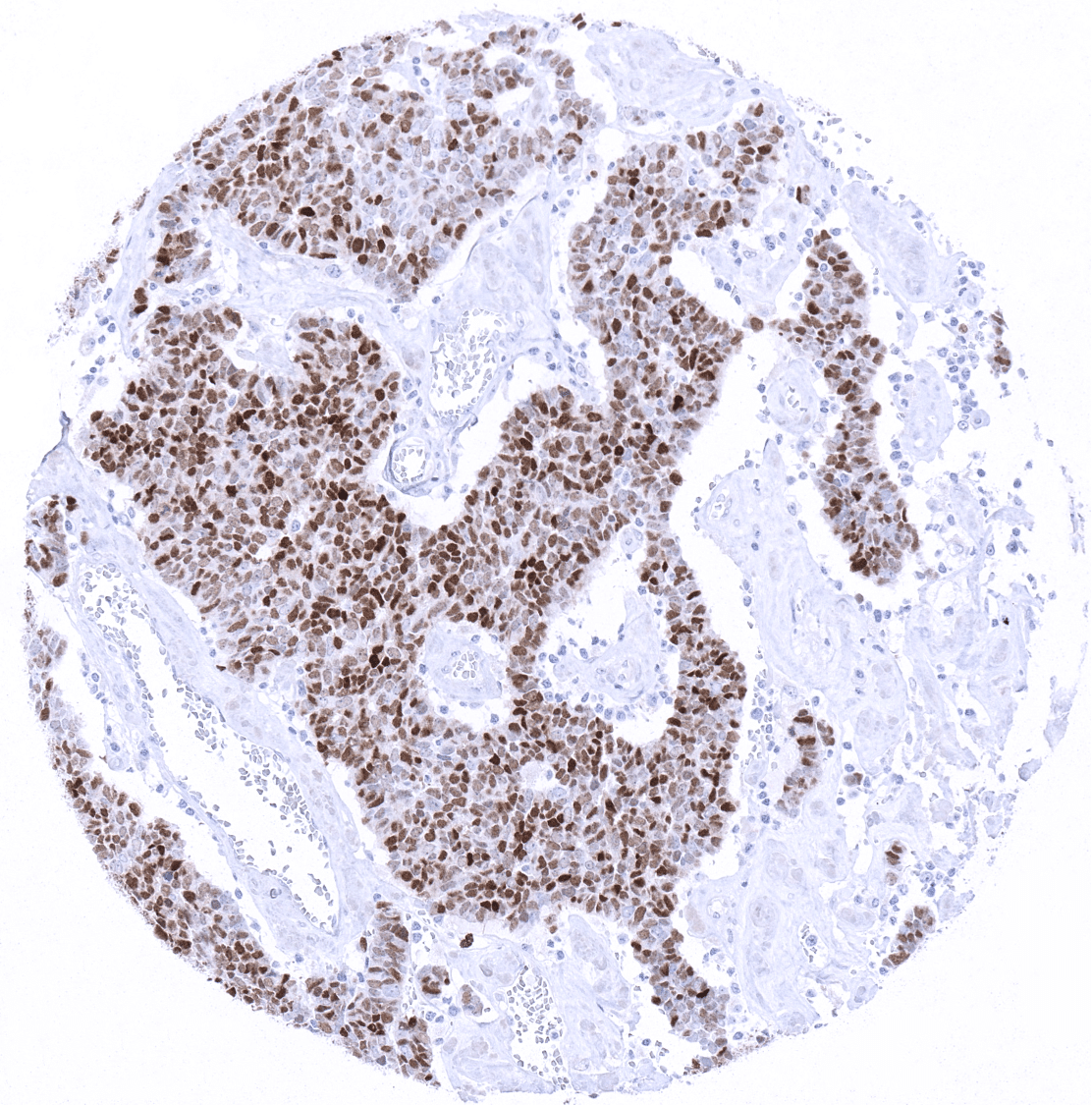
Staining Pattern in Relevant Tumor Types
Almost all pre-B and pre-T acute lymphoblastic leukemia/lymphoblastic lymphoma (ALL/LBL) show strong and diffuse TdT immunostaining. An often weak and only focal TdT positivity can also be seen in acute myelogenous leukemia / myeloid sarcoma, blastic plasmacytoid dendritic cell neoplasm, Burkitt lymphoma, as well as in some indolent T cell proliferations. Positive TdT immunostaining is seen in the vast majority of lymphocyte predominant thymomas and in 40-50% of Merkel cell carcinomas.
The TCGA findings on TdT RNA expression in different tumor categories have been summarized in the Human Protein Atlas.

Compatibility of Antibodies
No data available at the moment
Protocol Recommendations
IHC users have different preferences on how the stains should look like. Some prefer high staining intensity of the target stain and even accept some background. Others favor absolute specificity and lighter target stains. Factors that invariably lead to more intense staining include higher concentration of the antibody and visualization tools, longer incubation time, higher temperature during incubation, higher temperature and longer duration of the heat induced epitope retrieval (slide pretreatment). The impact of the pH during slide pretreatment has variable effects and depends on the antibody and the target protein. Accordingly, multiple different protocols can generate identical staining results.
All images and data shown here and in our image galleries are obtained by the manual protocol described below. Other protocols resulting in equivalent staining are described as well.
-Manual protocol
Freshly cut sections should be used (less than 10 days between cutting and staining). Heat-induced antigen retrieval for 5 minutes in an autoclave at 121°C in pH 9 Target Retrieveal Solution buffer. Apply MSVA-453R at a dilution of 1:25 at 37°C for 60 minutes. Visualization of bound antibody by the EnVision Kit (Dako, Agilent) according to the manufacturer’s directions.
Potential Research Applications
- TdT expression has also been described in non-hematological neoplasias. The role of TdT in these neoplasms is unclear. Many tumor types have so far not been analyzed for TdT expression.
Evidence for Antibody Specificity in IHC
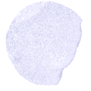
Specificity of MSVA-453R is documented by strong positive staining in cell types that are well documented to express TdT such cortical thymocytes and scattered cells in lymph nodes and tonsils while staining was absent in all tissues known to not express TdT including tissues notorious for non-specific IHC background such as kidney, colonic mucosa, and epidermis.