295,00 € – 995,00 €
Product details
Synonyms = EGFRvIII,EGFR mutant,EGFR variant,EGFRv3
Antibody type = Recombinant Rabbit monoclonal / IgG
Clone = HMV3805
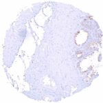
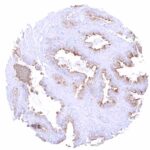
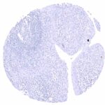
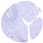
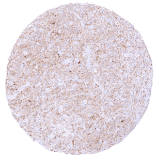
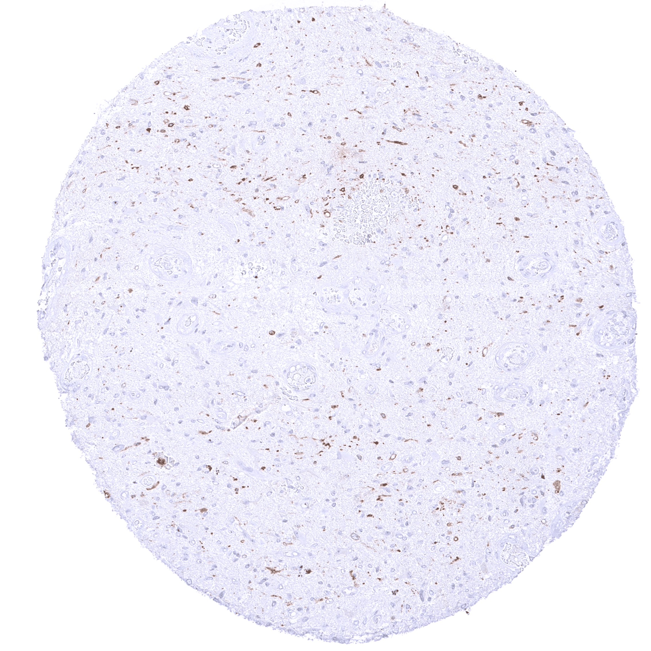
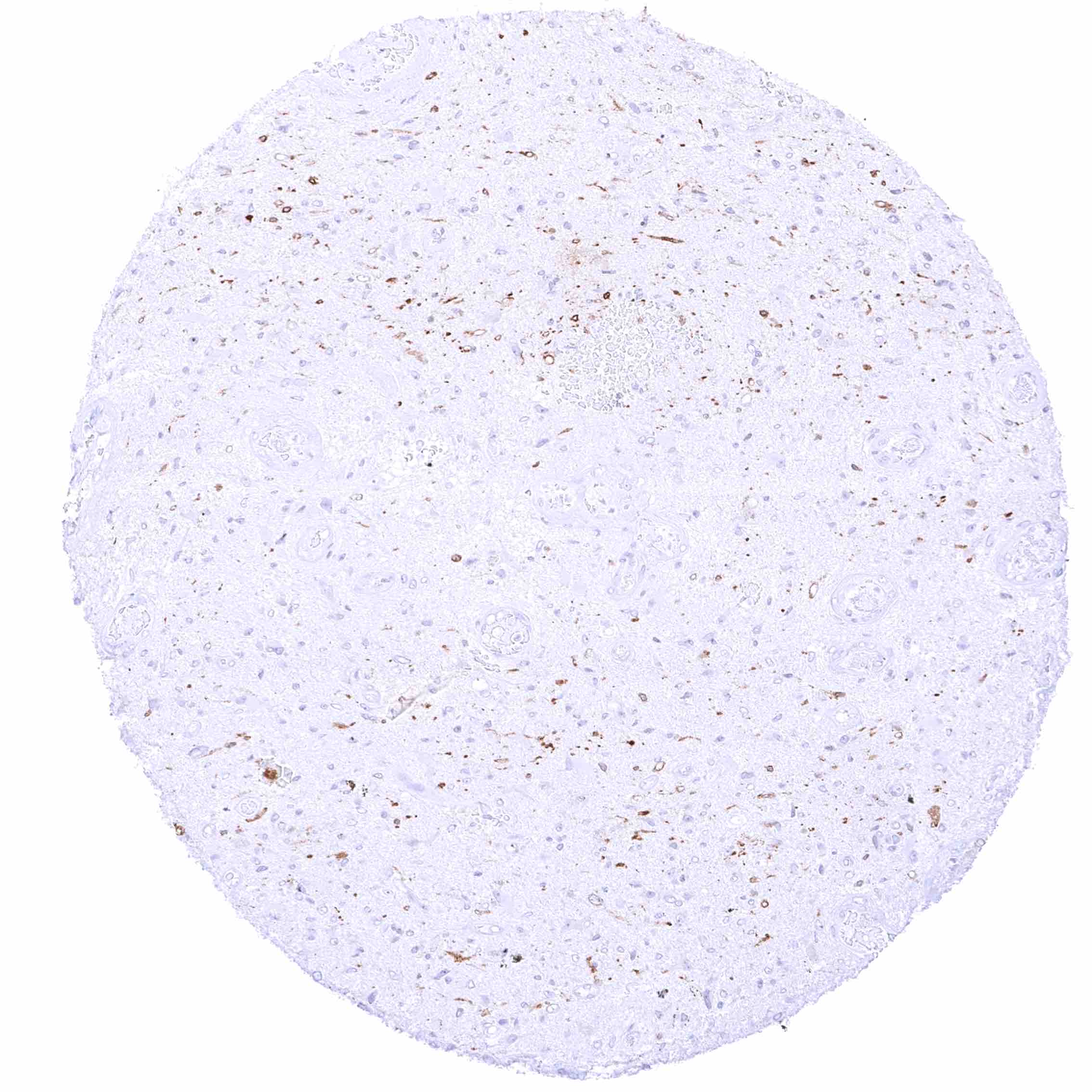
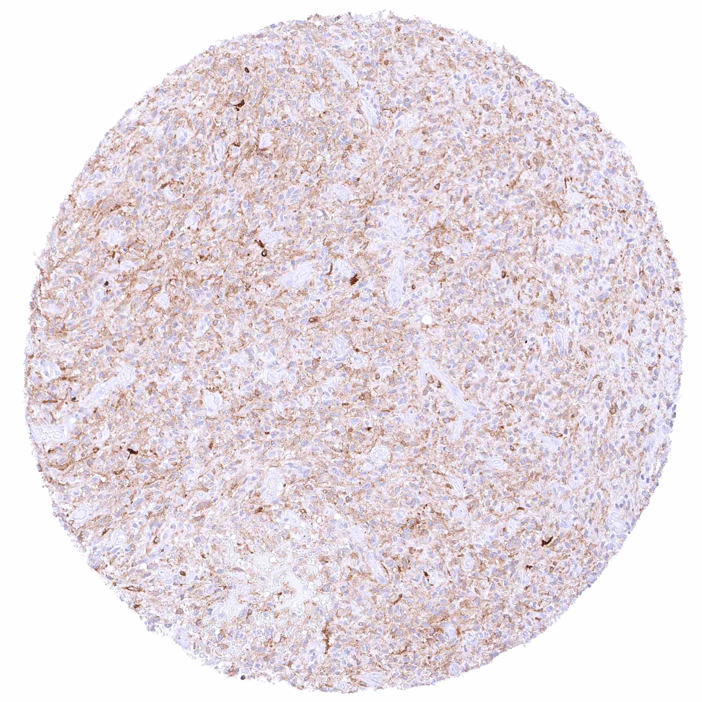
Positive control = Glioblastoma multiforme with known EGFRvIII mutation: Tumor cells must show a distinct EGFRvIII staining.
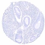
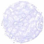
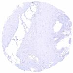
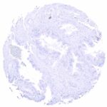
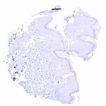
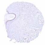
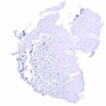
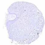
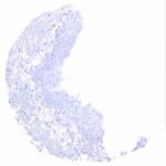
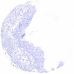
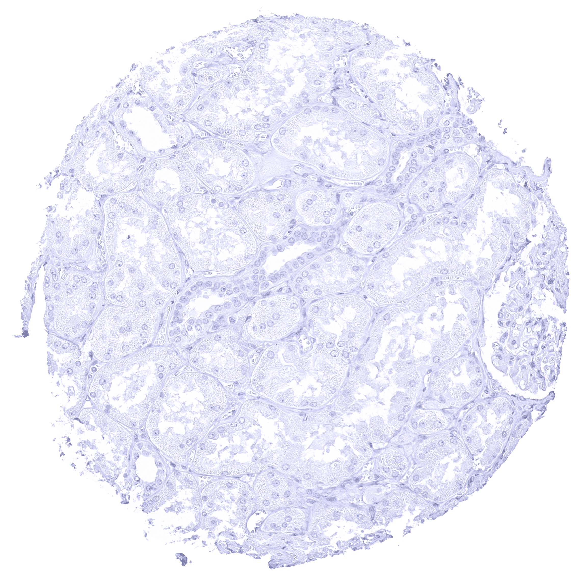
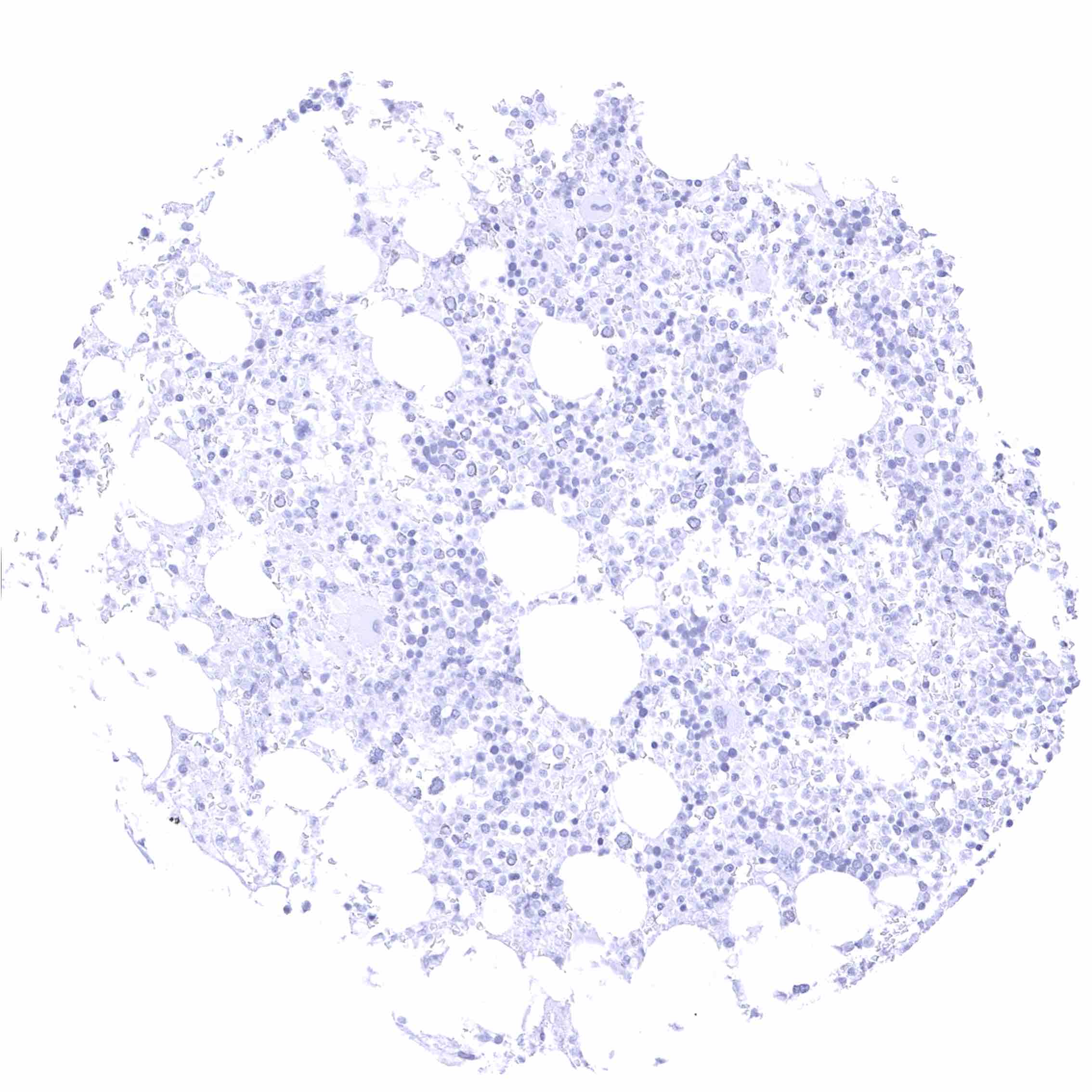
Negative control = Kidney*: EGFRvIII staining must be completely absent in all cells.
*Note: other normal tissues can serve as negative controls as well.
Cellular localization = Cytoplasmic
Reactivity = Human
Application = Immunohistochemistry
Dilution = 1:50 – 1:100
Intended Use = Research Use Only
Relevance of Antibody
EGFRvIII is a marker for a critical molecular event in glioblastoma.
Biology Behind
The epidermal growth factor receptor (EGFR) receptor protein is coded by the EGFR gene on chromosome 7p11.2. The EGFR variant type III (EGFRvIII) is formed by an 801 base pair in-frame deletion ranging from exons 2 to 7. EGFRvIII is the most common mutation of EGFR in glioblastoma multiforme. It only develops in EGFR amplified tumors and occurs in about 25% of all glioblastomas. EGFRvIII is a ligand-independent receptor. Although its signaling activity is thought to be lower than of activated wild-type EGFR, its constitutive activity results in a significant growth advantage to glioblastoma cells, especially in vivo. EGFRVIII is linked to shorter survival of glioblastoma multiforme.
Staining Pattern in Normal Tissues
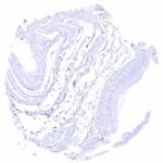
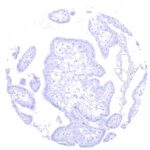
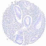
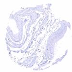
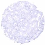
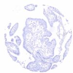
Images describing the EGFRvIII staining pattern in normal tissues obtained by the antibody HMV3805 are shown in our “Normal Tissue Gallery”.
| Brain | Cerebrum | Negative. |
| Cerebellum | Negative. | |
| Endocrine Tissues | Thyroid | Negative. |
| Parathyroid | Negative. | |
| Adrenal gland | Negative. | |
| Pituitary gland | Negative. | |
| Respiratory system | Respiratory epithelium | Negative. |
| Lung | Negative. | |
| Gastrointestinal Tract | Salivary glands | Negative. |
| Esophagus | Negative. | |
| Stomach | Negative. | |
| Duodenum | Negative. | |
| Small intestine | Negative. | |
| Appendix | Negative. | |
| Colon | Negative. | |
| Rectum | Negative. | |
| Liver | Negative. | |
| Gallbladder | Negative. | |
| Pancreas | Negative. | |
| Genitourinary | Kidney | Negative. |
| Urothelium | Negative. | |
| Male genital | Prostate | Negative. |
| Seminal vesicles | Negative. | |
| Testis | Negative. | |
| Epididymis | Negative. | |
| Female genital | Breast | Negative. |
| Uterus, myometrium | Negative. | |
| Uterus, ectocervix | Negative. | |
| Uterus endocervix | Negative. | |
| Uterus, endometrium | Negative. | |
| Fallopian Tube | Negative. | |
| Ovary | Negative. | |
| Placenta early | Negative. | |
| Placenta mature | Negative. | |
| Amnion | Negative. | |
| Chorion | Negative. | |
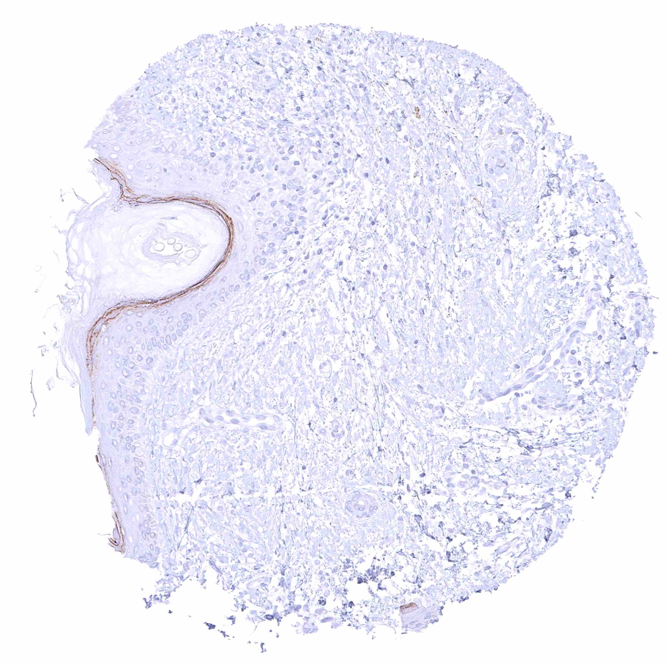
| Skin | Epidermis | Distinct EGFRvIII staining of the granular layer of keratinizing squamous epithelium. |
| Sebaceous glands | Negative. | |
| Muscle/connective tissue | Heart muscle | Negative. |
| Skeletal muscle | Negative. | |
| Smooth muscle | Negative. | |
| Vessel walls | Negative. | |
| Fat | Negative. | |
| Stroma | Negative. | |
| Endothelium | Negative. | |
| Bone marrow/ lymphoid tissue | Bone marrow | Negative. |
| Lymph node | Negative. | |
| Spleen | Negative. | |
| Thymus | Negative. | |
| Tonsil | Negative. | |
| Remarks | EGFRvIII staining of the granular layer of keratinizing squamous epithelium represents a (tolerable) cross-reactivity of the antibody. |
EGFRvIII is a pathogenic mutation which does not occur in normal tissues. A staining of the stratum granulosum of the epidermis by HMV3805 represents a (tolerable) cross-reactivity of the antibody.
Positive control = Glioblastoma multiforme with known EGFRvIII mutation: Tumor cells must show a distinct EGFRvIII staining.
Negative control = Kidney*: EGFRvIII staining must be completely absent in all cells.
*Note: other normal tissues can serve as negative controls as well.
Staining Pattern in Relevant Tumor Types
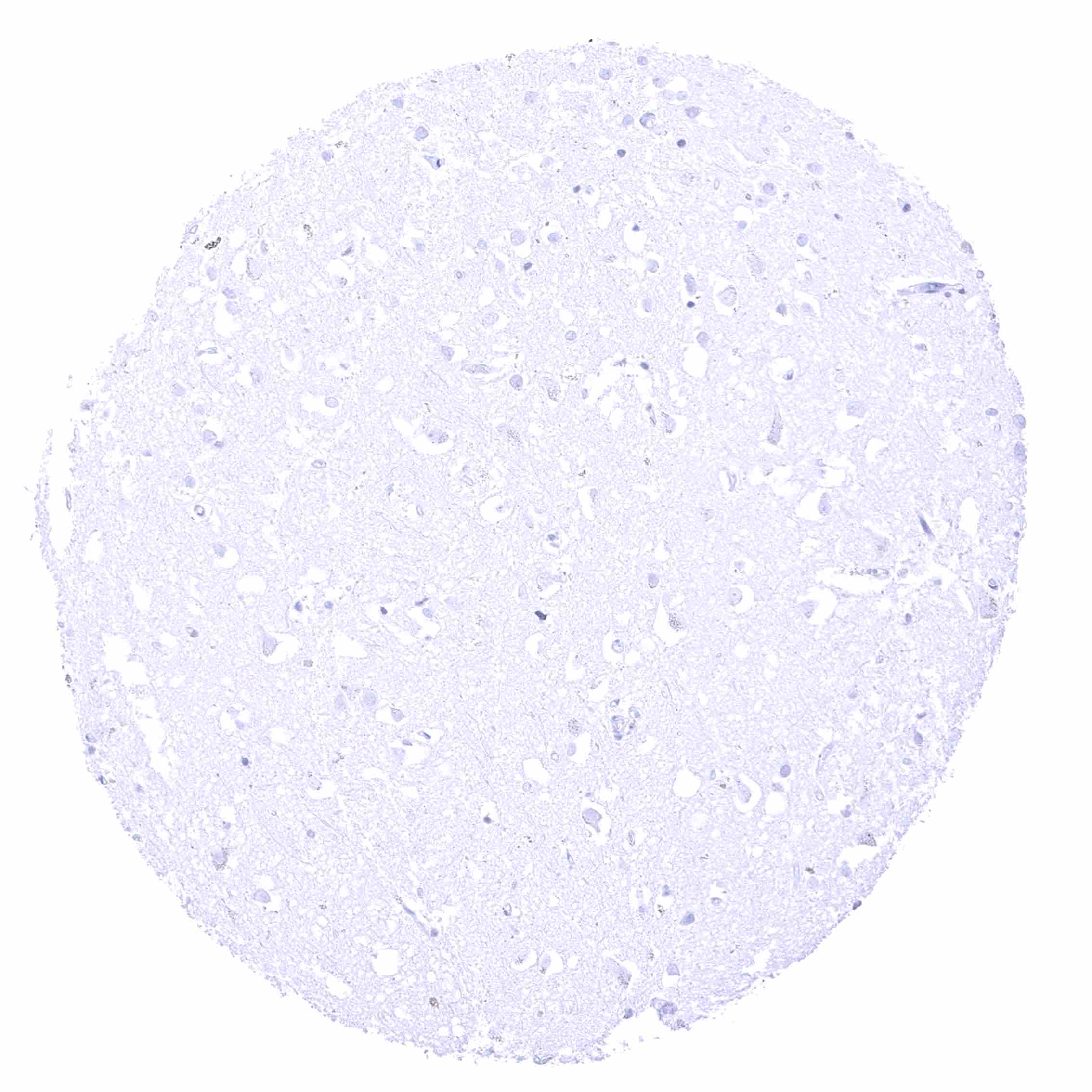
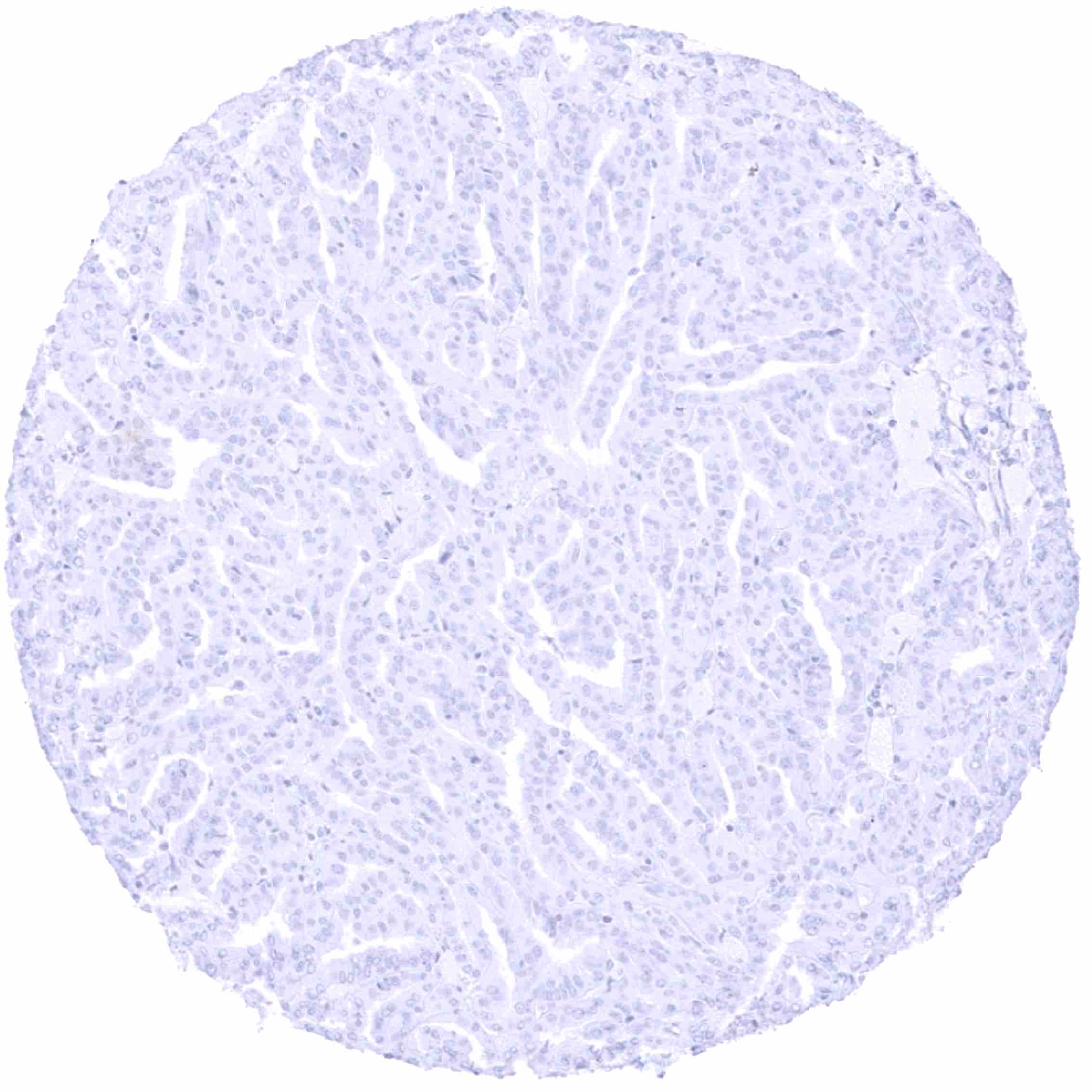
EGFRvIII mutations occur in 25% of glioblastoma multiforme. Very rarely, this variant can also be found in other cancers.
Compatibility of Antibodies
No data available at the moment
Protocol Recommendations
IHC users have different preferences on how the stains should look like. Some prefer high staining intensity of the target stain and even accept some background. Others favor absolute specificity and lighter target stains. Factors that invariably lead to more intense staining include higher concentration of the antibody and visualization tools, longer incubation time, higher temperature during incubation, higher temperature and longer duration of the heat induced epitope retrieval (slide pretreatment). The impact of the pH during slide pretreatment has variable effects and depends on the antibody and the target protein.
All images and data shown here and in our image galleries are obtained by the manual protocol described below. Other protocols resulting in equivalent staining are described as well.
-Manual protocol
Freshly cut sections should be used (less than 10 days between cutting and staining). Heat-induced antigen retrieval for 5 minutes in an autoclave at 121°C in pH 7,8 Target Retrieval Solution buffer. Apply HMV3805 at a dilution of 1:50 at 37°C for 60 minutes. Visualization of bound antibody by the EnVision Kit (Dako, Agilent) according to the manufacturer’s directions.
Potential Research Applications
- The prognostic and therapeutic implications of EGFRvIII mutations need to be further clarified.
- Compounds targeting EGFRvIII are under research.
- The functional effects of EGFRvIII mutations are still insufficiently understood.
Evidence for Antibody Specificity in IHC
There are two ways how the specificity of antibodies can be documented for immunohistochemistry on formalin fixed tissues. These are: 1. Comparison with a second independent method for target expression measurement across a large number of different tissue types (orthogonal strategy), and 2. Comparison with one or several independent antibodies for the same target and showing that all positive staining results are also seen with other antibodies for the same target (independent antibody strategy).
Orthogonal validation: In line with the lack of EGFRvIII in normal tissues EGFRvIII staining was absent in the vast majority of tissues. EGFRvIII positivity in 4 glioblastomas with previously documented EGFRvIII mutation further confirmed the validity of the staining by HMV3805. A distinct EGFRvIII positivity at the granular layer of keratinizing squamous epithelium of the skin was the only staining seen by HMV3805 in normal tissues.
Comparison of antibodies: EGFRvIII specific staining of HMV3805 is corroborated by comparison with an independent second antibody (termed “validation antibody”). All tumors stained by HMV3805 were also stained by the “validation antibody”. A distinct EGFRvIII positivity at the granular layer of keratinizing squamous epithelium of the skin was only seen by using HMV3805 and thus considered a (tolerable) cross-reactivity of HMV3805. EGFRvIII positivity of seminal vesicle epithelium was only seen by the “validation antibody” and was therefore considered a cross-reactivity of the “validation antibody”.