295,00 € – 995,00 €
Product details
Synonyms = Claudin 18, SFTA5, SFTPJ
Antibody type = Recombinant Rabbit monoclonal / IgG
Clone = HMV4729
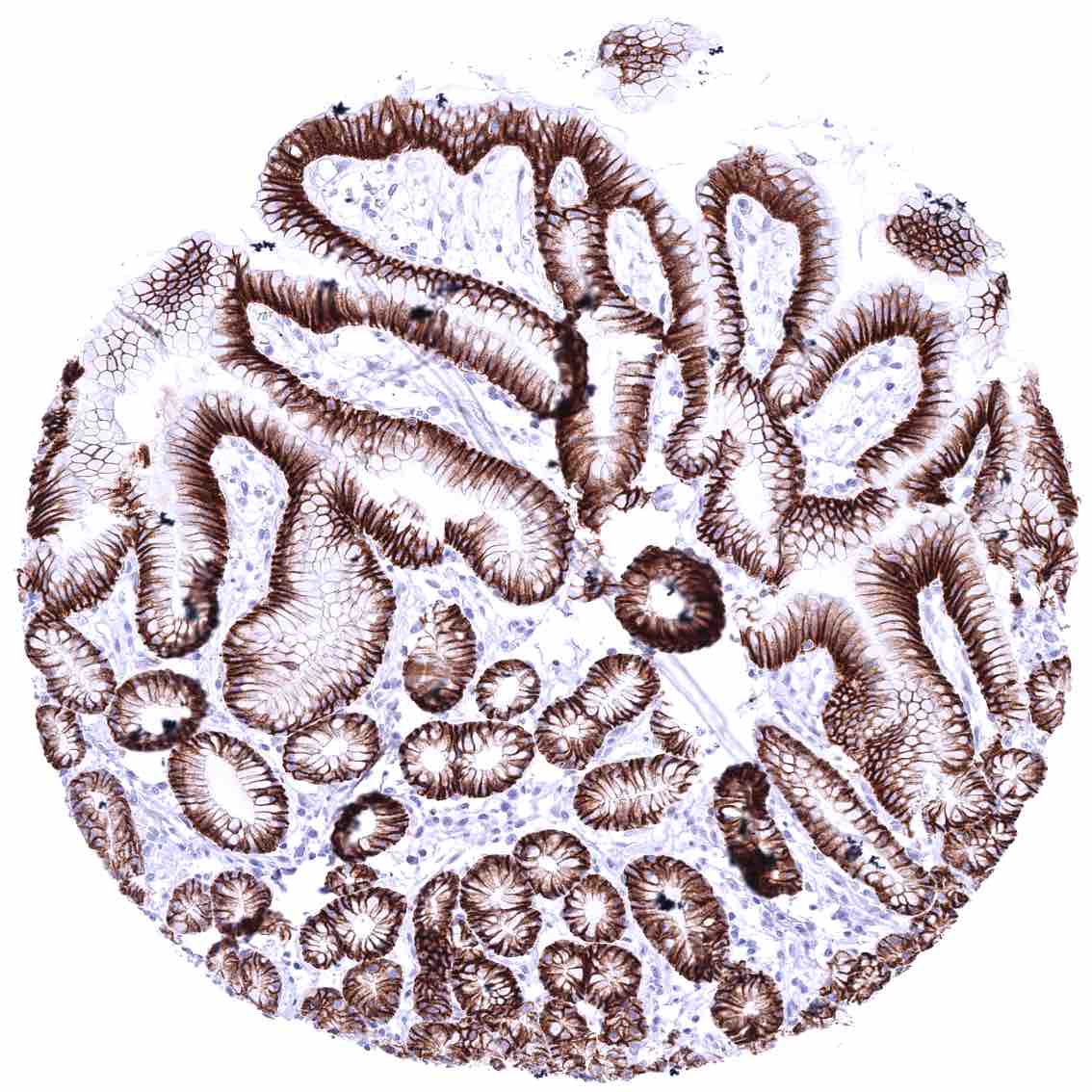
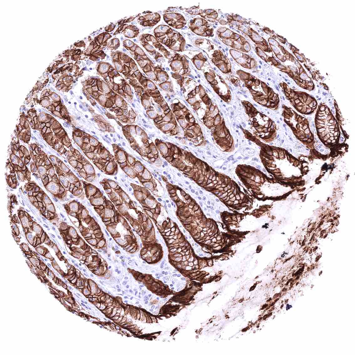
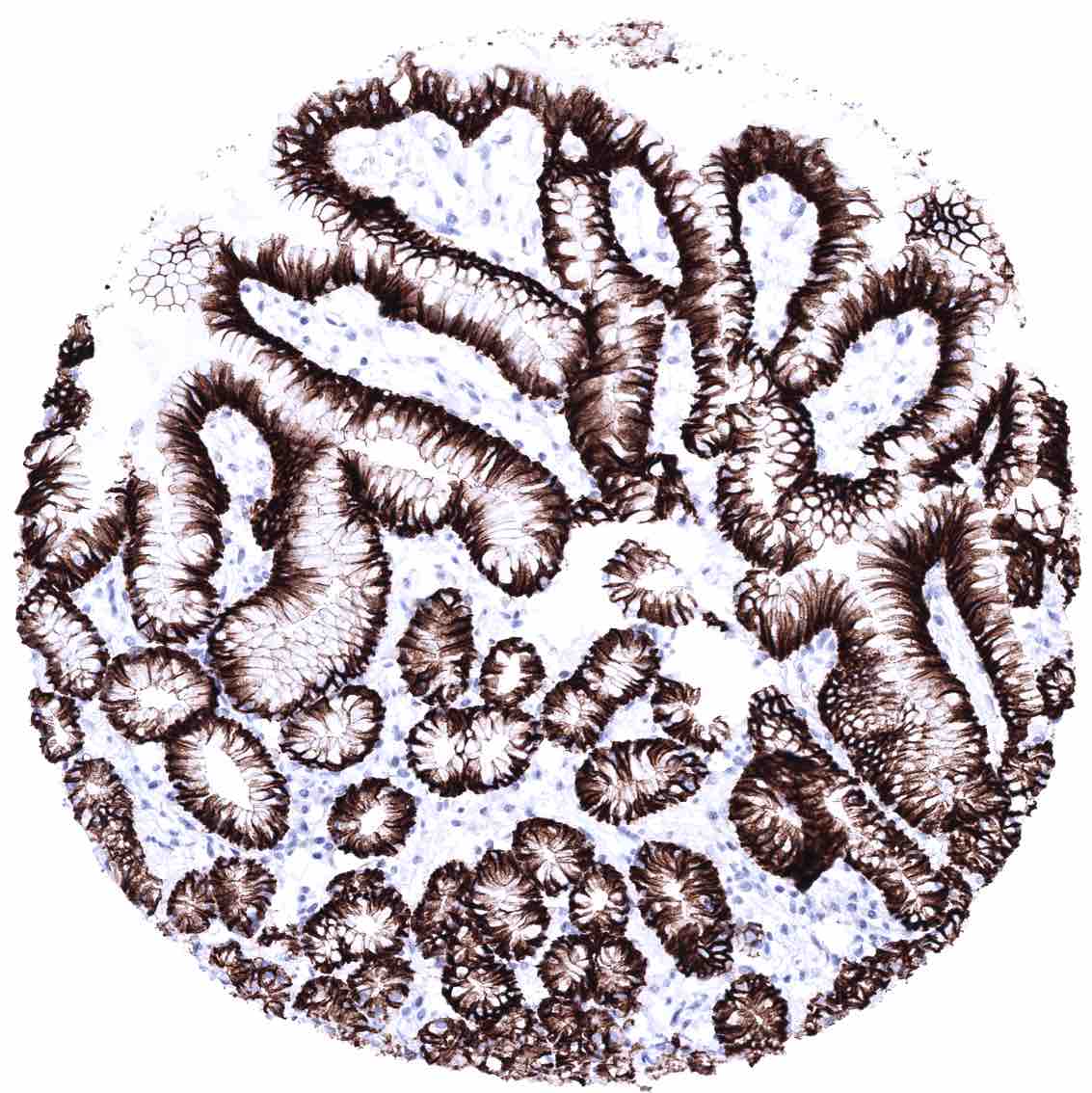
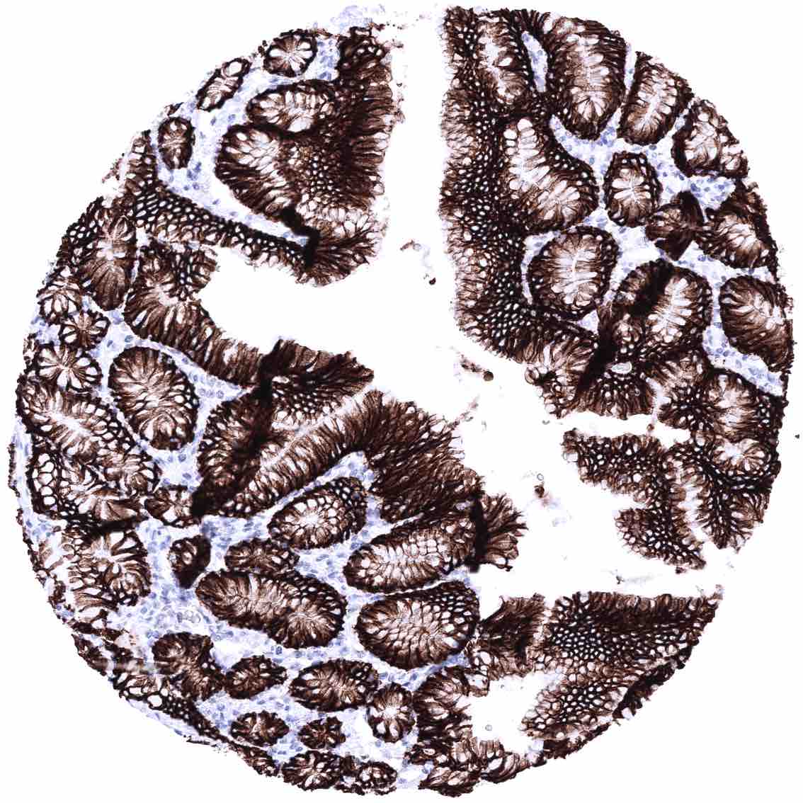
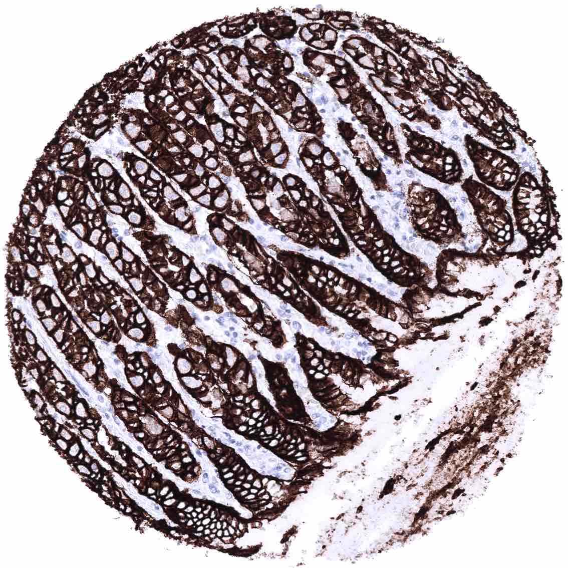
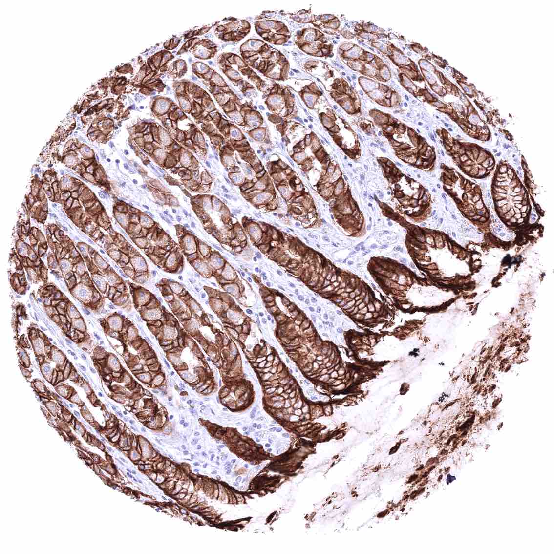
Positive control = Stomach: A strong membranous CLDN18.2 staining must be seen in all gastric epithelial cells.
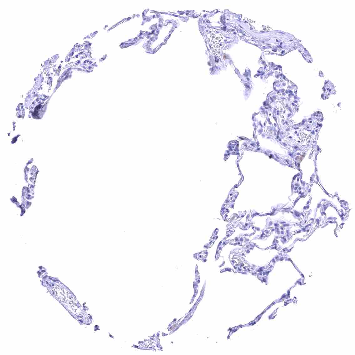
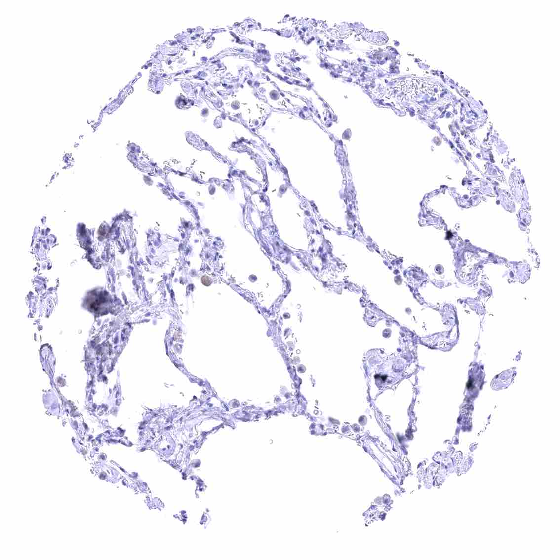
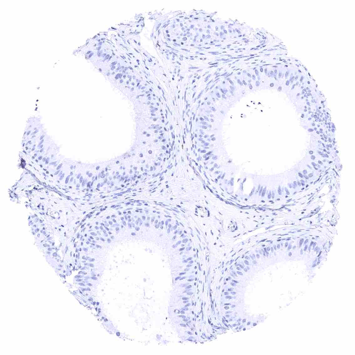
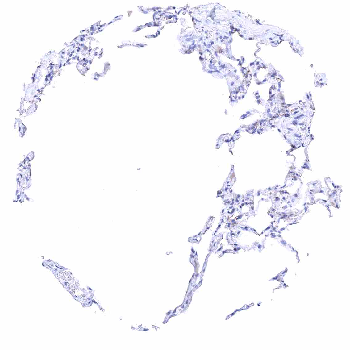
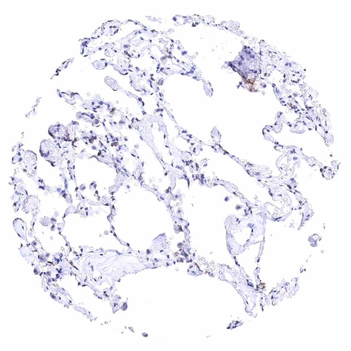
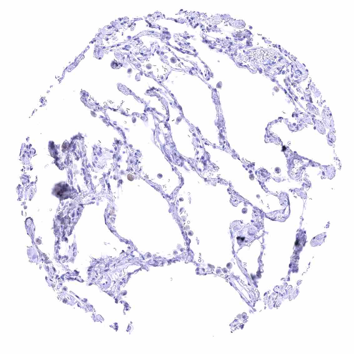
Negative control = Lung: CLDN18.2 staining must not be seen in pneumocytes.
Cellular localization = Membraneous
Reactivity = Human
Application = Immunohistochemistry
Dilution = 1:100 – 1:200
Intended Use = Research Use Only
Relevance of Antibody
Claudin 18.2 is a therapeutic target protein.
Biology Behind
Claudin 18.2 (CLDN18.2) is one of at least 26 members of the claudin family of membrane proteins that are crucial components of tight junctions, regulating paracellular permeability and maintaining cell polarity in epithelial and endothelial cells. CLDN18.2, is a 28 kDa splice variant coded by the CLDN18 gene on chromosome 3q22.3, which – in normal tissues – is exclusively expressed in gastric epithelial cells. This contrasts with CLDN18.1 which is expressed in pneumocytes of the lung. CLDN18.2 plays a crucial role in the barrier and selective permeability of the tight junctions in gastric epithelial cells by controlling the passage of ions such as Na+ and H+. CLDN18 knockdown mice develop severe gastritis within days, indicating that CLDN18.2 helps to prevent paracellular leakage of H+ ions. CLDN18.2 dysfunction may contribute to poor acidity regulation. Because of its membranous location, CLDN18.2 is a suitable therapeutic target in cancer. In healthy gastric epithelium, CLDN18.2 is confined to intercellular membranes, rendering it inaccessible to antibody binding. Changes in cell polarity occurring during malignant transformation expose CLDN18.2 on the tumor cell surface, allowing its recognition by therapeutic antibodies. The monoclonal CLDN18.2 antibody Zolbetuximab induces antibody-dependent cellular cytotoxicity and inhibits tumor growth in CLDN18.2 expressing cancers. It has been FDA approved for patients with locally advanced or metastatic gastric or gastroesophageal junction adenocarcinoma with high expression of CLDN18.2. Various other drugs targeting CLDN18.2 are being evaluated in clinical trials.
Staining Pattern in Normal Tissues
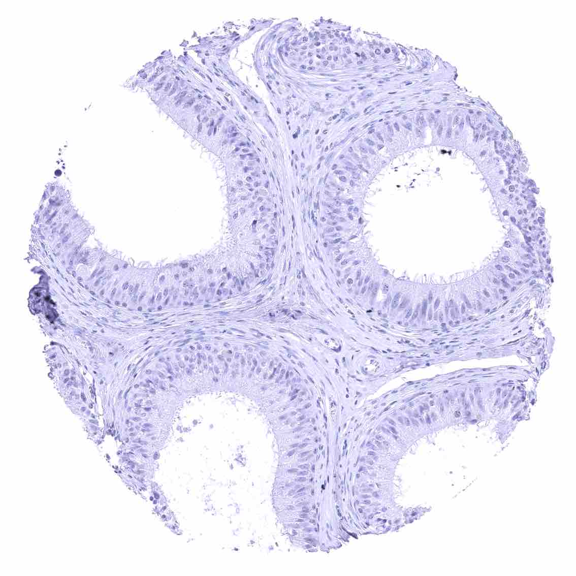
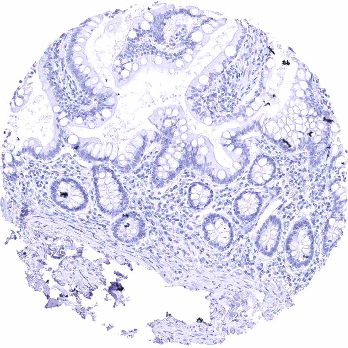
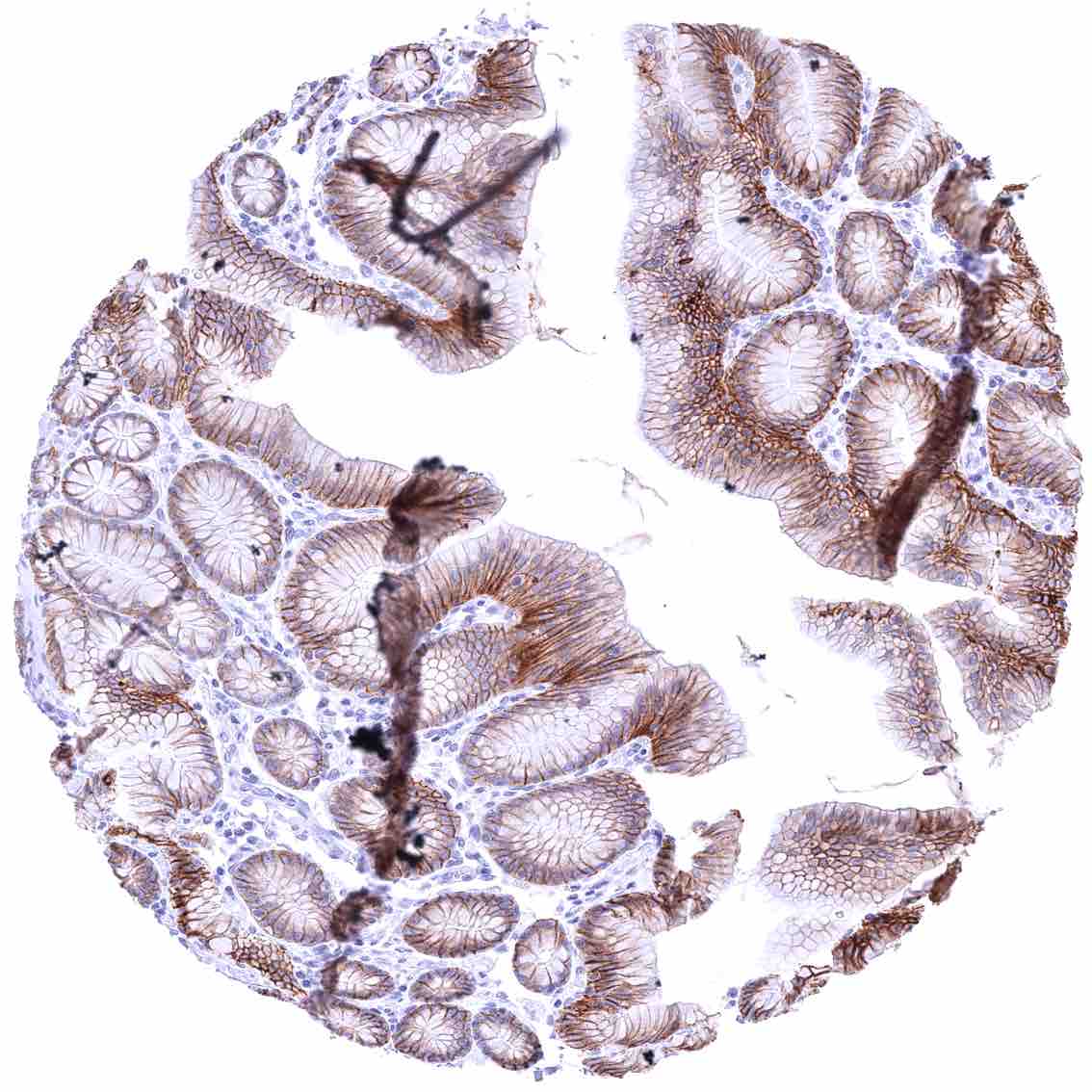
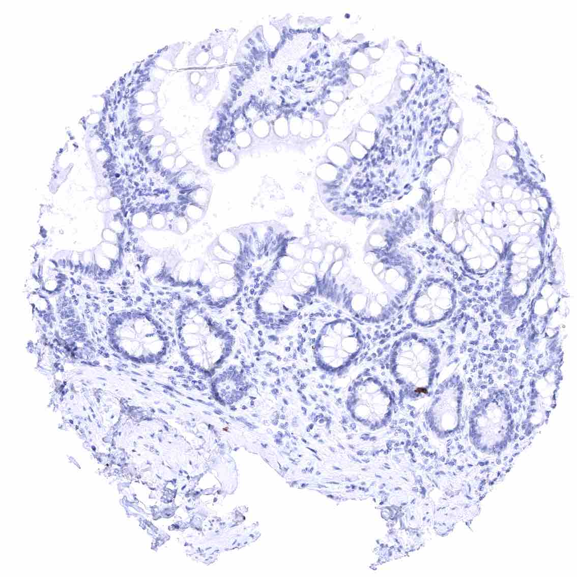
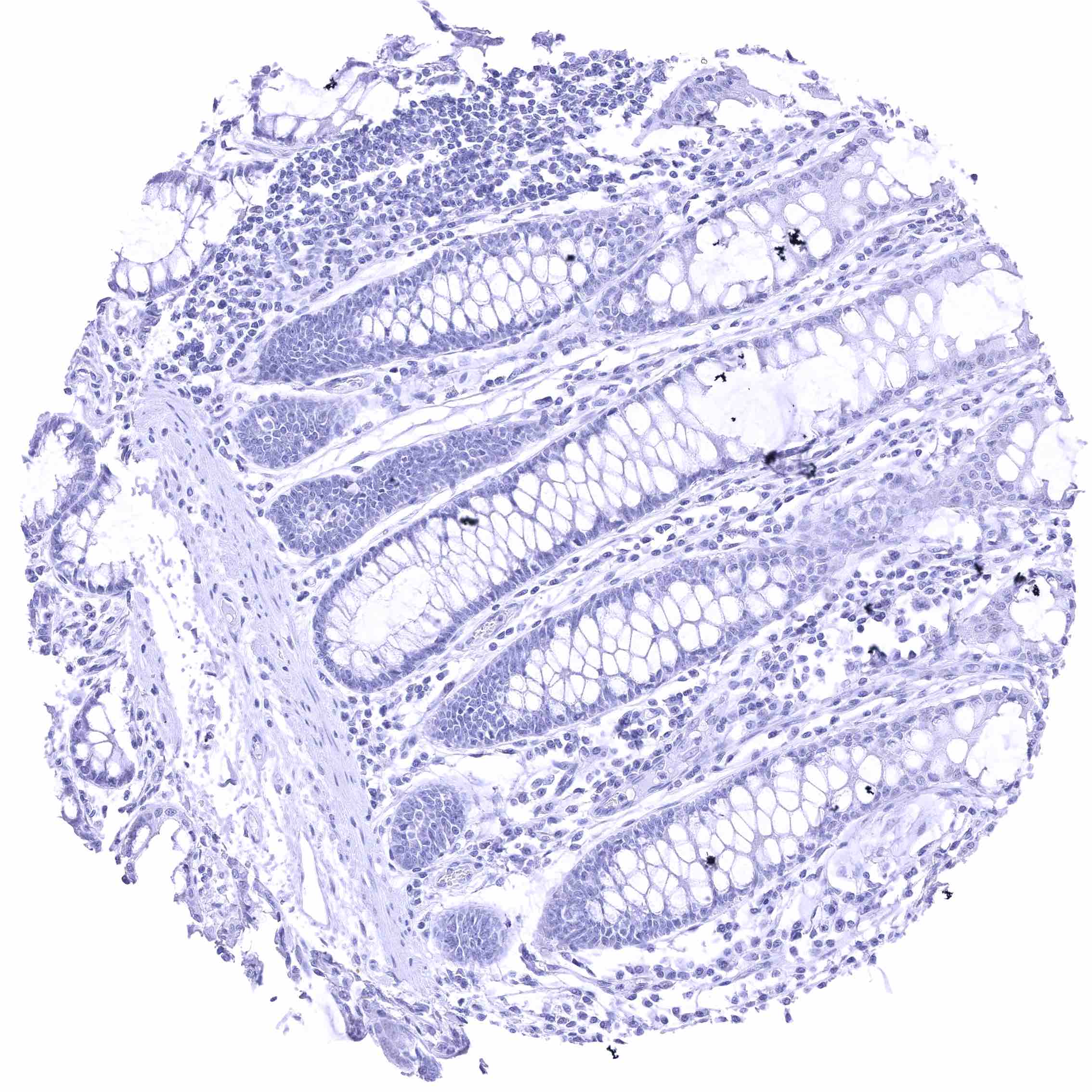
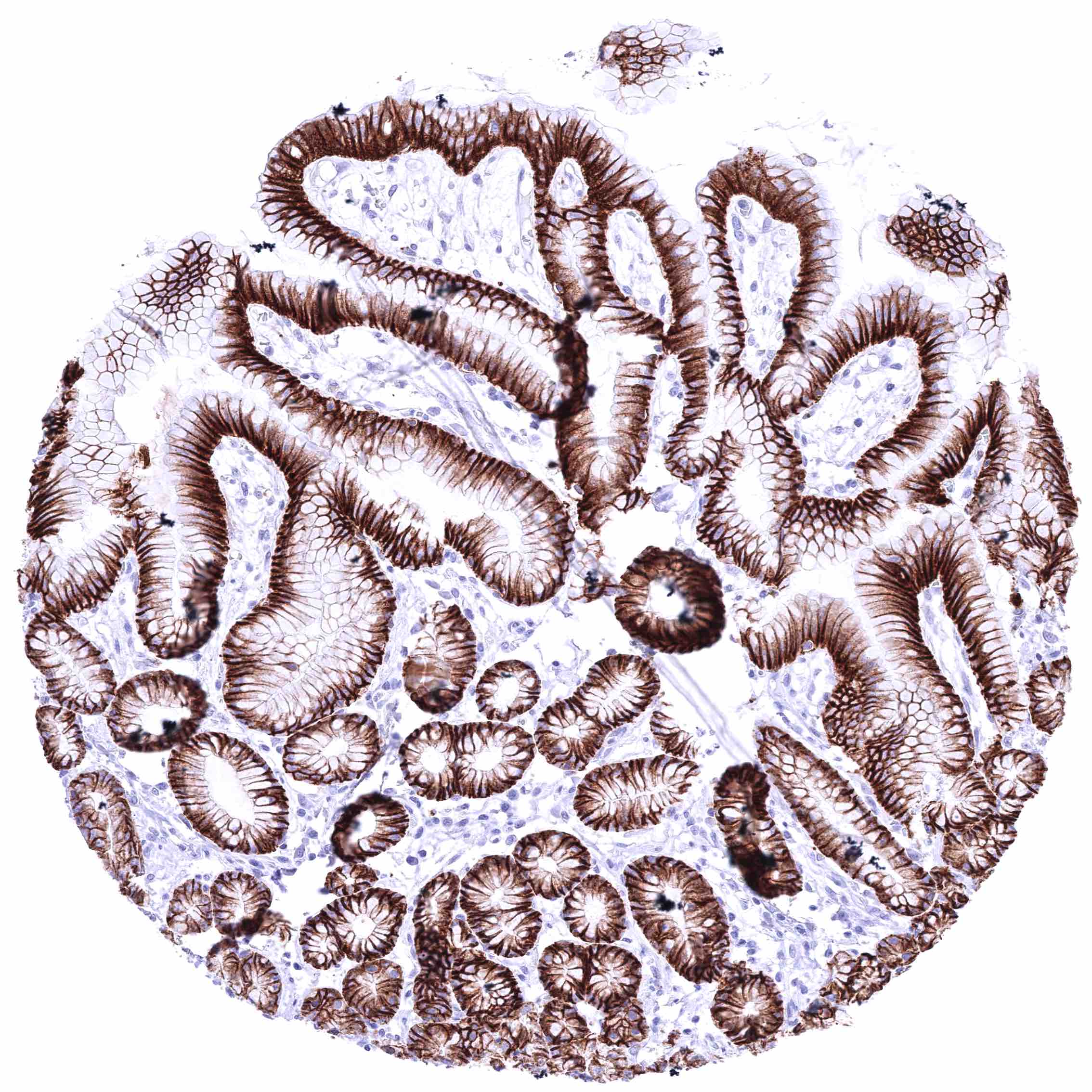
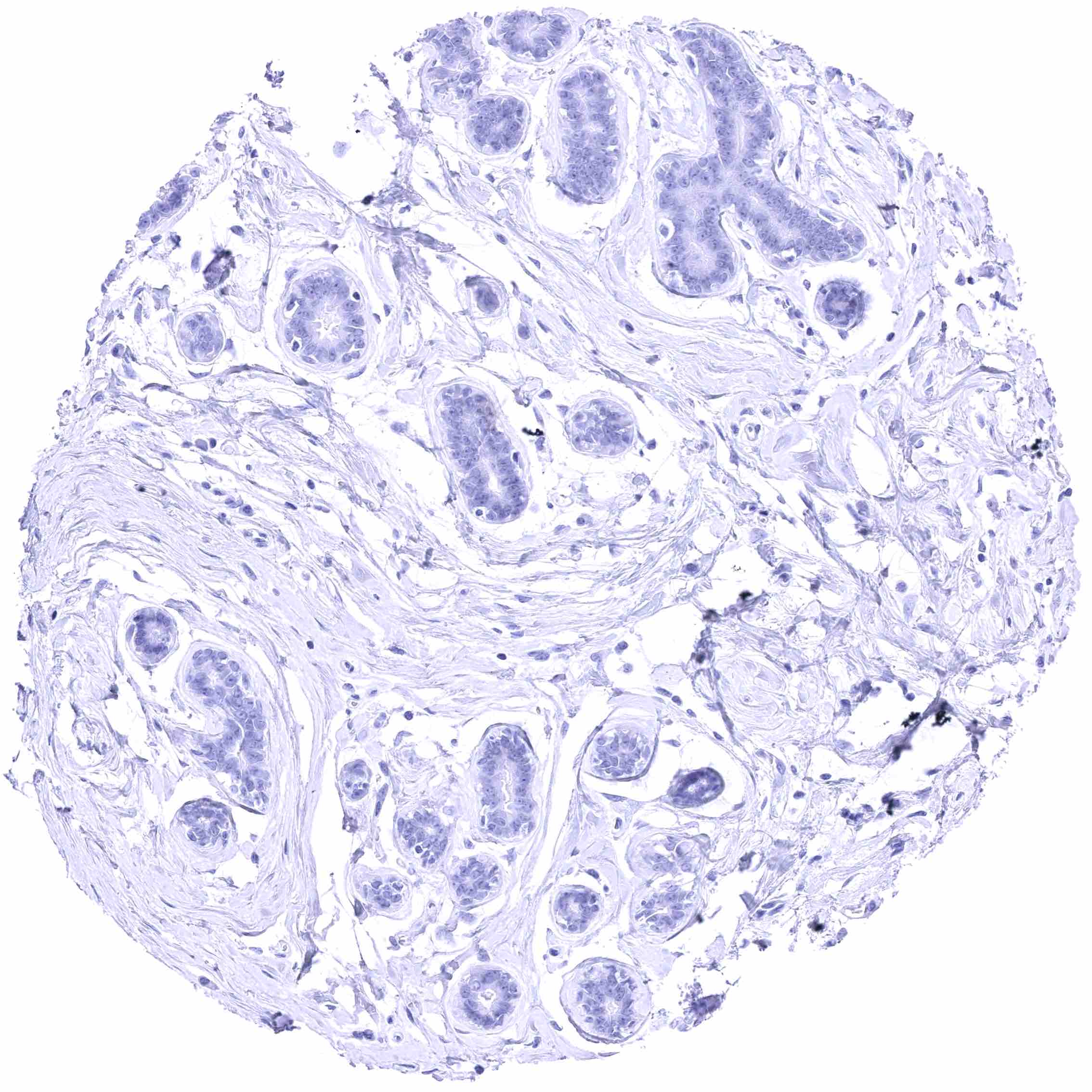
Images describing the Claudin 18.2 staining pattern in normal tissues obtained by the antibody HMV4279 are shown in our “Normal Tissue Gallery”.
| Brain | Cerebrum | Negative. |
| Cerebellum | Negative. | |
| Endocrine Tissues | Thyroid | Negative. |
| Parathyroid | Negative. | |
| Adrenal gland | Negative. | |
| Pituitary gland | Negative. | |
| Respiratory system | Respiratory epithelium | Negative. |
| Lung | Negative. | |
| Gastrointestinal Tract | Salivary glands | Negative. |
| Esophagus | Negative. | |
| Stomach | Distinct, membranous CLDN18.2 positivity of all epithelial cell types in antrum and corpus. | |
| Duodenum | Negative. | |
| Small intestine | Negative. | |
| Appendix | Negative. | |
| Colon | Negative. | |
| Rectum | Negative. | |
| Liver | Negative. | |
| Gallbladder | Negative. | |
| Pancreas | Negative. | |
| Genitourinary | Kidney | Negative. |
| Urothelium | Negative. | |
| Male genital | Prostate | Negative. |
| Seminal vesicles | Negative. | |
| Testis | Negative. | |
| Epididymis | Negative. | |
| Female genital | Breast | Negative. |
| Uterus, myometrium | Negative. | |
| Uterus, ectocervix | Negative. | |
| Uterus endocervix | Negative. | |
| Uterus, endometrium | Negative. | |
| Fallopian Tube | Negative. | |
| Ovary | Negative. | |
| Placenta early | Negative. | |
| Placenta mature | Negative. | |
| Amnion | Negative. | |
| Chorion | Negative. | |
| Skin | Epidermis | Negative. |
| Sebaceous glands | Negative. | |
| Muscle/connective tissue | Heart muscle | Negative. |
| Skeletal muscle | Negative. | |
| Smooth muscle | Negative. | |
| Vessel walls | Negative. | |
| Fat | Negative. | |
| Stroma | Negative. | |
| Endothelium | Negative. | |
| Bone marrow/ lymphoid tissue | Bone marrow | Negative. |
| Lymph node | Negative. | |
| Spleen | Negative. | |
| Thymus | Negative. | |
| Tonsil | Negative. | |
| Remarks |
Considering that RNA expression in the lung is driven by the alternative splice variant CLDN18.1, these findings are consistent with CLDN18 RNA expression data compiled in the Human Protein Atlas (Tissue expression Claudin 18.2).
Positive control = Stomach: A strong membranous CLDN18.2 staining must be seen in all gastric epithelial cells.
Negative control = Lung: CLDN18.2 staining must not be seen in pneumocytes.
Staining Pattern in Relevant Tumor Types
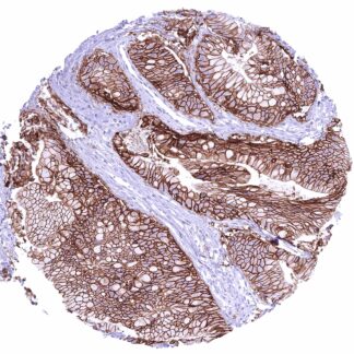
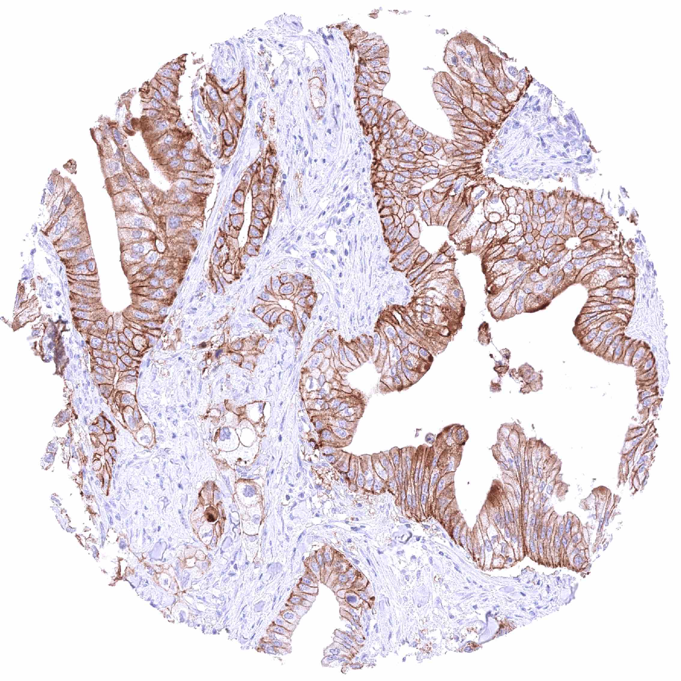
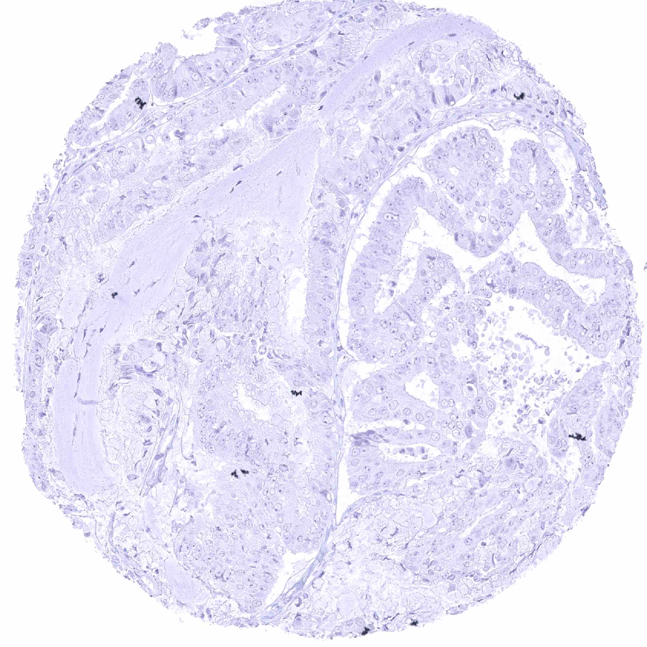
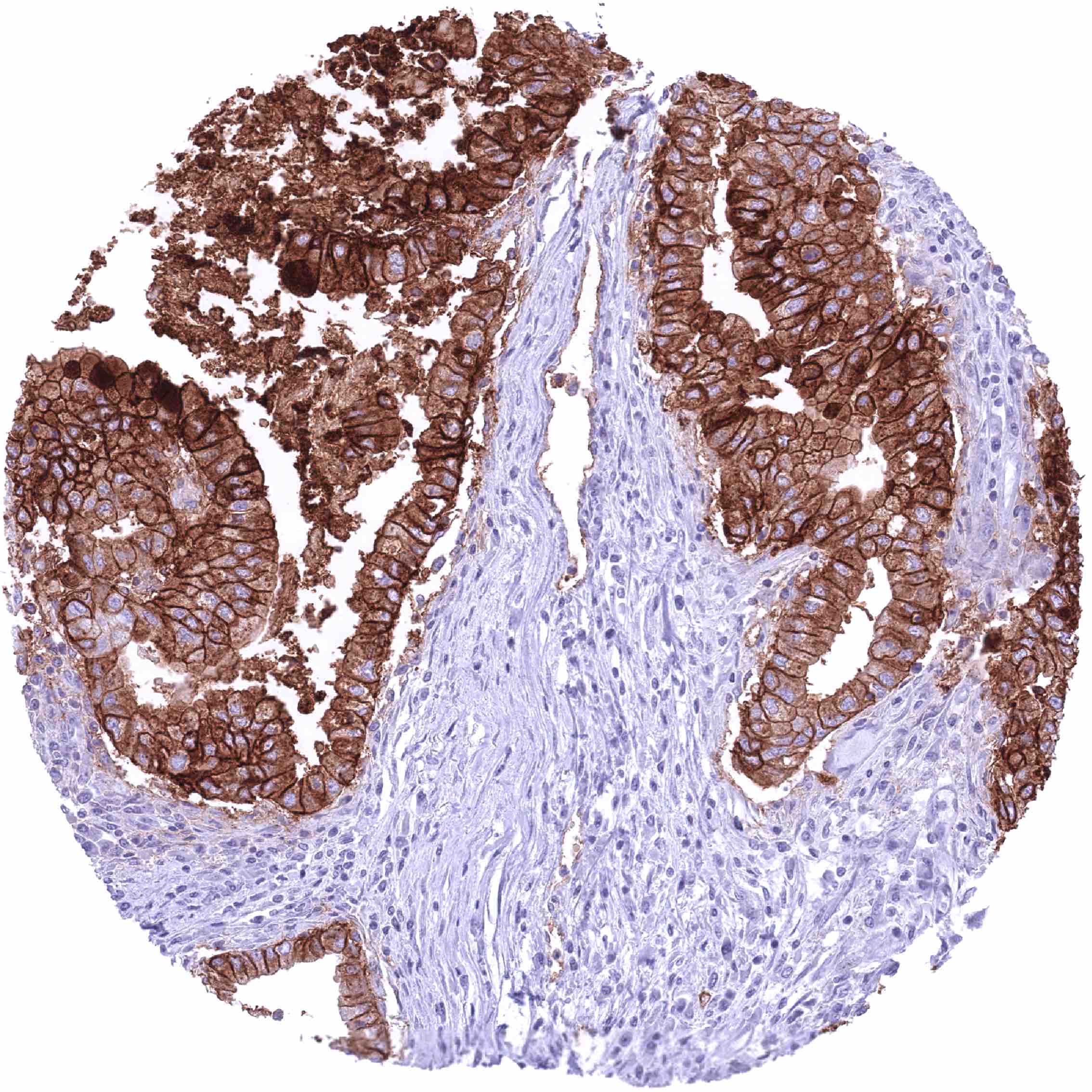
CLDN18.2 expression is most prevalent in gastric and pancreatic adenocarcinomas, but it can also be found in other cancer types.
The TCGA findings on Claudin 18.2 RNA expression in different tumor categories have been summarized in the Human Protein Atlas.
Compatibility of Antibodies
No data available at the moment
Protocol Recommendations
IHC users have different preferences on how the stains should look like. Some prefer high staining intensity of the target stain and even accept some background. Others favor absolute specificity and lighter target stains. Factors that invariably lead to more intense staining include higher concentration of the antibody and visualization tools, longer incubation time, higher temperature during incubation, higher temperature and longer duration of the heat induced epitope retrieval (slide pretreatment). The impact of the pH during slide pretreatment has variable effects and depends on the antibody and the target protein.
All images and data shown here and in our image galleries are obtained by the manual protocol described below. Other protocols resulting in equivalent staining are described as well.
Manual protocol
Freshly cut sections should be used (less than 10 days between cutting and staining). Heat-induced antigen retrieval for 5 minutes in an autoclave at 121°C in pH 7,8 Target Retrieval Solution buffer. Apply HMV4729 at a dilution of 1:150 at 37°C for 60 minutes. Visualization of bound antibody by the EnVision Kit (Dako, Agilent) according to the manufacturer’s directions.
Potential Research Applications
- What approach (antibodies, ADCs, others) works best for targeting CLDN18.2 positive cancers?
- What is the prevalence of CLDN18.2 expression in different cancer types?
- Can CLDN18.2 IHC be used as a diagnostic marker for gastric/pancreatic cancer?
- What is the best approach to predict response to current and future anti-CLDN18.2 drugs?
Evidence for Antibody Specificity in IHC
There are two ways how the specificity of antibodies can be documented for immunohistochemistry on formalin fixed tissues. These are: 1. Comparison with a second independent method for target expression measurement across a large number of different tissue types (orthogonal strategy), and 2. Comparison with one or several independent antibodies for the same target and showing that all positive staining results are also seen with other antibodies for the same target (independent antibody strategy).
Orthogonal validation: For the antibody HMV4729specificity is strongly supported by a comparison of its staining pattern with CLDN18 RNA expression data in normal tissues which were collected in three independent RNA screening studies, including the Human Protein Atlas (HPA) RNA-seq tissue dataset, the FANTOM5 project, and the Genotype-Tissue Expression (GTEx) project, and which are summarized in the Human Protein Atlas (Tissue expression Claudin 18.2). In line with these RNA data, CLDN18.2 immunostaining by HMV4729 was only seen in gastric epithelial cells. That CLDN18.2 immunostaining by HMV4729 was absent in the lung – the only other tissue with detectable CLDN18 RNA – is obviously due to the fact that the other splice variant (CLDN18.1) is expressed in the lung.
Comparison of antibodies: True expression of CLDN18.2 in all cells with CLDN18.2 positivity by HMV4729 is corroborated by a confirmation of each individual staining seen by HMV4729 in 76 different normal tissue types by a commercially available independent second antibody (termed “validation antibody”).