
295,00 € – 995,00 €
Product details
Synonyms = Arc 1; cadherin 1 type 1 E-cadherin; Cadherin1; CAM 120/80; CD324; CDH1; CDHE; E-Cad/CTF3; E-cadherin; ECAD; Epithelial cadherin; epithelial calcium dependent adhesion protein; Liver cell adhesion molecule (LCAM); Uvomorulin (UVO)
Antibody type = Recombinant Rabbit monoclonal / IgG
Clone = MSVA-035R
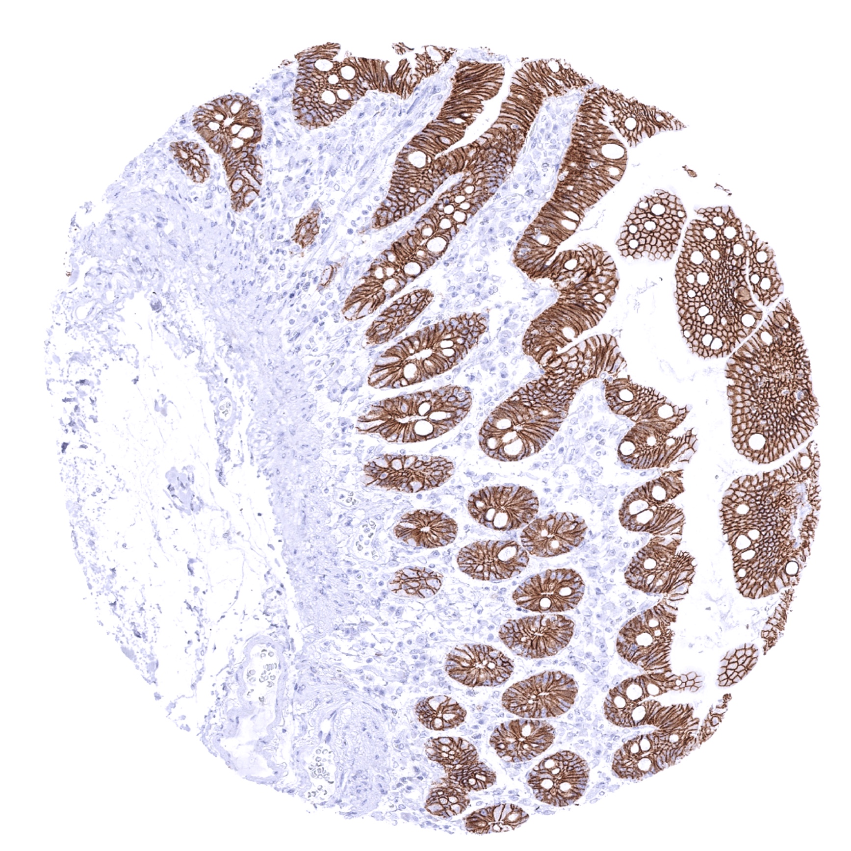
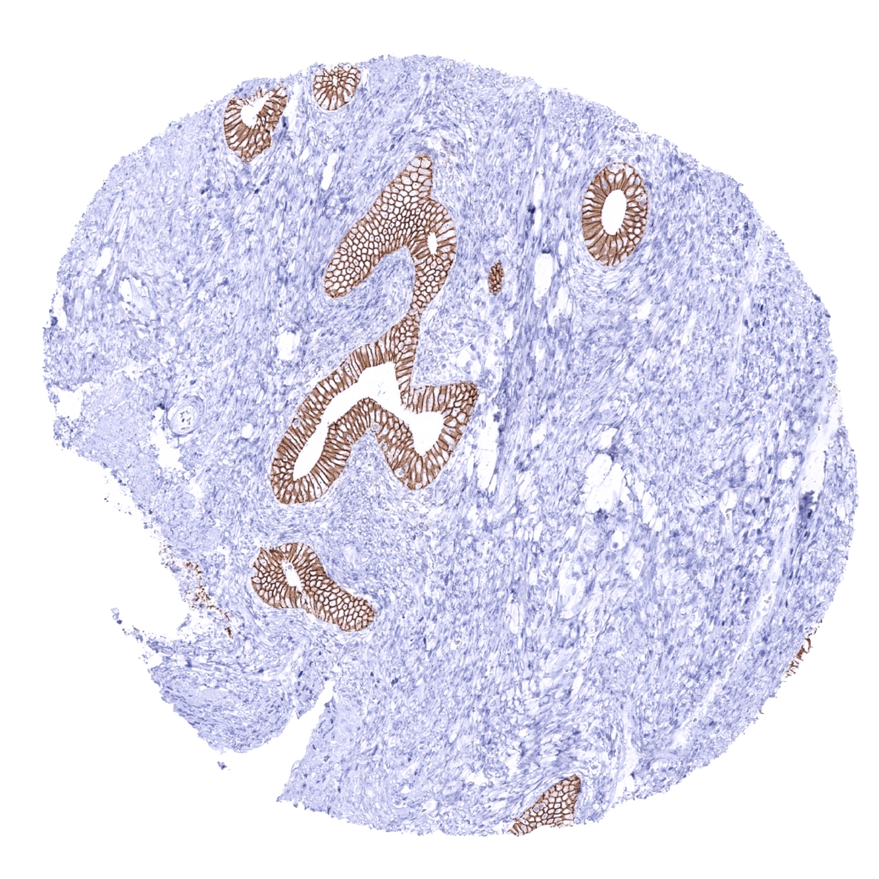
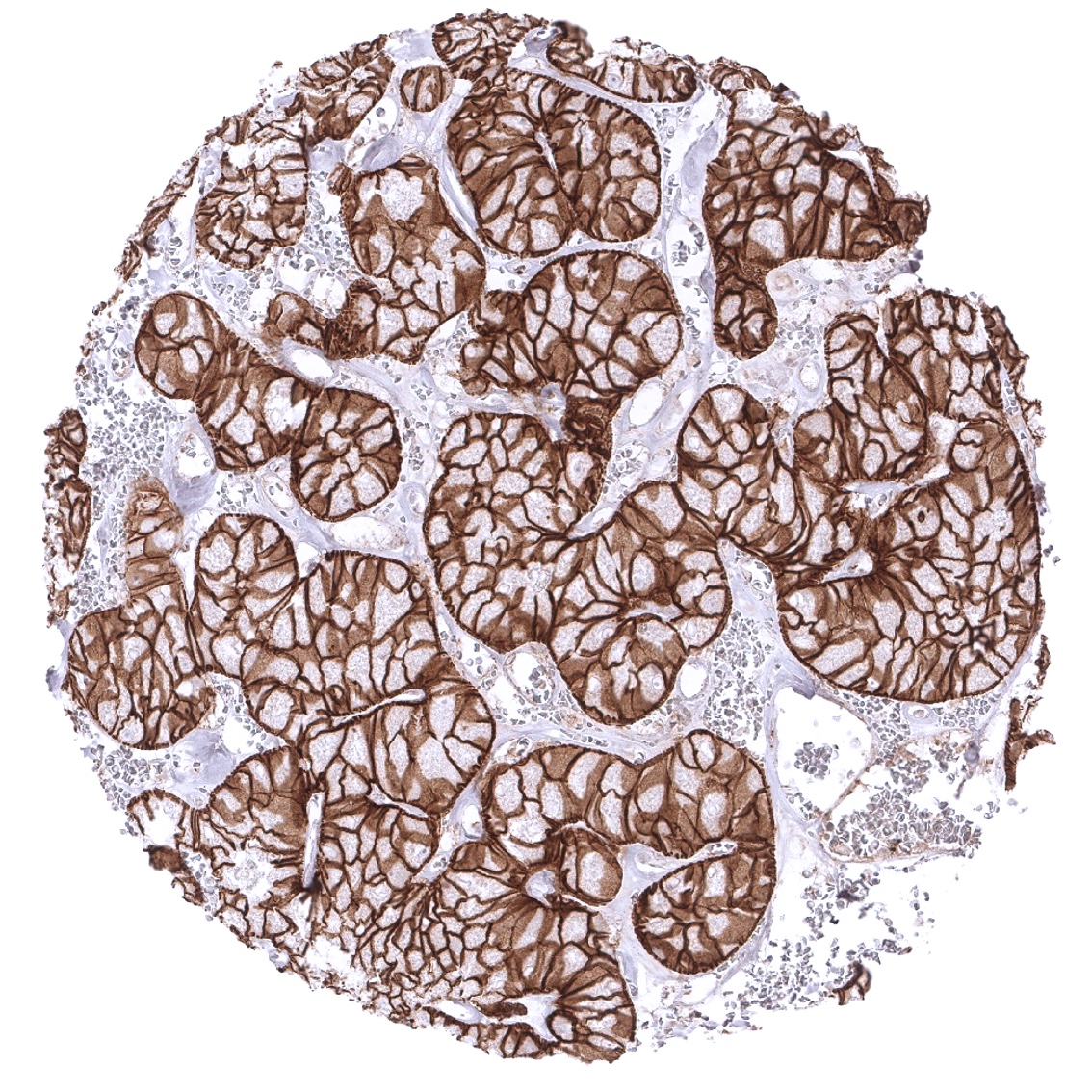
Positive control = In the liver, at least a moderate membranous staining should be seen in hepatocytes while bile duct epithelia stain strongly. In the colon, only epithelium should display a strong membranous staining reaction.
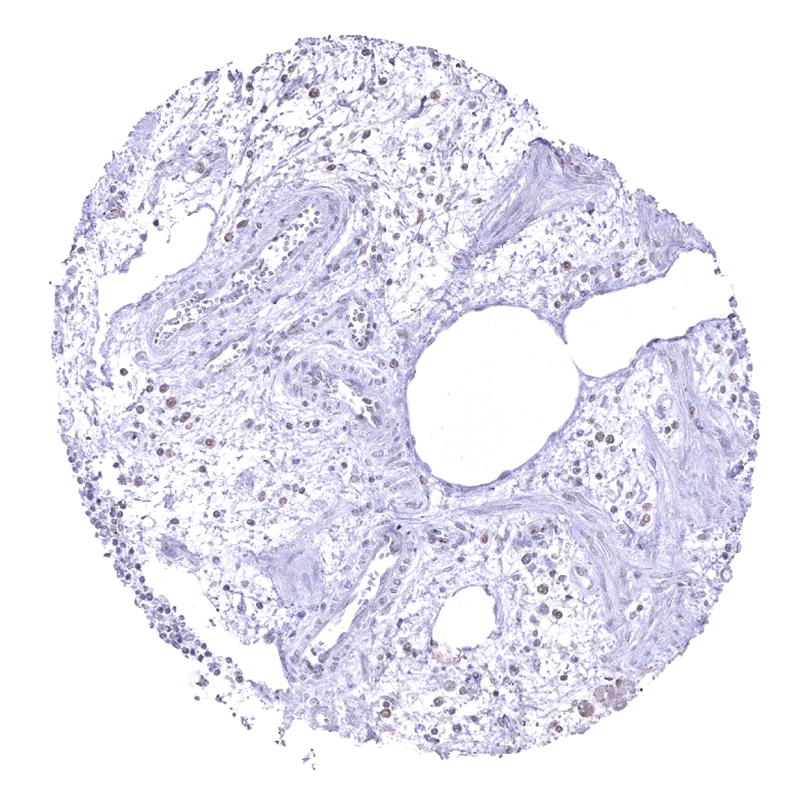
Negative control = Tonsil or appendix: No staining reaction must be seen in stromal cells such as lymphocytes, plasma cells, smooth muscle cells or endothelial cells.
Cellular localization = Cell surface
Reactivity = Human
Application = Immunohistochemistry
Dilution = 1:100 – 1:200
Intended Use = Research Use Only
Relevance of Antibody
E-Cadherin is an Important cell adhesion protein and therapeutic target.
Biology Behind
The E-cadherin gene (CHD1, Cadherin-1), located at 16q22.1 codes for a calcium-dependent membranous glycoprotein with an important role in cellular adhesion and polarity maintenance. The pivotal role of E-cadherin is highlighted by its expression starting at the 2-cell stage of mammalian development. In adult tissues, E-cadherin is expressed in virtually all epithelial tissues, where it is constantly regenerated with a 5-hour half-life on the cell surface. Loss of E-cadherin function or expression plays a relevant role in cancer development and progression. E-cadherin downregulation diminishes cellular adhesion in epithelial tissues. As a result, increased cell motility may facilitate invasive growth and metastasis. Reduced E-cadherin function is commonly seen in cancers with particularly discoherent features such as invasive lobular breast cancer, pseudopapillary neoplasm of the pancreas and plasmacytoid urothelial carcinoma. Heterozygous germline alteration of the CDH1 gene is associated with hereditary diffuse gastric cancer syndrome and invasive lobular carcinoma of the breast.
Staining Pattern in Normal Tissues
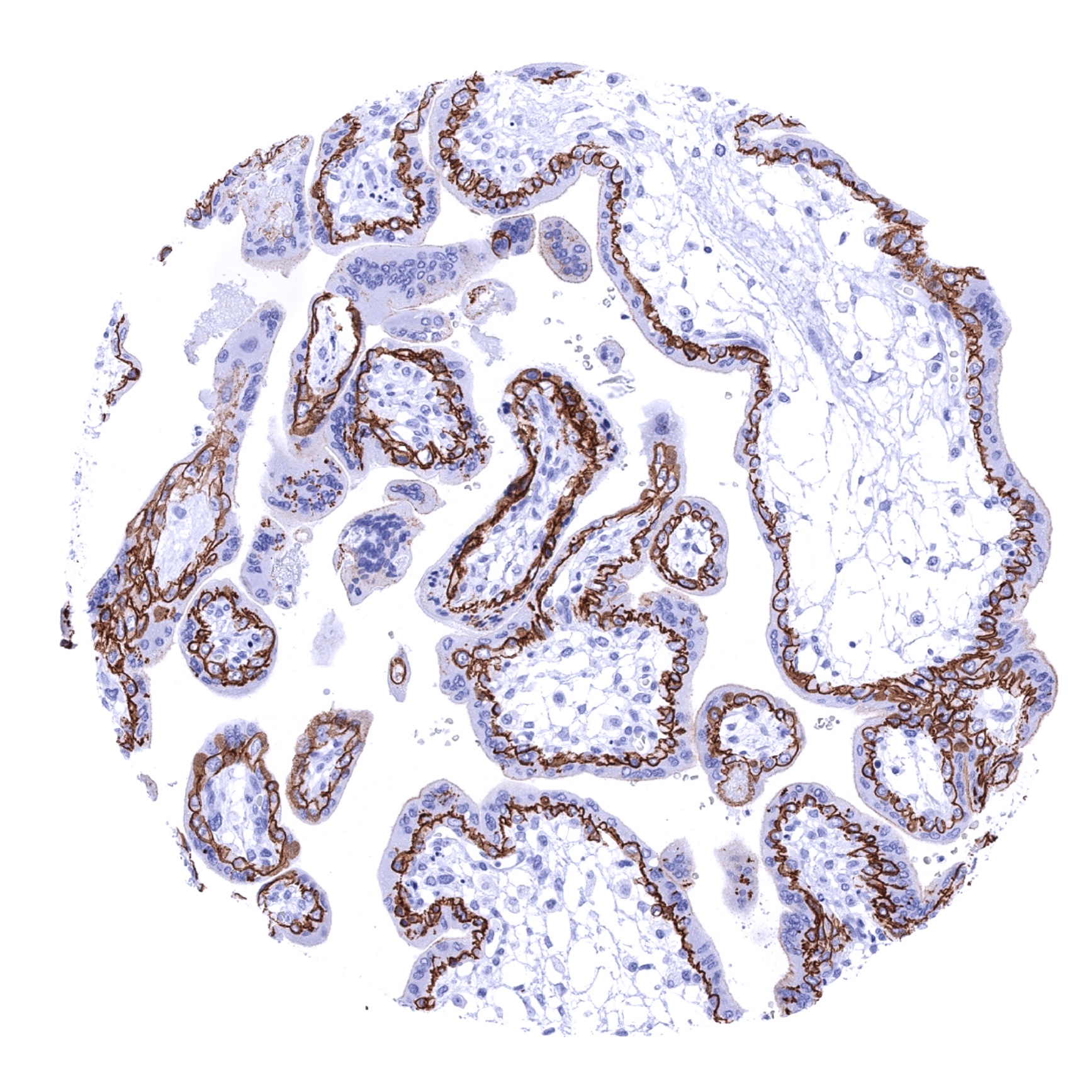
Membranous E-Cadherin staining is seen in almost all epithelial cells of skin, oral cavity, tonsils, lip, salivary glands, esophagus, stomach, duodenum, ileum, appendix, colon, rectum, anal canal, gallbladder, liver, pancreas, uterus cervix, fallopian tube, breast, thyroid gland, kidney pelvis, urinary bladder, prostate, seminal vesicle, epididymis, adenohypophysis, paranasale sinus, and the lung. In the kidney, E-Cadherin expression is only seen in collecting ducts and the distal but not in the proximal tubuli. In the placenta, only the cytotrophoblastic but not the syncytiotrophoblastic layer show E-Cadherin staining. In the thymus, a positive staining occurs in corpuscles of Hassall´s and in a fraction of thymic epithelial cells. A fraction of small vessels stain E-Cadherin-positive in the spleen. In lymphatic tissue a weak to moderate fibrillar staining reflecting fibroblastic reticular cells is occasionally seen. E-Cadherin immunostaining is absent in aorta, heart, striated muscle, myometrium of the uterus, muscular wall of the gastrointestinal tract, kidney pelvis and urinary bladder, corpus spongiosum of the penis, ovarian stroma, fat, testis, corpus luteum of the ovary, adrenal gland, cerebellum and cerebrum.
The findings described above are this consistent with the RNA data described in the Human Protein Atlas (Tissue expression E-Cadherin)
Immunostaining by using MSVA-035R was detected in all organs/tissues containing epithelial cells (except the adrenal gland).
Positive control = In the liver, at least a moderate membranous staining should be seen in hepatocytes while bile duct epithelia stain strongly. In the colon, only epithelium should display a strong membranous staining reaction.
Negative control = Tonsil or appendix: No staining reaction must be seen in stromal cells such as lymphocytes, plasma cells, smooth muscle cells or endothelial cells.
Staining Pattern in Relevant Tumor Types
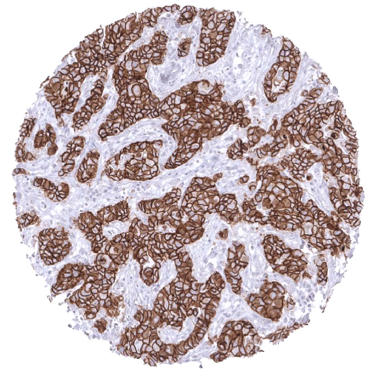
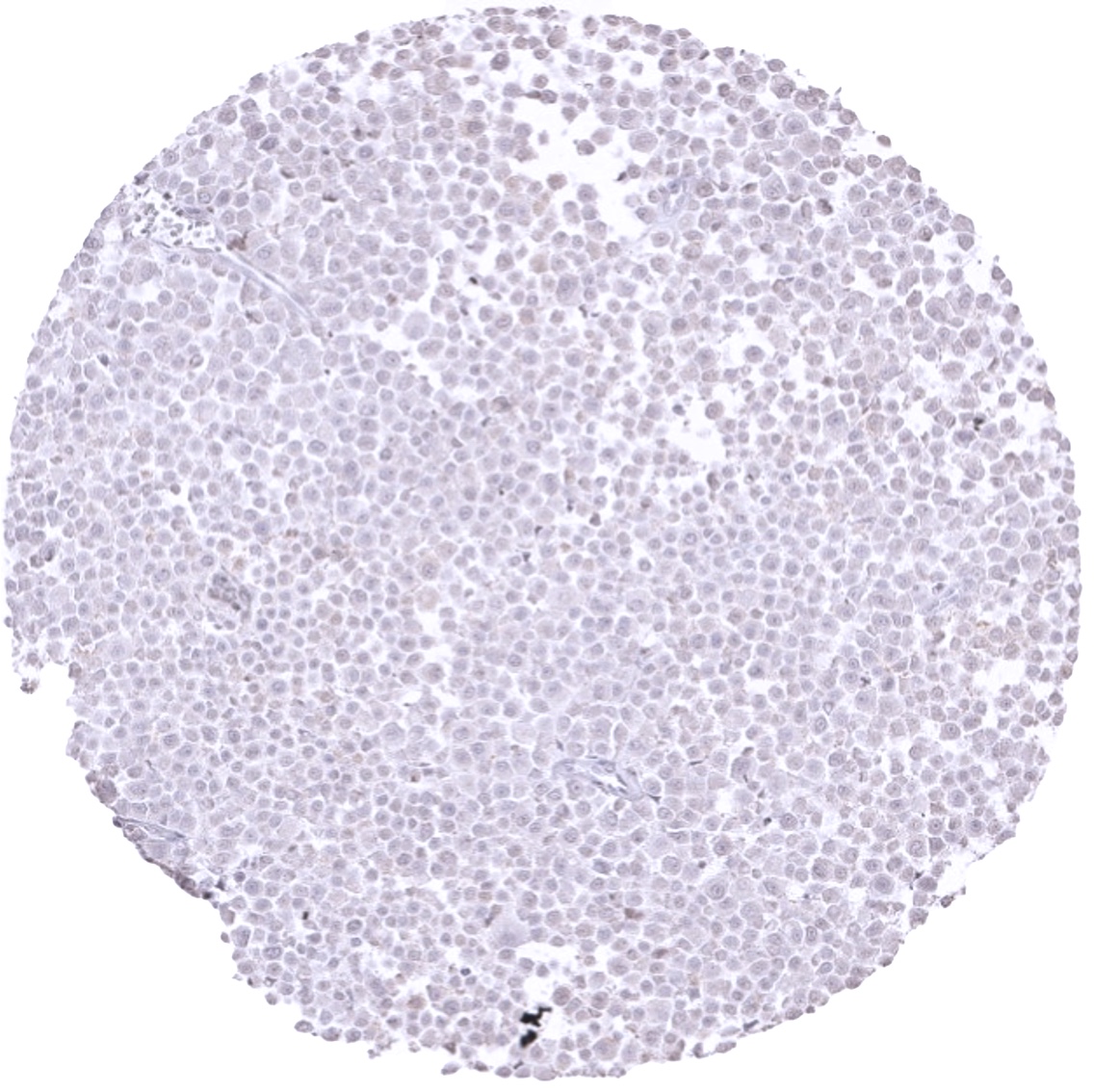
E-Cadherin immunostaining in neoplasms largely parallels the findings in normal tissues. Cancers derived from E-Cadherin expressing normal tissues are typically E-Cadherin positive. Neoplasias derived from E-Cadherin negative normal tissues are usually E-Cadherin negative. Loss of E-Cadherin expression is characteristic of lobular breast cancer and also occurs in other tumor entities that are characterized for a discohesive tumor growth such as for example plasmacytoid urothelial cancer or diffuse adenocarcinoma of the stomach. Both, down-regulation of E-Cadherin expression in cancers of E-Cadherin positive tissues and E-Cadherin neo-expression in cancers arising from E-Cadherin negative tissues are often linked to cancer progression and may herald poor prognosis.
The TCGA findings on E-Cadherin RNA expression in different tumor categories have been summarized in the Human Protein Atlas.

Compatibility of Antibodies
No data available at the moment
Protocol Recommendations
IHC users have different preferences on how the stains should look like. Some prefer high staining intensity of the target stain and even accept some background. Others favor absolute specificity and lighter target stains. Factors that invariably lead to more intense staining include higher concentration of the antibody and visualization tools, longer incubation time, higher temperature during incubation, higher temperature and longer duration of the heat induced epitope retrieval (slide pretreatment). The impact of the pH during slide pretreatment has variable effects and depends on the antibody and the target protein.
All images and data shown here and in our image galleries are obtained by the manual protocol described below. Other protocols resulting in equivalent staining are described as well.
-Manual protocol
Freshly cut sections should be used (less than 10 days between cutting and staining). Heat-induced antigen retrieval for 5 minutes in an autoclave at 121°C in pH 7,8 Target Retrieval Solution buffer. Apply MSVA-035R at a dilution of 1:150 at 37°C for 60 minutes. Visualization of bound antibody by the EnVision Kit (Dako, Agilent) according to the manufacturer’s directions.
Potential Research Applications
- The prognostic role of increased or decreased E-cadherin expression is not well understood in many cancers.
- The role of E-cadherin in epithelial-mesenchymal transition is under intensive research.
- E-cadherin is a potential therapeutic target.
Evidence for Antibody Specificity in IHC
There are two ways how the specificity of antibodies can be documented for immunohistochemistry on formalin fixed tissues. These are: 1. comparison with a second independent method for target expression measurement across a large number of different tissue types (orthogonal strategy), and 2. Comparison with one or several independent antibodies for the same target and showing that all positive staining results are also seen with other antibodies for the same target (independent antibody strategy).
Orthogonal validation: For the antibody MSVA-035R specificity is suggested by the concordance of the immunostaining data with data from three independent RNA screening studies, including the Human Protein Atlas (HPA) RNA-seq tissue dataset, the FANTOM5 project, and the Genotype-Tissue Expression (GTEx) project, which are all summarized in the Human Protein Atlas (Tissue expression E-Cadherin). Immunostaining by using MSVA-035R was detected in all organs/tissues containing epithelial cells (except the adrenal gland).
Comparison of antibodies: True E-cadherin staining by MSVA-035R was suggested by a comparison of the MSVA-035R staining with data obtained by an independent commercially available E-cadherin antibody here called “validation antibody”. This comparison resulted in comparable stainings in all tissues and cell types analyzed. Examples of comparative stainings are shown below, especially for tissues and cell types for which E-cadherin expression is not intuitive.


