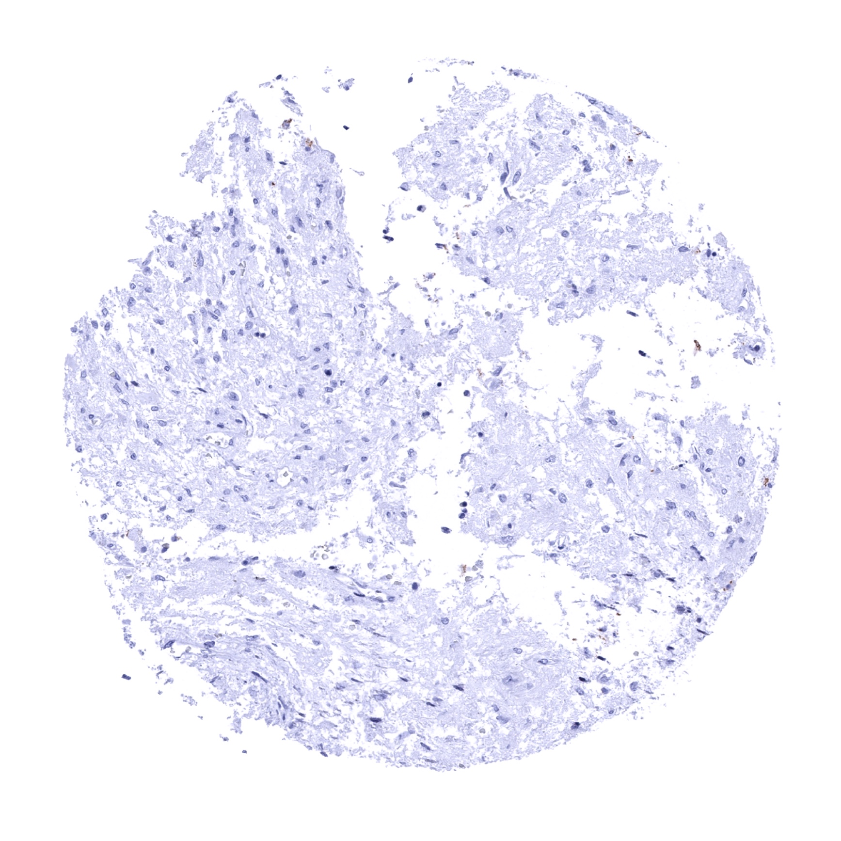
Adrenal gland - E-Cadherin staining is absent in adrenocortical cells.
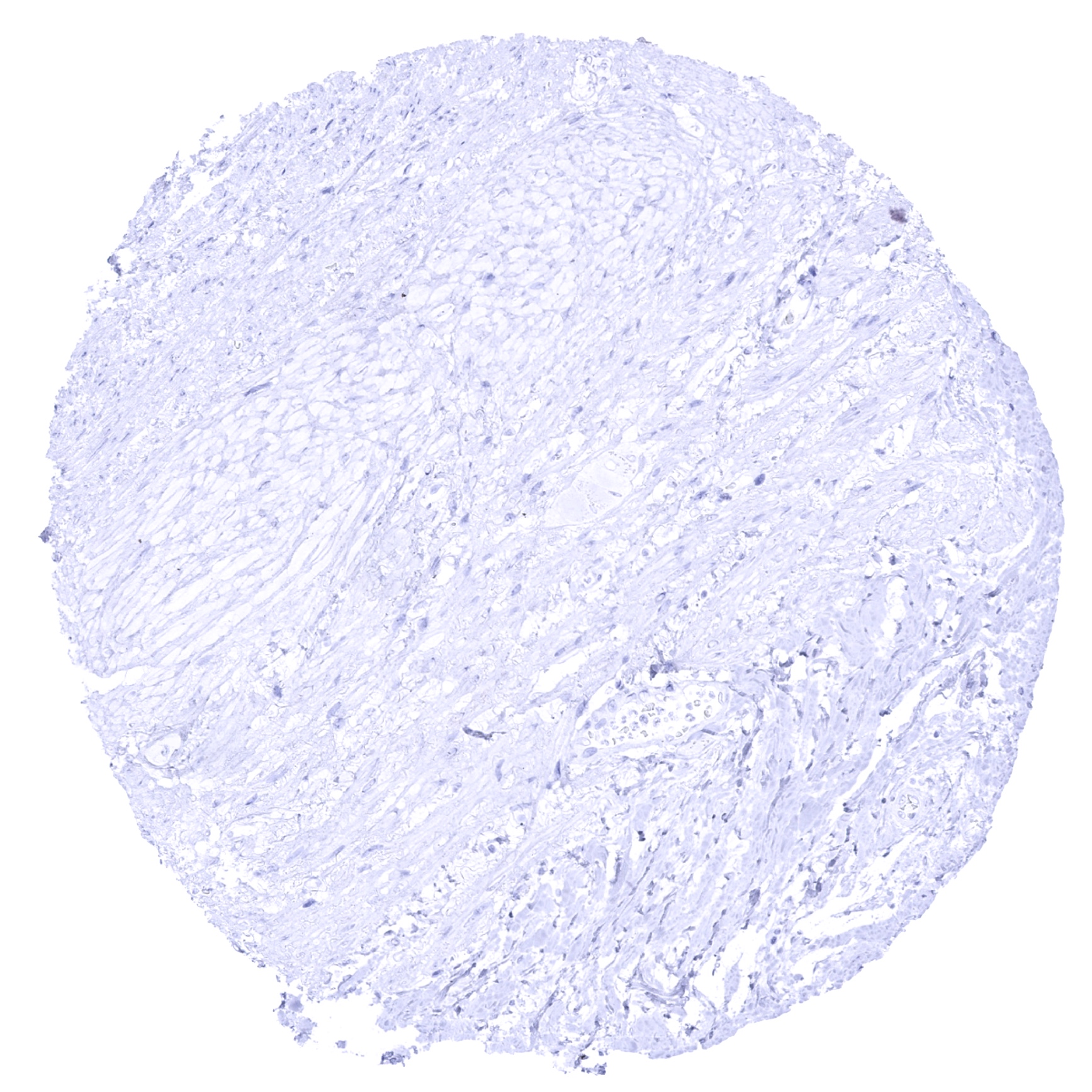
Aorta, media

Appendix, mucosa
Appendix, muscular wall
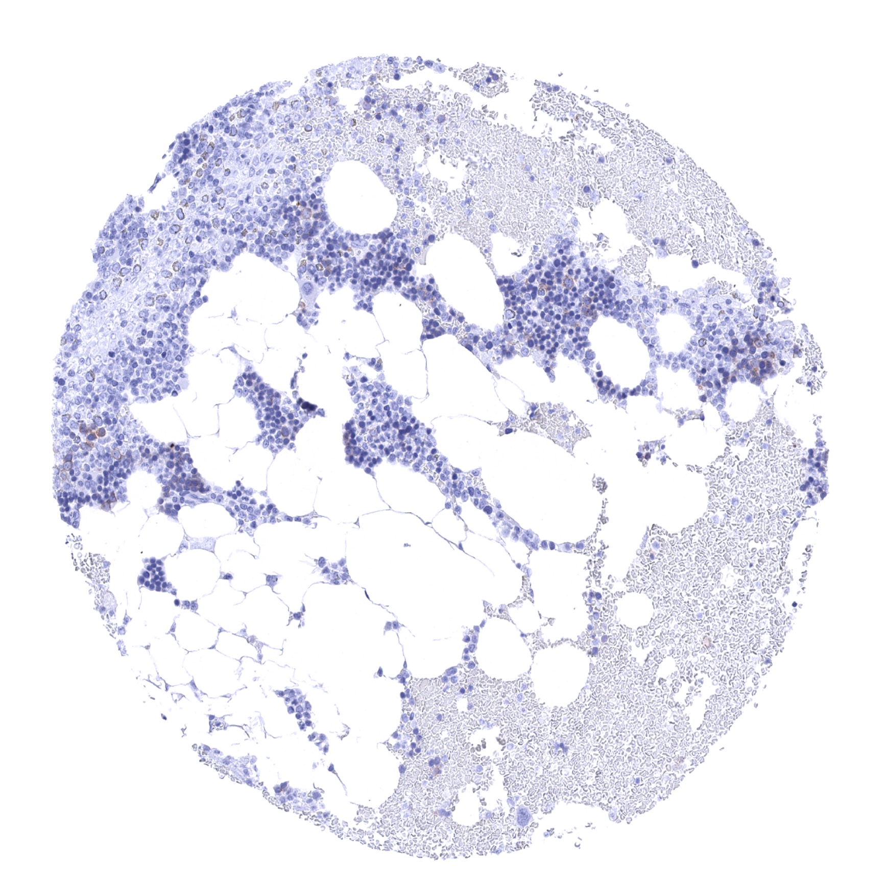
Bone marrow
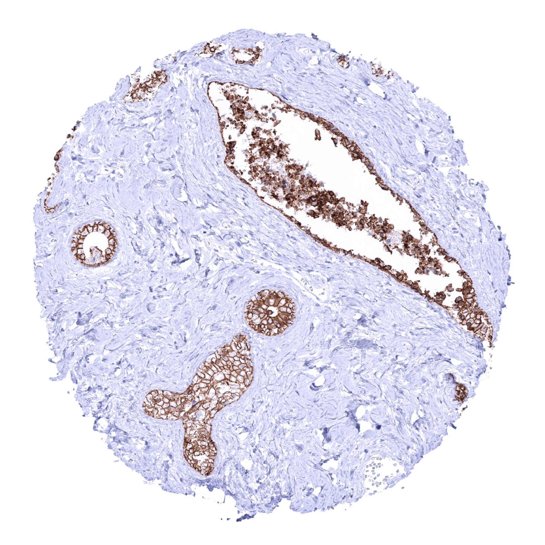
Breast

Bronchus, mucosa
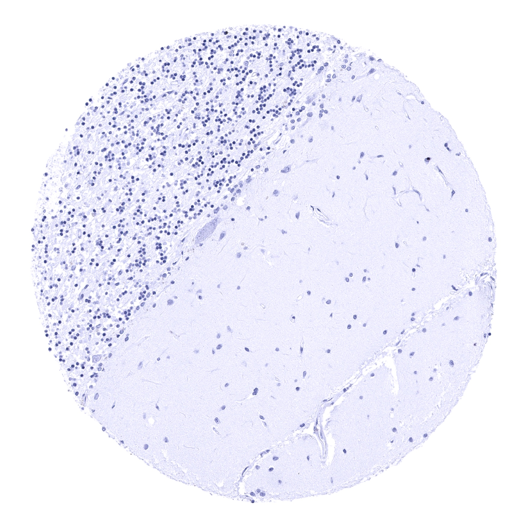
Cerebellum (molecular layer, Purkinje cell layer, granule cell layer, white matter)
Cerebellum, grey (Stratum neuronorum)
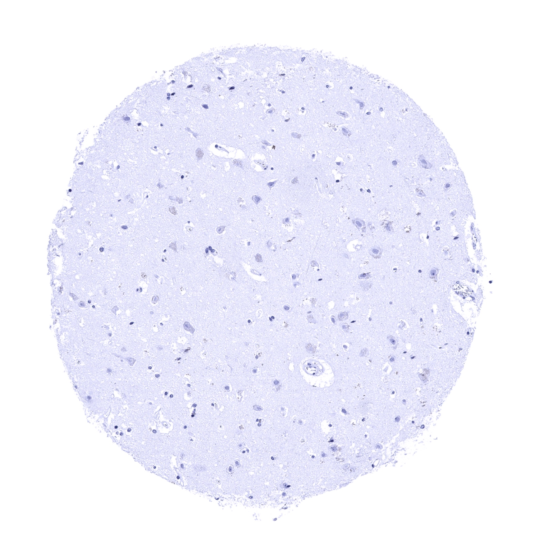
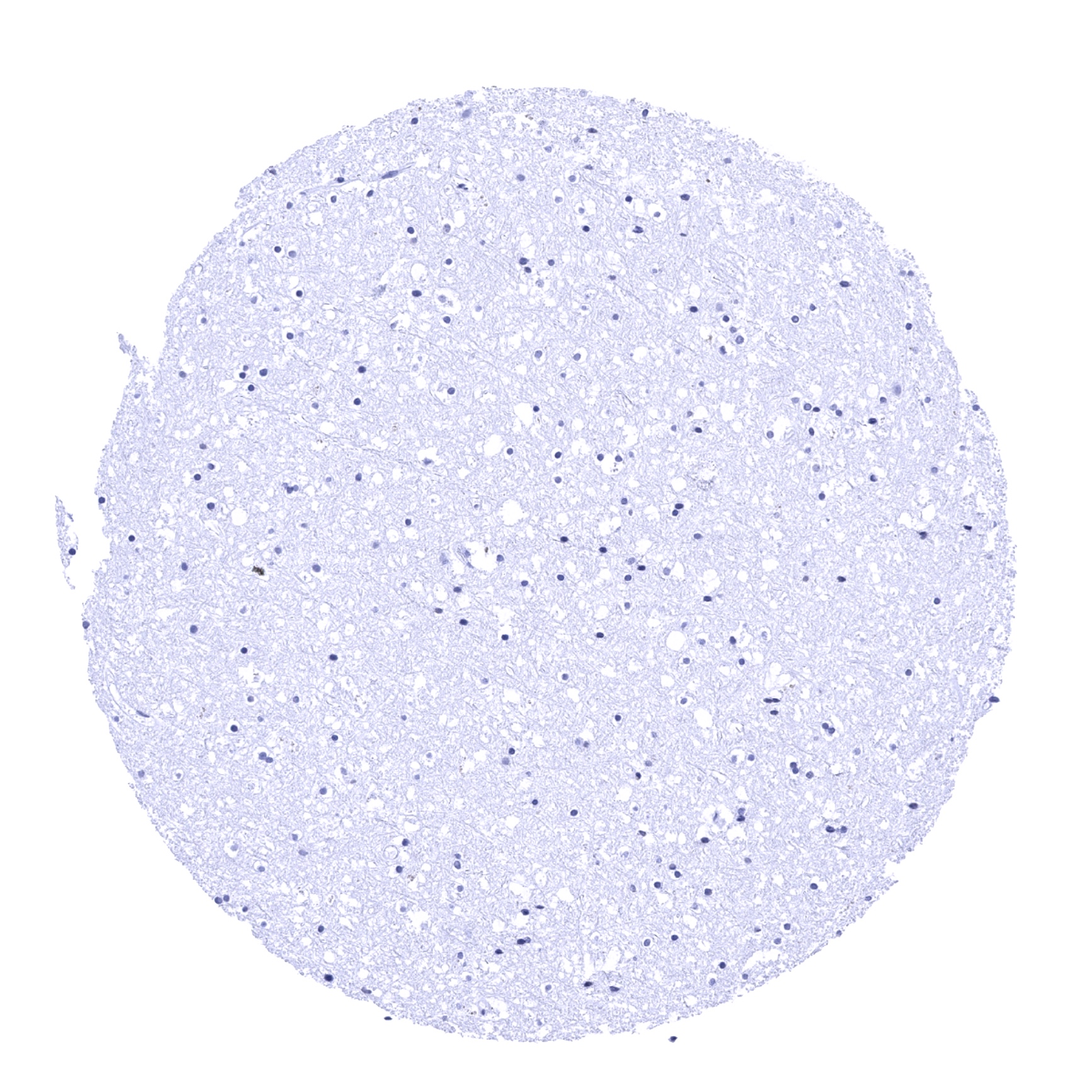
Cerebrum, grey matter

Cerebrum, grey matter
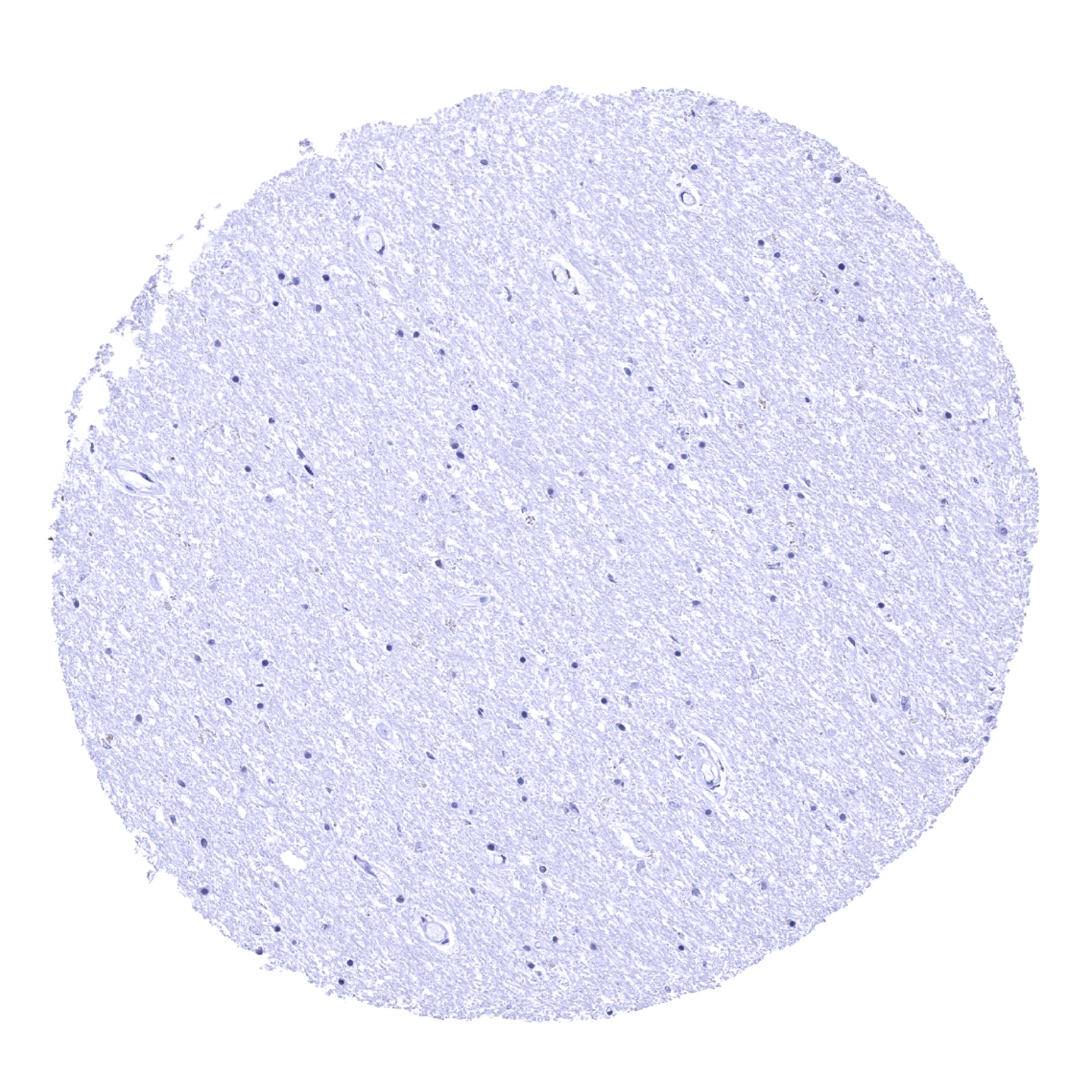
Cerebrum, white matter
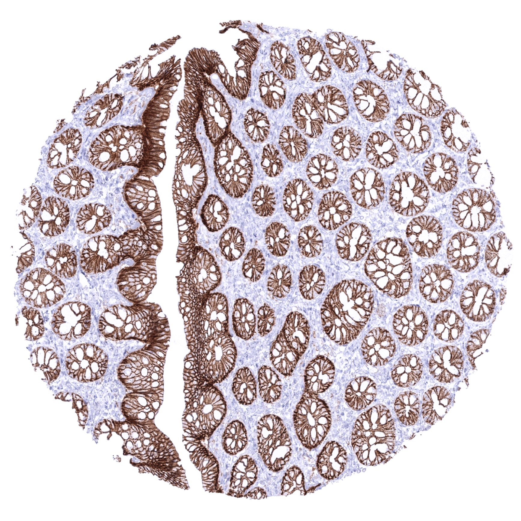
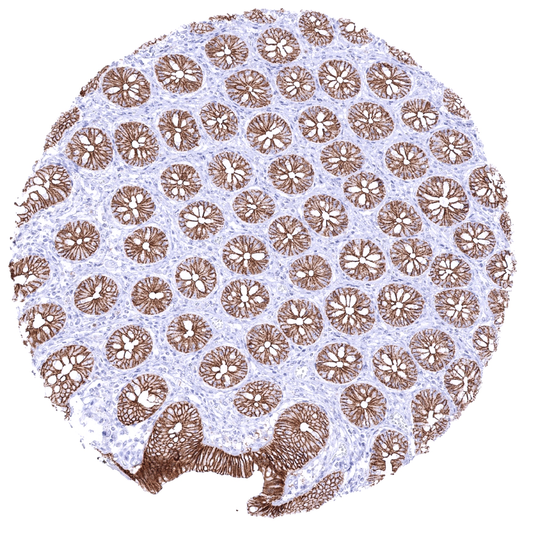
Colon descendens, mucosa
Colon descendens, muscular wall

Duodenum, Brunner gland

Duodenum, mucosa
Epididymis

Epididymis
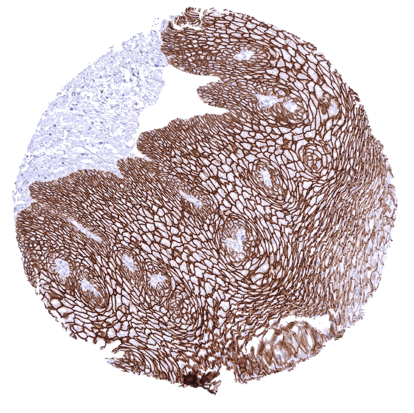
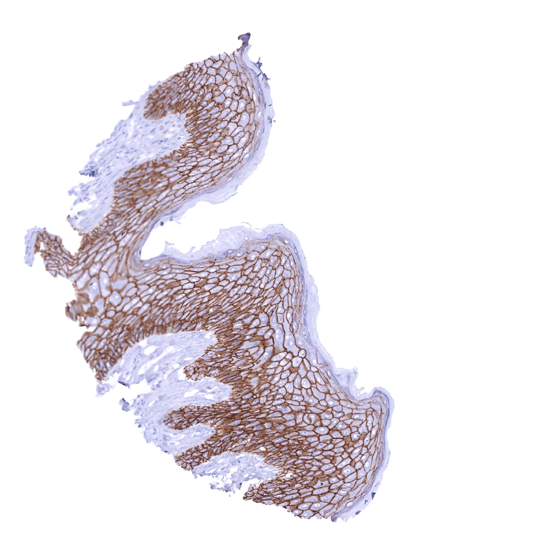
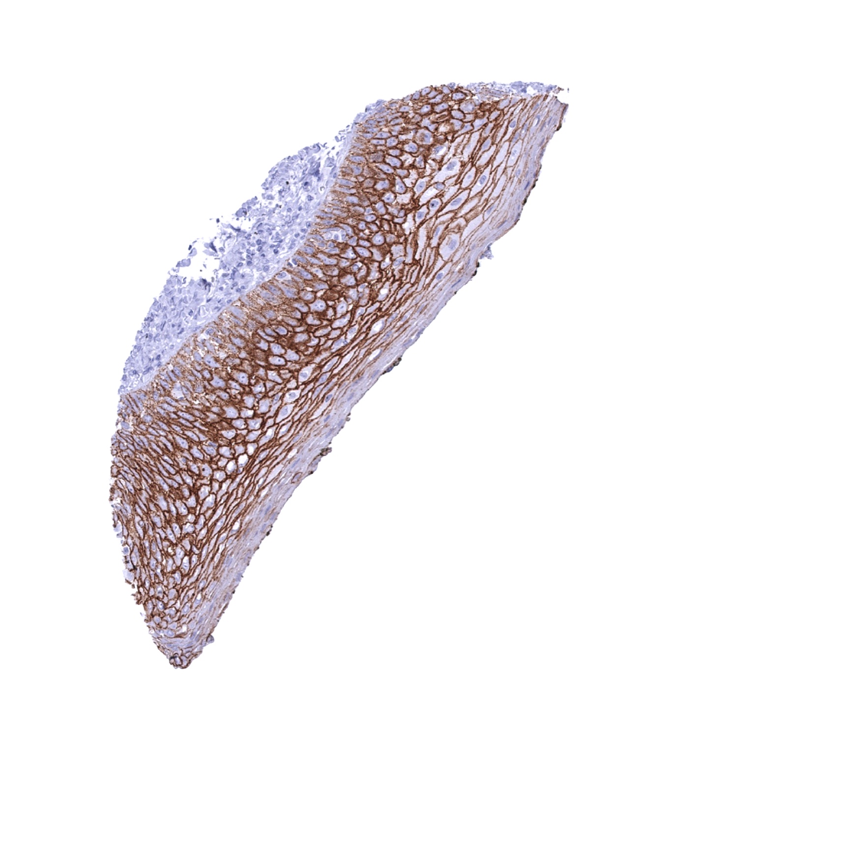
Esophagus, squamous epithelium

Fallopian tube, mucosa
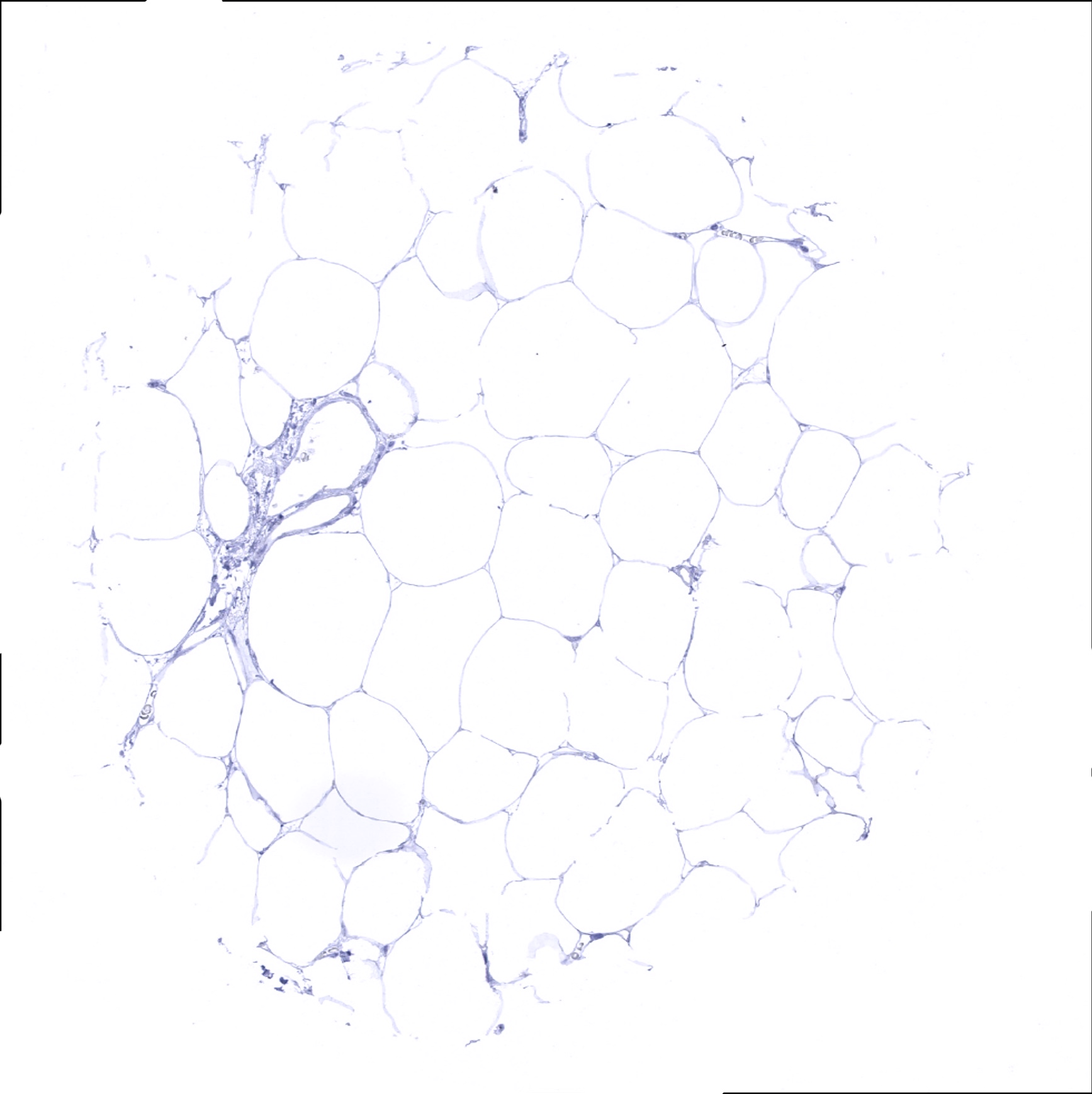
Fat
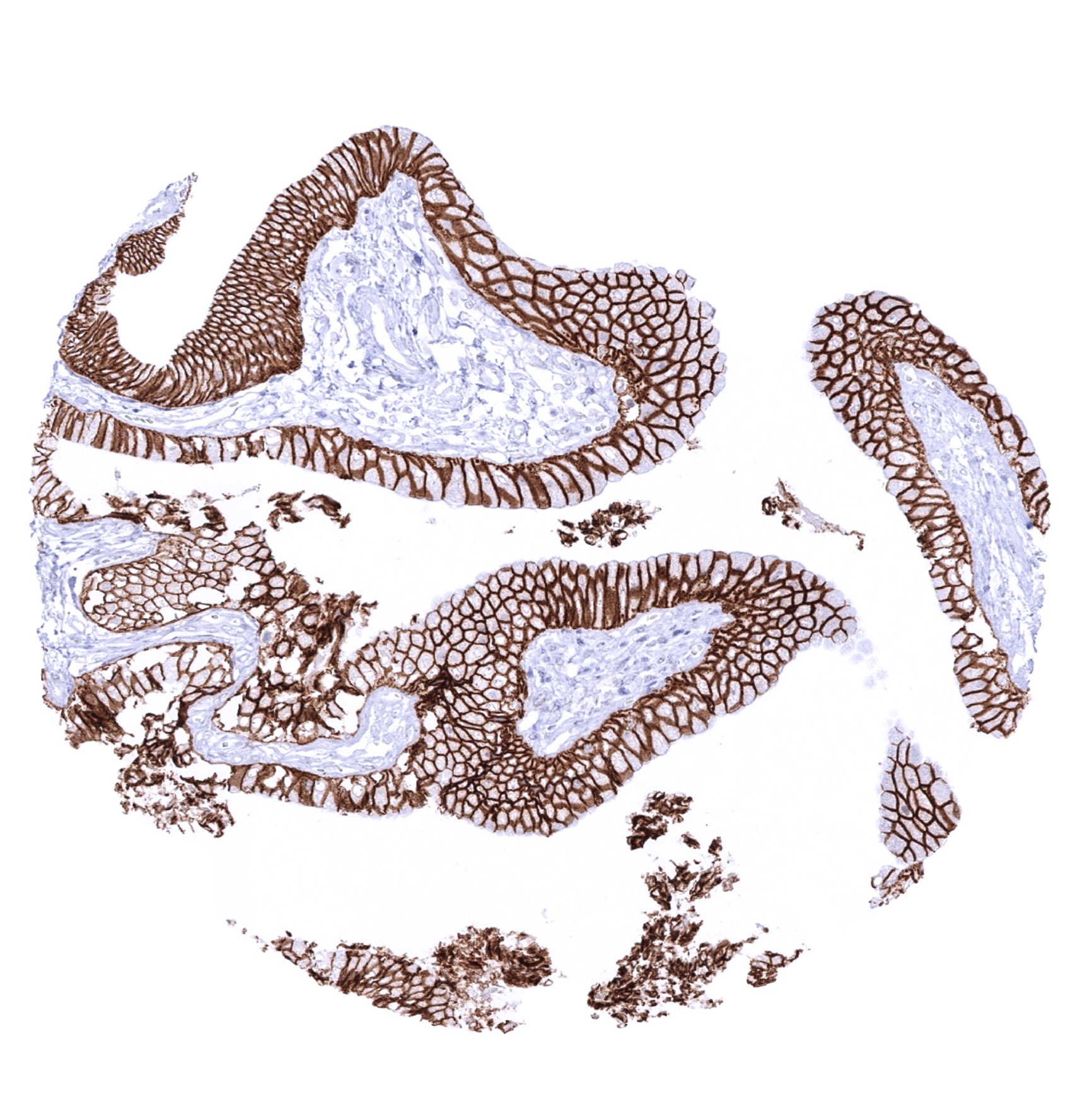
Gallbladder, epithelium
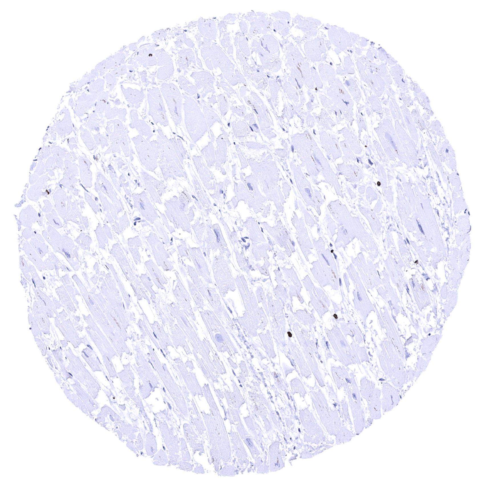
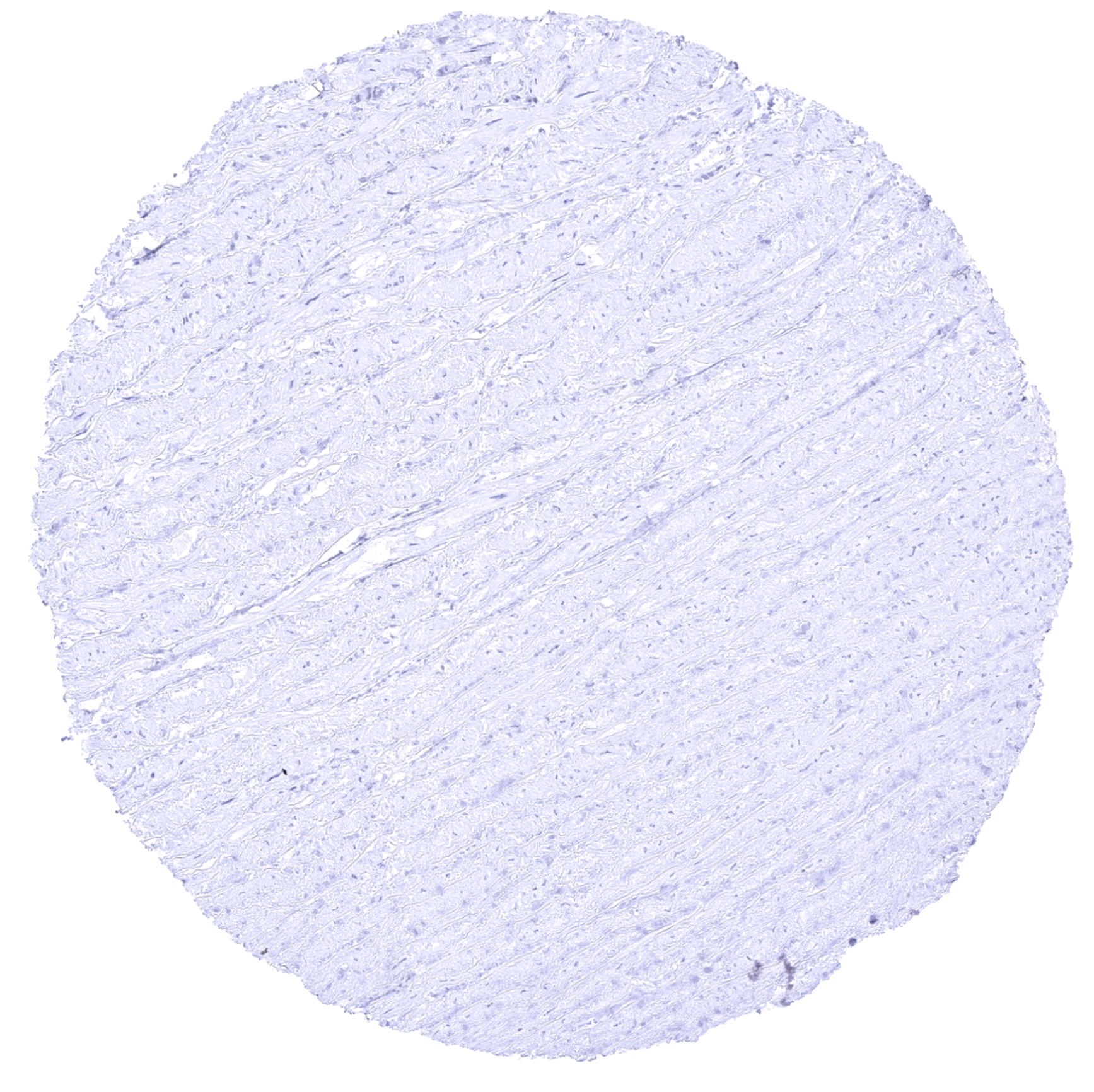
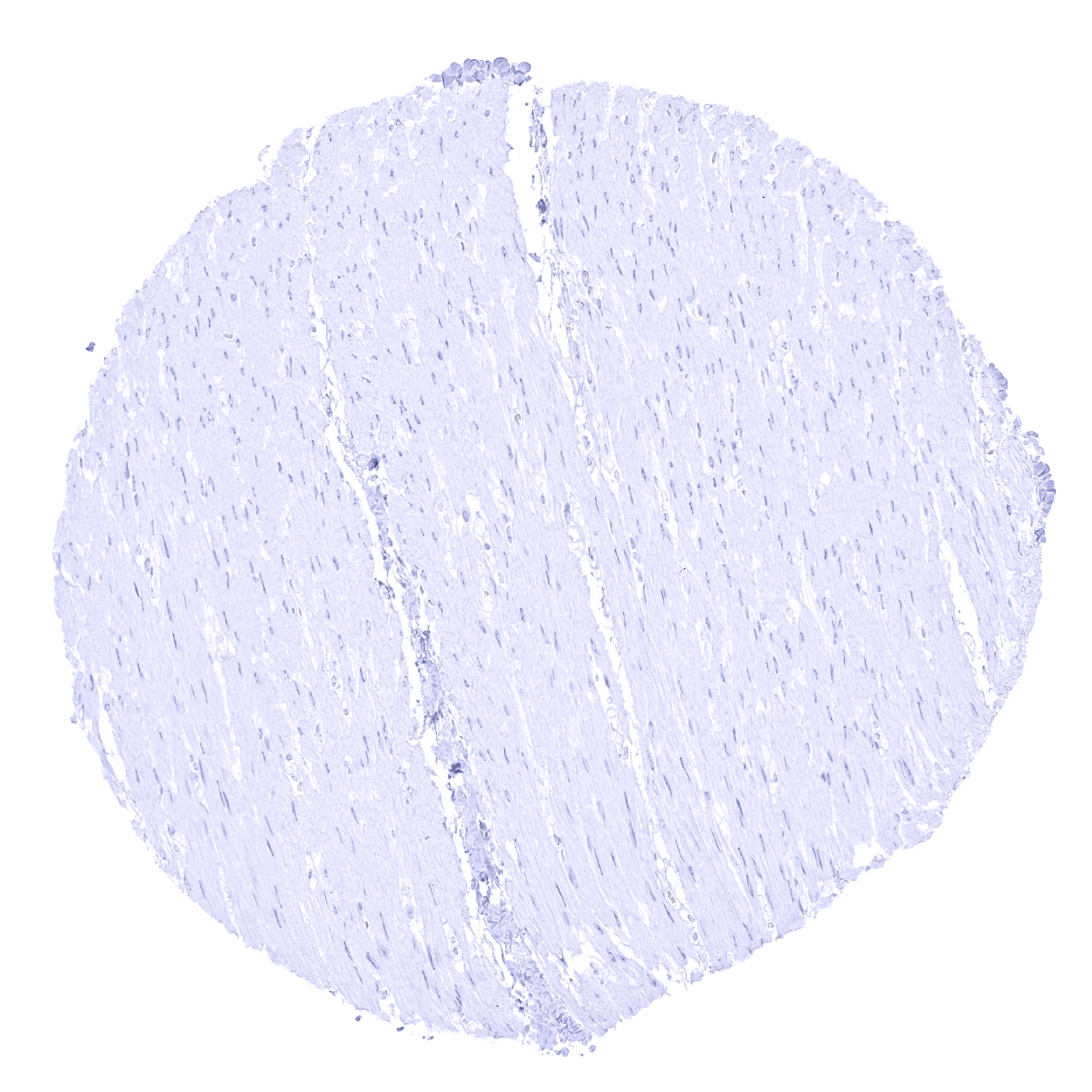
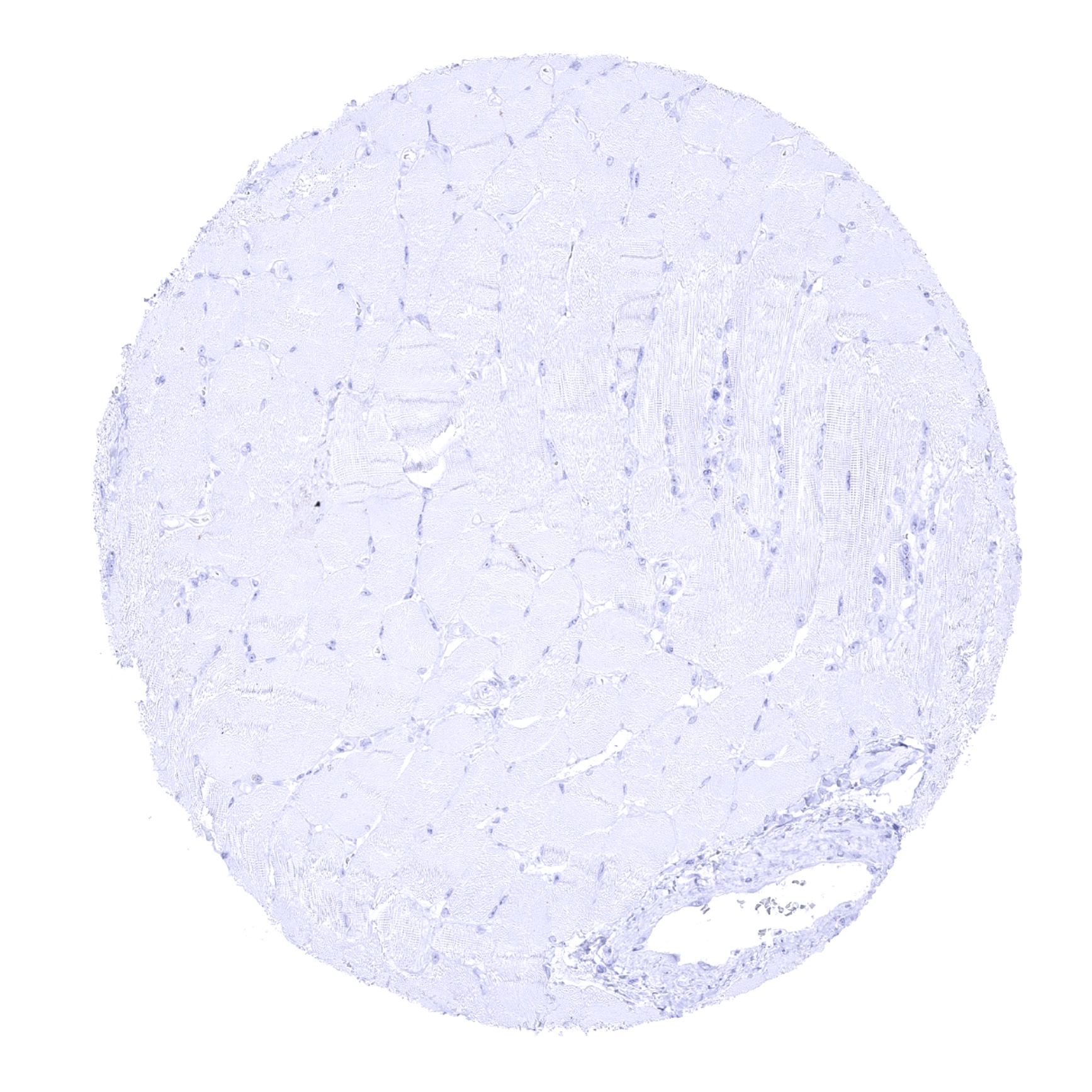
Heart muscle

Ileum, mucosa

Kidney, cortex - E-Cadherin staining is only seen in collecting ducts and in distal tubuli but not in proximal tubuli.

Kidney, medulla
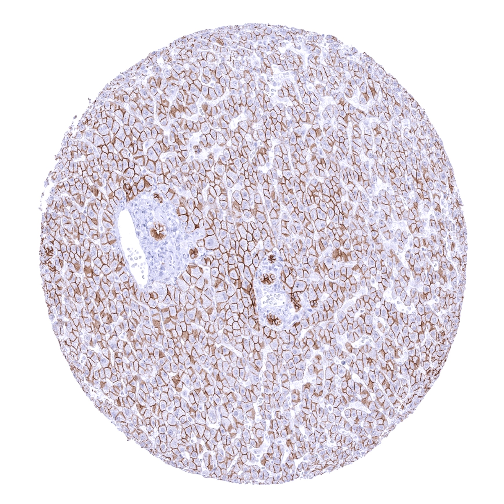
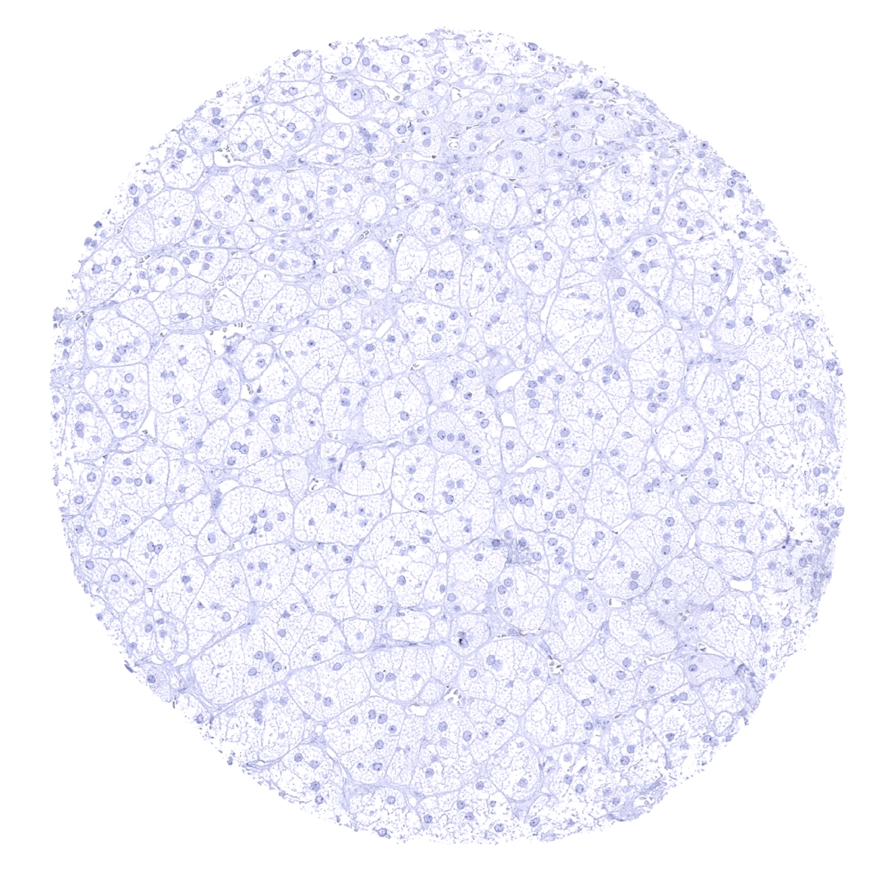
Liver - At least a moderate membranous staining is seen in hepatocytes while bile duct epithelia stain strongly.

Lung
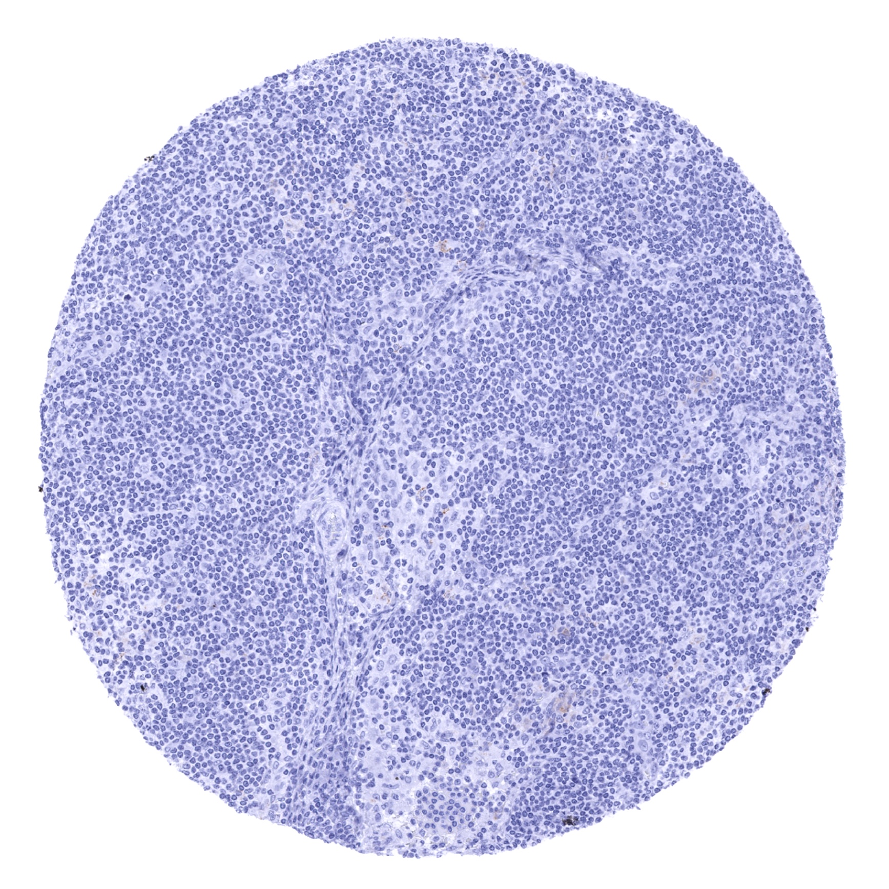
Lymph node
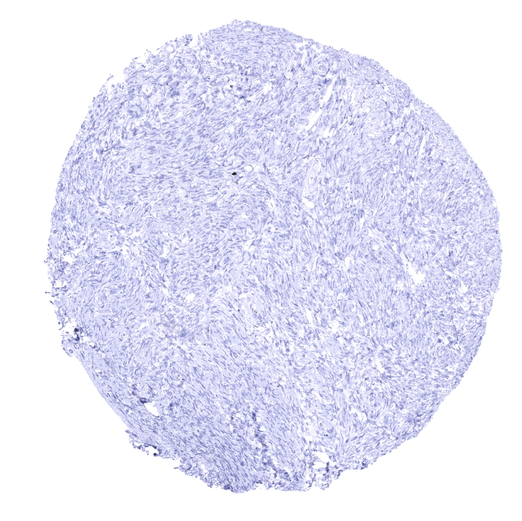
Ovary, stroma
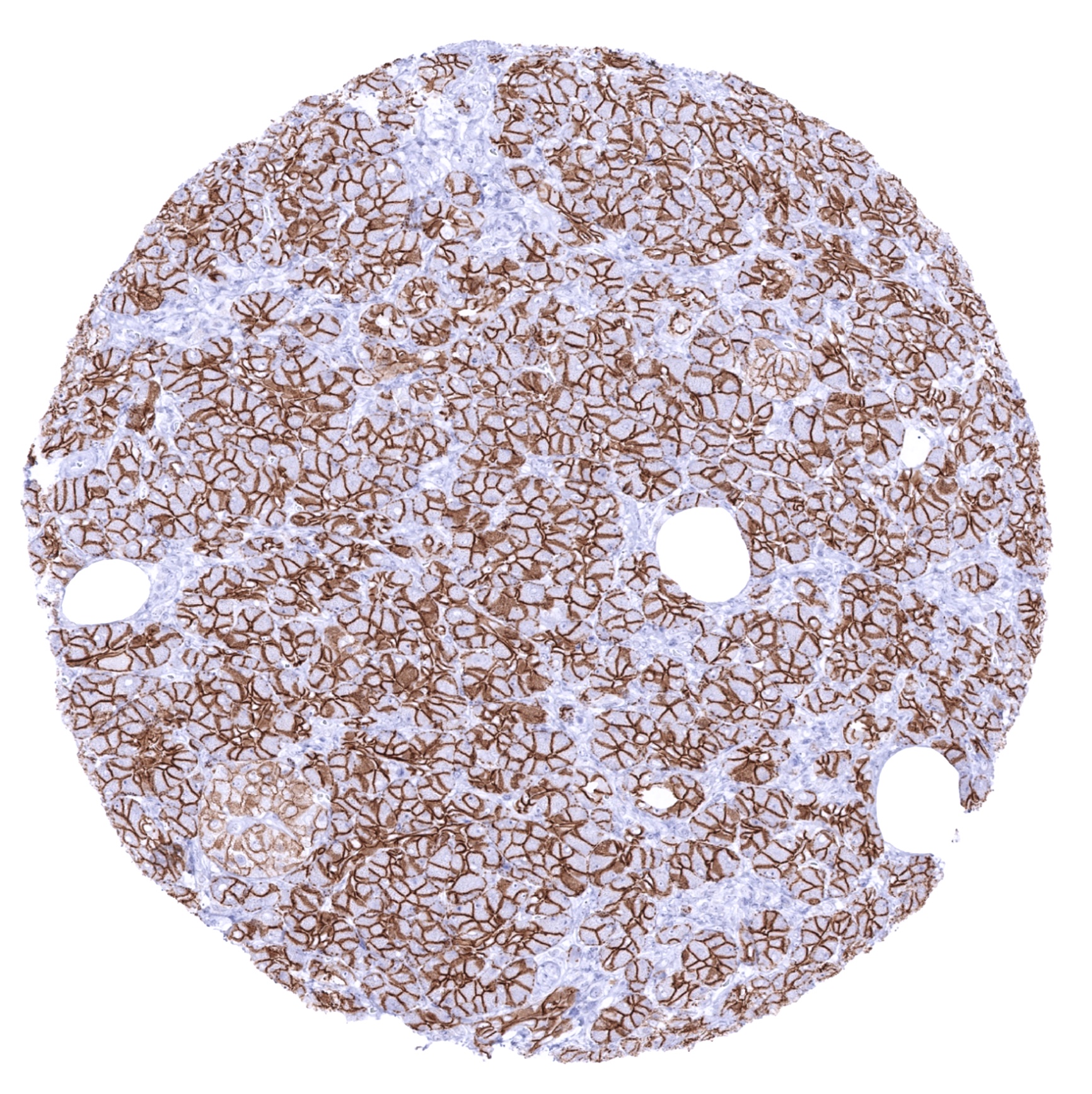
Pancreas - E-cadherin staining is strong in acinar cells but somehat weaker in islet cells.

Parathyroid gland
Parotid gland
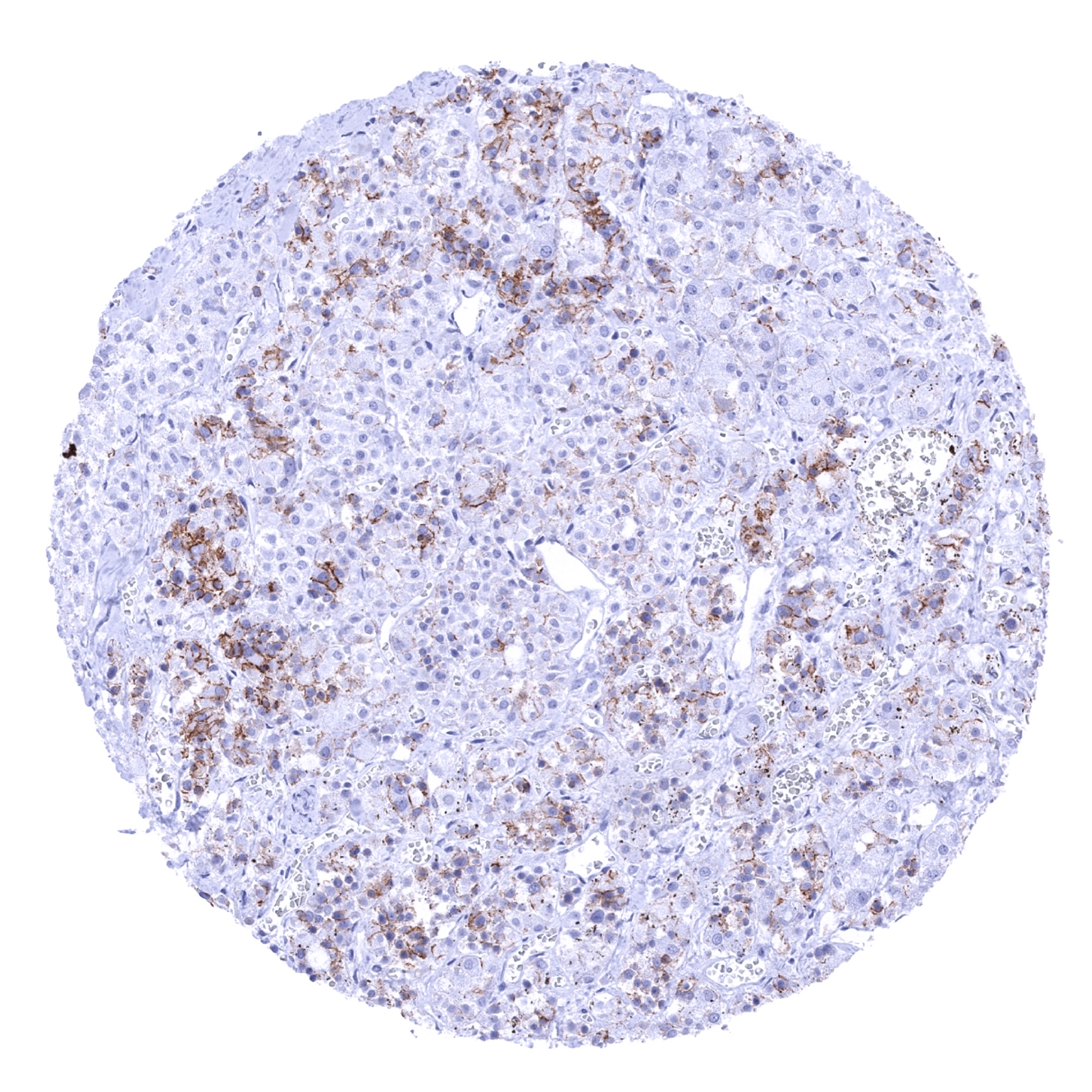
Pituitary gland, anterior lobe - A fraction of epithelial cells shows E-cadherin staining in the adenohypophysis.
Pituitary gland, posterior lobe

Placenta (amnion and chorion) - Both amnion and chorion cells show E-Cadherin staining.
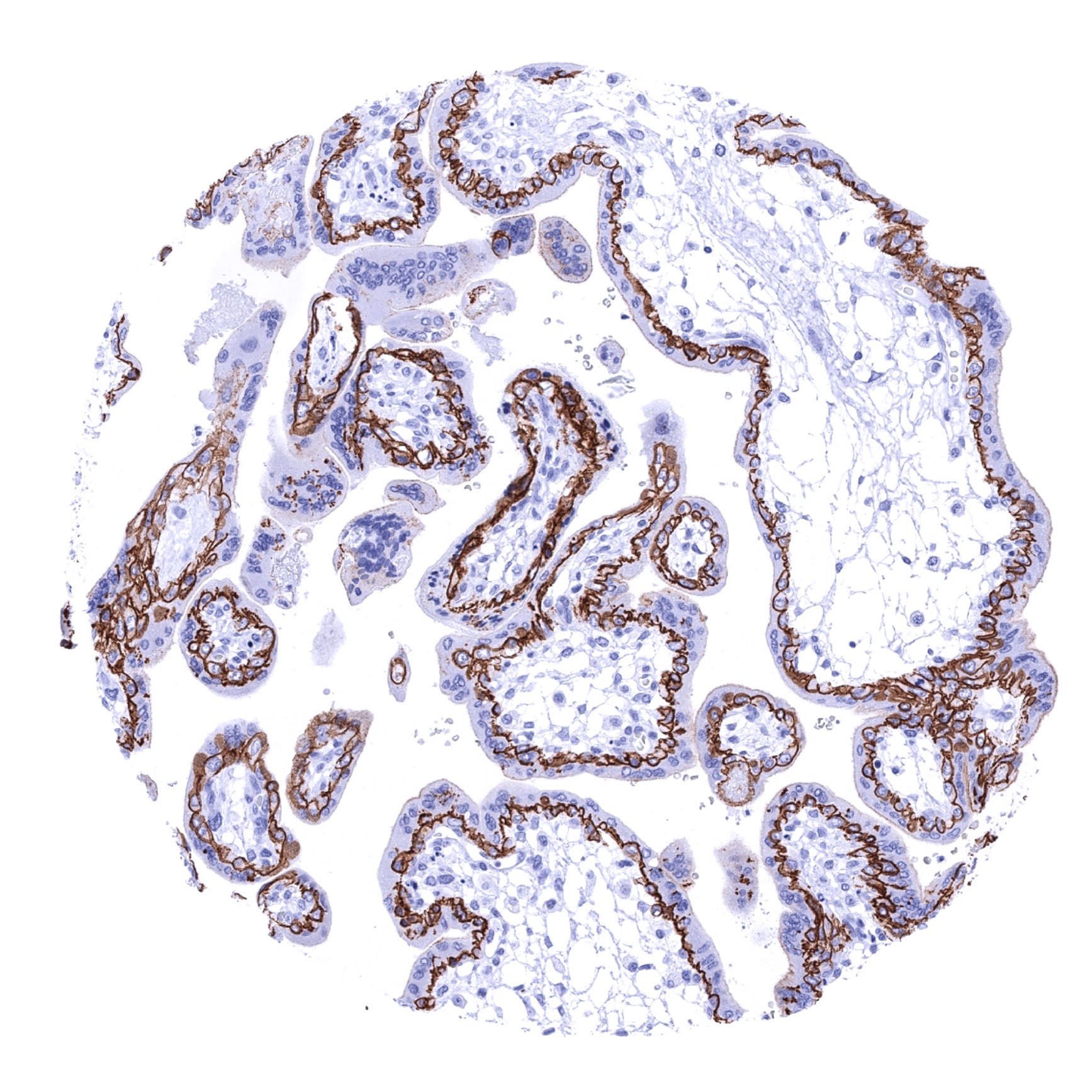
Placenta, early - The cytotrophoblast but not the syncytiotrophoblast shows membranous E-Cadherin staining.

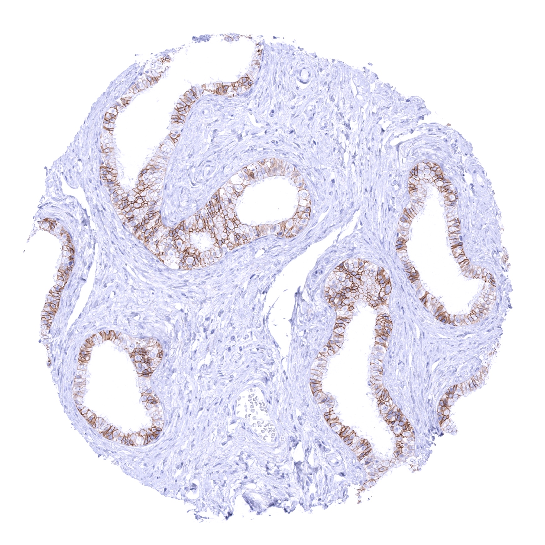
Prostate
Rectum, mucosa
Seminal vesicle
Sinus paranasales
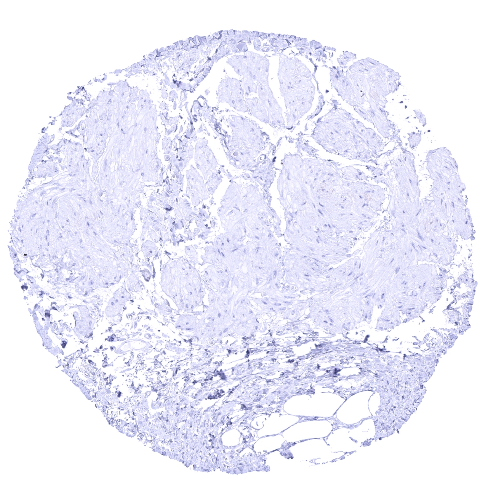
Skeletal muscle
Skin
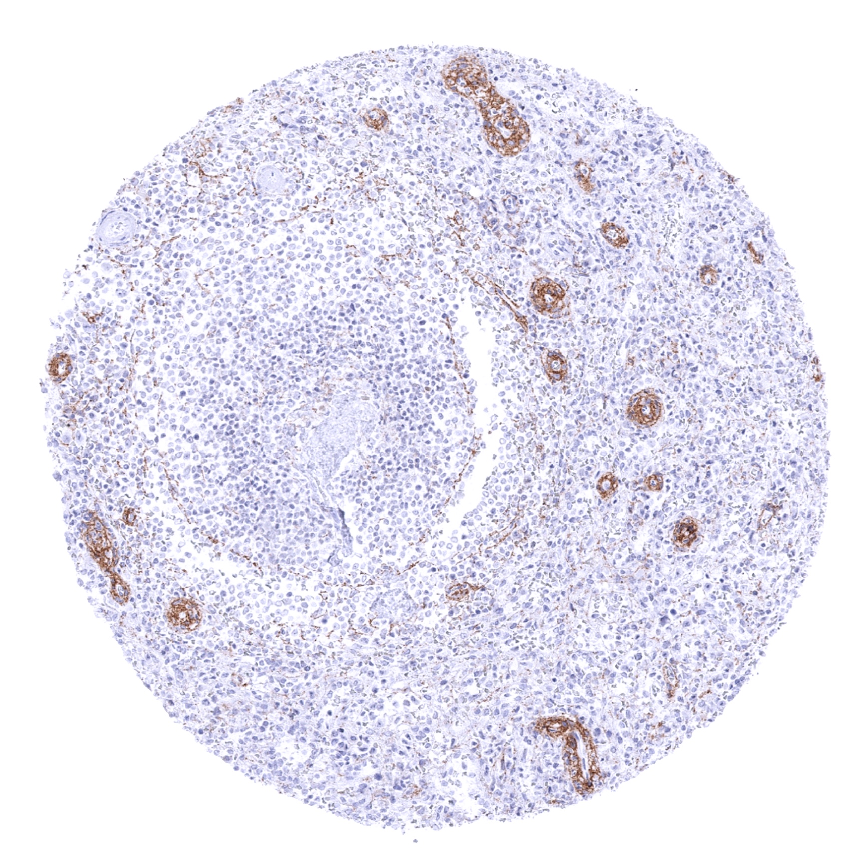
Spleen - A fraction of small vessels stain E-Cadherin-positive in the spleen.

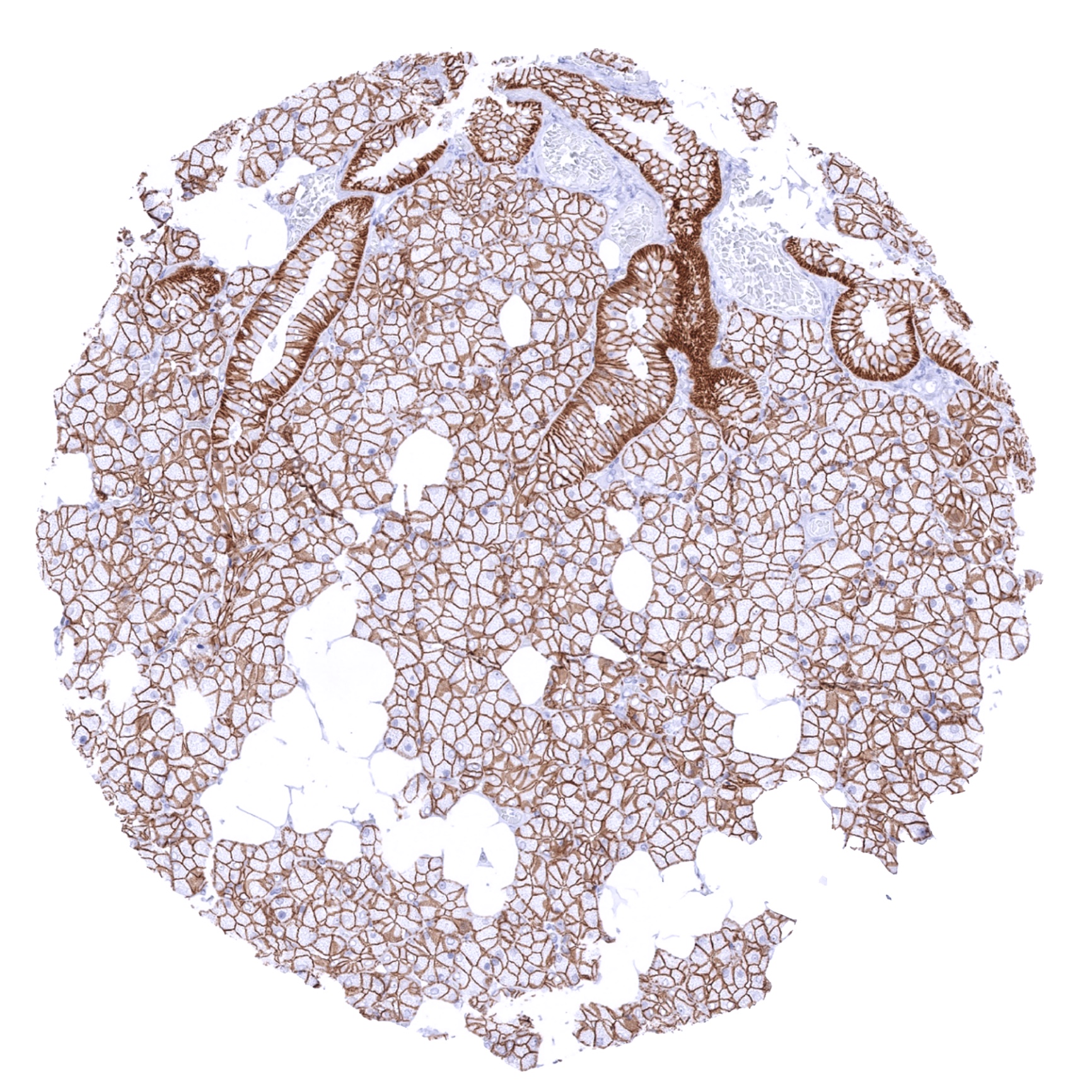
Stomach, antrum
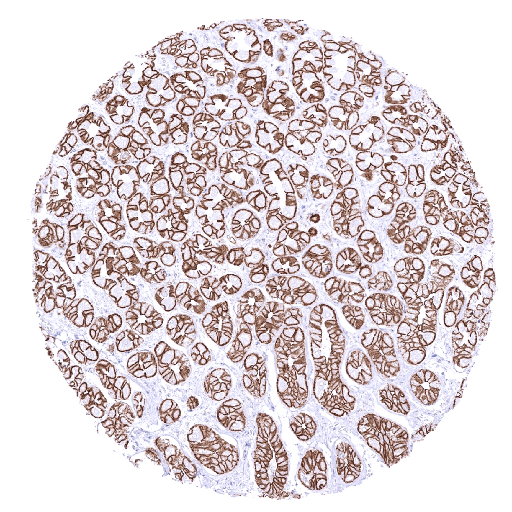
Stomach, antrum
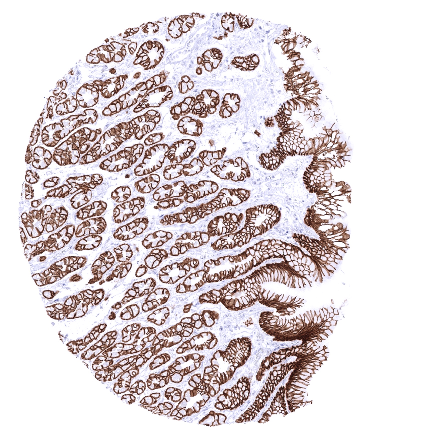
Stomach, corpus
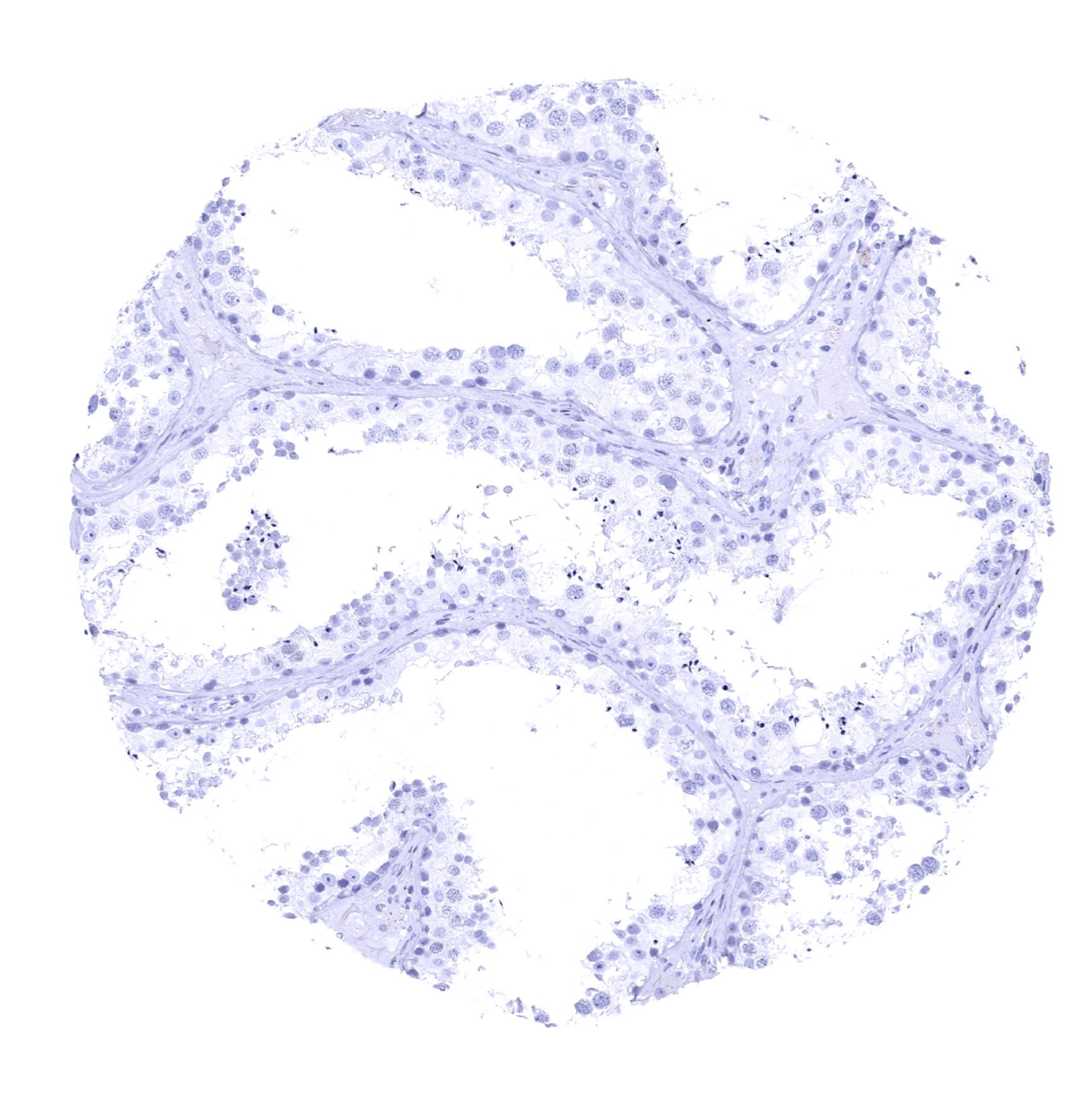
Testis
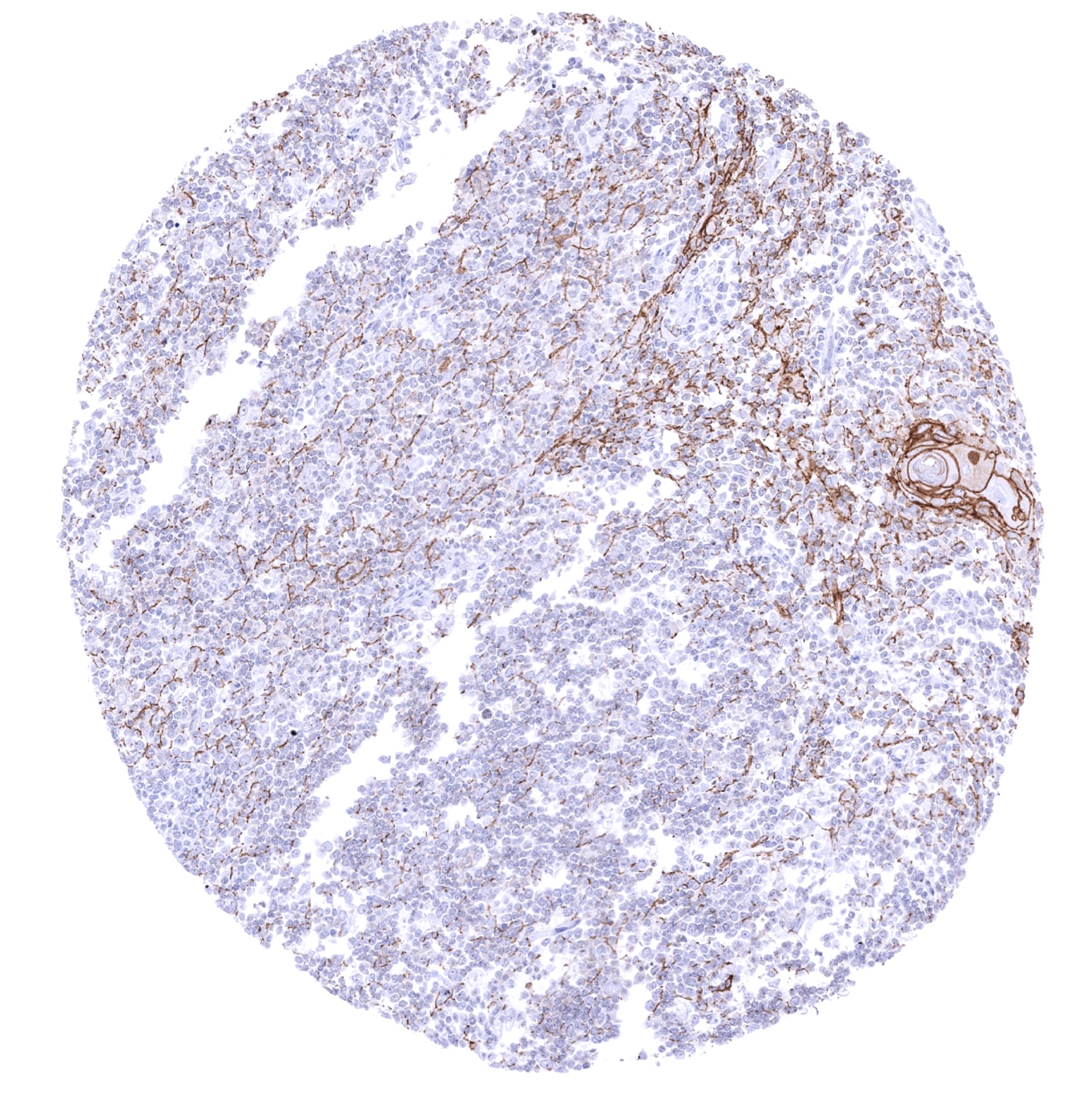
Thymus - E-Cadherin staining occurs in corpuscles of Hassall´s and in a fraction of thymic epithelial cells
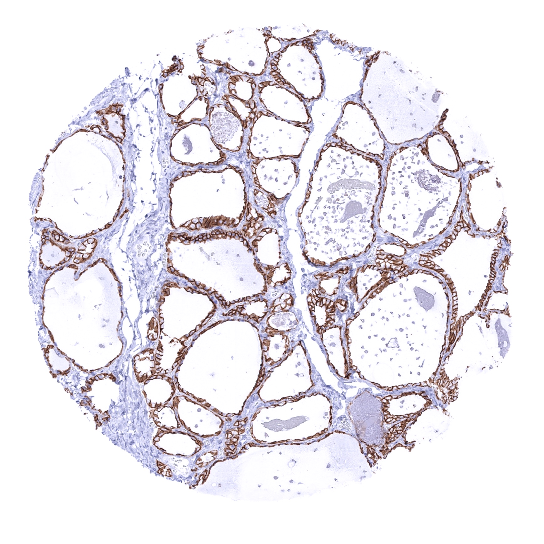
Thyroid gland
Tonsil, surface epithelium

Tonsil
Urinary bladder, muscular wall

Urinary bladder, urothelium

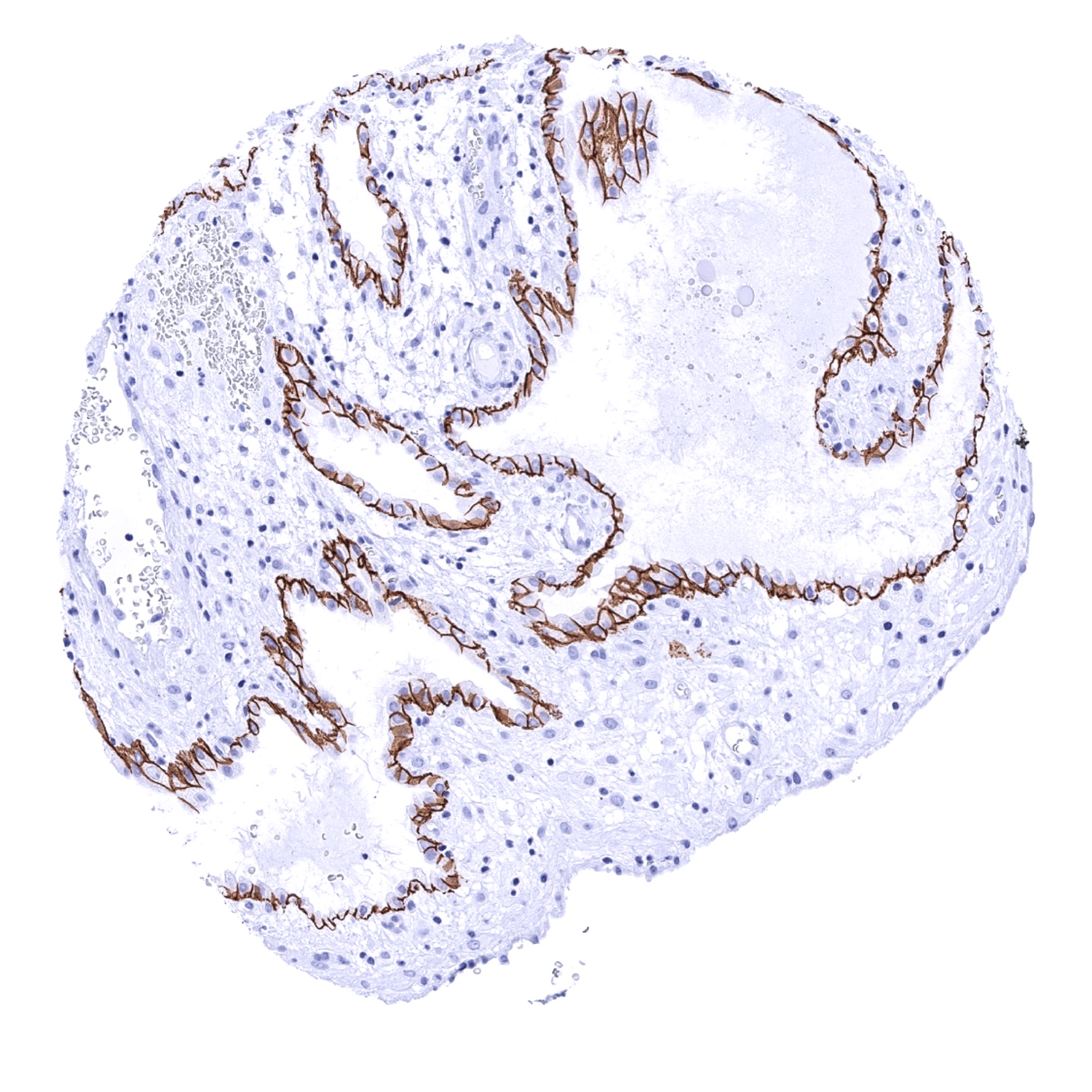
Uterus, ectocervix

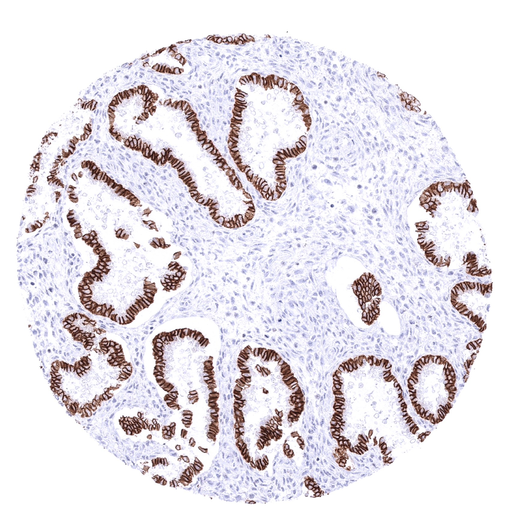
Uterus, endocervix
Uterus, endometrium (pregnancy)
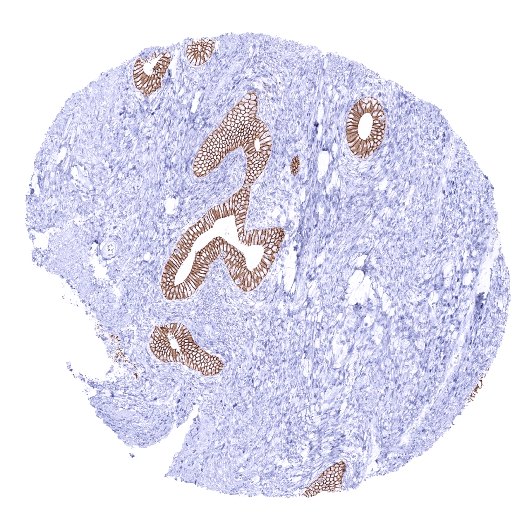
Uterus, endometrium (proliferation)
Uterus, endometrium (secretion)
Uterus, myometrium