295,00 € – 895,00 €
Product details
Synonyms = hPTH; Parathormone; Parathyrin; Parathyroid hormone 1 (PTH1); Parathyroid hormone (PTH)
Antibody type = Recombinant Rabbit monoclonal / IgG
Clone = MSVA-525R
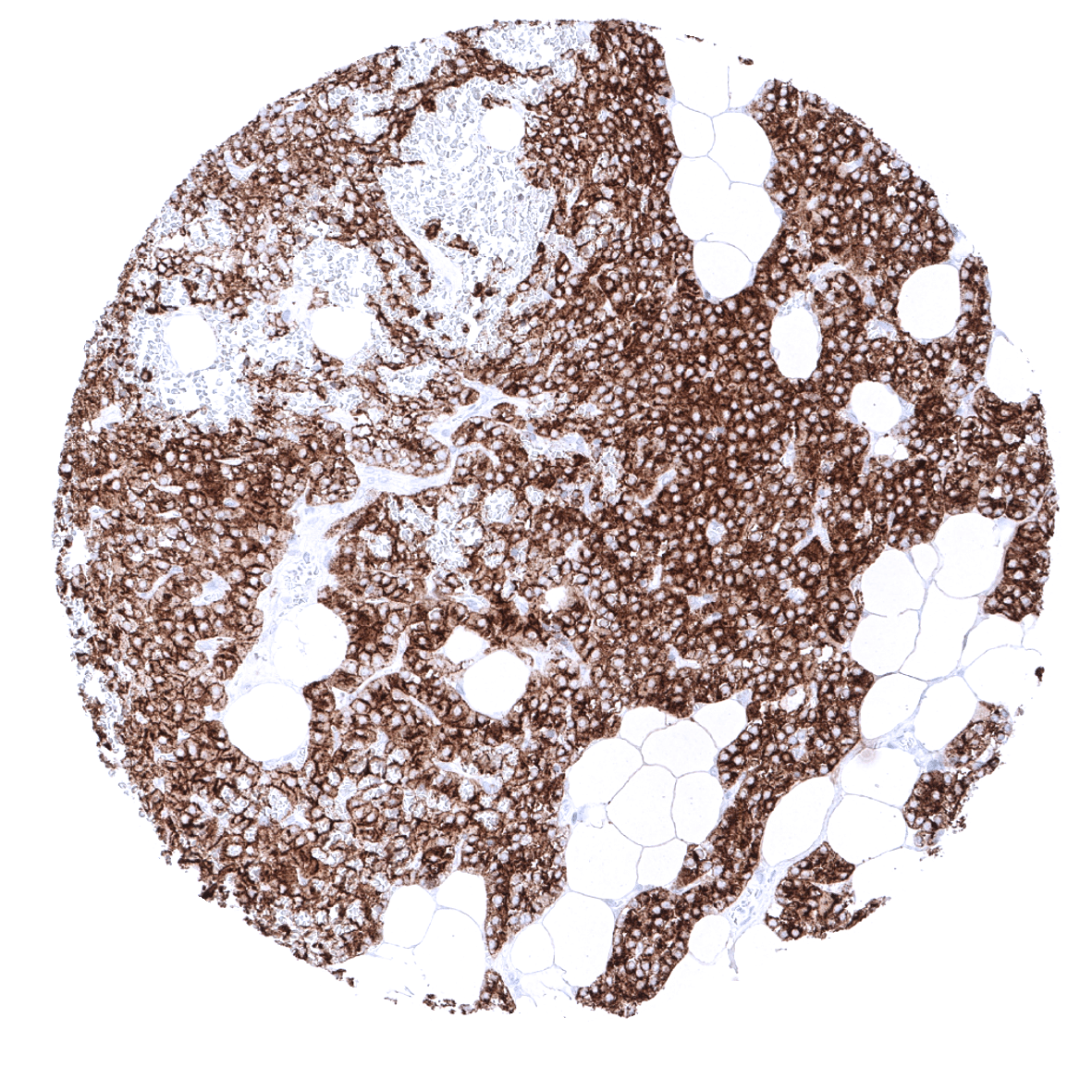
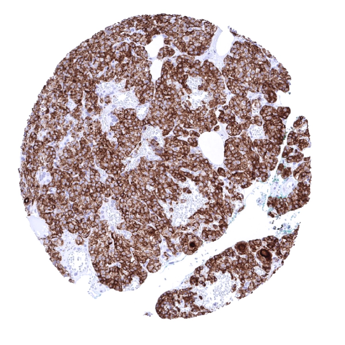
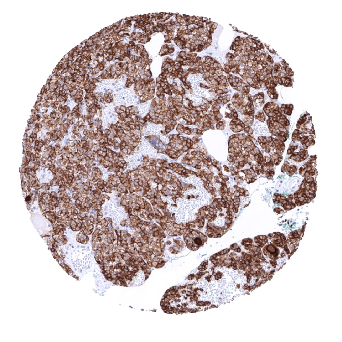
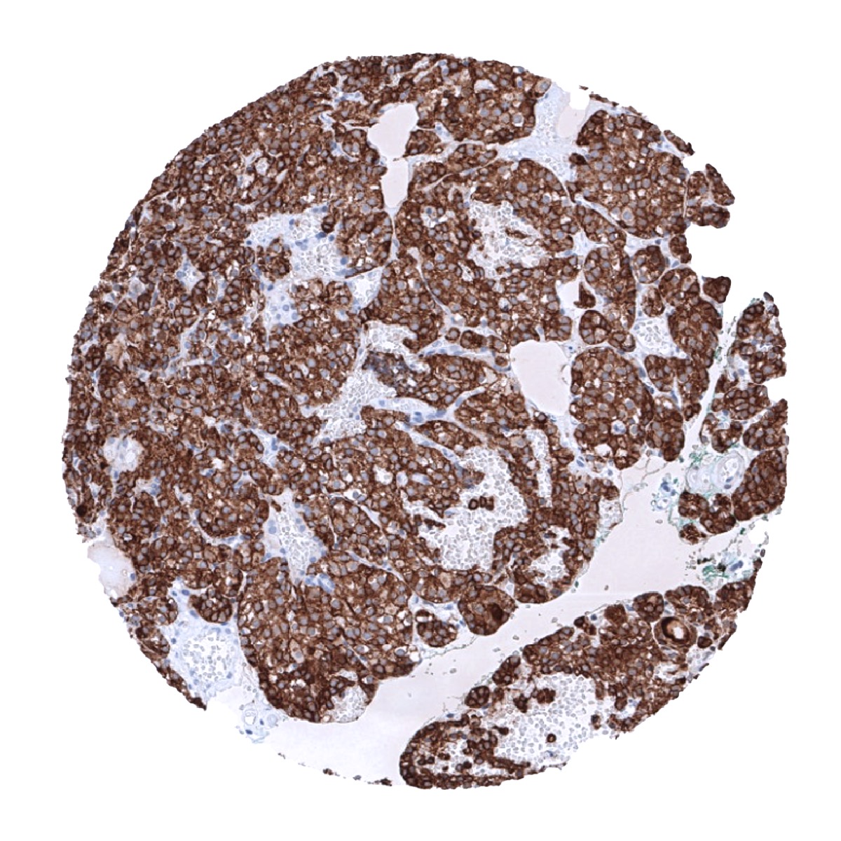
Positive control = Pituitary gland: A strong PTH immunostaining should be seen in all epithelial cells.
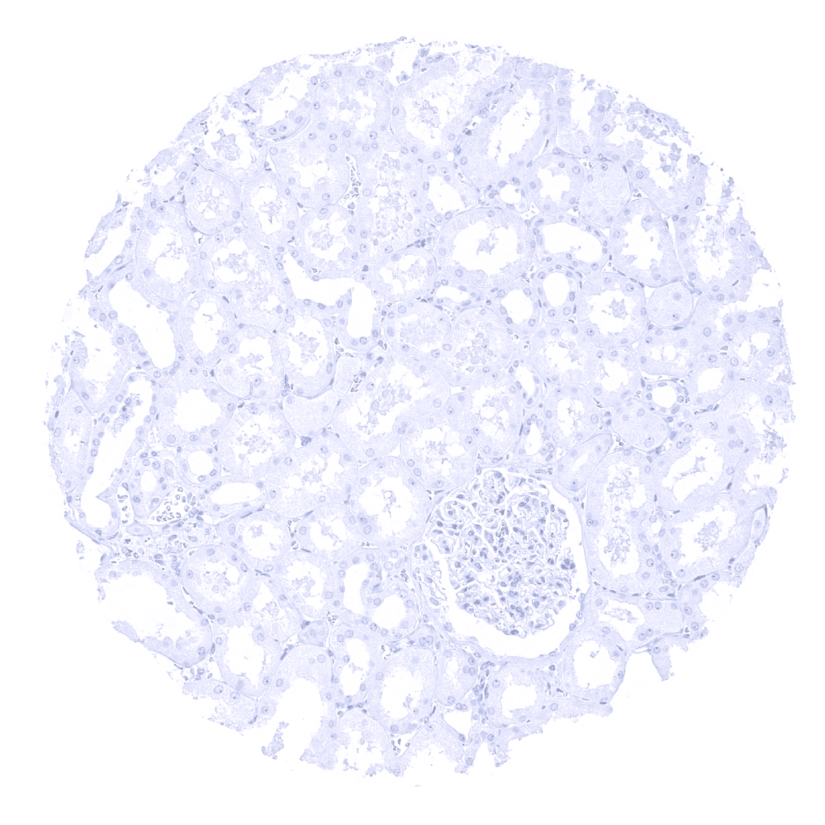
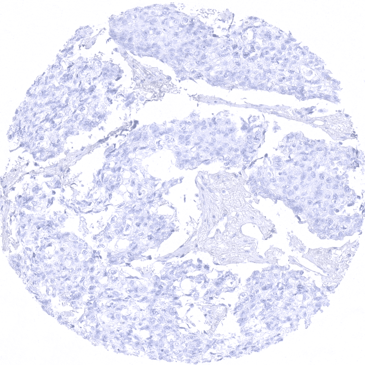
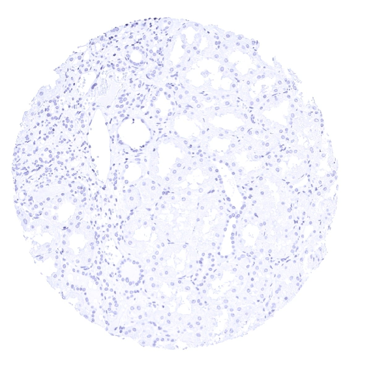
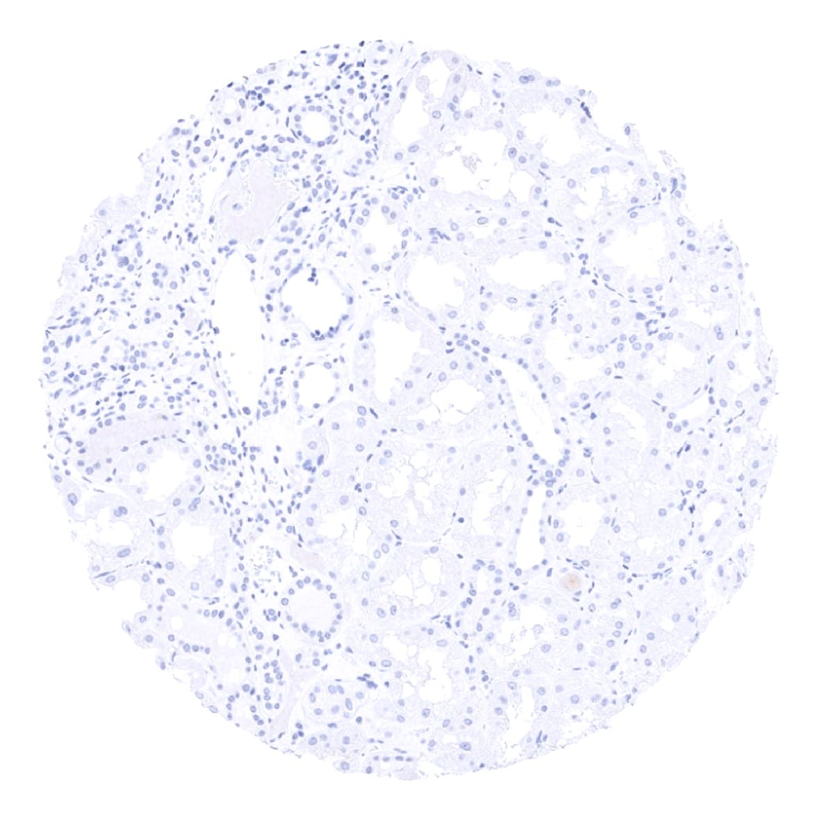
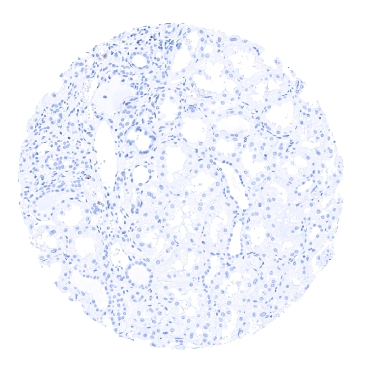
Negative control = Kidney: PTH immunostaining should be absent in all cells.
Cellular localization = Cytoplasmic
Reactivity = Human
Application = Immunohistochemistry
Dilution = 1:100 – 1:200
Intended Use = Research Use Only
Relevance of Antibody
PTH is expressed in parathyroid tissue.
Biology Behind
Parathyroid hormone (PTH), a 9,5 kDa hormone protein which is coded by the PTH gene at 11p15.3 and produced by the chief cells of the parathyroid glands. It regulates the serum calcium concentration by exerting effects on bone, kidney, and intestine. PTH is secreted in response to low blood serum calcium (Ca2+) levels. In order to elevate a low serum calcium level and to release more ionic calcium (Ca2+) into the blood, PTH indirectly stimulates osteoclast activity by increasing RANKL expression of osteoblasts. The half-life of PTH is about 4 minutes. Disorders that yield too little or too much PTH, such as hypoparathyroidism, hyperparathyroidism, and paraneoplastic syndromes can cause bone disease, hypocalcaemia, and hypercalcaemia. In the kidney, circulating parathyroid hormone I) increases the reabsorption of filtered calcium ions in the distal tubules and the renal collecting ducts, II) inhibits the reabsorption of phosphate from the tubular fluid, resulting in a decrease of the plasma phosphate concentration, and III) stimulates the conversion of 25-hydroxy vitamin D into the active form of vitamin D (1,25-dihydroxy vitamin D) which stimulates calcium uptake from the intestine.
Staining Pattern in Normal Tissues
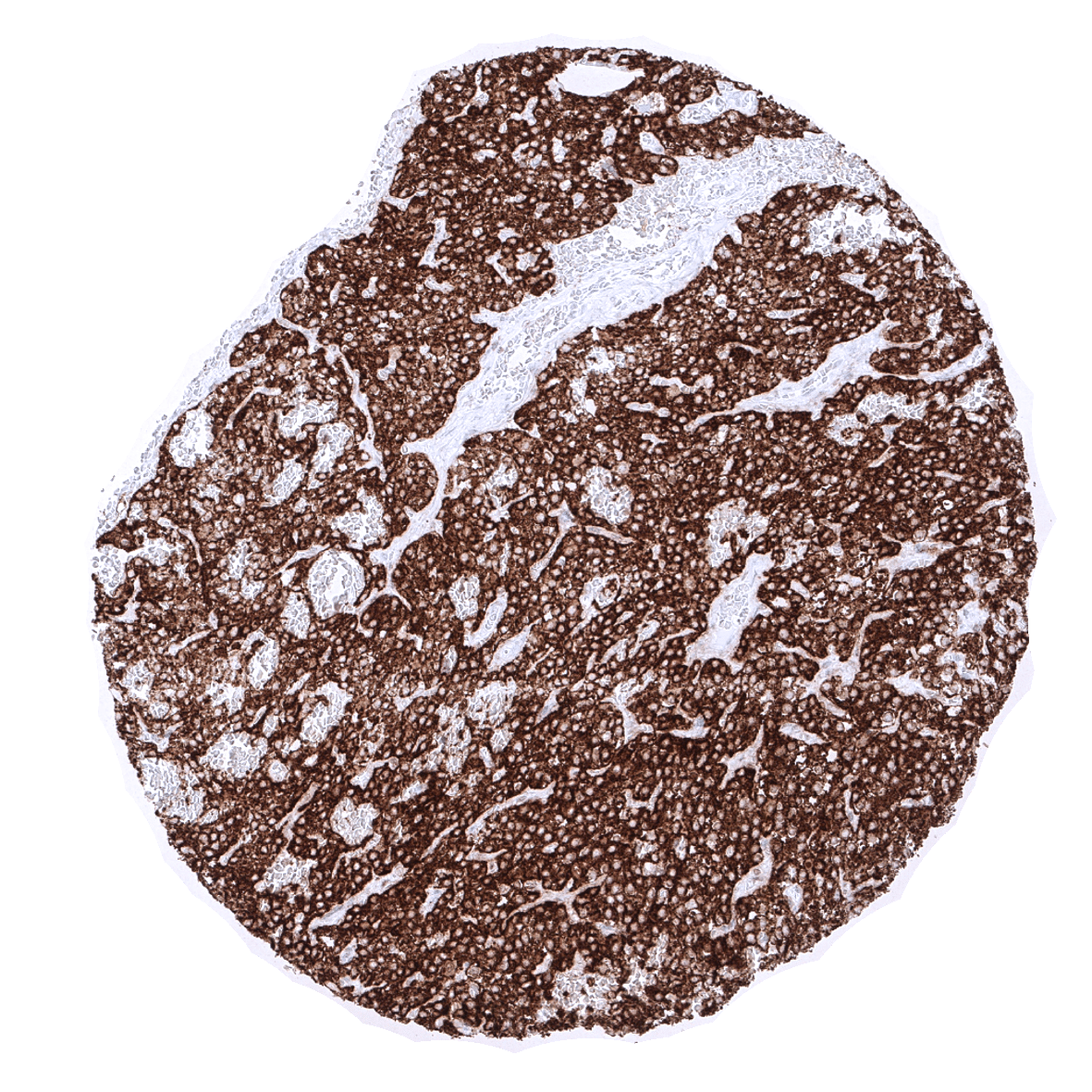
In normal tissues, PTH immunostaining is exclusively seen in epithelial cells of the parathyroid gland.
These findings are largely consistent to the RNA and protein data described in the Human Protein Atlas (Tissue expression PTH) even though RNA data are provided suggesting that PTH may also occur in the thyroid. As PTH RNA expression in the thyroid was described in the GTEx and the FANTOM5 databases but not in the Human Protein Atlas (HPA) RNA-seq tissue dataset, it is concluded, that PTH RNA is not regularly expressed in the normal thyroid gland. It is well possible, that some small and difficult to identify parathyroid glands were erroneously included in some RNA analyses of thyroid tissues.
Suggested positive tissue control: Pituitary gland: A strong PTH immunostaining should be seen in all epithelial cells.
Suggested negative tissue control: Kidney: PTH immunostaining should be absent in all cells.
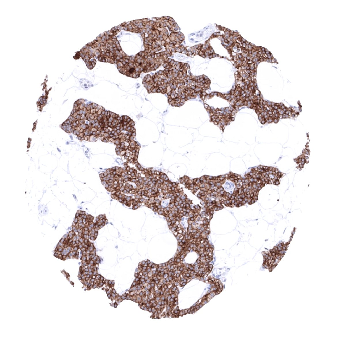
Staining Pattern in Relevant Tumor Types
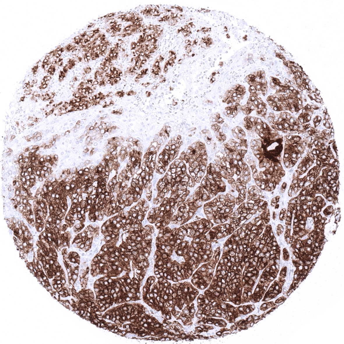
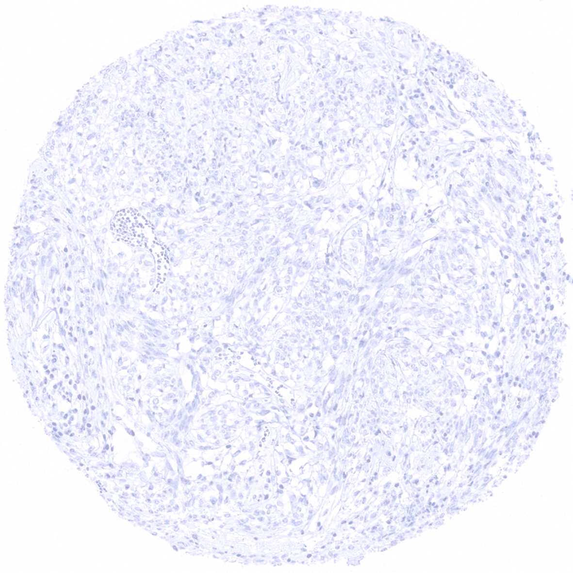
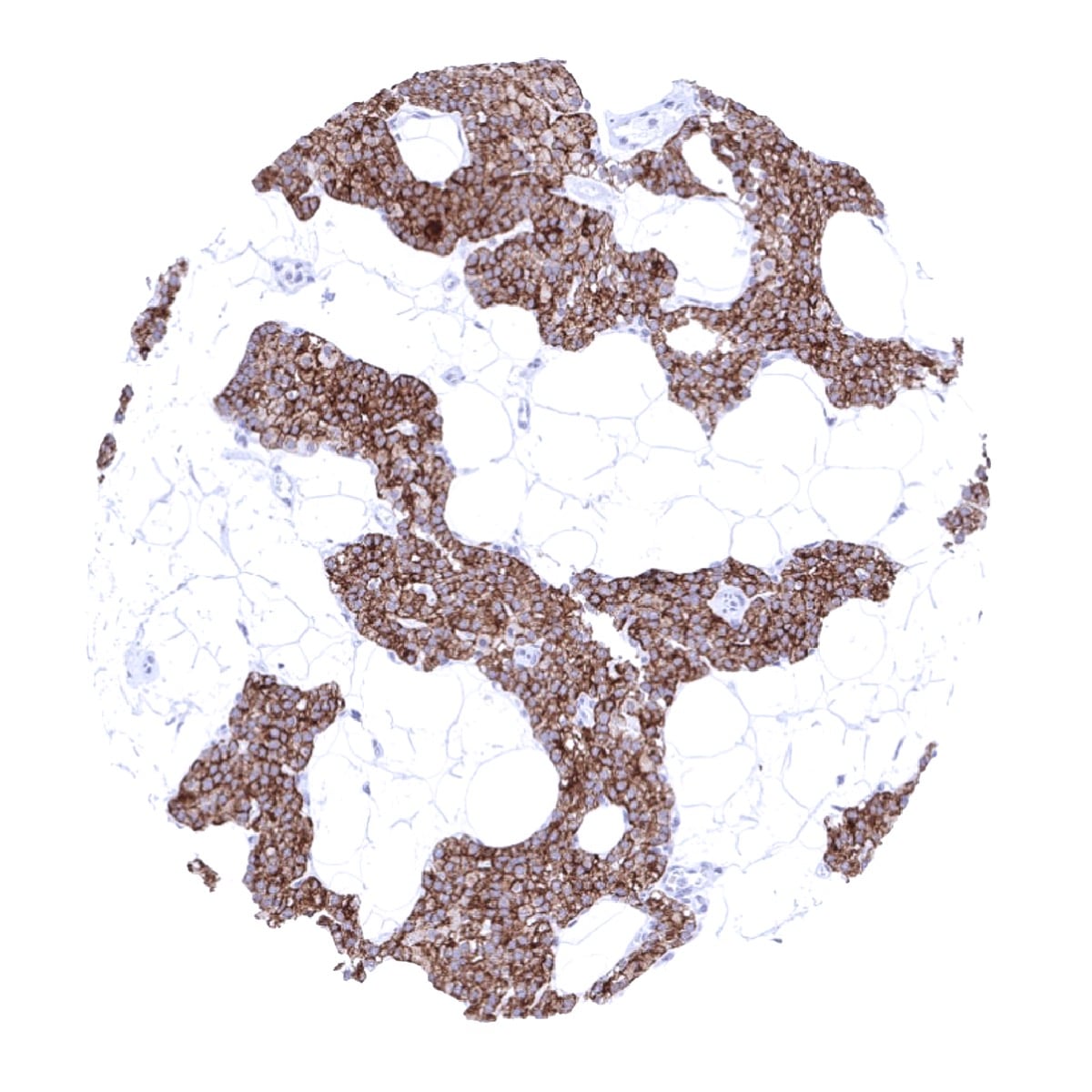
PTH is regularly expressed in benign and malignant neoplasms of the thyroid but largely absent in other tumors.
The TCGA findings on PTH RNA expression in different tumor categories have been summarized in the Human Protein Atlas.
Compatibility of Antibodies
No data available at the moment
Protocol Recommendations
IHC users have different preferences on how the stains should look like. Some prefer high staining intensity of the target stain and even accept some background. Others favor absolute specificity and lighter target stains. Factors that invariably lead to more intense staining include higher concentration of the antibody and visualization tools, longer incubation time, higher temperature during incubation, higher temperature and longer duration of the heat induced epitope retrieval (slide pretreatment). The impact of the pH during slide pretreatment has variable effects and depends on the antibody and the target protein.
All images and data shown here and in our image galleries are obtained by the manual protocol described below. Other protocols resulting in equivalent staining are described as well.
Manual protocol
Freshly cut sections should be used (less than 10 days between cutting and staining). Heat-induced antigen retrieval for 5 minutes in an autoclave at 121°C in pH 9,0 Target Retrieval Solution buffer. Apply MSVA-525R at a dilution of 1:150 at 37°C for 60 minutes. Visualization of bound antibody by the EnVision Kit (Dako, Agilent) according to the manufacturer’s directions.


Agilent / Dako – Autostainer Link 48
Pretreatment in PT-Link for 30 minutes at 95°C (pH high); FLEX peroxidase blocking for 5 minutes (room temperature), MSVA-525R 1:150 for 20 minutes (room temperature), FLEX+ mouse/rabbit (LINKER) for 15 minutes (room temperature), horseradish peroxidase (HRP) for 20 minutes (room temperature), FLEX DAB+Sub-Chromo for 10 minutes (room temperature), FLEX hematoxylin for 5 minutes (room temperature).
These images reflect stainings by the protocol described above. It is of note that a comparable staining result can also be obtained by different protocols. In general, a longer pretreatment, a longer incubation time of the primary antibody, a higher antibody concentration, and a longer incubation time of FLEX+LINKER result in stronger staining, potentially at the cost of more background staining. Modifications of the protocol with a strengthening effect on staining intensity in combination with changes of other parameters that result in lower staining intensity can result in a comparable result as shown above.
Leica – BOND RX
Dewax at 72°C for 30 seconds; Pretreatment in Bond Epitope Retrieval Solution (ER2 – EDTA pH9) for 20 minutes at 100°C; Peroxidase blocking for 5 minutes (room temperature), MSVA-525R 1:150 for 15 minutes (room temperature), Post primary (rabbit anti mouse) for 8 minutes (room temperature), Polymer (goat anti rabbit) for 8 minutes (room temperature), mixed DAB refine for 10 minutes (room temperature), hematoxylin for 5 minutes (room temperature).
These images reflect stainings by the protocol described above. It is of note that a comparable staining result can also be obtained by different protocols. In general, a longer pretreatment, a longer incubation time of the primary antibody, a higher antibody concentration, a higher temperature during incubation, and a longer incubation time of Post primary and or the Polymer result in stronger staining, potentially at the cost of more background staining. Modifications of the protocol with a strengthening effect on staining intensity in combination with changes of other parameters that result in lower staining intensity can result in a comparable result as shown above.
Roche – Ventana Discovery ULTRA
Pretreatment for 64 minutes at 100°C (pH 8,4); CM peroxidase blocking for 12 minutes (room temperature), MSVA-525R 1:150 for 20 minutes at 36°C, secondary antibody (anti-rabbit HQ) for 12 minutes at 36°C, anti-HQ HRP for 12 minutes at room temperature, DAB at room temperature, hematoxylin II at room temperature for 8 minutes, bluing reagent at room temperature for 4 minutes.

These images depict staining results obtained by the protocol described above. It is of note, that the Ventana machines generally require higher antibody concentrations than other commonly used autostainers because the antibodies are automatically diluted during the procedure. Various other protocols can result in an identical result as shown above. A longer pretreatment, a longer incubation time of the primary antibody, a higher antibody concentration, a higher temperature during incubation, and a longer incubation time of secondary antibody and or the anti-HQ HRP result in stronger staining, potentially at the cost of more background staining.
Potential Research Applications
- Data on a possible paraneoplastic expression of PTH in cancer are sparse and controversial.
Evidence for Antibody Specificity in IHC
There are two ways, how the specificity of antibodies can be documented for immunohistochemistry on formalin fixed tissues. These are: 1. comparison with a second independent method for target expression measurement across a large number of different tissue types (orthogonal strategy), and 2. Comparison with one or several independent antibodies for the same target and showing that all positive staining results are also seen with other antibodies for the same target (independent antibody strategy).
Comparison of MSVA-525R immunostaining with RNA expression
RNA expression data derived from the Human Protein Atlas (HPA) RNA-seq tissue dataset, the FANTOM5 project, and the Genotype-Tissue Expression (GTEx) project are summarized in the Human Protein Atlas (Tissue expression PTH). In agreement with the immunostaining pattern observed by MSVA-525R in normal tissues, unequivocal PTH RNA expression is only seen in the parathyroid gland. The thyroid is the only other organ, for which a PTH RNA expression is suggested by RNA data. As PTH RNA expression in the thyroid was described in the GTEx and the FANTOM5 databases but not in the Human Protein Atlas (HPA) RNA-seq tissue dataset, it is concluded, that PTH RNA is not regularly expressed in the normal thyroid gland. It is well possible, that some small and difficult to identify parathyroid glands were erroneously included in some RNA analyses of thyroid tissues.
The quality of the antibody is further documented by absence of staining in tissues notorious for non-specific IHC background such as kidney, colonic mucosa, and epidermis.
