
295,00 € – 995,00 €
Product details
Synonyms = GCTM-2 antigen, Gp200, Podocalyxin-like protein 1, PDXL, Gp200, PCLP1, Pcx
Antibody type = Mouse monoclonal
Clone = MSVA-645M
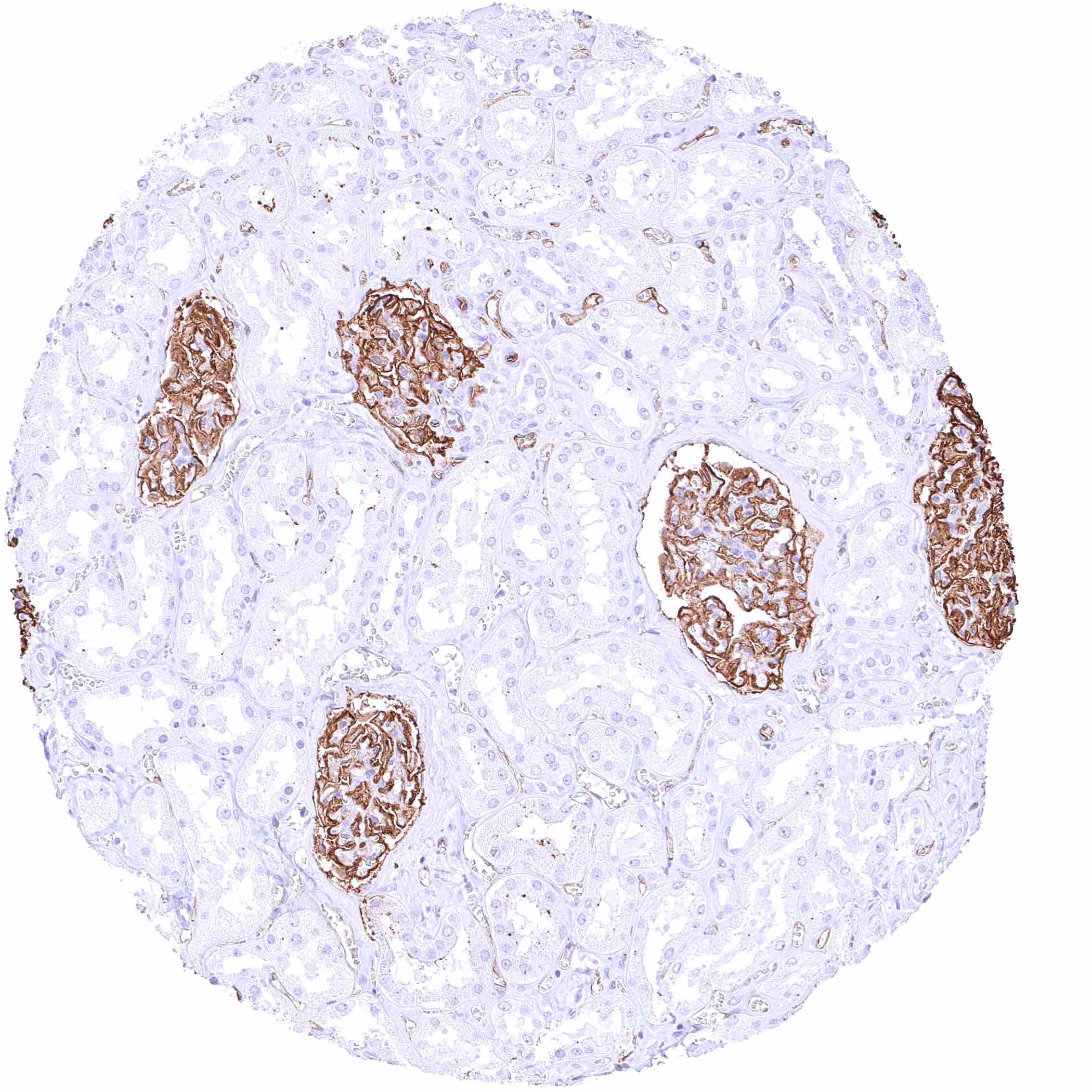
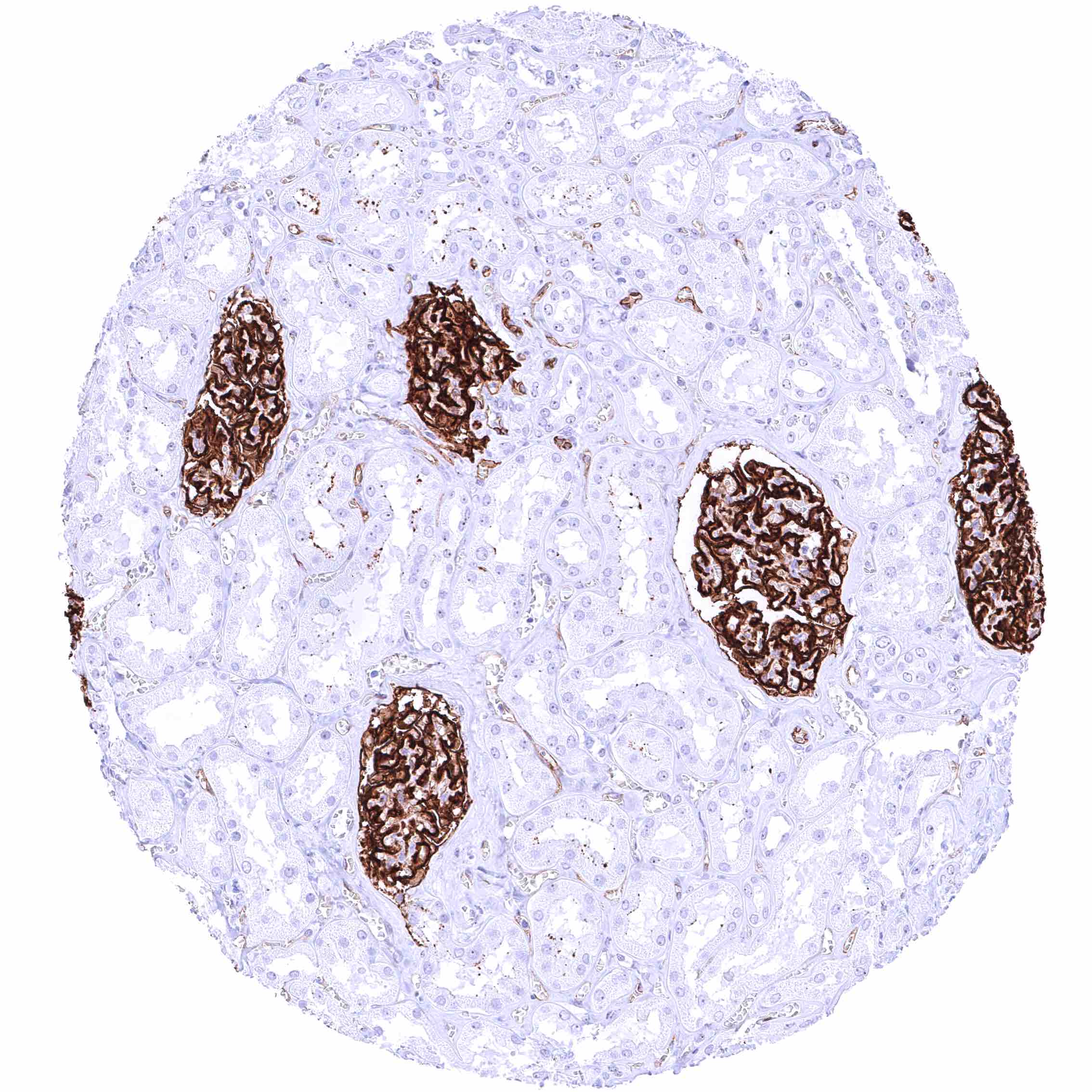
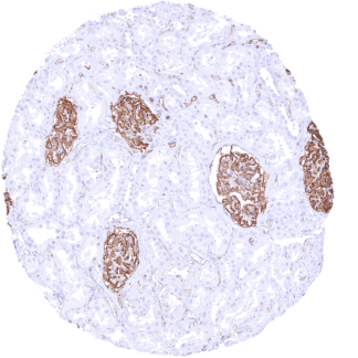
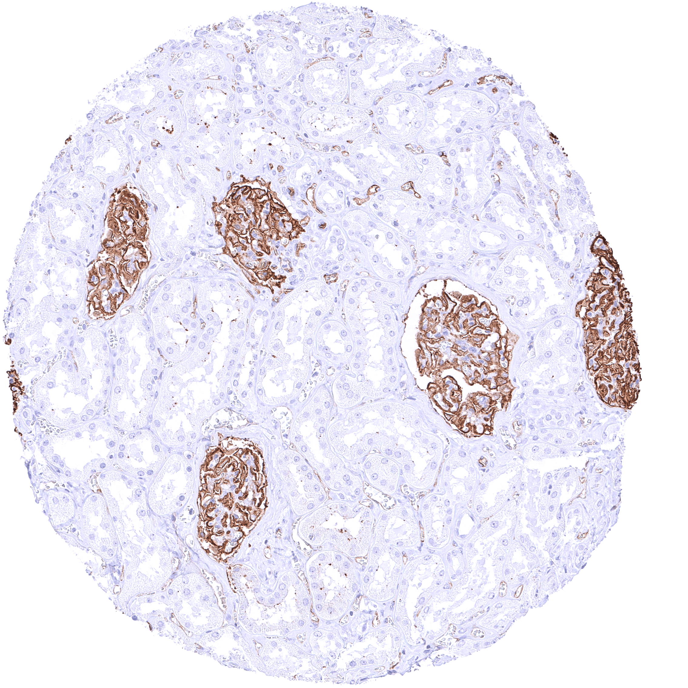
Positive control = Kidney: A strong PODXL must be seen in glomerular podocytes while at least a weak to moderate PODXL staining must be seen in endothelial cells of small intertubular vessels.
Negative control = Kidney: Tubuli and collecting ducts must be PODXL negative.
Cellular localization = Membranous
Reactivity = Human
Application = Immunohistochemistry
Dilution = 1:100 – 1:200
Intended Use = Research Use Only
Relevance of Antibody
Podocalyxin is a therapeutic target protein.
Biology Behind
Podocalyxin (PODXL) is a CD34-related transmembrane sialomucin protein which is coded by the PODXL gene located on chromosome 7q32.2. PODXL is primarily expressed in podocytes of the kidneys, endothelial cells, mesothelial cells, and a few selected epithelial cell types. PODXL knock-out mice die within the first 24 hours of life from renal failure because podocyte foot processes do not form, and intercellular junctions between adjacent podocyte foot processes are abnormal. Germline mutations in PODXL are associated with congenital nephrotic syndrome. Overall PODXL has an important role in tissue development and for optimal glomerular function. PODXL also plays an important role in cancer as it can be mutated and aberrantly expressed in several cancer types. Many cancers with high PODXL expression are derived from PODXL negative normal epithelial cells. PODXL neo-expression was found to be associated with aggressive cancer, drug resistance, and immune evasion. In cancer cells, PODXL can interact with several proteins and downstream signaling pathways that are critical for promoting cancer metastasis and subsequent invasion. Because of its membranous location, PODXL is being evaluated for a role as a therapeutic target protein.
Staining Pattern in Normal Tissues
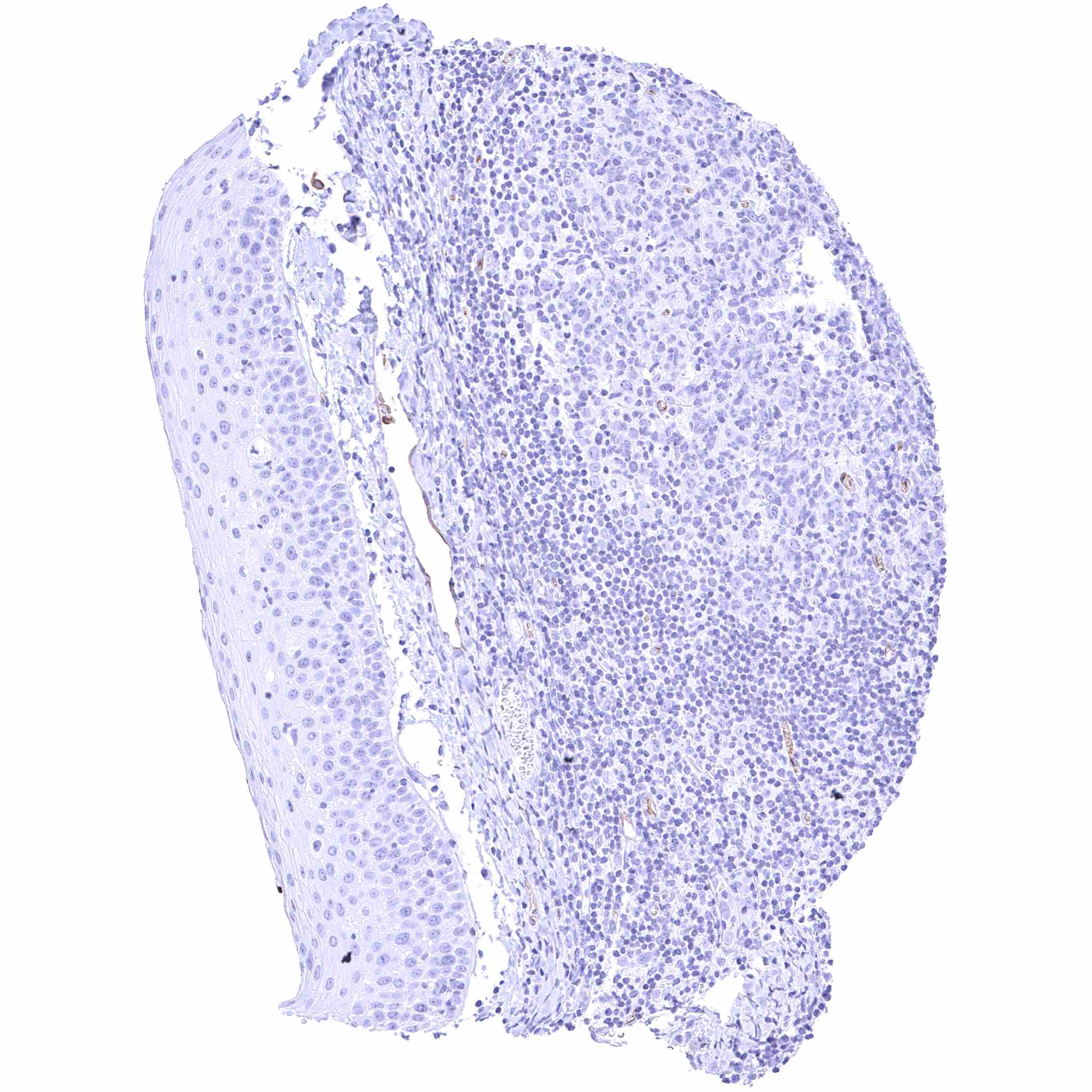
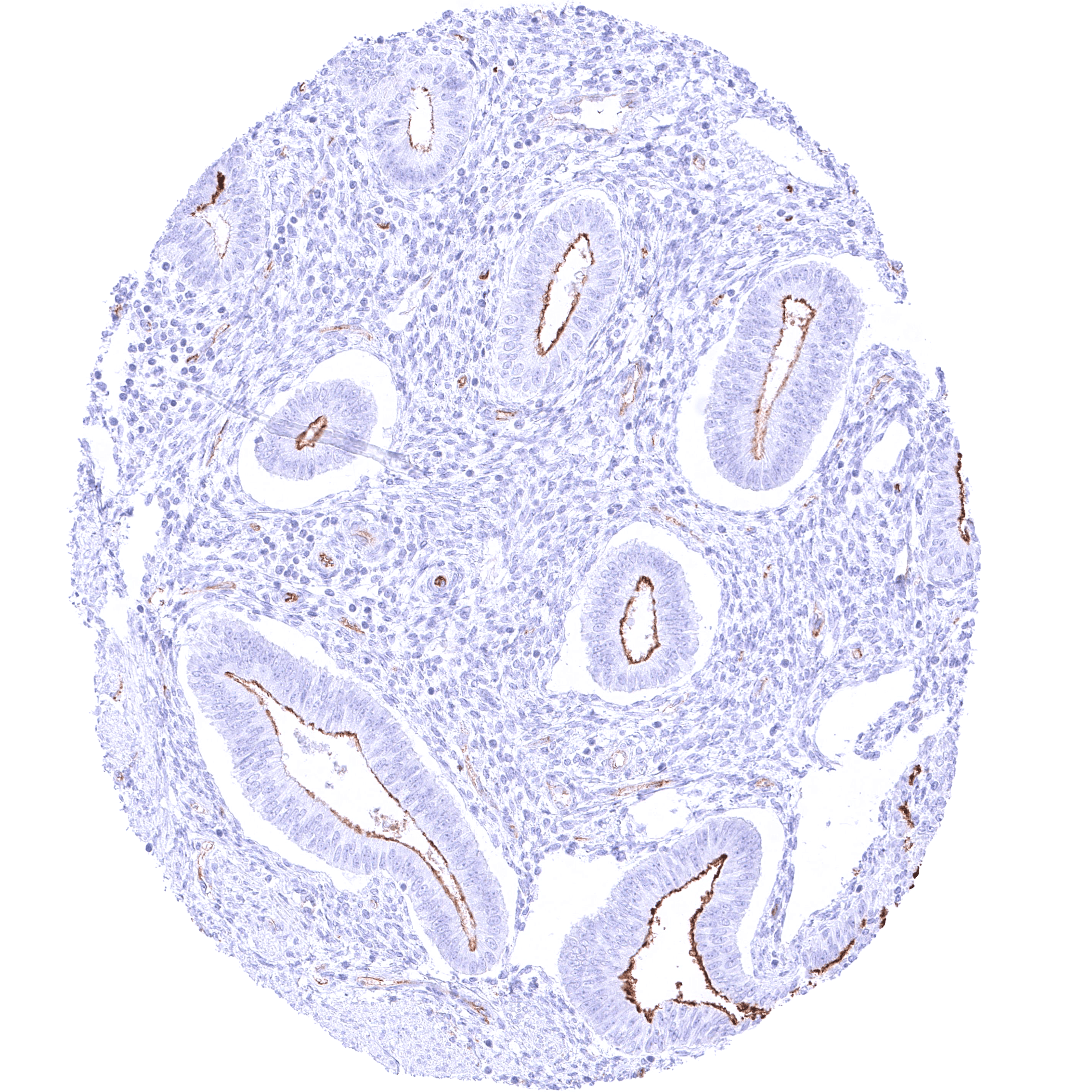
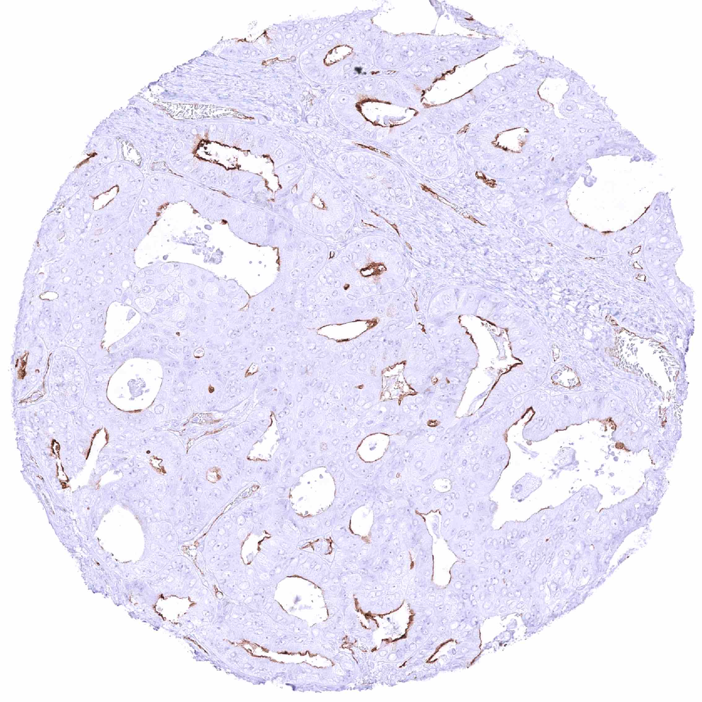
PODXL is most prominently expressed in podocytes of the kidney and also occurs rather abundantly in endothelial cells, mesothelial cells, and a few selected epithelial cell types.
Images describing the PODXL staining pattern in normal tissues obtained by the antibody MSVA-645M are shown in our “Normal Tissue Gallery”.
| Brain | Cerebrum | Negative. |
| Cerebellum | Negative. | |
| Endocrine Tissues | Thyroid | Negative. |
| Parathyroid | Negative. | |
| Adrenal gland | Negative. | |
| Pituitary gland | Negative. | |
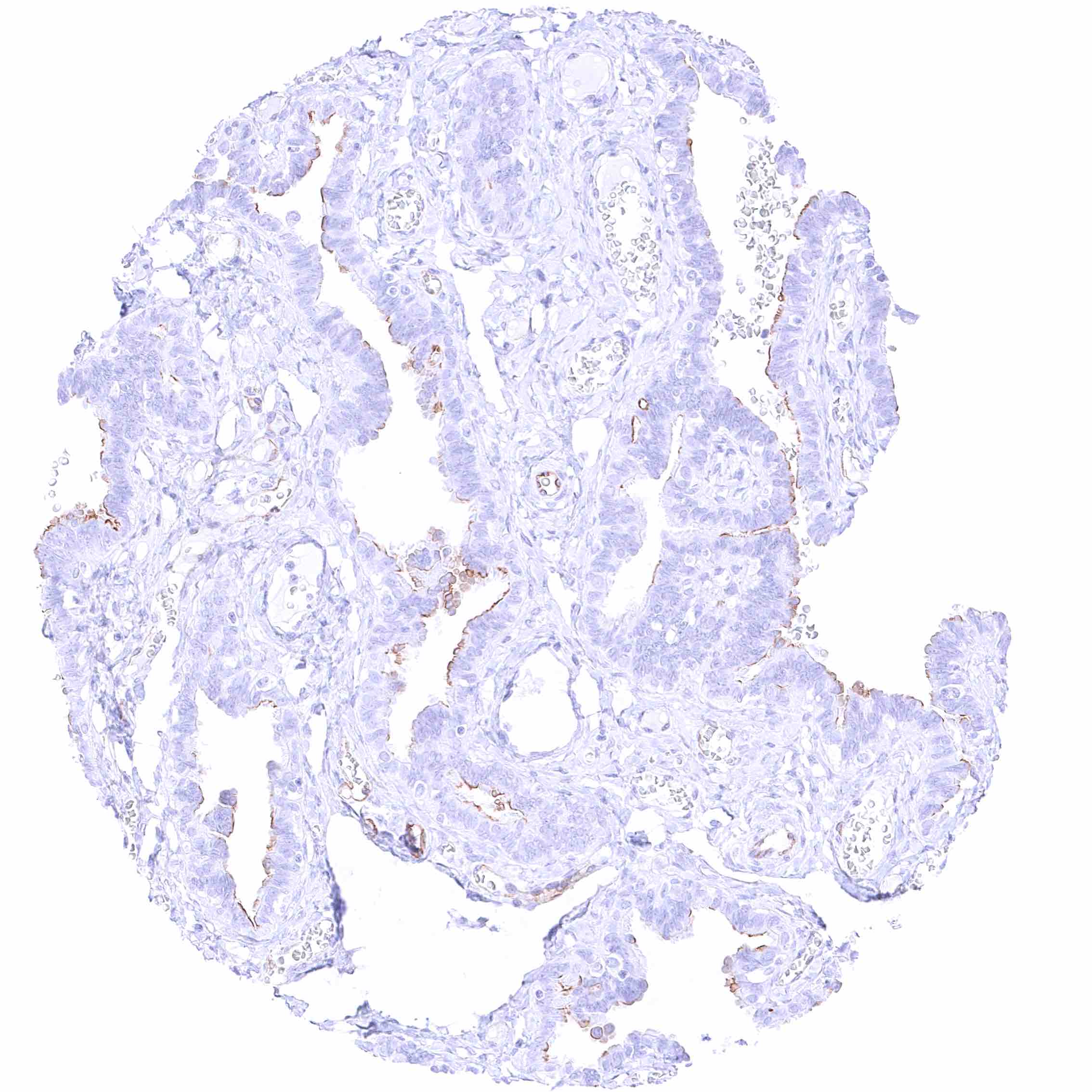
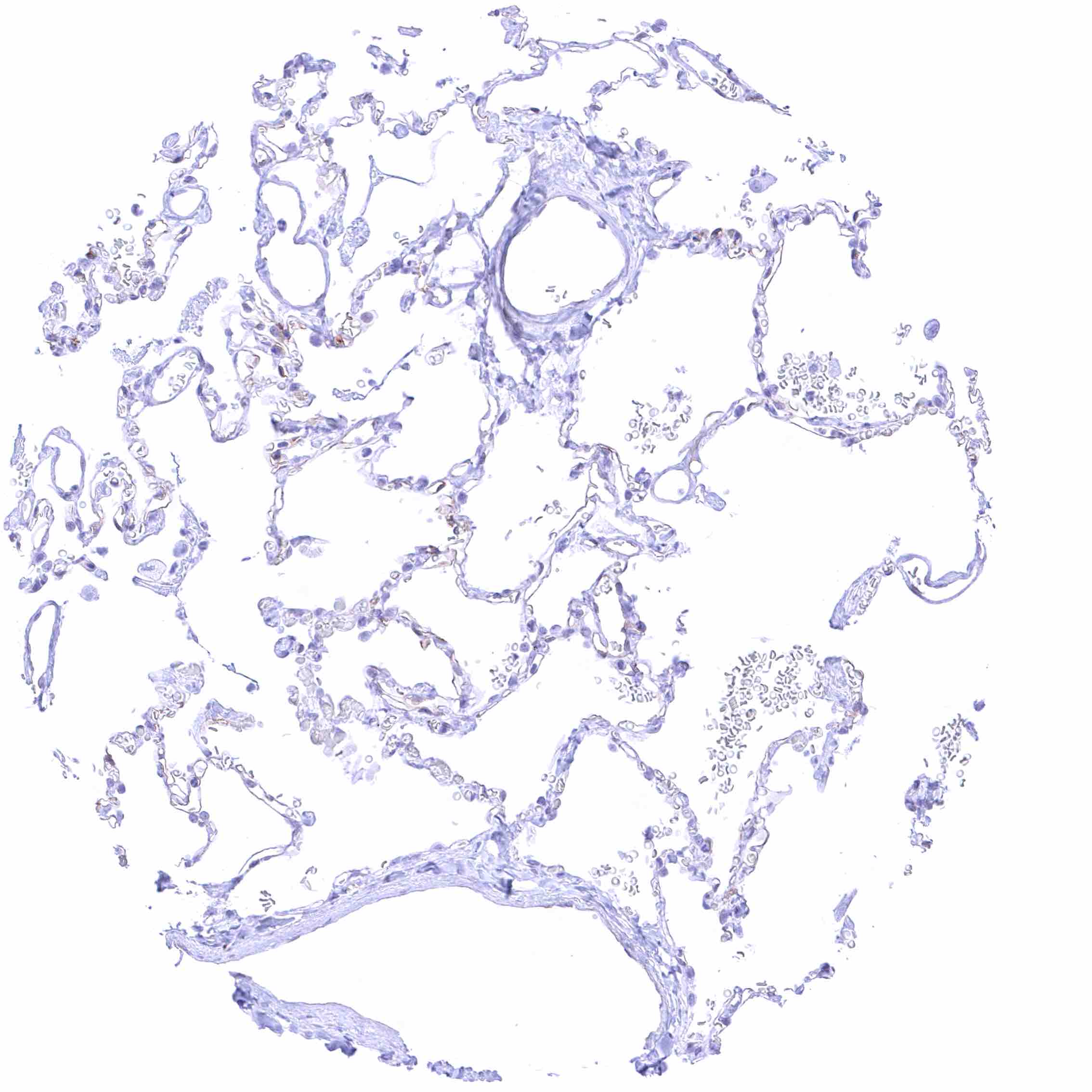
| Respiratory system | Respiratory epithelium | Moderate PODXL staining of luminal/apical membranes of a subset of bronchial glandular cells. Respiratory epithelium is PODXL negative. |
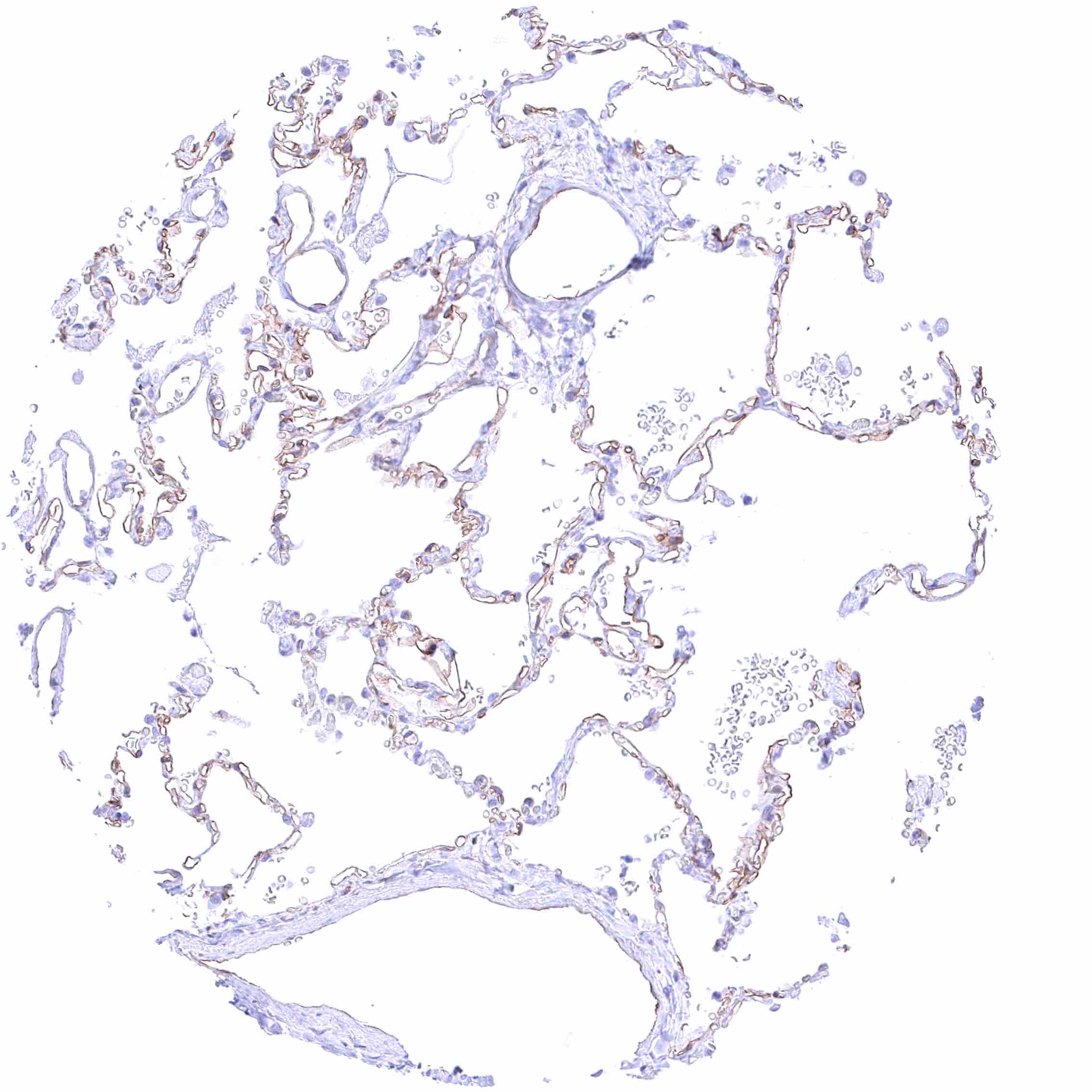
| Lung | Distinct PODXL staining of alveolar capillaries. | |
| Gastrointestinal Tract | Salivary glands | Negative. |
| Esophagus | Negative. | |
| Stomach | Negative. | |
| Duodenum | Negative. | |
| Small intestine | Negative. | |
| Appendix | Negative. | |
| Colon | Negative. | |
| Rectum | Negative. | |
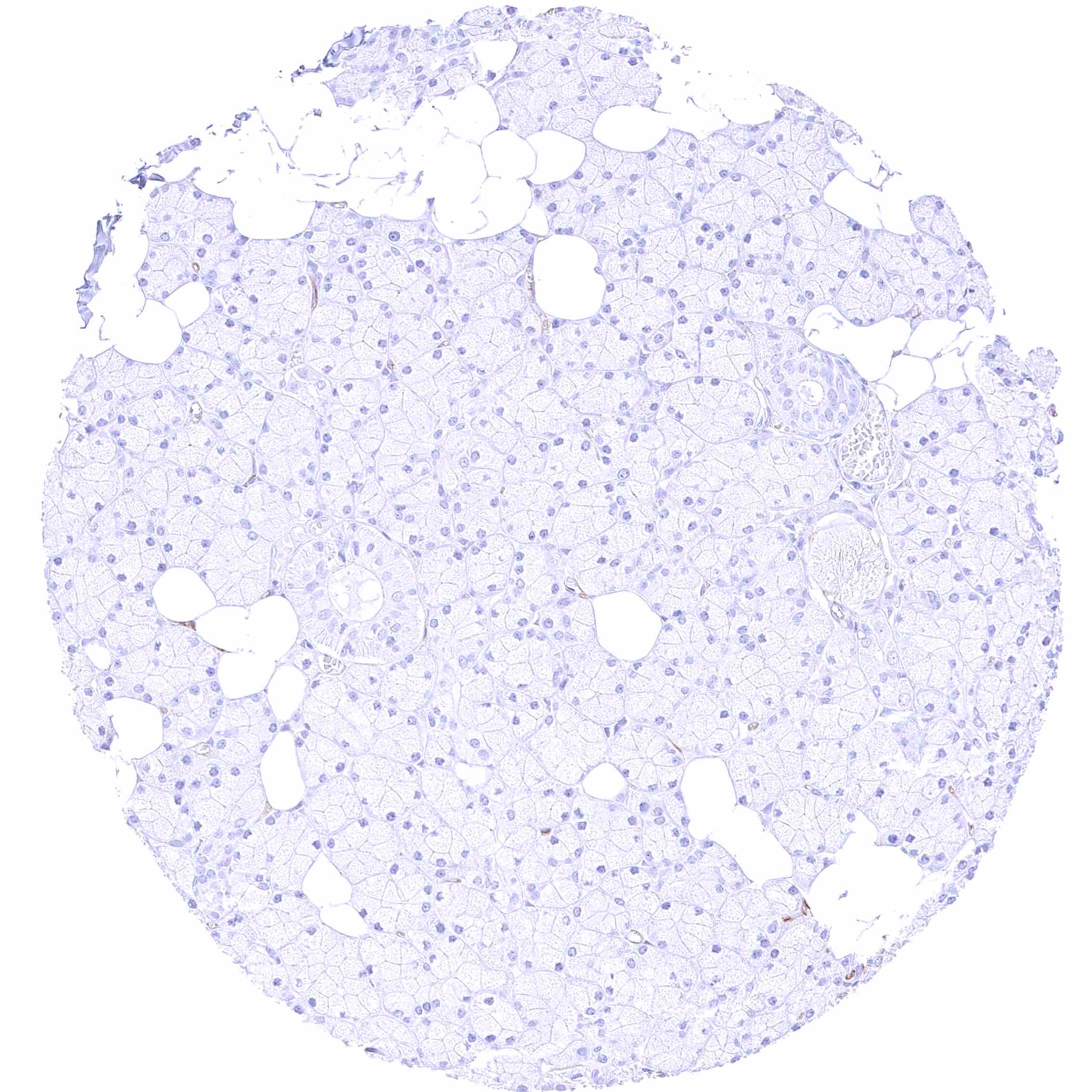
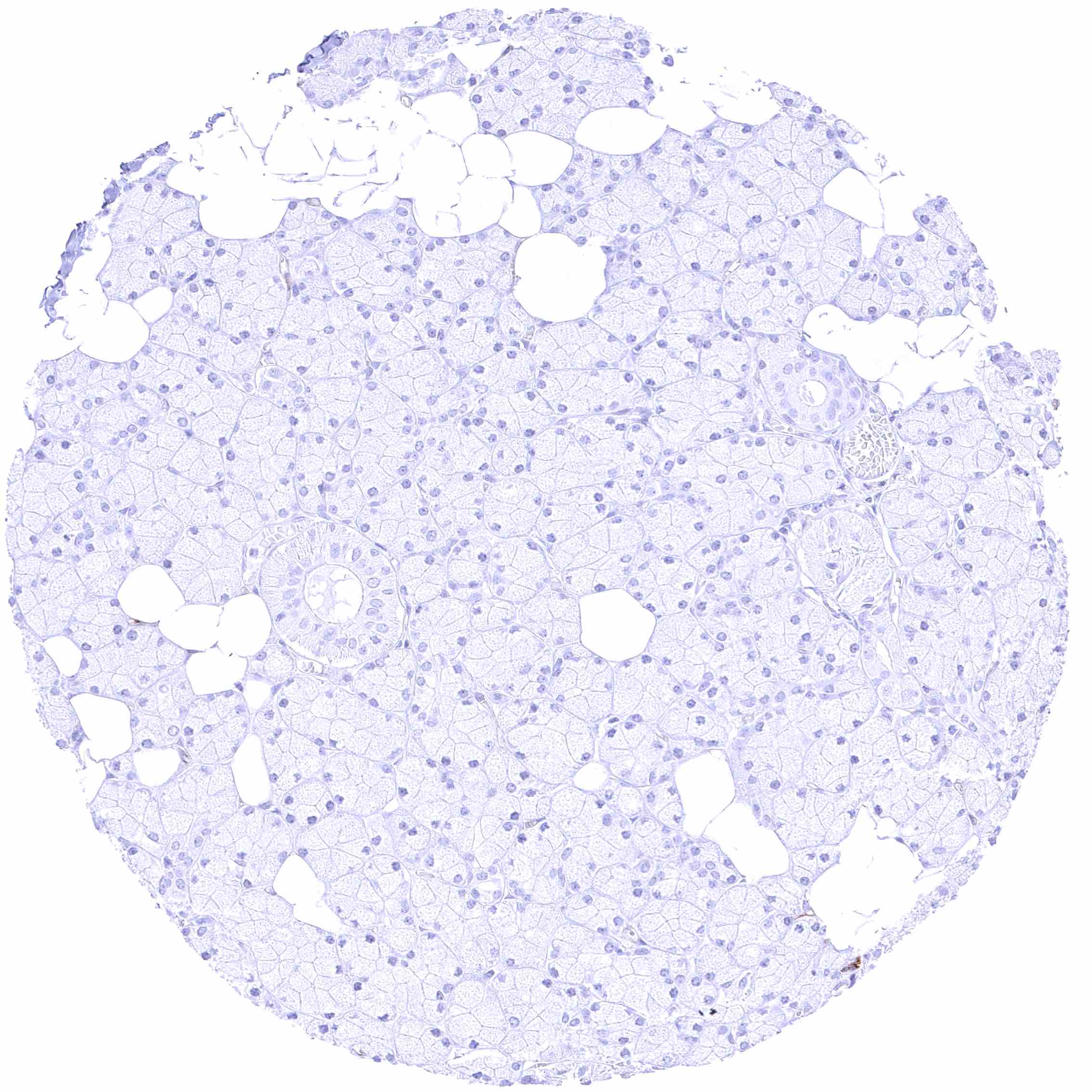
| Liver | Negative. | |
| Gallbladder | Negative. | |
| Pancreas | Negative. | |
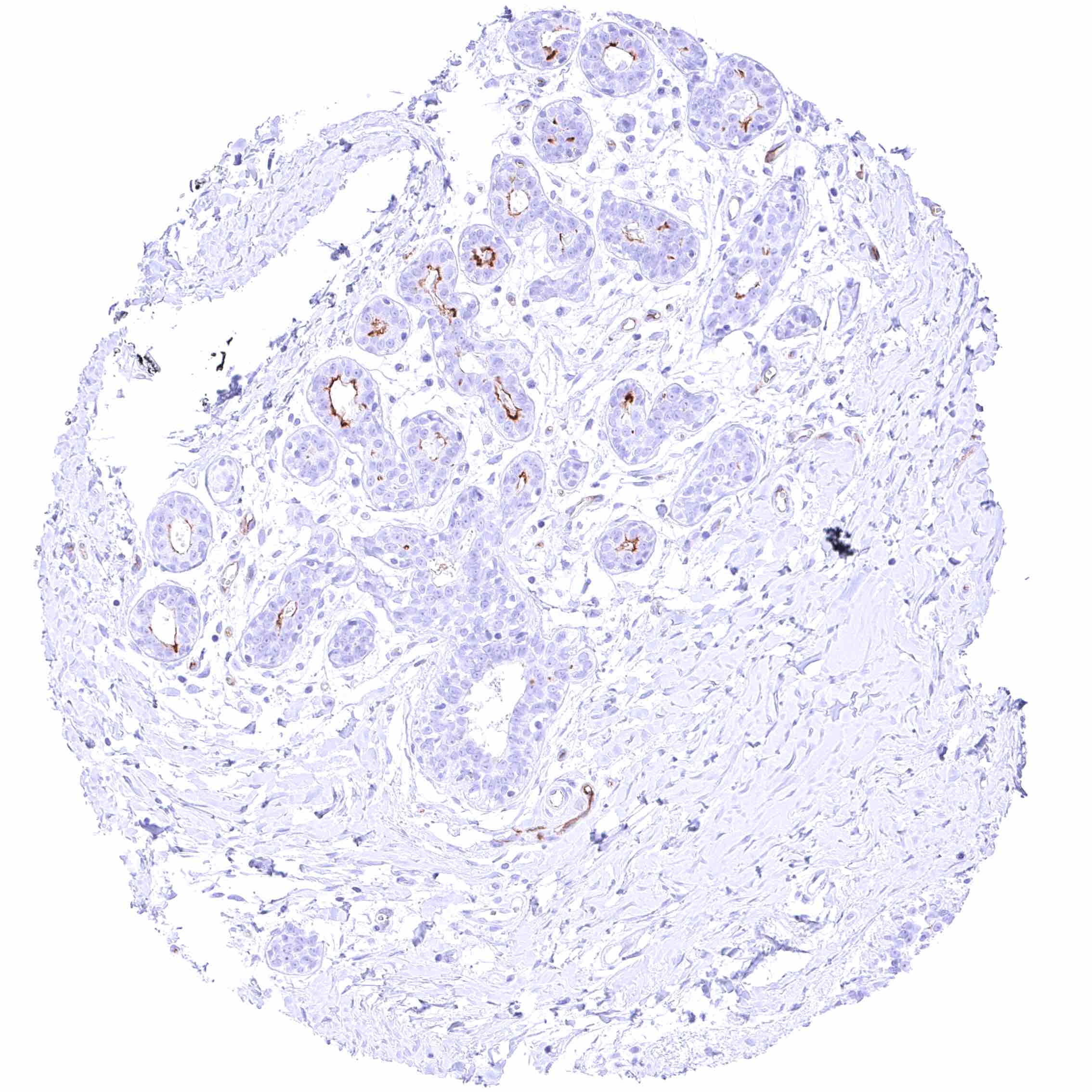
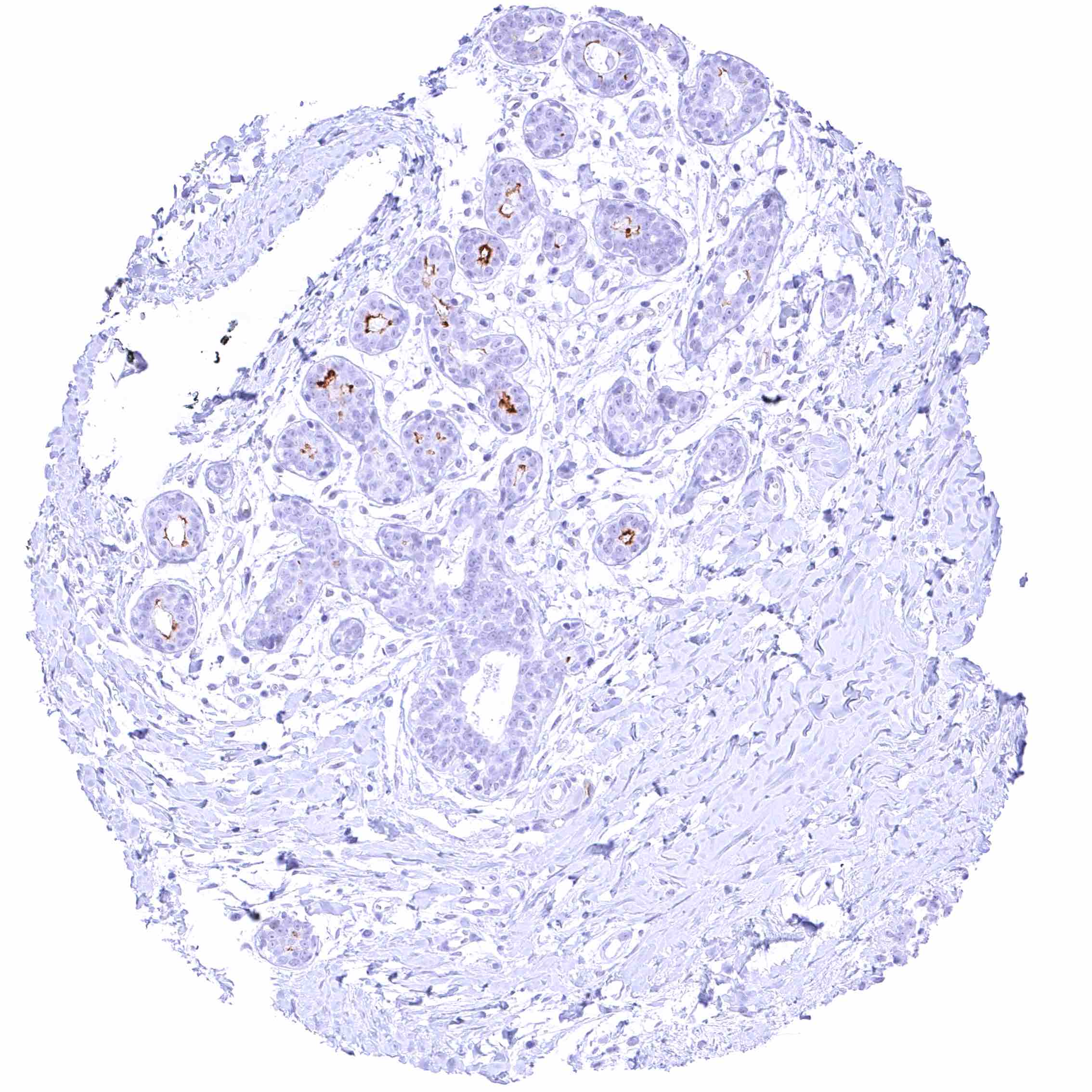
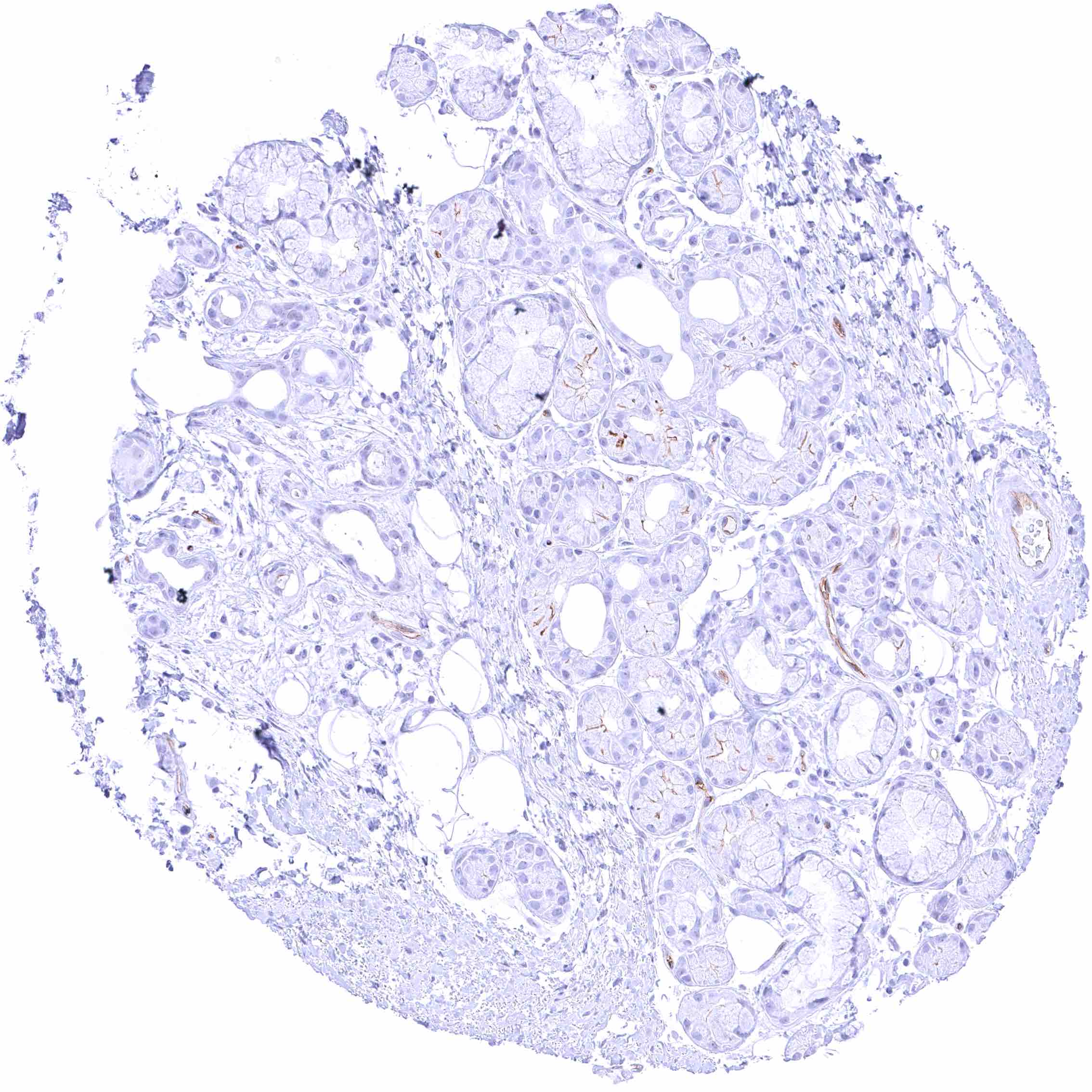
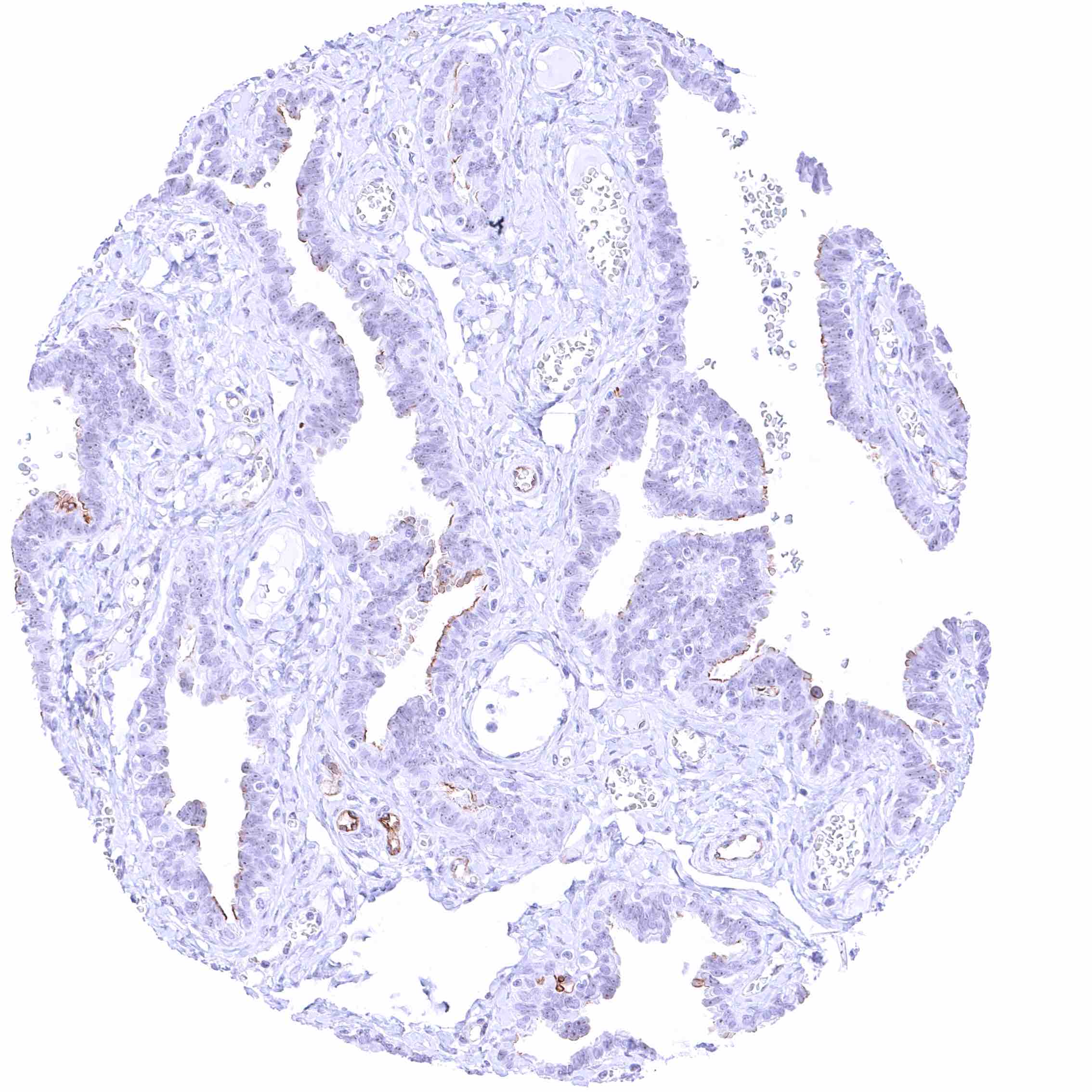
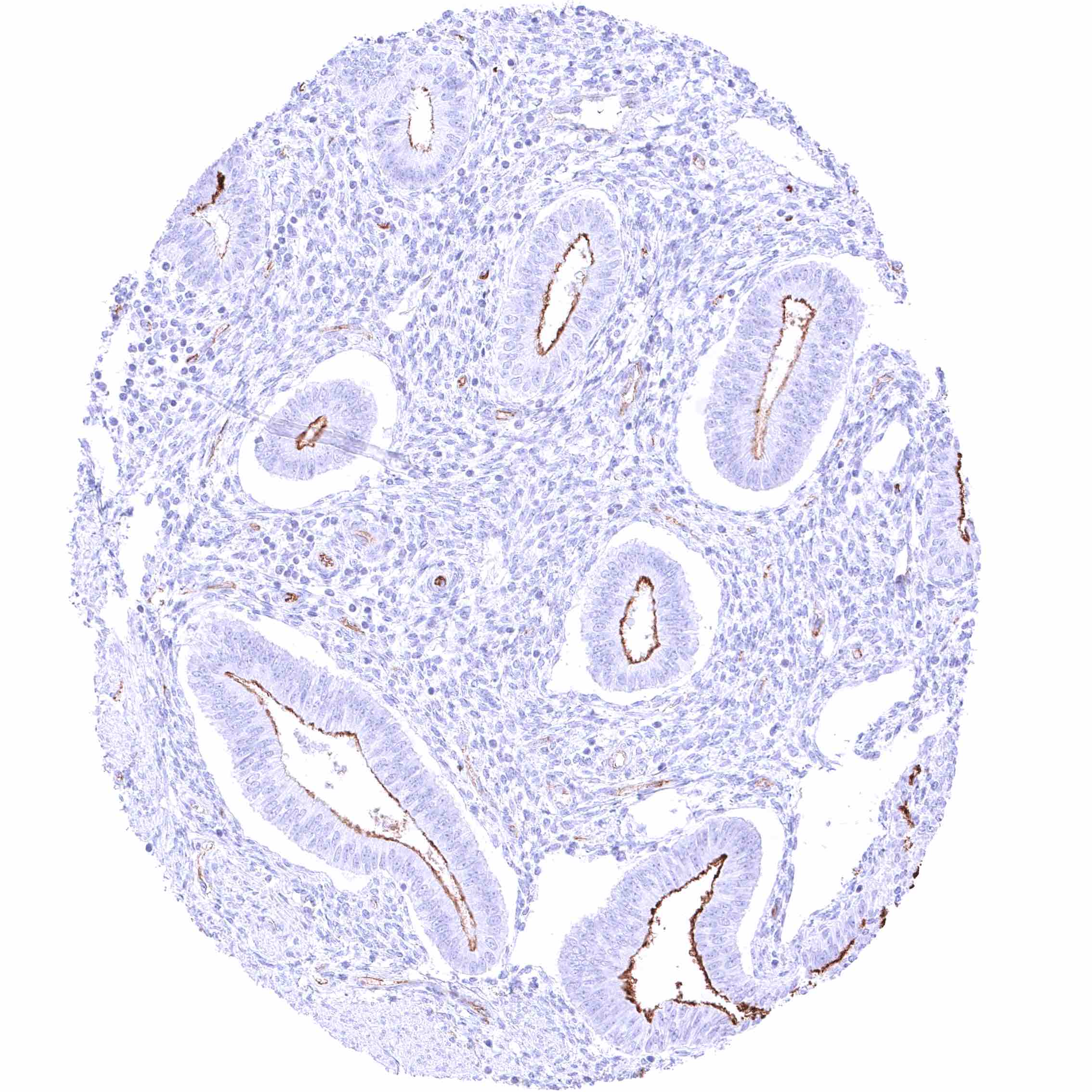
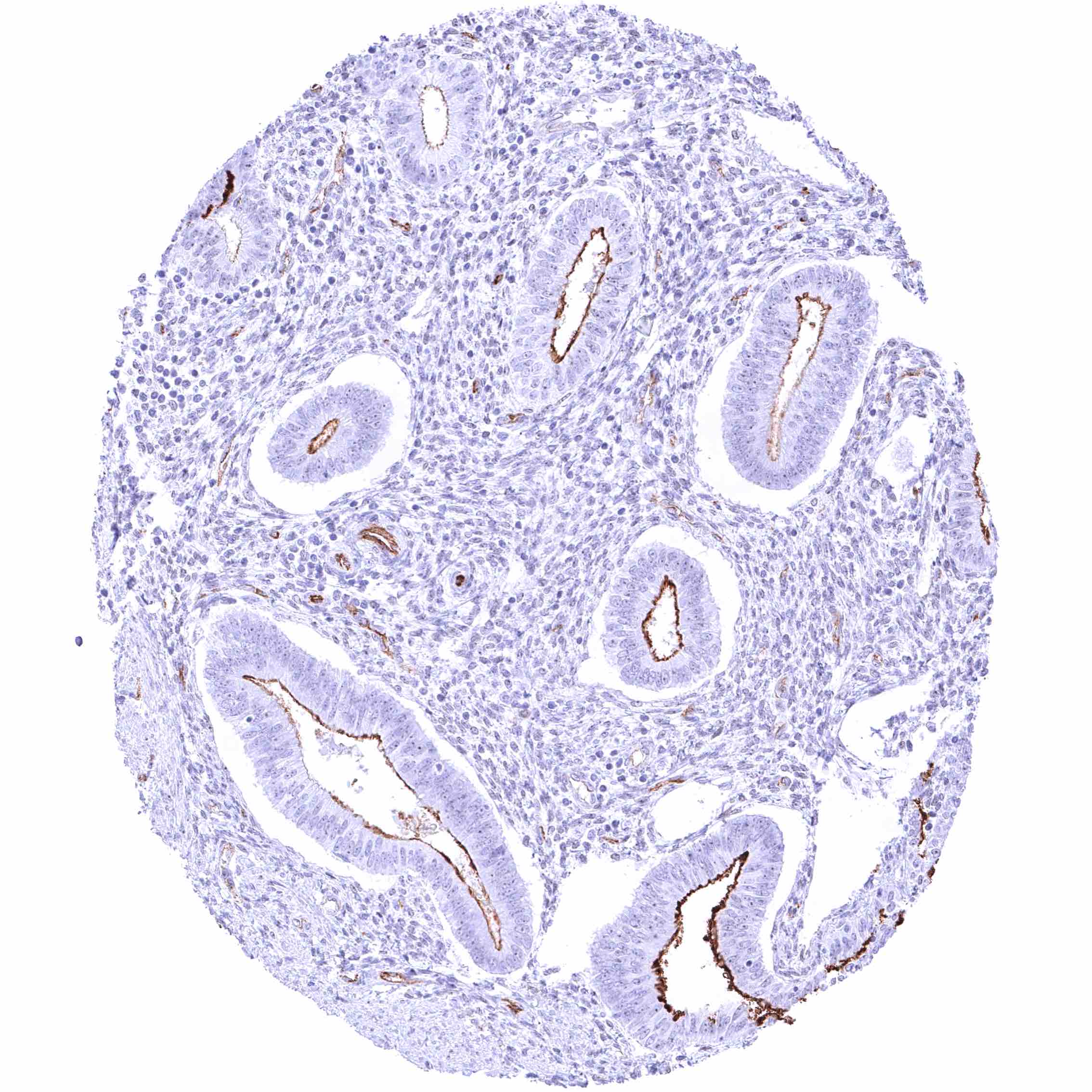
| Genitourinary | Kidney | Intense PODXL staining of glomerular podocytes (and probably also endothelial cells) while is less prominent on endothelial staining of intertubular vessels. |
| Urothelium | Negative. | |
| Male genital | Prostate | Negative. |
| Seminal vesicles | Moderate to strong PODXL staining of luminal/apical membranes of a subset of epithelial cells. | |
| Testis | Negative. | |
| Epididymis | Moderate PODXL staining of luminal/apical membranes of columnar cells in the cauda but not the caput epididymis. | |
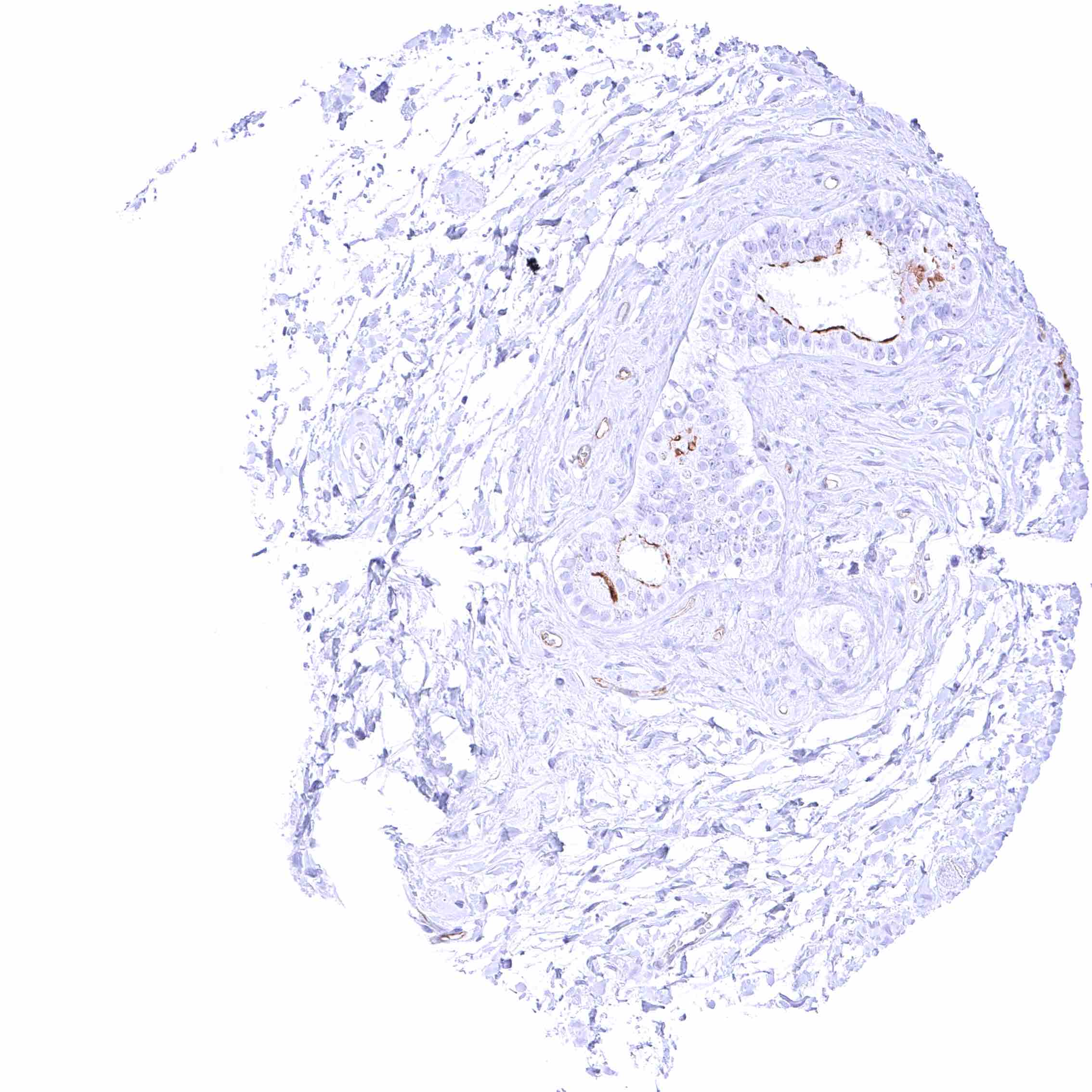
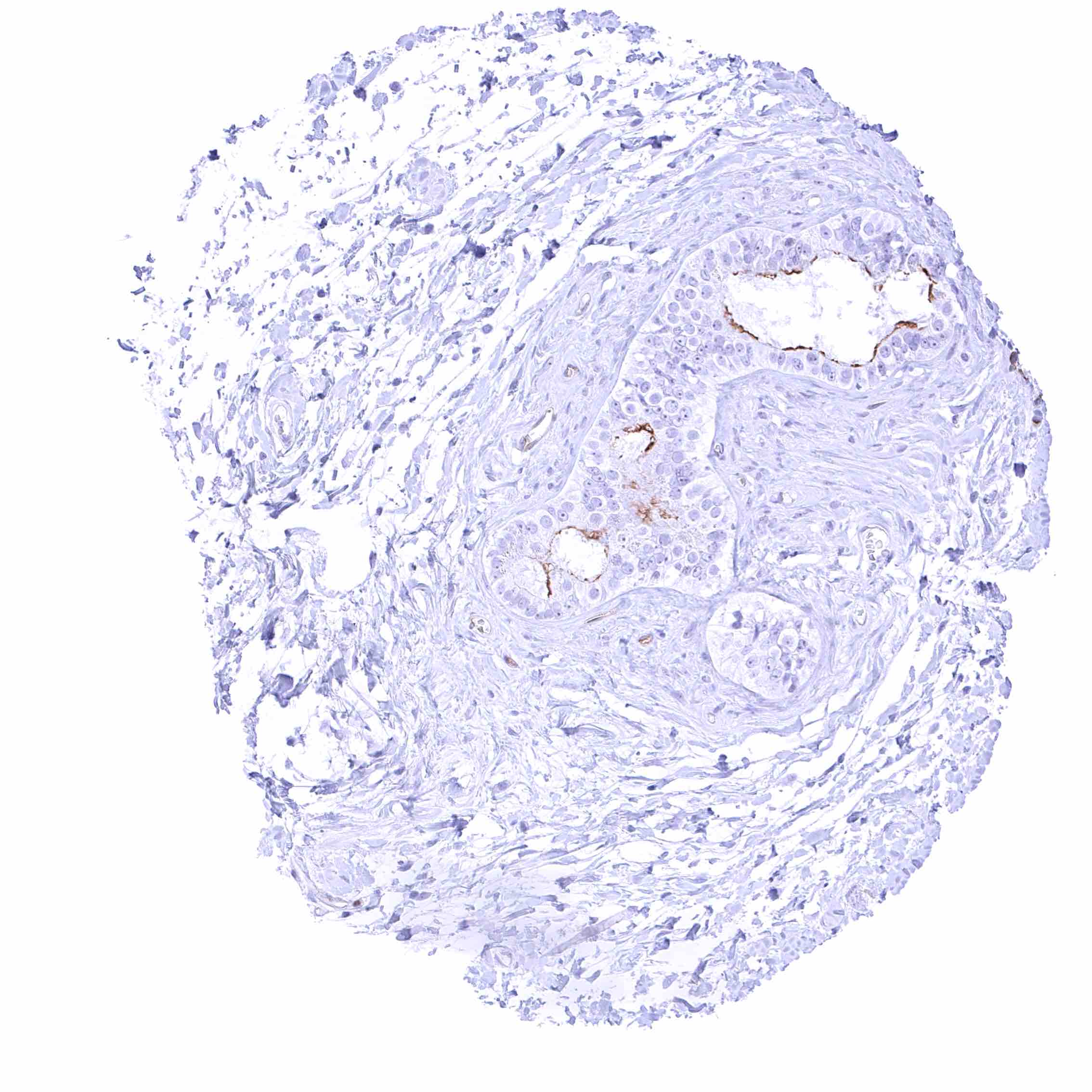
| Female genital | Breast | Moderate PODXL staining of apical membranes of luminal epithelial cells. |
| Uterus, myometrium | Negative. | |
| Uterus, ectocervix | Negative. | |
| Uterus endocervix | Negative. | |
| Uterus, endometrium | Moderate PODXL staining of apical membranes of resting or proliferating endometrium glands while this may not be visible in secreting endometrium or during pregnancy. During pregnancy, a weak to moderate membranous staining of few decidua cells can occur. | |
| Fallopian Tube | Moderate PODXL staining of apical membranes of a subset of epithelial cells. | |
| Ovary | Negative. | |
| Placenta early | Negative. | |
| Placenta mature | Negative. | |
| Amnion | Negative. | |
| Chorion | Negative. | |
| Skin | Epidermis | Negative. |
| Sebaceous glands | Negative. | |
| Muscle/connective tissue | Heart muscle | Negative. |
| Skeletal muscle | Negative. | |
| Smooth muscle | Negative. | |
| Vessel walls | Negative. | |
| Fat | Negative. | |
| Stroma | Negative. | |
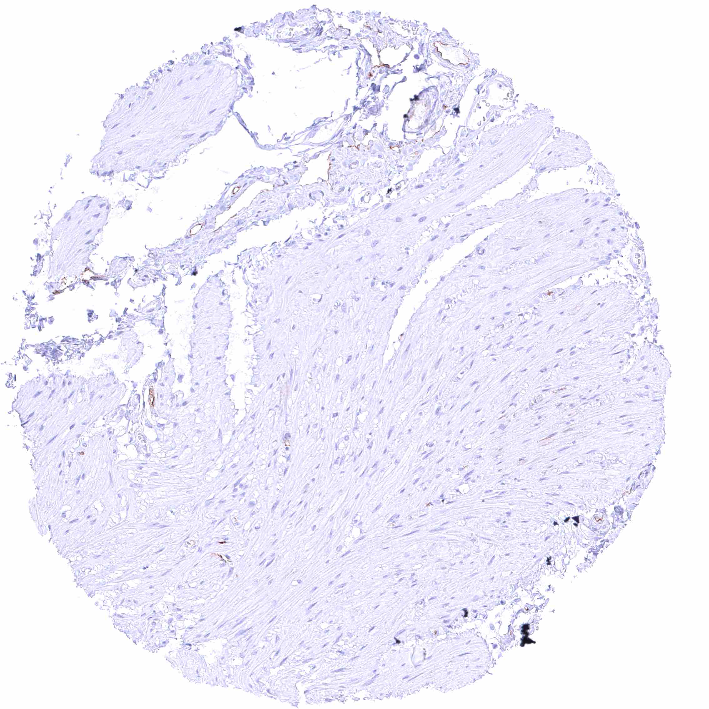
| Endothelium | Distinct PODXL staining of endothelial cells of predominantly small vessels in all tissues. | |
| Bone marrow/ lymphoid tissue | Bone marrow | Negative. |
| Lymph node | Negative. | |
| Spleen | Negative. | |
| Thymus | Negative. | |
| Tonsil | Negative. | |
| Remarks | PODXL staining of endothelial cells of predominantly small vessels is seen in all tissues. |
These findings are largely consistent with the RNA data described in the Human Protein Atlas (Tissue expression PODXL) where a rather ubiquitous expression is described (endothelial cells occur in all tissues).
Positive control = Kidney: A strong PODXL must be seen in glomerular podocytes while at least a weak to moderate PODXL staining must be seen in endothelial cells of small intertubular vessels.
Negative control = Kidney: Tubuli and collecting ducts must be PODXL negative.
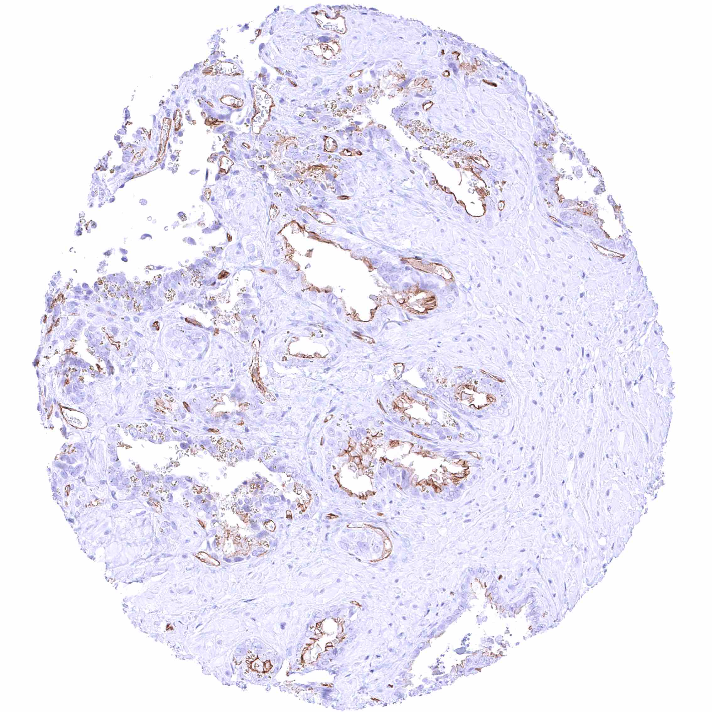


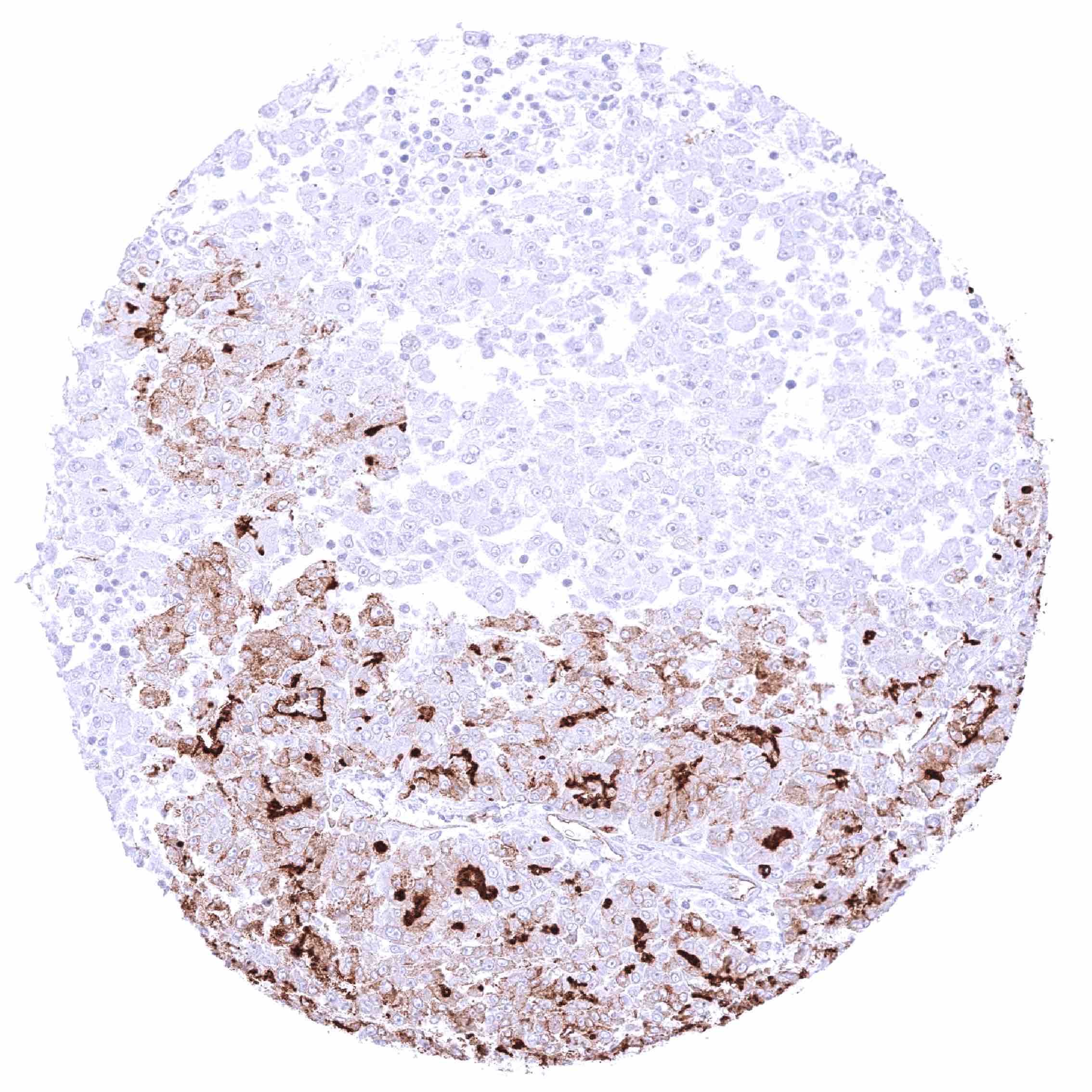
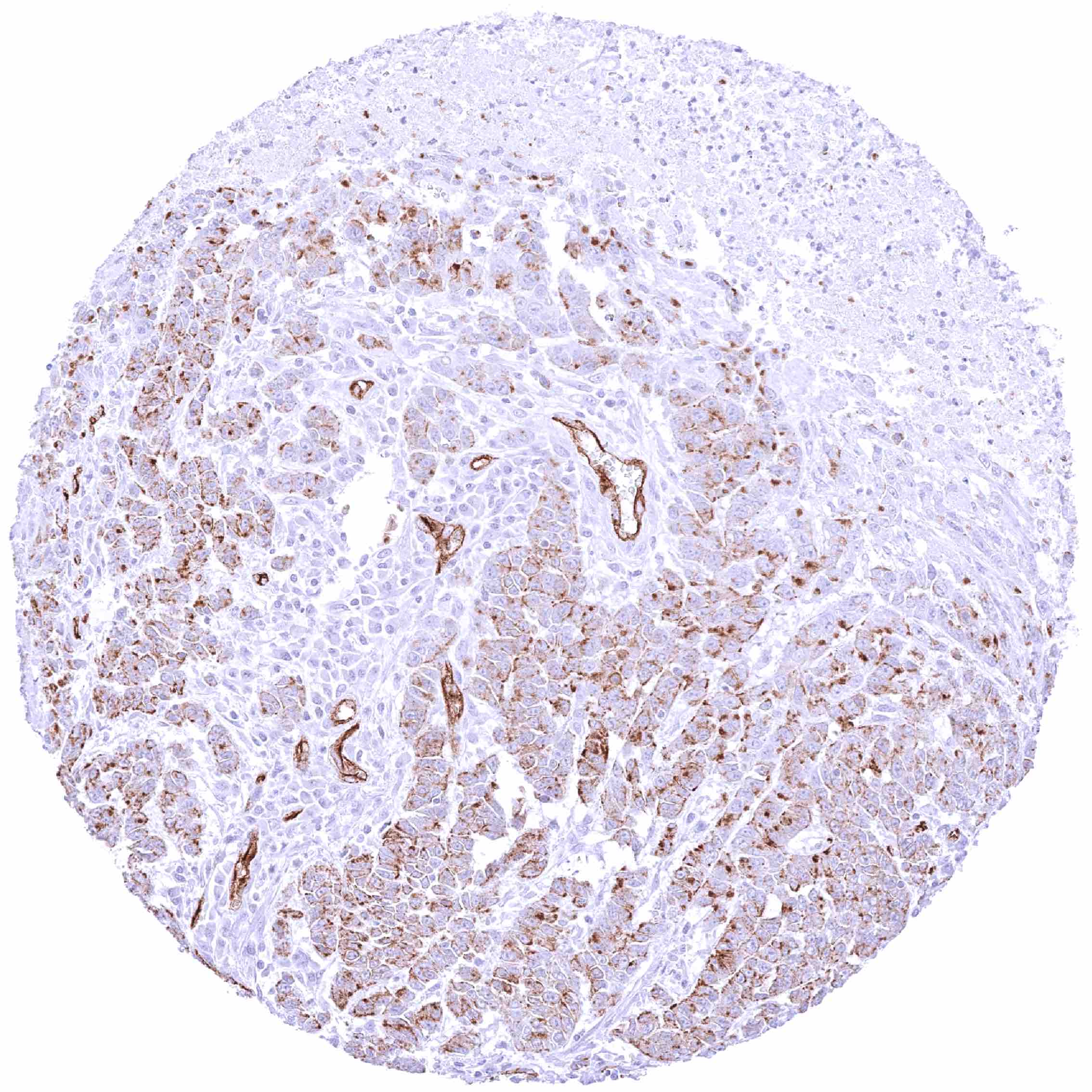
Staining Pattern in Relevant Tumor Types
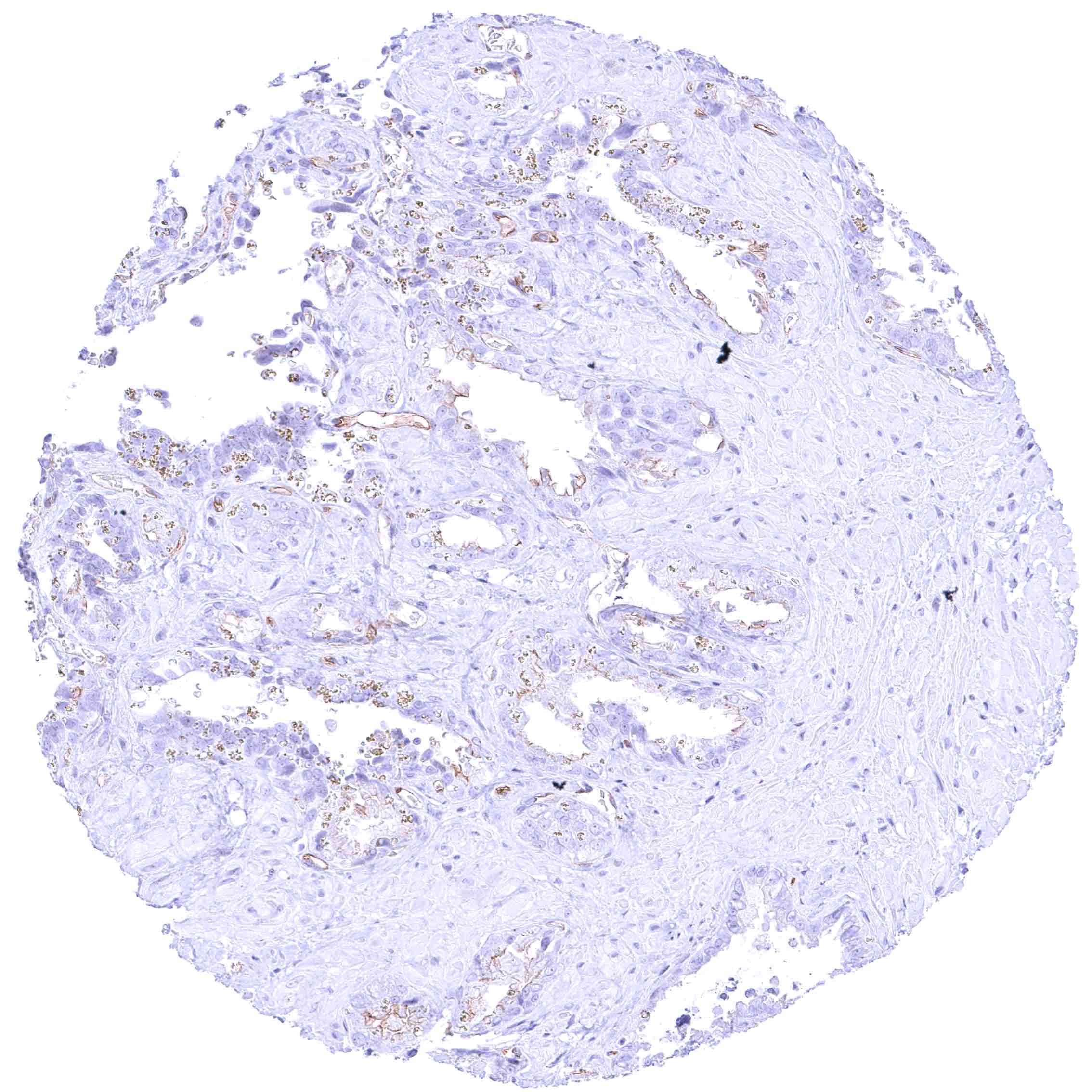
PODXL expression occurs in many different tumor types.
The TCGA findings on PODXL RNA expression in different tumor categories have been summarized in the Human Protein Atlas.
Compatibility of Antibodies
No data available at the moment
Protocol Recommendations
IHC users have different preferences on how the stains should look like. Some prefer high staining intensity of the target stain and even accept some background. Others favor absolute specificity and lighter target stains. Factors that invariably lead to more intense staining include higher concentration of the antibody and visualization tools, longer incubation time, higher temperature during incubation, higher temperature and longer duration of the heat induced epitope retrieval (slide pretreatment). The impact of the pH during slide pretreatment has variable effects and depends on the antibody and the target protein.
All images and data shown here and in our image galleries are obtained by the manual protocol described below. Other protocols resulting in equivalent staining are described as well.
Manual protocol
Freshly cut sections should be used (less than 10 days between cutting and staining). Heat-induced antigen retrieval for 5 minutes in an autoclave at 121°C in pH 7,8 Target Retrieval Solution buffer. Apply MSVA-645M at a dilution of 1:150 at 37°C for 60 minutes. Visualization of bound antibody by the EnVision Kit (Dako, Agilent) according to the manufacturer’s directions.
Potential Research Applications
- How can PODXL-targeted therapies be effectively developed and optimized for different tumor types?
- What specific molecular pathways are affected by PODXL in cancer?
- What is the prevalence and clinical significance of PODXL expression across tumor types?
- Is there any diagnostic/prognostic/predictive utility of PODXL IHC?
Evidence for Antibody Specificity in IHC
There are two ways how the specificity of antibodies can be documented for immunohistochemistry on formalin fixed tissues. These are: 1. Comparison with a second independent method for target expression measurement across a large number of different tissue types (orthogonal strategy), and 2. Comparison with one or several independent antibodies for the same target and showing that all positive staining results are also seen with other antibodies for the same target (independent antibody strategy).
Orthogonal validation: For the antibody MSVA-645M specificity is in line with RNA expression data from normal tissues which were collected in three independent RNA screening studies, including the Human Protein Atlas (HPA) RNA-seq tissue dataset, the FANTOM5 project, and the Genotype-Tissue Expression (GTEx) project, and which are summarized in the Human Protein Atlas (Tissue expression PODXL). Although PODXL RNA expression was ubiquitous, due to its presence in capillaries of all organs, the highest level of PODXL RNA expression in the kidney and the absence of its expression in the bone marrow are well in agreement with our MSVA-645M IHC data.
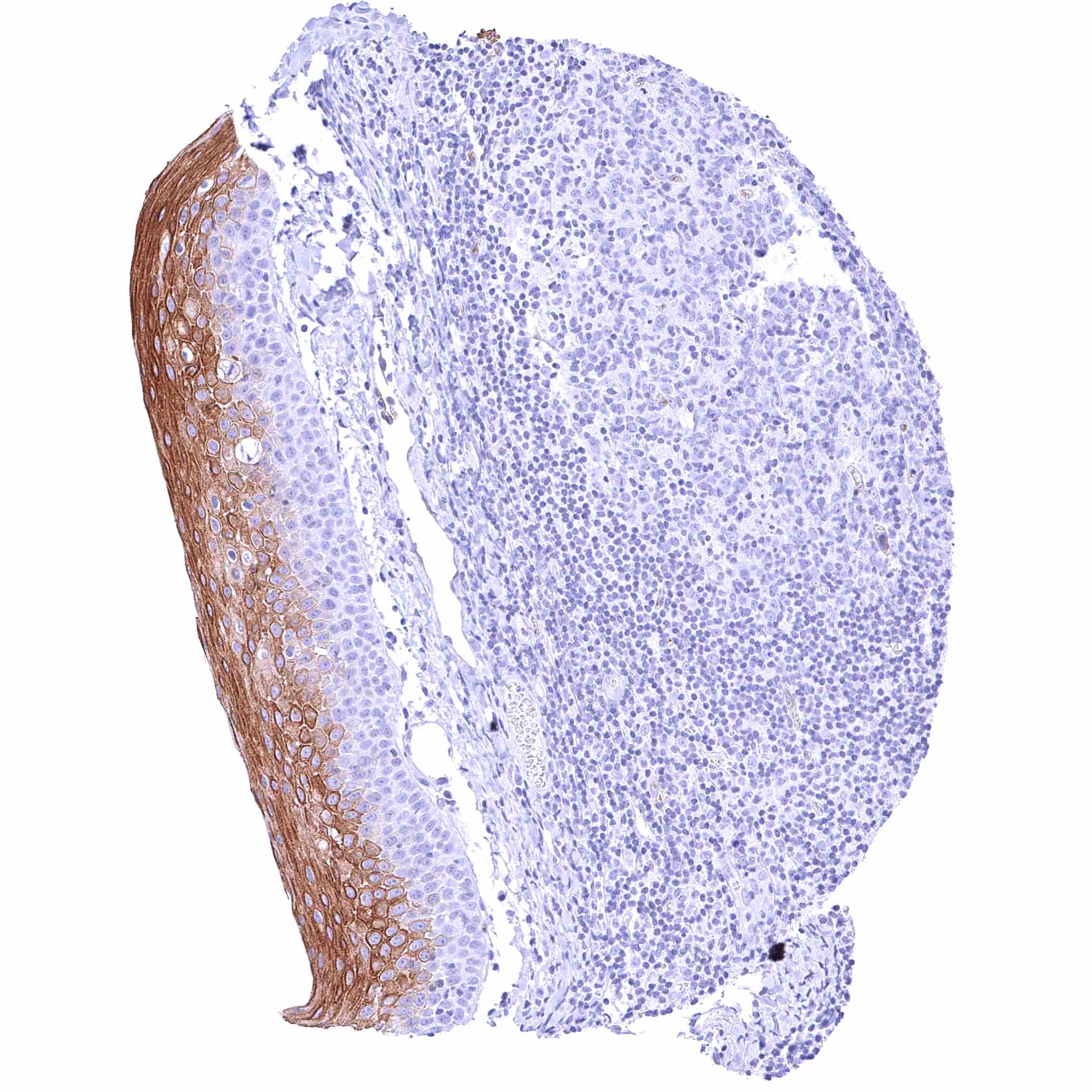
Comparison of antibodies: True expression of PODXL in all cell types with PODXL positivity by MSVA-645M is corroborated by an identical staining obtained by a commercially available independent second antibody (termed “validation antibody”). An additional staining of squamous epithelia by the validation antibody which was not seen by MSVA-645M was considered an antibody specific cross-reactivity of our validation antibody.