295,00 € – 995,00 €
Product details
Synonyms = Amplified in squamous cell carcinoma (AIS); Chronic ulcerative stomatitis protein (CUSP); EEC3; Keratinocyte transcription factor KET; LMS; NBP; p40; P51/P63; p53 like transcription factor; p53-related protein p63; RHS; SHFM4; TAp63alpha; TP53CP; TP53L; TP63; TP73; TP73L; Transformation-related protein 63; Trp53rp1; Trp6;3; Tumor protein 63; Tumor protein p53-like; tumor protein p73-like
Antibody type = Recombinant Rabbit monoclonal / IgG
Clone = MSVA-063R
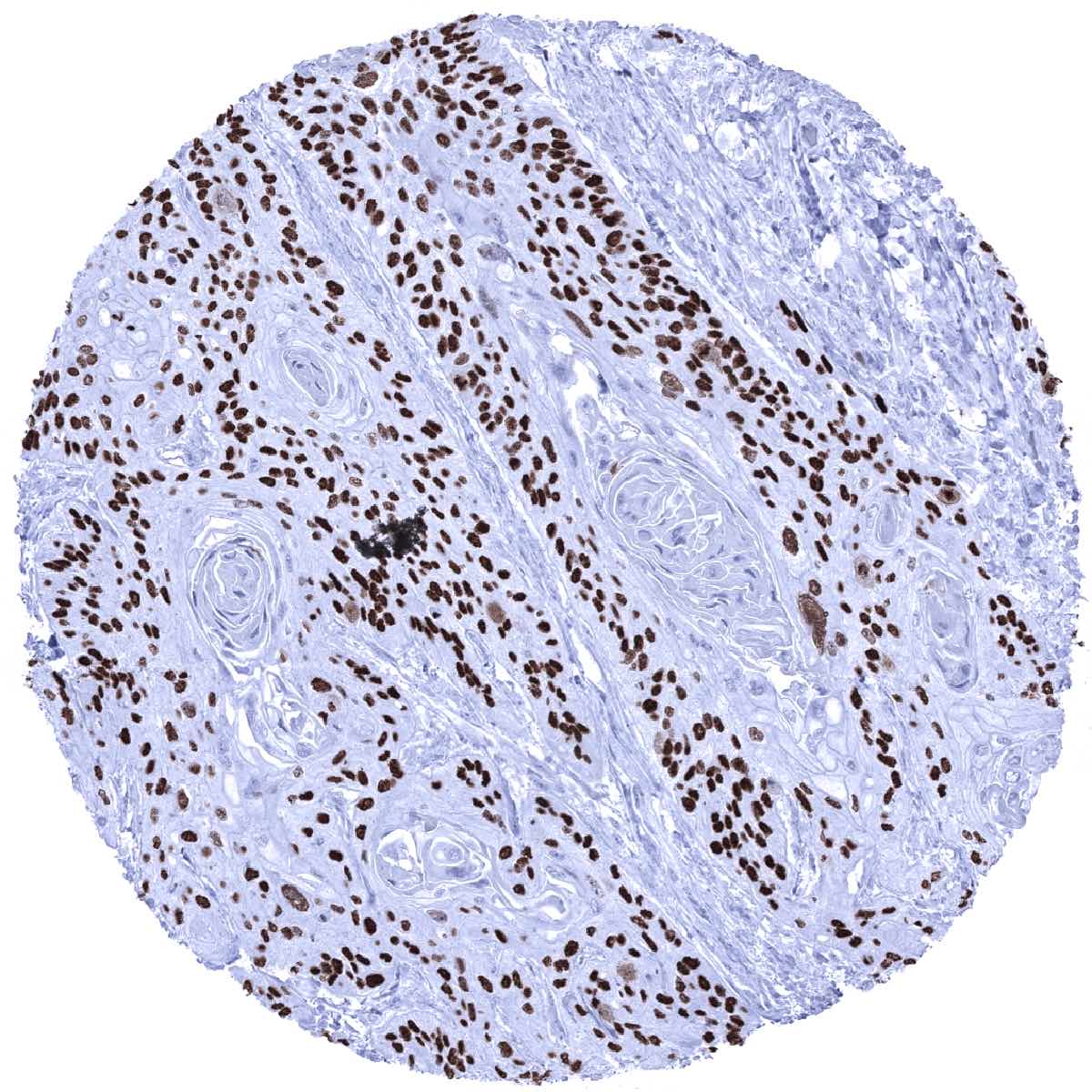
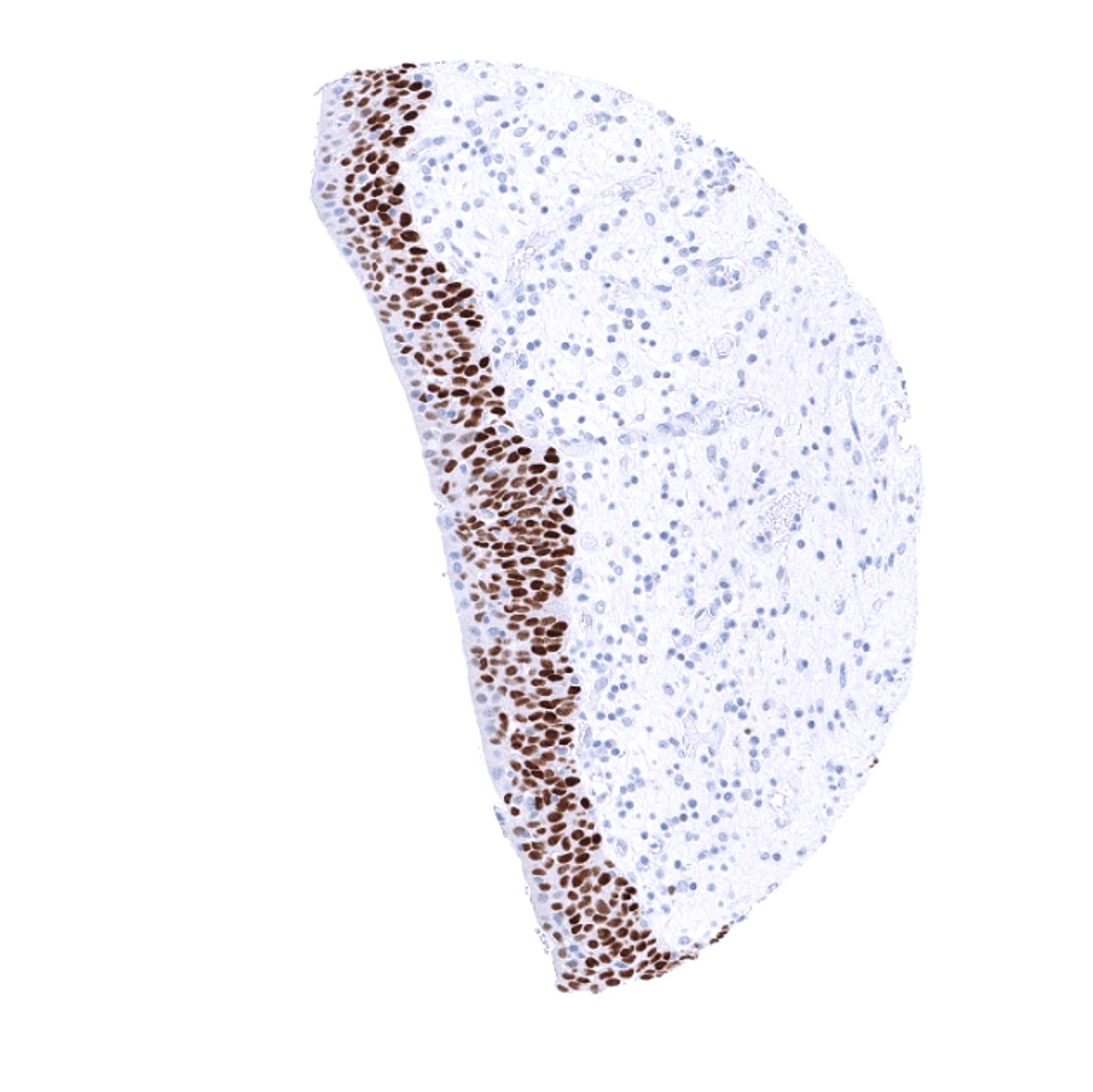
Positive control = Tonsil: Virtually all squamous epithelial cells must show a moderate to strong, nuclear staining, while few scattered lymphocytes and endothelial cells must show a at least a weak staining.
Negative control = Tonsil: The vast majority of lymphocytes should be p63 negative.
Cellular localization = Nuclear
Reactivity = Human
Application = Immunohistochemistry
Dilution = 1:100 – 1:200
Intended Use = Research Use Only
Relevance of Antibody
p63 is expressed in cells with squamous, urothelial, or myoepithelial differentiation.
Biology Behind
Tumor protein 63 (p63) is a transcription factor of the p53 gene family encoded by the TP63 gene located at chromosome 3q28. Together with p73 it makes up the p53 gene family based on their structural similarity. p63 regulates the activity of a multitude of genes involved in growth and development of the ectoderm and derived structures and tissues, such as basal layer keratins and cell cycle control genes. Accordingly, p63 expression is found in basal cell layers of various organs, squamous epithelial cells of many organs and urothelium. p63 (syn. TAp63) is closely related to p40 (syn. ΔNp63) as both proteins represent isoforms of the p63 gene with distinct molecular functions. While “full length” p63 (TAp63) activates p53 target genes such as p21 or BAX, the shorter transcript p40 (ΔNp63) inhibits activation of p53 and “full length” p63. p63 -/- mice suffer from severe developmental defects which include the lack of limbs, teeth, and mammary glands.
Staining Pattern in Normal Tissues
| Brain | Cerebrum | Negative. |
| Cerebellum | Negative. | |
| Endocrine Tissues | Thyroid | Negative. |
| Parathyroid | Negative. | |
| Adrenal gland | Negative. | |
| Pituitary gland | Negative. | |
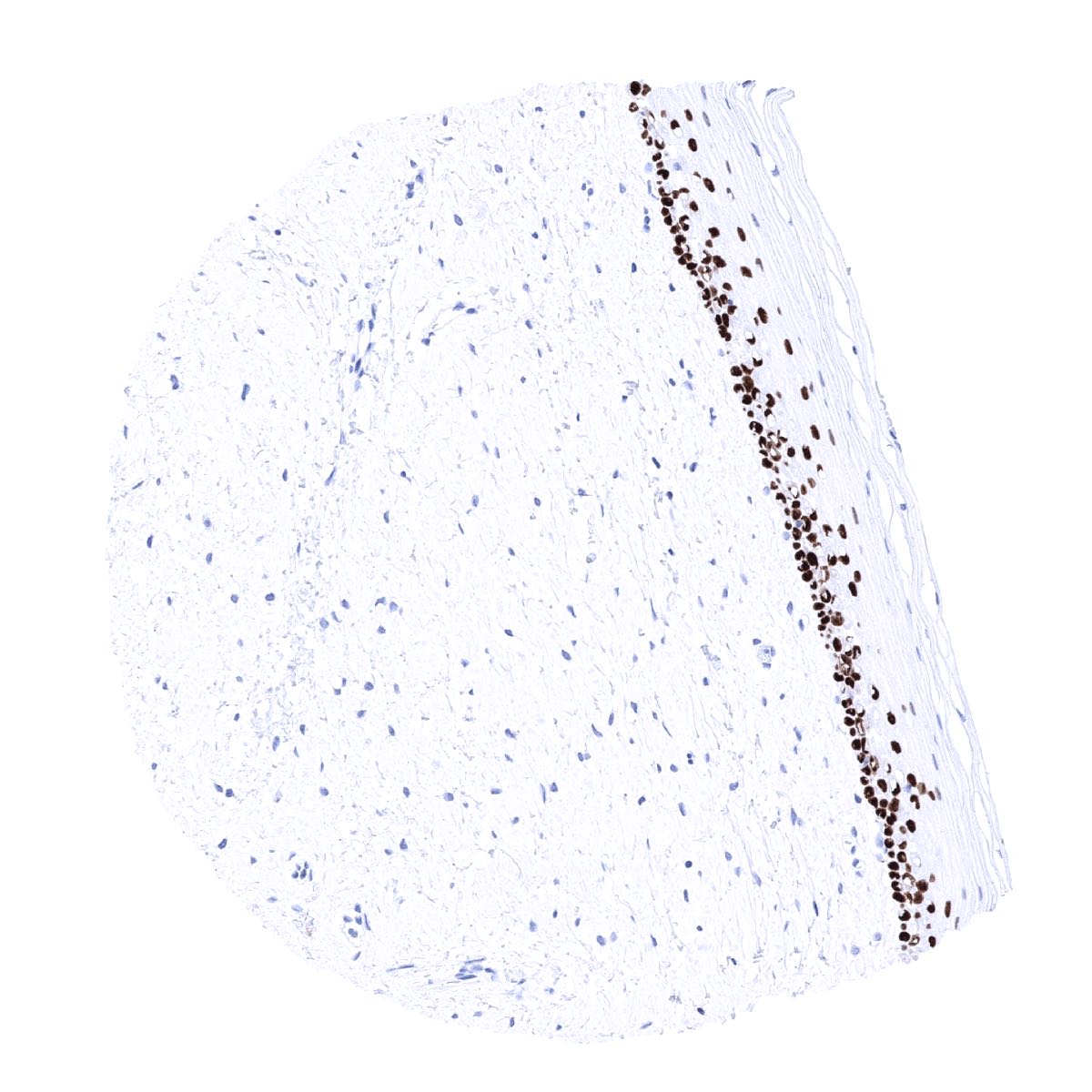
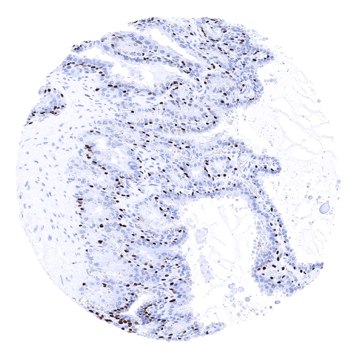
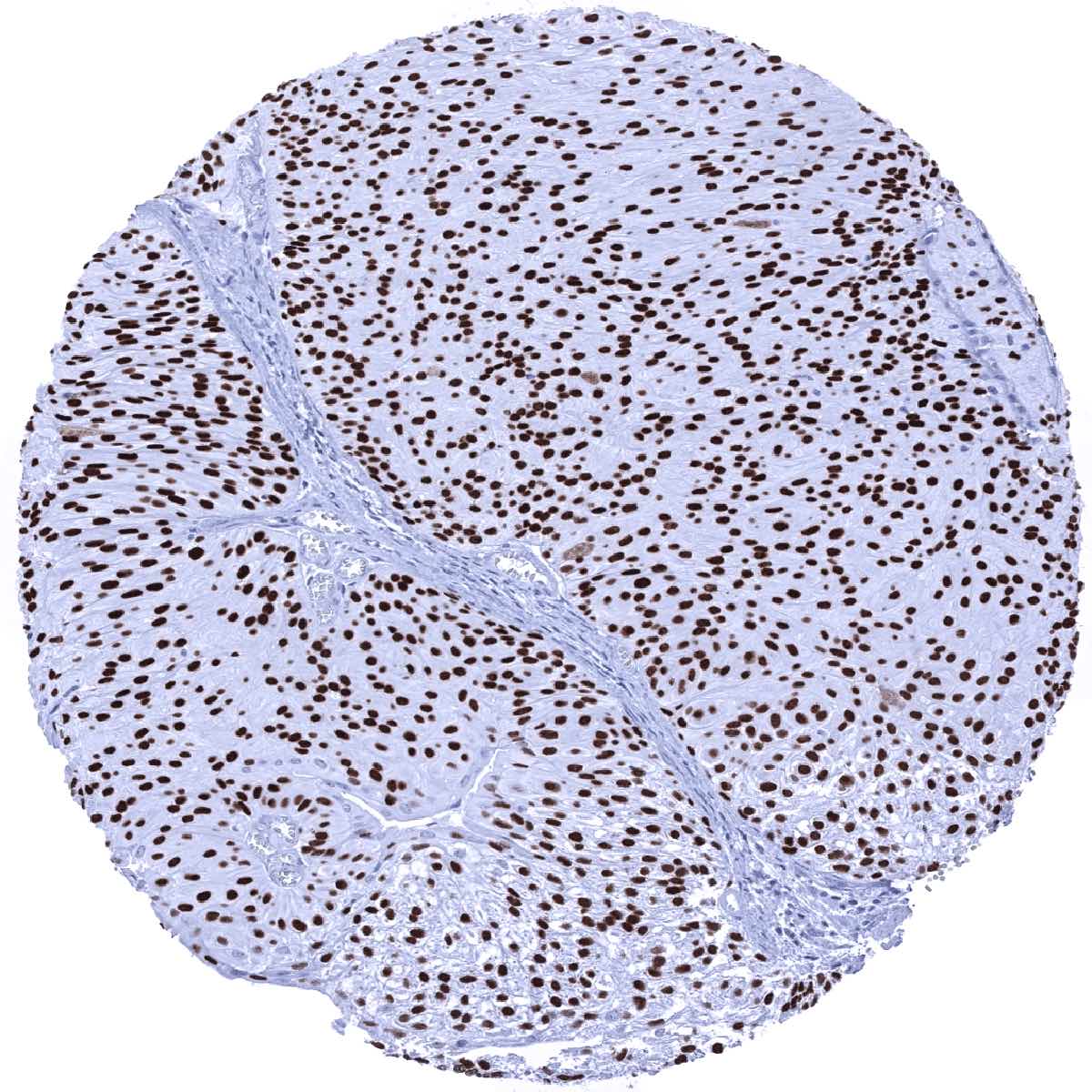
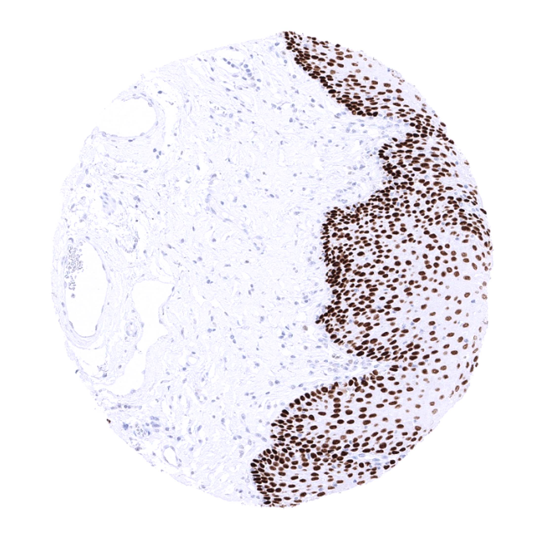
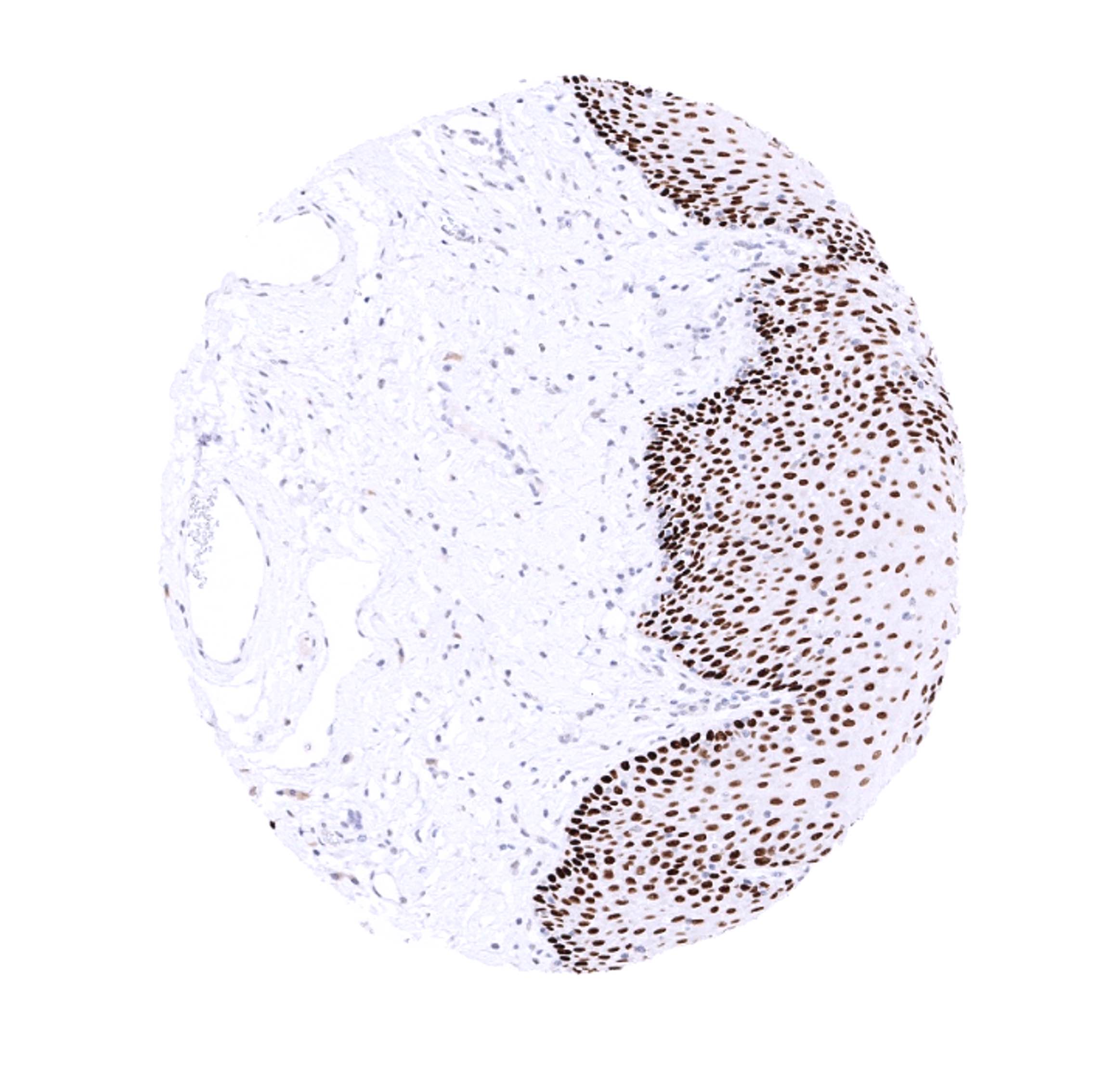
| Respiratory system | Respiratory epithelium | Strong nuclear p63 positivity of basal cells.
|
| Lung | Negative. | |
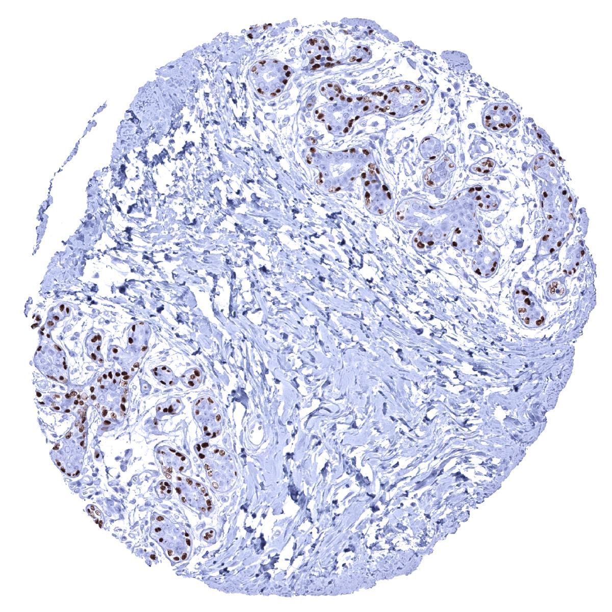
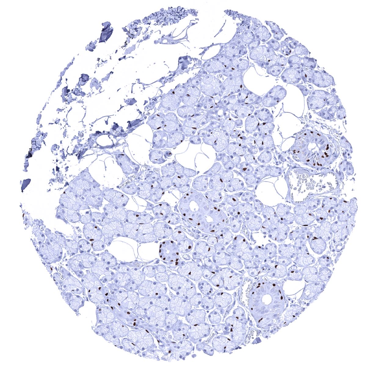
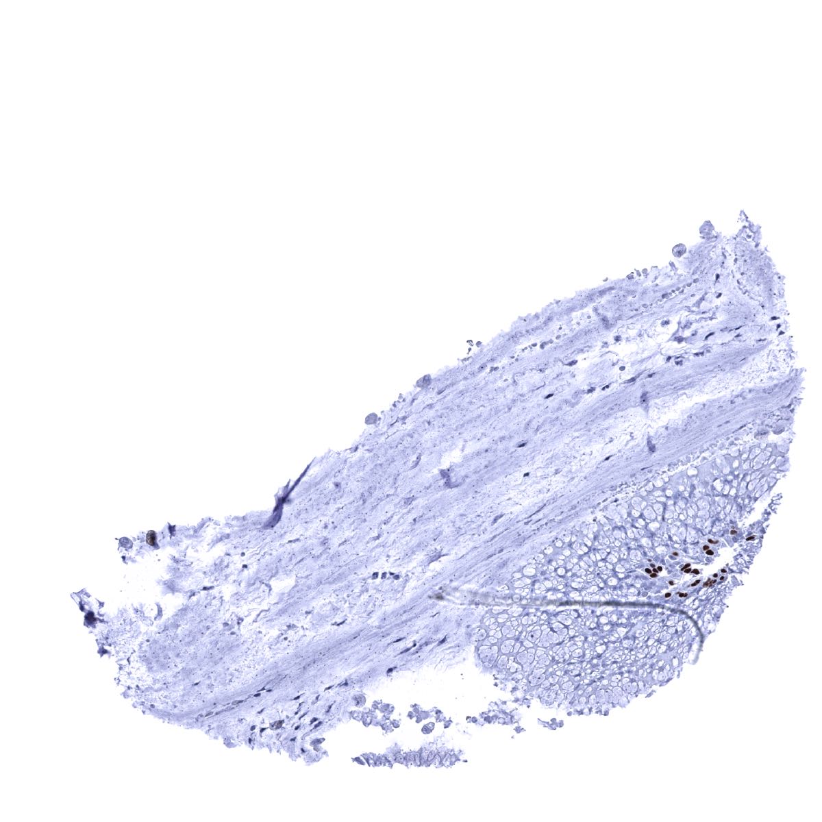
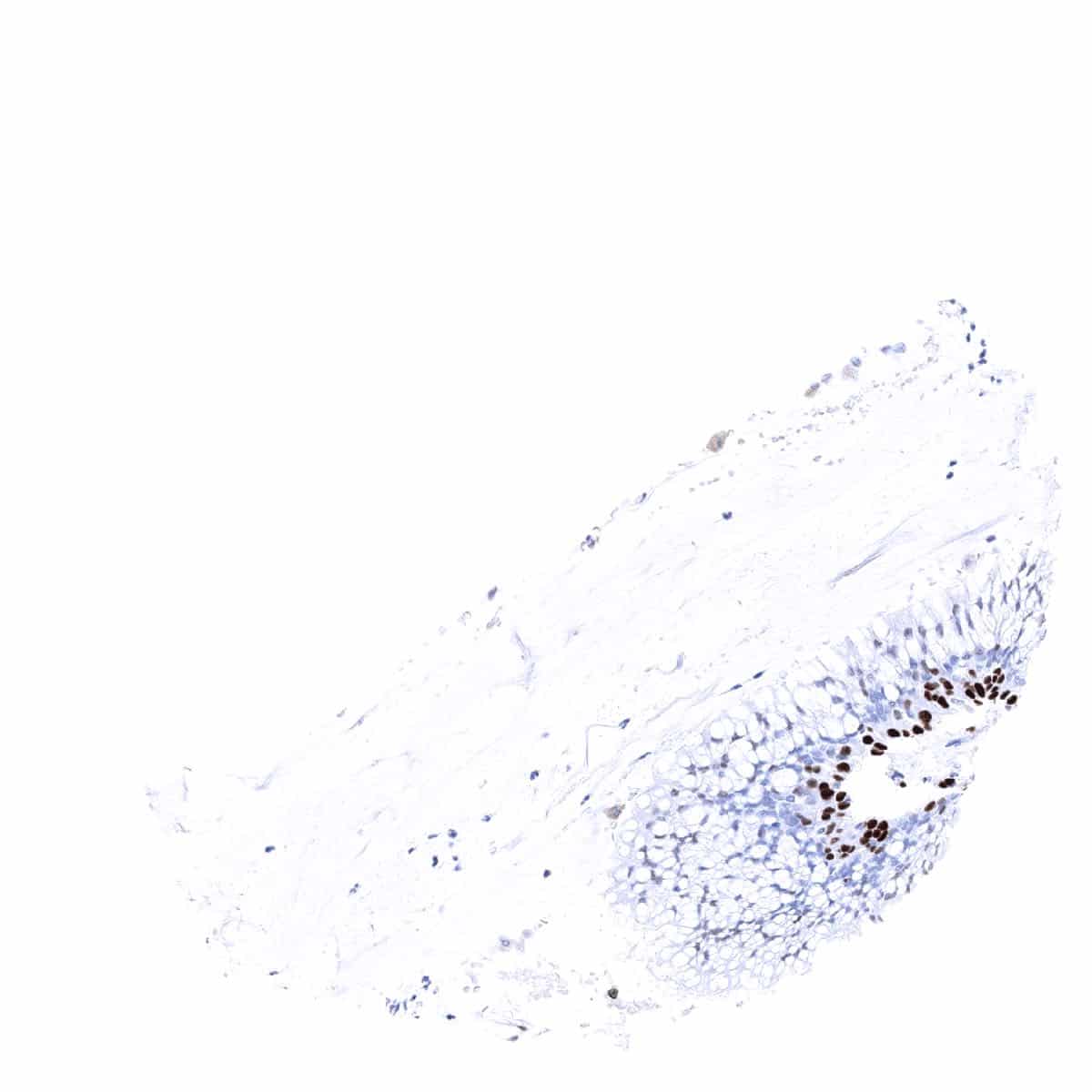
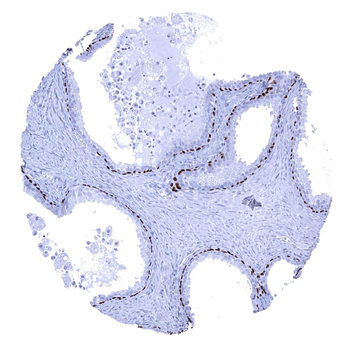
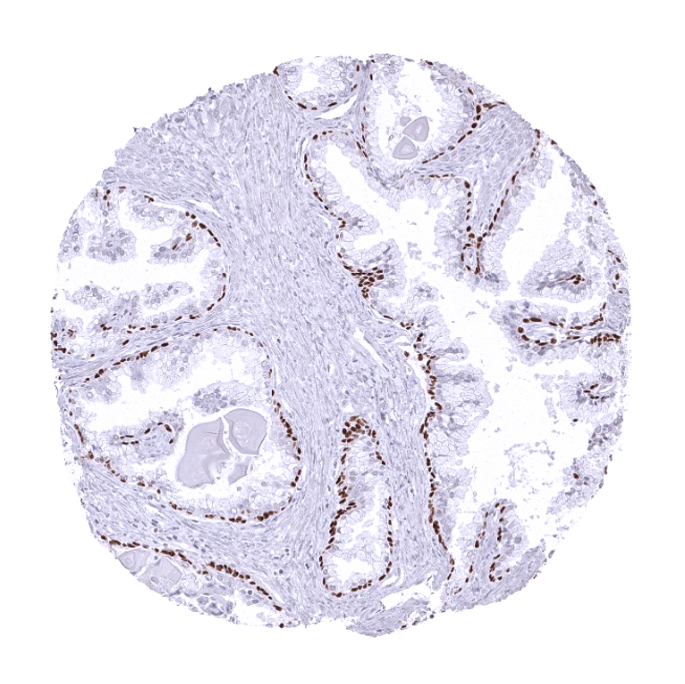
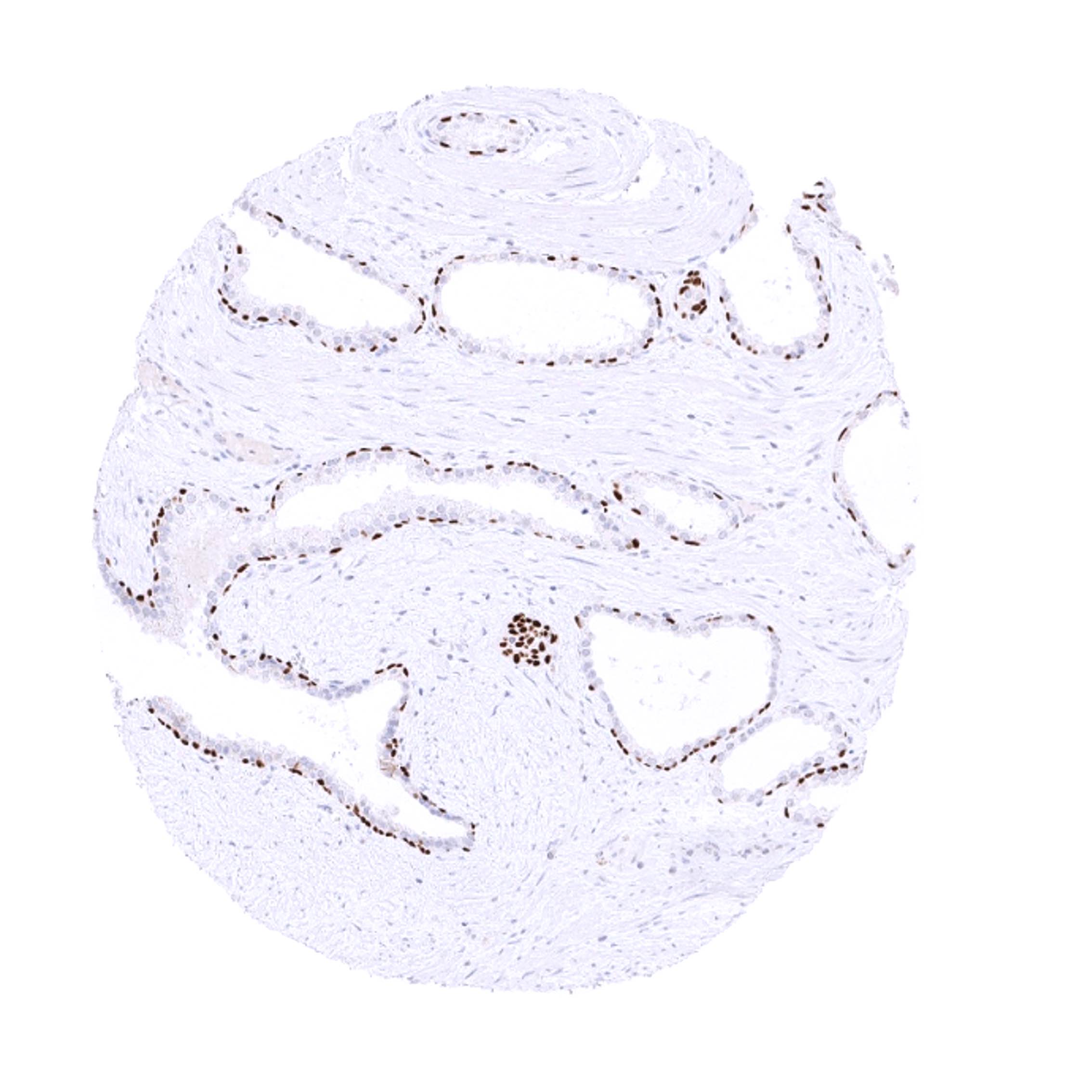
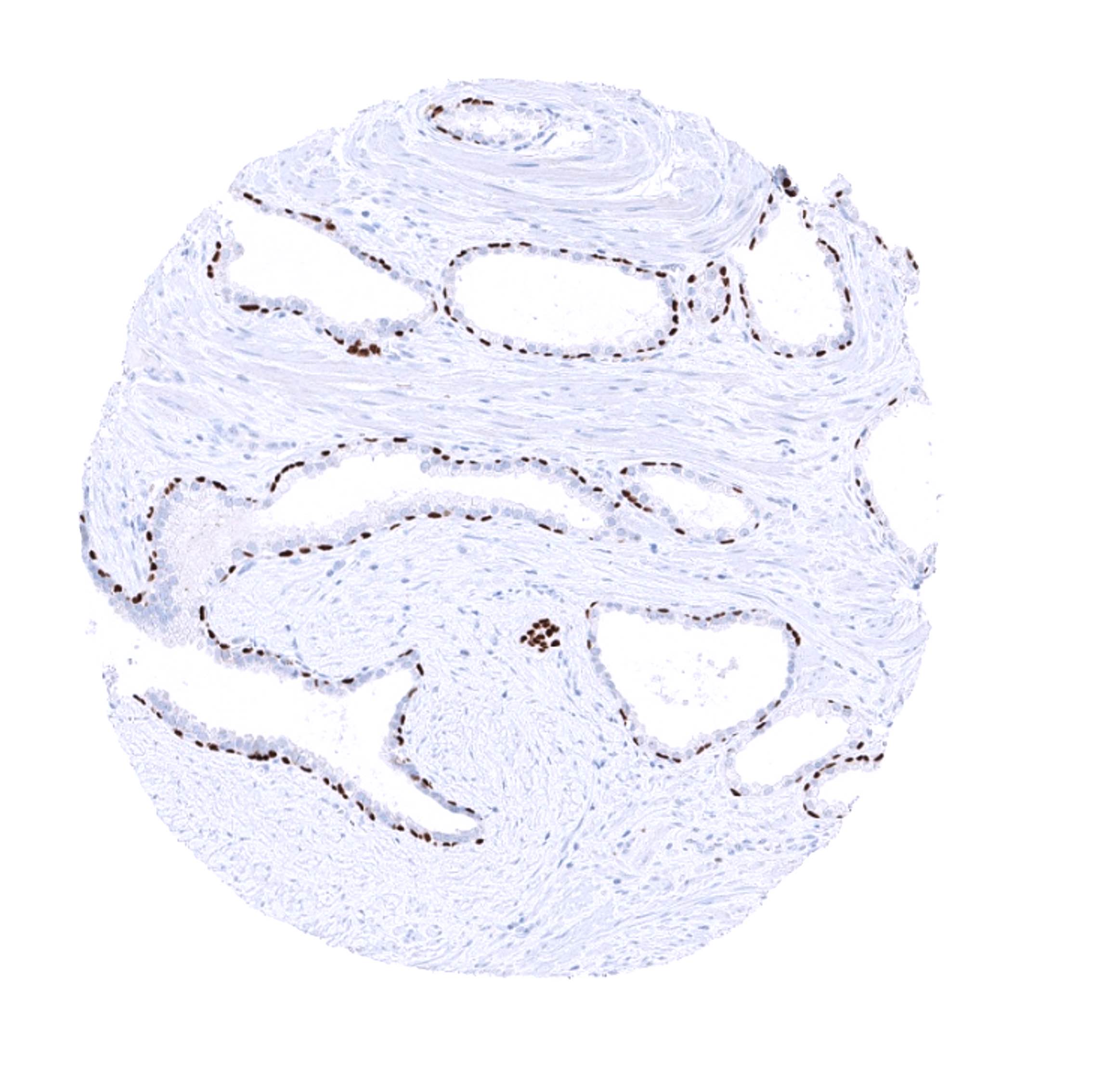
| Gastrointestinal Tract | Salivary glands | Strong nuclear p63 positivity of myoepithelial cells. |
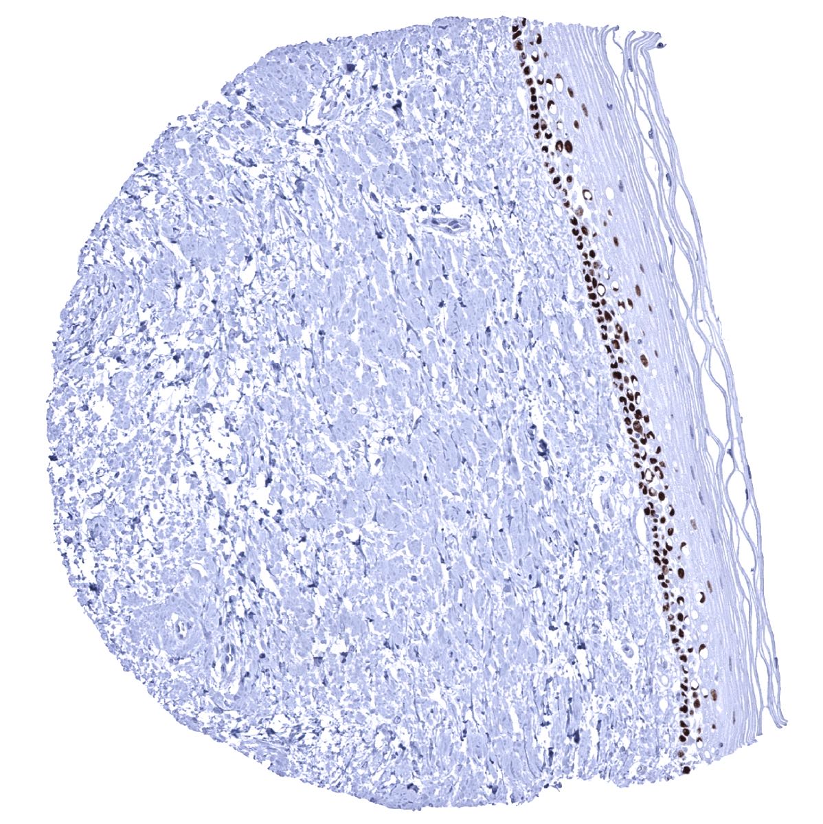
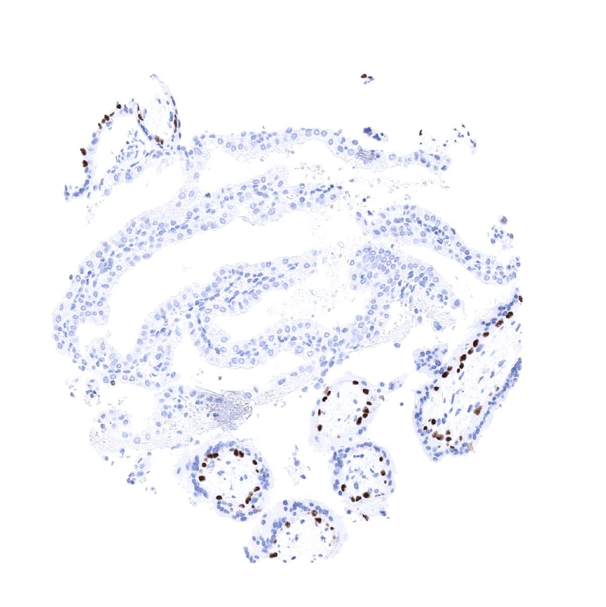
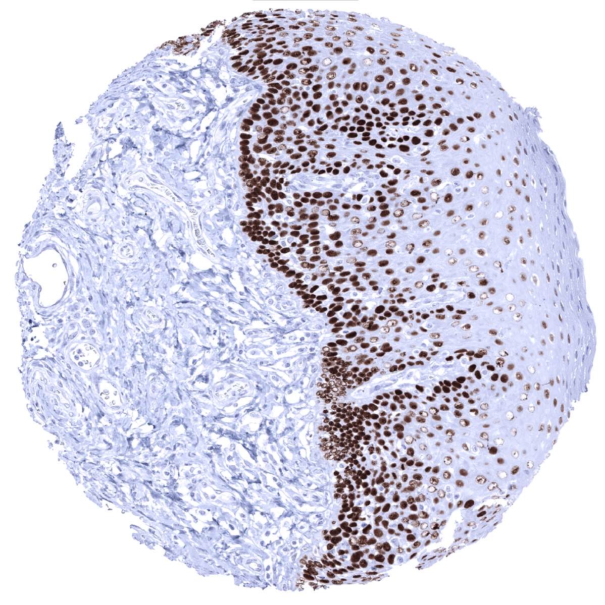
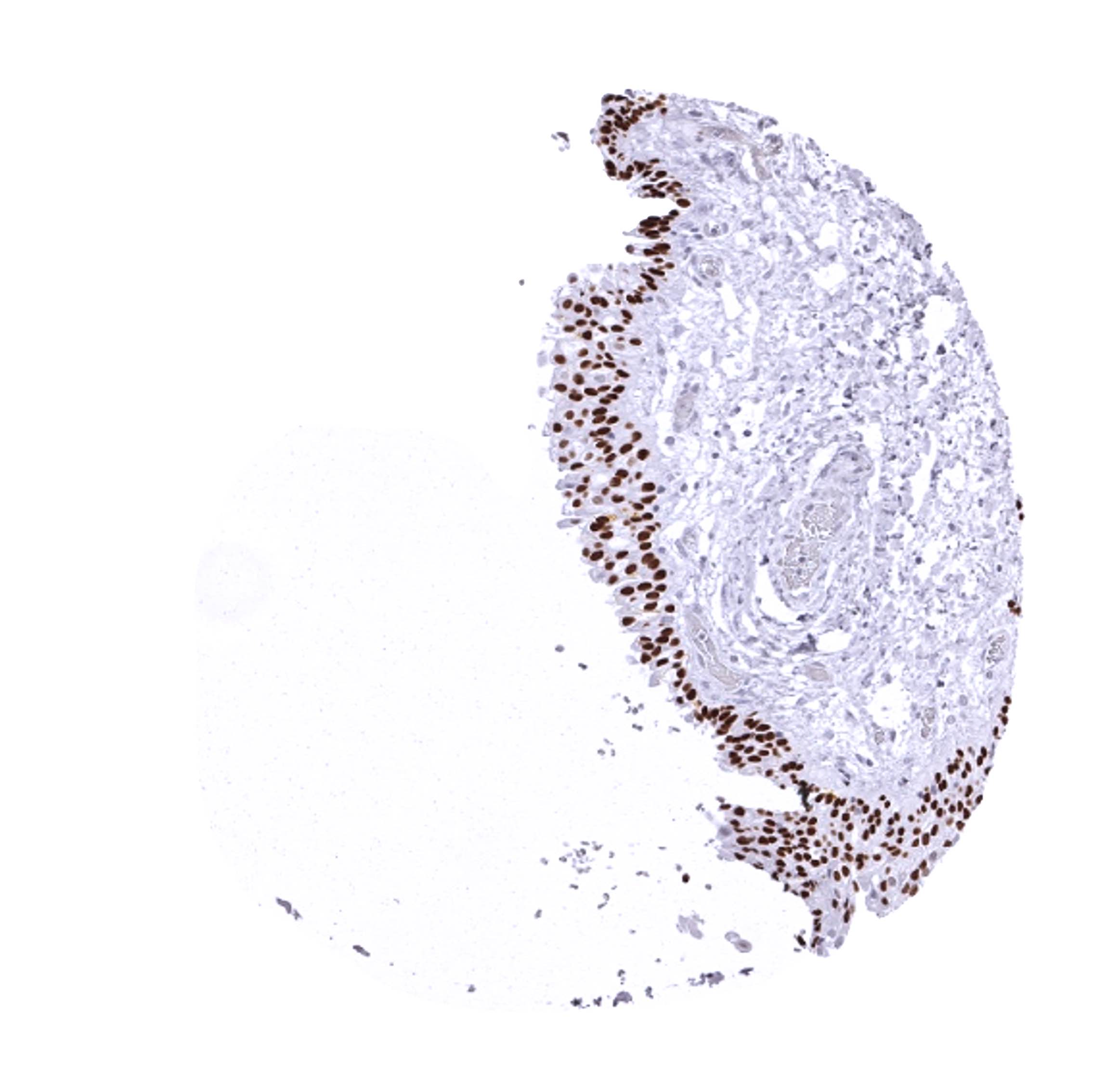
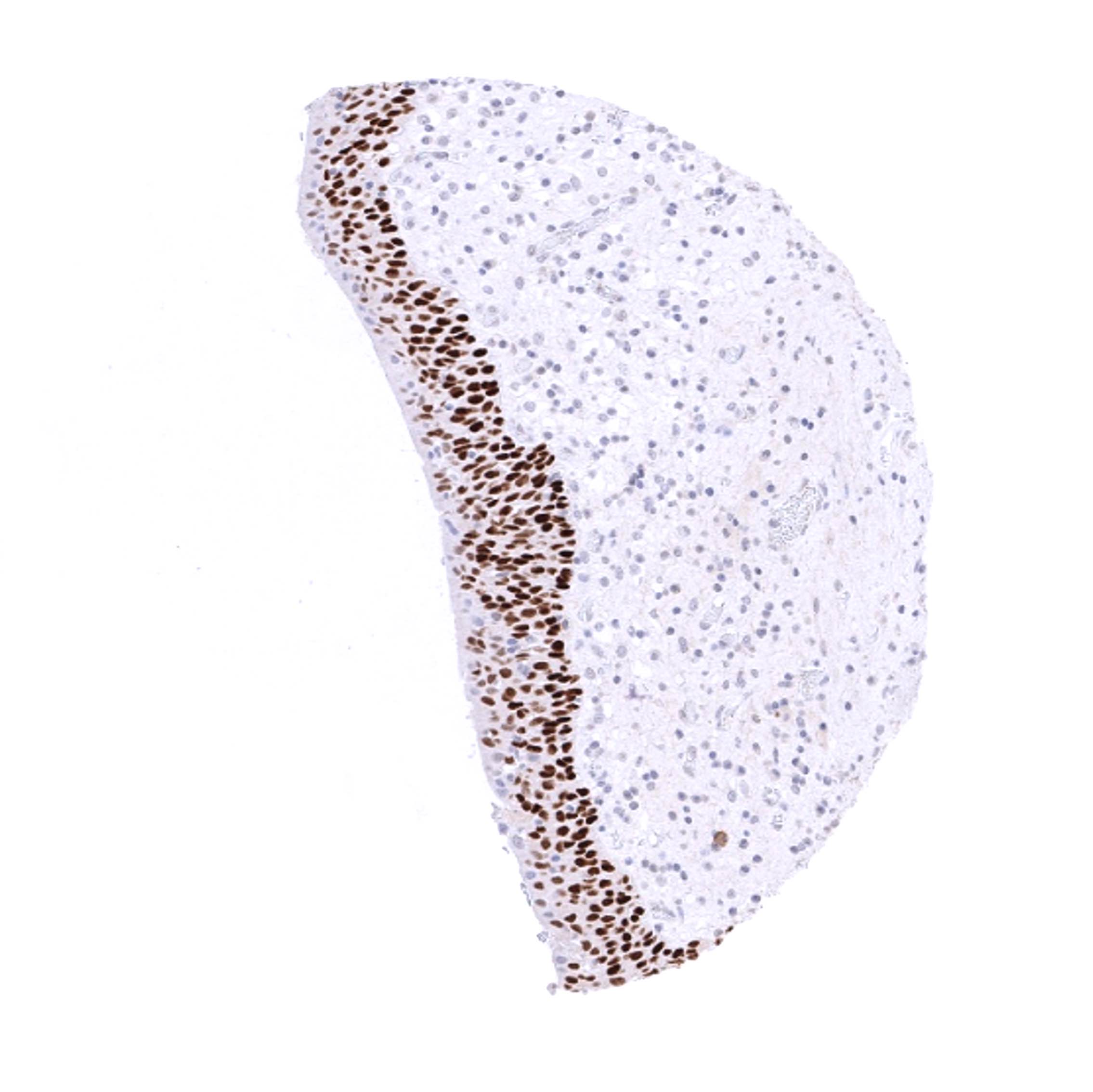
| Esophagus | Nuclear p63 staining in squamous epithelial cells is strongest in the basal cell layers while the intensity gradually decreases towards the top layers. | |
| Stomach | Negative. | |
| Duodenum | Negative. | |
| Small intestine | Negative. | |
| Appendix | Negative. | |
| Colon | Negative. | |
| Rectum | Negative. | |
| Liver | Negative. | |
| Gallbladder | Negative. | |
| Pancreas | Negative. | |
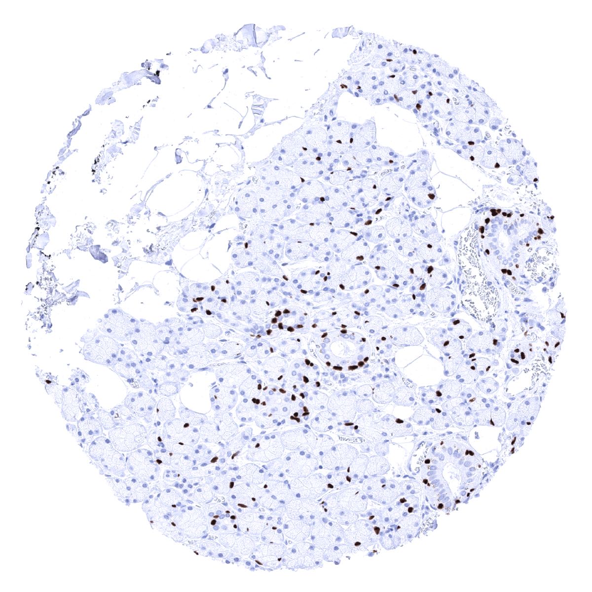
| Genitourinary | Kidney | Negative. |
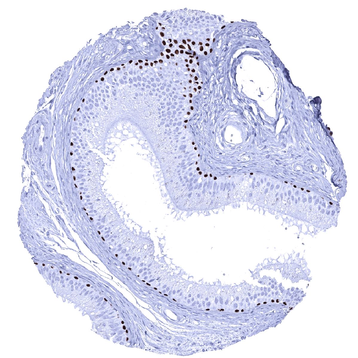
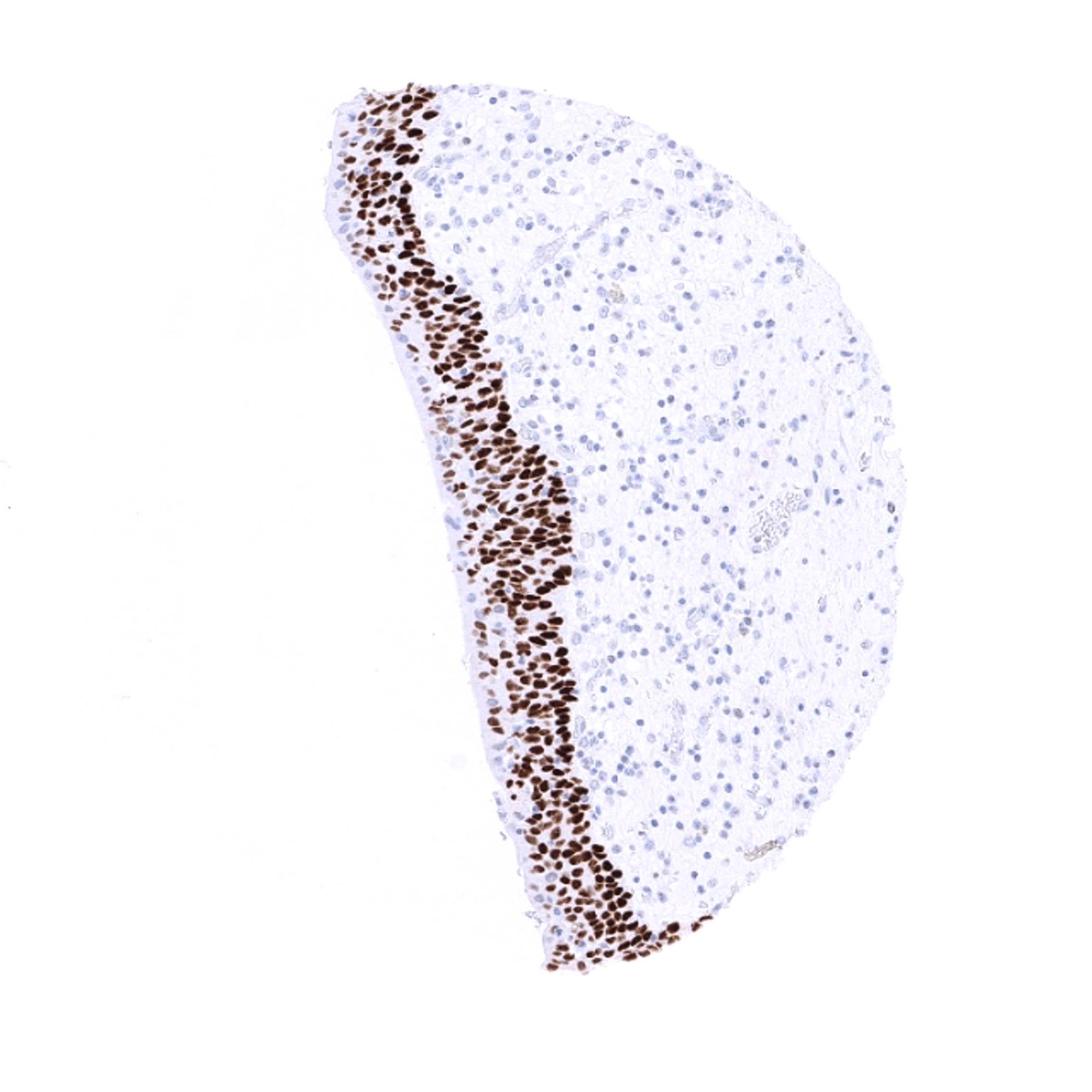
| Urothelium | Strong nuclear p63 staining in all urothelial cells except umbrella cells. | |
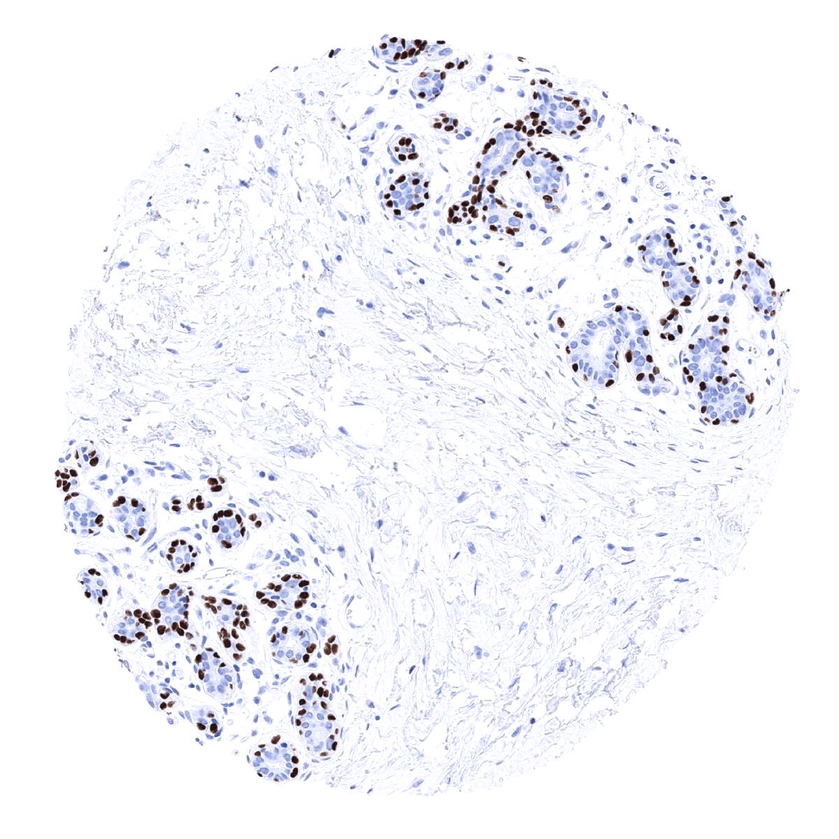
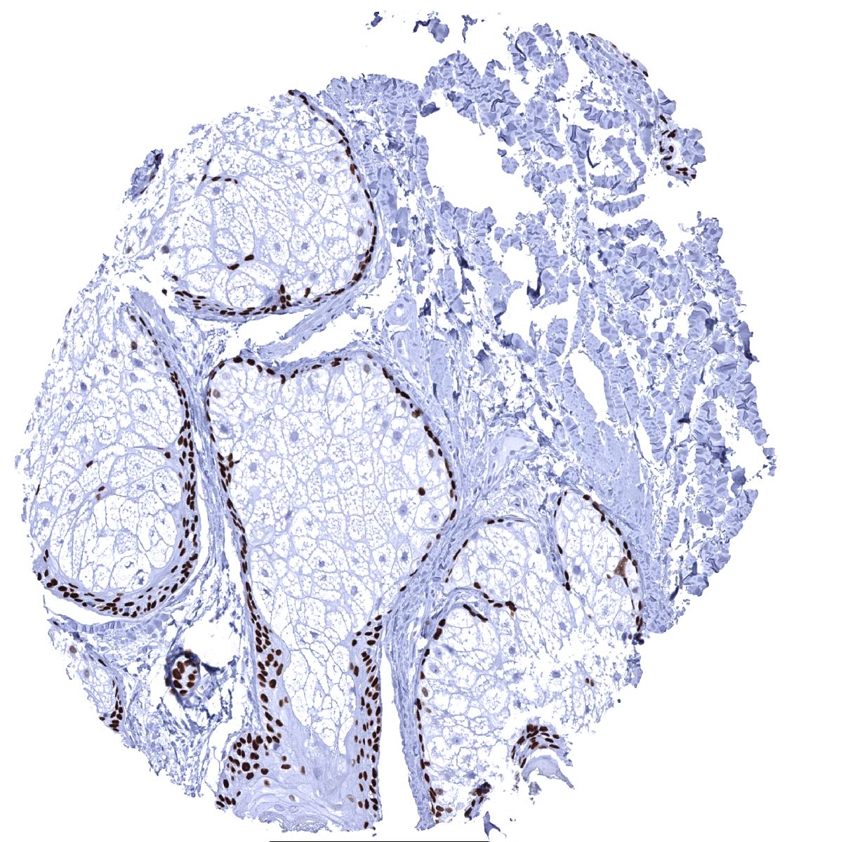
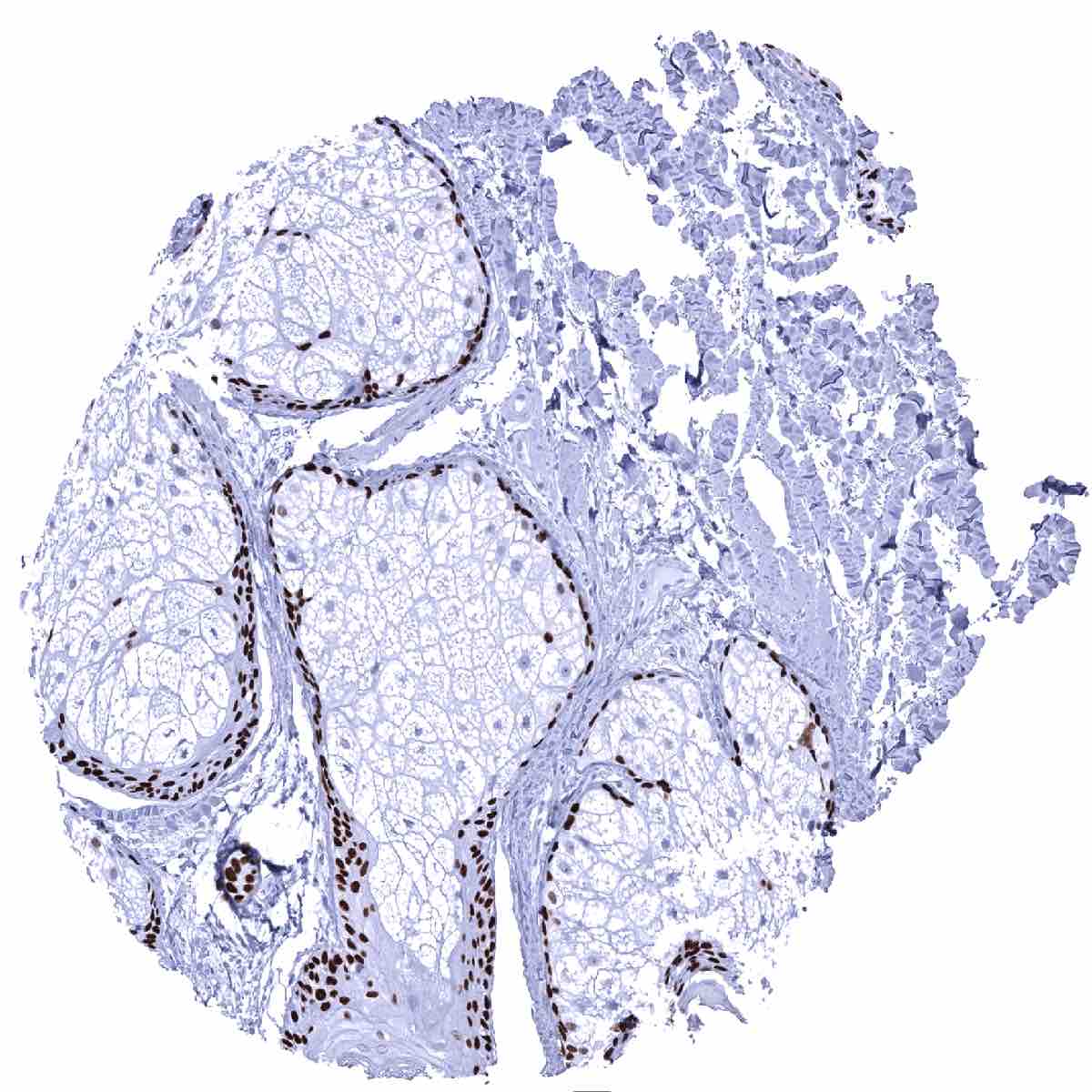
| Male genital | Prostate | Strong nuclear p63 positivity of basal cells. |
| Seminal vesicles | Strong nuclear p63 positivity of basal cells. | |
| Testis | Negative. | |
| Epididymis | Strong nuclear p63 positivity of basal cells. | |
| Female genital | Breast | Strong nuclear p63 positivity of myoepithelial cells. |
| Uterus, myometrium | Negative. | |
| Uterus, ectocervix | Nuclear p63 staining in squamous epithelial cells is strongest in the basal cell layers while the intensity gradually decreases towards the top layers. | |
| Uterus endocervix | Negative. | |
| Uterus, endometrium | Negative. | |
| Fallopian Tube | Negative. | |
| Ovary | Negative. | |
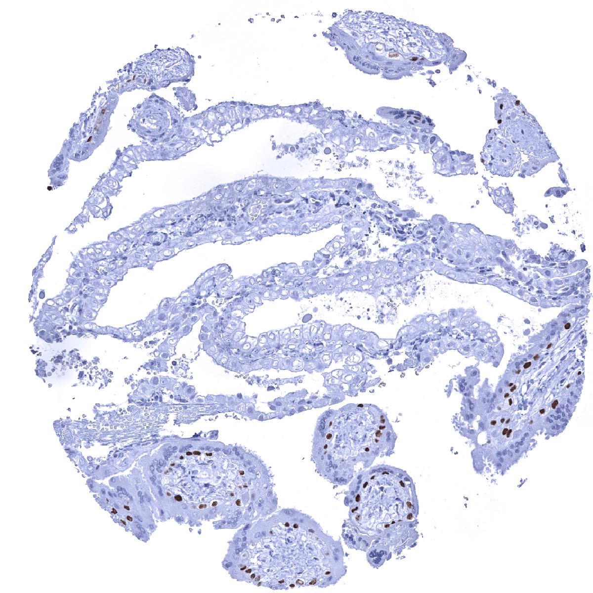
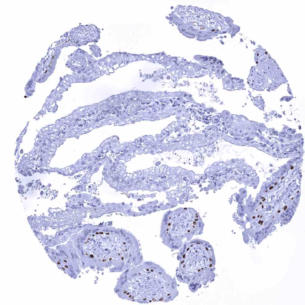
| Placenta early | Strong nuclear p63 positivity of chorion cells and of a fraction of cytotrophoblast cells. | |
| Placenta mature | Strong nuclear p63 positivity of chorion cells and of a fraction of cytotrophoblast cells. | |
| Amnion | Negative. | |
| Chorion | Strong nuclear p63 positivity of chorion cells. | |
| Skin | Epidermis | Nuclear p63 staining in squamous epithelial cells is strongest in the basal cell layers while the intensity gradually decreases towards the top layers. |
| Sebaceous glands | Strong nuclear p63 staining in peripheric germinative cells of sebaceous glands. | |
| Muscle/connective tissue | Heart muscle | Negative. |
| Skeletal muscle | Negative. | |
| Smooth muscle | Negative. | |
| Vessel walls | Negative. | |
| Fat | Negative. | |
| Stroma | Negative. | |
| Endothelium | Negative. | |
| Bone marrow/ lymphoid tissue | Bone marrow | Negative. |
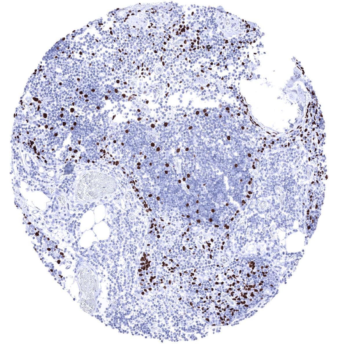
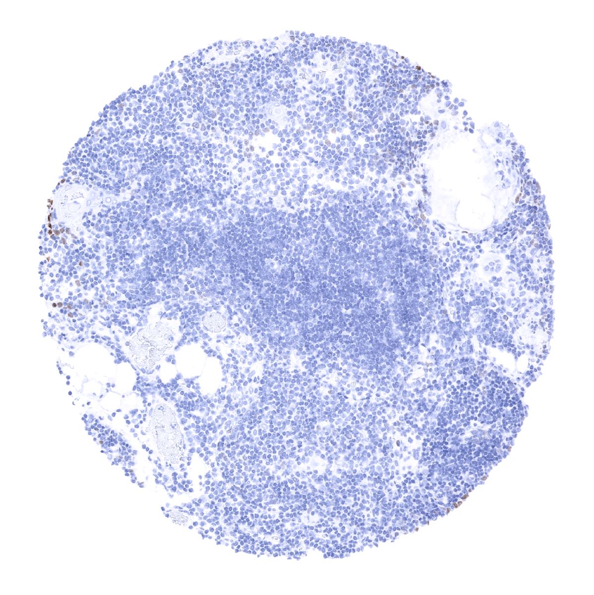
| Lymph node | A rather faint p63 staining occurs in few lymphocytes and in high endothelial venules. | |
| Spleen | Negative. | |
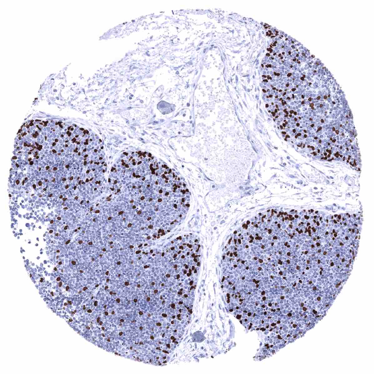
| Thymus | Strong nuclear p63 staining of epithelial cells. | |
| Tonsil | Nuclear p63 staining in squamous epithelial cells is strongest in the basal cell layers while the intensity gradually decreases towards the top layers. | |
| Remarks | A faint p63 staining occurs in small subset of lymphocytes |
The findings described above are this consistent with the RNA data described in the Human Protein Atlas (Tissue expression p63)
Positive control = Tonsil: Virtually all squamous epithelial cells must show a moderate to strong, nuclear staining, while few scattered lymphocytes and endothelial cells must show a at least a weak staining.
Negative control = Tonsil: The vast majority of lymphocytes should be p63 negative.
Staining Pattern in Relevant Tumor Types
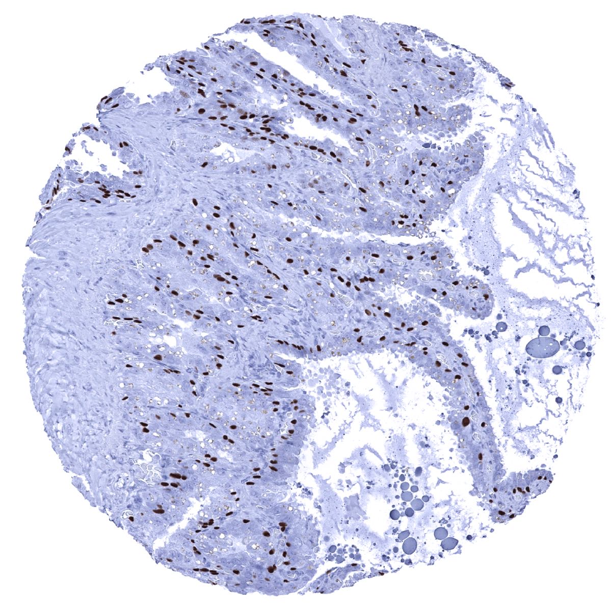
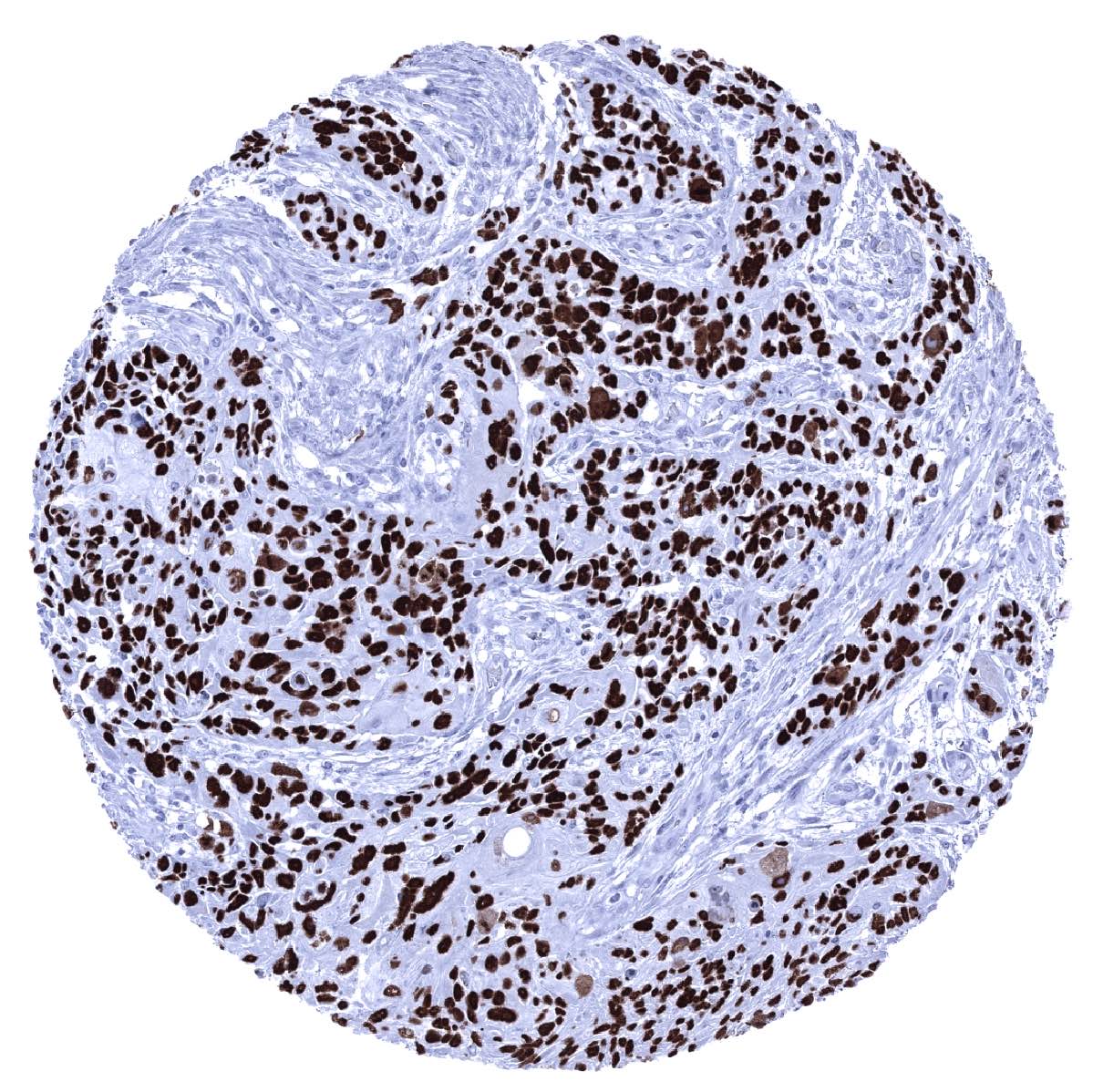
p63 immunostaining is predominantly seen in cancers that are derived from p63 positive normal cell types. The most commonly positive cancers include squamous cell carcinomas of all origins, urothelial carcinomas, thymic tumors, basal cell carcinomas, and various salivary gland tumors. p63 can also be expressed in a small fraction of tumors from entities that are derived from p63 negative normal tissues. In some of these tumors, p63 neo-expression is linked to focal squamous cell differentiation which can for example occur in endometroid cancer and malignant mixed Mullerian tumors of the uterus, ovarian, pancreatic and cholangiocellular carcinomas. In other tumors, occasional p63 positive cells may reflect stemness properties.
The TCGA findings on p63 RNA expression in different tumor categories have been summarized in the Human Protein Atlas.
Compatibility of Antibodies
No data available at the moment
Protocol Recommendations
IHC users have different preferences on how the stains should look like. Some prefer high staining intensity of the target stain and even accept some background. Others favor absolute specificity and lighter target stains. Factors that invariably lead to more intense staining include higher concentration of the antibody and visualization tools, longer incubation time, higher temperature during incubation, higher temperature and longer duration of the heat induced epitope retrieval (slide pretreatment). The impact of the pH during slide pretreatment has variable effects and depends on the antibody and the target protein.
All images and data shown here and in our image galleries are obtained by the manual protocol described below. Other protocols resulting in equivalent staining are described as well.
Manual protocol
Freshly cut sections should be used (less than 10 days between cutting and staining). Heat-induced antigen retrieval for 5 minutes in an autoclave at 121°C in pH 7,8 Target Retrieval Solution buffer. Apply MSVA-063R at a dilution of 1:150 at 37°C for 60 minutes. Visualization of bound antibody by the EnVision Kit (Dako, Agilent) according to the manufacturer’s directions.

Agilent / Dako – Autostainer Link 48
Pretreatment in PT-Link for 30 minutes at 95°C (pH high); FLEX peroxidase blocking for 5 minutes (room temperature), MSVA-063R 1:100 for 20 minutes (room temperature), FLEX+ mouse/rabbit (LINKER) for 15 minutes (room temperature), horseradish peroxidase (HRP) for 20 minutes (room temperature), FLEX DAB+Sub-Chromo for 10 minutes (room temperature), FLEX hematoxylin for 5 minutes (room temperature).

These images reflect stainings by the protocol described above. It is of note that a comparable staining result can also be obtained by different protocols. In general, a longer pretreatment, a longer incubation time of the primary antibody, a higher antibody concentration, and a longer incubation time of FLEX+LINKER result in stronger staining, potentially at the cost of more background staining. Modifications of the protocol with a strengthening effect on staining intensity in combination with changes of other parameters that result in lower staining intensity can result in a comparable result as shown above.
Leica – BOND RX
Dewax at 72°C for 30 seconds; Pretreatment in Bond Epitope Retrieval Solution (ER2 – EDTA pH9) for 60 minutes at 100°C; Peroxidase blocking for 5 minutes (room temperature), MSVA-063R 1:100 for 20 minutes (room temperature), Post primary (rabbit anti mouse) for 8 minutes (room temperature), Polymer (goat anti rabbit) for 8 minutes (room temperature), mixed DAB refine for 10 minutes (room temperature), hematoxylin for 5 minutes (room temperature).
These images reflect stainings by the protocol described above. It is of note that a comparable staining result can also be obtained by different protocols. In general, a longer pretreatment, a longer incubation time of the primary antibody, a higher antibody concentration, a higher temperature during incubation, and a longer incubation time of Post primary and or the Polymer result in stronger staining, potentially at the cost of more background staining. Modifications of the protocol with a strengthening effect on staining intensity in combination with changes of other parameters that result in lower staining intensity can result in a comparable result as shown above.
Roche – Ventana Discovery ULTRA
Pretreatment for 64 minutes at 100°C (pH 8,4); CM peroxidase blocking for 12 minutes (room temperature), MSVA-063R 1:100 for 20 minutes at 36°C, secondary antibody (anti-rabbit HQ) for 12 minutes at 36°C, anti-HQ HRP for 12 minutes at room temperature, DAB at room temperature, hematoxylin II at room temperature for 8 minutes, bluing reagent at room temperature for 4 minutes.

These images depict staining results obtained by the protocol described above. It is of note, that the Ventana machines generally require higher antibody concentrations than other commonly used autostainers because the antibodies are automatically diluted during the procedure. Various other protocols can result in an identical result as shown above. A longer pretreatment, a longer incubation time of the primary antibody, a higher antibody concentration, a higher temperature during incubation, and a longer incubation time of secondary antibody and or the anti-HQ HRP result in stronger staining, potentially at the cost of more background staining.
Potential Research Applications
- A comprehensive study analyzing p63 expression in various different tumor entities would be helpful to assess the diagnostic significance of p63 IHC.
- The roles of p63 in multiple aspects of cancer, including tumorigenesis, cancer progression, and metastasis as well as how they impact other diseases are still not completely discovered.
Evidence for Antibody Specificity in IHC
There are two ways how the specificity of antibodies can be documented for immunohistochemistry on formalin fixed tissues. These are: 1. Comparison with a second independent method for target expression measurement across a large number of different tissue types (orthogonal strategy), and 2. Comparison with one or several independent antibodies for the same target and showing that all positive staining results are also seen with other antibodies for the same target (independent antibody strategy).
Orthogonal validation: For the antibody MSVA-063R, specificity is suggested by the perfect concordance of the immunostaining data with data from three independent RNA screening studies, including the Human Protein Atlas (HPA) RNA-seq tissue dataset, the FANTOM5 project, and the Genotype-Tissue Expression (GTEx) project, which are all summarized in the Human Protein Atlas (Tissue expression p63). P63 RNA expression was only seen in organs covered by squamous epithelium (esophagus, vagina, cervix, skin, tonsil) or urothelium (urinary bladder) as well as in organs for which a p63 positive cell type had been observed by MSVA-063R such as myoepithelial cells in salivary and breast glands, basal cells in the prostate, salivary glands, epididymis, and respiratory epithelium, epithelial cells in the thymus, as well as chorion and cytotrophoblast cells in the placenta.
Comparison of antibodies: A p63 specific staining of MSVA-063R is also supported by identical staining patterns – across all analyzed tissues – found by using a commercially available independent second p63 antibody (termed “Validation Antibody”). Comparative images on squamous epithelium, urothelium, salivary glands, breast, prostate, salivary glands, epididymis, respiratory epithelium, thymus, and the placenta are shown below.
Antibody Comparison: MSVA-063R vs another commercially available p63 antibody called “Validation Antibody”