
195,00 € – 695,00 €
Product details
Synonyms = AAT4; Myosin heavy chain 11 (MYH11); Myosin heavy chain 11 smooth muscle; Smooth muscle myosin heavy chain 11; Myosin-11; SM1; SM2; SMHC; SMMHC
Antibody type = Recombinant Rabbit monoclonal / IgG
Clone = MSVA-375R
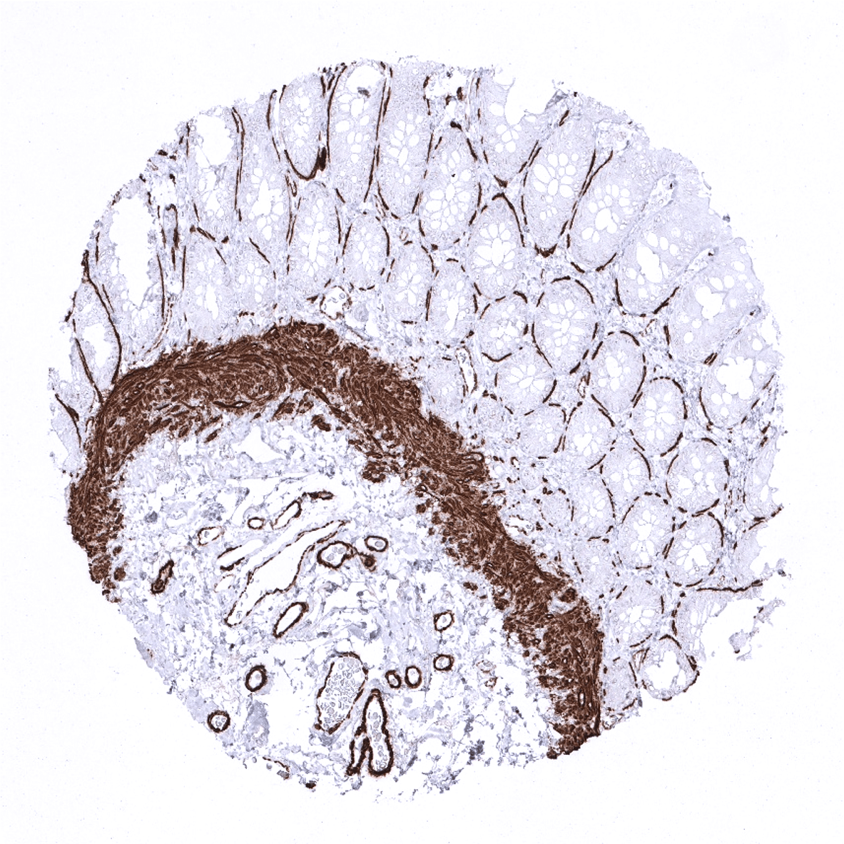
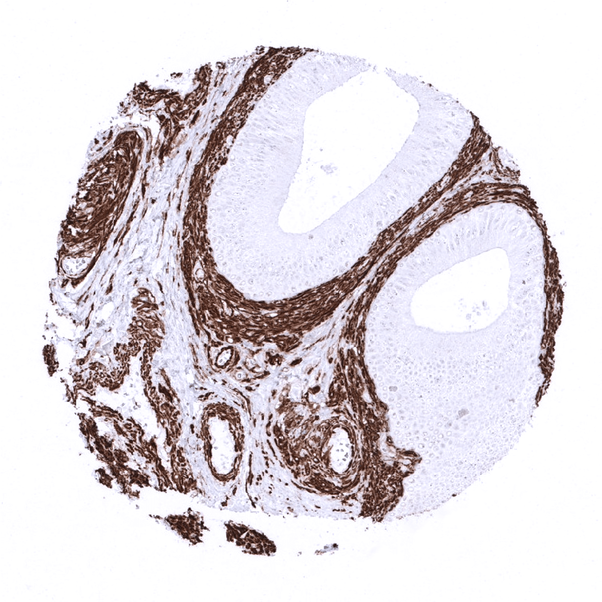
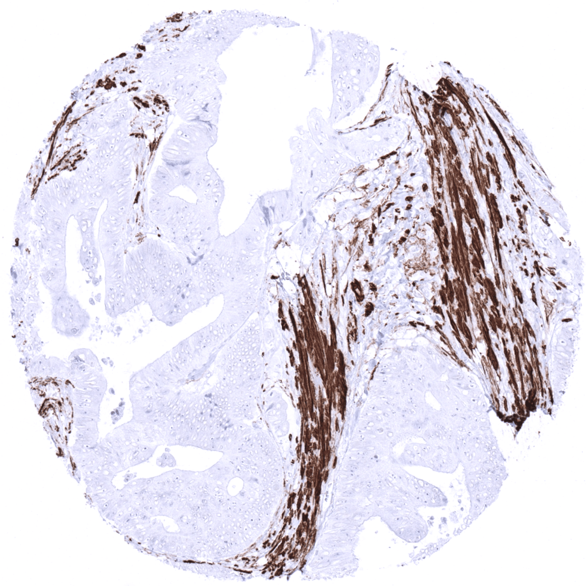
Positive control = Colon: A strong MYH11 staining should be seen in muscularis mucosa, muscular propria, small and medium sized vessels and the fine smooth muscle layer surrounding crypt glands.
Negative control = Colon: Epithelial cells must always be MYH11 negative.
Cellular localization = Cytoplasmic
Reactivity = Human
Application = Immunohistochemistry
Dilution = 1:100 – 1:200
Intended Use = Research Use Only
Relevance of Antibody
Biology Behind
Myosin-11 also termed smooth muscle myosin heavy chain (SMMHC) is a smooth muscle myosin belonging to the myosin heavy chain family. It is a contractile protein, converting chemical into mechanical energy through the ATP hydrolysis. Alternative splicing generates multiple isoforms the composition of which varies during muscle cell maturation. Mutations in MYH11 have been found in about 1% of patients with a familial predisposition to thoracic aortic aneurysms leading to acute aortic dissections (TAAD). The MYH11 gene is involved in the genomic rearrangements inv(16)(p13q22) and t(16;16)(p13;q22 which are both characteristic of acute myeloid leukemia (AML) with abnormal bone marrow eosinophils (AML-M4Eo). As a result a fusion protein composed of core binding factor beta (CBF-beta) with SMMHC (CBFbeta-SMMHC; Inv(16)(p13q22)) is found in the nuclei of nearly all AML M4Eo.
Staining Pattern in Normal Tissues
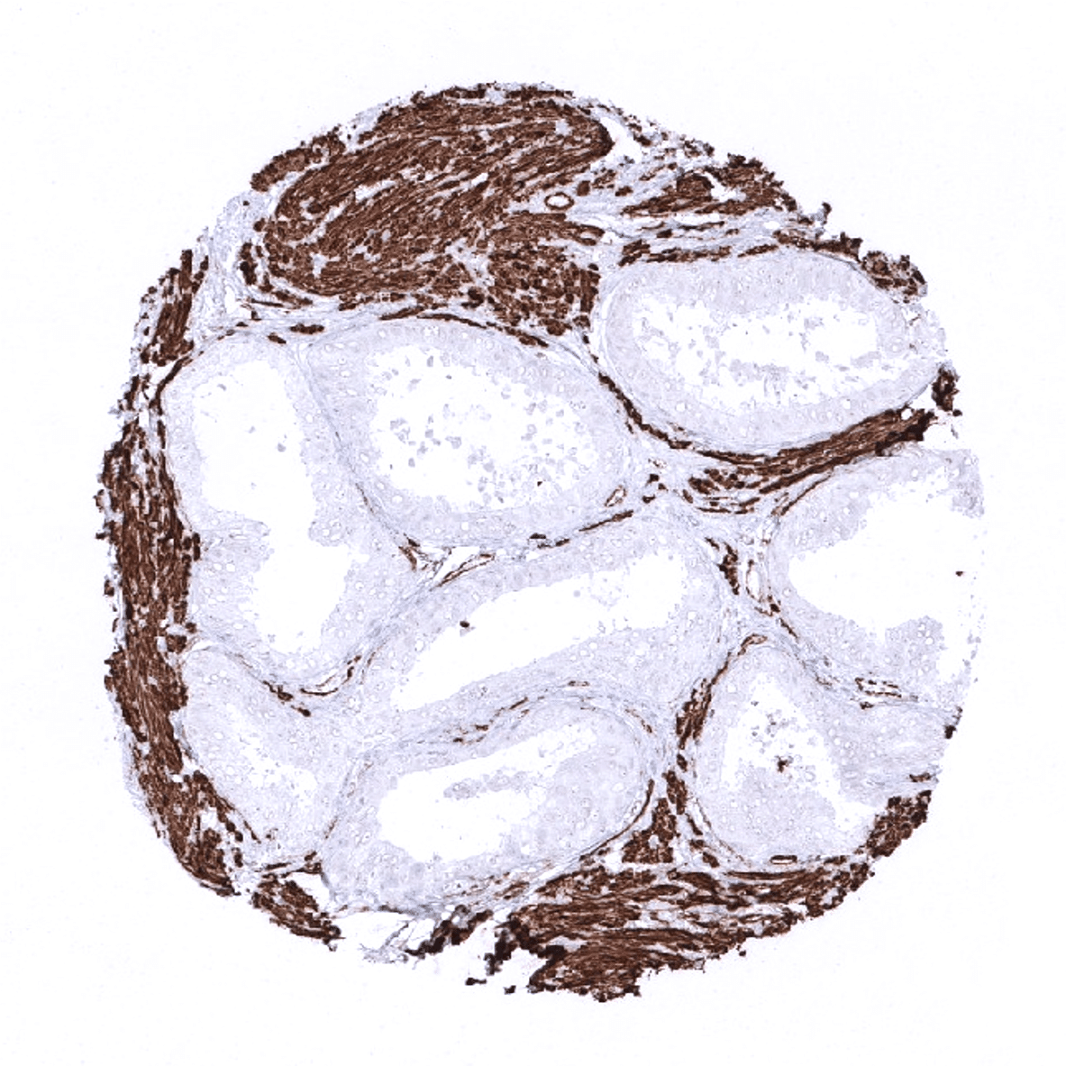
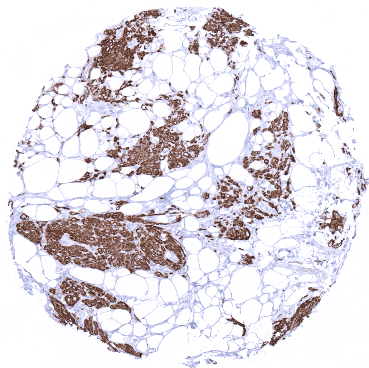
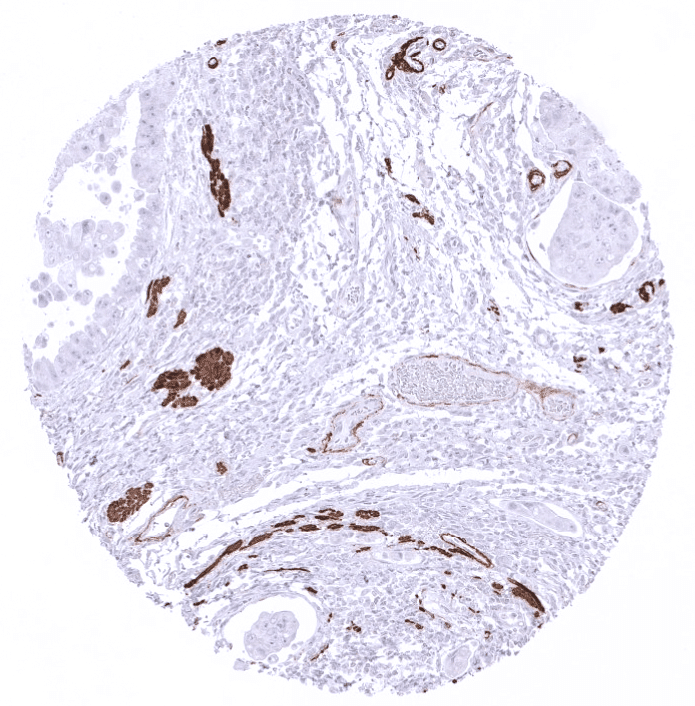
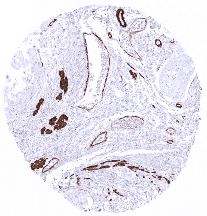
Using antibody MSVA-375R, MYH11 is found expressed in the cytoplasm and cell membrane of smooth muscle cells occurring in various organs as well as in small, medium sized, and large vessels including the aortic media. MYH11 immunostaining is also seen in myoepithelial cells of the breast, myoepithelial cells and basal cells of excretory ducts in salivary glands. In the colorectum, MYH11 delineates a discrete but distinct pericryptal cell layer. MYH11 is also expressed in follicular dendritic cells.
These findings are largely consistent with the RNA and protein data described in the Human Protein Atlas (Tissue expression MYH11).
Suggested positive tissue control: Colon: A strong MYH11 staining should be seen in muscularis mucosa, muscular propria, small and medium sized vessels and the fine smooth muscle layer surrounding crypt glands.
Suggested negative tissue control: Colon: Epithelial cells must always be MYH11 negative.

Staining Pattern in Relevant Tumor Types
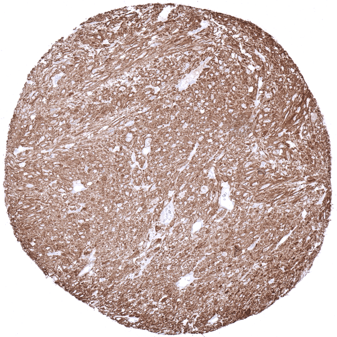
In tumors, MYH11 is almost exclusively seen in tumors derived from smooth muscle such as leiomyoma, leiomyosarcoma, and few others. Nuclear MYH11 staining is seen in AML-M4Eo.
The TCGA findings on MYH11 RNA expression in different tumor categories have been summarized in the Human Protein Atlas.

Compatibility of Antibodies
No data available at the moment
Protocol Recommendations
IHC users have different preferences on how the stains should look like. Some prefer high staining intensity of the target stain and even accept some background. Others favor absolute specificity and lighter target stains. Factors that invariably lead to more intense staining include higher concentration of the antibody and visualization tools, longer incubation time, higher temperature during incubation, higher temperature and longer duration of the heat induced epitope retrieval (slide pretreatment). The impact of the pH during slide pretreatment has variable effects and depends on the antibody and the target protein.
All images and data shown here and in our image galleries are obtained by the manual protocol described below. Other protocols resulting in equivalent staining are described as well.
-Manual protocol
Freshly cut TMA sections are deparaffinized and exposed to heat-induced antigen retrieval for 5 minutes in an autoclave at 121°C in pH 9 Target Retrieval Solution buffer. Apply MSVA-375R at a dilution of 1:50 at 37°C for 60 minutes. Visualization of bound antibody by the EnVision Kit (Dako, Agilent) according to the manufacturer’s directions.
-Impact of pH

The strongest Myosin Heavy Chain 11 staining by MSVA-375R is obtained at a pH 9,0. However, pH 7,8 results in only a slight reduction of the staining intensity as compared to pH9. We thus consider pH7,8 as optimal for manual staining because of the better tissue preservation at pH7,8 than at pH 9,0.
Potential Research Applications
- MYH11 immunostaining can be used as a marker for myoepithelial cells in multicolor immunohistochemistry approaches for automated tumor detection.
- A comprehensive study analyzing MYH11 expression in various different tumor entities would be helpful to assess the diagnostic significance of MYH11 IHC.
Evidence for Antibody Specificity in IHC
Specificity of MSVA-375R is documented by strong positive staining in cell types that are well documented to express myosin heavy chain 11 such as smooth muscle cells in various organs, blood vessels of all size, and follicular dendritic cellsof lymph nodes in combination with absence of staining in all tissues known to not express myosin heavy chain 11 such as epithelial cells of all kinds including tissues notorious for non-specific IHC background such as kidney, colonic mucosa, and epidermis.
