295,00 € – 995,00 €
Product details
Synonyms = Minichromosome maintenance complex component 5, CDC46, MGORS8, P1-CDC46
Antibody type = Recombinant Rabbit monoclonal / IgG
Clone = MSVA-505R
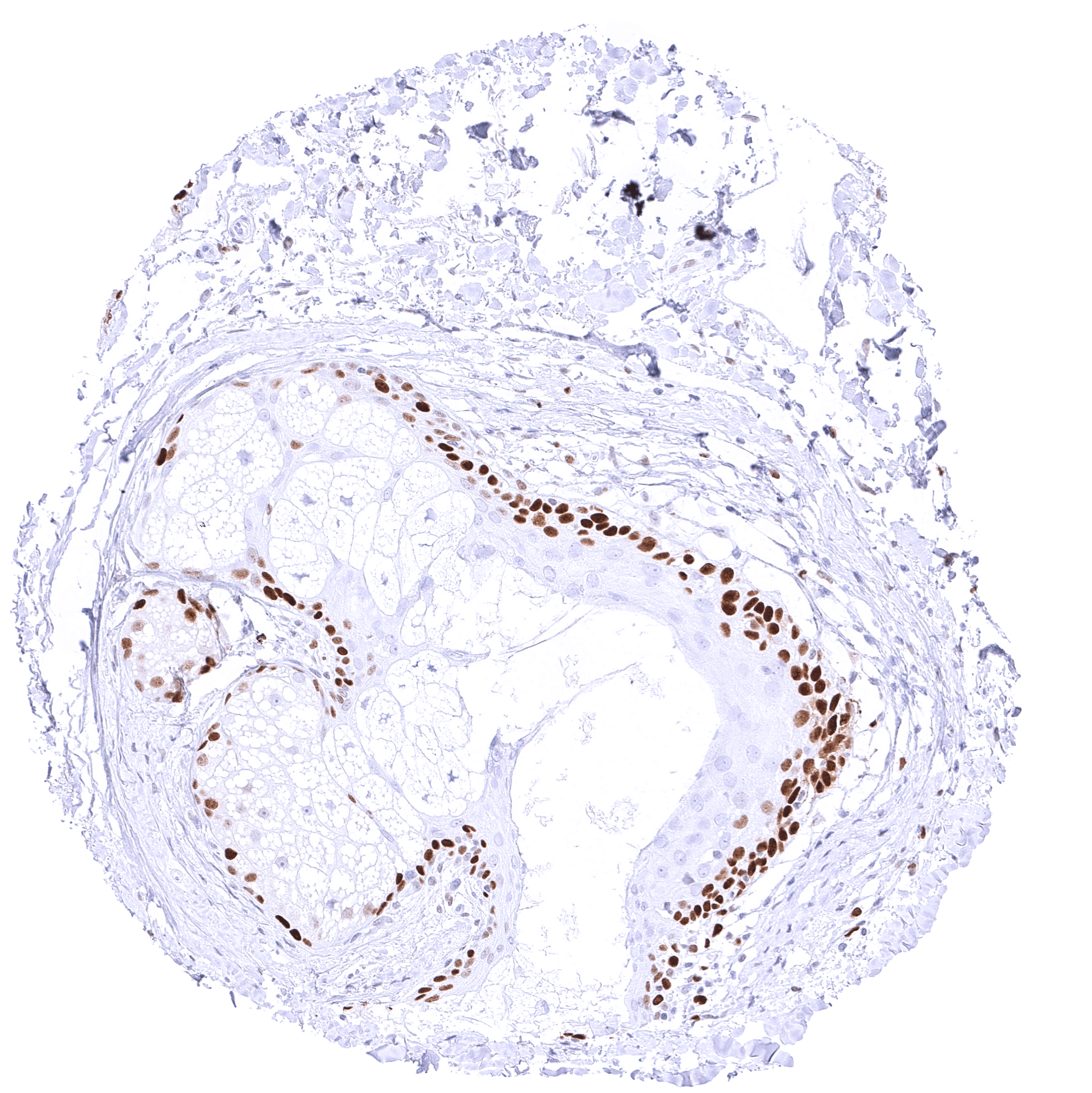
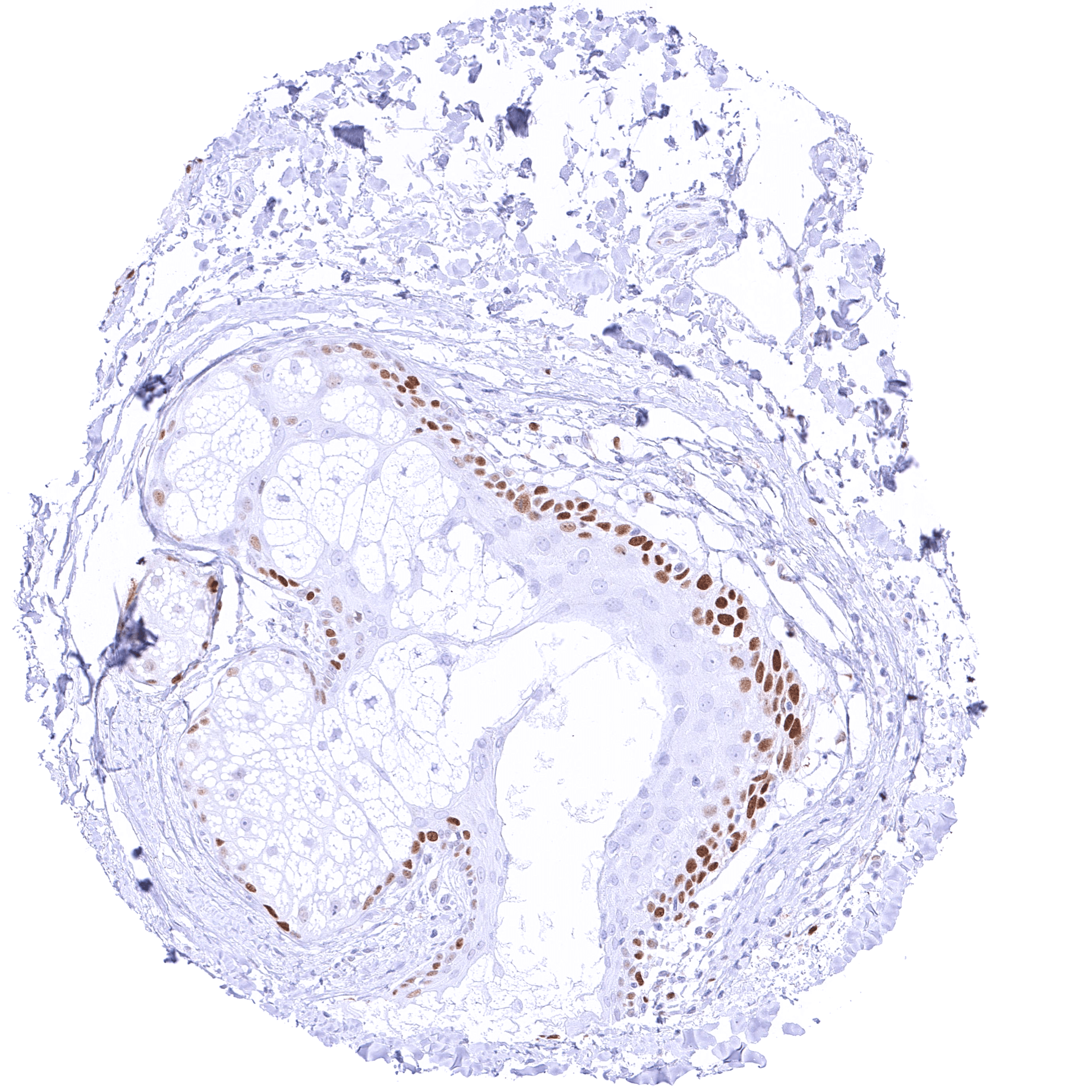
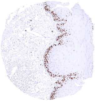
Positive control = Colon: A strong nuclear MCM5 immunostaining should be seen in virtually all crypt base cells.
Negative control = Colon: MCM5 immunostaining should be less intense or absent in surface epithelial cells and absent in most stroma cells.
Cellular localization = Intracellular
Reactivity = Human
Application = Immunohistochemistry
Dilution = 1:50 – 1:100
Intended Use = Research Use Only
Relevance of Antibody
MCM5 is a highly sensitive marker for proliferating cells.
Biology Behind
The MCM5 gene is located at 22q12.3 and codes for a nuclear protein which belongs to the highly conserved mini-chromosome maintenance proteins (MCM) 2-7 that play a key role in genome replication. They form a ring-shaped hexameric protein complex which unwinds double-stranded DNA, forms a replication fork during the initiation of DNA replication, and helps to recruit other DNA replication related proteins. The MCM2-7 limits DNA replication to a single occurrence per cell division and is critical for maintaining genome integrity. The MCM proteins are expressed in all cells in the G1, S, G2 and M-phase of the cell cycle but in contrast to the better established proliferation marker Ki-67, MCMs are already expressed in early G1 phase. This results in the detection of more proliferating cells as compared to Ki67 immunohistochemistry which might be advantageous in tumor types with low proliferative activity. Whether the role of MCM5 specifically differs from those of other MCM proteins is not clear. Interaction analyses have revealed that MCM5 directly interacts with MCM2, 3, and 7.
Staining Pattern in Normal Tissues
Images describing the MCM5 staining pattern in normal tissues obtained by the antibody MSVA-505R are shown in our “Normal Tissue Gallery”.
| Brain | Cerebrum | Negative. |
| Cerebellum | Negative. | |
| Endocrine Tissues | Thyroid | Weak to moderate nuclear MCM5 staining of a small fraction of follicular cells. |
| Parathyroid | Distinct nuclear MCM5 staining in a very small fraction of epithelial cells. | |
| Adrenal gland | A variable MCM5 staining occurs in a small fraction of adrenocortical cells. Spindle shaped nuclei are also found positive. | |
| Pituitary gland | Negative. | |
| Respiratory system | Respiratory epithelium | Significant MCM5 staining in a fraction of (mostly basal/suprabasal) respiratory epithelial cells. |
| Lung | Distinct MCM5 staining of a subset of pneumocytes. | |
| Gastrointestinal Tract | Salivary glands | Distinct MCM5 staining in a fraction of epithelial cells. |
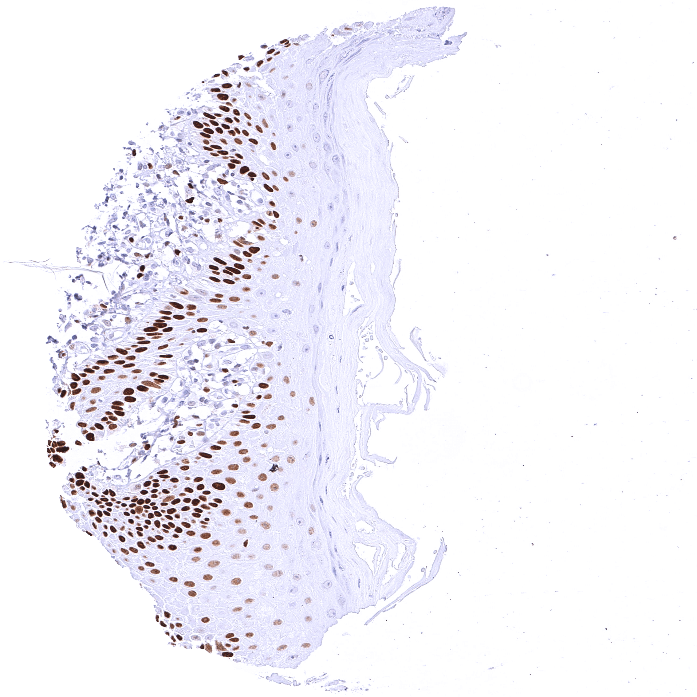
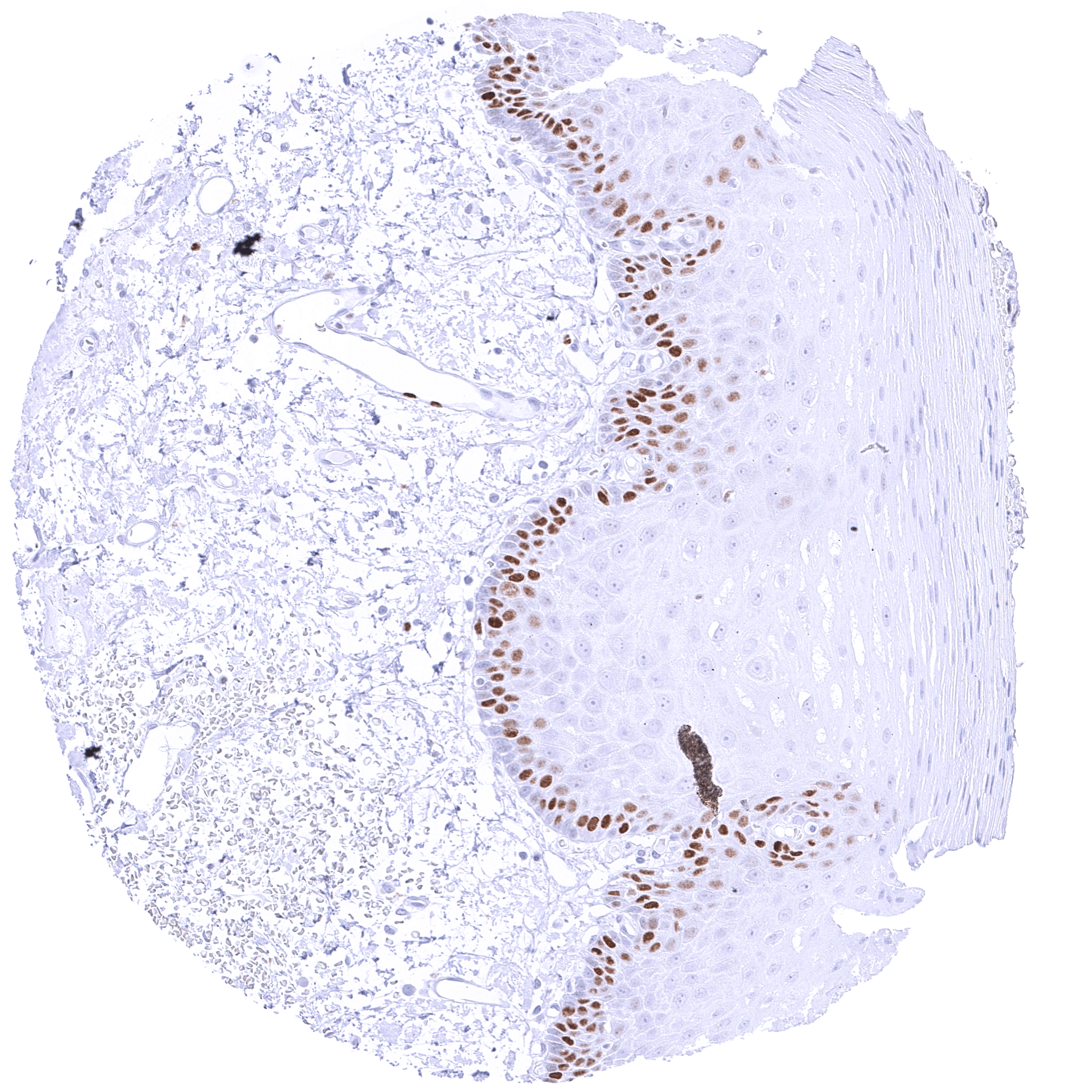
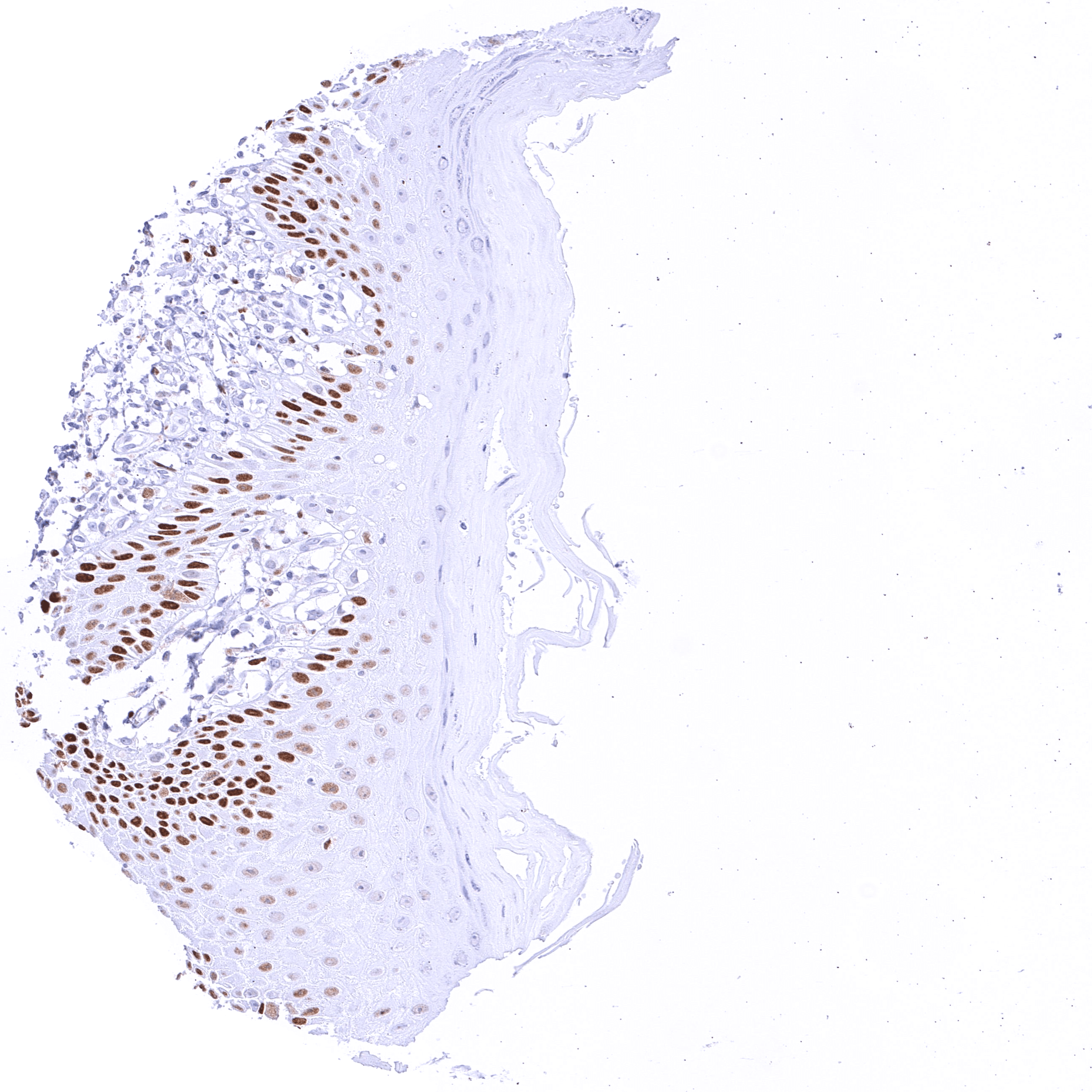
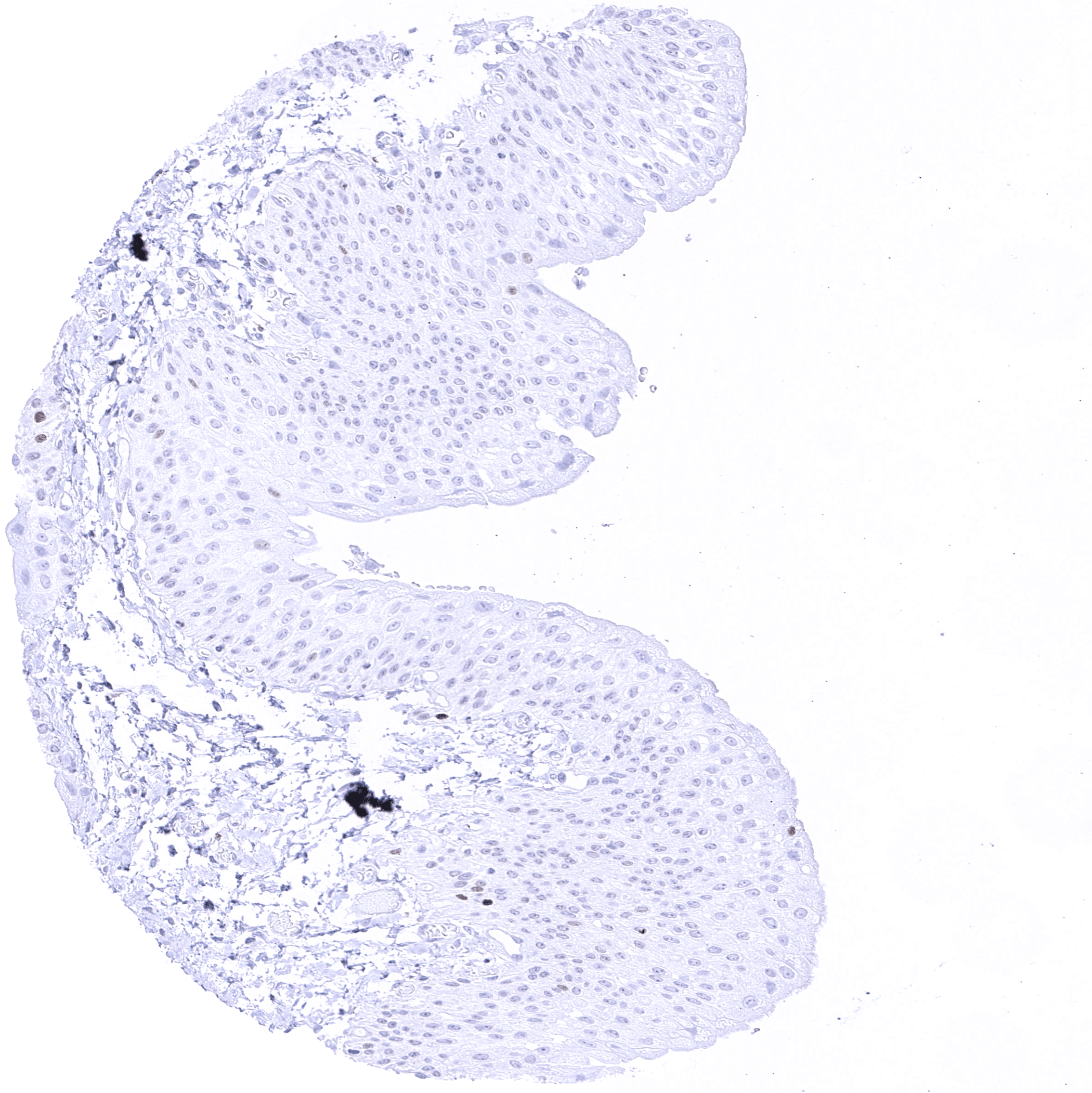
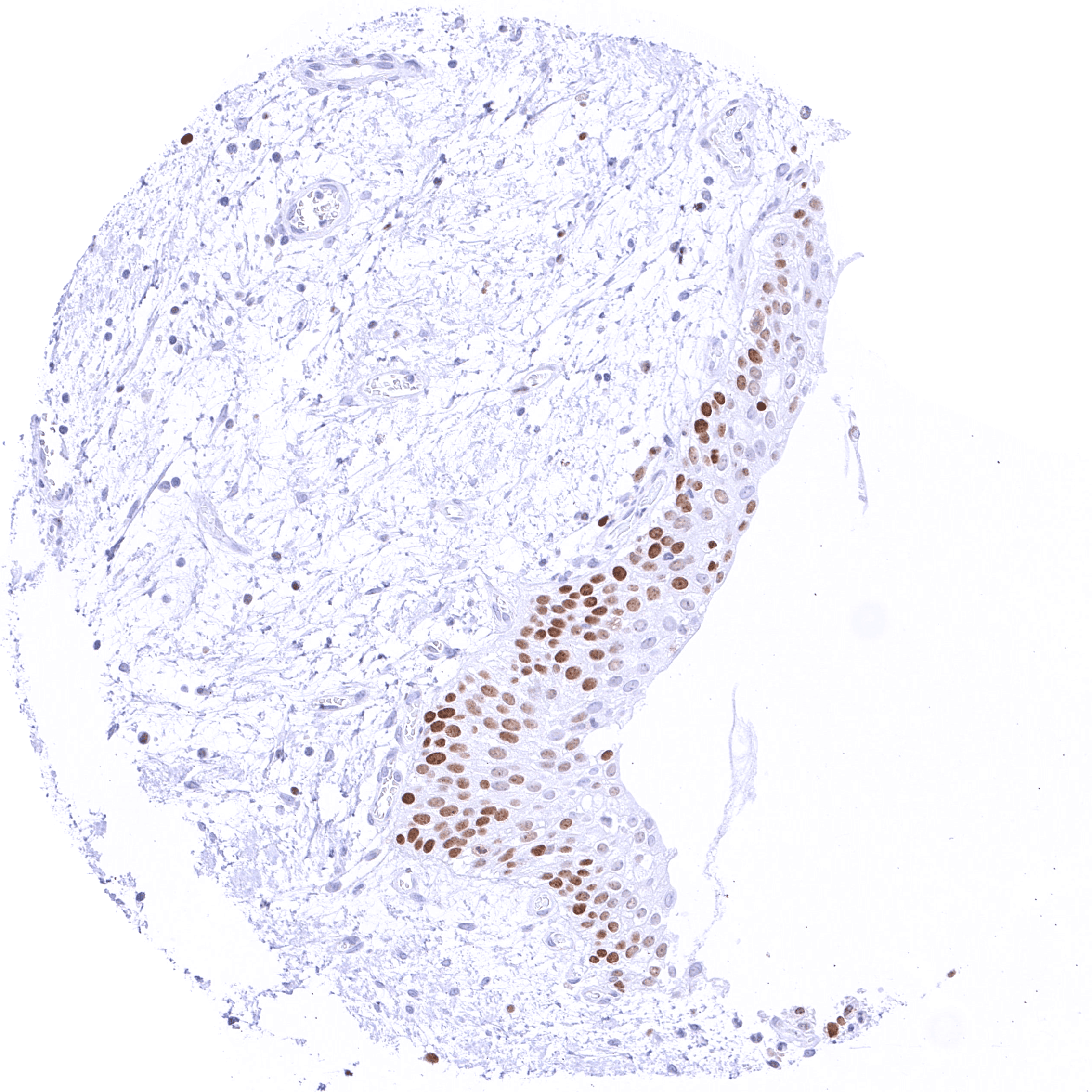
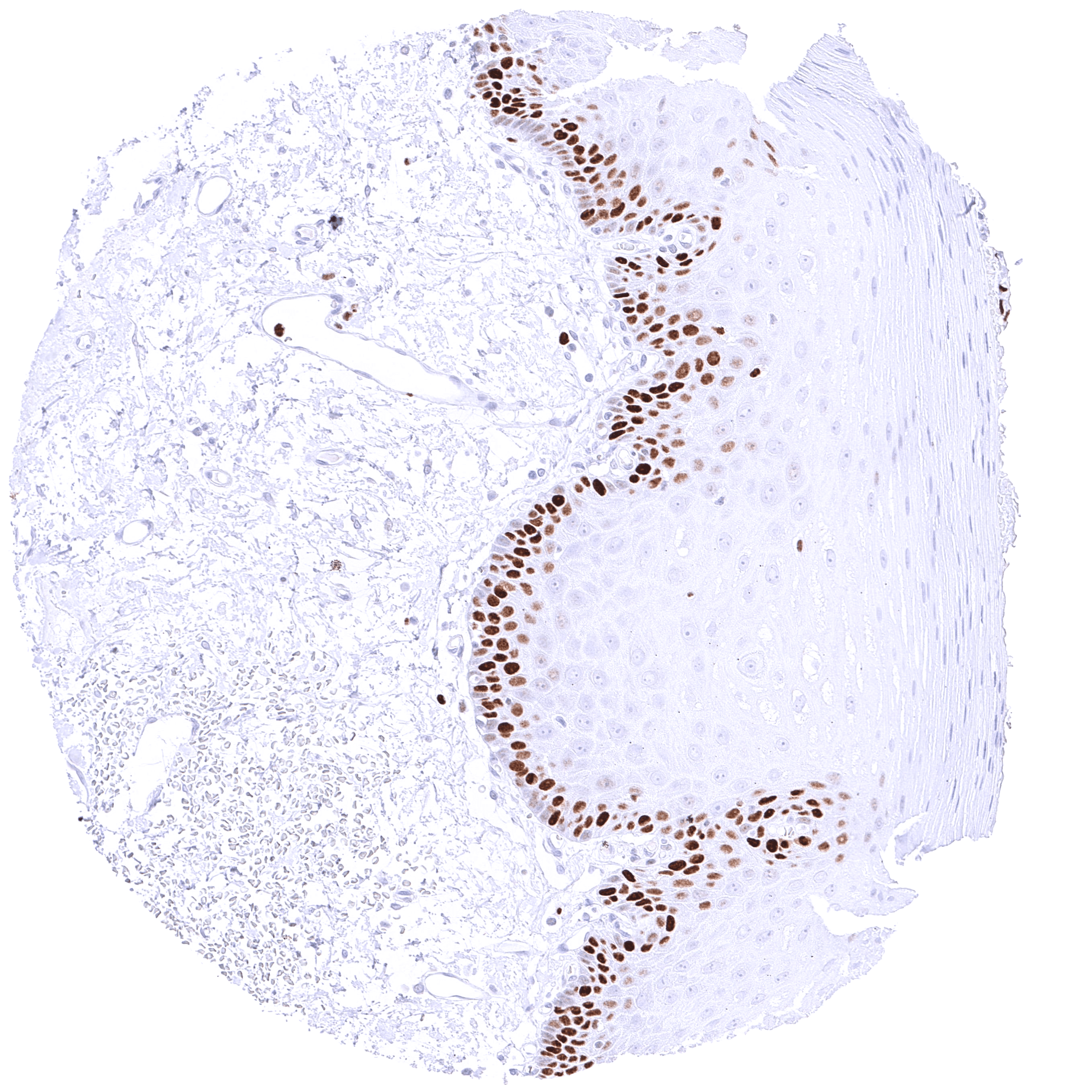
| Esophagus | Distinct MCM5 staining of suprabasal and (much less intense) basal cells of the squamous epithelium. | |
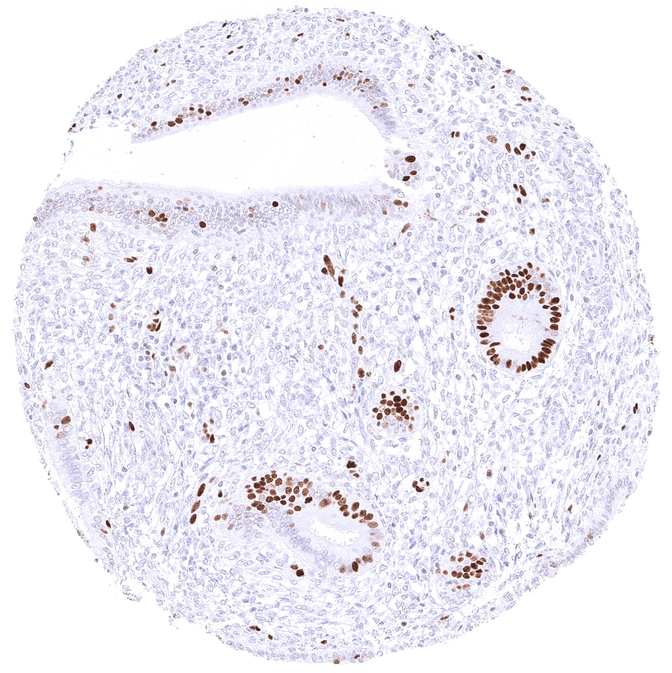
| Stomach | Strong nuclear MCM5 immunostaining of many mucous neck cells. | |
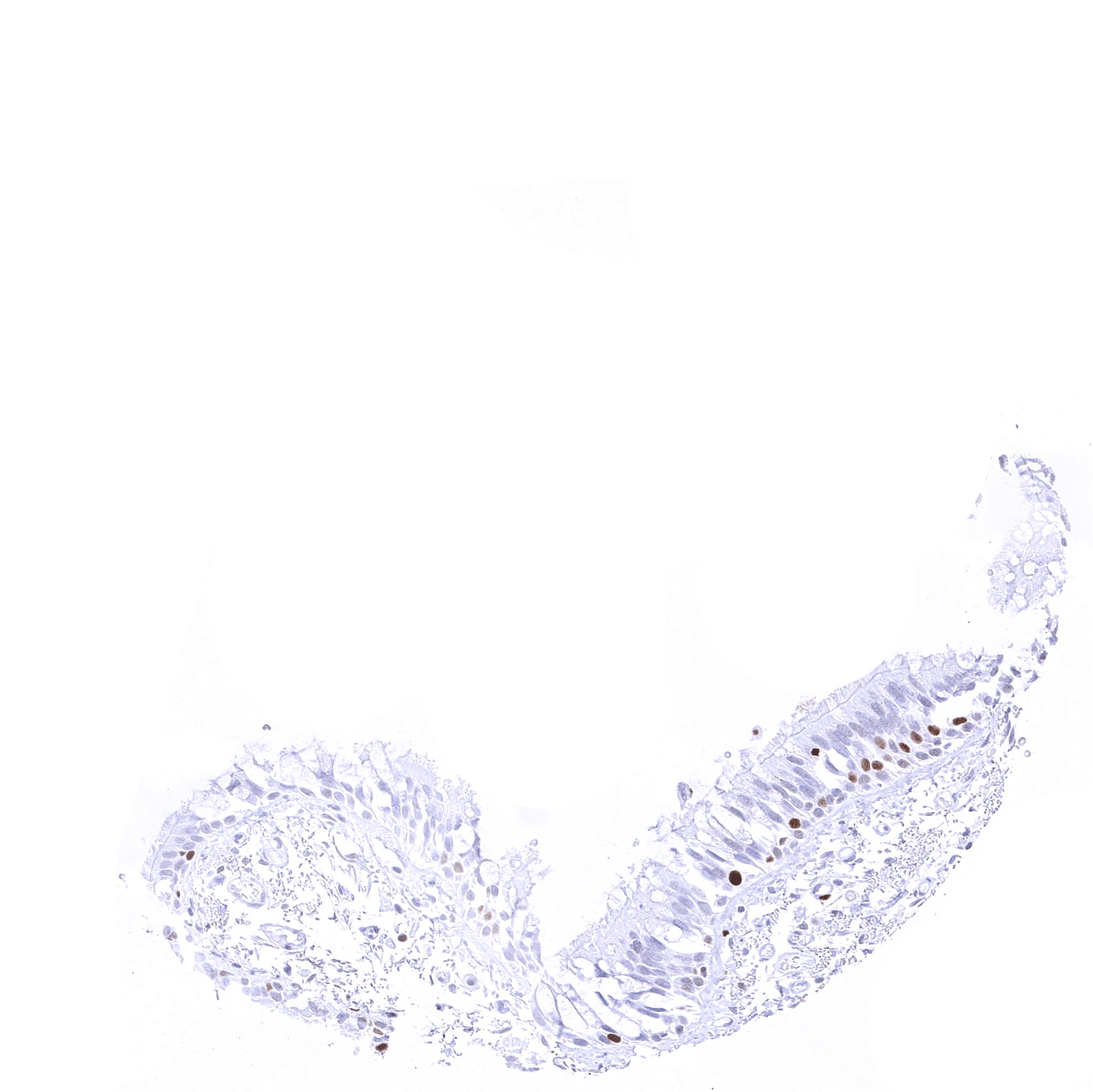
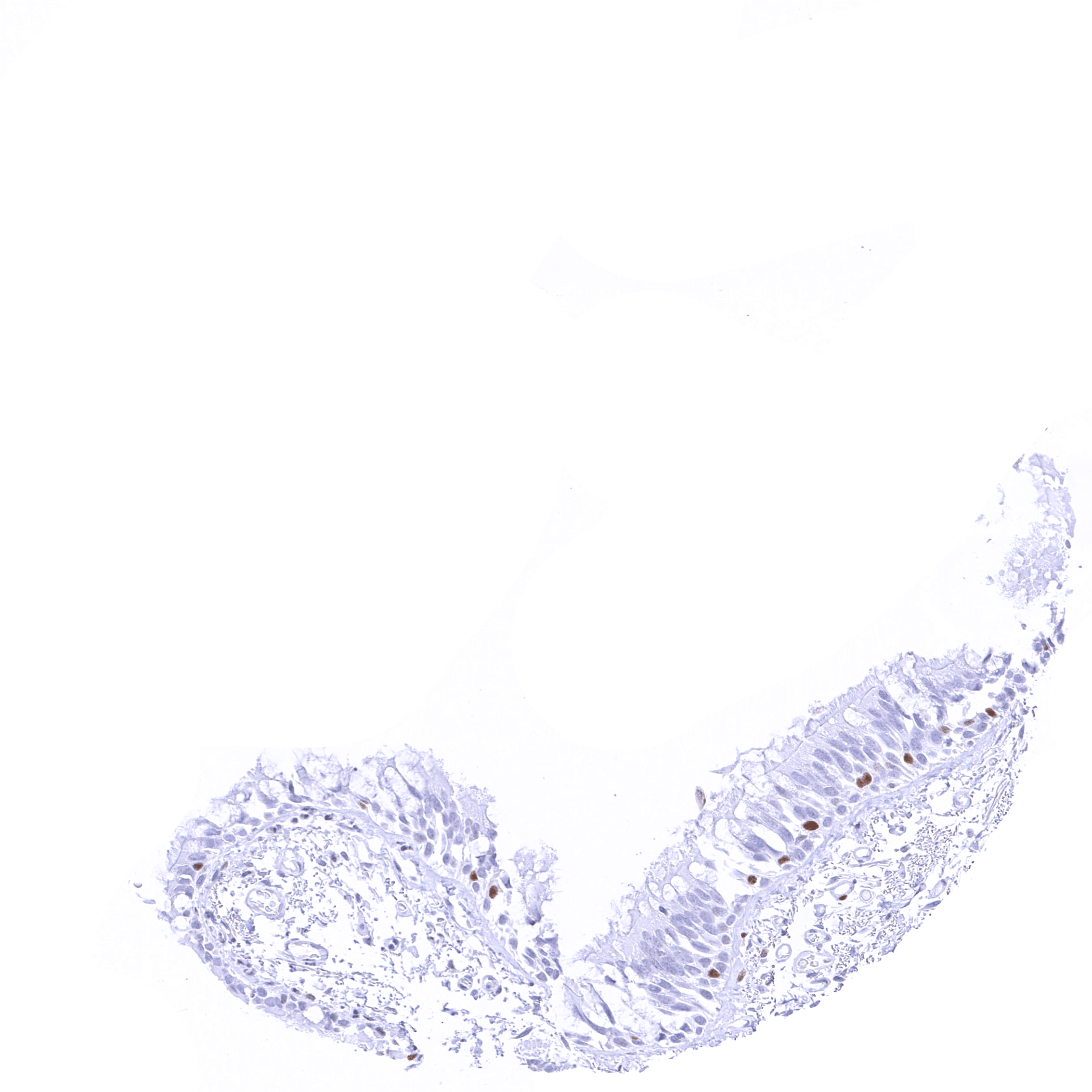
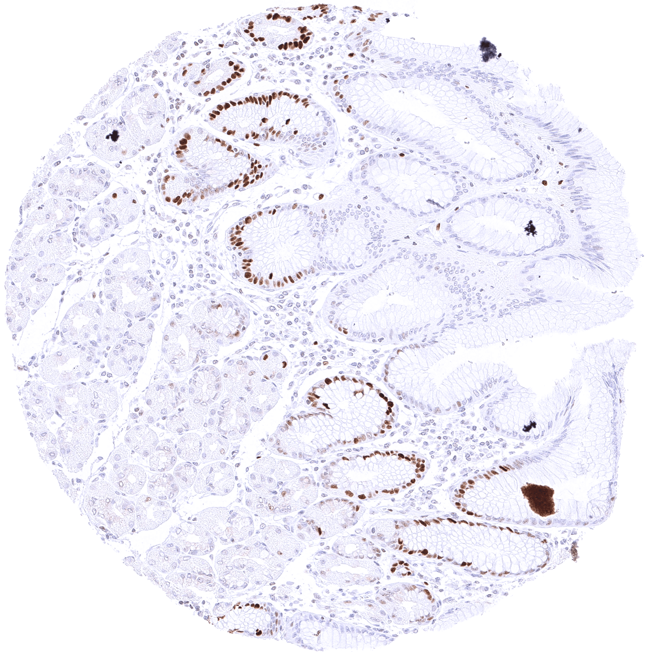
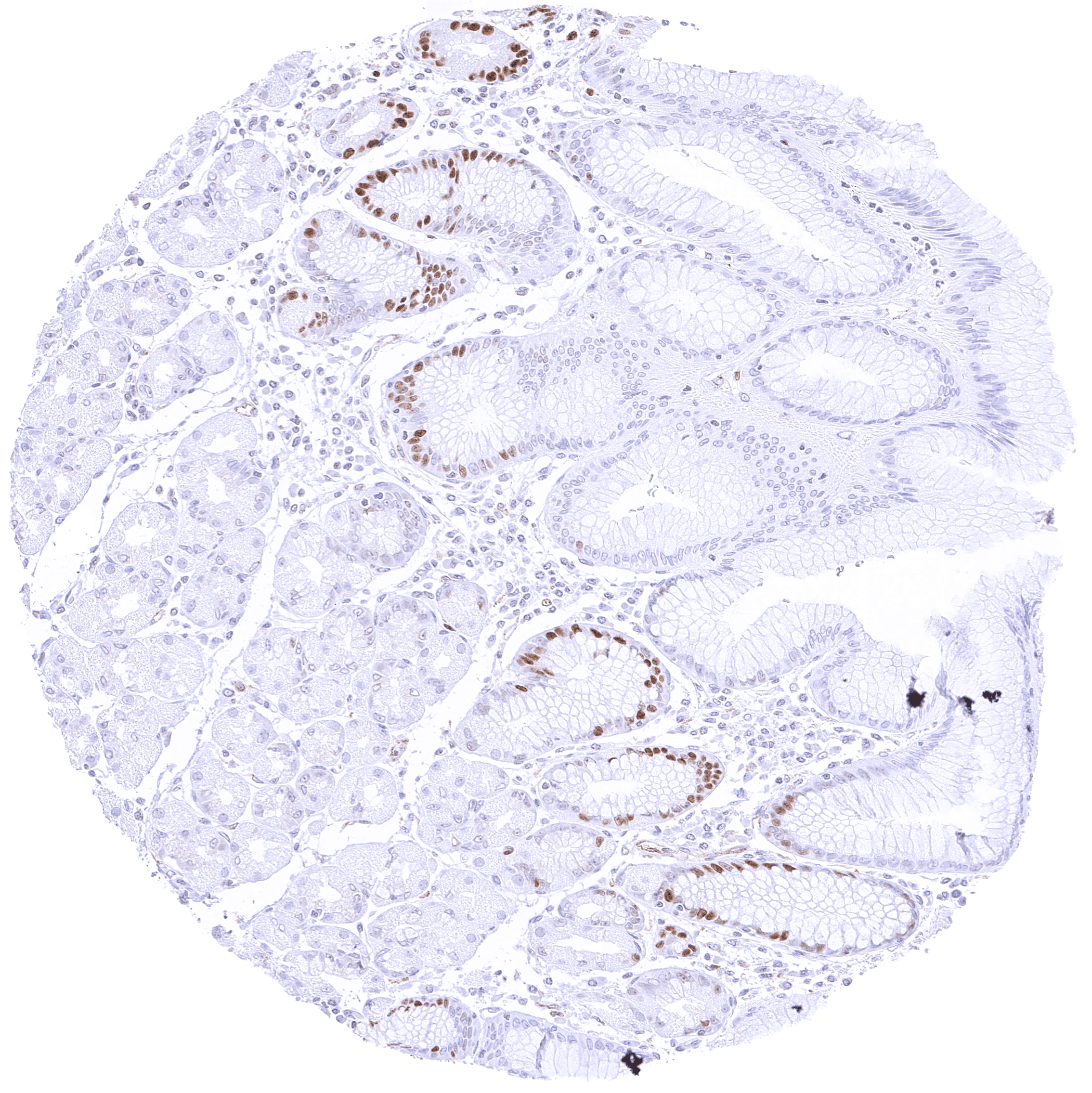
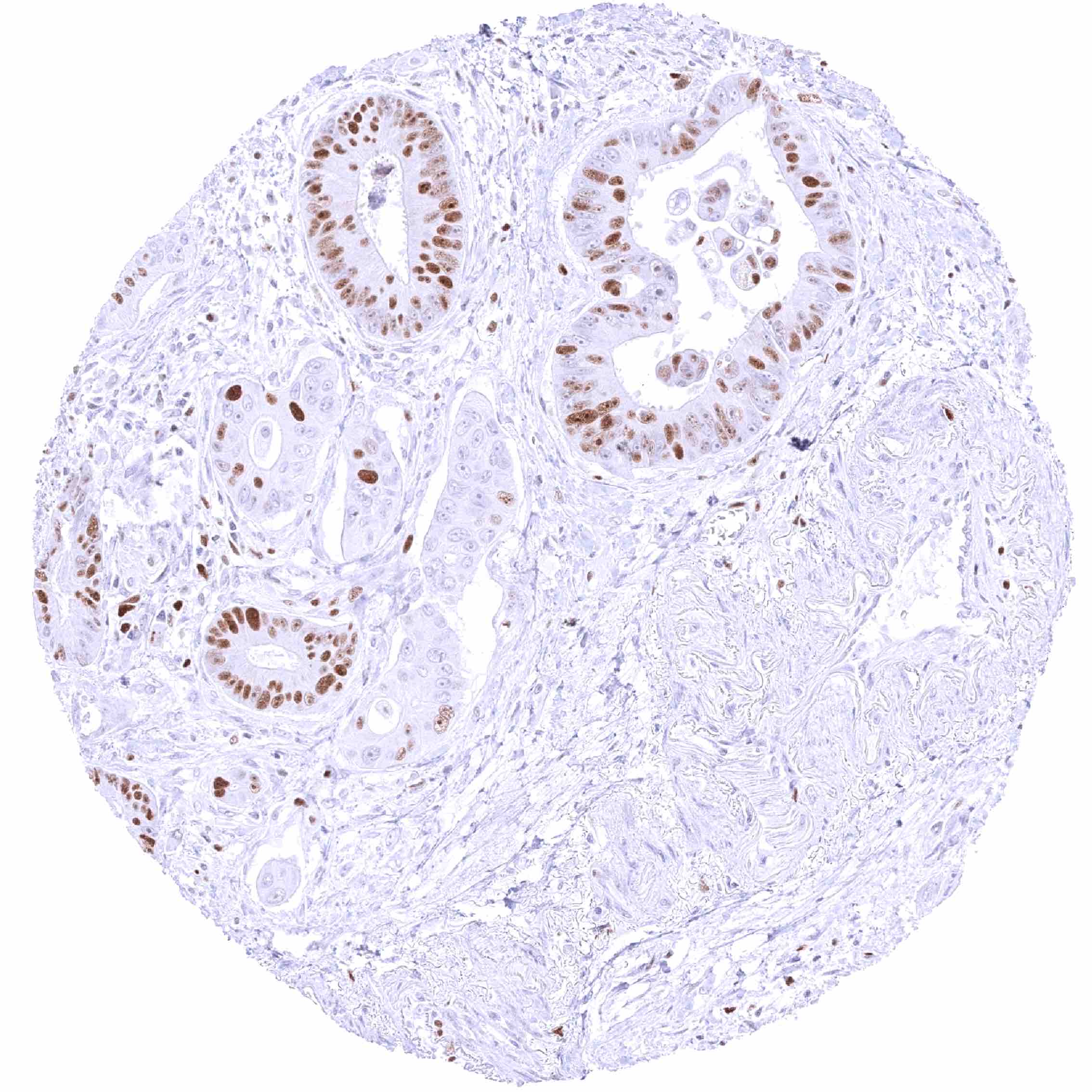
| Duodenum | MCM5 staining predominates in epithelial cells of the crypts. MCM5 staining is largely absent in Brunner glands. | |
| Small intestine | MCM5 staining predominates in epithelial cells of the crypts. | |
| Appendix | Nuclear MCM5 staining predominates in in epithelial cells of the crypts. Many lymphocytes are also positive. | |
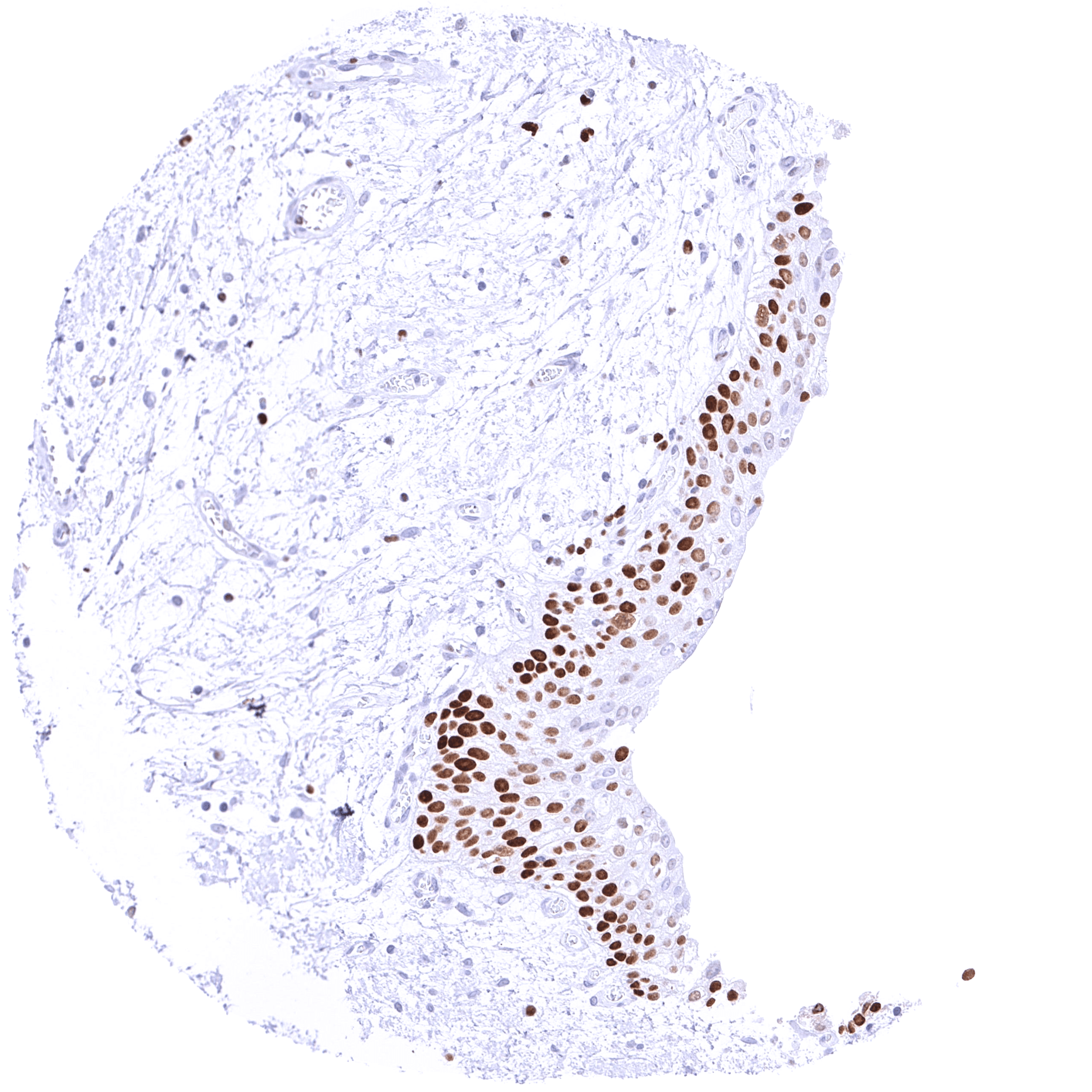
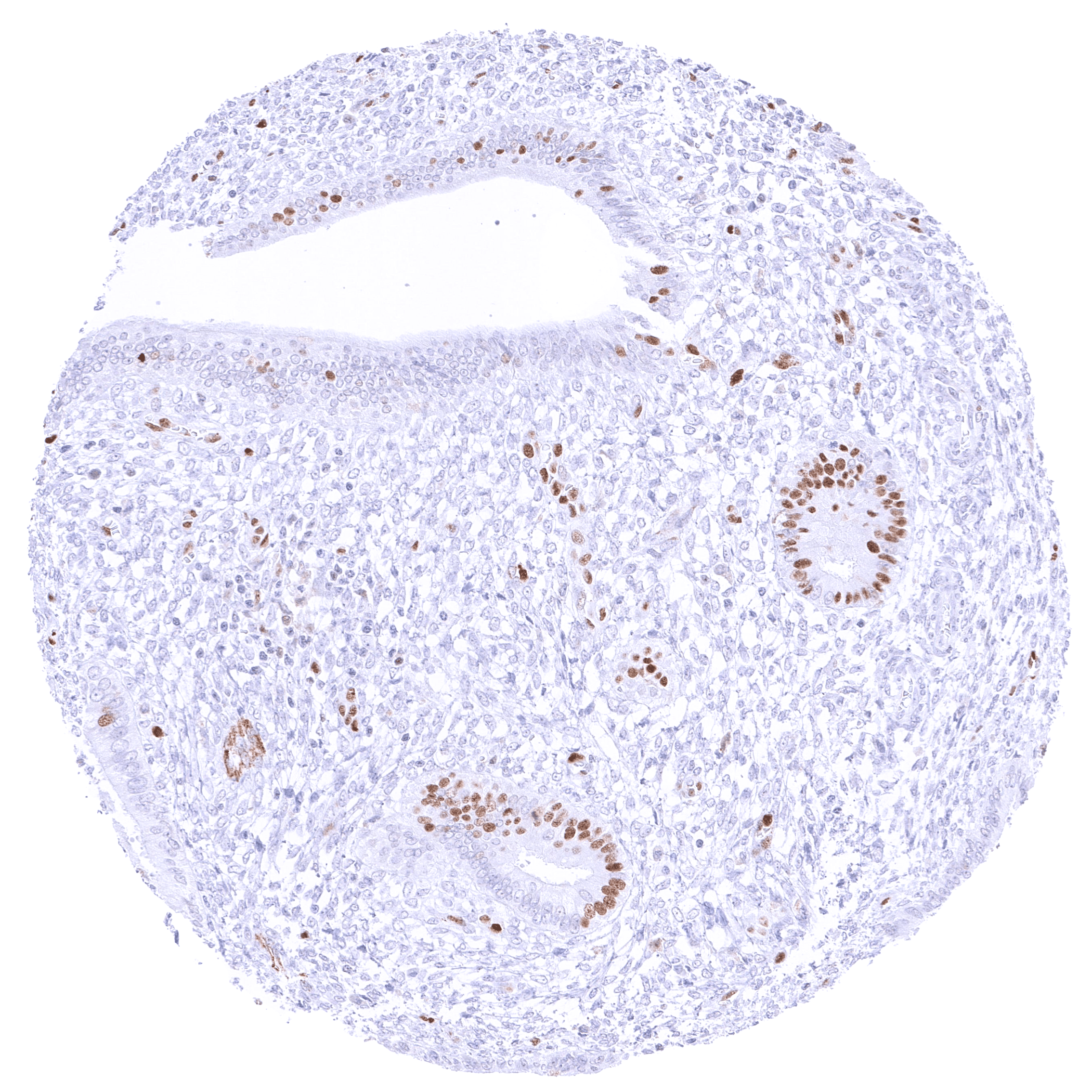
| Colon | Nuclear MCM5 staining predominates in in epithelial cells of the crypts. Some lymphocytes are also positive. | |
| Rectum | Nuclear MCM5 staining predominates in in epithelial cells of the crypts. Some lymphocytes are also positive. | |
| Liver | Faint nuclear MCM5 staining in a small fraction of hepatocytes. | |
| Gallbladder | A variable number of MCM5 positive cells can be seen in the gallbladder epithelium. | |
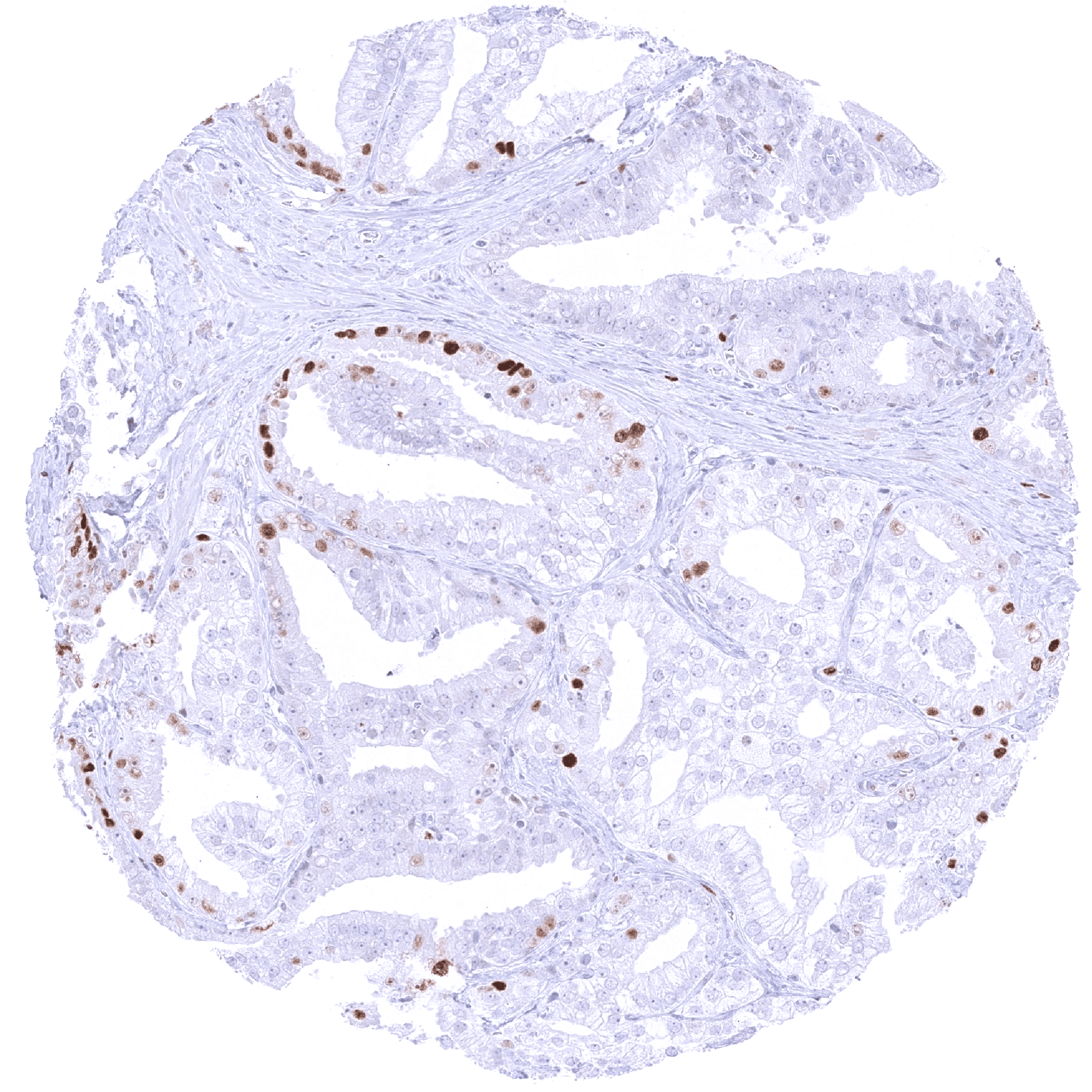
| Pancreas | Moderate to strong MCM5 staining in a rather small fraction of epithelial cells. | |
| Genitourinary | Kidney | Few epithelial cells are MCM5 positive. |
| Urothelium | A variable fraction of urothelial cells in all cell layers can show nuclear MCM5 staining. | |
| Male genital | Prostate | Nuclear MCM5 staining of a subset of epithelial cells. It is more common in basal than in acinar cells. |
| Seminal vesicles | Strong nuclear MCM5 staining of a rather small fraction of epithelial cells. | |
| Testis | Most spermatogonia and spermatocytes show strong MCM5 positivity. | |
| Epididymis | Distinct MCM5 staining in a fraction of epithelial cells (More common in the caput than in the cauda). | |
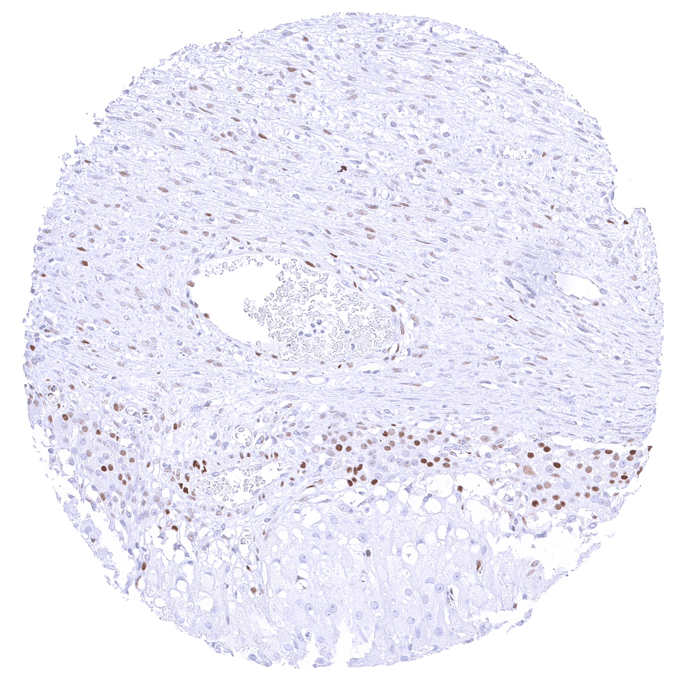
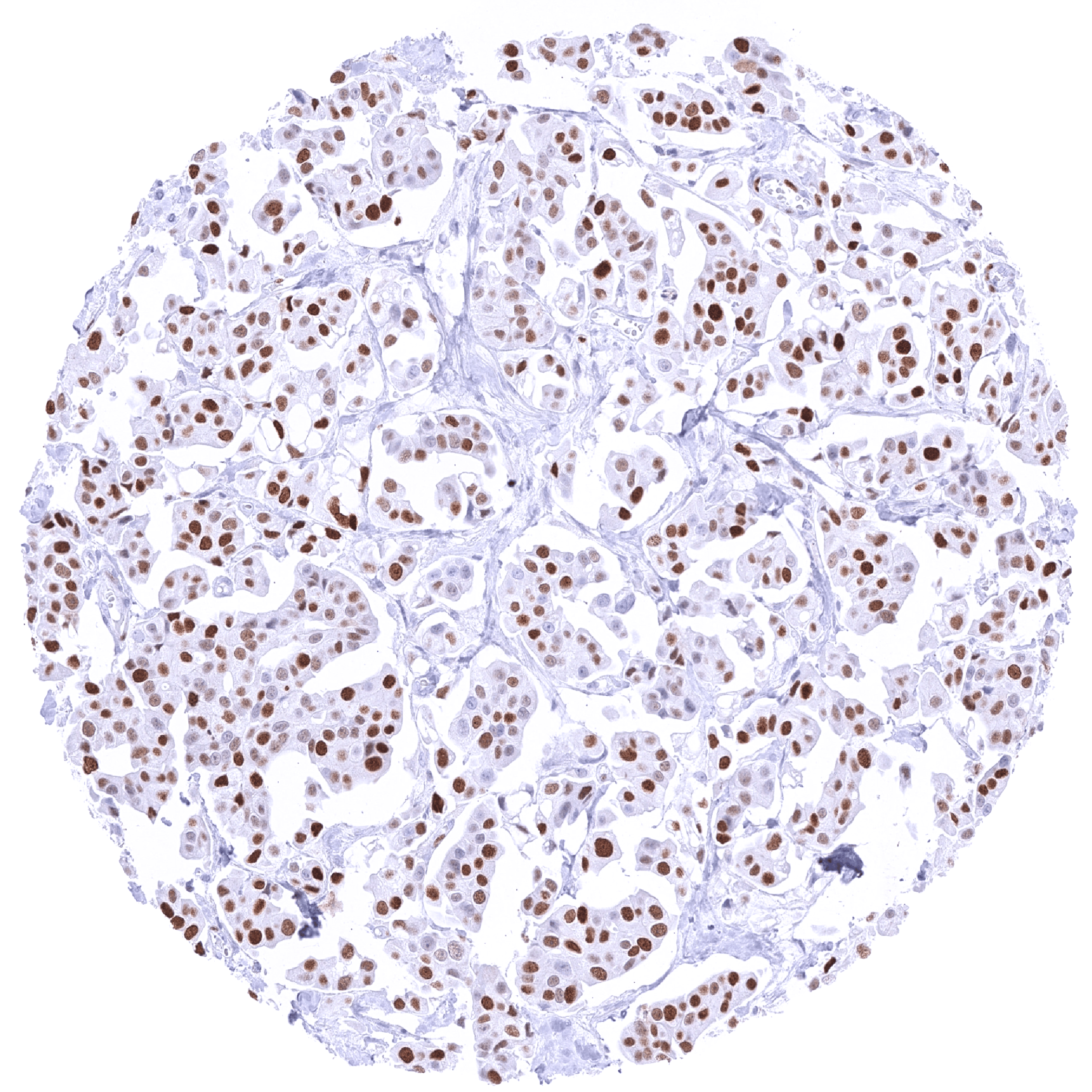
| Female genital | Breast | Strong MCM5 staining of most epithelial cells. |
| Uterus, myometrium | Faint MCM5 staining in a variable fraction of muscle cells. | |
| Uterus, ectocervix | Distinct MCM5 staining of suprabasal cells of the squamous epithelium. | |
| Uterus endocervix | Only few epithelial cells show (often weak) MCM5 staining. | |
| Uterus, endometrium | Many epithelial cells and a fraction of stromal cells are MCM5 positive. The fraction of MCM5 positive epithelial cells is lower in the secretion phase than in the proliferation phase. | |
| Fallopian Tube | Strong nuclear MCM5 staining of a significant subset of epithelial cells. | |
| Ovary | Strong MCM5 staining of virtually all granulosa and theca interna cells. Weak nuclear MCM5 staining of stroma cells.Weak or absent MCM5 staining of corpus luteum cells. | |
| Placenta early | Strong MCM5 staining of a large fraction of cytotrophoblast cells. | |
| Placenta mature | Strong MCM5 staining of a large fraction of cytotrophoblast cells. | |
| Amnion | ||
| Chorion | Moderate to strong MCM5 staining in a fraction of chorion cells. | |
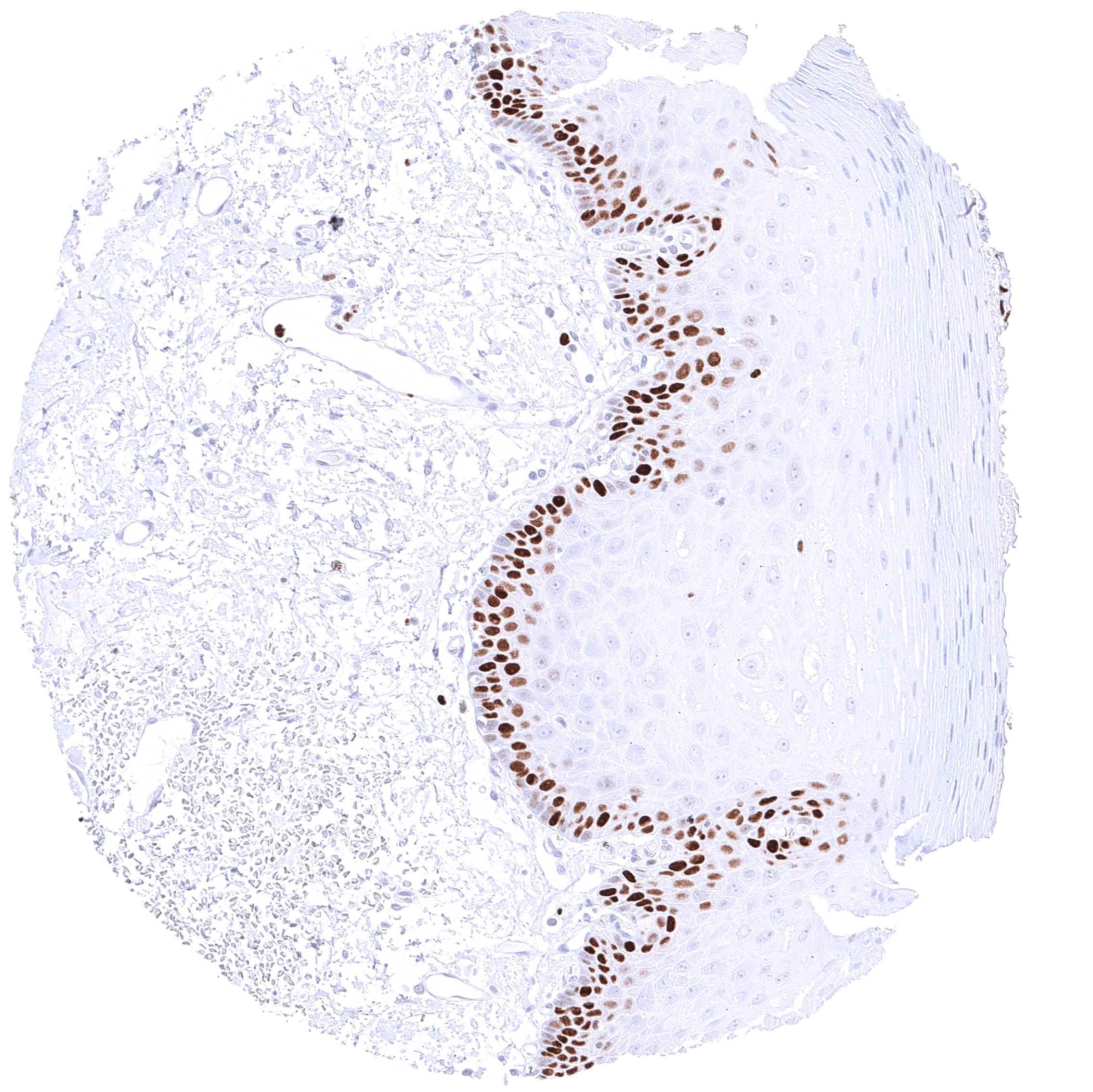
| Skin | Epidermis | Suprabasal and basal cells of the squamous epithelium show a distinct nuclear MCM5 positivity. |
| Sebaceous glands | Intense nuclear MCM5 positivity of peripheral germinative cells. | |
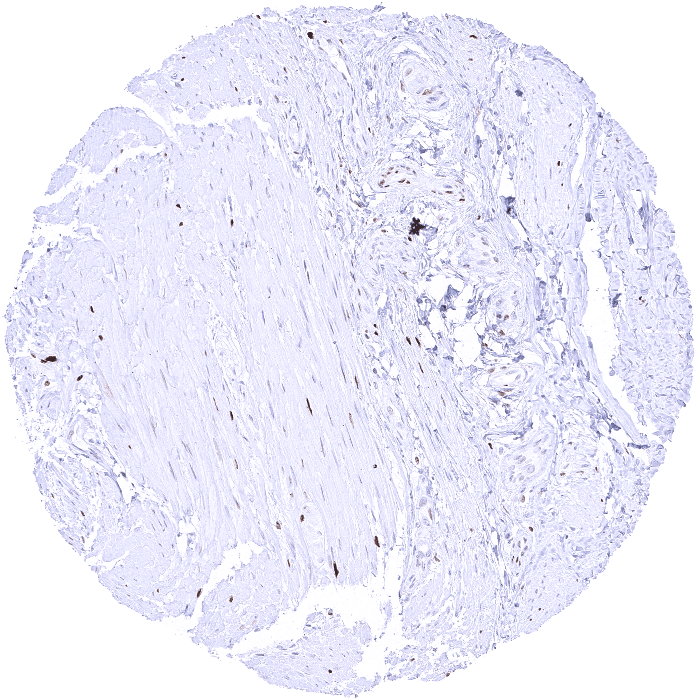
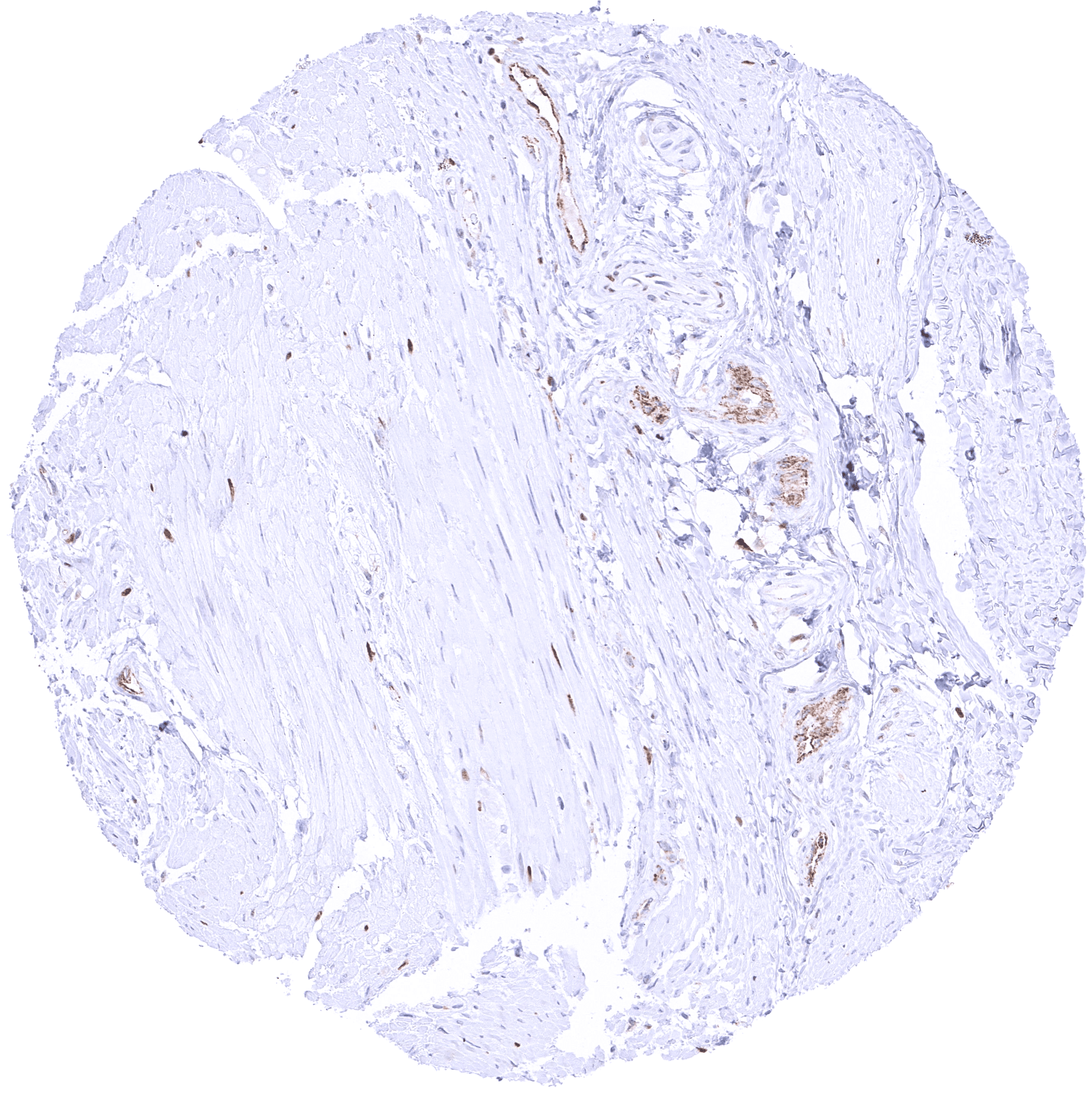
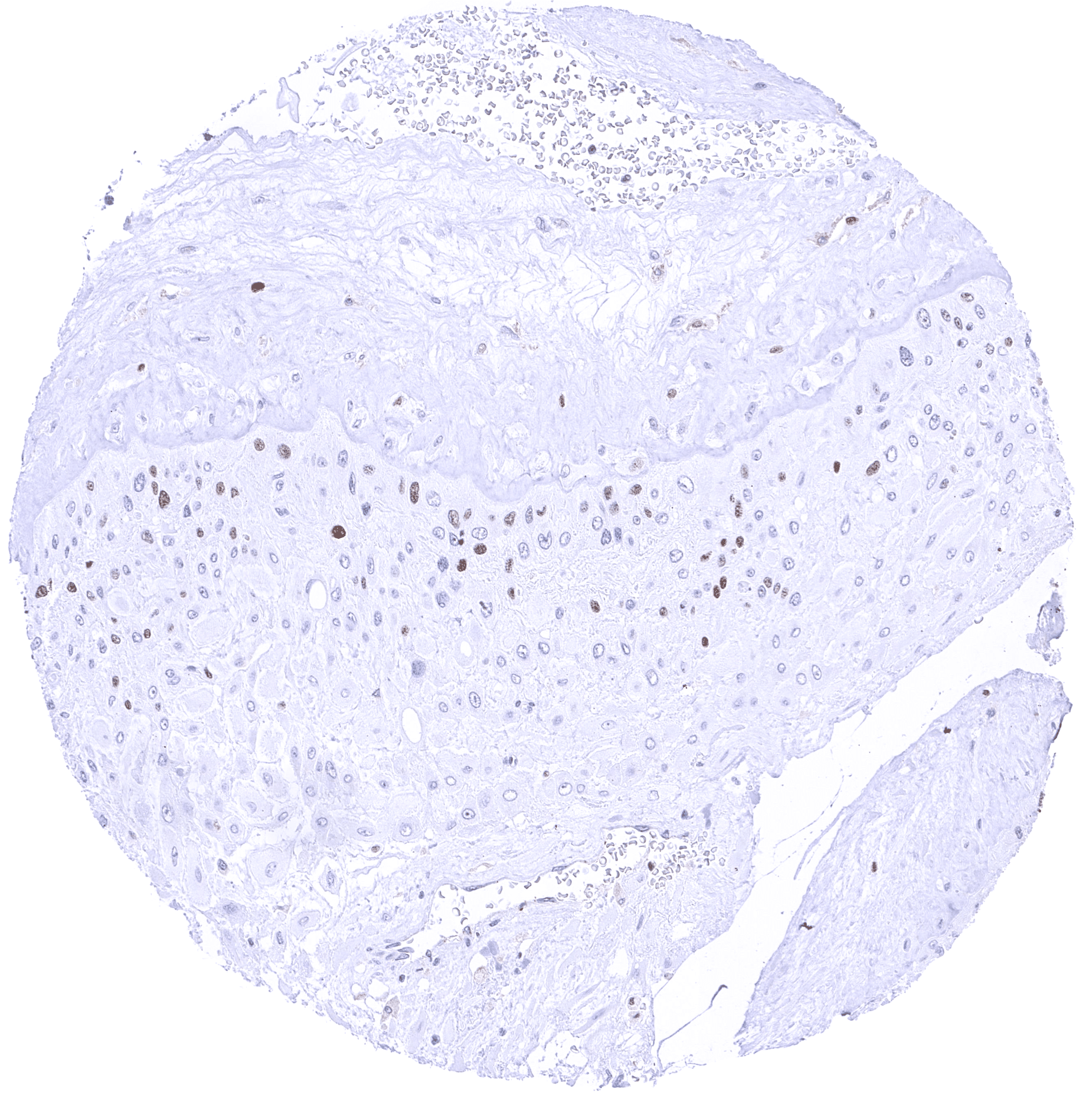
| Muscle/connective tissue | Heart muscle | Faint MCM5 staining of few muscle cells. |
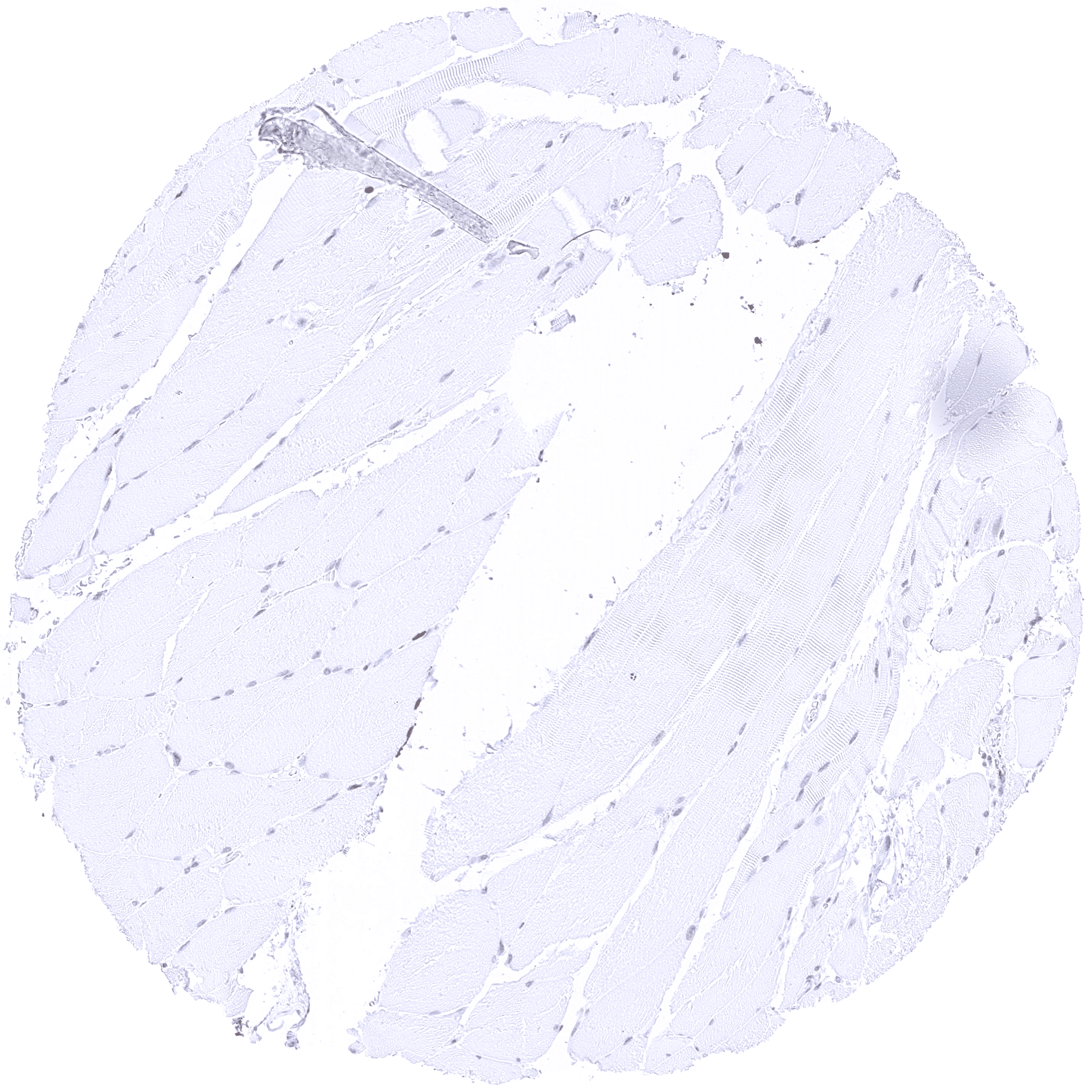
| Skeletal muscle | Distinct MCM5 staining in a large fraction of skeletal muscle cells. | |
| Smooth muscle | Distinct MCM5 staining in some muscle cells. | |
| Vessel walls | Negative. | |
| Fat | Negative. | |
| Stroma | Usually negative. | |
| Endothelium | Usually negative. | |
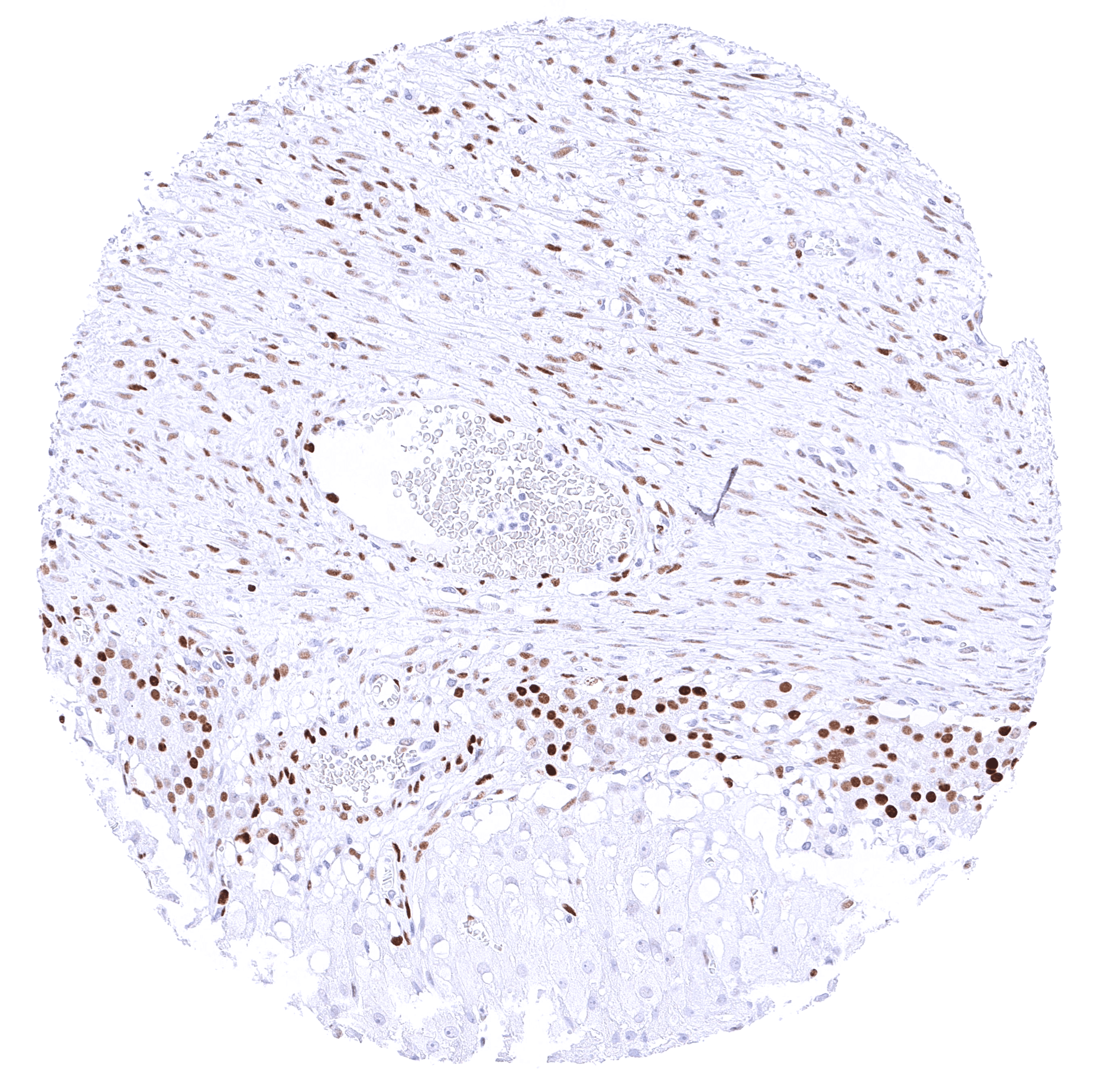
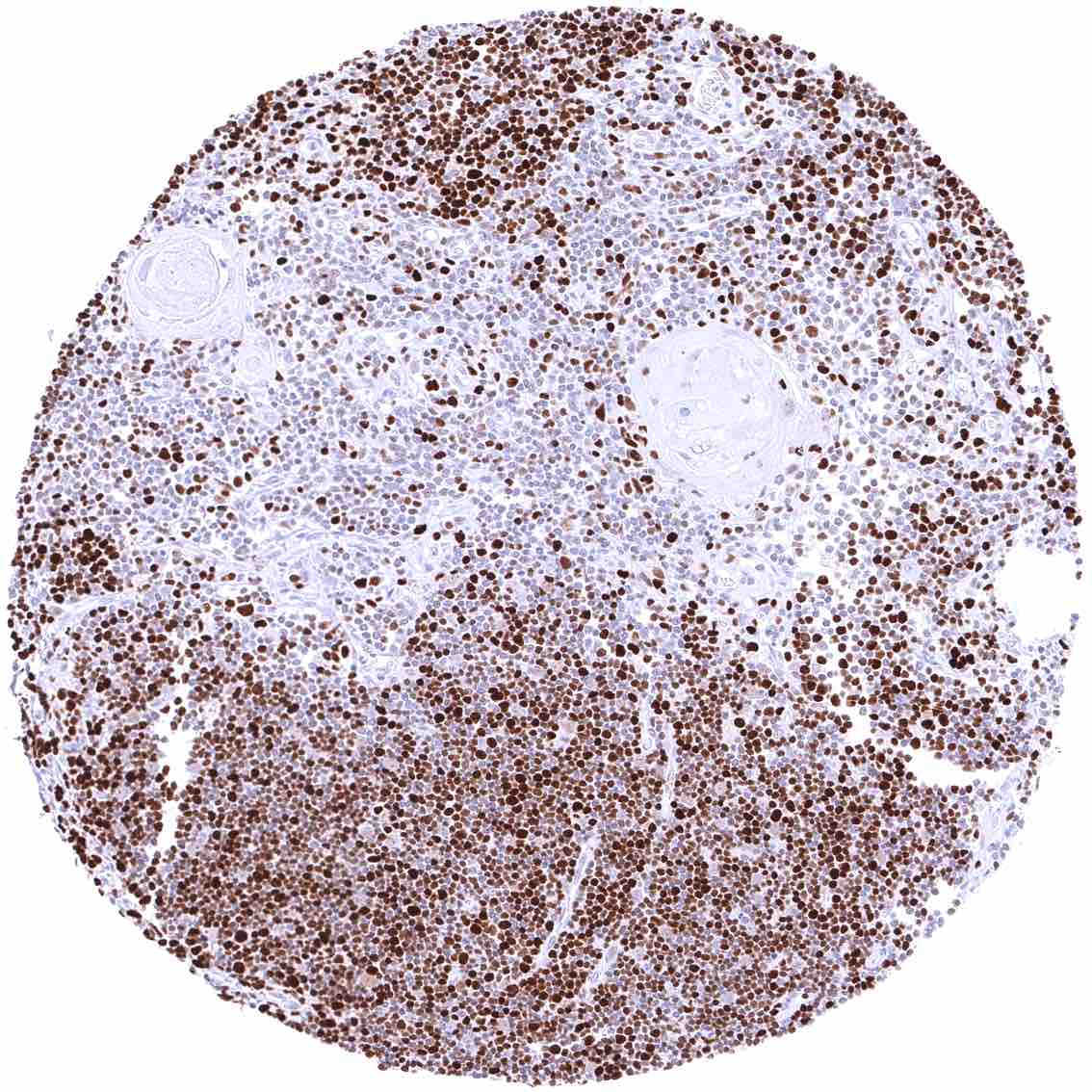
| Bone marrow/lymphoid tissue | Bone marrow | Strong MCM5 staining in most bone marrow cells. |
| Lymph node | Many lymphocytes are MCM5 positive. MCM5 staining is strongest and most common in cells of germinal centres. | |
| Spleen | A strong MCM5 positivity occurs in few scattered cells of all the red pulp. | |
| Thymus | Strong MCM5 positivity of most cells of the thymic cortex. Medullary cells show markedly less staining. | |
| Tonsil | Many lymphocytes are MCM5 positive. MCM5 staining is strongest and most common in cells of germinal centres. Suprabasal and basal cells of the squamous surface epithelium show a distinct nuclear MCM5 positivity. | |
| Remarks | Strong nuclear MCM5 staining of few cells along nerve fibers in many tissues. |
MCM5 is a ubiquitously expressed protein. The findings described above are thus consistent with the the Human Protein Atlas (Tissue expression MCM5).
Positive control = Colon: A strong nuclear MCM5 immunostaining should be seen in virtually all crypt base cells.
Negative control = Colon: MCM5 immunostaining should be less intense or absent in surface epithelial cells and absent in most stroma cells.
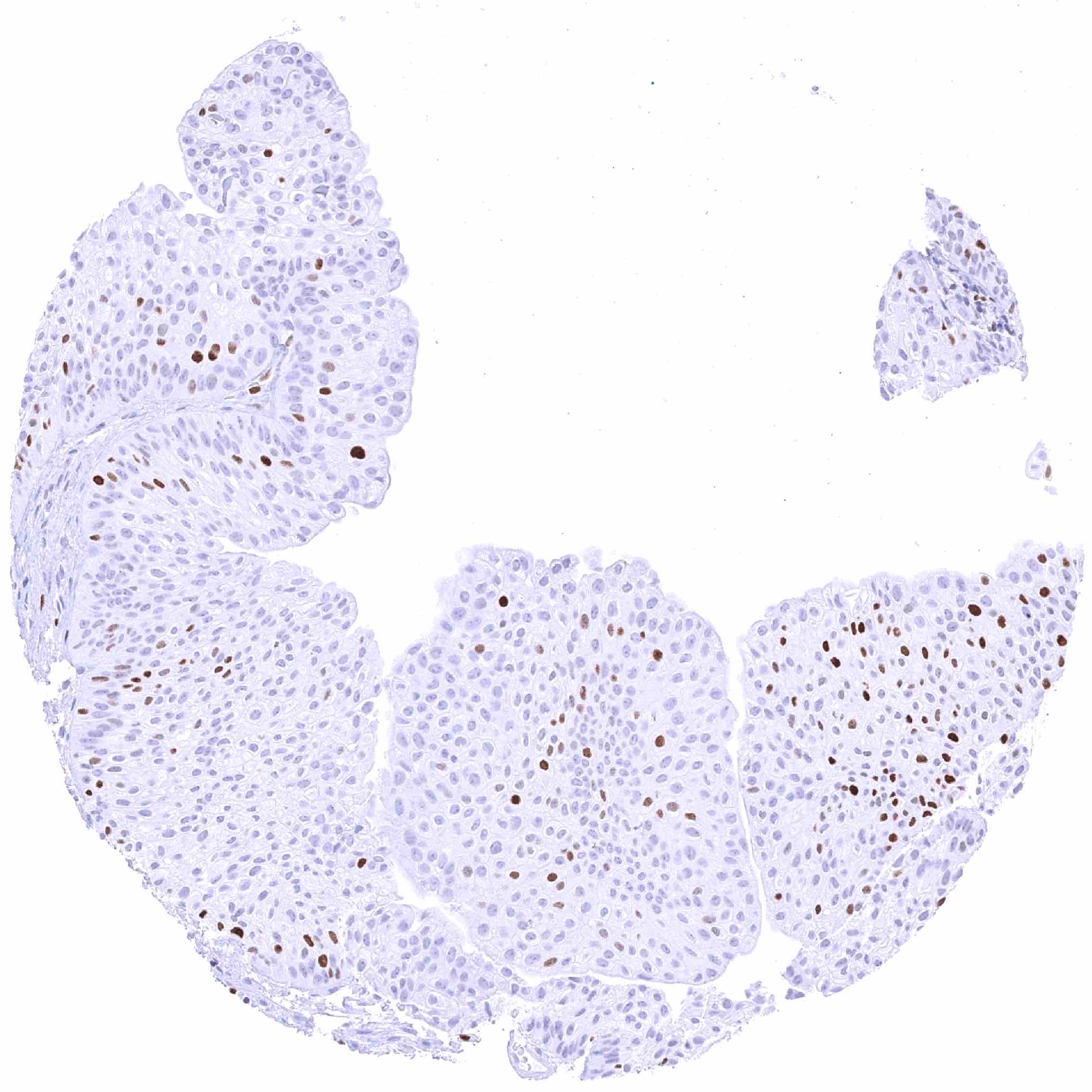
Staining Pattern in Relevant Tumor Types
A nuclear MCM5 immunostaining in a fraction of tumor cells is always seen in cancerous tissues.
The TCGA findings on MCM5 RNA expression in different tumor categories have been summarized in the Human Protein Atlas.
Compatibility of Antibodies
No data available at the moment
Protocol Recommendations
IHC users have different preferences on how the stains should look like. Some prefer high staining intensity of the target stain and even accept some background. Others favor absolute specificity and lighter target stains. Factors that invariably lead to more intense staining include higher concentration of the antibody and visualization tools, longer incubation time, higher temperature during incubation, higher temperature and longer duration of the heat induced epitope retrieval (slide pretreatment). The impact of the pH during slide pretreatment has variable effects and depends on the antibody and the target protein.
All images and data shown here and in our image galleries are obtained by the manual protocol described below. Other protocols resulting in equivalent staining are described as well.
Manual protocol
Freshly cut sections should be used (less than 10 days between cutting and staining). Heat-induced antigen retrieval for 5 minutes in an autoclave at 121°C in pH 7,8 Target Retrieval Solution buffer. Apply MSVA-505R at a dilution of 1:100 at 37°C for 60 minutes. Visualization of bound antibody by the EnVision Kit (Dako, Agilent) according to the manufacturer’s directions.
Potential Research Applications
- The prognostic role of the percentage of MCM5 positive cells is yet unknown.
- It is unclear whether MCM5 quantification is equally or better suited than the established Ki67-Li for prognosis assessment in tumors with rather low proliferation rate.
Evidence for Antibody Specificity in IHC
In principle, there are two ways how the specificity of antibodies can be documented for immunohistochemistry on formalin fixed tissues. These are: 1. Comparison with a second independent method for target expression measurement across a large number of different tissue types (orthogonal strategy), and 2. Comparison with one or several independent antibodies for the same target and showing that all positive staining results are also seen with other antibodies for the same target (independent antibody strategy).
Orthogonal validation: For the antibody MSVA-505R, specificity of staining is in line with data from three independent RNA screening studies, including the Human Protein Atlas (HPA) RNA-seq tissue dataset, the FANTOM5 project, and the Genotype-Tissue Expression (GTEx) project, which are all compiled in the Human Protein Atlas (Tissue expression MCM5). In agreement with MSVA-505R immunostaining data, MCM5 RNA expression is highest in lymphatic and hematopoetic tissues but occurs in virtually all tissues. However, it must be understood that orthogonal validation is not optimal for assessing ubiquitously expressed proteins
Comparison of antibodies: A specific staining of the MCM5 antibody MSVA-505R is supported by an identical staining pattern – often restricted to proliferating cell types – seen by an independent commercially available second MCM5 antibody (termed “validation antibody”). Staining with the validation antibody also confirmed MCM5 positivity in all cell types with positivity for MSVA-505R. This applies for tissues and cell types known for high proliferative activity such as neck cells and crypt cells in the gastrointestinal tract and germinal centres of lymph nodes but also for cell types which are not notorious for cell proliferation such as skeletal muscle, myometrium, ovarian stroma, or pneumocytes of the lung.