Login / Register
MS Validated Antibodies - Logo
  • About us
  • Products
    • Primary antibodies
    • Gallery
  • Distributors
  • Validation
    • Tissue Microarrays
  • Publications
    • Large Scale Studies
    • Further Publications
    • Events
    • Quiz
  • Contact
  • About us
  • Products
    • Primary antibodies
    • Gallery
  • Distributors
  • Validation
    • Tissue Microarrays
  • Publications
    • Large Scale Studies
    • Further Publications
    • Events
    • Quiz
  • Contact
×

Normal Tissue Gallery MCM5

Path Created with Sketch.
Go to MCM5 Cancer Tissue GalleryGo back to Product Page
Adrenal gland – A variable MCM5 staining occurs in a small fraction of adrenocortical cells. Spindle shaped nuclei are also found positive.
Aorta, media
Aorta, media
Appendix, mucosa – Nuclear MCM5 staining predominates in in epithelial cells of the crypts. Many lymphocytes are also positive.
Appendix, muscular wall – Distinct MCM5 staining in some muscle cells.
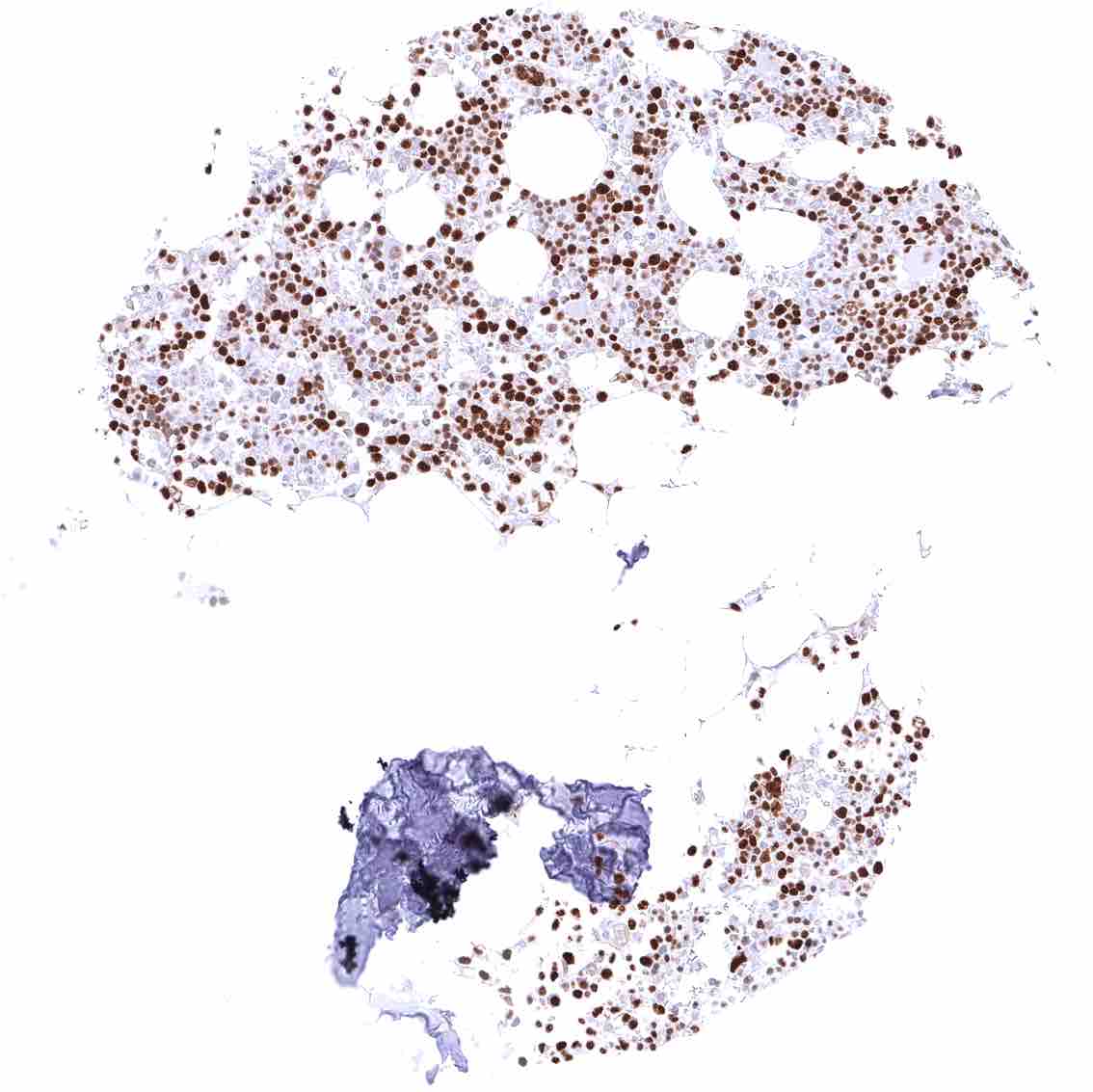
Bone marrow – Strong MCM5 staining in most bone marrow cells.
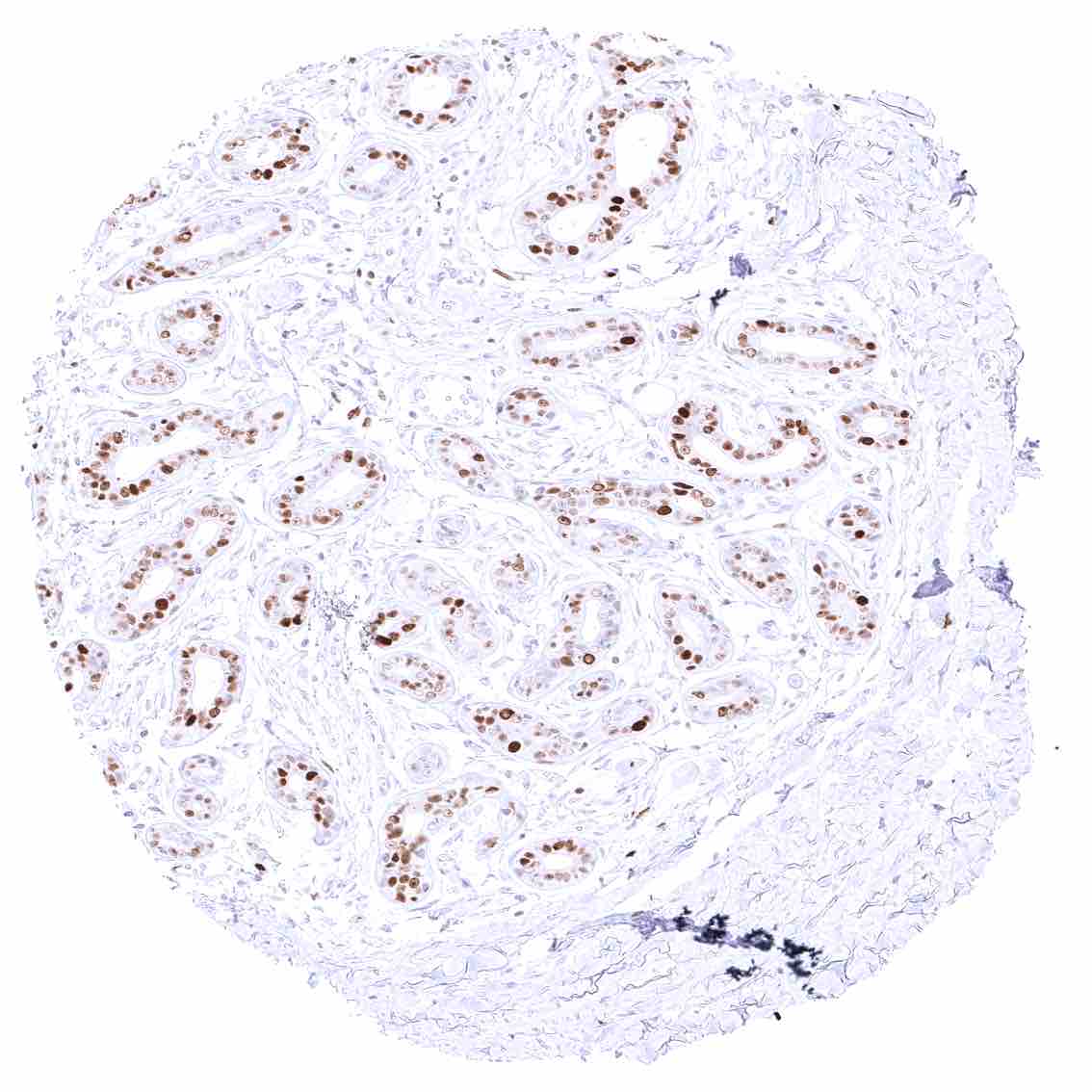
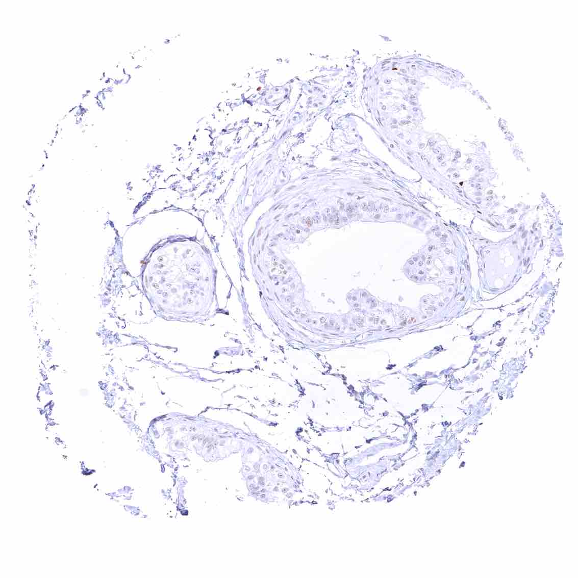
Breast – Strong MCM5 staining of most epithelial cells.
Bronchus, mucosa – Distinct nuclear MCM5 staining in a fraction of basal-suprabasal respiratory epithelial cells.
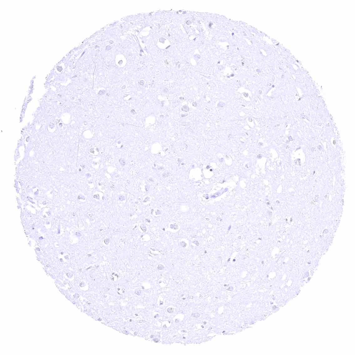
Cerebellum (molecular layer, Purkinje cell layer, granule cell layer)
Cerebellum (white matter)
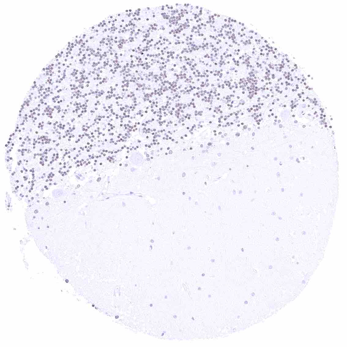
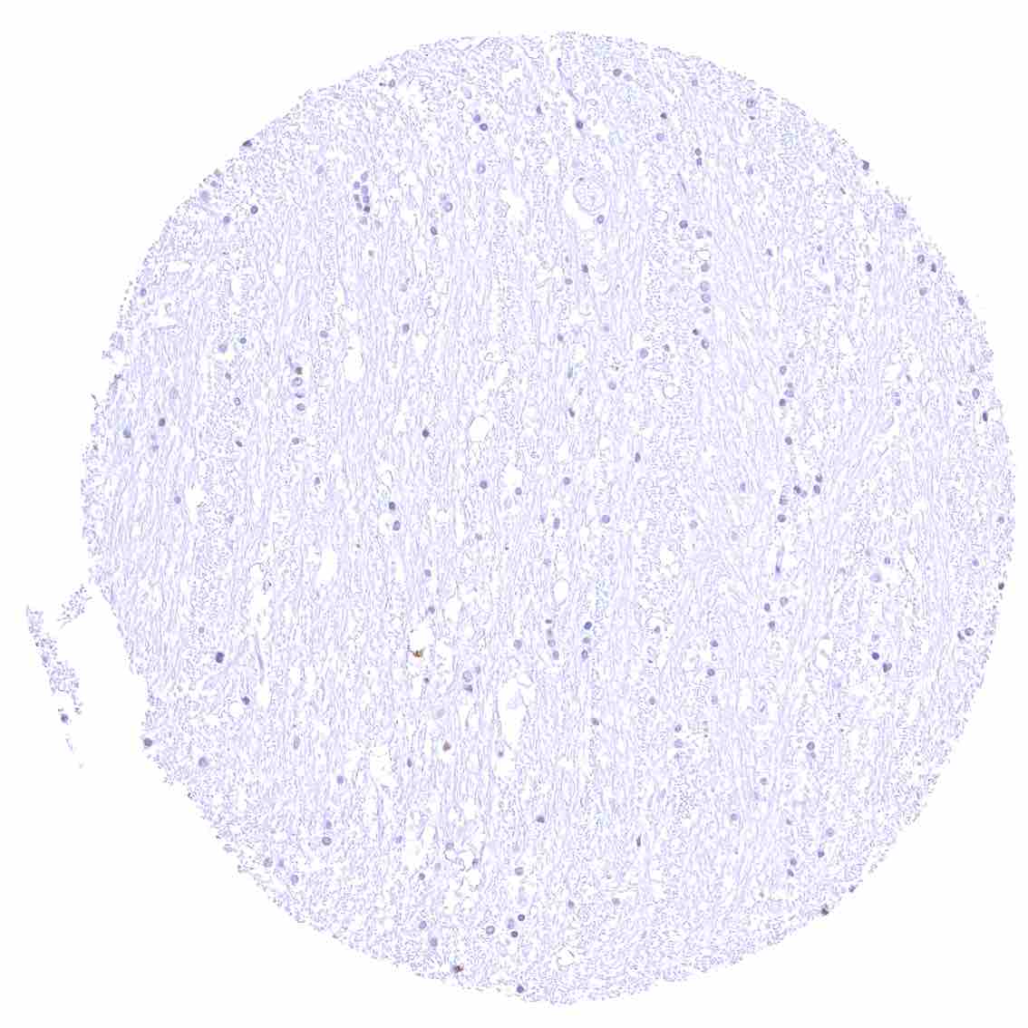
Cerebrum (grey matter)
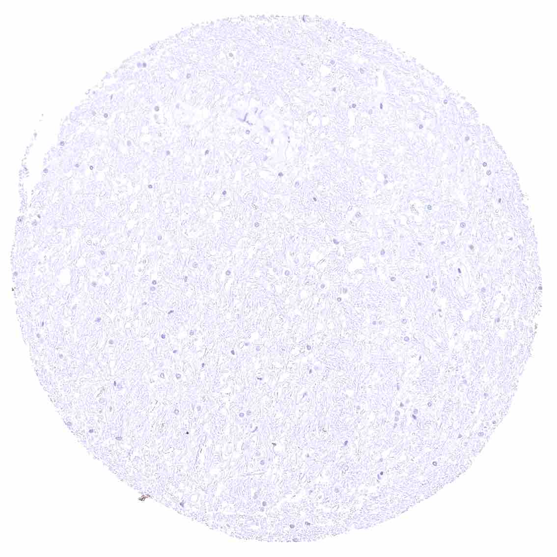
Cerebrum (white matter)
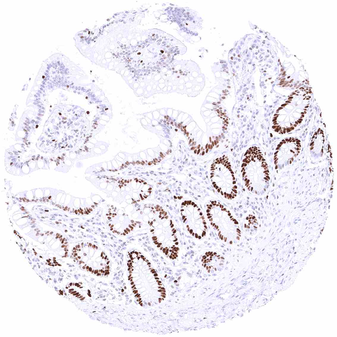
Colon descendens, mucosa – Nuclear MCM5 staining predominates in in epithelial cells of the crypts. Some lymphocytes are also positive.
Colon descendens, muscular wall – Strong nuclear MCM5 staining of few cells along nerve fibers.
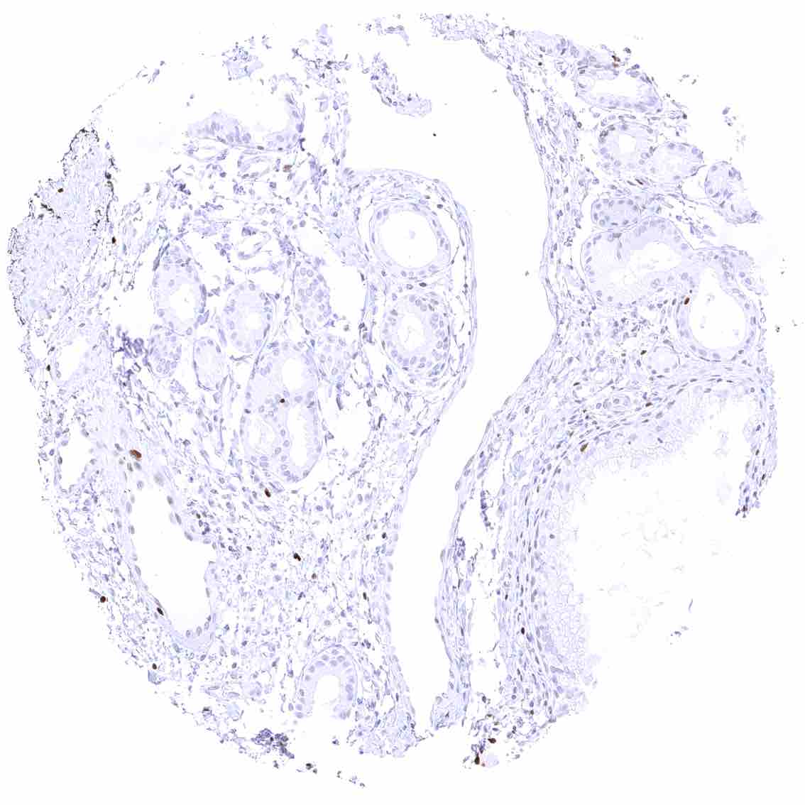
Duodenum, Brunner gland.jpeg
Duodenum, Brunner gland
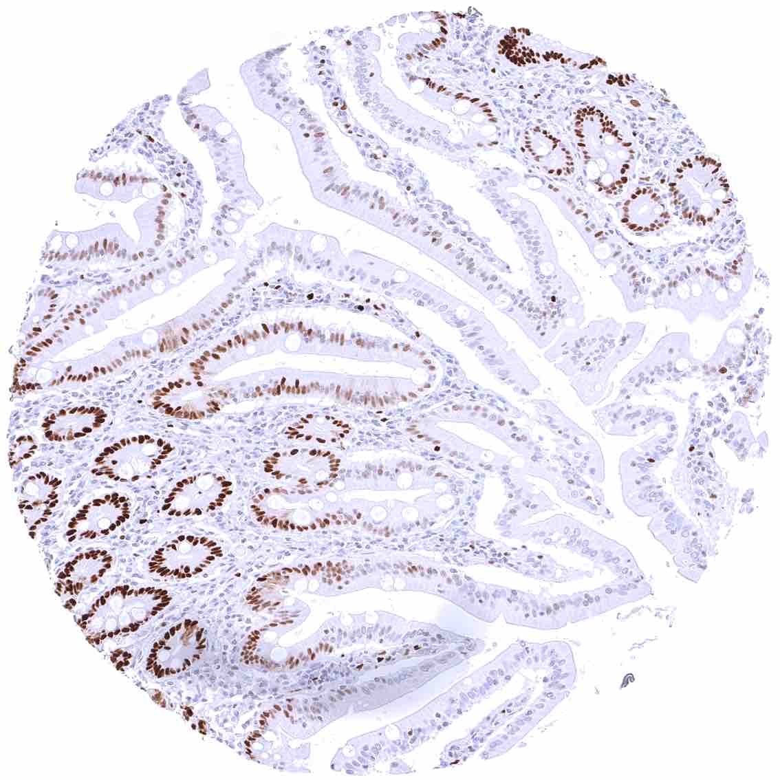
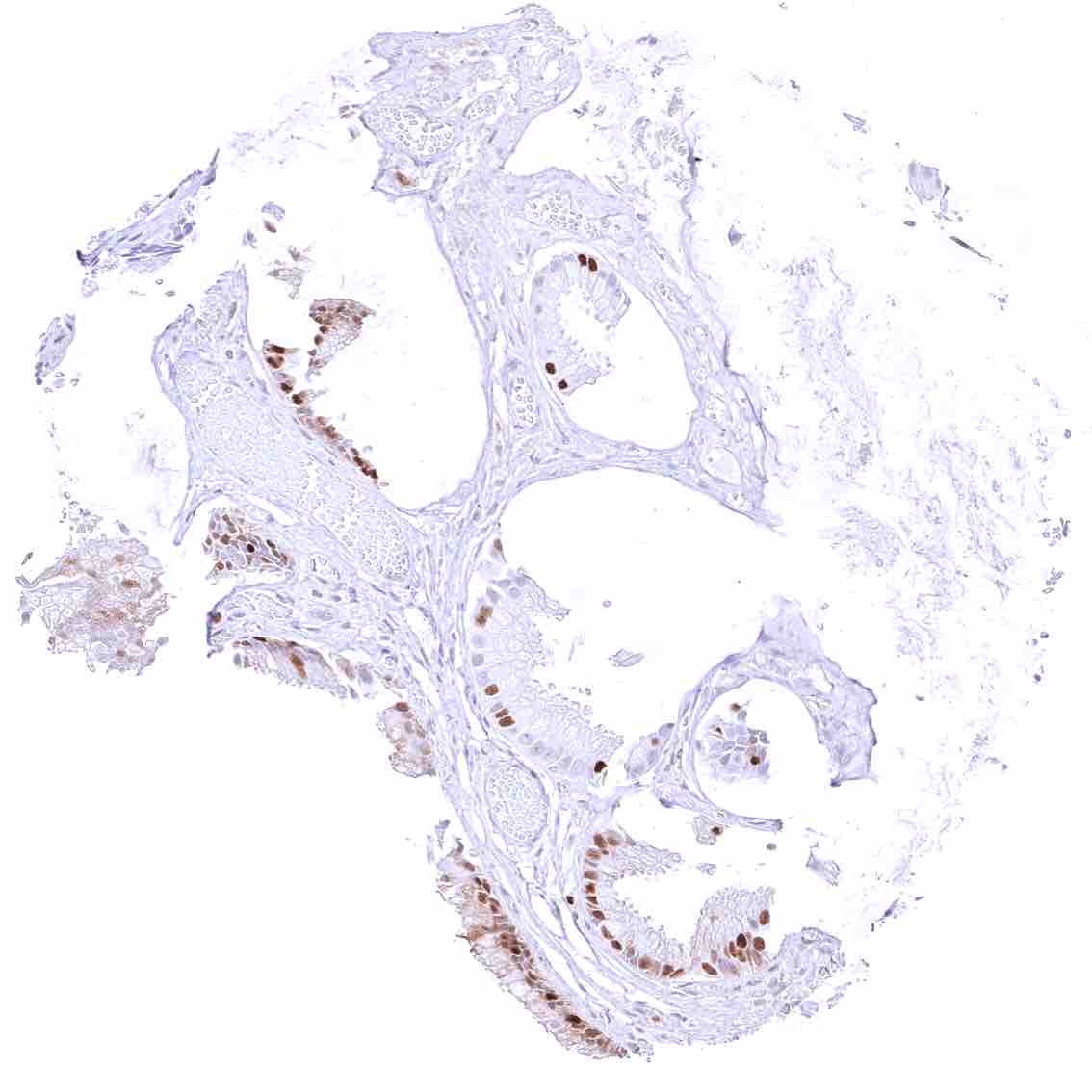
Duodenum, mucosa – MCM5 staining predominates in epithelial cells of the crypts.
Epididymis (Caput) – Distinct MCM5 staining in a fraction of epithelial cells.
Epididymis (Cauda) – Weak MCM5 staining of a fraction of epithelial cells.
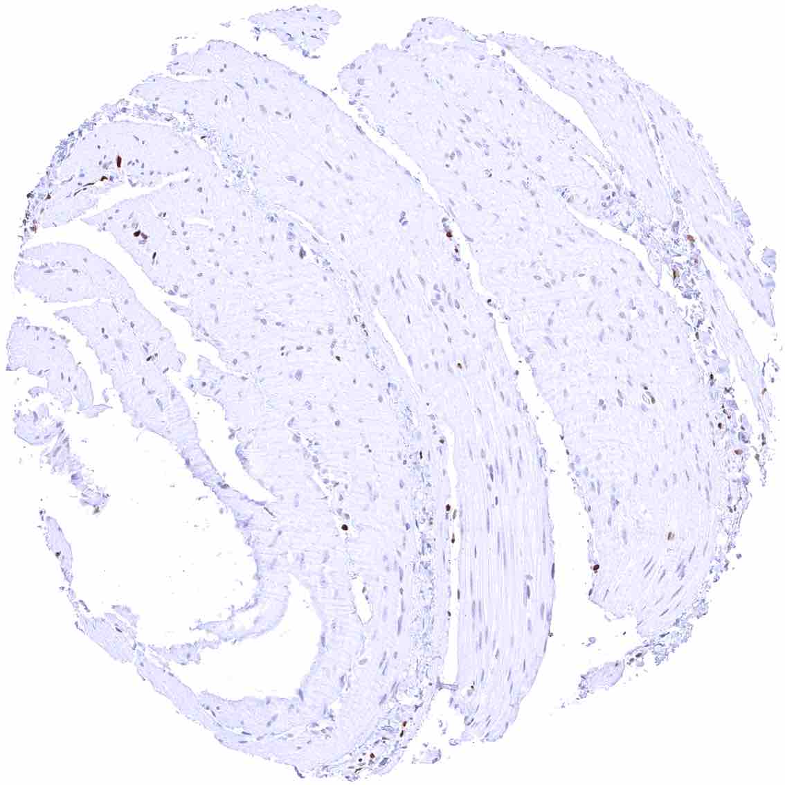
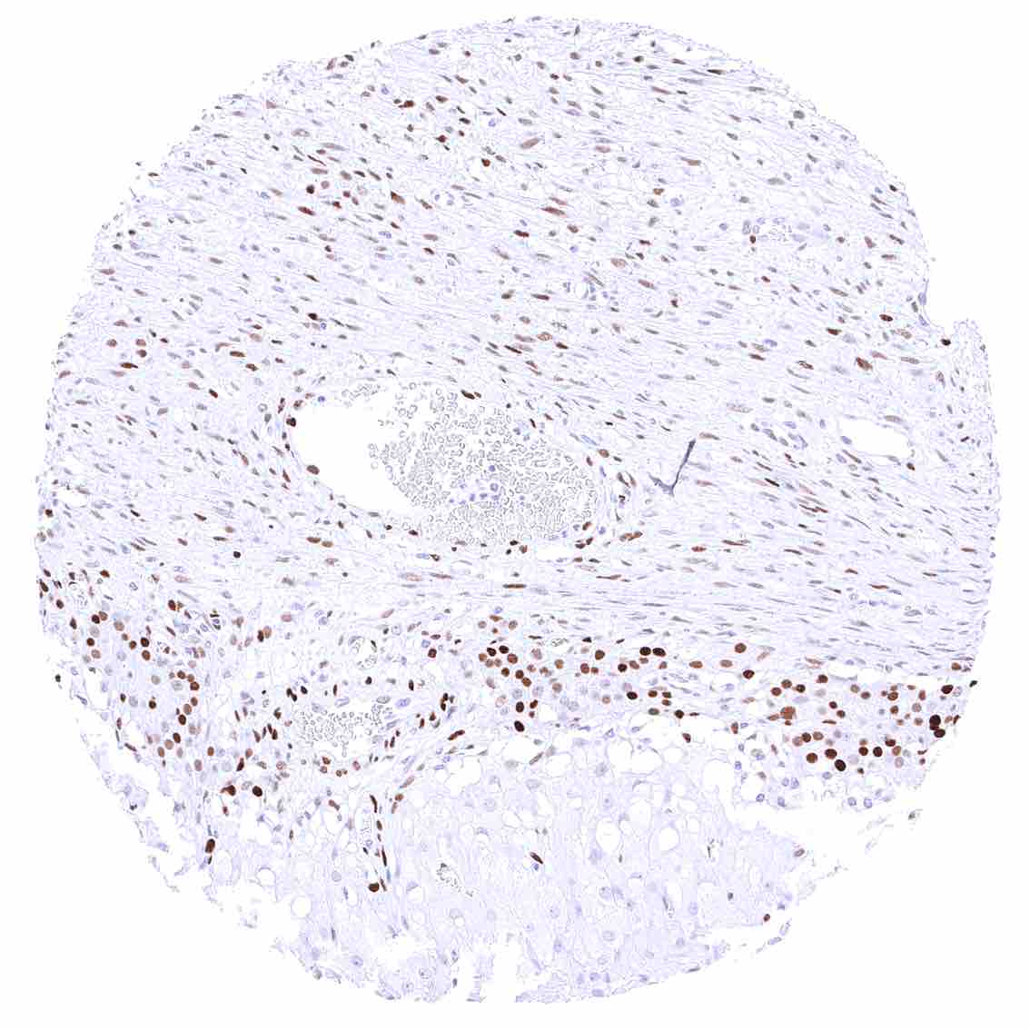
Esophagus, muscular wall – Faint MCM5 staining in few muscle cells.
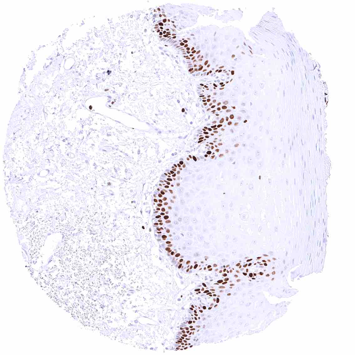
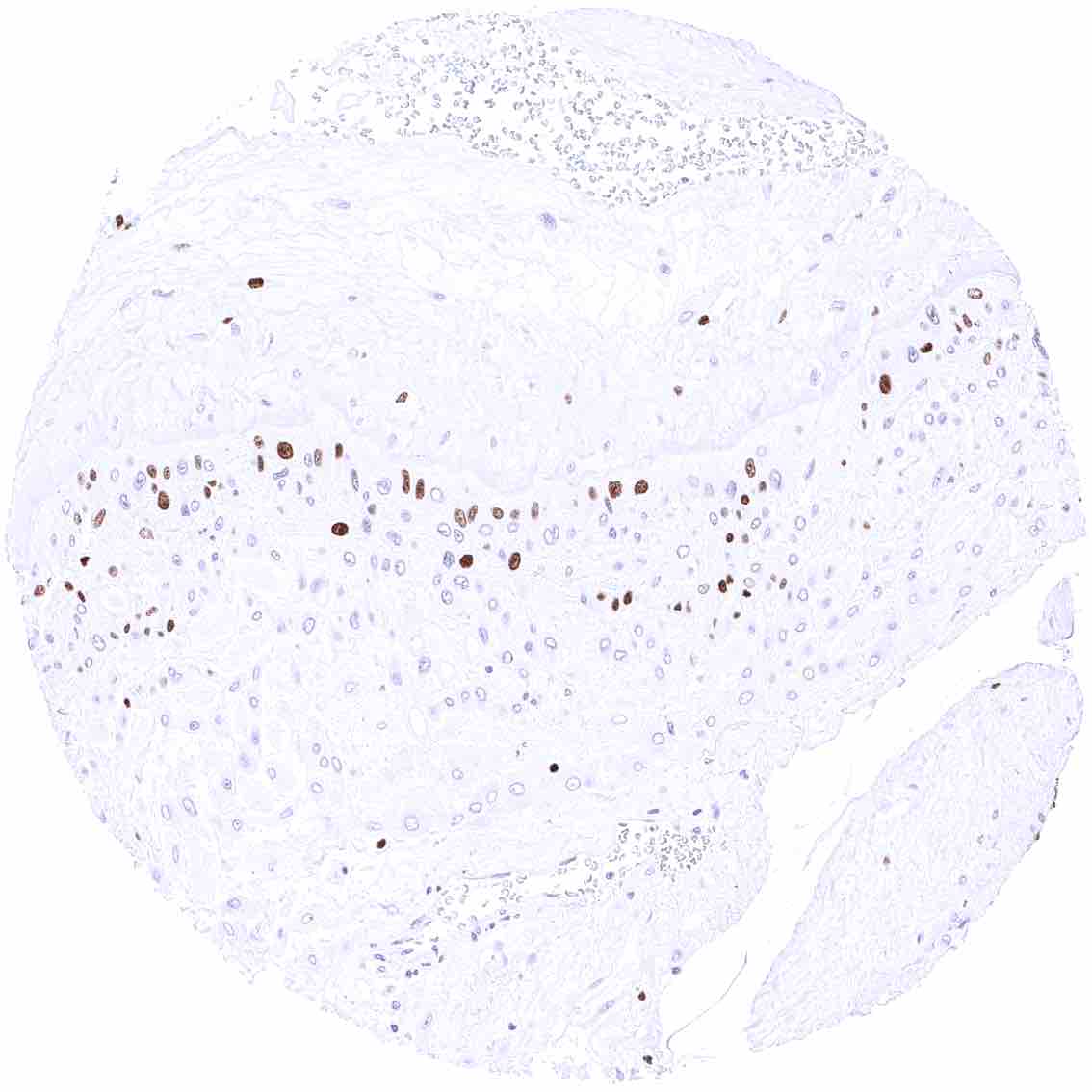
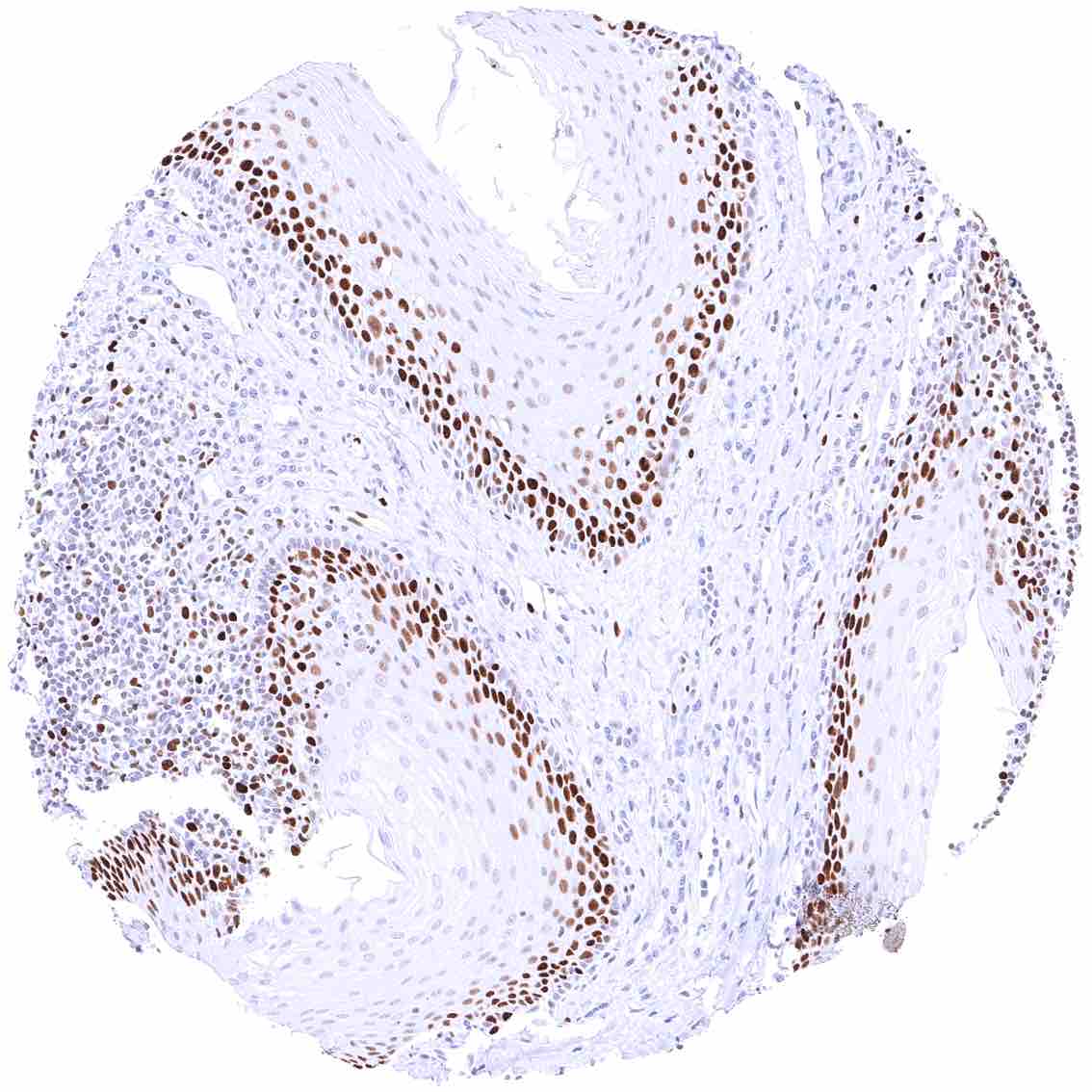
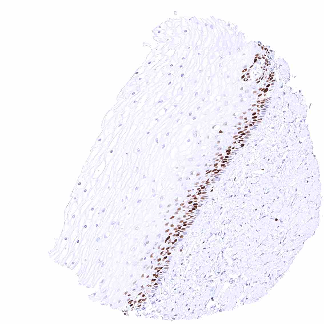
Esophagus, squamous epithelium – Distinct MCM5 staining of suprabasal and (much less intense) basal cells of the squamous epithelium.
Fallopian tube, mucosa – Strong nuclear MCM5 staining of a significant subset of epithelial cells.
Fat
Fat
Gallbladder, epithelium – A variable number of MCM5 positive cells can be seen in the gallbladder epithelium.
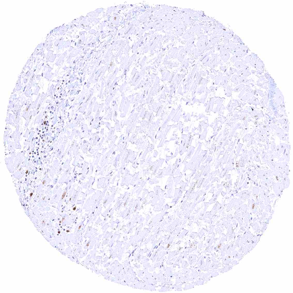
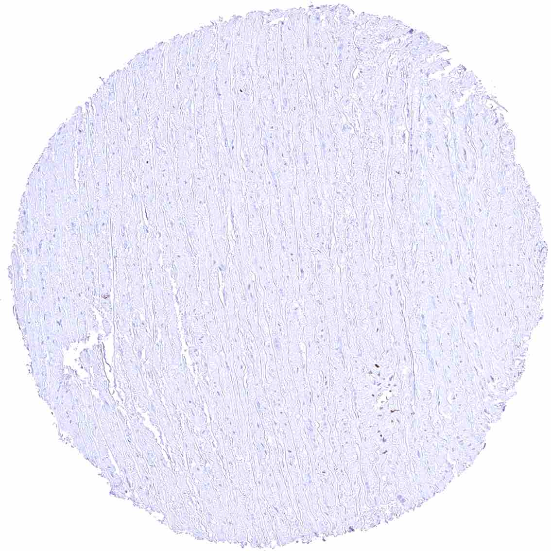
Heart muscle – Faint MCM5 staining of few muscle cells.
Ileum, mucosa – MCM5 staining predominates in epithelial cells of the crypts.
Ileum, muscular wall
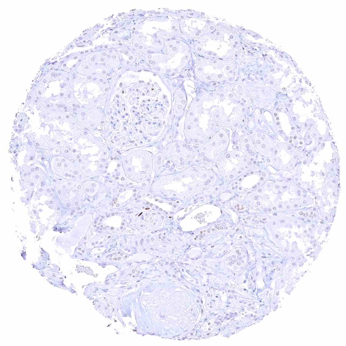
Kidney, cortex – Few epithelial cells are MCM5 positive.
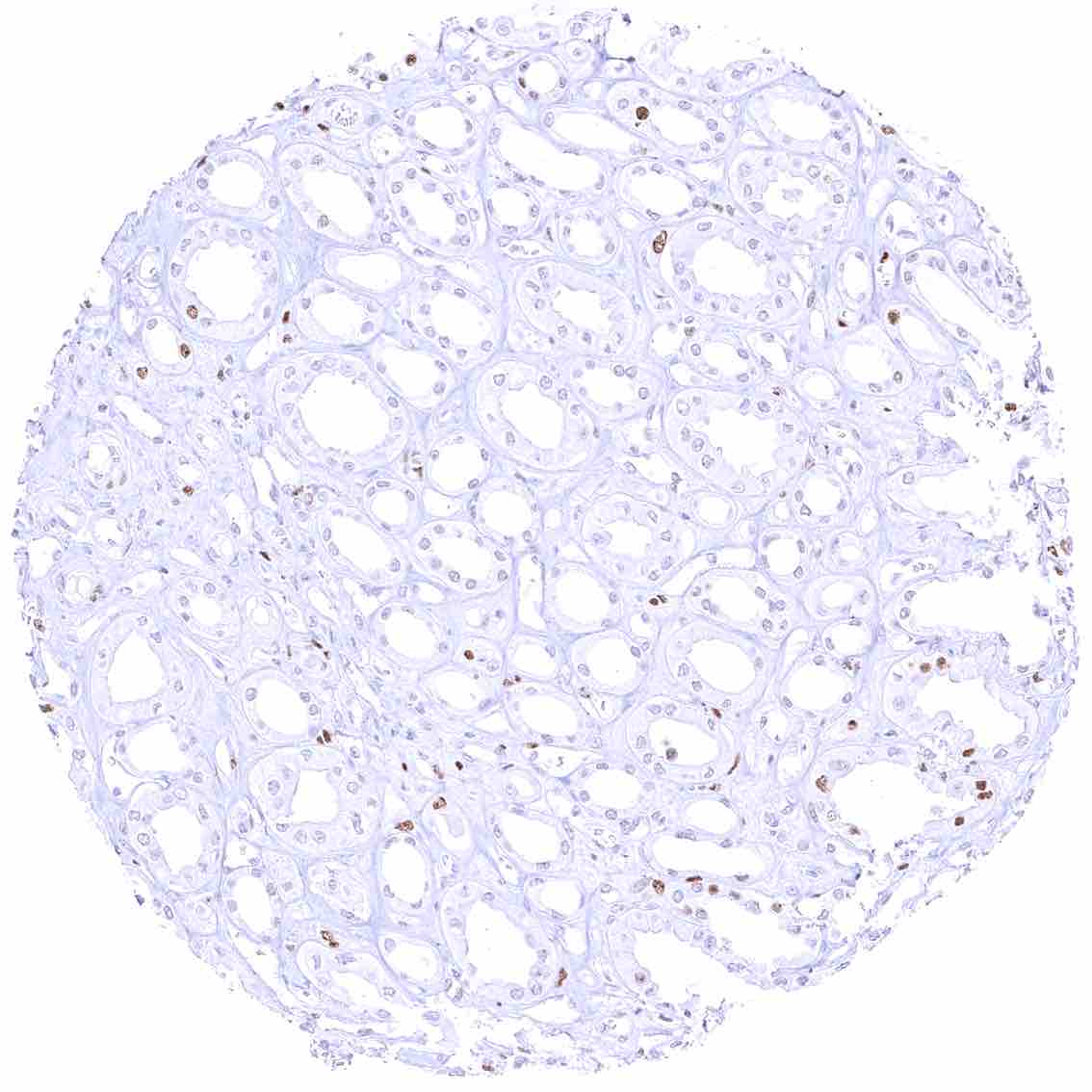
Kidney, medulla – Few epithelial cells are MCM5 positive.
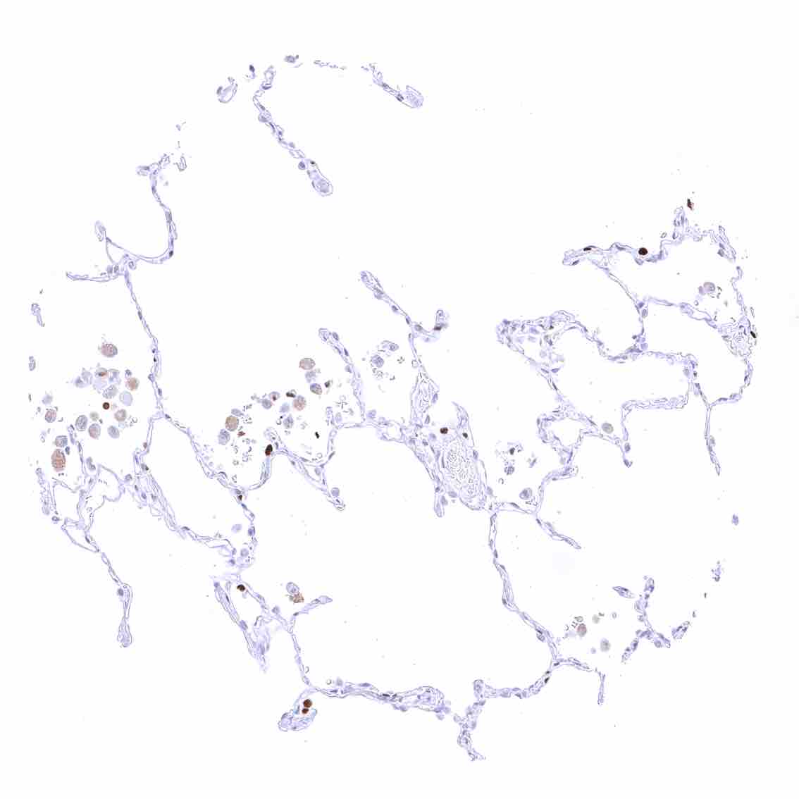
Kidney, pelvis, urothelium – A variable fraction of urothelial cells in all cell layers can show nuclear MCM5 staining.
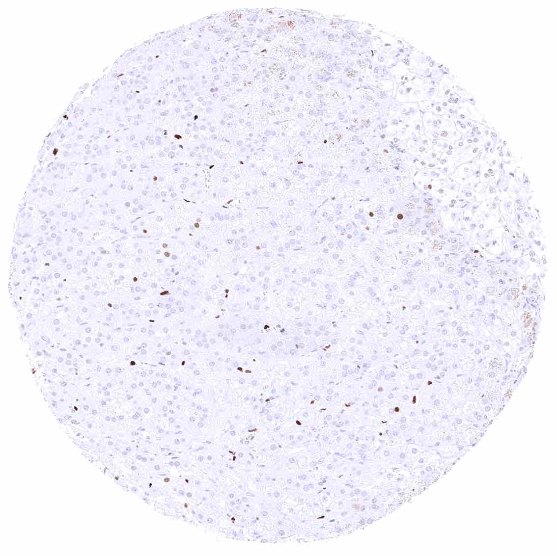
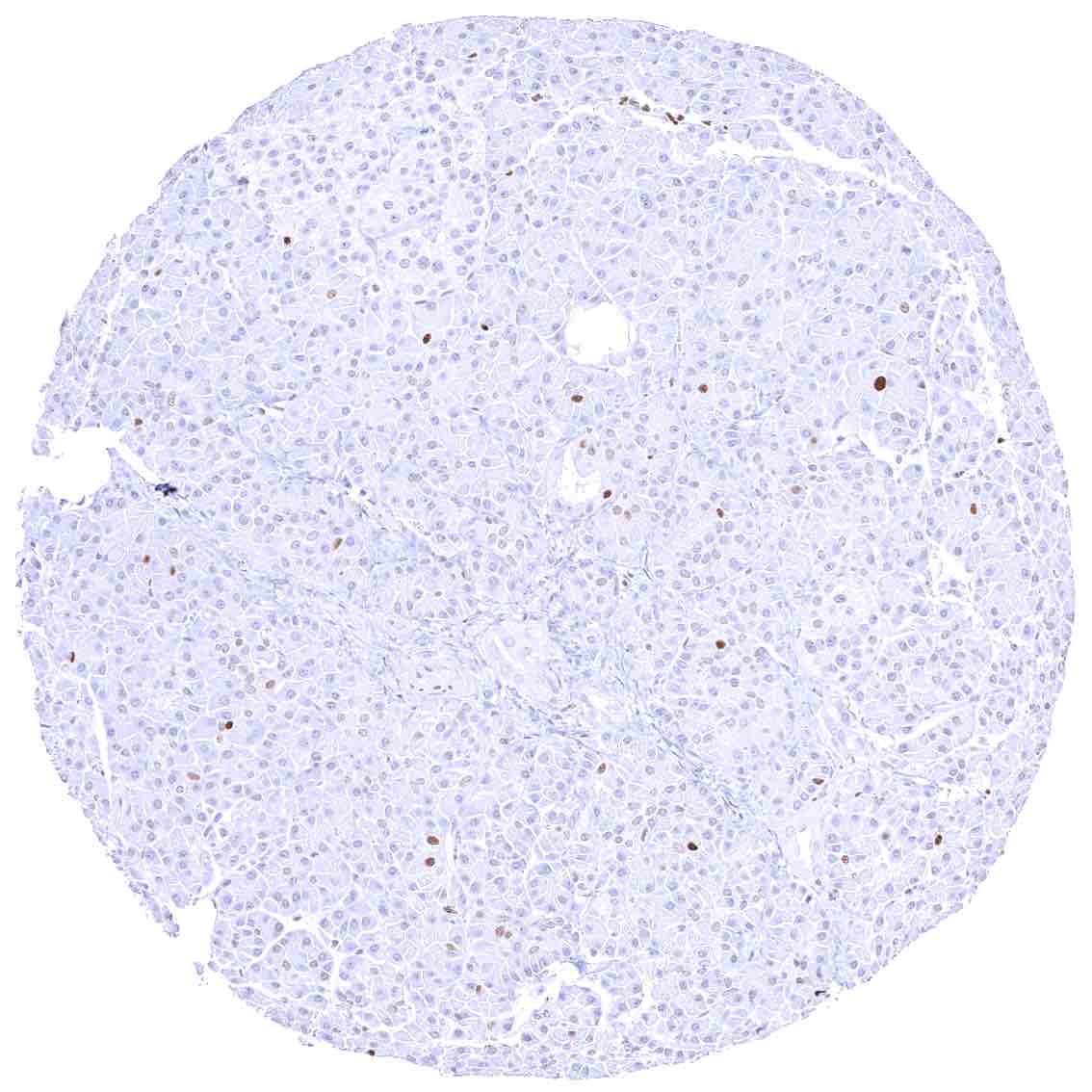
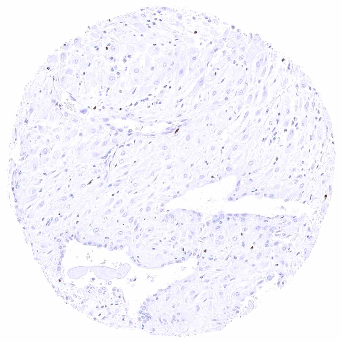
Liver – Faint nuclear MCM5 staining in a small fraction of hepatocytes.
Lung – Distinct MCM5 staining of a subset of pneumocytes
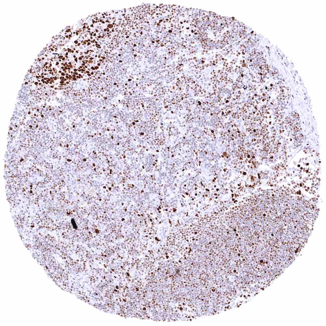
Lymph node – Many lymphocytes are MCM5 positive. MCM5 staining is strongest and most common in cells of germinal centres.
Ovary, corpus luteum – Weak or absent MCM5 staining of corpus luteum cells.
Ovary, follicular cyst – Strong MCM5 staining of virtually all granulosa and theca interna cells.
Ovary, stroma – Weak nuclear MCM5 staining of stroma cells.
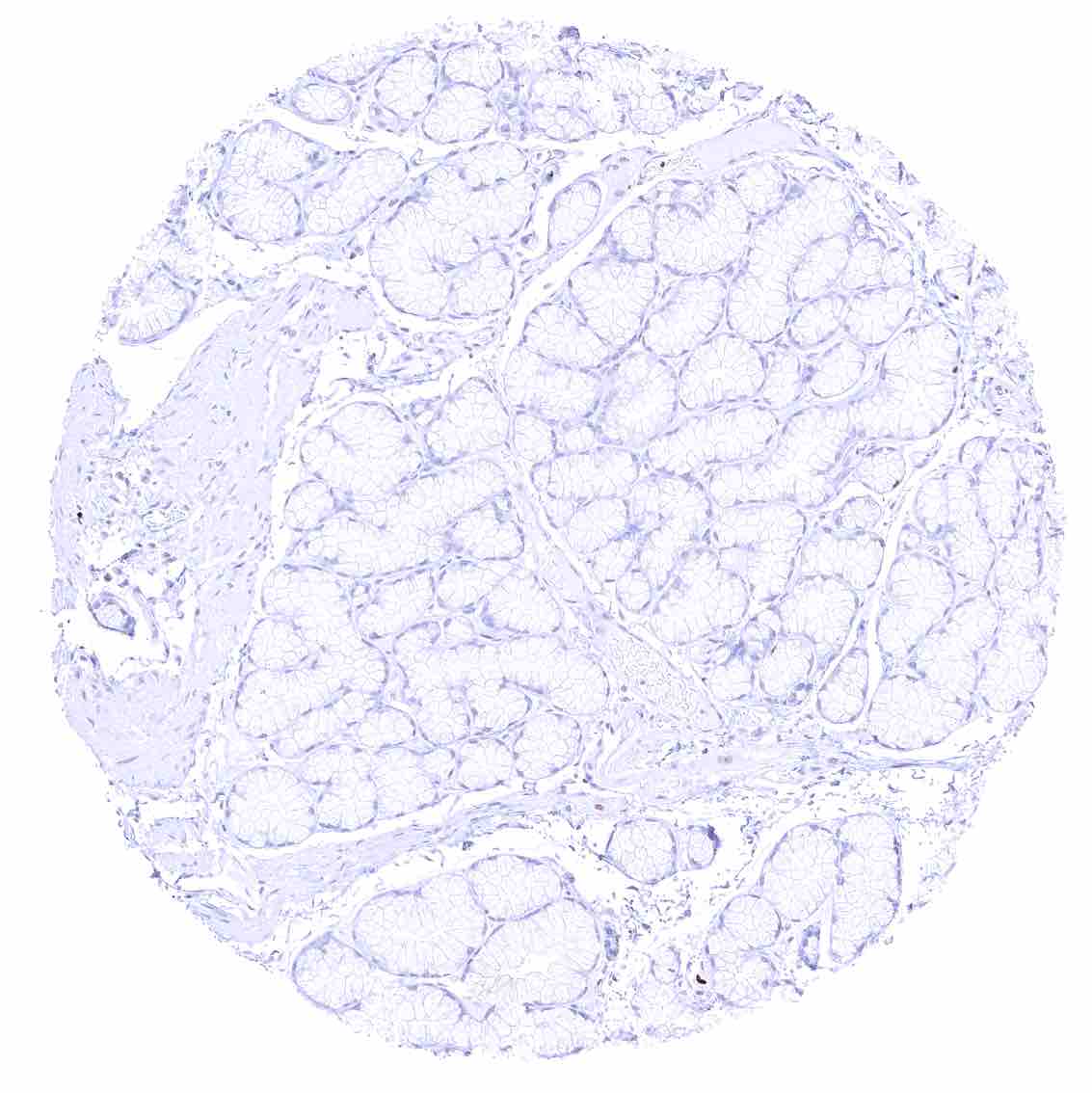
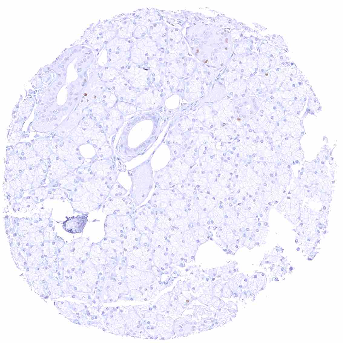
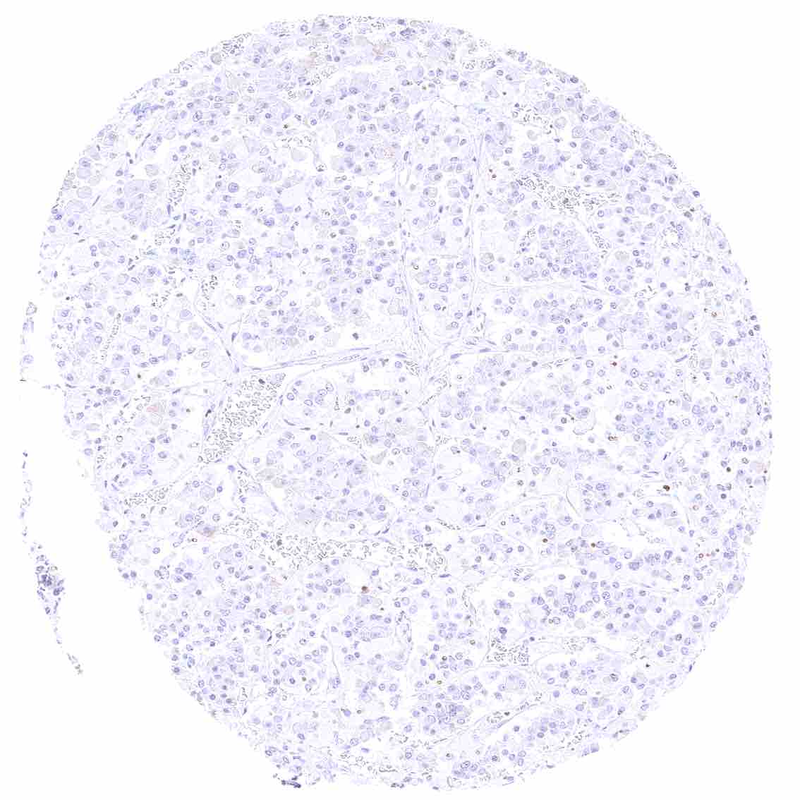
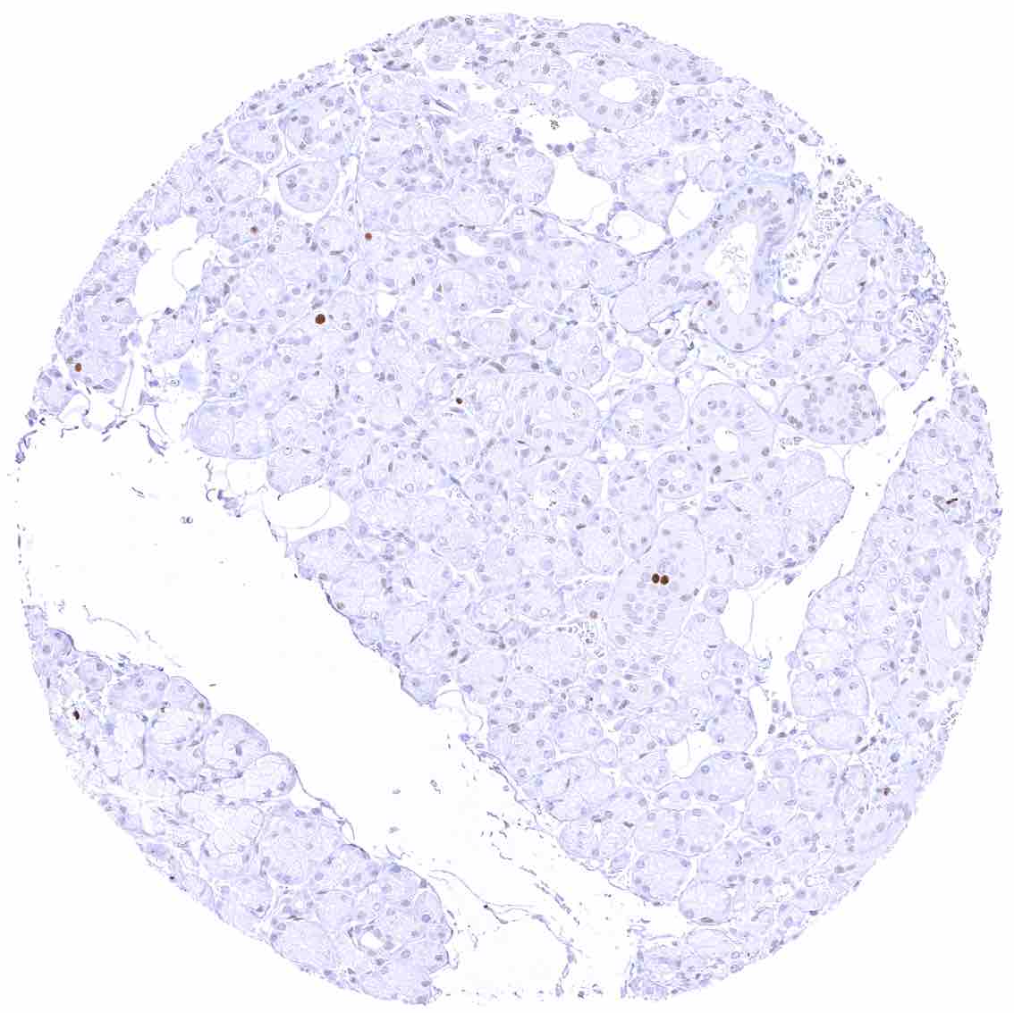
Pancreas – Moderate to strong MCM5 staining in a rather small fraction of epithelial cells.
Parathyroid gland – Distinct nuclear MCM5 staining in a very small fraction of epithelial cells.
Parotid gland
Parotid gland
Pituitary gland, anterior lobe
Pituitary gland, posterior lobe .jpeg
Pituitary gland, posterior lobe
Placenta (chorion) – Moderate to strong MCM5 staining in a fraction of chorion cells. .jpeg
Placenta (chorion) – Moderate to strong MCM5 staining in a fraction of chorion cells.
Placenta, early – Strong MCM5 staining of a large fraction of cytotrophoblast cells.
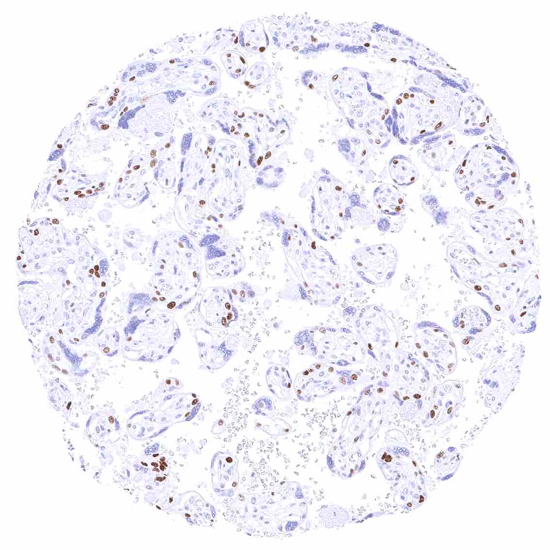
Placenta, mature – Strong MCM5 staining of a large fraction of cytotrophoblast cells. .jpeg
Placenta, mature – Strong MCM5 staining of a large fraction of cytotrophoblast cells.
Prostate – Nuclear MCM5 staining of a subset of epithelial cells. It is more common in basal than in acinar cells.
Rectum, mucosa – Nuclear MCM5 staining predominates in in epithelial cells of the crypts. Some lymphocytes are also positive.
Seminal vesicle – Strong nuclear MCM5 staining of a rather small fraction of epithelial cells.
Sinus paranasales.jpeg
Sinus paranasales
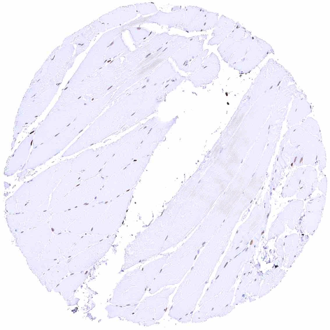
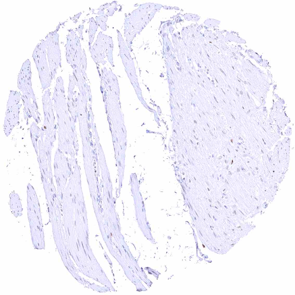
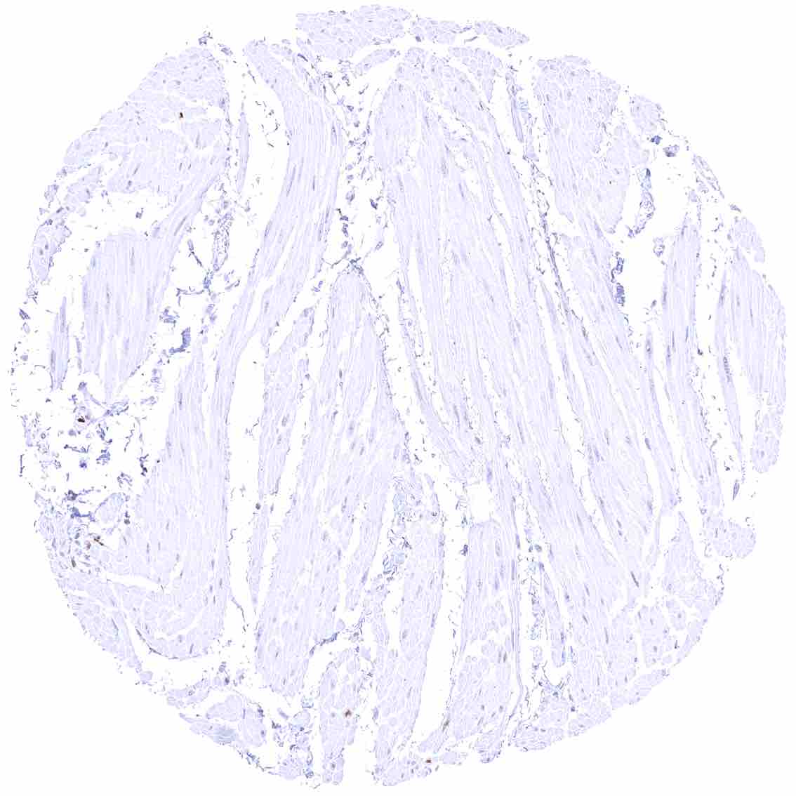
Skeletal muscle – Distinct MCM5 staining in a large fraction of skeletal muscle cells. .jpeg
Skeletal muscle – Distinct MCM5 staining in a large fraction of skeletal muscle cells.
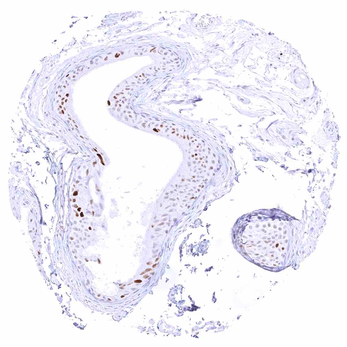
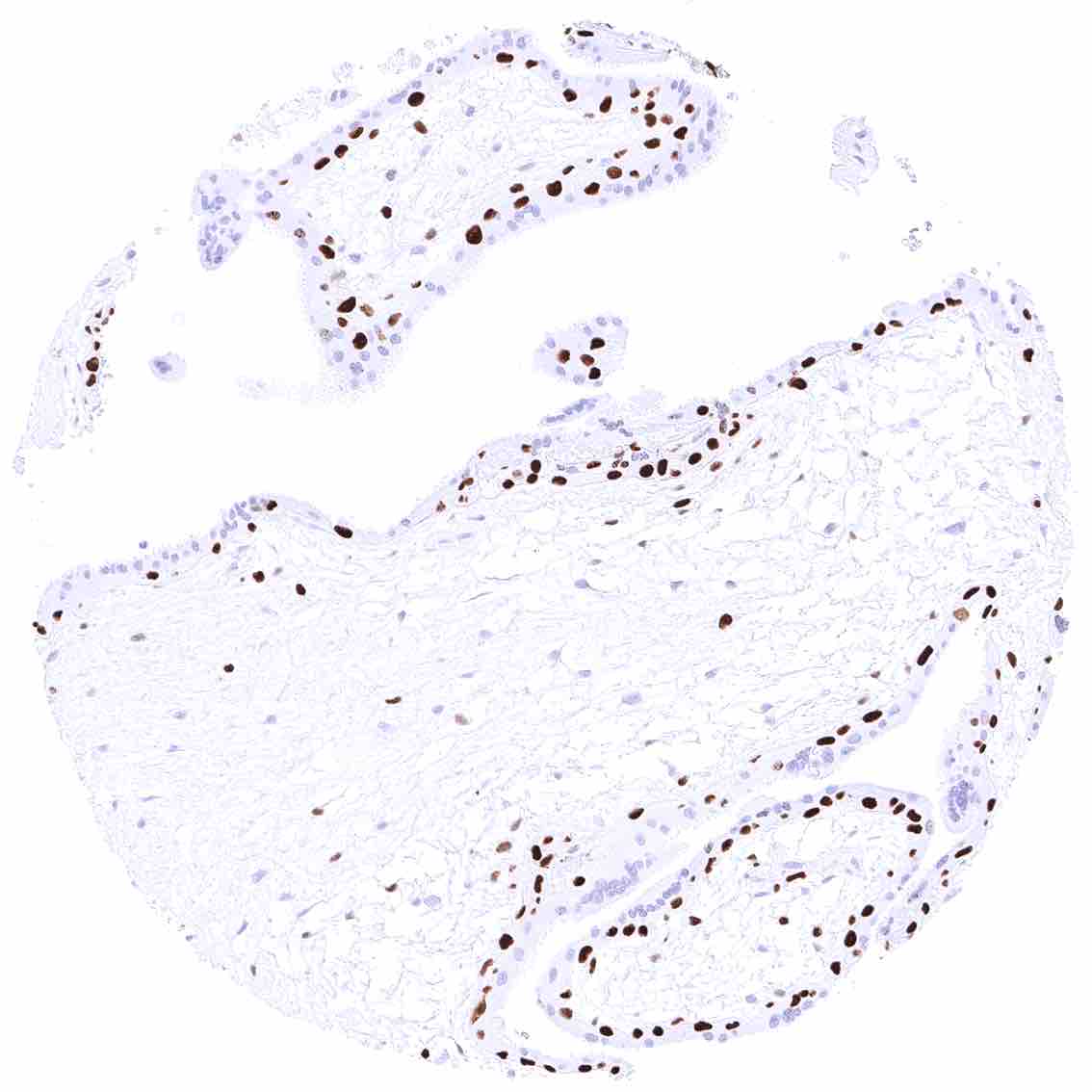
Skin – Suprabasal and basal cells of the squamous epithelium with a distinct nuclear MCM5 staining.
Skin, sebaceous glands – Intense nuclear MCM5 positivity of peripheral germinative cells.
Spleen – A strong MCM5 positivity occurs in few scattered cells of all the red pulp. .jpeg
Spleen – A strong MCM5 positivity occurs in few scattered cells of all the red pulp.
Stomach, antrum – Strong nuclear MCM5 immunostaining of many mucous neck cells. .jpeg
Stomach, antrum – Strong nuclear MCM5 immunostaining of many mucous neck cells.
Stomach, corpus – Strong nuclear MCM5 immunostaining of many mucous neck cells.
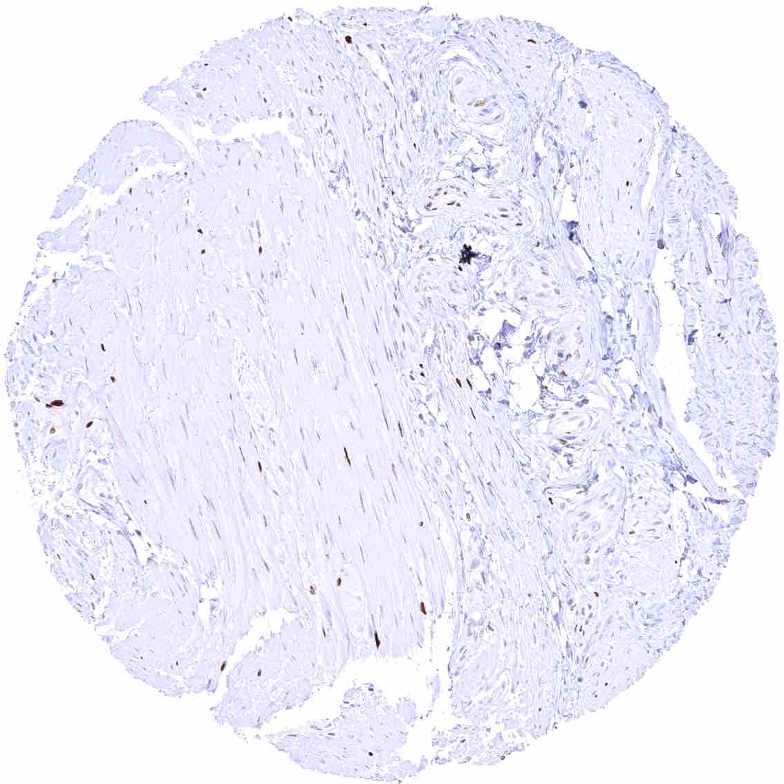
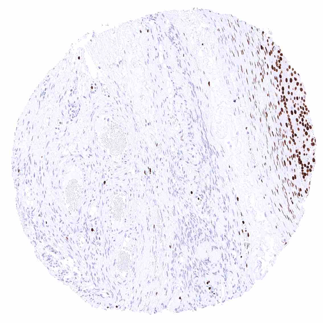
Stomach, muscular wall – Strong nuclear MCM5 staining of few cells along nerve fibers.
Submandibular gland – Distinct MCM5 staining in a fraction of epithelial cells.
Testis – Most spermatogonia and spermatocytes show strong nuclear MCM5 positivity.
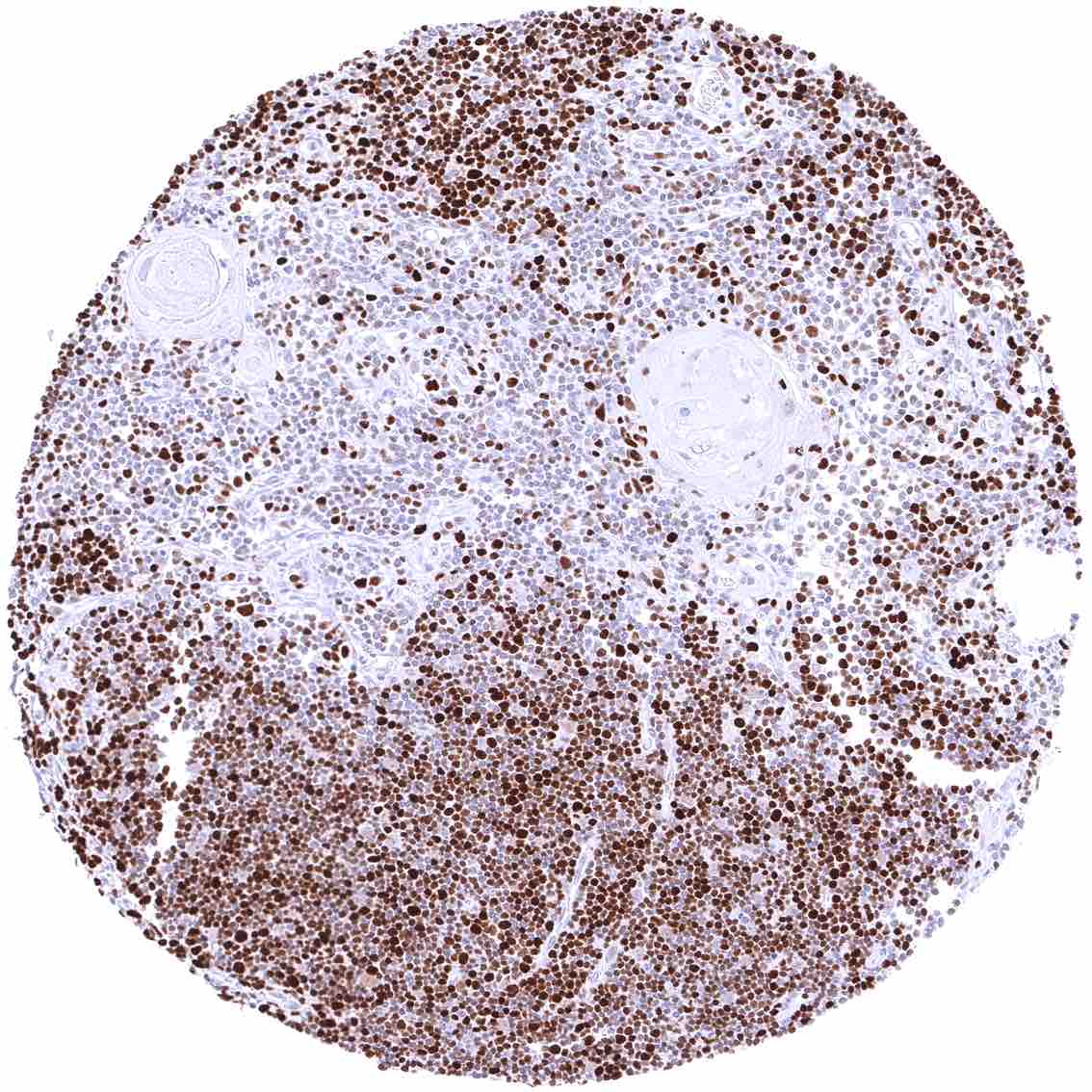
Thymus – Strong MCM5 positivity of most cells of the thymic cortex. Medullary cells show markedly less staining.
Thyroid gland – Weak to moderate nuclear MCM5 staining of a small fraction of follicular cells.
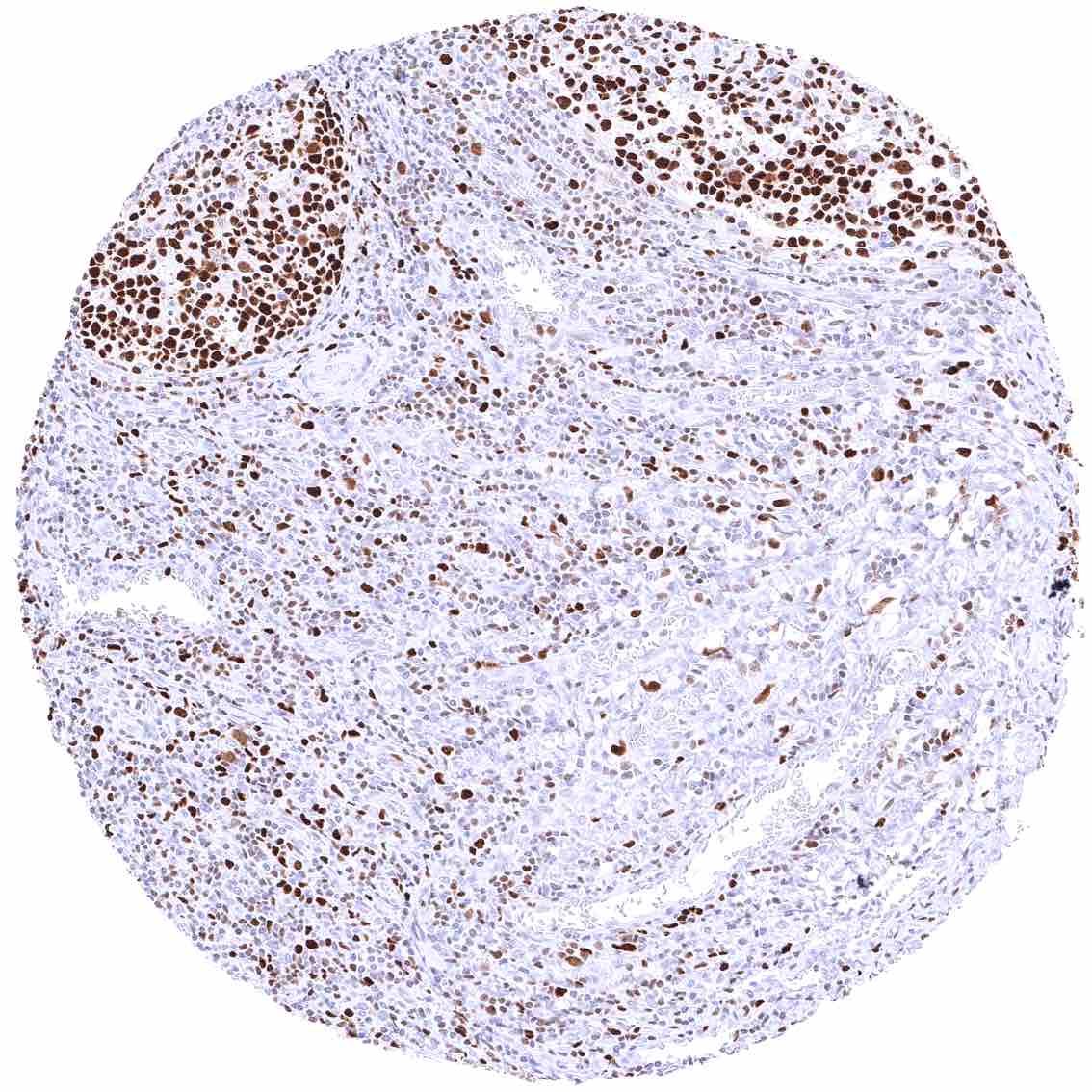
Tonsil – Many lymphocytes are MCM5 positive. MCM5 staining is strongest and most common in cells of germinal centres.
Tonsil, surface epithelium
Urinary bladder, muscular wall
Urinary bladder, urothelium – A variable fraction of urothelial cells in all cell layers can show nuclear MCM5 staining.
Urinary bladder, urothelium – Only few urothelial cells show (weak) nuclear MCM5 staining in this sample.
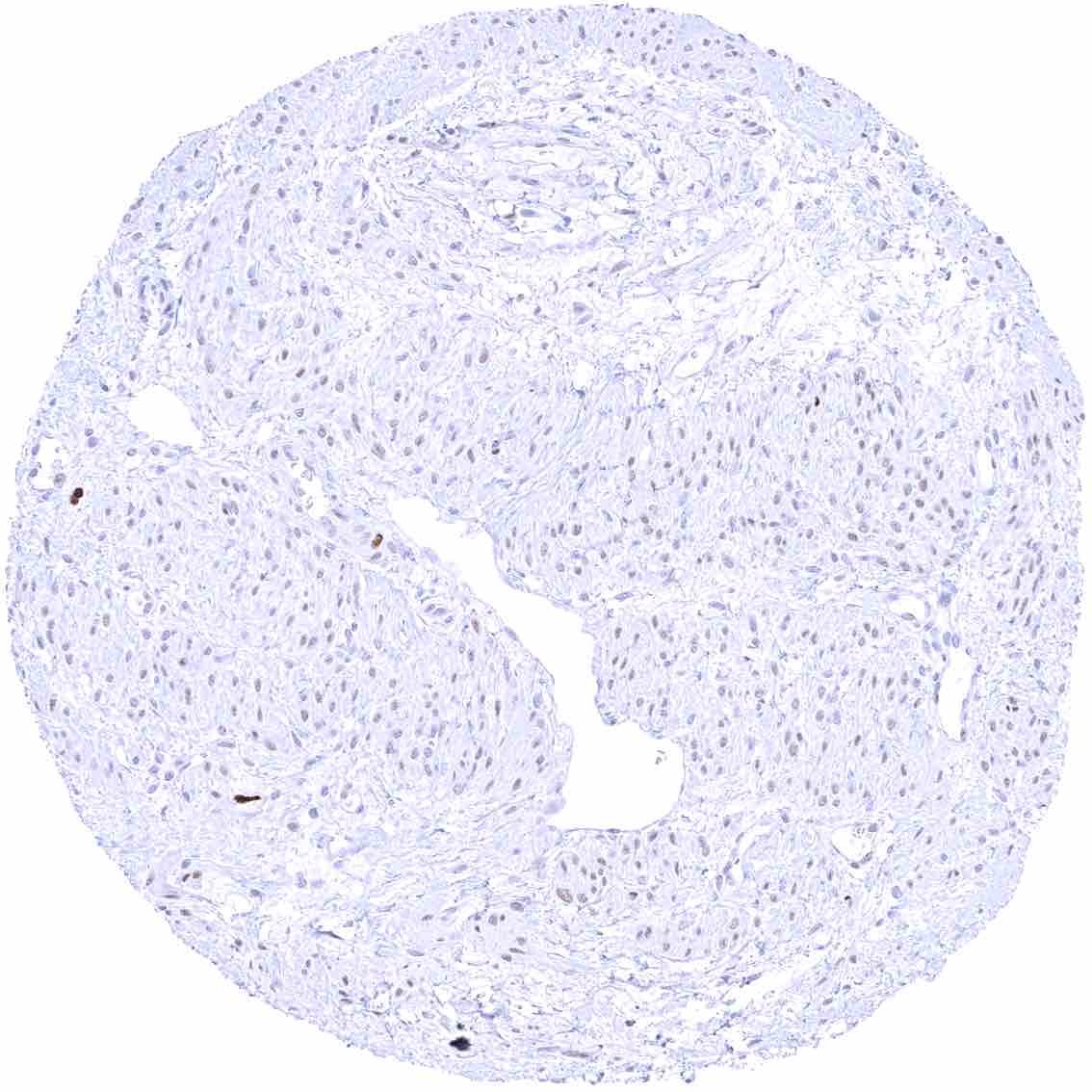
Uterus, ectocervix – Distinct MCM5 staining of suprabasal and (less intense) basal cells of the squamous epithelium.
Uterus, endocervix – Only few epithelial cells show weak nuclear MCM5 staining.
Uterus, endometrium (pregnancy)
Uterus, endometrium (proliferation) – Strong nuclear MCM5 positivity of many epithelial cells and a fraction of stromal cells.
Uterus, endometrium (secretion) – Nuclear MCM5 positivity of a subset epithelial cells.
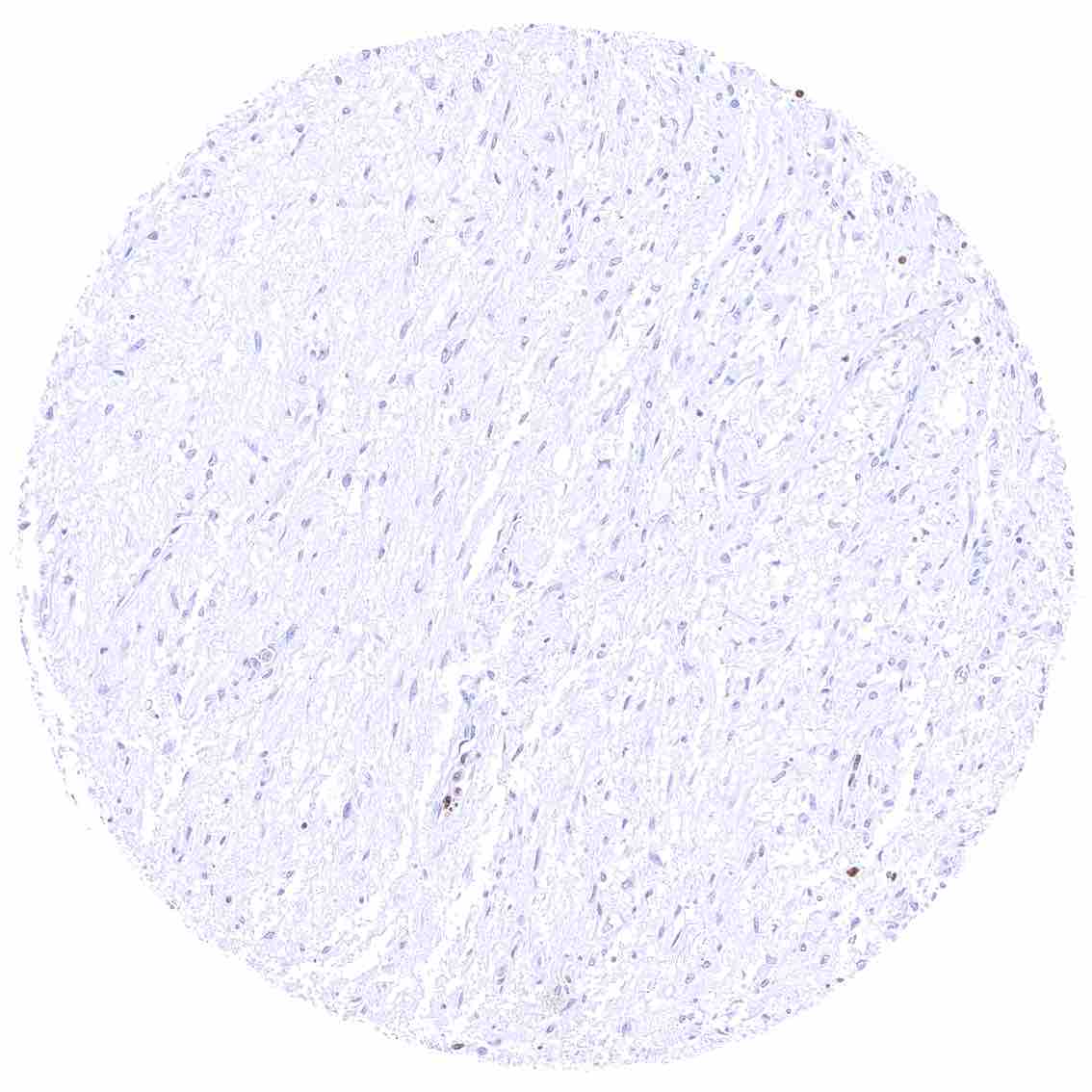
Uterus, myometrium – Faint MCM5 staining in a variable fraction of muscle cells.
  • Linkedin
  • My Account
  • Search
    ×
  • Cart 0
© 2025 MS Validated Antibodies
  • Imprint
  • Privacy Policy
  • Distributors