295,00 € – 995,00 €
Product details
Synonyms = tripartite motif containing 28 , KAP1 , PPP1R157 , RNF96 , TF1B , TIF1B
Antibody type = Recombinant Rabbit monoclonal / IgG
Clone = HMV335
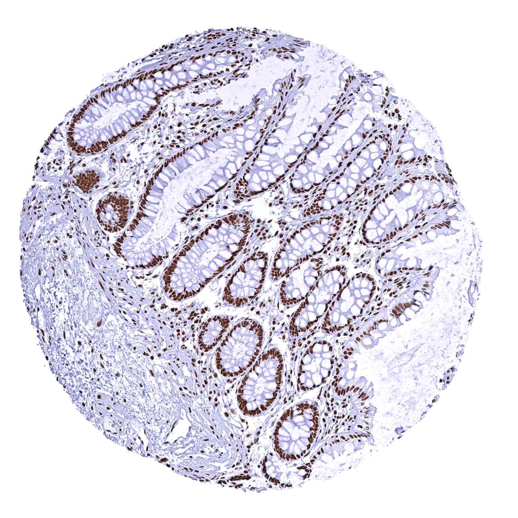
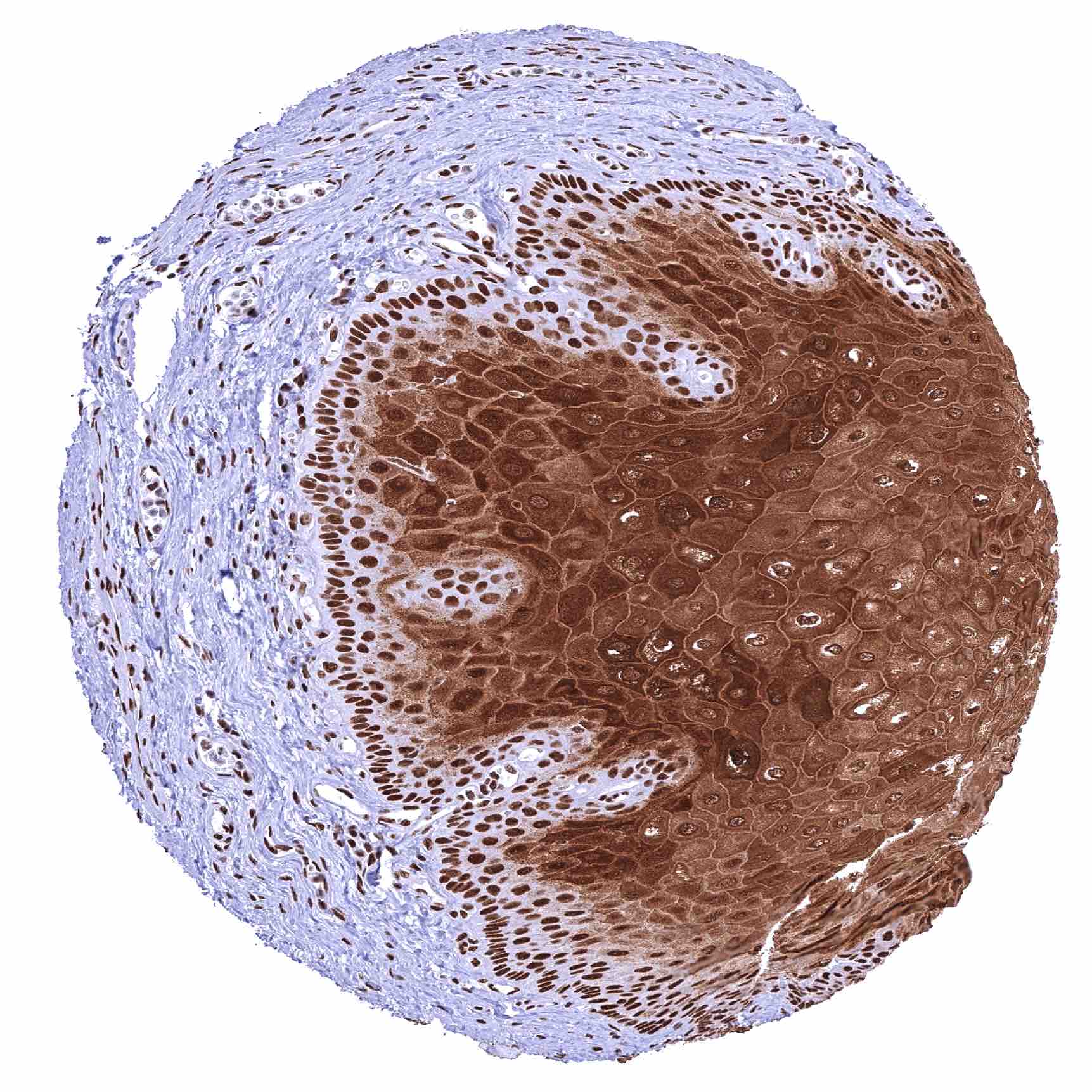
Positive control = Colon: A strong nuclear TRIM28are staining should be seen in all cells.
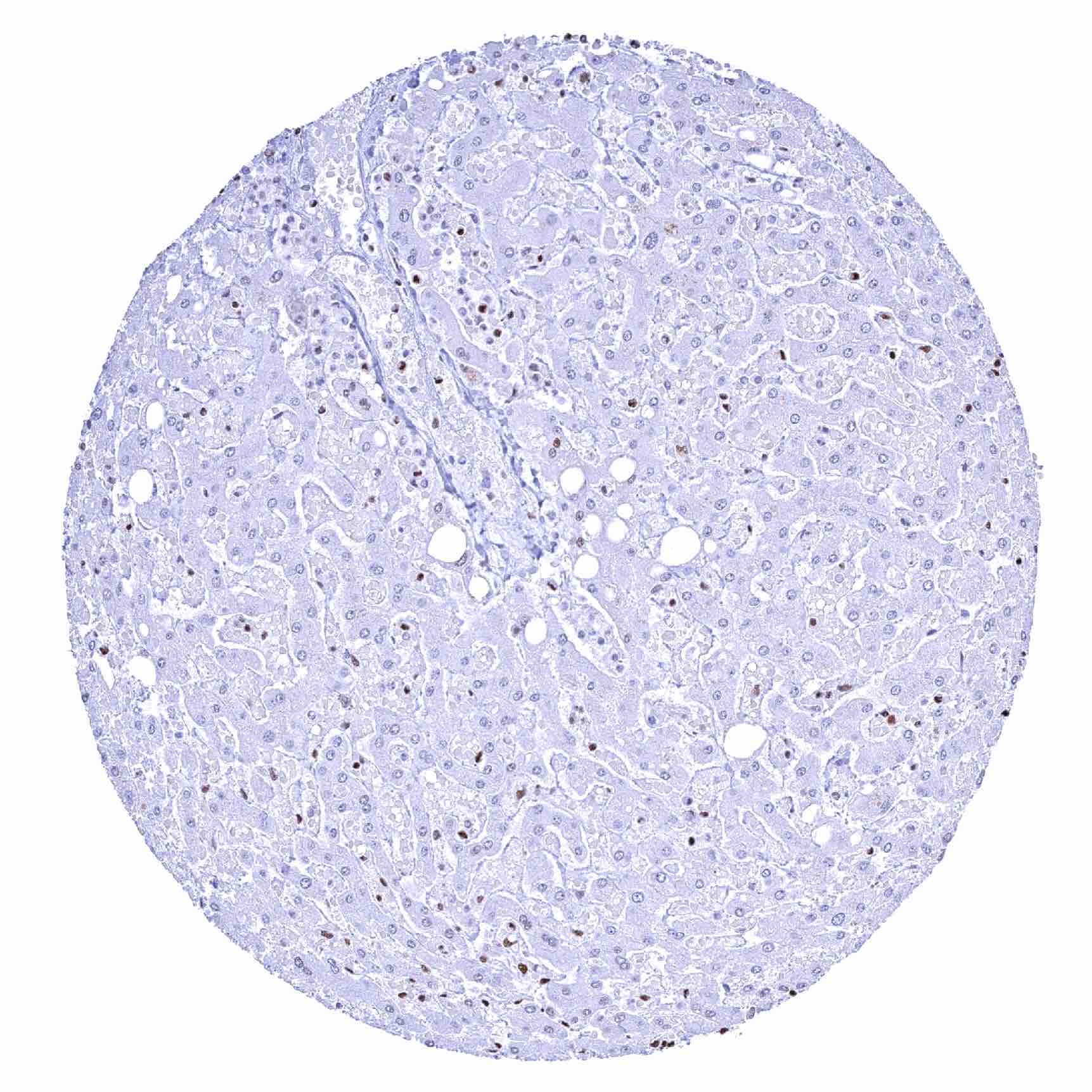
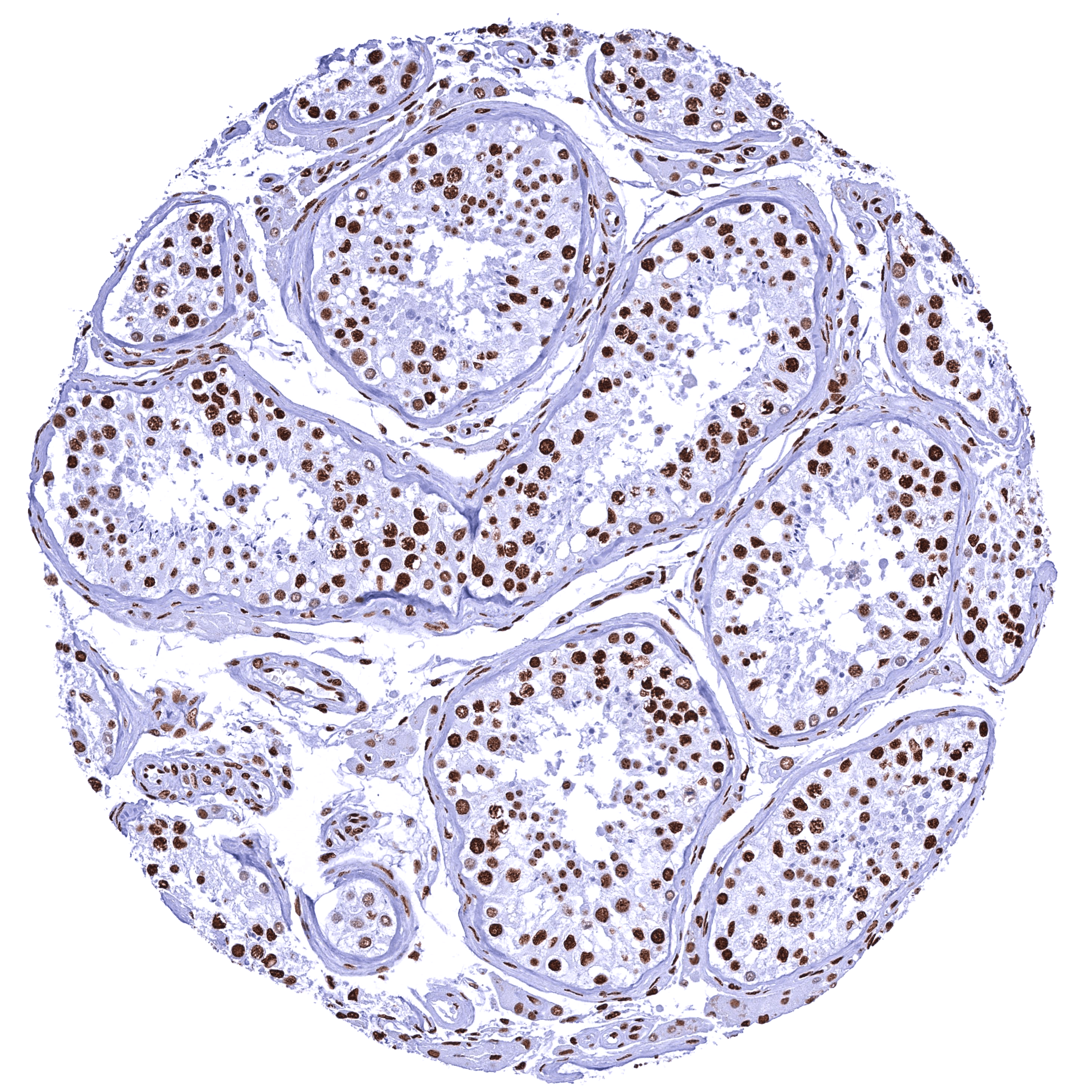
Negative control = Testis: Spermatids are TRIM28 negative while all other cells show a strong nuclear TRIM28 staining of all cells.
Cellular localization = Nucleus
Reactivity = Human
Application = Immunohistochemistry
Dilution = 1:100 – 1:200
Intended Use = Research Use Only
Relevance of Antibody
KAP1 / TRIM28 is a multifunctional regulatory nuclear protein.
Biology Behind
Tripartite motif-containing 28 (TRIM28), also termed KAP1 (KRAB-associated protein-1), or transcriptional mediator 1b (TIF1b) is a regulatory protein coded by the TRIM28 gene at chromosome 19q13.43. The protein plays a role in transcription regulation by interaction with the Krüppel-associated box repression domain found in many transcription factors and binding to specific chromatin regions. In addition to regulating gene transcription, KAP1 has a variety of regulatory intracellular functions, such as response to DNA damage, maintaining stem cell pluripotency, cellular differentiation and proliferation, viral suppression, and apoptosis. KAP1 is ubiquitously expressed, and its function depends on posttranscriptional modification.
Staining Pattern in Normal Tissues
In normal tissues, TRIM28 is ubiquitously seen in nuclei of all tissues.
Images describing the KAP1 / TRIM28 staining pattern in normal tissues obtained by the antibody HMV335 are shown in our “Normal Tissue Gallery”.
| Brain | Cerebrum | Strong nuclear TRIM28 staining in all cells. |
| Cerebellum | Strong nuclear TRIM28 staining in all cells. | |
| Endocrine Tissues | Thyroid | Strong nuclear TRIM28 staining in all cells. |
| Parathyroid | Strong nuclear TRIM28 staining in all cells. | |
| Adrenal gland | Strong nuclear TRIM28 staining in all cells. | |
| Pituitary gland | Strong nuclear TRIM28 staining in all cells. | |
| Respiratory system | Respiratory epithelium | Strong nuclear TRIM28 staining in all cells. |
| Lung | Strong nuclear TRIM28 staining in all cells. | |
| Gastrointestinal Tract | Salivary glands | Strong nuclear TRIM28 staining in all cells. |
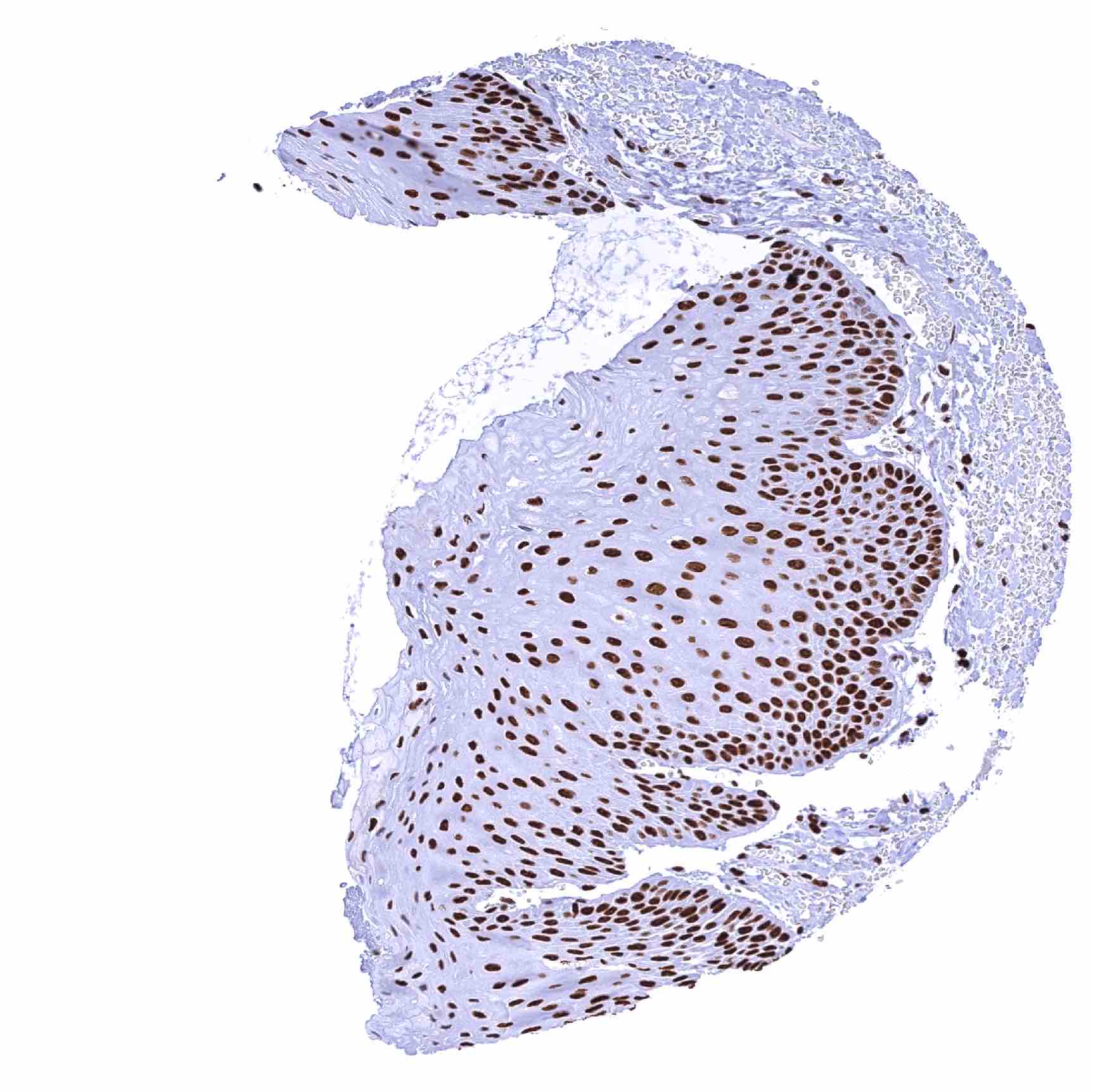
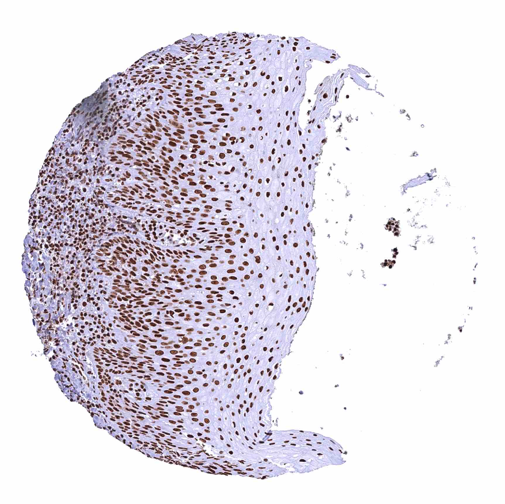
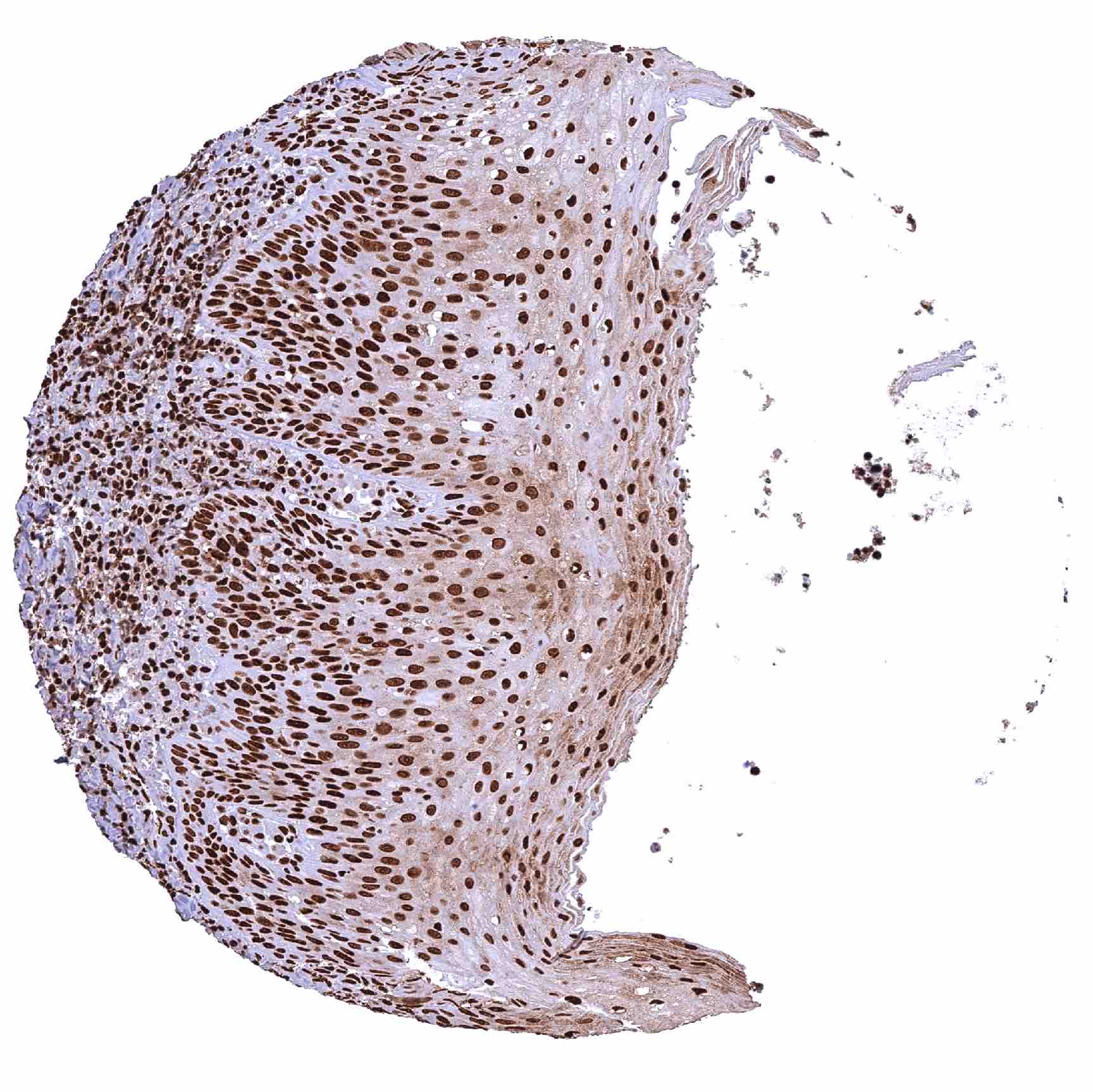
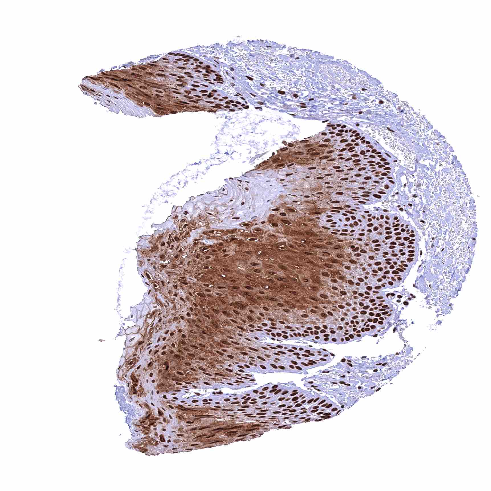
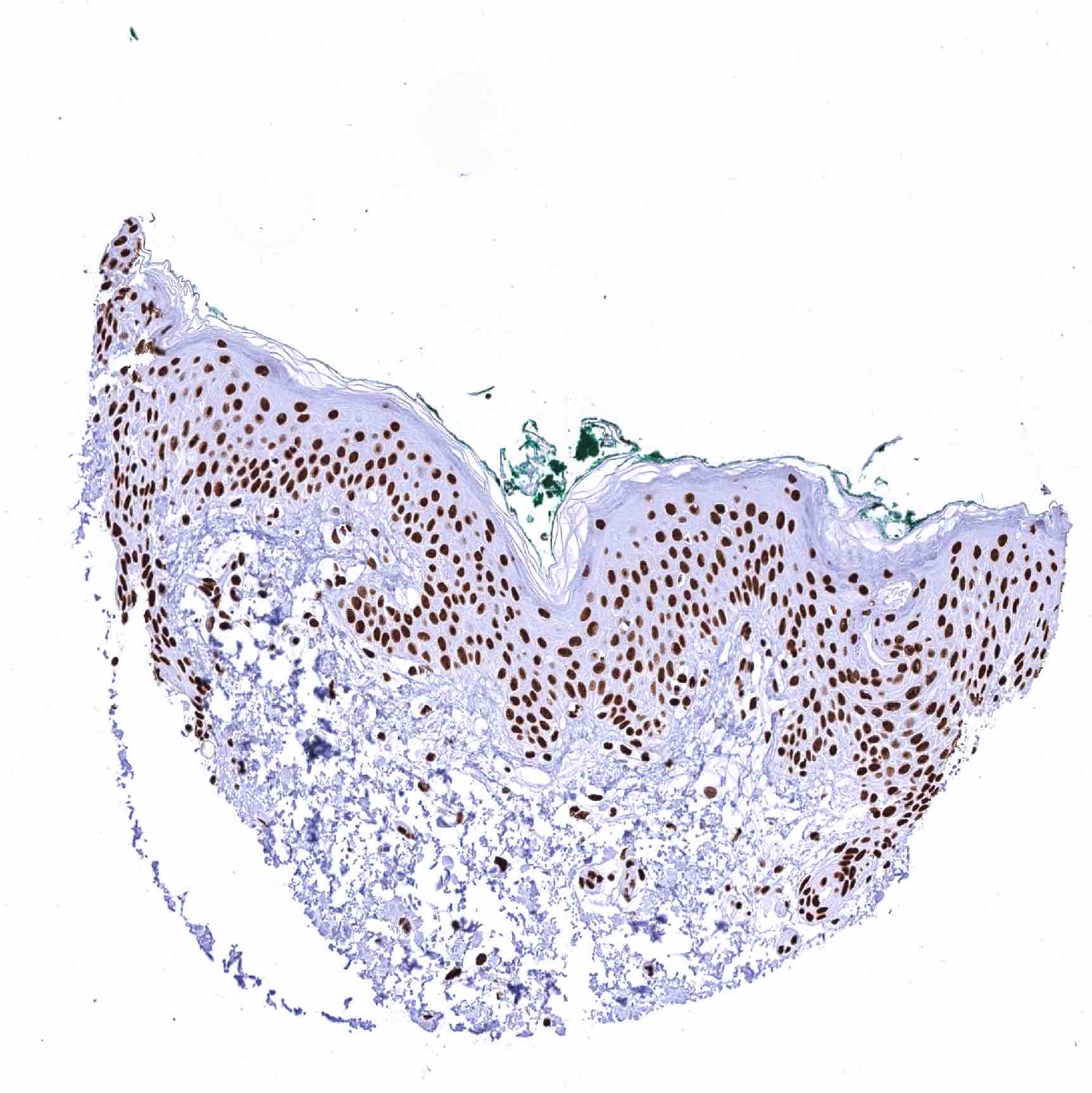
| Esophagus | Slight decrease of the staining intensity from basal to superficial cell layers of squamous eithelium. | |
| Stomach | Strong nuclear TRIM28 staining in all cells. | |
| Duodenum | Strong nuclear TRIM28 staining in all cells. | |
| Small intestine | Strong nuclear TRIM28 staining in all cells. | |
| Appendix | Strong nuclear TRIM28 staining in all cells. | |
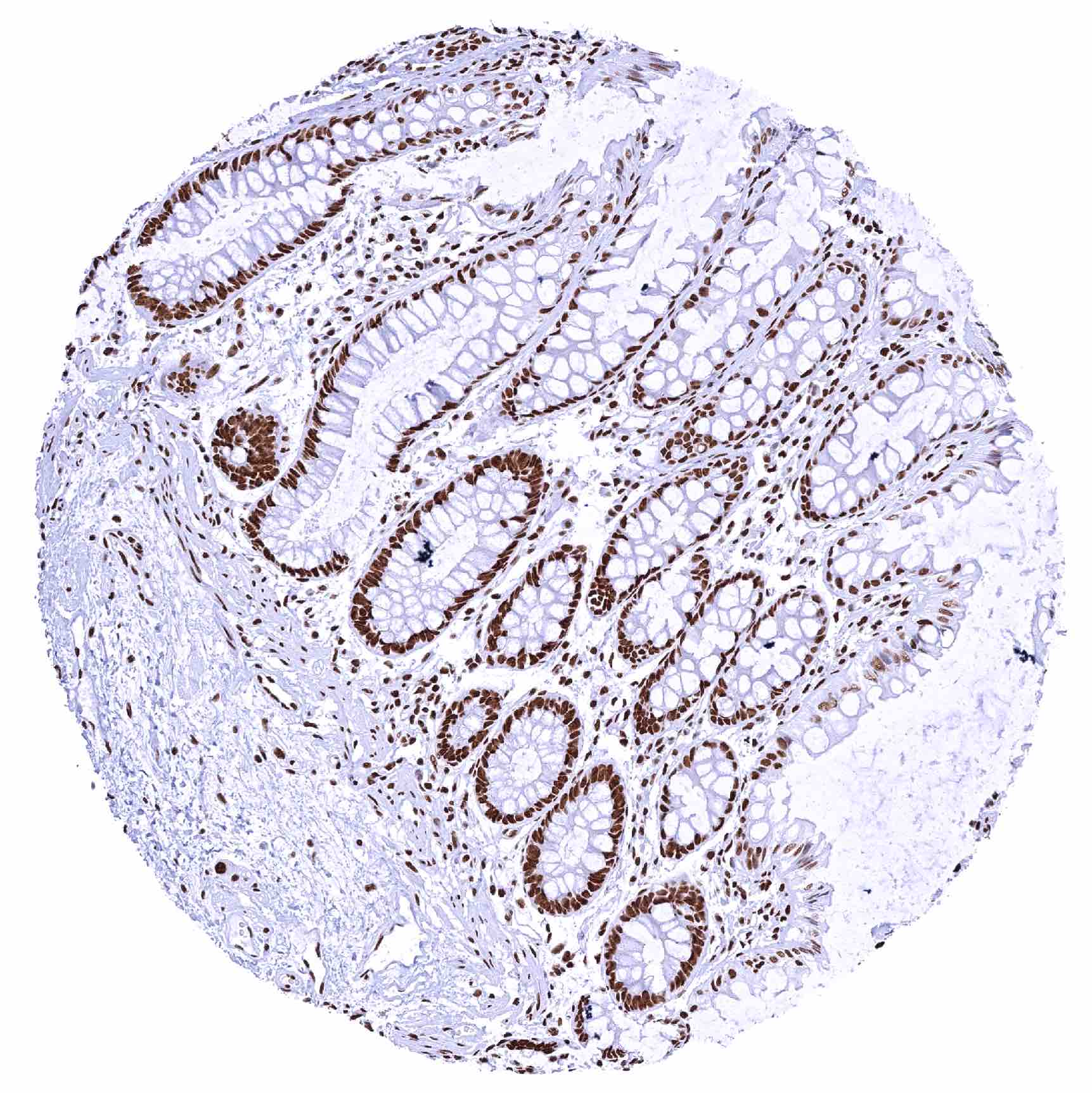
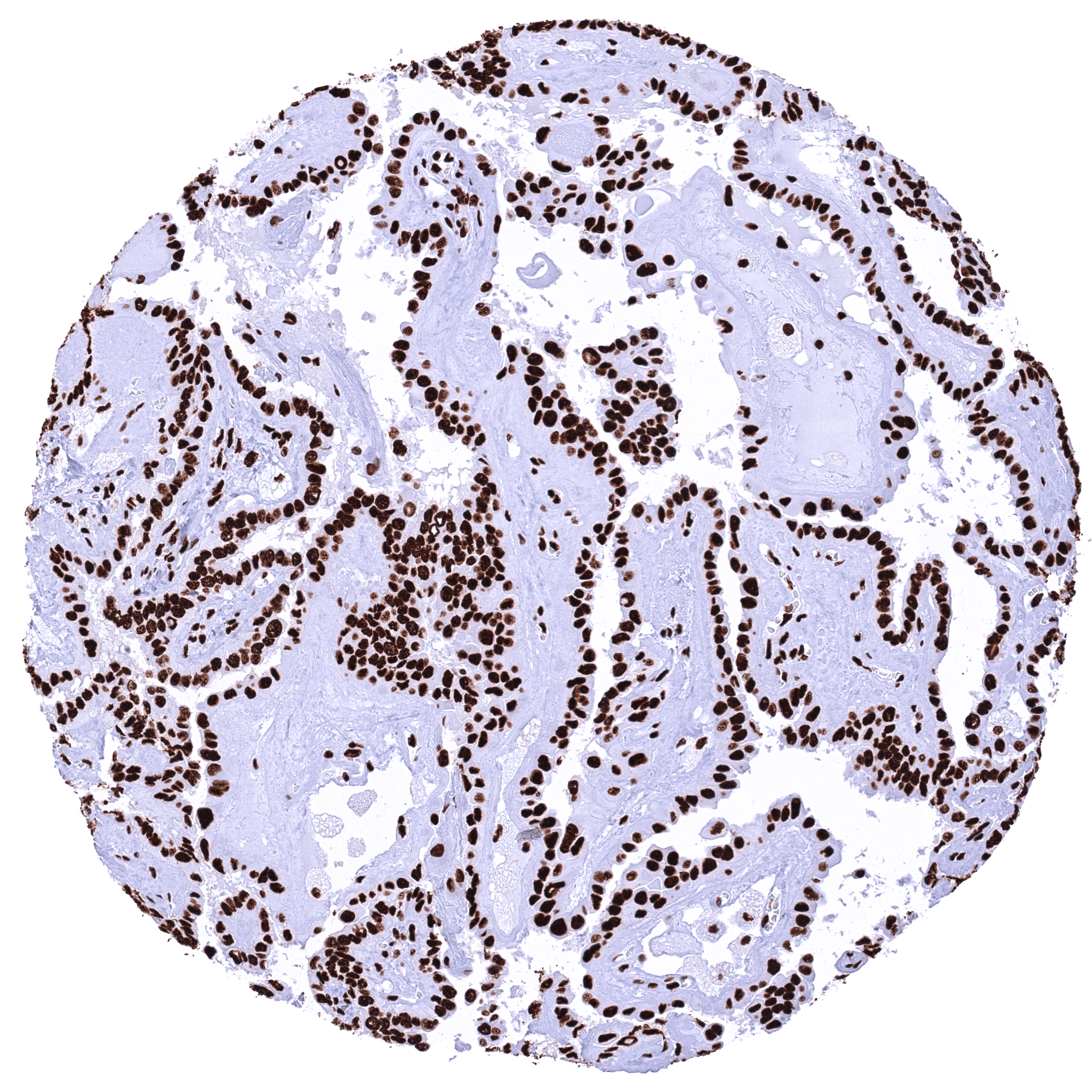
| Colon | Slight decrease of the nuclear TRIM28 staining intensity from crypt base to the surface epithelium. | |
| Rectum | Slight decrease of the nuclear TRIM28 staining intensity from crypt base to the surface epithelium. | |
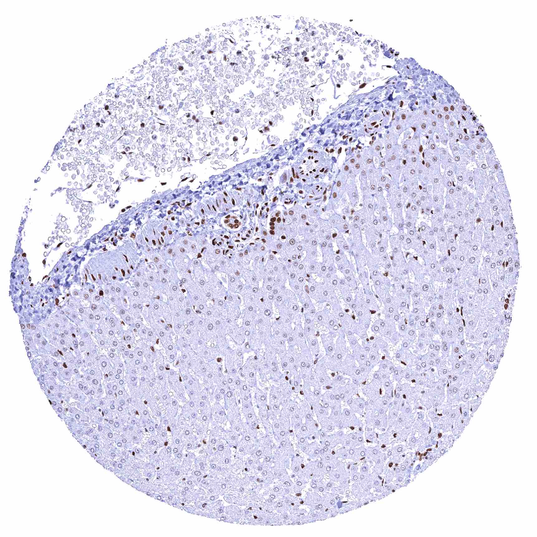
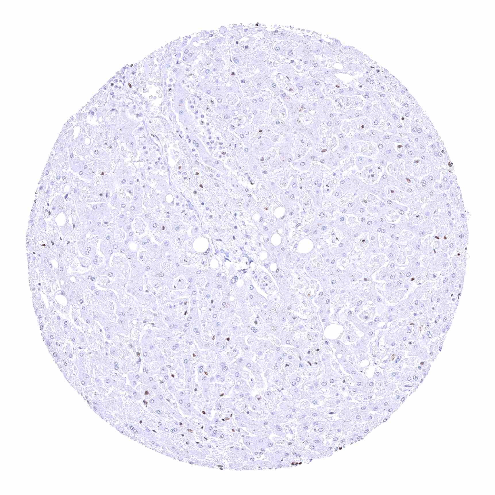
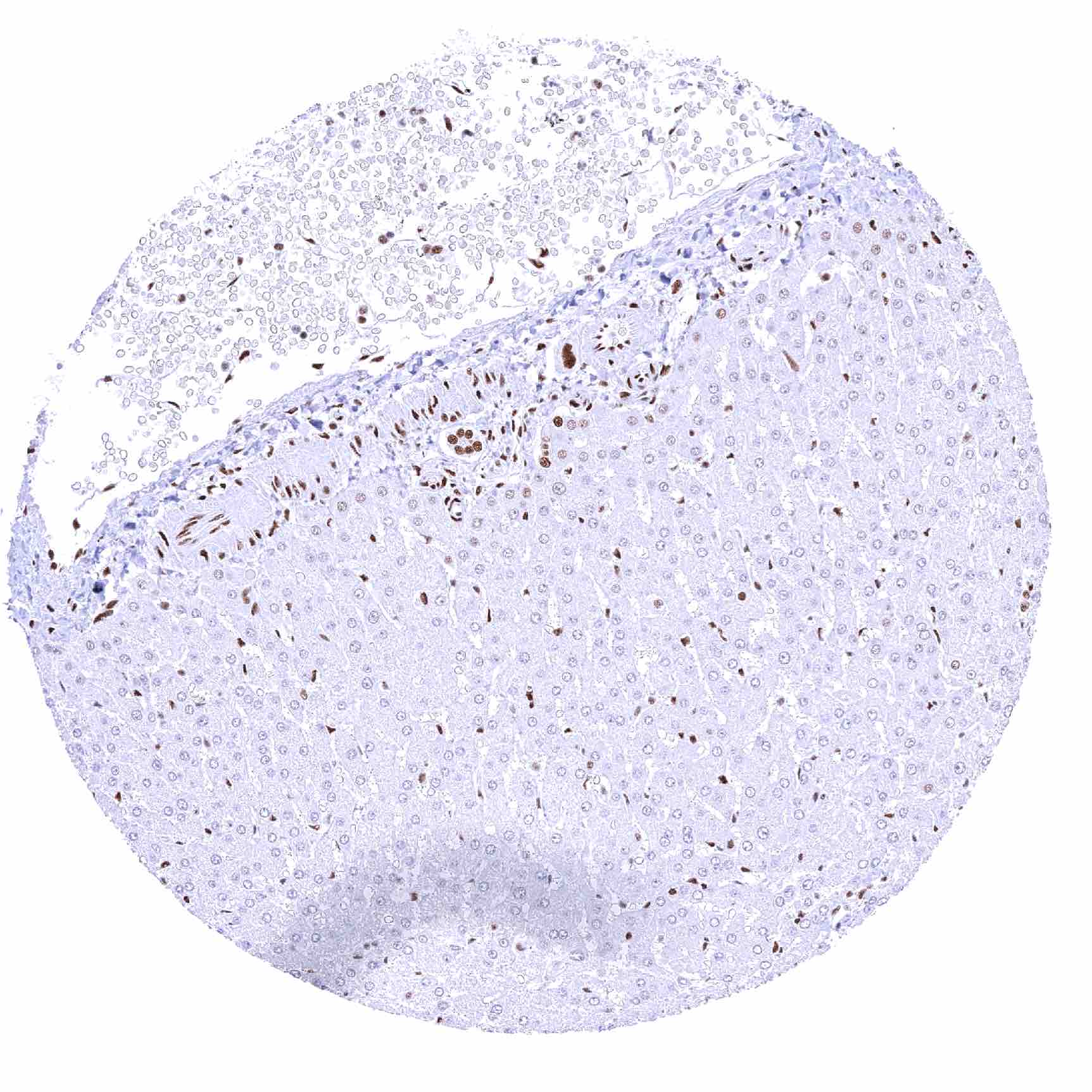
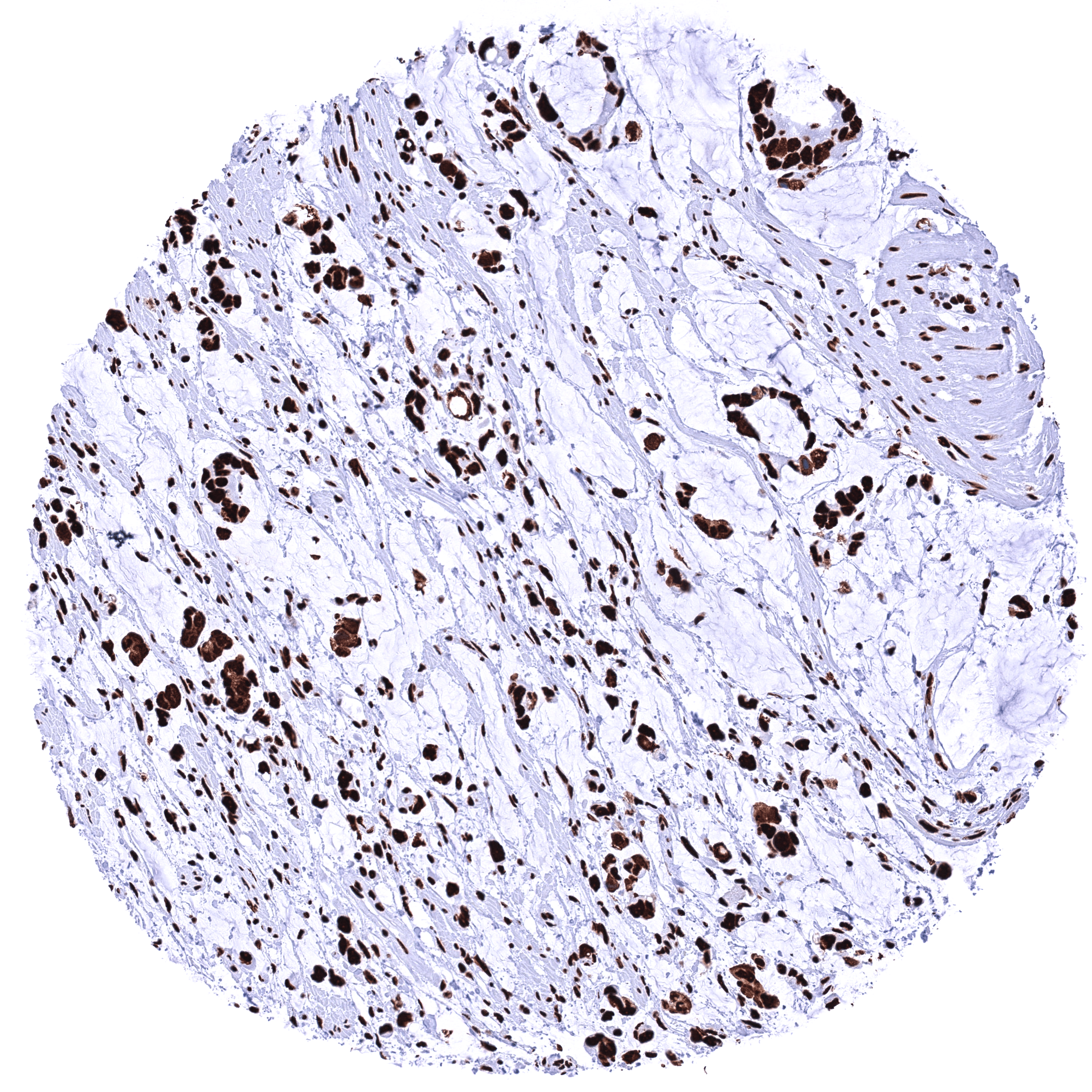
| Liver | The intensity of nuclear TRIM28 staining is less intense in hepatocytes than in other cell types (in some saples even completely missing). | |
| Gallbladder | Strong nuclear TRIM28 staining in all cells. | |
| Pancreas | Strong nuclear TRIM28 staining in all cells. | |
| Genitourinary | Kidney | Strong nuclear TRIM28 staining in all cells. |
| Urothelium | Strong nuclear TRIM28 staining in all cells. | |
| Male genital | Prostate | Strong nuclear TRIM28 staining in all cells. |
| Seminal vesicles | Strong nuclear TRIM28 staining in all cells. | |
| Testis | Strong nuclear TRIM28 staining in all cells. Exception: Spermatids are TRIM28 negative. | |
| Epididymis | Strong nuclear TRIM28 staining in all cells. | |
| Female genital | Breast | Strong nuclear TRIM28 staining in all cells. |
| Uterus, myometrium | Strong nuclear TRIM28 staining in all cells. | |
| Uterus, ectocervix | Slight decrease of the staining intensity from basal to superficial cell layers of squamous eithelium. | |
| Uterus endocervix | Strong nuclear TRIM28 staining in all cells. | |
| Uterus, endometrium | Strong nuclear TRIM28 staining in all cells. | |
| Fallopian Tube | Strong nuclear TRIM28 staining in all cells. | |
| Ovary | Strong nuclear TRIM28 staining in all cells. | |
| Placenta early | Strong nuclear TRIM28 staining in all cells. | |
| Placenta mature | Strong nuclear TRIM28 staining in all cells. | |
| Amnion | Strong nuclear TRIM28 staining in all cells. | |
| Chorion | Strong nuclear TRIM28 staining in all cells. | |
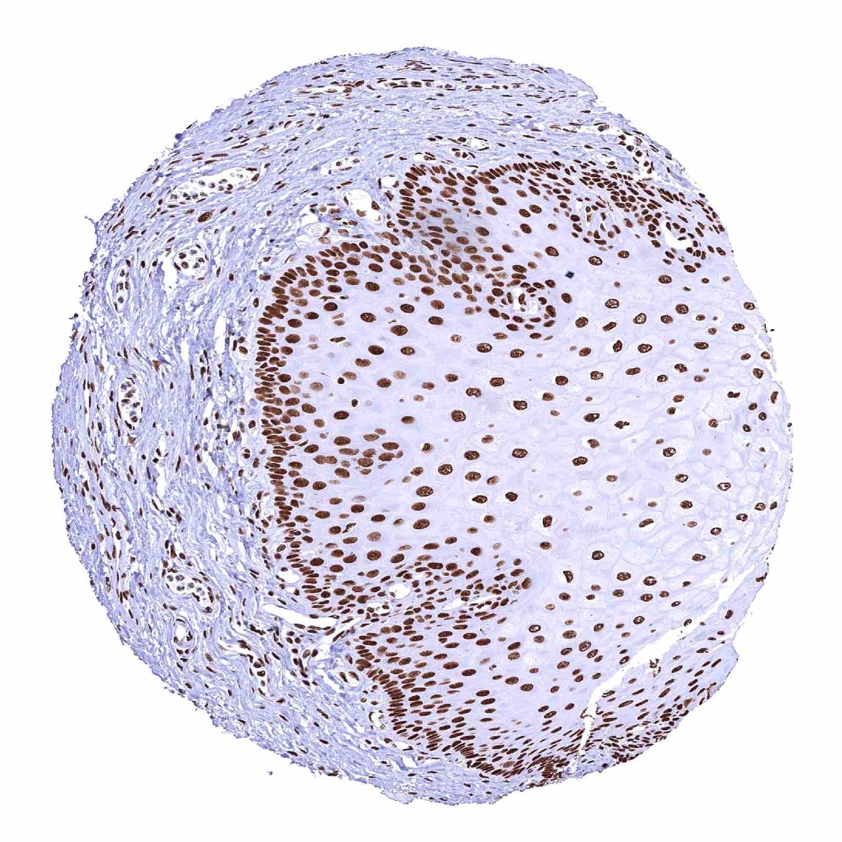
| Skin | Epidermis | Strong nuclear TRIM28 staining in all cells. |
| Sebaceous glands | Strong nuclear TRIM28 staining in all cells. | |
| Muscle/connective tissue | Heart muscle | Strong nuclear TRIM28 staining in all cells. |
| Skeletal muscle | Strong nuclear TRIM28 staining in all cells. | |
| Smooth muscle | Strong nuclear TRIM28 staining in all cells. | |
| Vessel walls | Strong nuclear TRIM28 staining in all cells. | |
| Fat | Strong nuclear TRIM28 staining in all cells. | |
| Stroma | Strong nuclear TRIM28 staining in all cells. | |
| Endothelium | Strong nuclear TRIM28 staining in all cells. | |
| Bone marrow/ lymphoid tissue | Bone marrow | Strong nuclear TRIM28 staining in all cells. |
| Lymph node | Strong nuclear TRIM28 staining in all cells. | |
| Spleen | Strong nuclear TRIM28 staining in all cells. | |
| Thymus | Strong nuclear TRIM28 staining in all cells. | |
| Tonsil | Strong nuclear TRIM28 staining in all cells. | |
| Remarks |
These findings are largely consistent with the RNA data described in the Human Protein Atlas (Tissue expression KAP1 / TRIM28).
Positive control = Colon: A strong nuclear TRIM28are staining should be seen in all cells.
Negative control = Testis: Spermatids are TRIM28 negative while all other cells show a strong nuclear TRIM28 staining of all cells.
Staining Pattern in Relevant Tumor Types
A strong TRIM28 staining is seen in the cells of most cancers. RNA data suggest that the expression level is highest in testicular cancers. Only a few cancers completely lack TRIM28 staining.
The TCGA findings on KAP1 / TRIM28 RNA expression in different tumor categories have been summarized in the Human Protein Atlas.
Compatibility of Antibodies
No data available at the moment
Protocol Recommendations
IHC users have different preferences on how the stains should look like. Some prefer high staining intensity of the target stain and even accept some background. Others favor absolute specificity and lighter target stains. Factors that invariably lead to more intense staining include higher concentration of the antibody and visualization tools, longer incubation time, higher temperature during incubation, higher temperature and longer duration of the heat induced epitope retrieval (slide pretreatment). The impact of the pH during slide pretreatment has variable effects and depends on the antibody and the target protein.
All images and data shown here and in our image galleries are obtained by the manual protocol described below. Other protocols resulting in equivalent staining are described as well.
Manual protocol
Freshly cut sections should be used (less than 10 days between cutting and staining). Heat-induced antigen retrieval for 5 minutes in an autoclave at 121°C in pH 7,8 Target Retrieval Solution buffer. Apply HMV335 at a dilution of 1:200 at 37°C for 60 minutes. Visualization of bound antibody by the EnVision Kit (Dako, Agilent) according to the manufacturer’s directions.
Potential Research Applications
- The role of TRIM28 in cancer is not sufficiently understood.
- The role of TRIM28 in viral diseases needs to be further investigated.
- The role of TRIM28 in transcription regulation awaits further clarification.
Evidence for Antibody Specificity in IHC
There are two ways how the specificity of antibodies can be documented for immunohistochemistry on formalin fixed tissues. These are: 1. Comparison with a second independent method for target expression measurement across a large number of different tissue types (orthogonal strategy), and 2. Comparison with one or several independent antibodies for the same target and showing that all positive staining results are also seen with other antibodies for the same target (independent antibody strategy).
Orthogonal validation: For the antibody HMV335, specificity of its nuclear staining is consistent by the good concordance of the immunostaining data with data from three independent RNA screening studies, including the Human Protein Atlas (HPA) RNA-seq tissue dataset, the FANTOM5 project, and the Genotype-Tissue Expression (GTEx) project, which are all summarized in the Human Protein Atlas (Tissue expression KAP1 / TRIM28). In agreement with HMV335 immunostaining data, TRIM28 expression is ubiquitous and somehow predominated in the bone marrow and lymphoid tissues. However, orthogonal validation is not optimal for assessing ubiquitously expressed proteins.
Comparison of antibodies: True expression of TRIM28 in all cell types with nuclear TRIM28 positivity by HMV335 is corroborated by an identical nuclear staining obtained by a commercially available independent second antibody (termed “validation antibody”). Most of all, both antibodies showed absent or reduced saining in hepatocytes of some liver samples.