295,00 € – 995,00 €
Product details
Synonyms = CD27 ligand (CD27L); CD27LG; Ki24; Surface antigen CD70; Tumor necrosis factor ligand superfamily member 7 (TNFSF7)
Antibody type = Recombinant Rabbit monoclonal
Clone = MSVA-070R
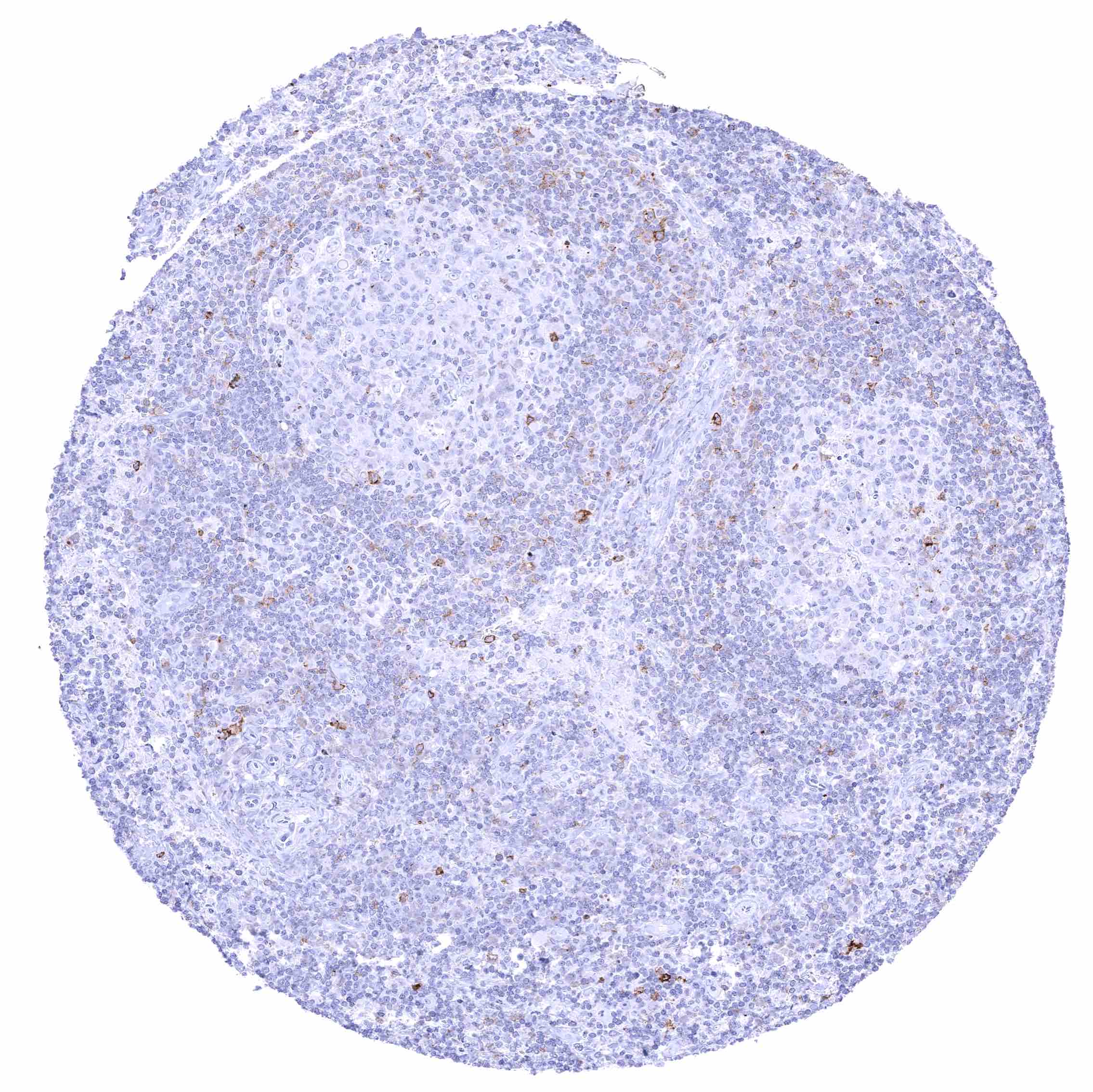
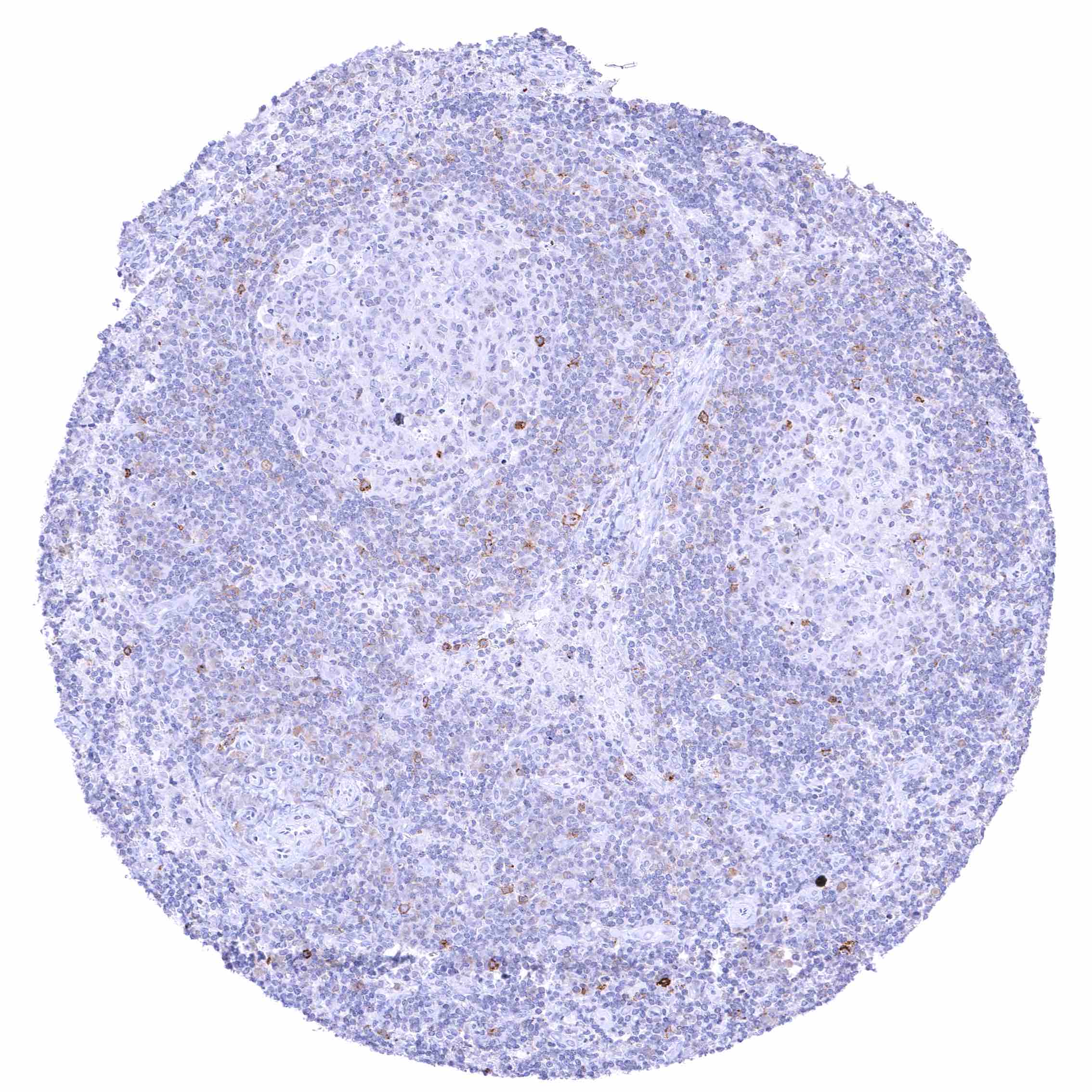
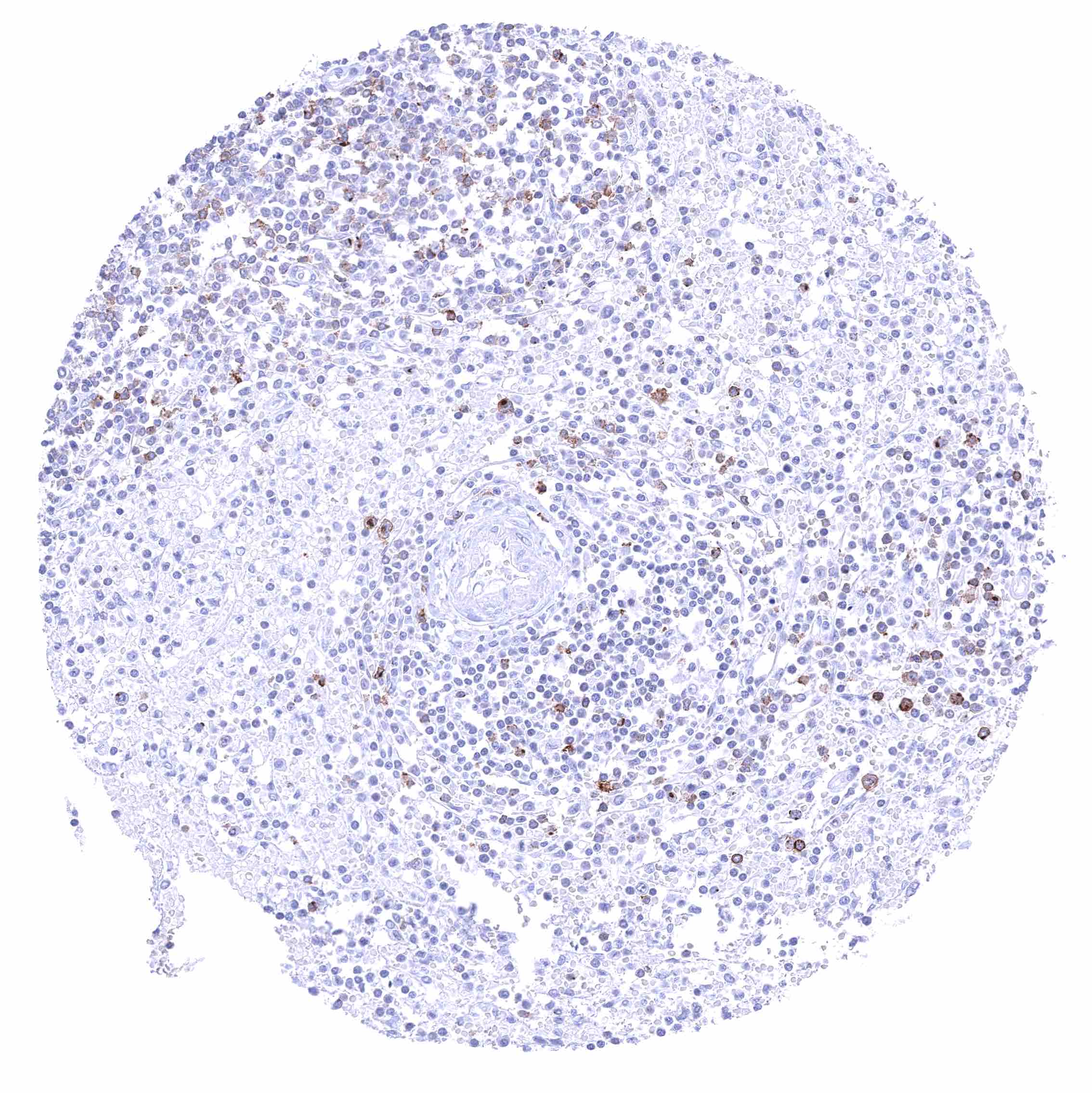
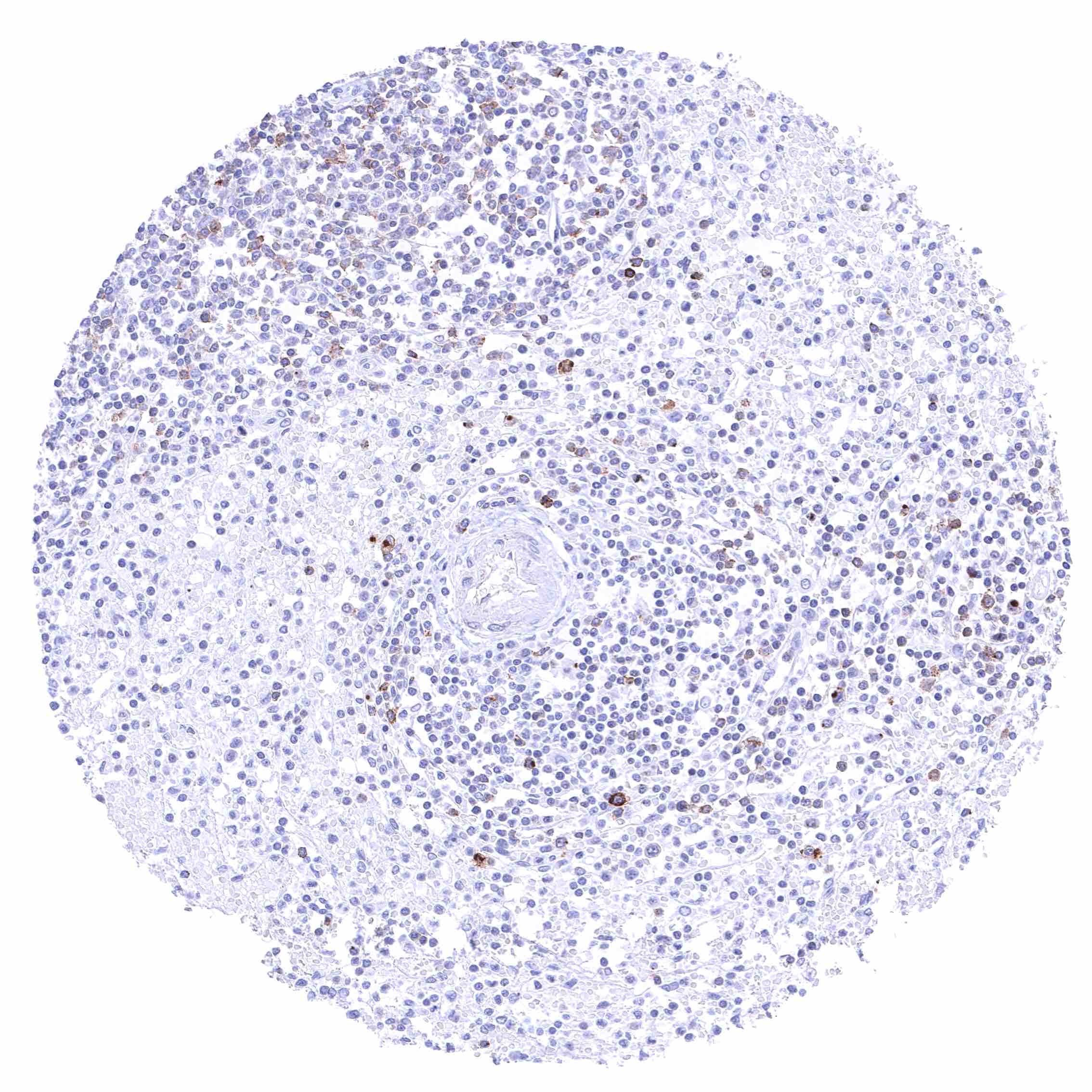
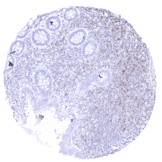
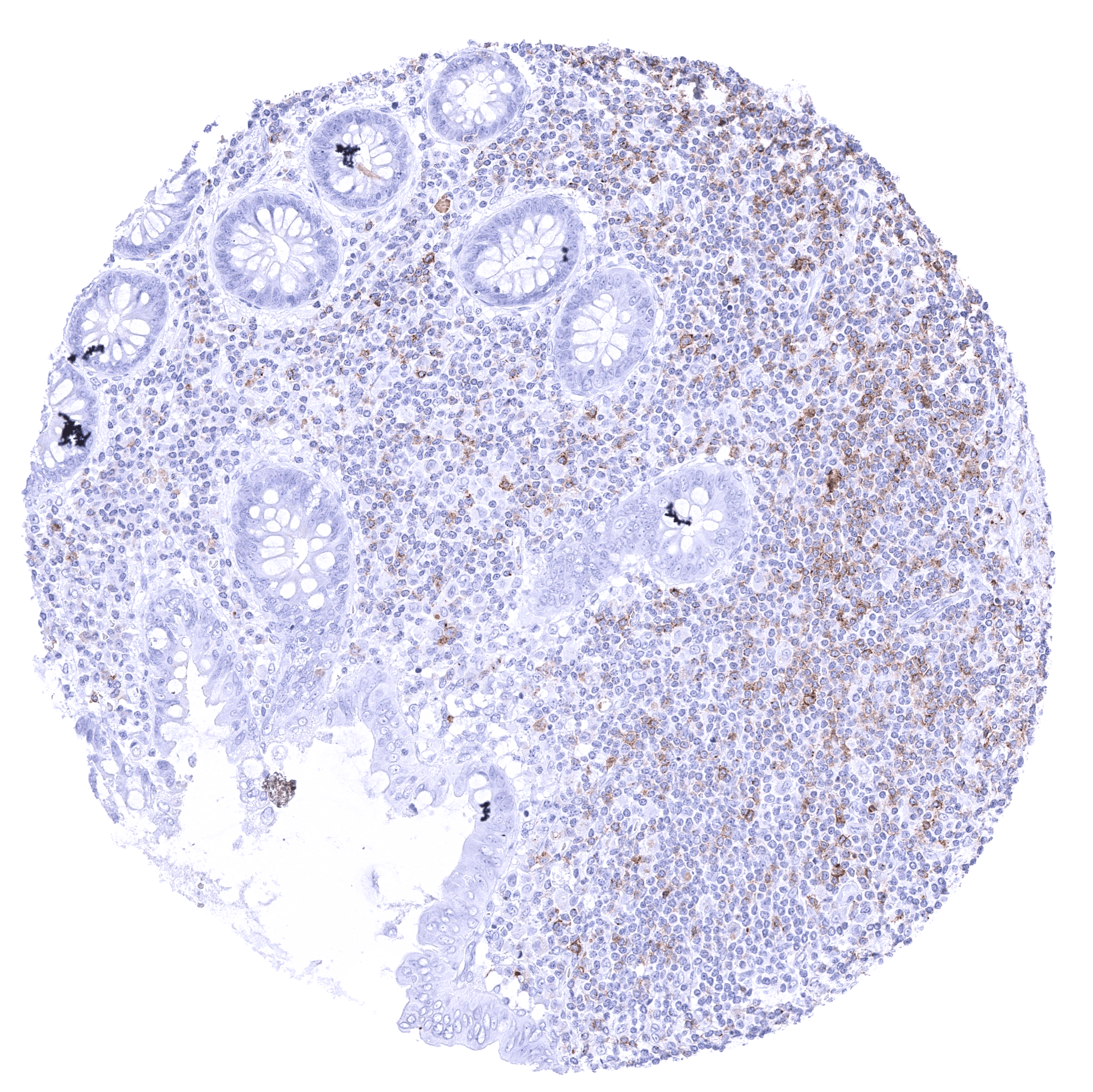
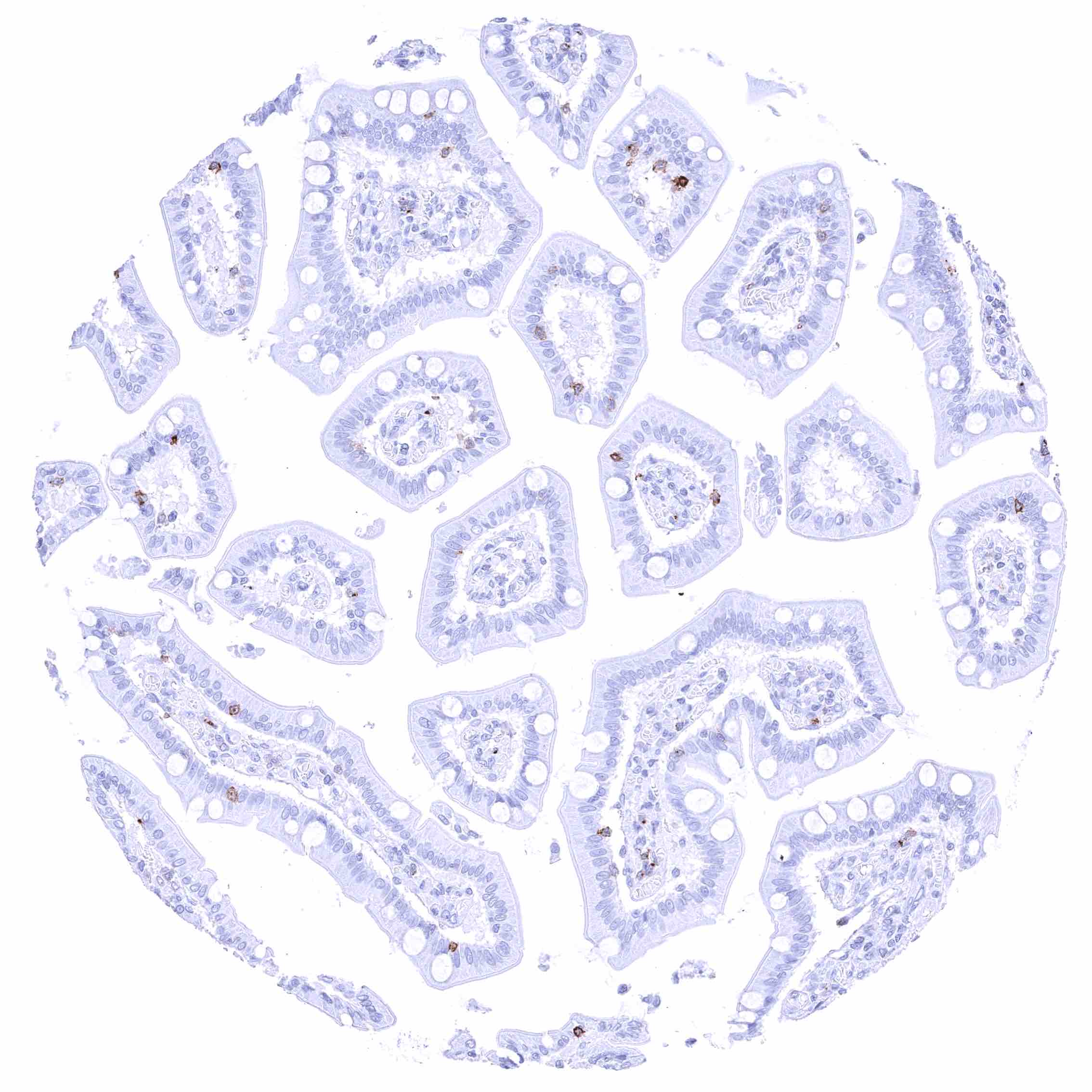
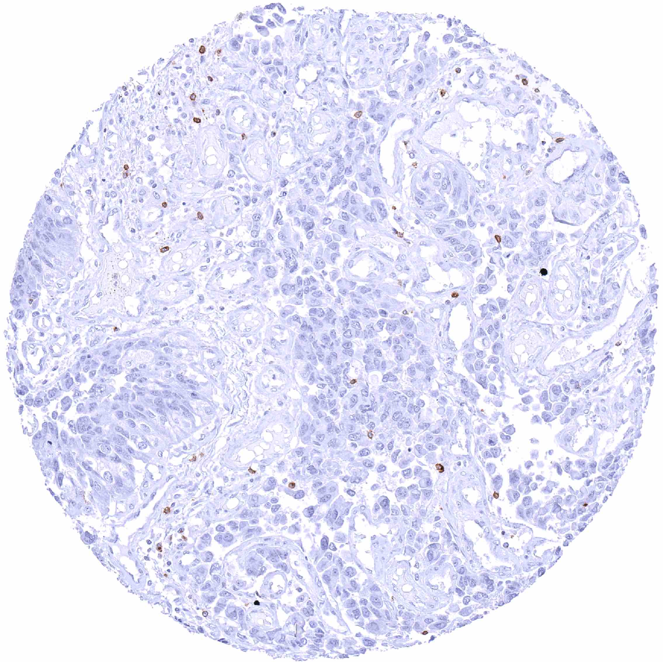
Positive control = Appendix: A distinct membranous CD70 staining should be seen in a subset of inflammatory cells (especially interfollicular).
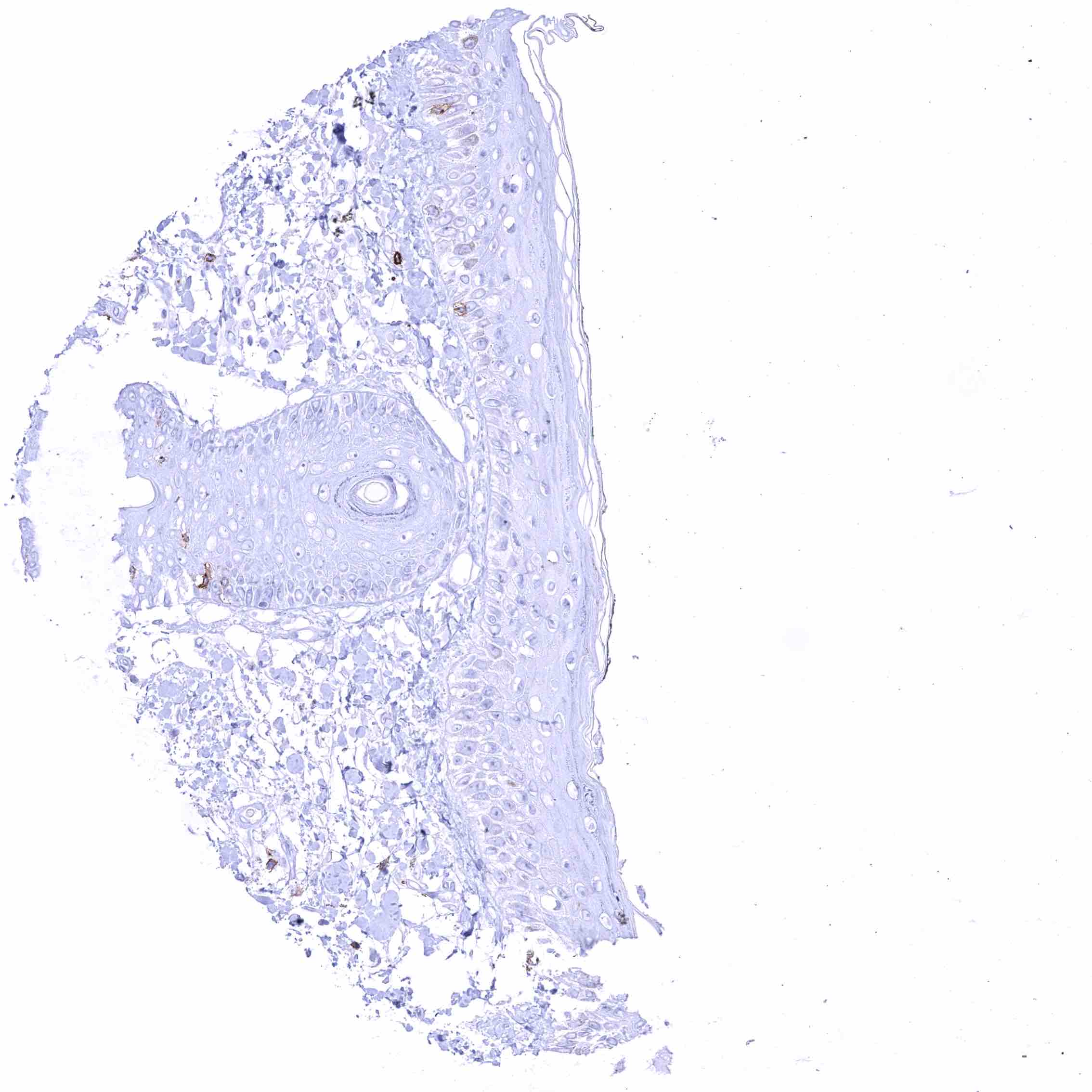
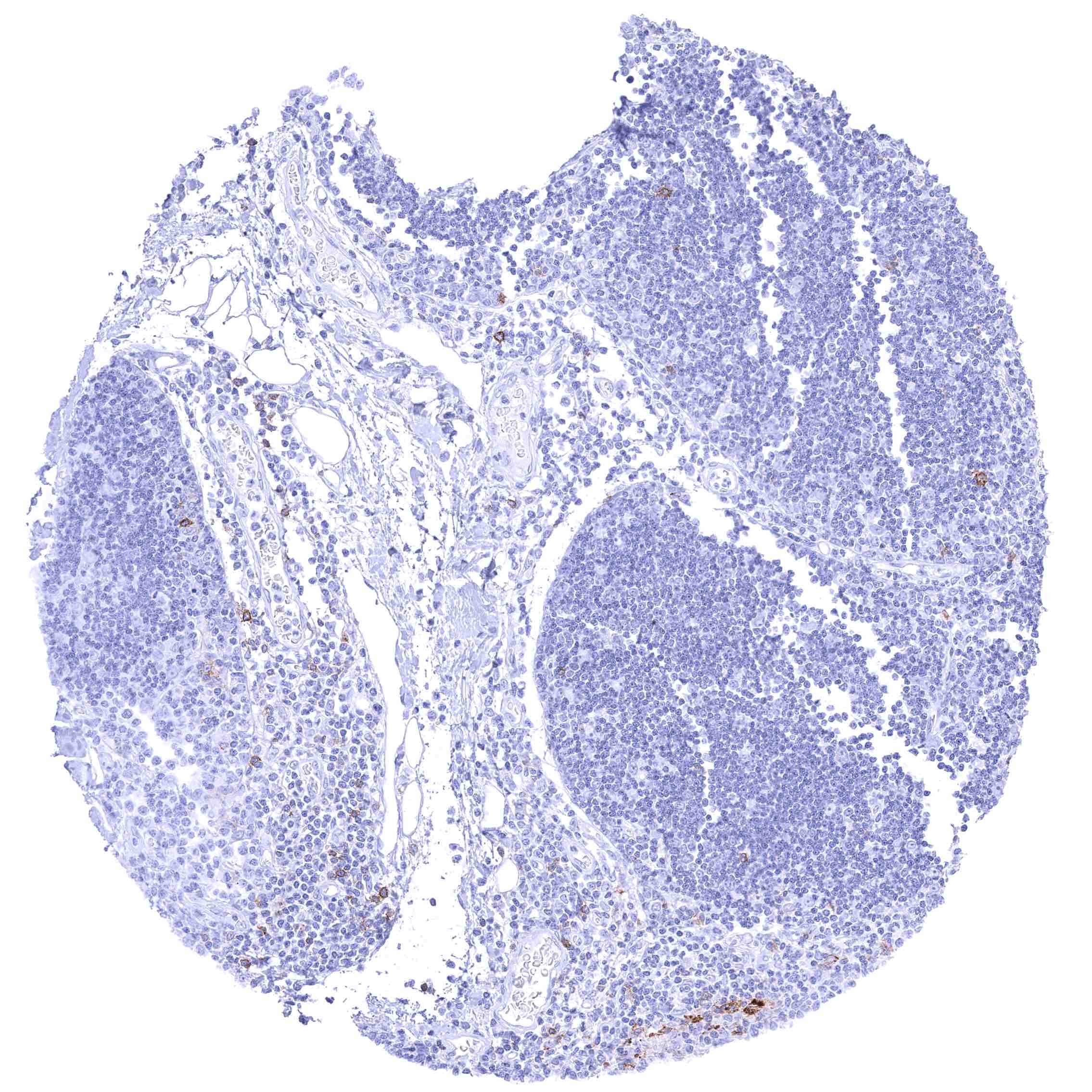
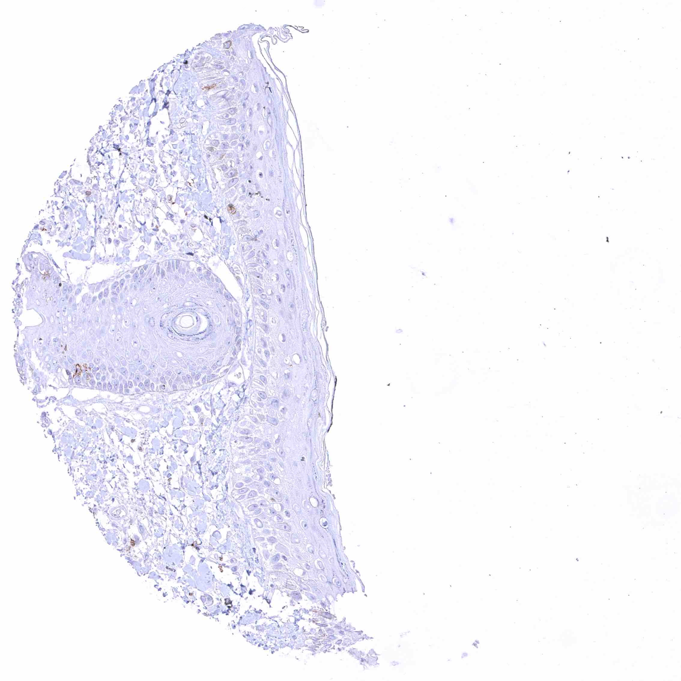
Negative control = Appendix: CD70 staining should be absent in epithelial cells.
Cellular localization = Membranous
Reactivity = Human
Application = Immunohistochemistry
Dilution = 1:100 – 1:200
Intended Use = Research Use Only
Relevance of Antibody
CD70 is an immune regulator and therapeutic target protein.
Biology Behind
CD70 (Cluster of Differentiation 70) is a type II transmembrane protein which is coded by the TNFSF7 gene located on chromosome 19p13.3. The CD70 protein is a member of the tumor necrosis factor receptor family. Under physiologic conditions, CD70 is transiently expressed on antigen-activated B and T cells, NK cells and mature dendritic cells where it acts as a costimulatory molecule and interacts with CD27. Upon contact with CD70, the extracellular domain of CD27 is cleaved off and disposed as a soluble fragment (called sCD27) into body fluids. The CD70/CD27 axis plays an important role in the regulation of the immune response. Its role may be particularly important in cancer. CD70/CD27 signaling is believed to support tumoral growth by constraining inflammatory T-cell expansion, guiding the proliferation of immune-suppressive T-regulatory cells, and empowering immune evasion. Neo-expression of CD70 has been described to occur in various cancer types. Accordingly, CD70 is considered a promising drug target for oncology.
Staining Pattern in Normal Tissues
Among normal tissues, CD70 expression is limited to subsets of cells of lymphatic organs.
Images describing the CD70 staining pattern in normal tissues obtained by the antibody MSVA-070R are shown in our “Normal Tissue Gallery”.
| Brain | Cerebrum | Negative. |
| Cerebellum | Negative. | |
| Endocrine Tissues | Thyroid | Negative. |
| Parathyroid | Negative. | |
| Adrenal gland | Negative. | |
| Pituitary gland | Negative. | |
| Respiratory system | Respiratory epithelium | Negative. |
| Lung | Negative. | |
| Gastrointestinal Tract | Salivary glands | Negative. |
| Esophagus | Negative. | |
| Stomach | Negative. | |
| Duodenum | Negative. | |
| Small intestine | Negative. | |
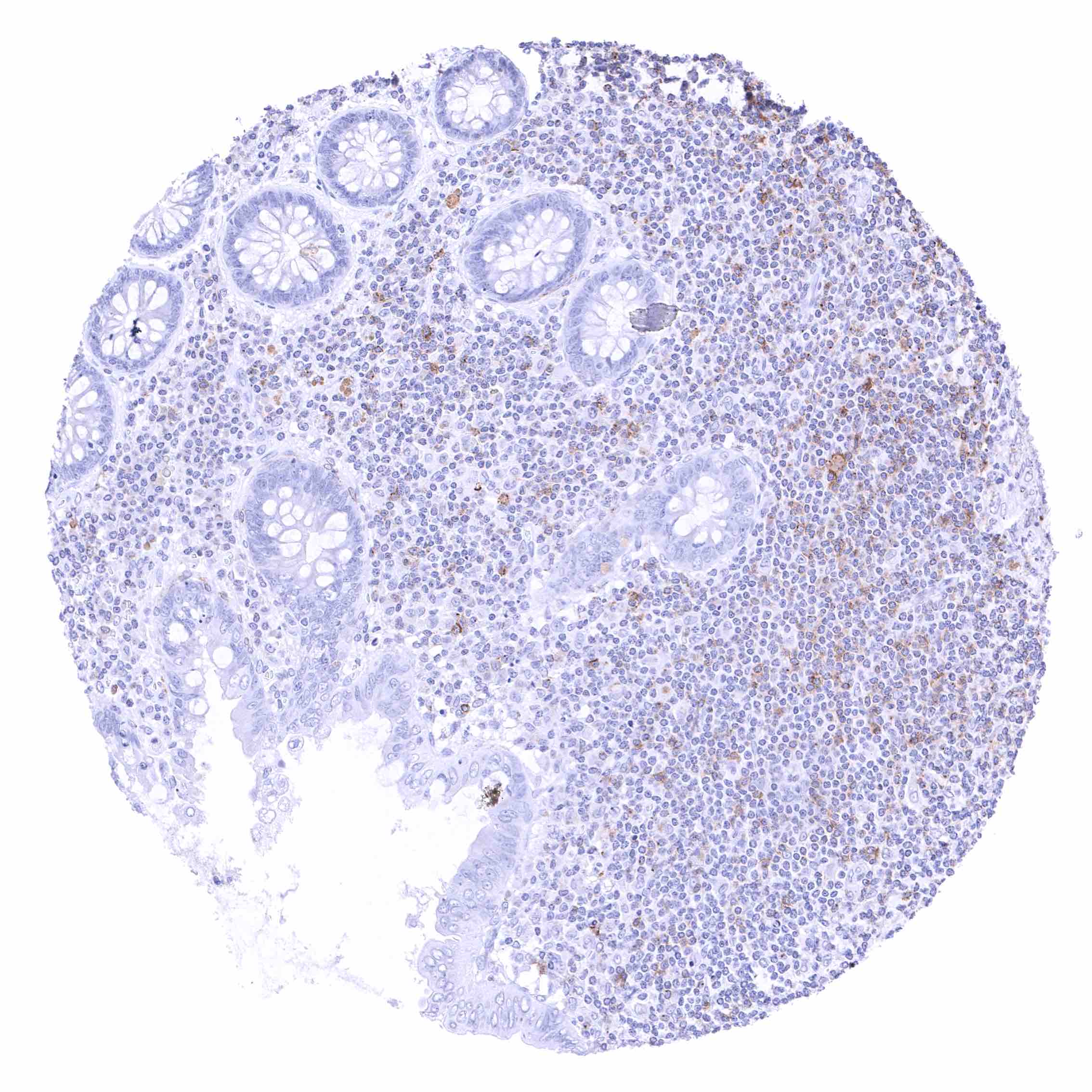
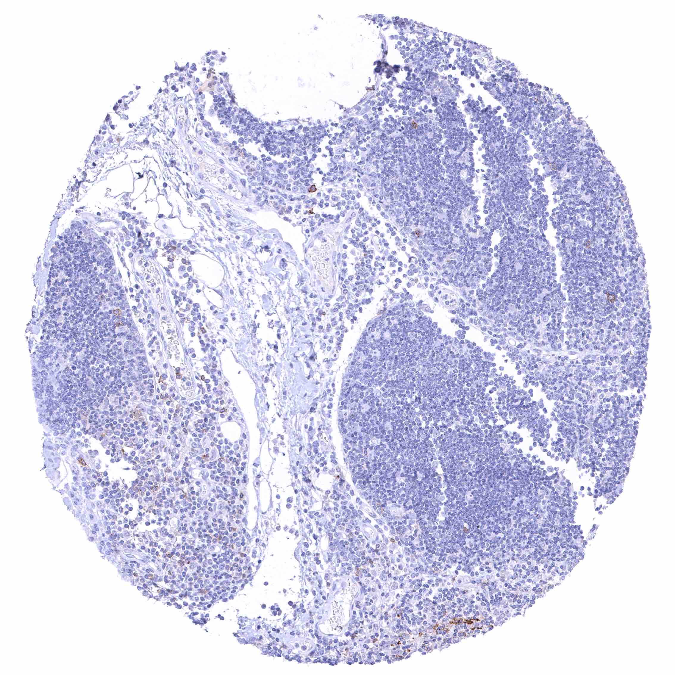
| Appendix | Distinct CD70 positivity of a fraction of lymphocytic cells, predominantly interfollicular. Epithelial cells are CD70 negative. | |
| Colon | Negative. | |
| Rectum | Negative. | |
| Liver | Negative. | |
| Gallbladder | Negative. | |
| Pancreas | Negative. | |
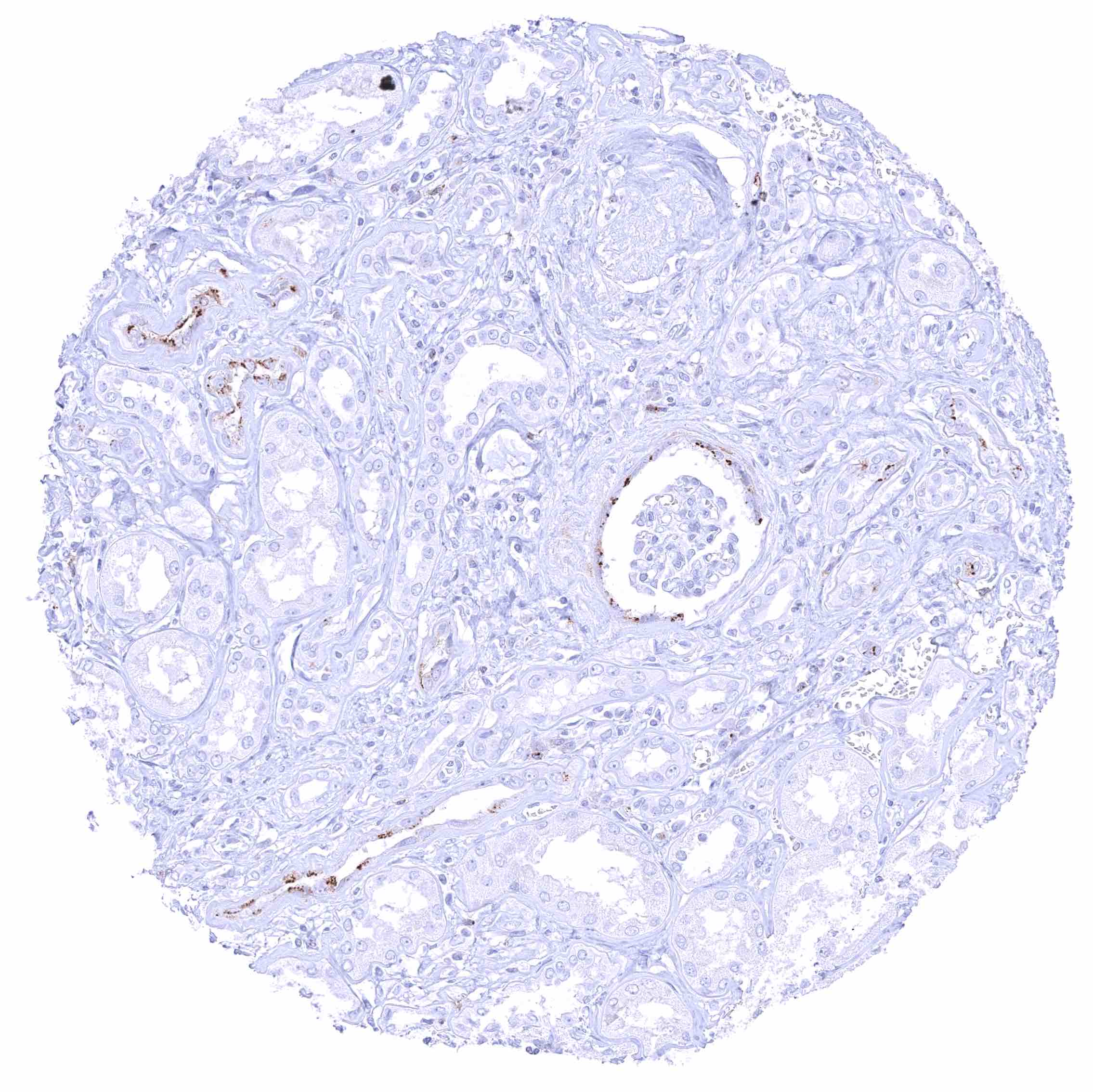
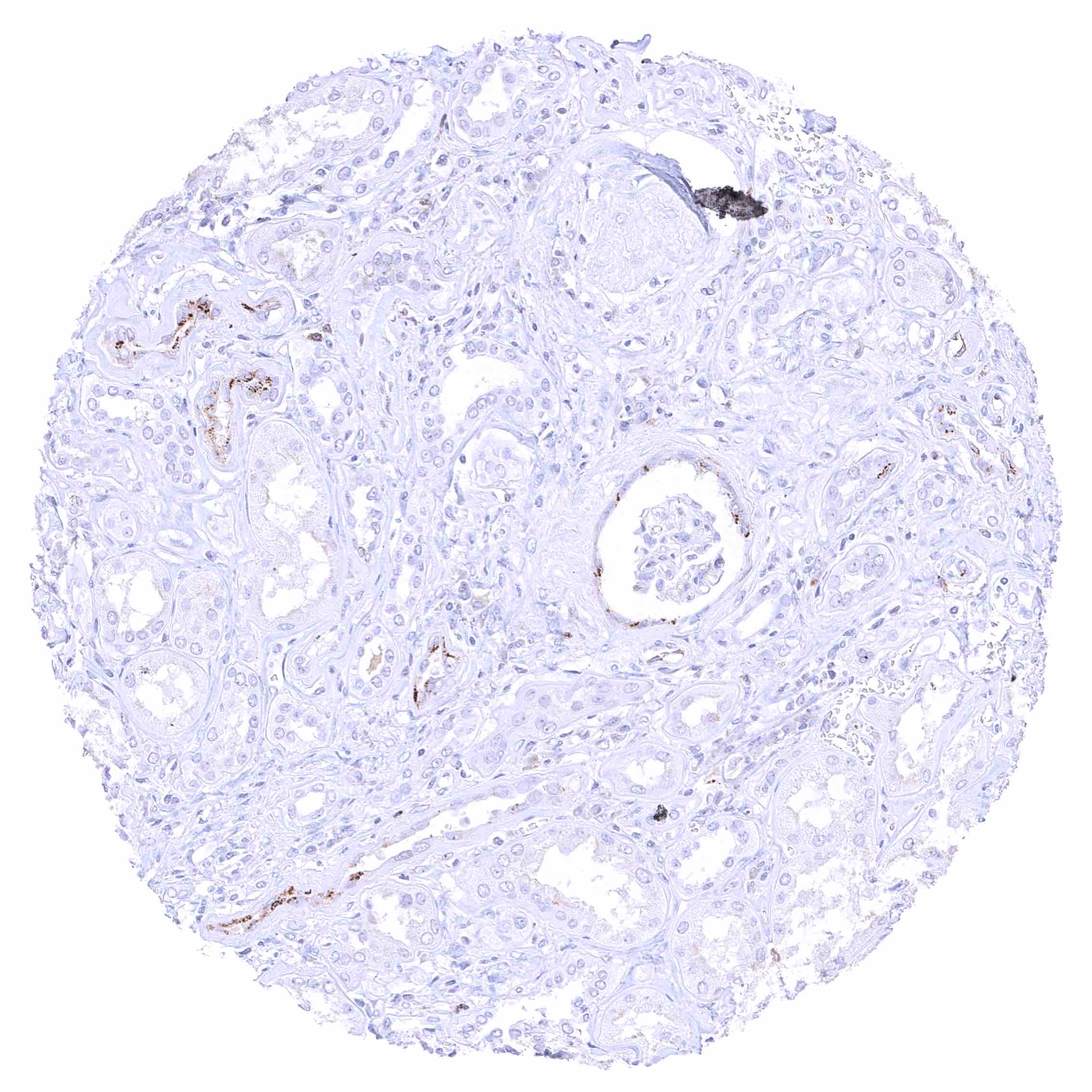
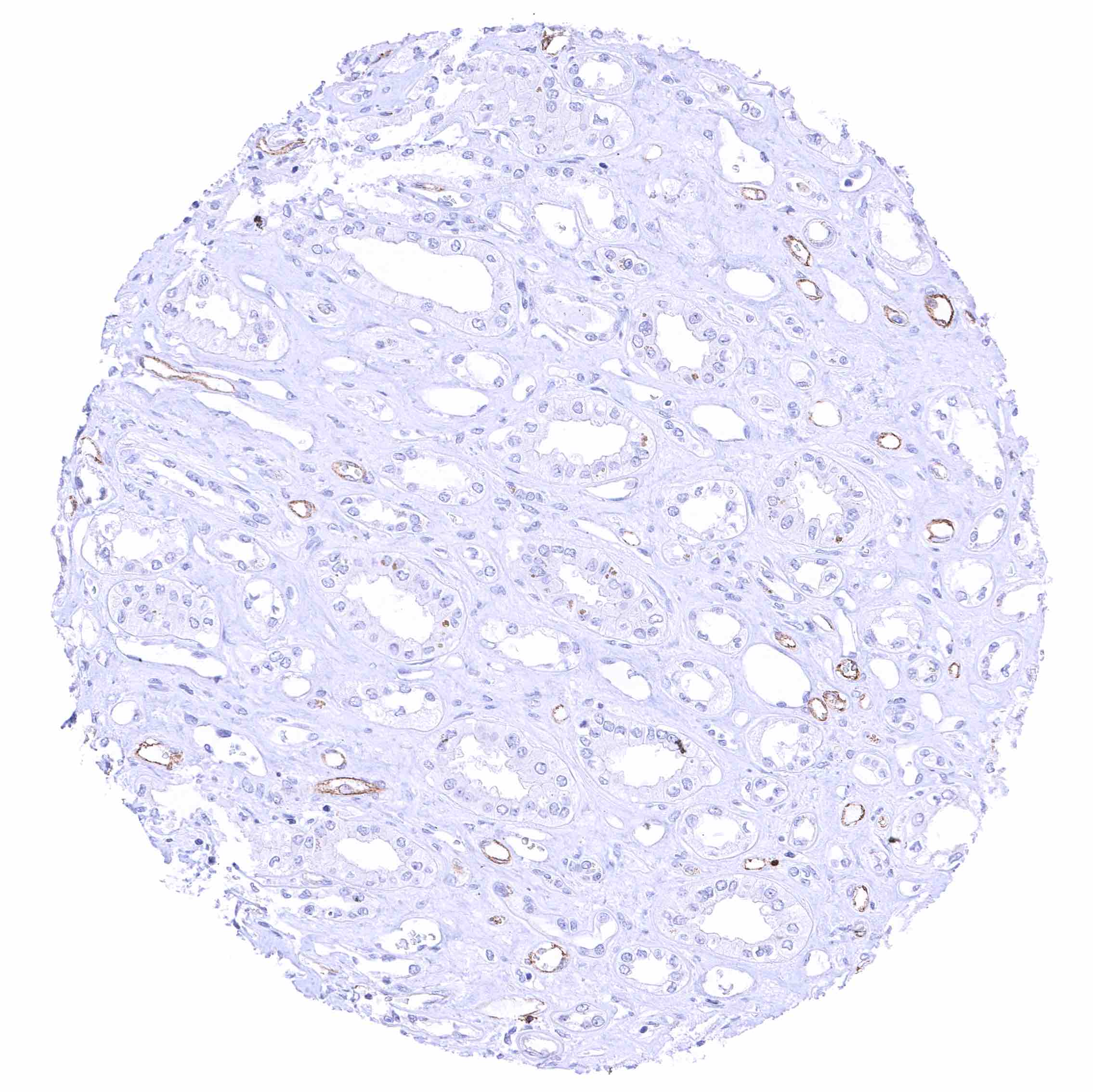
| Genitourinary | Kidney | A distinct CD70 staining of the luminal surface membrane can be seen in individual tubuli and at the parietal layer of the Bowman capsule. It is possible, that these stainings only (or preferably) occur in case of tissue damage. |
| Urothelium | Negative. | |
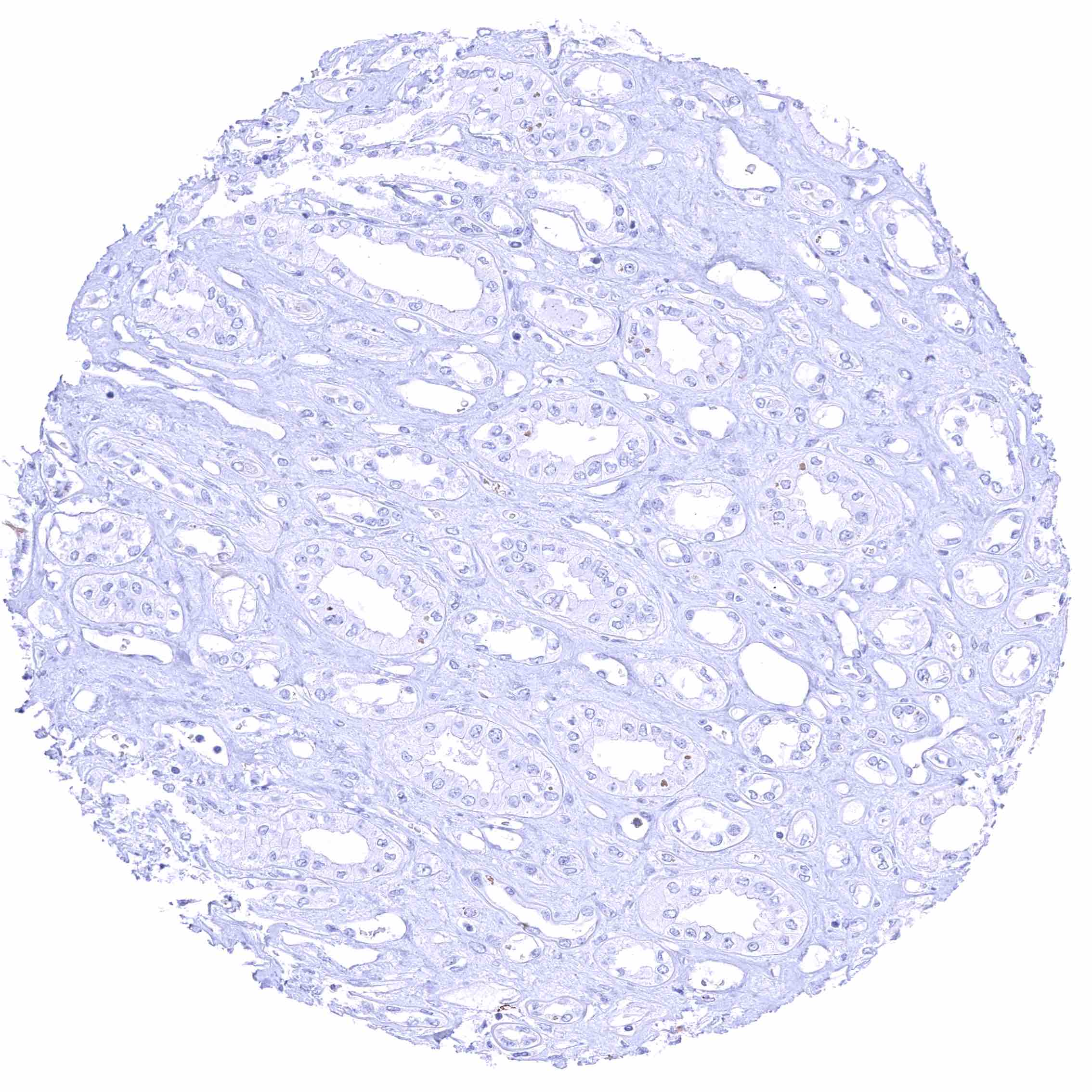
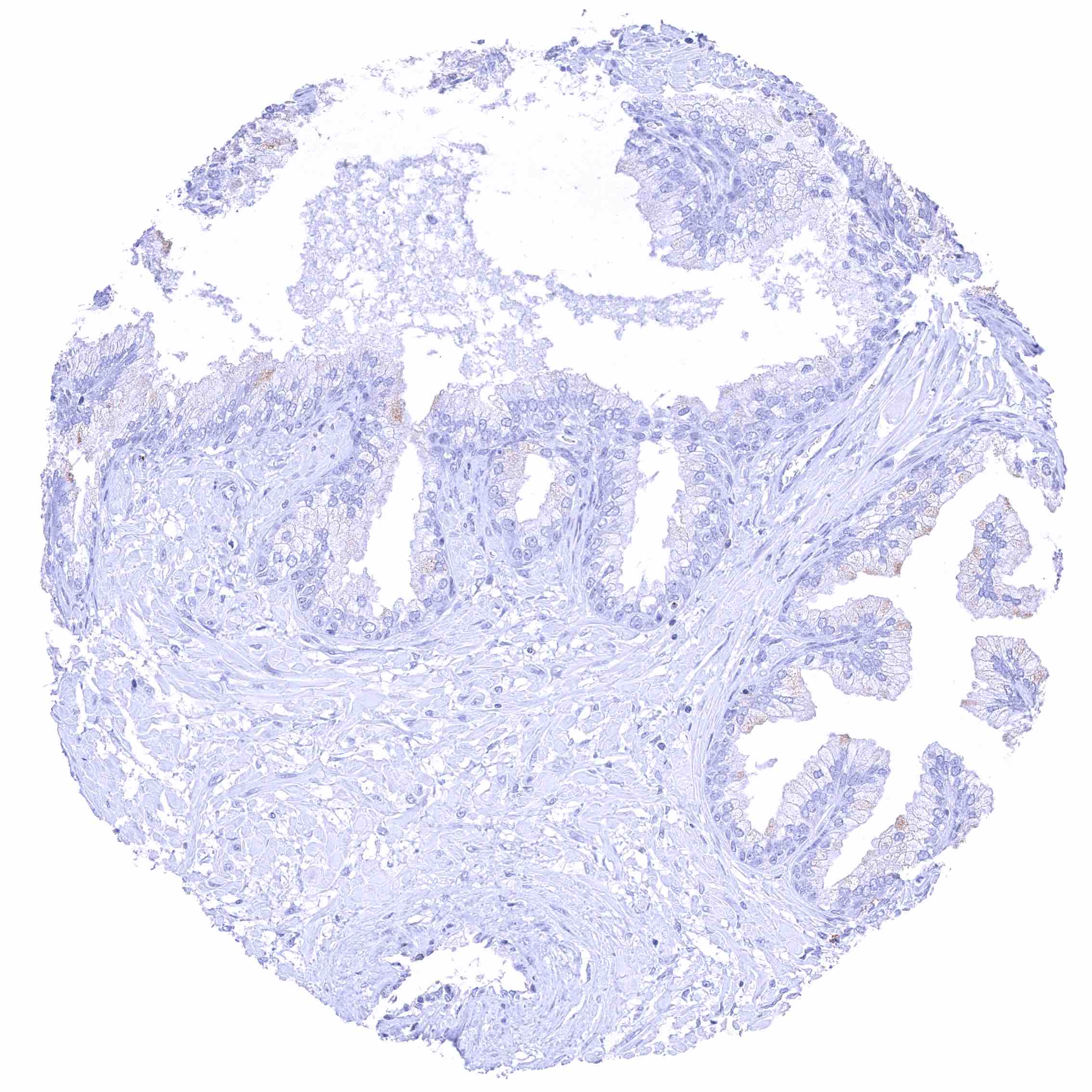
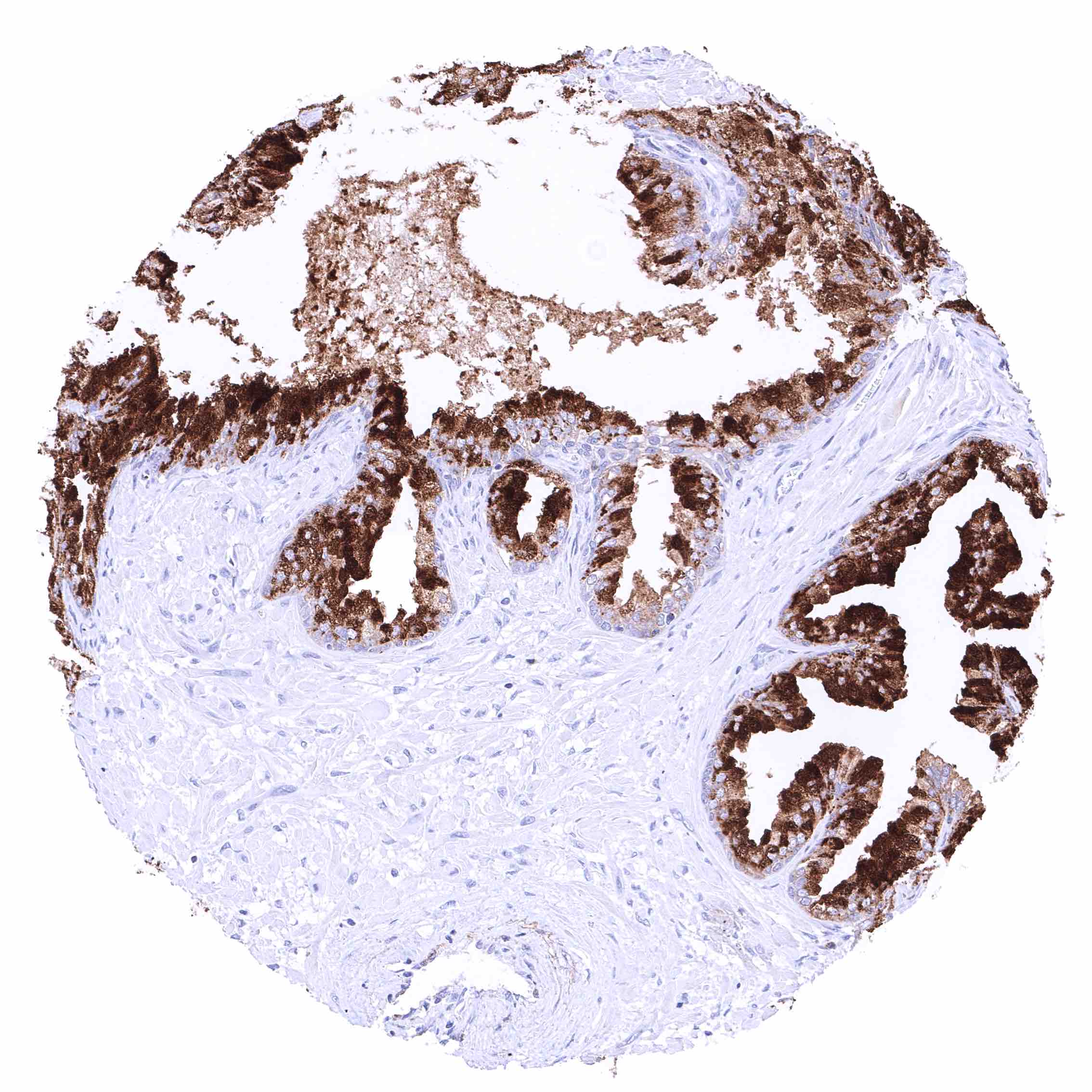
| Male genital | Prostate | Negative. |
| Seminal vesicles | Negative. | |
| Testis | Negative. | |
| Epididymis | Negative. | |
| Female genital | Breast | Negative. |
| Uterus, myometrium | Negative. | |
| Uterus, ectocervix | Negative. | |
| Uterus endocervix | Negative. | |
| Uterus, endometrium | Negative. | |
| Fallopian Tube | Negative. | |
| Ovary | Negative. | |
| Placenta early | Negative. | |
| Placenta mature | Negative. | |
| Amnion | Negative. | |
| Chorion | Negative. | |
| Skin | Epidermis | Negative. |
| Sebaceous glands | Negative. | |
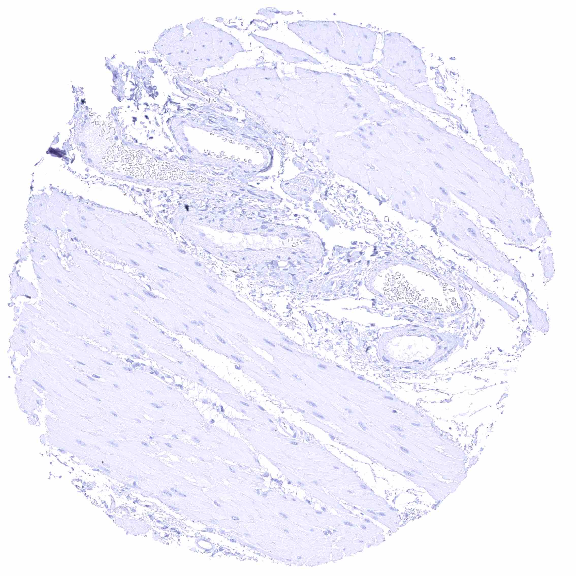
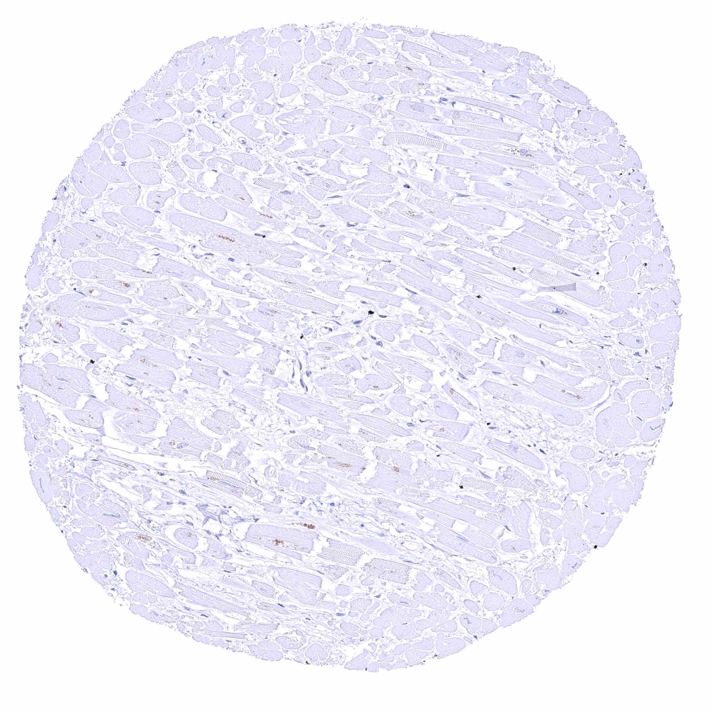
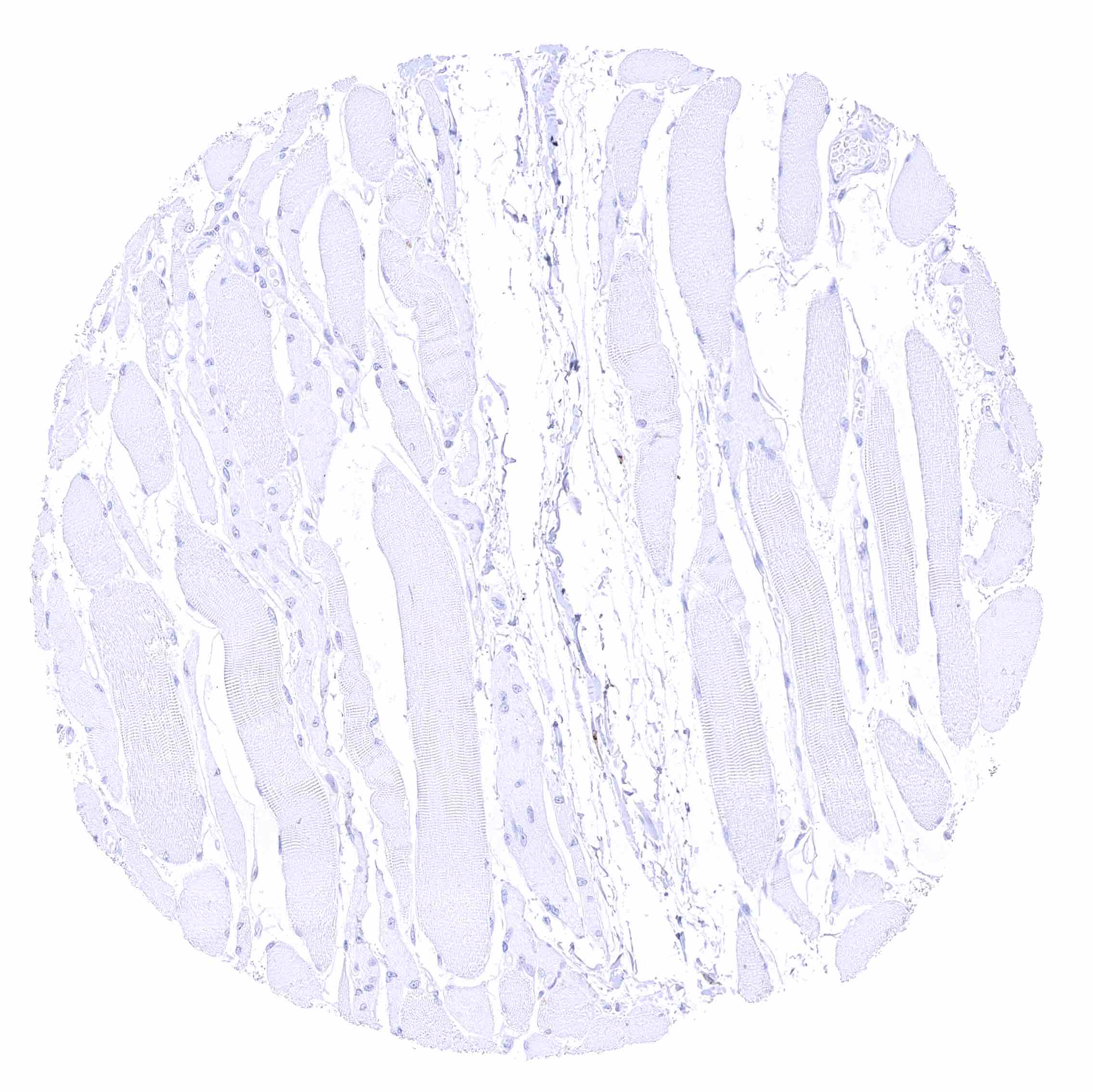
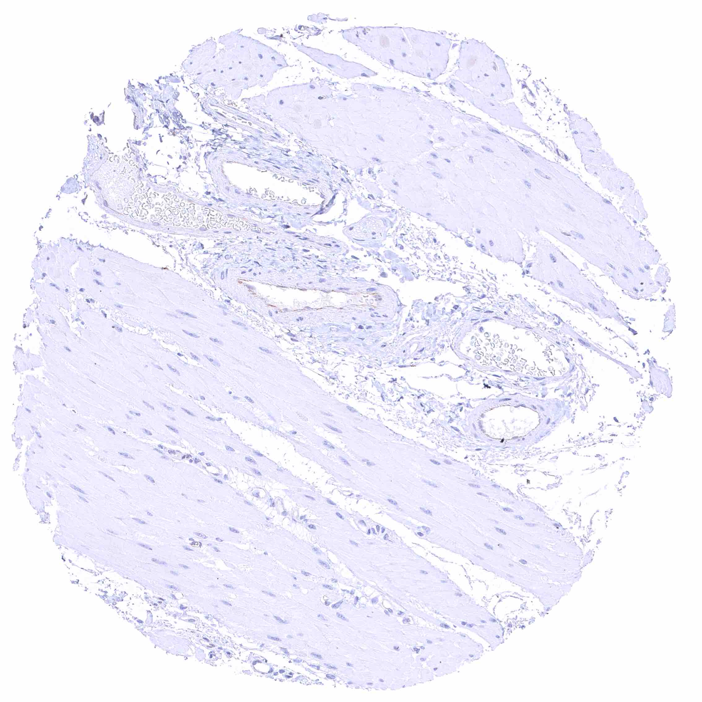
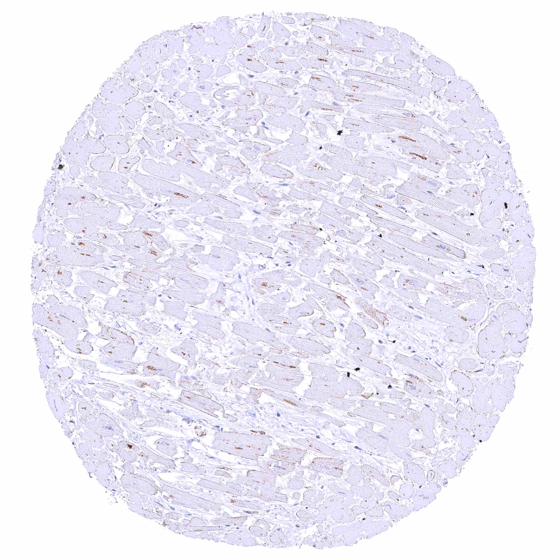
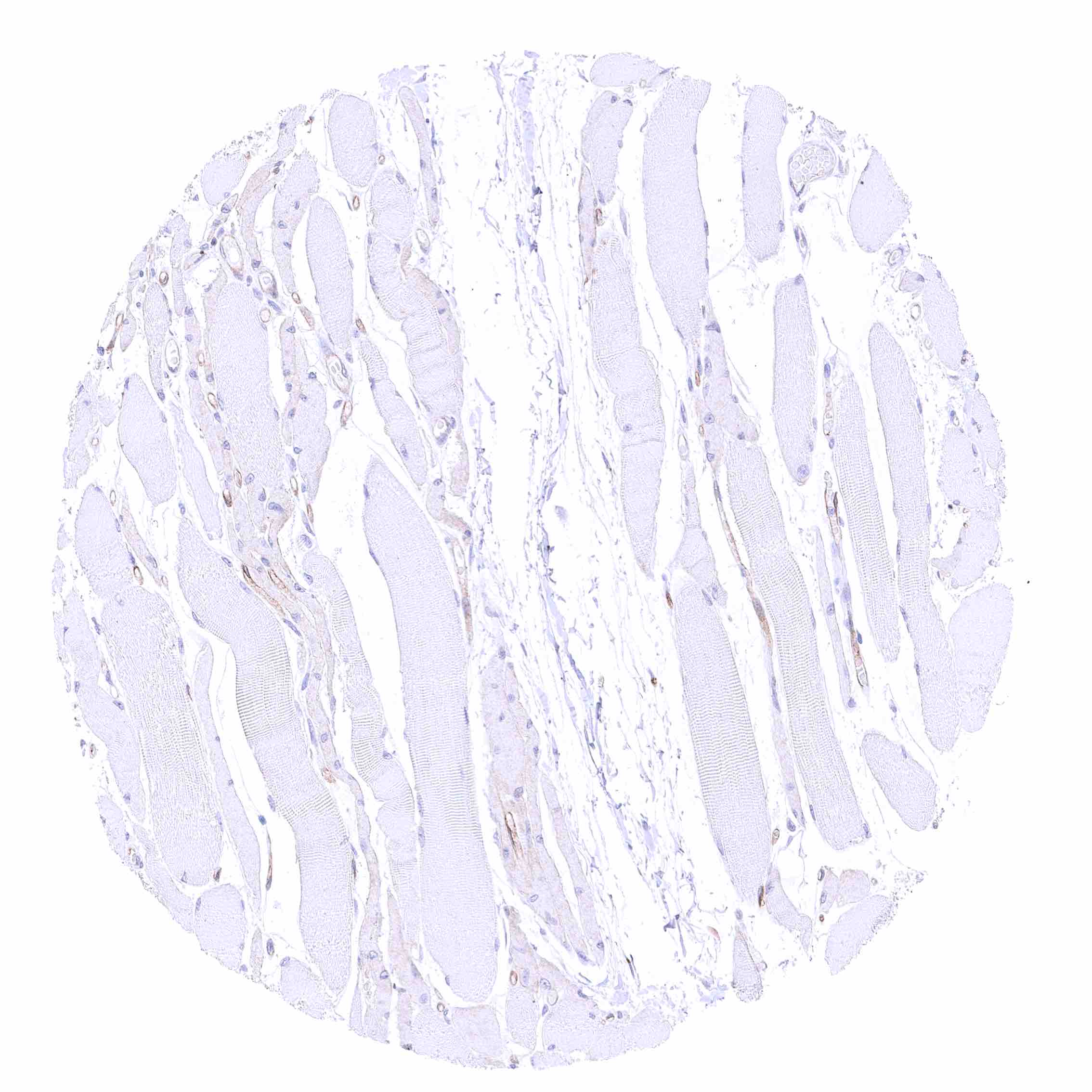
| Muscle/connective tissue | Heart muscle | Negative. |
| Skeletal muscle | Negative. | |
| Smooth muscle | Negative. | |
| Vessel walls | Negative. | |
| Fat | Negative. | |
| Stroma | Negative. | |
| Endothelium | Negative. | |
| Bone marrow/lymphoid tissue | Bone marrow | Weak CD70 staining in few cells. |
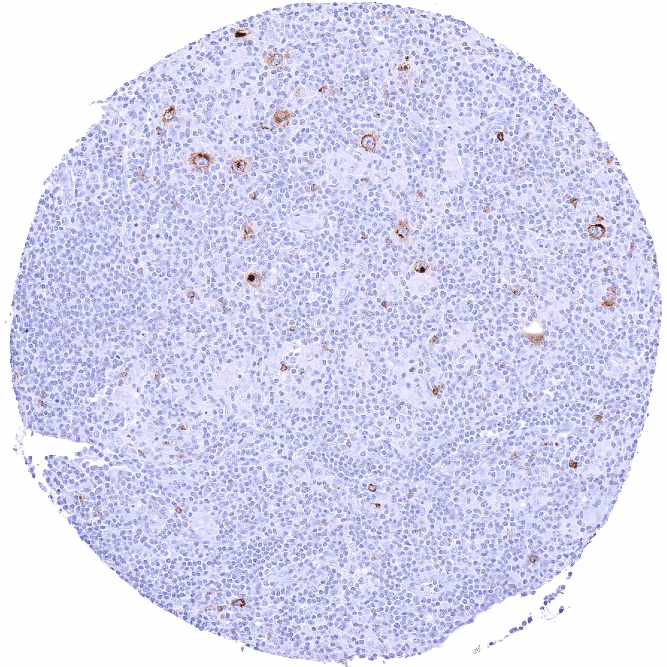
| Lymph node | Membranous CD70 staining of variable intensity in subsets of inflammatory cells (mostly lymphocytes). Most CD70 positive cells are interfollicular. | |
| Spleen | Membranous CD70 staining of variable intensity in a fraction of cells. | |
| Thymus | Membranous CD70 staining of variable intensity in a small subset of cells. | |
| Tonsil | Membranous CD70 staining of variable intensity in subsets of inflammatory cells (mostly lymphocytes). Most CD70 positive cells are interfollicular. Squamous epithelium remains CD70 negative. | |
| Remarks | A CD70 positivity of a subset of inflammatory cells is regularly seen in tissues samples that contain inflammatory cells. |
These findings are largely consistent with the RNA data described in the Human Protein Atlas (Tissue expression CD70).
Positive control = Appendix: A distinct membranous CD70 staining should be seen in a subset of inflammatory cells (especially interfollicular).
Negative control = Appendix: CD70 staining should be absent in epithelial cells.


Staining Pattern in Relevant Tumor Types
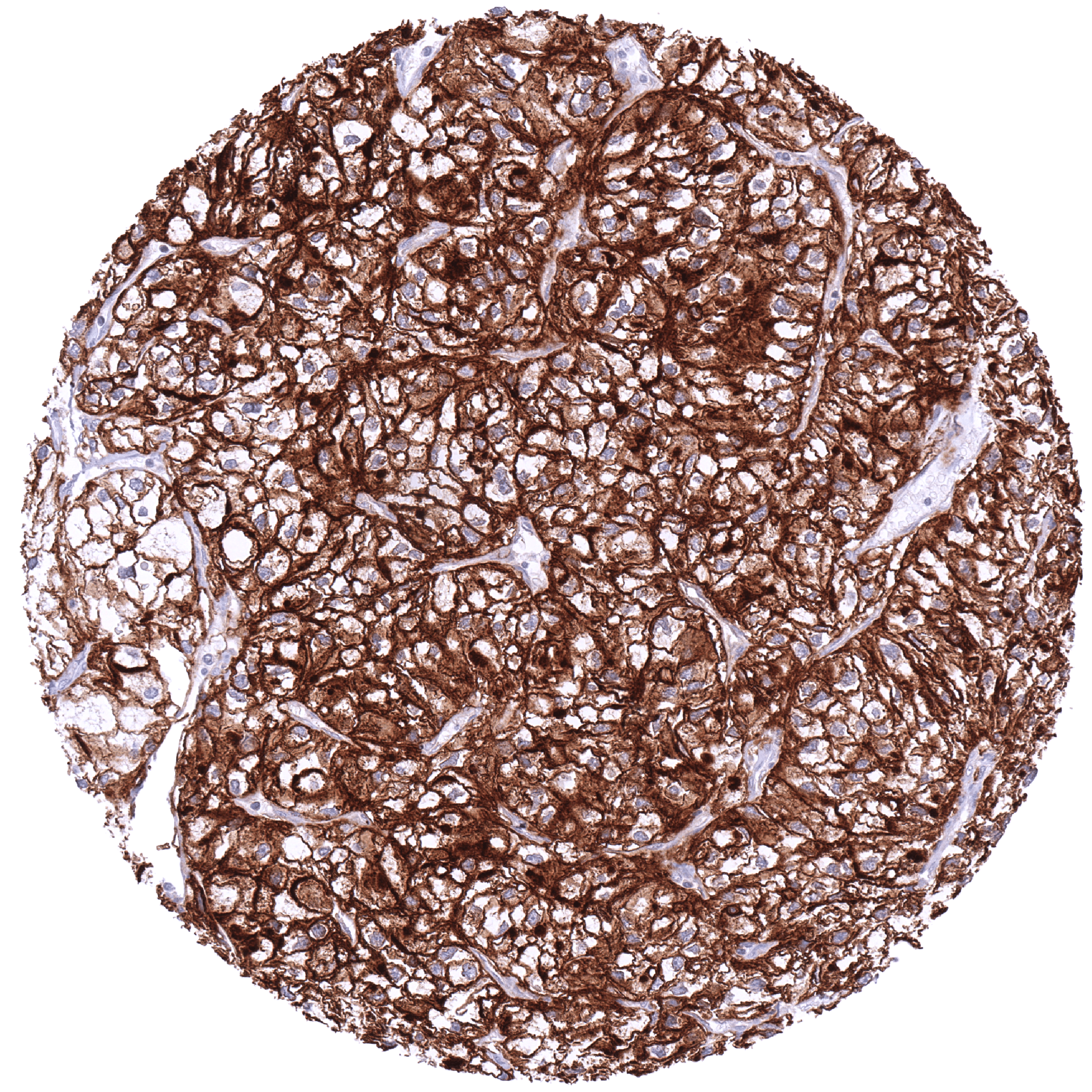
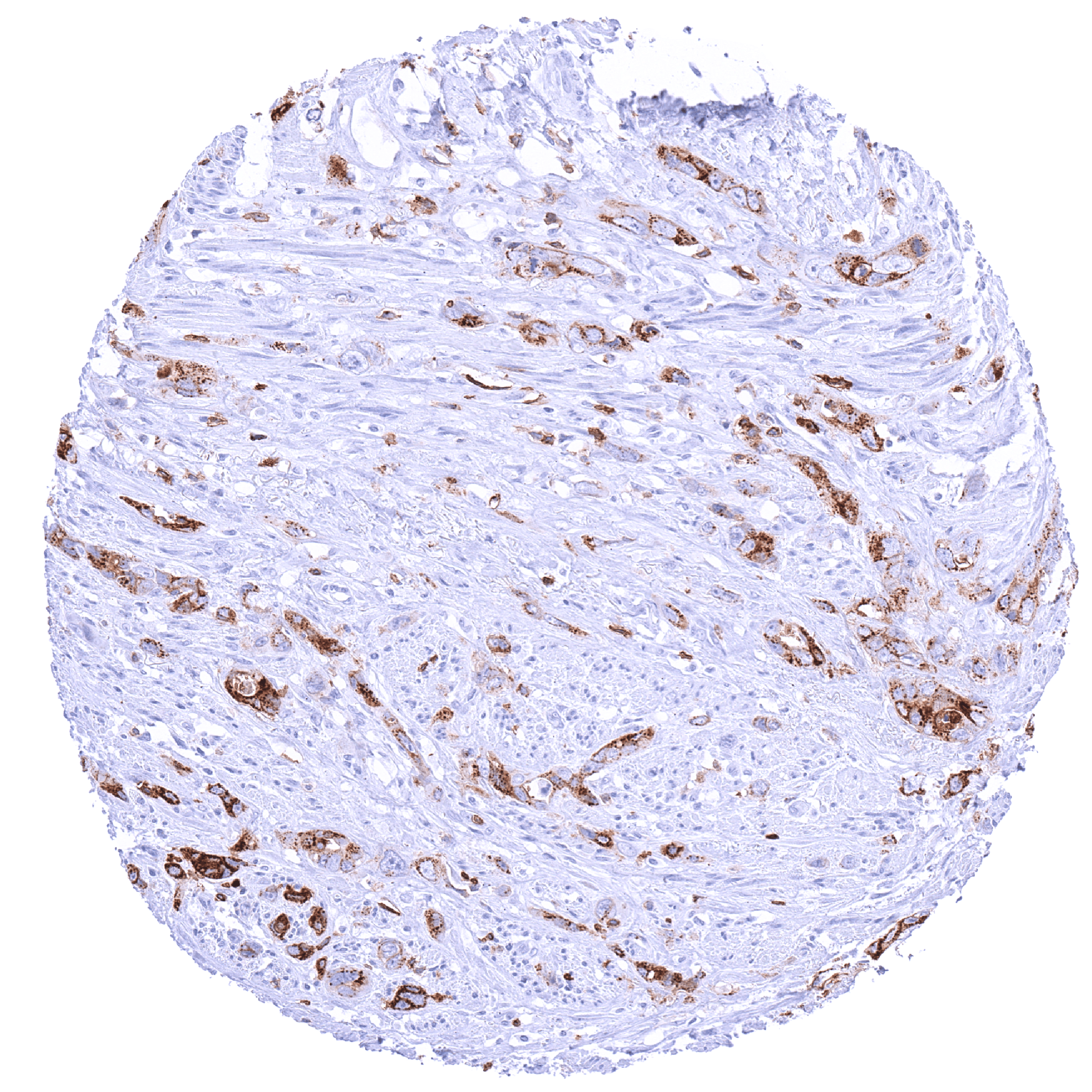
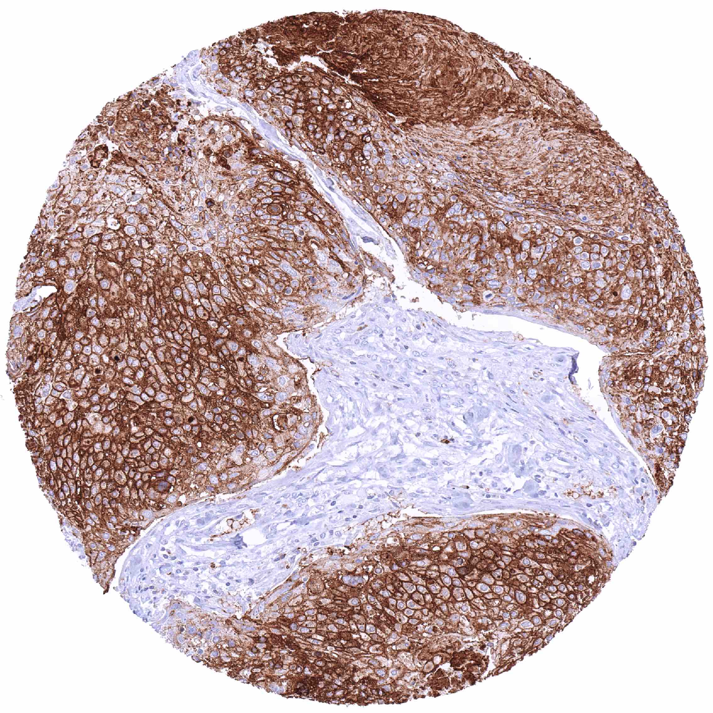
CD70 expression is most commonly seen in different types of lymphoma and in clear cell renal cell carcinoma. Other cancer types have also been described to express CD70 at lower frequency.
The TCGA findings on CD70 RNA expression in different tumor categories have been summarized in the Human Protein Atlas.
Compatibility of Antibodies
No data available at the moment
Protocol Recommendations
IHC users have different preferences on how the stains should look like. Some prefer high staining intensity of the target stain and even accept some background. Others favor absolute specificity and lighter target stains. Factors that invariably lead to more intense staining include higher concentration of the antibody and visualization tools, longer incubation time, higher temperature during incubation, higher temperature and longer duration of the heat induced epitope retrieval (slide pretreatment). The impact of the pH during slide pretreatment has variable effects and depends on the antibody and the target protein.
All images and data shown here and in our image galleries are obtained by the manual protocol described below. Other protocols resulting in equivalent staining are described as well.
Manual protocol
Freshly cut sections should be used (less than 10 days between cutting and staining). Heat-induced antigen retrieval for 5 minutes in an autoclave at 121°C in pH 7,8 Target Retrieval Solution buffer. Apply MSVA-070R at a dilution of 1:150 at 37°C for 60 minutes. Visualization of bound antibody by the EnVision Kit (Dako, Agilent) according to the manufacturer’s directions.
Potential Research Applications
- Various drugs targeting CD70 are in clinical trials.
- The prevalence and clinical significance of CD70 expression on tumor cells in different cancer types is unclear.
- The role and clinical significance of CD70 expressing cancer associated fibroblasts is unclear.
- The exact mechanism of CD70/CD27 signaling needs to be further evaluated.
Evidence for Antibody Specificity in IHC
There are two ways how the specificity of antibodies can be documented for immunohistochemistry on formalin fixed tissues. These are: 1. Comparison with a second independent method for target expression measurement across a large number of different tissue types (orthogonal strategy), and 2. Comparison with one or several independent antibodies for the same target and showing that all positive staining results are also seen with other antibodies for the same target (independent antibody strategy).
Orthogonal validation: For the antibody MSVA-070R specificity is supported by the good concordance of the immunostaining data with data from three independent RNA screening studies, including the Human Protein Atlas (HPA) RNA-seq tissue dataset, the FANTOM5 project, and the Genotype-Tissue Expression (GTEx) project, which are all summarized in the Human Protein Atlas (Tissue expression CD70). CD70 immunostaining by using MSVA-070R was preferably detected in lymphatic tissues for which the highest levels of RNA expression had been recorded. All other RNA findings (low level CD70 expression in many different organs) can be explained by the regular occurrence of inflammatory cells in these tissues.
Comparison of antibodies: True expression of CD70 in cell types with documented CD70 immunostaining by MSVA-070R is further validated by identical staining patterns obtained by a second, independent CD70 antibody, termed “validation antibody”. The additional membranous staining of heart muscle cells and endothelial cells in many different organs as well as an intense cytoplasmic staining of prostatic epithelial cells which were only seen by using the validation antibody were considered antibody specific cross-reactivities of the validation antibody.