295,00 € – 995,00 €
Product details
Synonyms = Glutathione S-transferase P, GST class-pi, GSTP1-1, FAEES3, GST3, Glutathione S-transferase P, GST class-pi, EC:2.5.1.18,
Antibody type = Mouse monoclonal
Clone = MSVA-035M
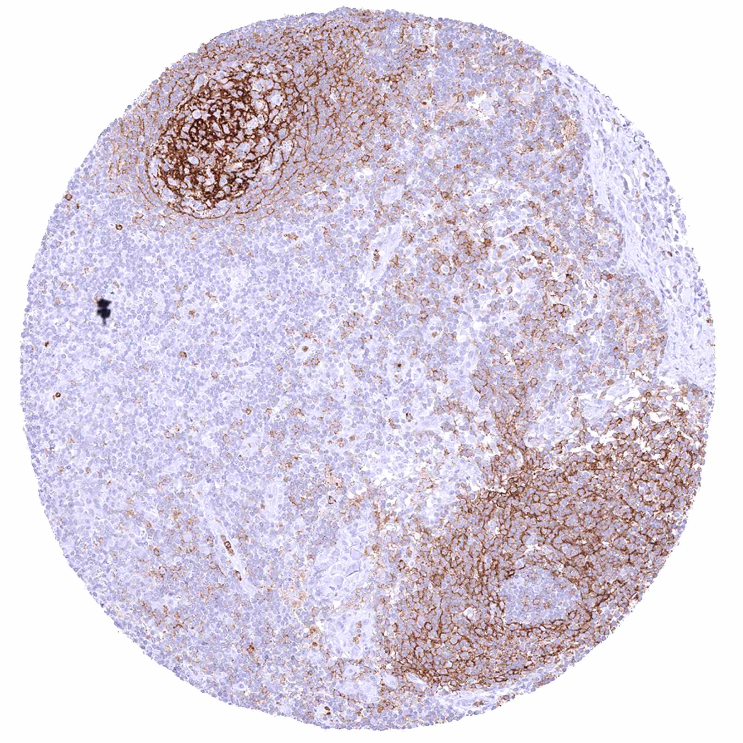
Positive control = Tonsil: A strong CD35 must be seen in follicular dendritic cells while there is a less intense CD35 staining of a subset of lymphocytes.
Negative control = Tonsil: Squamous epithelium and most lymphocytic cells must be CD35 negative.
Cellular localization = Membranous
Reactivity = Human
Application = Immunohistochemistry
Dilution = 1:100 – 1:200
Intended Use = Research Use Only
Relevance of Antibody
CD35 is a critical protein for interaction with the complement system.
Biology Behind
Complement receptor type 1 (CR1) also known as CD35 (cluster of differentiation 35) is a monomeric type I membrane glycoprotein which is coded by the CR1 gene on chromosome 1q32. CR1 is needed for connecting cells to immune complexes and other particles that contain activated complement on their surface. It plays a role in T-cell and B-cell mediated immune regulation and it is a critical factor for the actions of the complement system as it interacts with C3b and C4b to promote neutrophil-mediated phagocytosis. The complement system was initially considered a “first line of defense” against microbial intruders but it is now known to also participate in many other cellular processes including lipid metabolism, tissue regeneration, clearance of immune complexes, and cancer where it has roles in enhancing invasion, metastasis, proliferation and inhibition of apoptosis. Due to its role in complement activation, innate immunity, and chronic inflammation, CD35 appears to be involved in several different disease types. Polymorphisms of the CR1 gene have been shown to impact susceptibility for several diseases such as infections with various agents including Covid-19 or lepropsy, several cancer types, mesangiocapillary glomerulonephritis, systemic lupus erythematosus, and sarcoidosis. Antigens located on the CR1 protein constitute the Knops blood group system.
Staining Pattern in Normal Tissues
In normal tissues, CD35 is most highly expressed in normal follicular dendritic cells, but it can also be seen on lymphocytes of various types, macrophages, granulocytes, and erythrocytes.
Images describing the CD35 staining pattern in normal tissues obtained by the antibody MSVA-035M are shown in our “Normal Tissue Gallery”.
| Brain | Cerebrum | Negative. |
| Cerebellum | Negative. | |
| Endocrine Tissues | Thyroid | Negative. |
| Parathyroid | Negative. | |
| Adrenal gland | Negative. | |
| Pituitary gland | Negative. | |
| Respiratory system | Respiratory epithelium | Negative. |
| Lung | Negative. | |
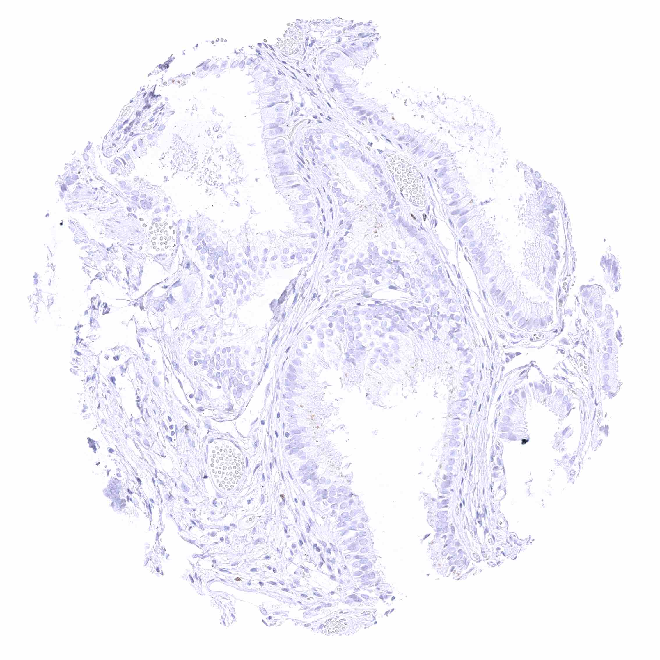
| Gastrointestinal Tract | Salivary glands | Negative. |
| Esophagus | Negative. | |
| Stomach | Negative. | |
| Duodenum | Negative. | |
| Small intestine | Negative. | |
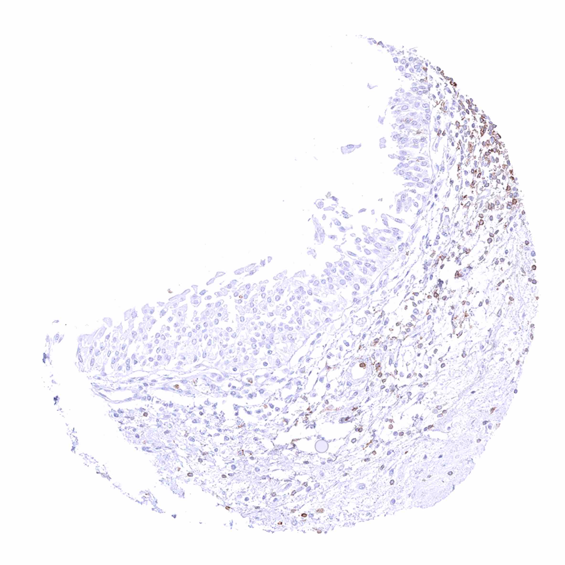
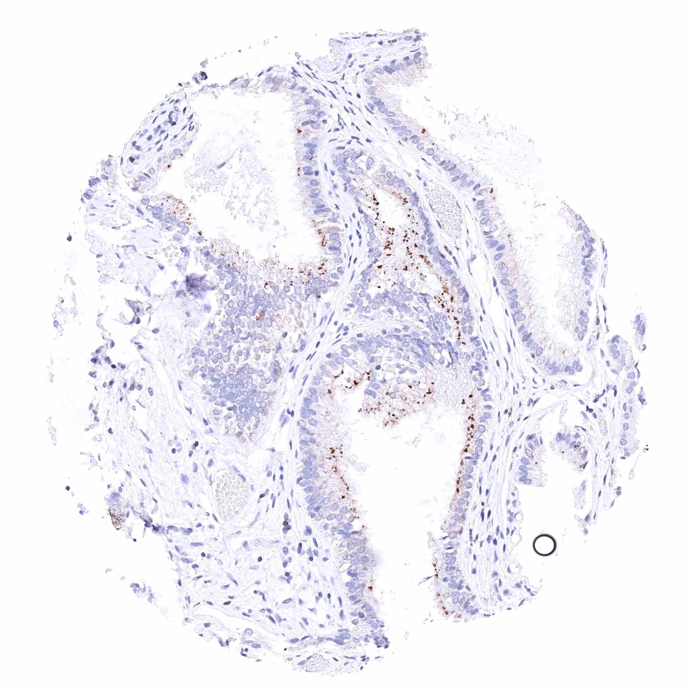
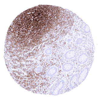
| Appendix | Strong membranous CD35 staining of follicular dendritic cells and of a large subset of lymphocytes. The epithelium is CD35 negative. | |
| Colon | Negative. | |
| Rectum | Negative. | |
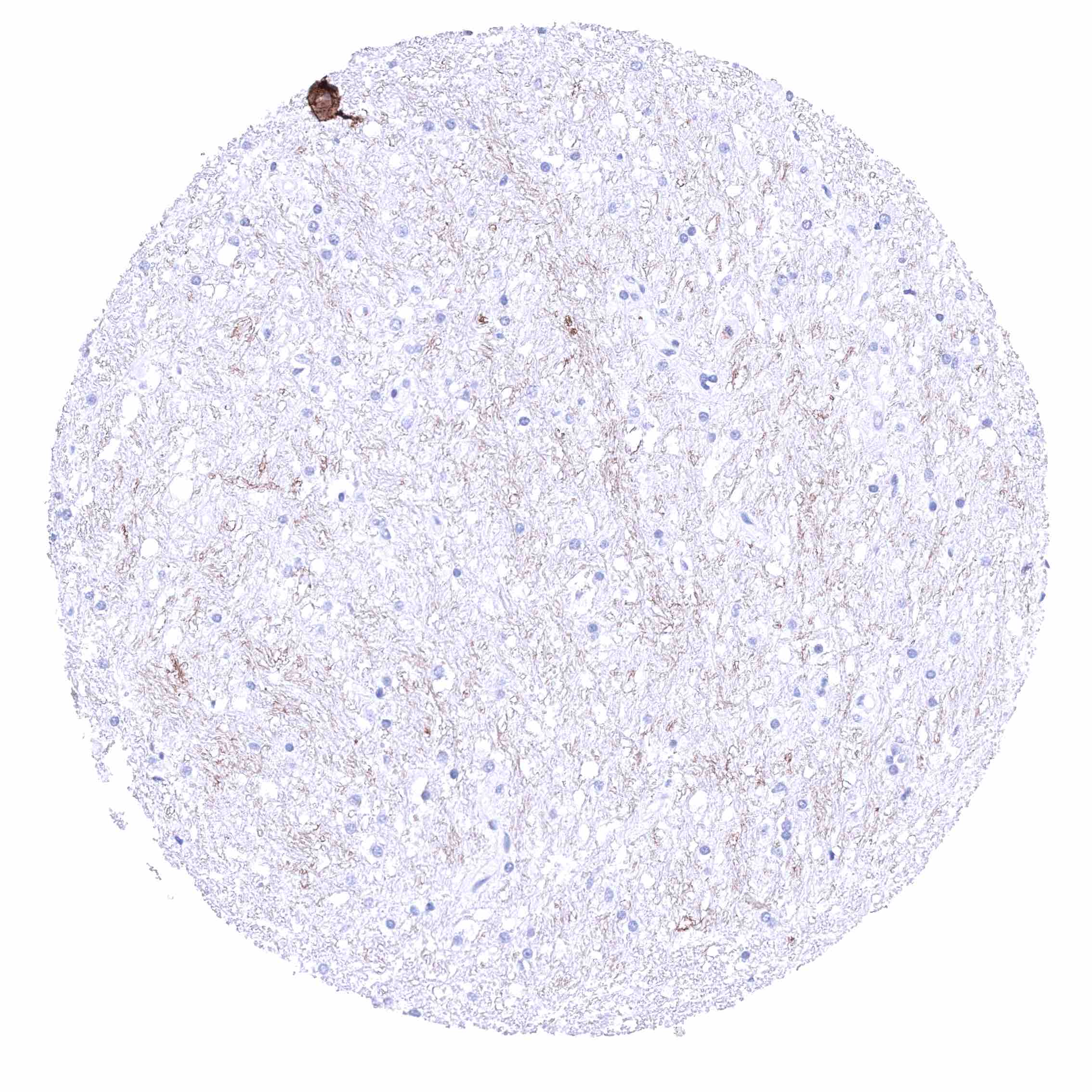
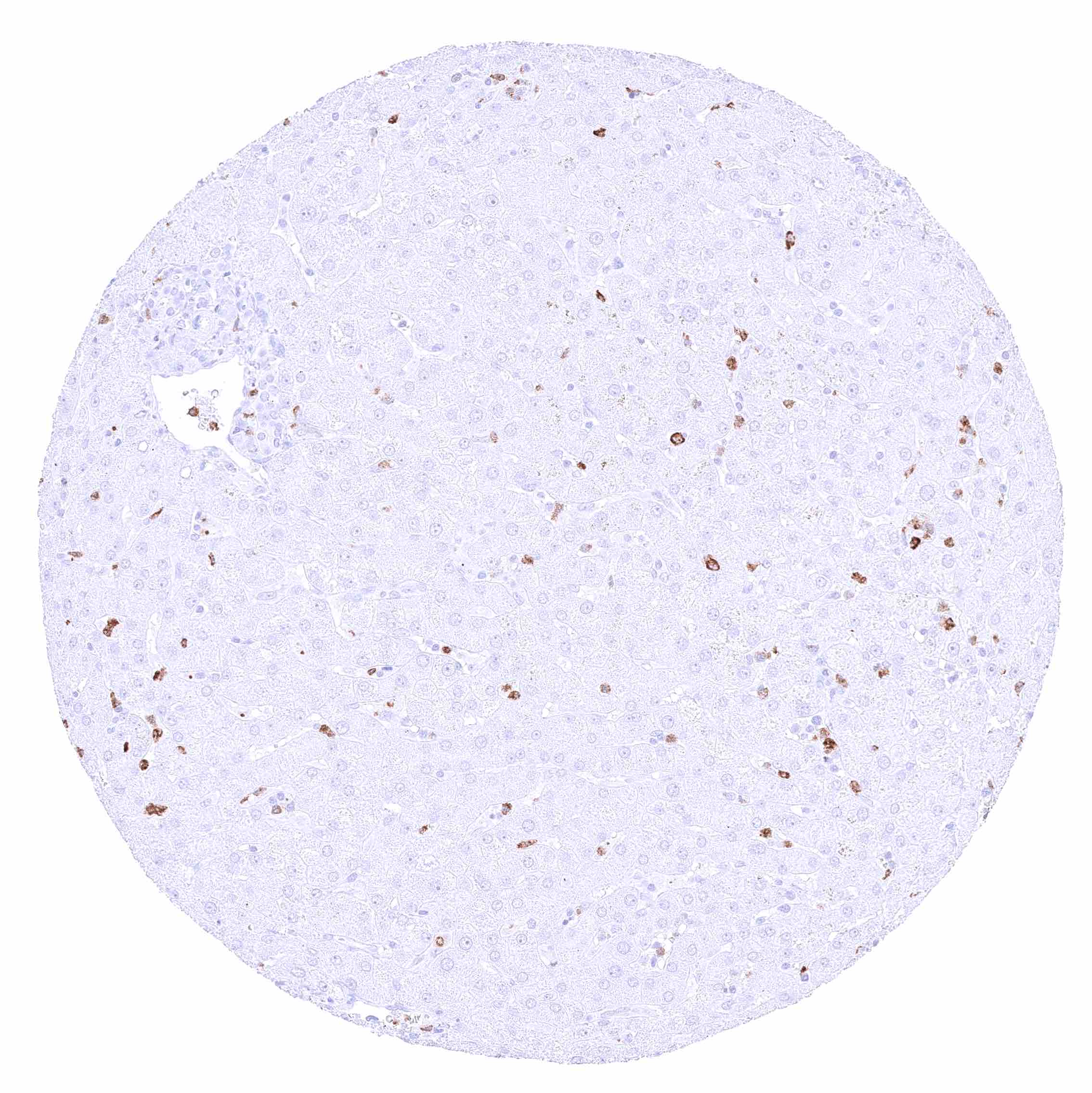
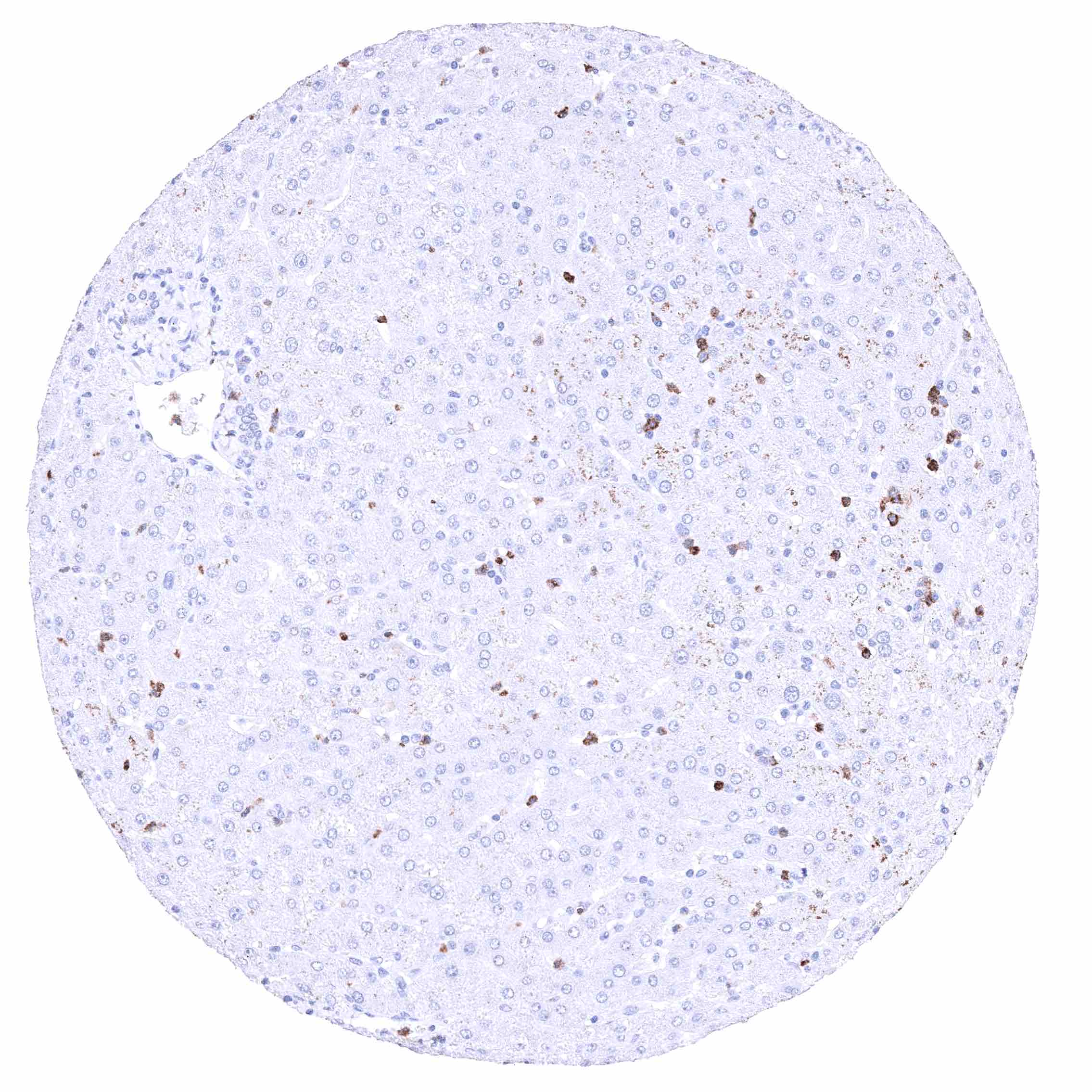
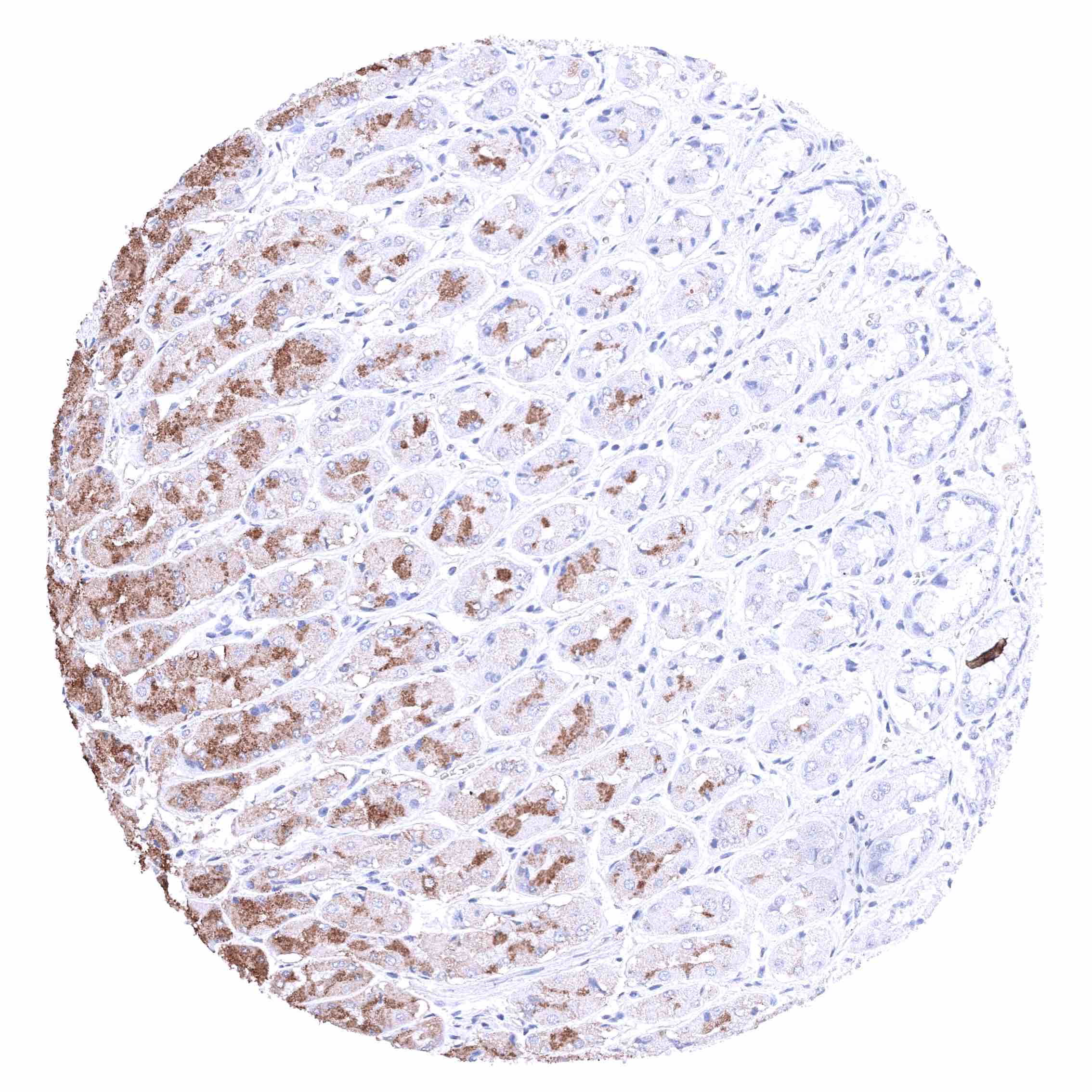
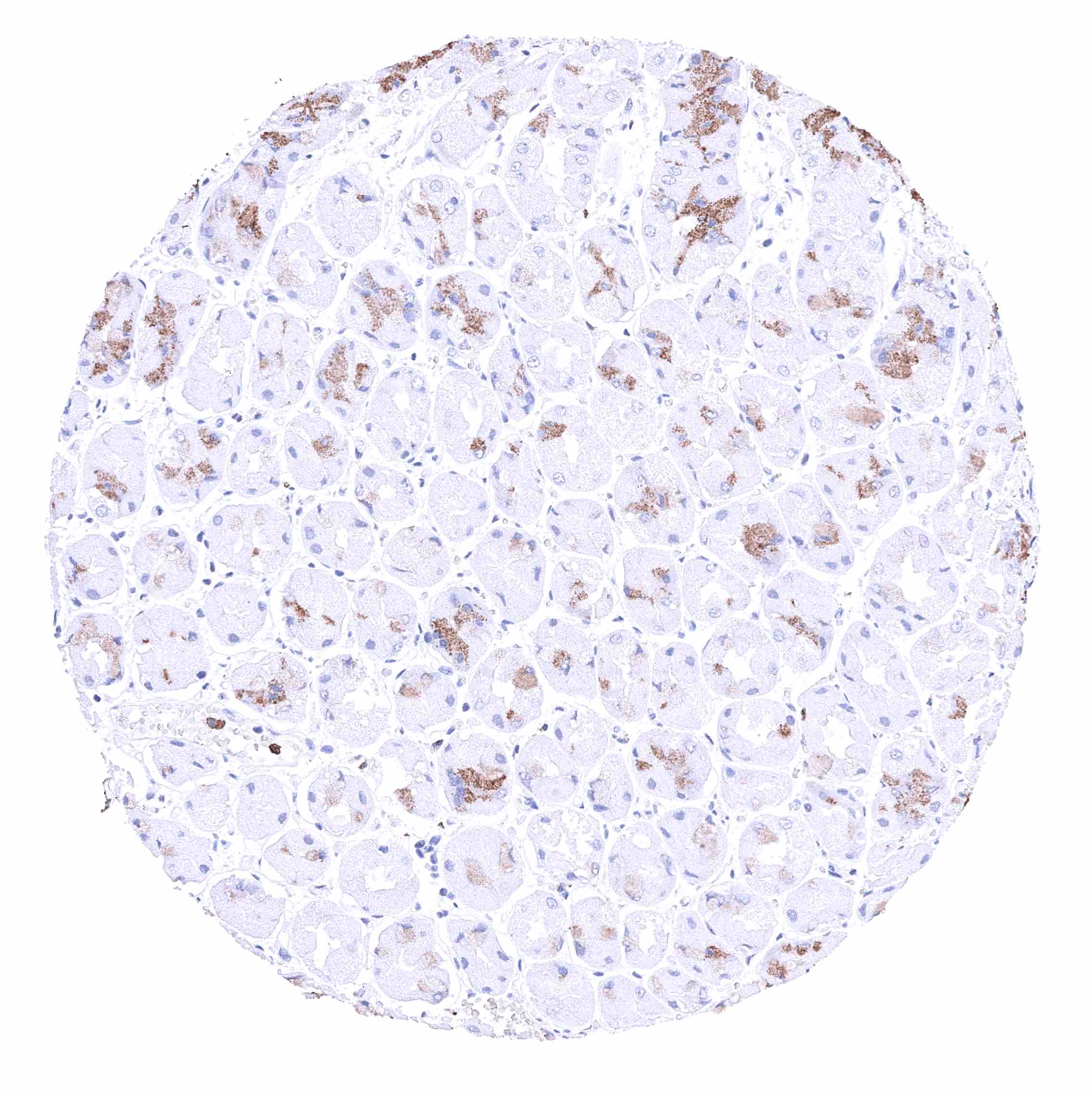
| Liver | Distinct membranous CD35 positivity of Kupffer cells. | |
| Gallbladder | Negative. | |
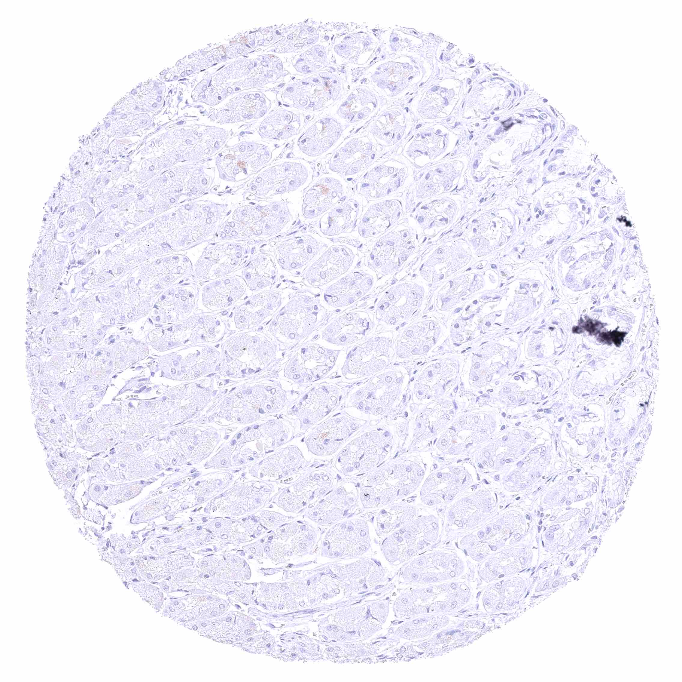
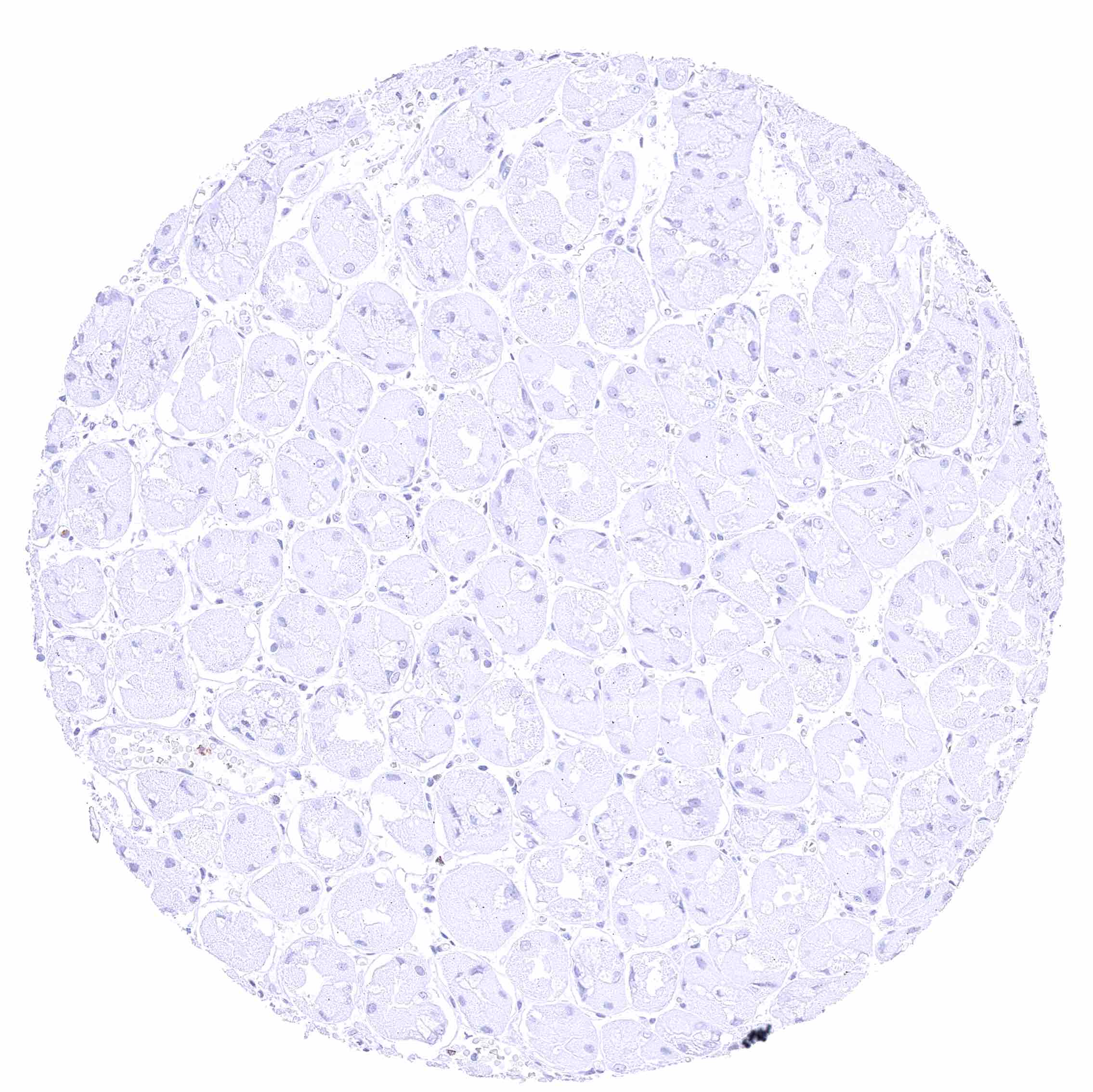
| Pancreas | Negative. | |
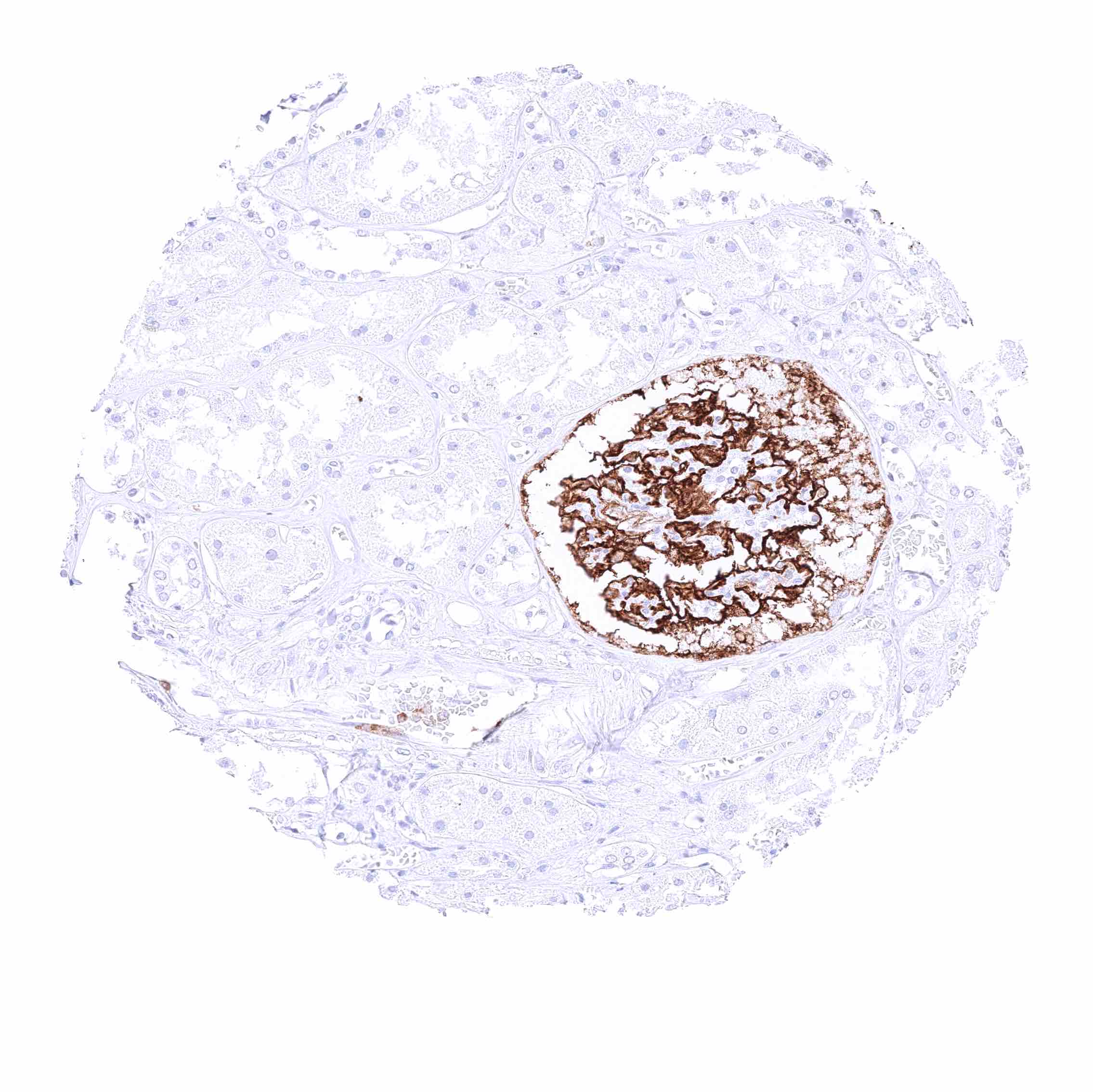
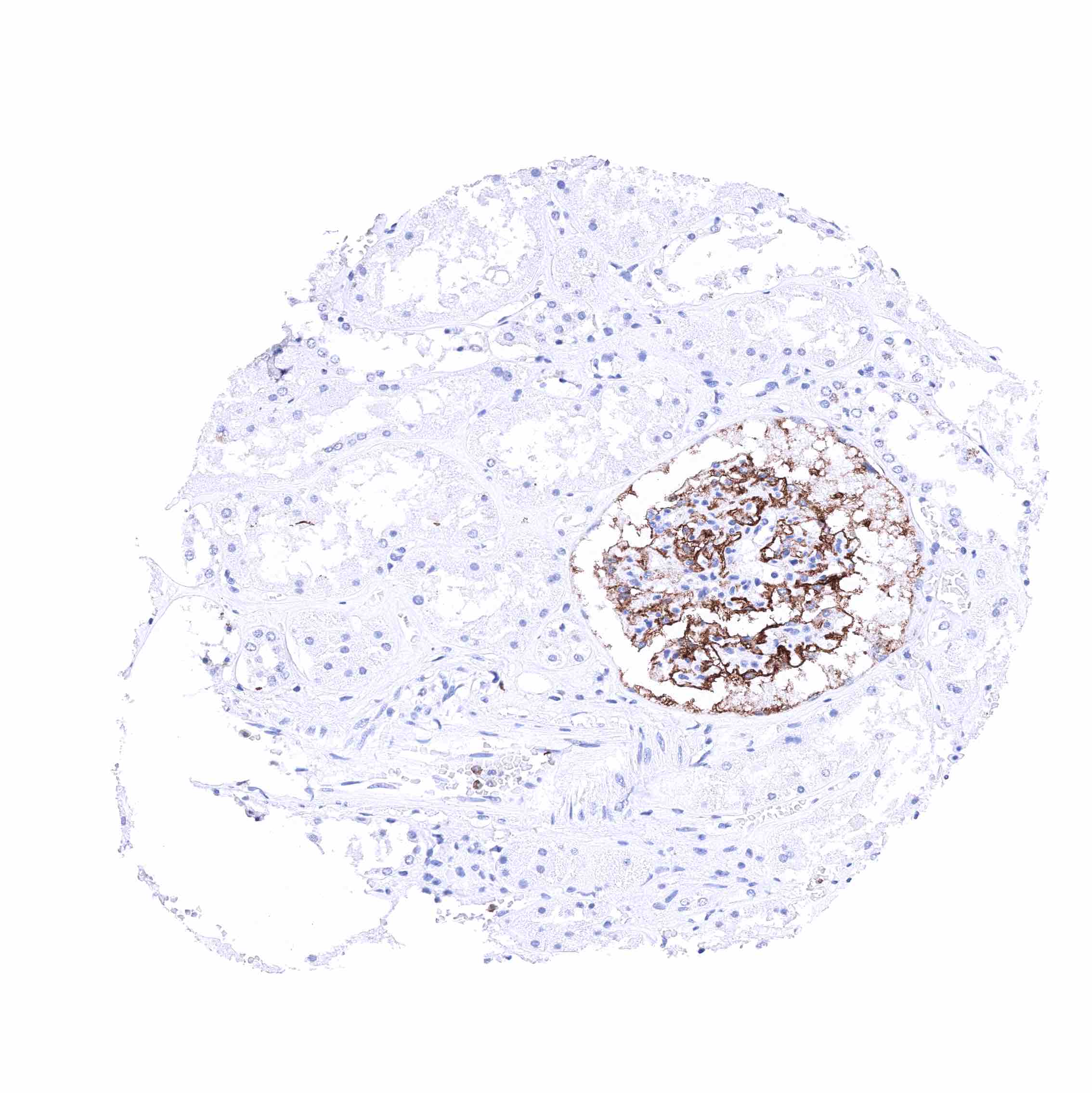
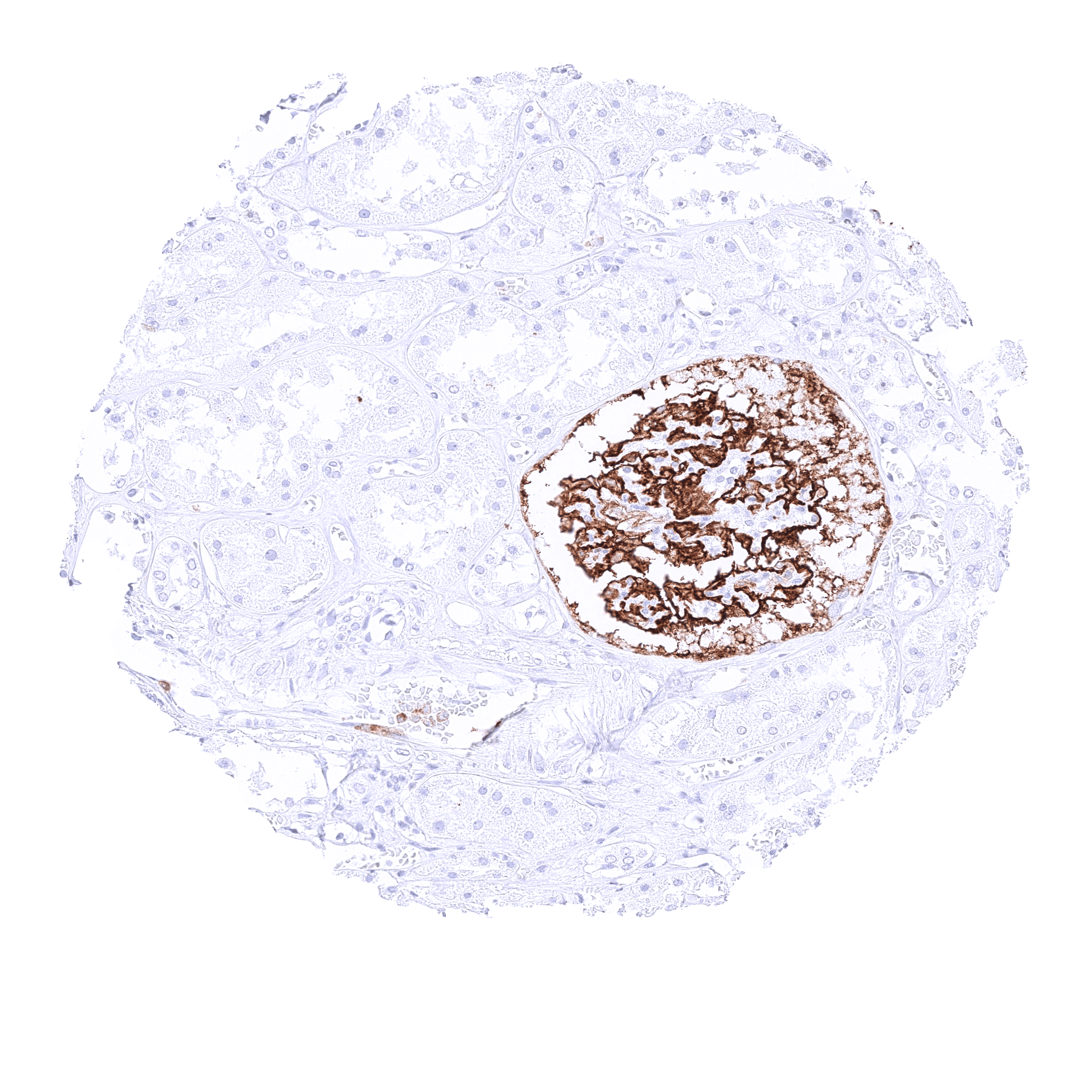
| Genitourinary | Kidney | Strong membranous CD35 positivity of visceral (podocytes) and peripheral cells of the Bowman capsule. |
| Urothelium | Negative. | |
| Male genital | Prostate | Negative. |
| Seminal vesicles | Negative. | |
| Testis | Negative. | |
| Epididymis | Negative. | |
| Female genital | Breast | Negative. |
| Uterus, myometrium | Negative. | |
| Uterus, ectocervix | Negative. | |
| Uterus endocervix | Negative. | |
| Uterus, endometrium | Negative. | |
| Fallopian Tube | Negative. | |
| Ovary | Negative. | |
| Placenta early | Negative. | |
| Placenta mature | Negative. | |
| Amnion | ||
| Chorion | Negative. | |
| Skin | Epidermis | Negative. |
| Sebaceous glands | Negative. | |
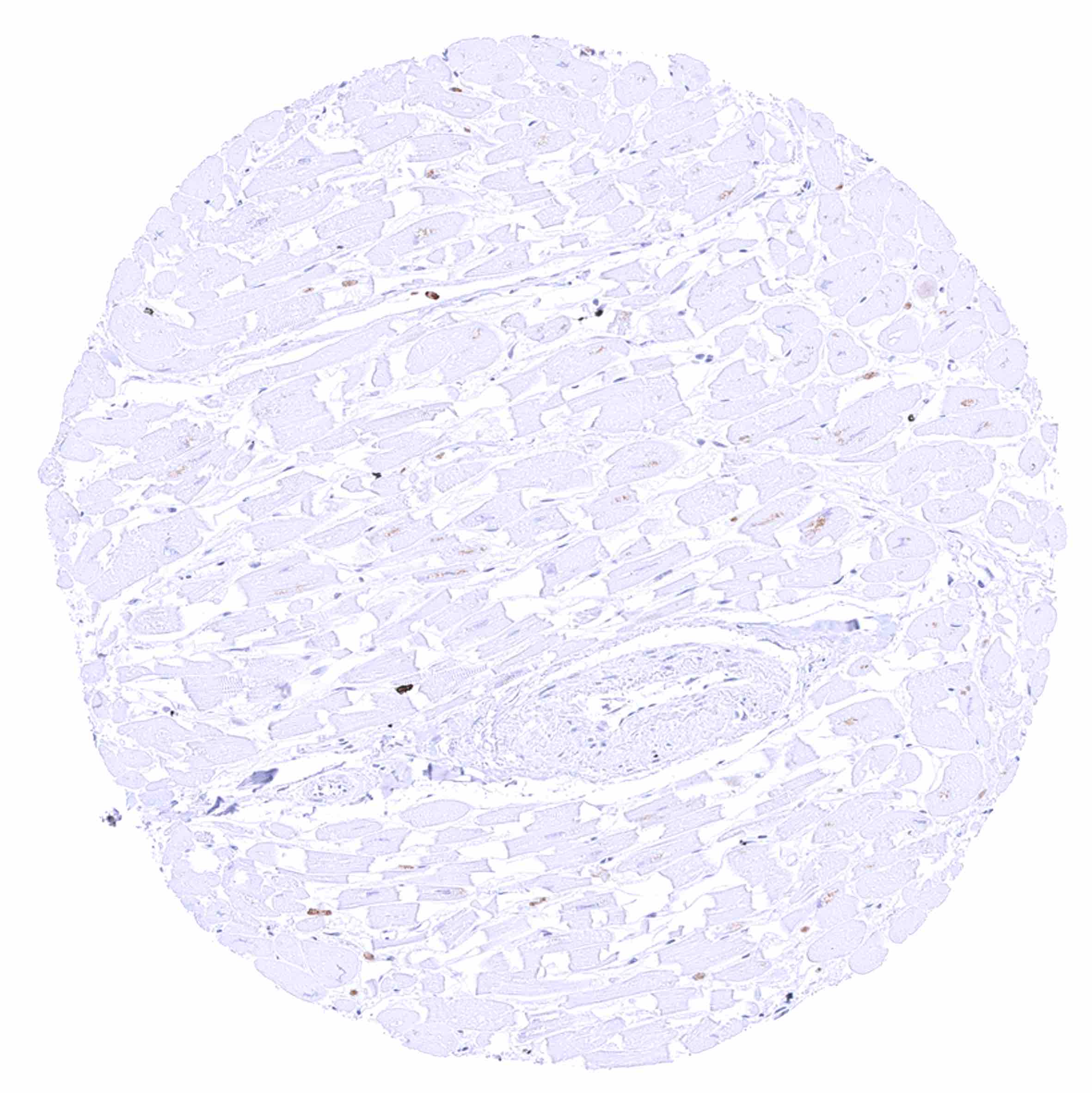
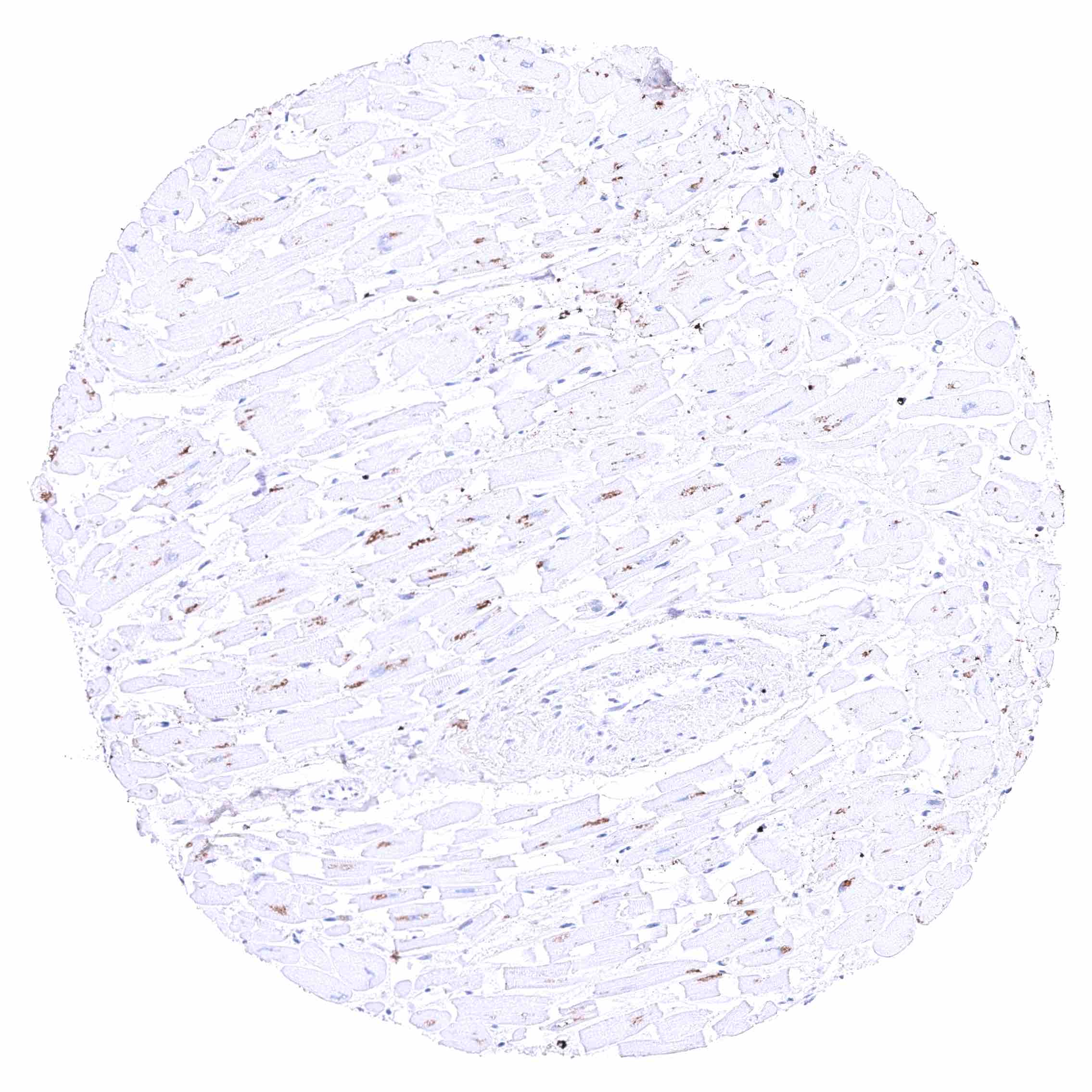
| Muscle/connective tissue | Heart muscle | Negative. |
| Skeletal muscle | Negative. | |
| Smooth muscle | Negative. | |
| Vessel walls | Negative. | |
| Fat | Negative. | |
| Stroma | Negative. | |
| Endothelium | Negative. | |
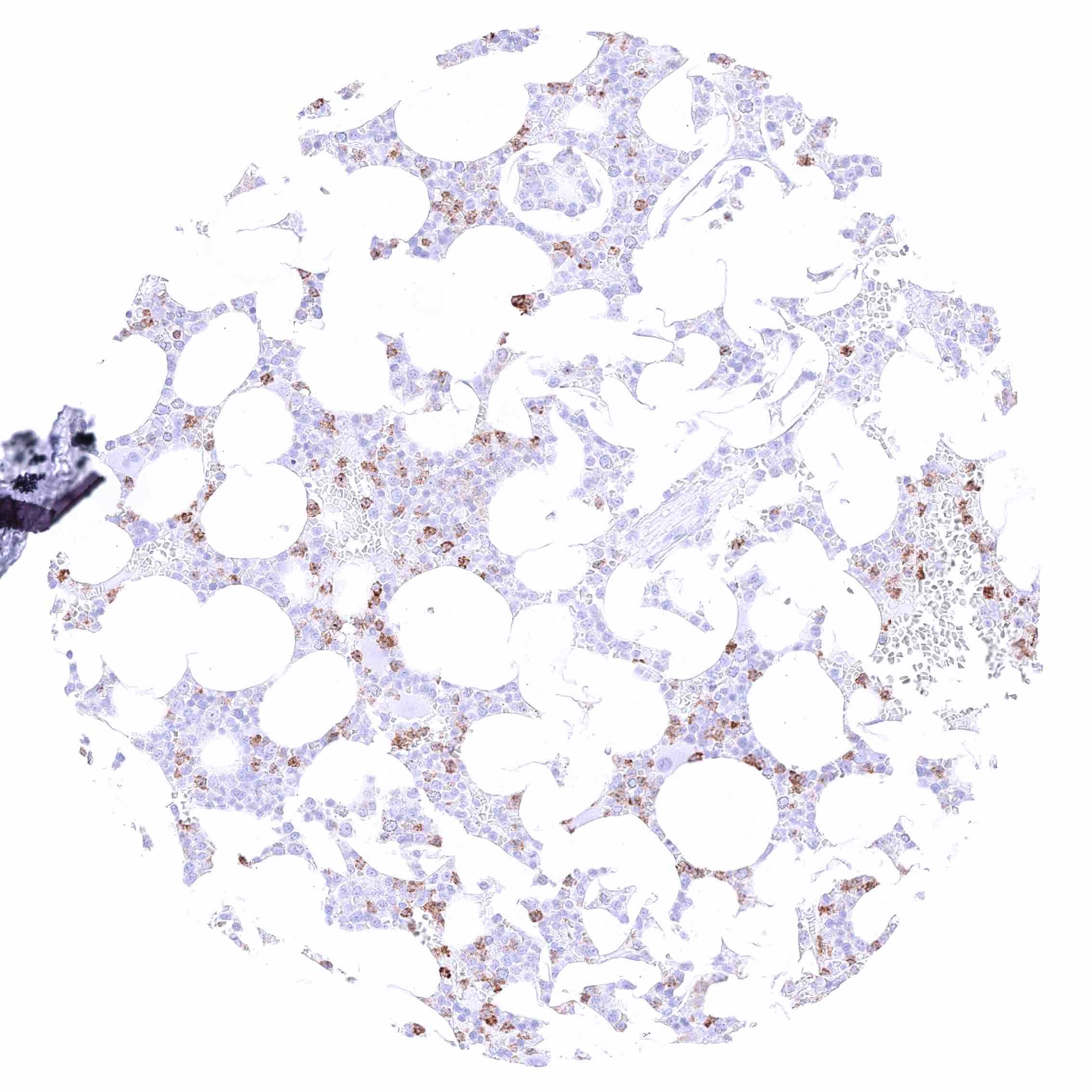
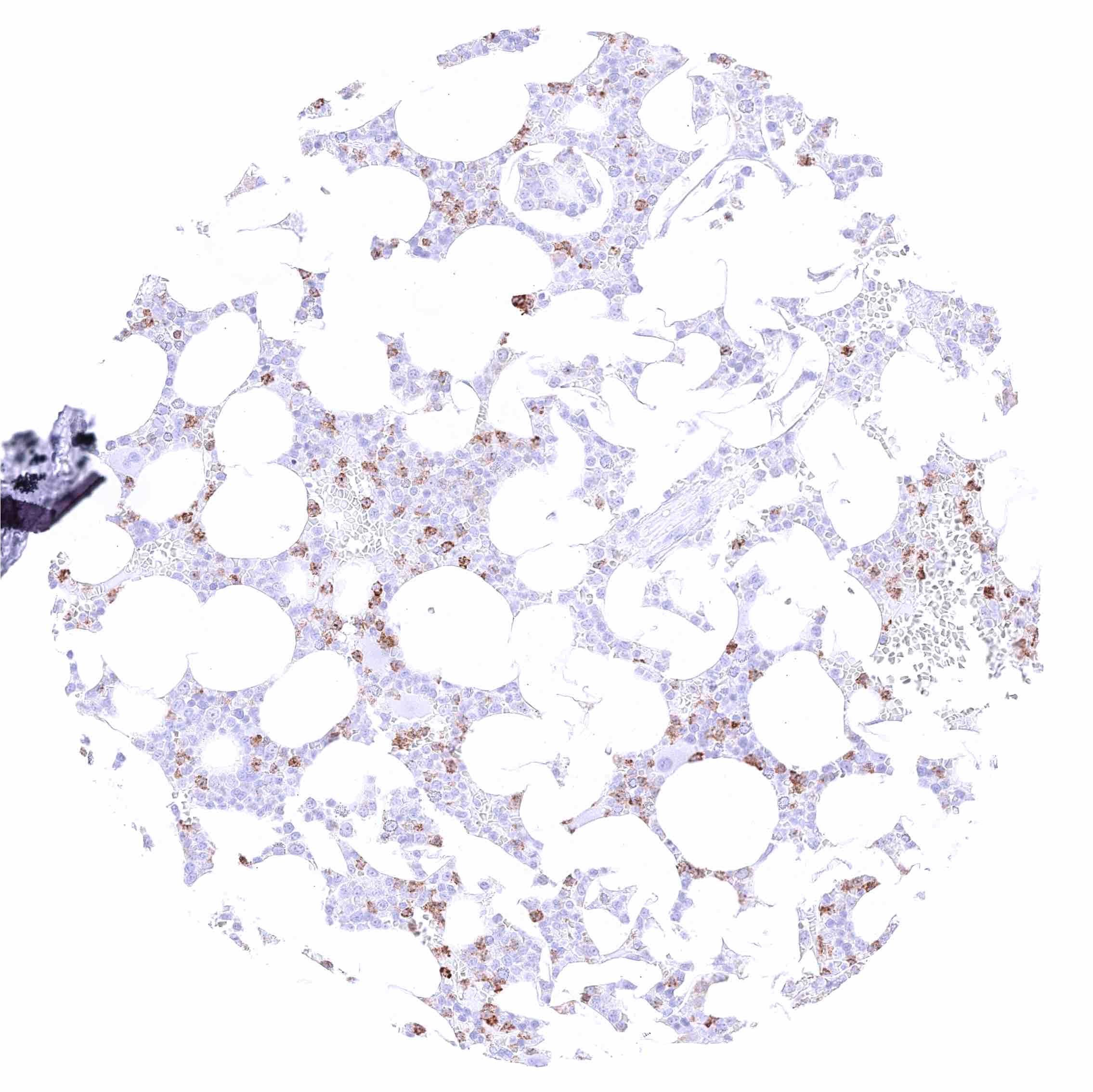
| Bone marrow/ lymphoid tissue | Bone marrow | Strong membranous CD35 staining of a subset of cells. |
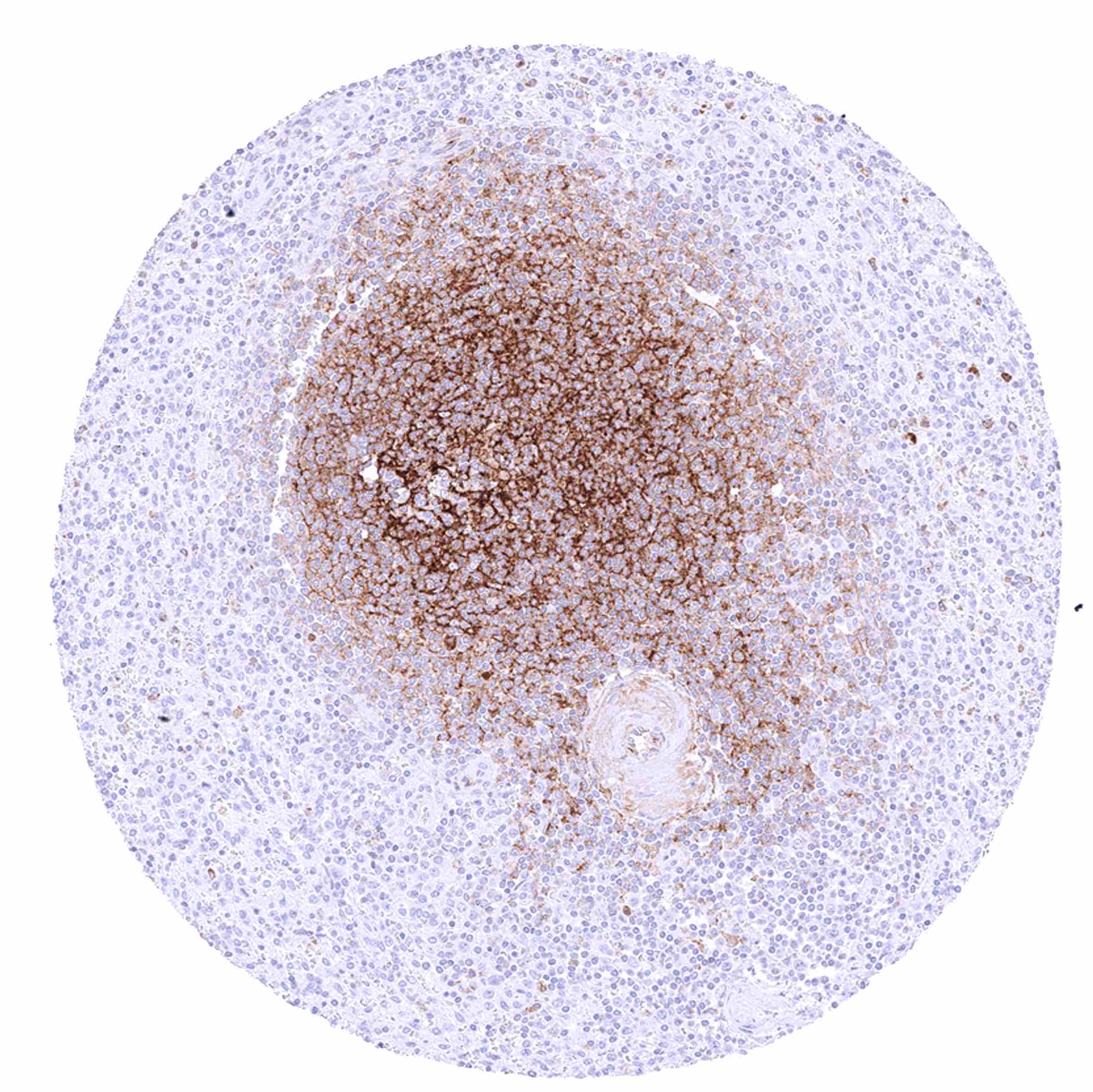
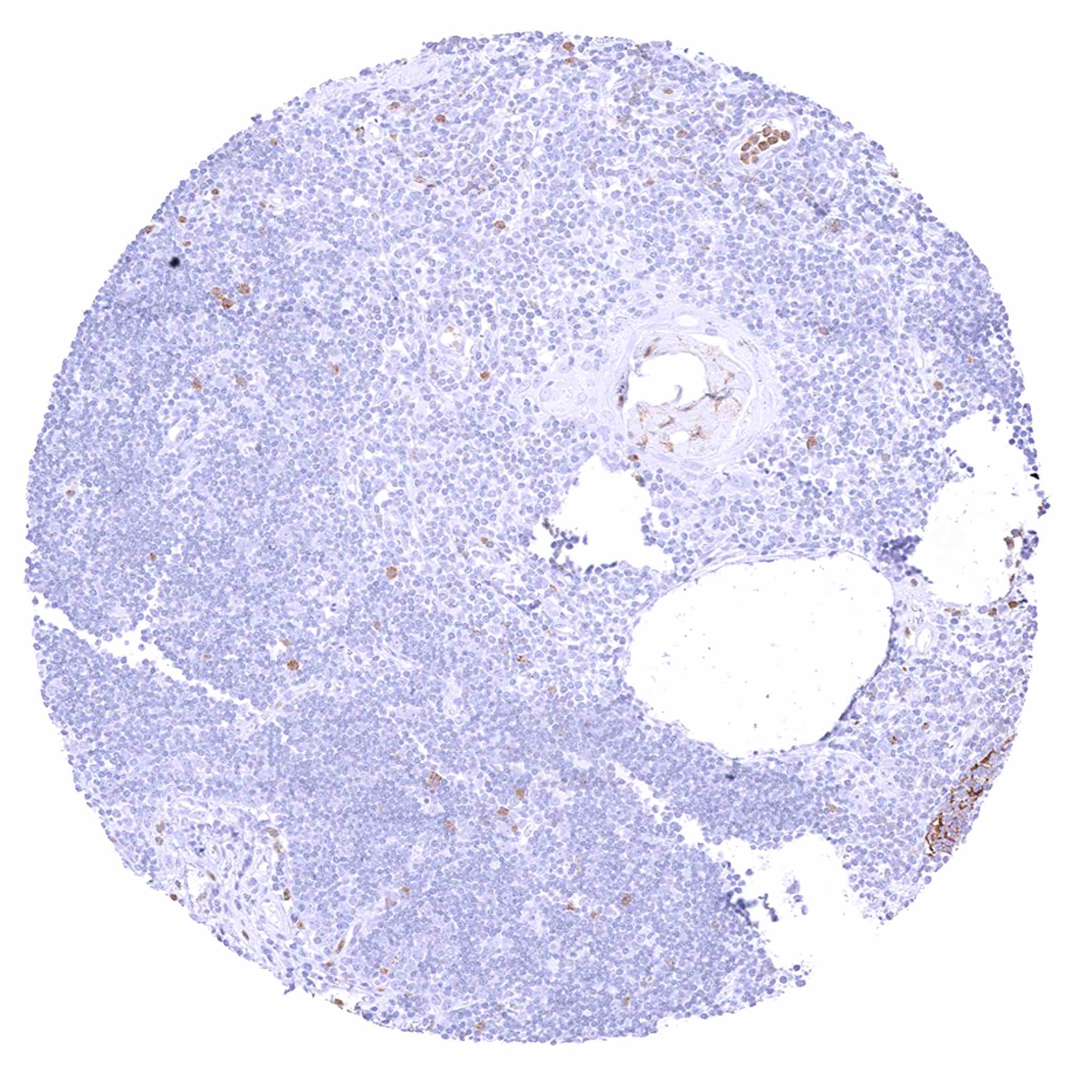
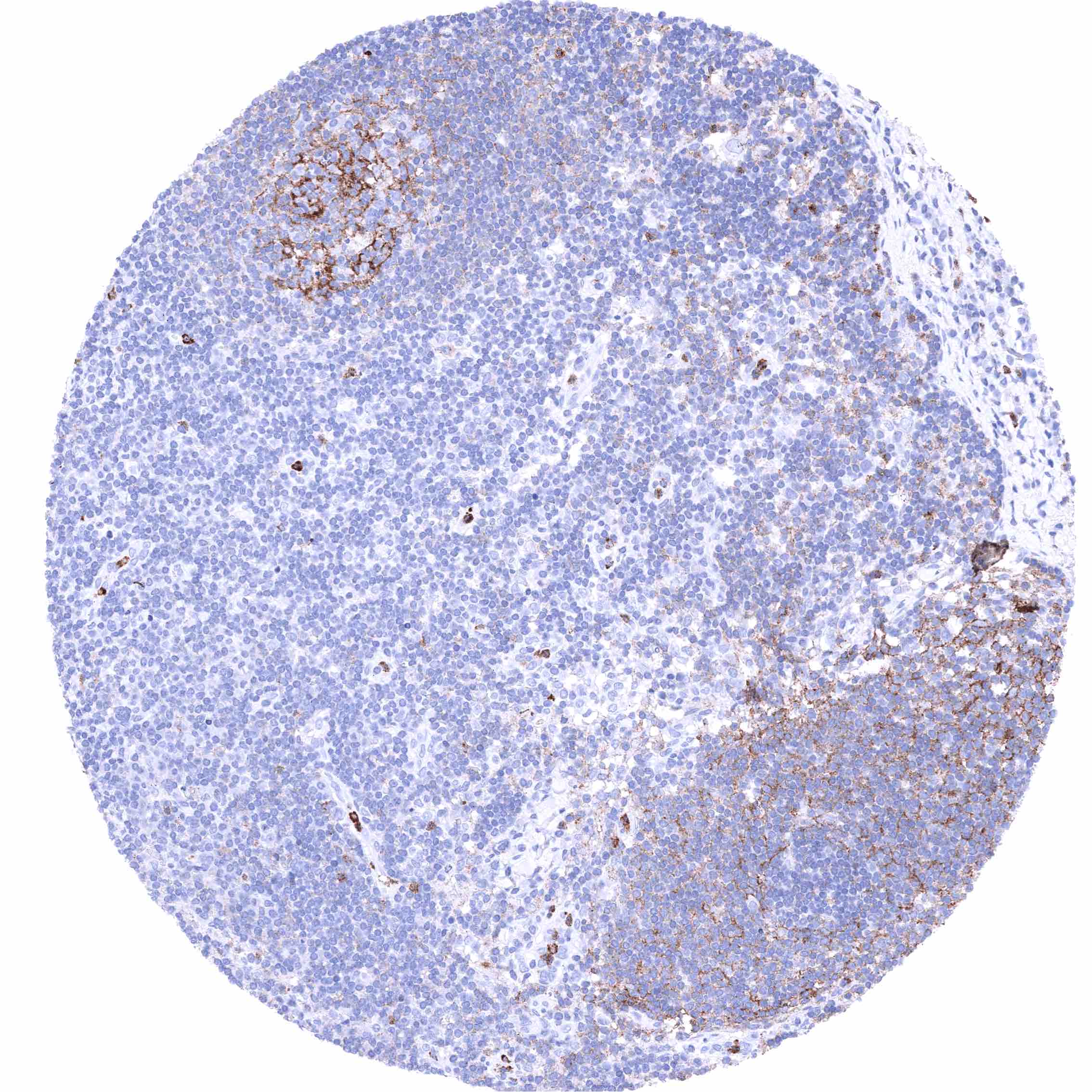
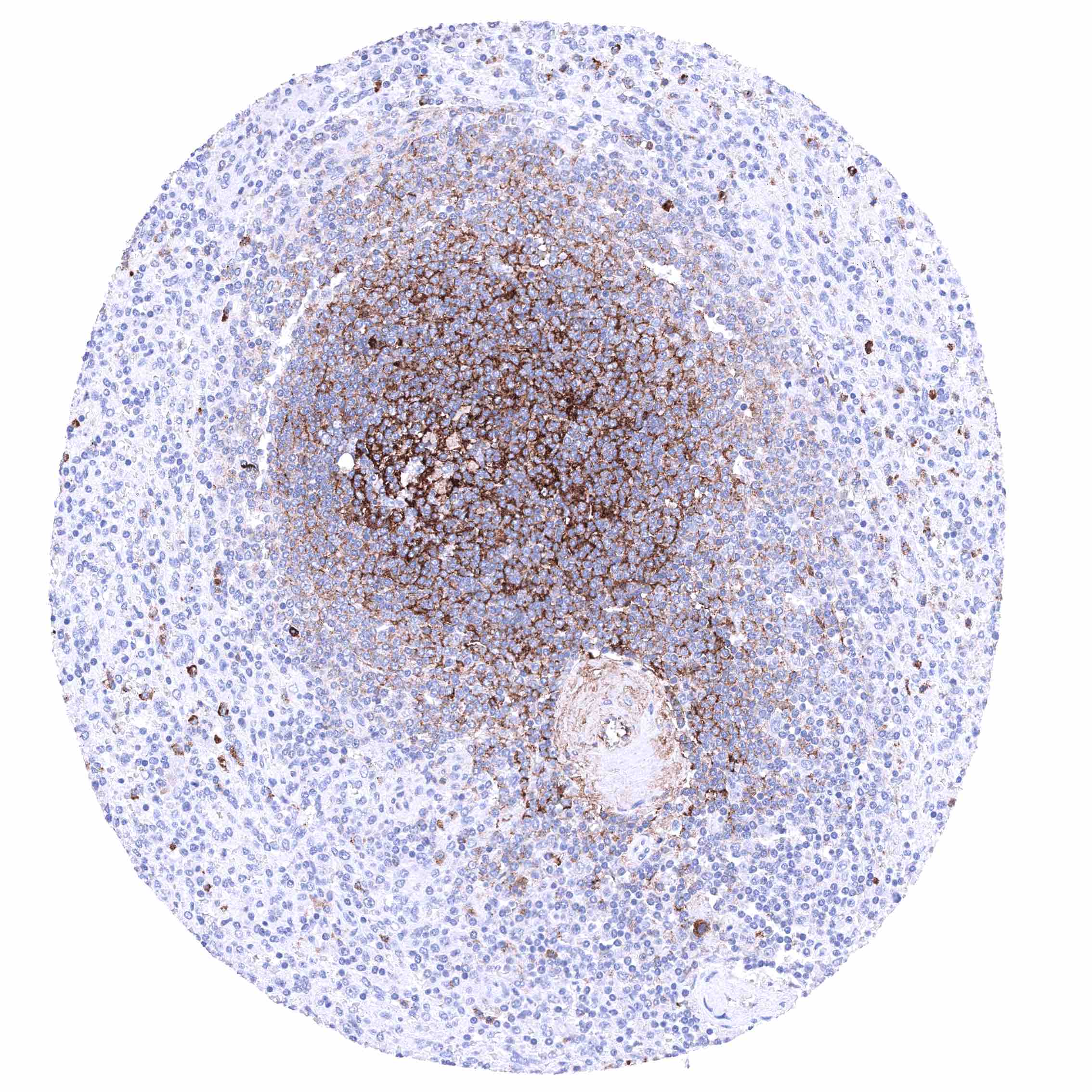
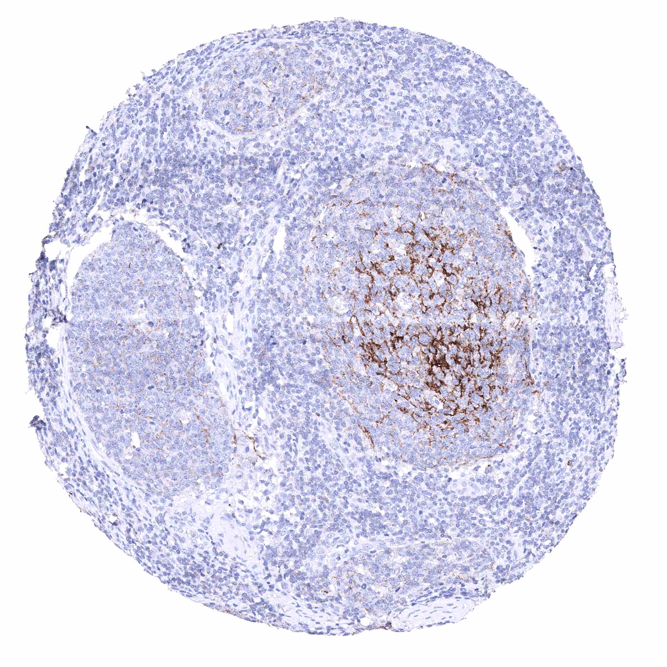
| Lymph node | Strong membranous CD35 staining of follicular dendritic cells and of a subset of lymphocytes. | |
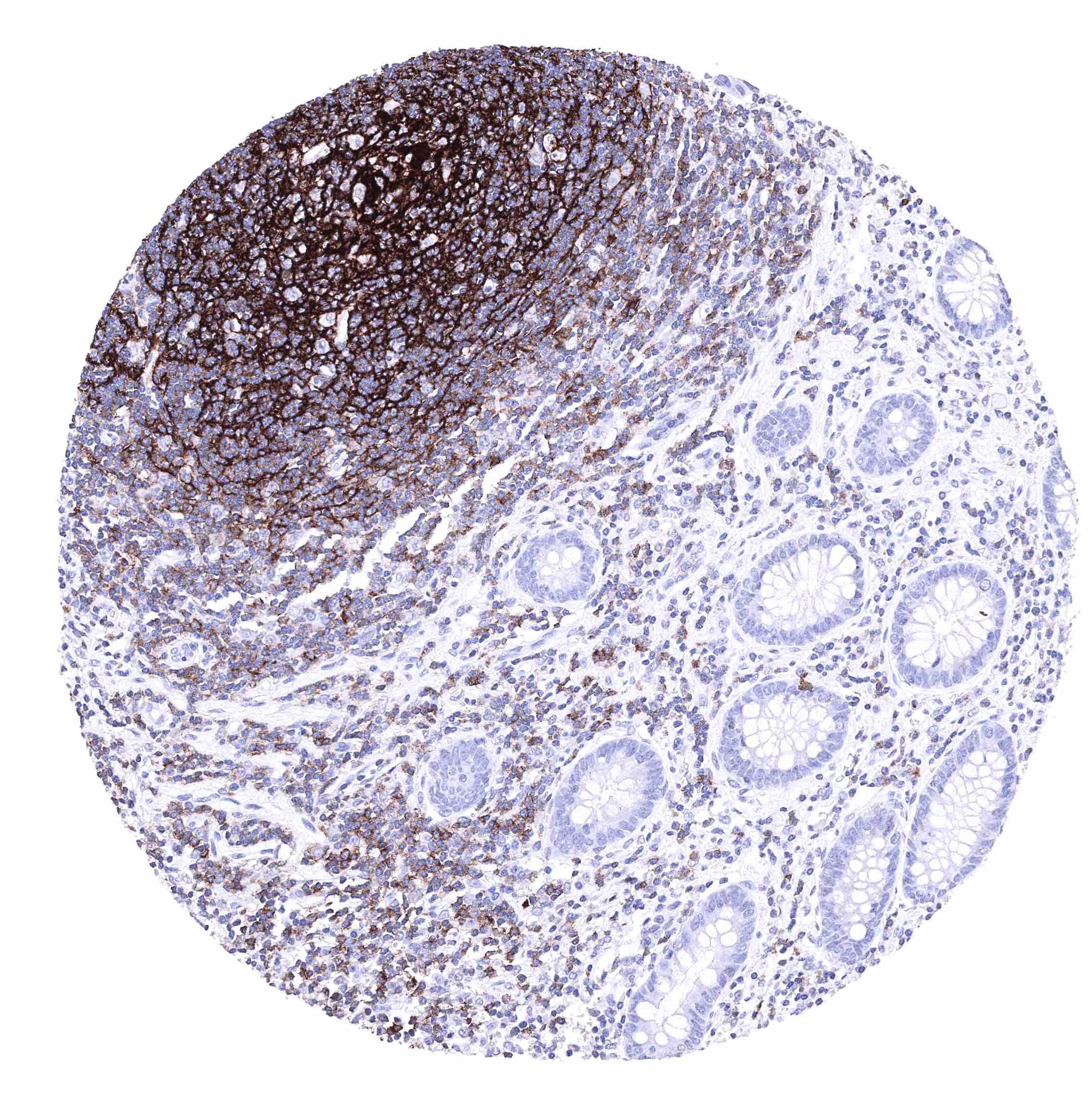
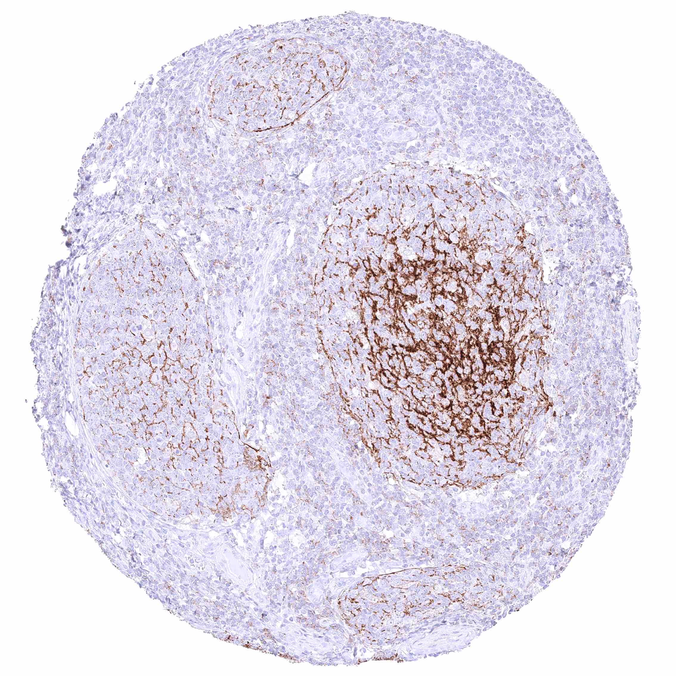
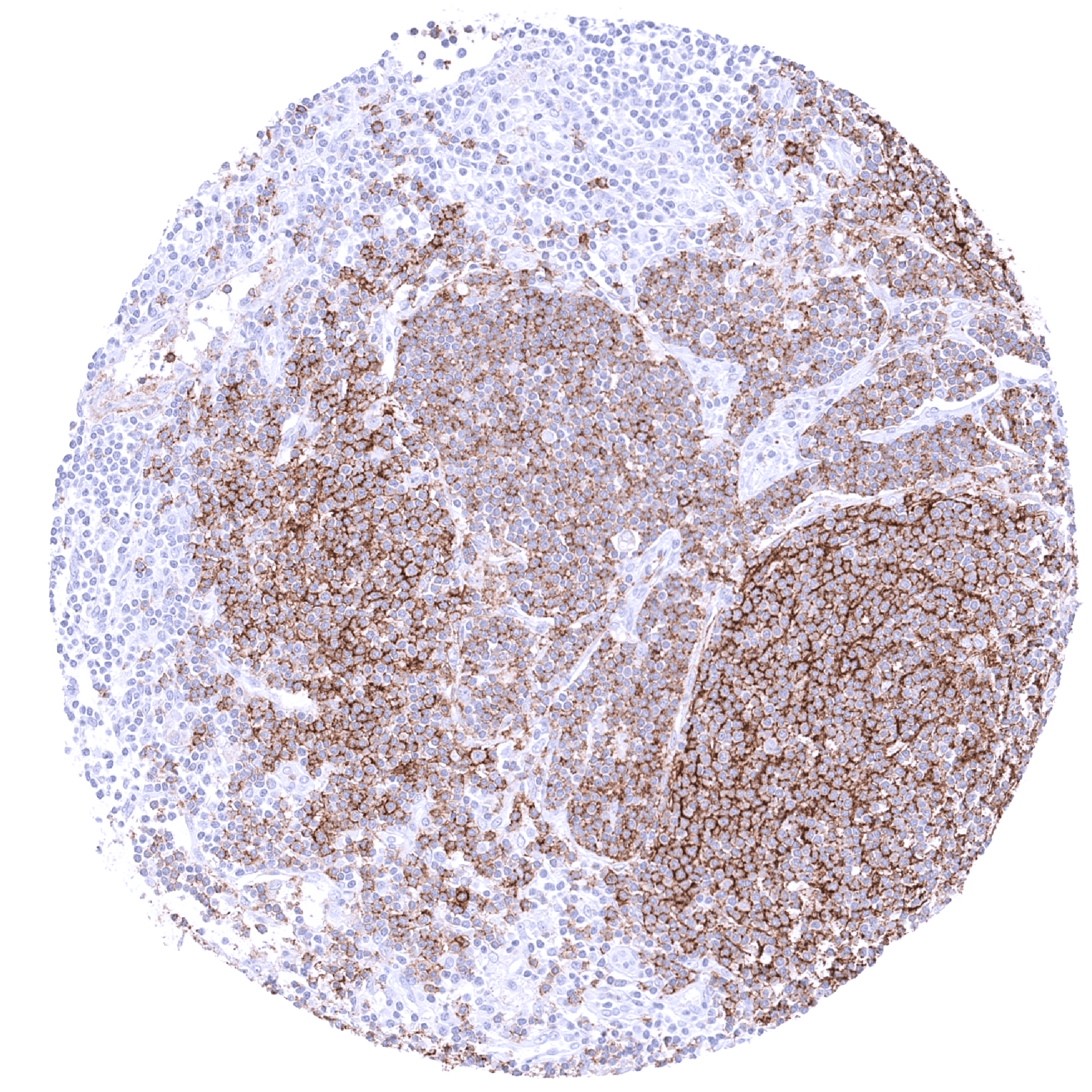
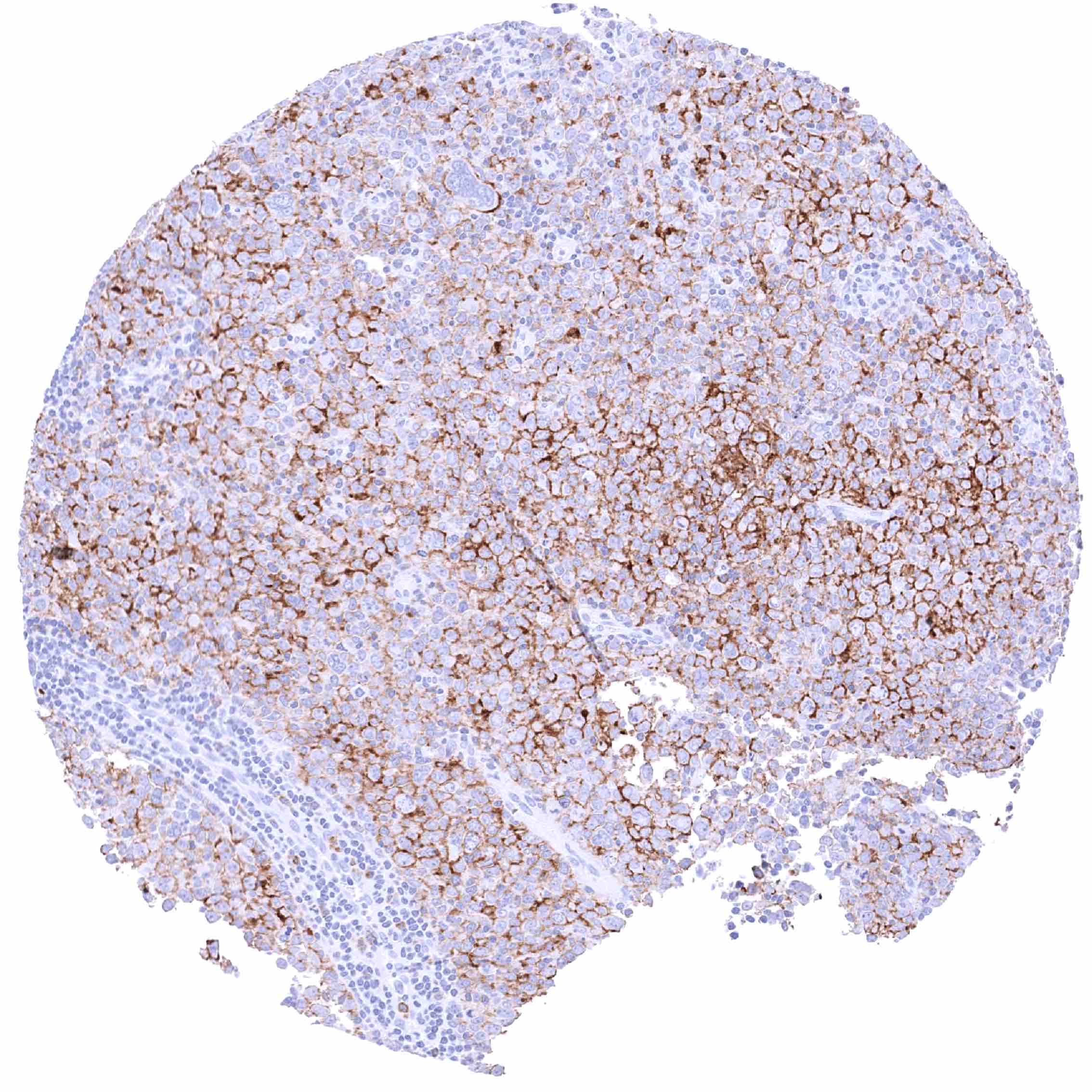
| Spleen | Strong membranous CD35 staining of most cells in the white pulpa. | |
| Thymus | Distinct membranous CD35 staining of only few lymphocytes. | |
| Tonsil | Strong membranous CD35 staining of follicular dendritic cells and of a subset of lymphocytes. The squamous epithelium is CD35 negative. | |
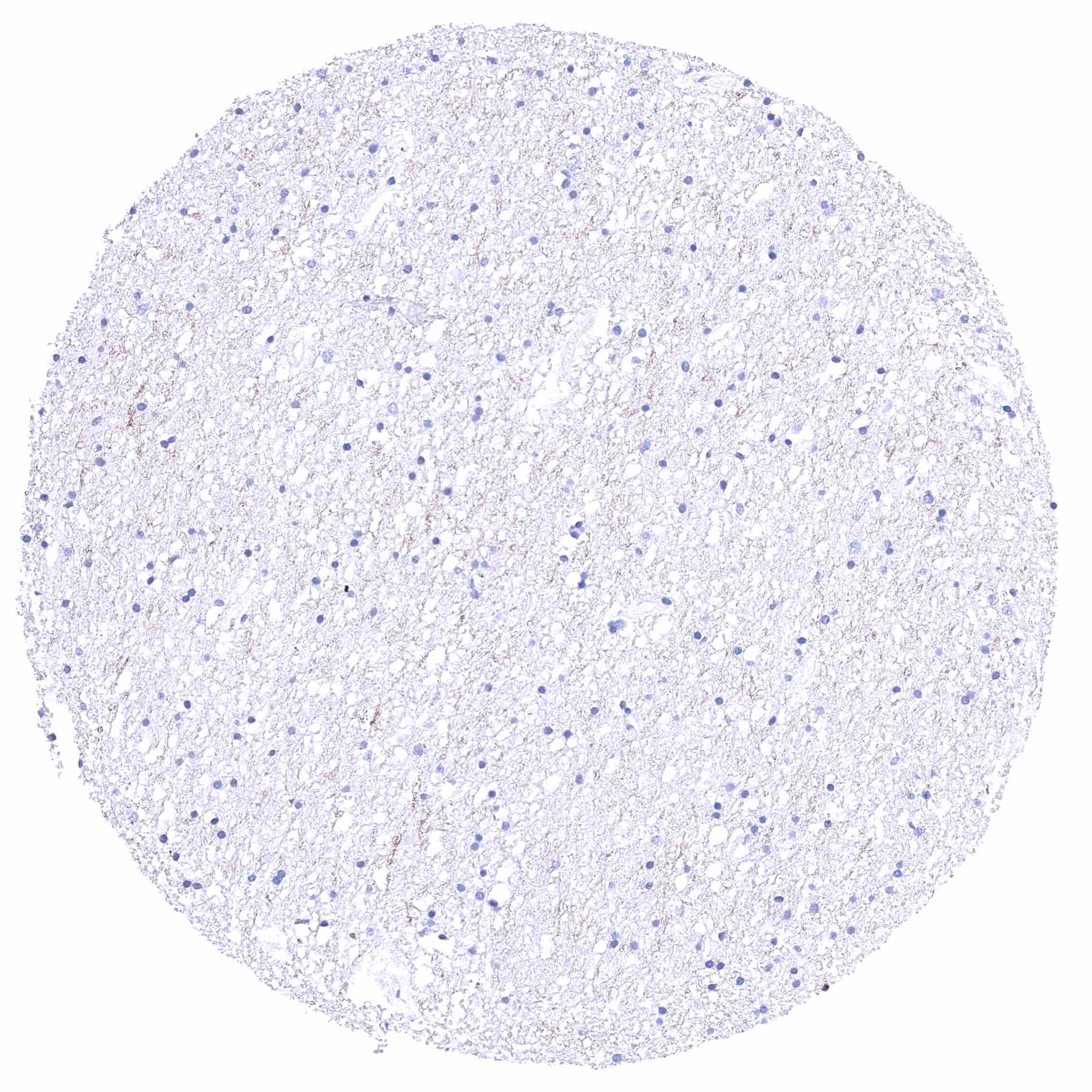
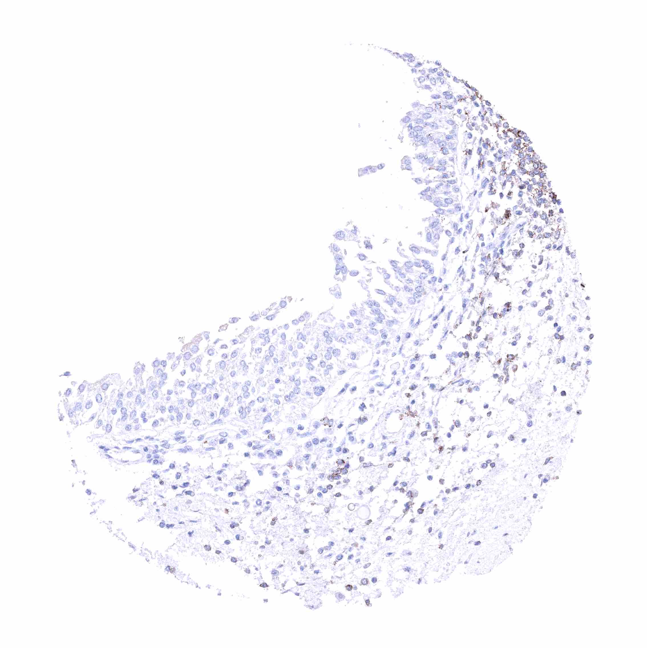
| Remarks | Scattered CD35 positive inflammatory cells can be seen in all tissues. |
These findings are largely consistent with the RNA data described in the Human Protein Atlas (Tissue expression CD35).
Positive control = Tonsil: A strong CD35 must be seen in follicular dendritic cells while there is a less intense CD35 staining of a subset of lymphocytes.
Negative control = Tonsil: Squamous epithelium and most lymphocytic cells must be CD35 negative.

Staining Pattern in Relevant Tumor Types
CD35 positive inflammatory cells can be found in all cancers at a variable extent. CD35 expression is variable in malignant lymphomas. For example, it was proposed to be reduced in B-CLL.
The TCGA findings on CD35 RNA expression in different tumor categories have been summarized in the Human Protein Atlas.
Compatibility of Antibodies
No data available at the moment
Protocol Recommendations
IHC users have different preferences on how the stains should look like. Some prefer high staining intensity of the target stain and even accept some background. Others favor absolute specificity and lighter target stains. Factors that invariably lead to more intense staining include higher concentration of the antibody and visualization tools, longer incubation time, higher temperature during incubation, higher temperature and longer duration of the heat induced epitope retrieval (slide pretreatment). The impact of the pH during slide pretreatment has variable effects and depends on the antibody and the target protein.
All images and data shown here and in our image galleries are obtained by the manual protocol described below. Other protocols resulting in equivalent staining are described as well.
Manual protocol
Freshly cut sections should be used (less than 10 days between cutting and staining). Heat-induced antigen retrieval for 5 minutes in an autoclave at 121°C in pH 7,8 Target Retrieval Solution buffer. Apply MSVA-035M at a dilution of 1:150 at 37°C for 60 minutes. Visualization of bound antibody by the EnVision Kit (Dako, Agilent) according to the manufacturer’s directions.
Potential Research Applications
- The diagnostic role of CD35 IHC in lymphoma needs to be further evaluated.
- The role of CD35 polymorphisms for the risk of developing specific diseases needs to be further investigated.
- The role of CD35 in cancer should be further elucidated.
Evidence for Antibody Specificity in IHC
There are two ways how the specificity of antibodies can be documented for immunohistochemistry on formalin fixed tissues. These are: 1. Comparison with a second independent method for target expression measurement across a large number of different tissue types (orthogonal strategy), and 2. Comparison with one or several independent antibodies for the same target and showing that all positive staining results are also seen with other antibodies for the same target (independent antibody strategy).
Orthogonal validation: For the antibody MSVA-035M, specificity is suggested by the good concordance of the immunostaining data with data from three independent RNA screening studies, including the Human Protein Atlas (HPA) RNA-seq tissue dataset, the FANTOM5 project, and the Genotype-Tissue Expression (GTEx) project, which are all summarized in the Human Protein Atlas (Tissue expression CD35). In agreement with MSVA-035M immunostaining data, CD35 RNA expression predominated in appendix, spleen, tonsil, lymph node, and the bone marrow while it was markedly lower in the thymus. Also in agreement with CD35 RNA data, occasional CD35 positive inflammatory cells were found in numerous different organs.
Comparison of antibodies: True expression of CD35 in all cell types with CD35 staining by MSVA-035M is strongly corroborated by a confirmation of all stainings obtained by MSVA-035M by another commercially available independent second antibody (termed “validation antibody”). These especially applies for positive staining in varous types of inflammatory cells. Additional stainings only seen by an independent commercially available “validation antibody” such as of lipofuscin pigment in the heart, a subset of gastric glands, pigment in gallbladder epithelium, and of fibers in the white matter of the cerebrum and the cerebellum were considered antibody specific cross-reactivities of the “validation antibody”.