295,00 € – 995,00 €
Product details
Synonyms = tumor protein p53 binding protein 1 , 53BP1 , TDRD30 , p202 , p53BP1
Antibody type = Recombinant Rabbit monoclonal / IgG
Clone = HMV324
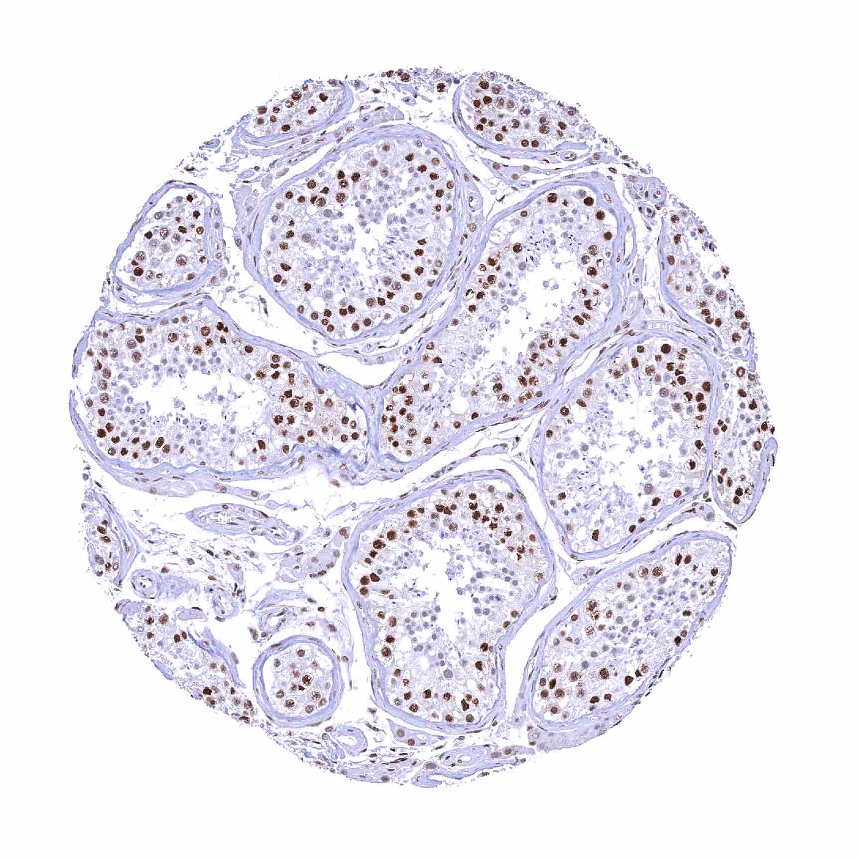
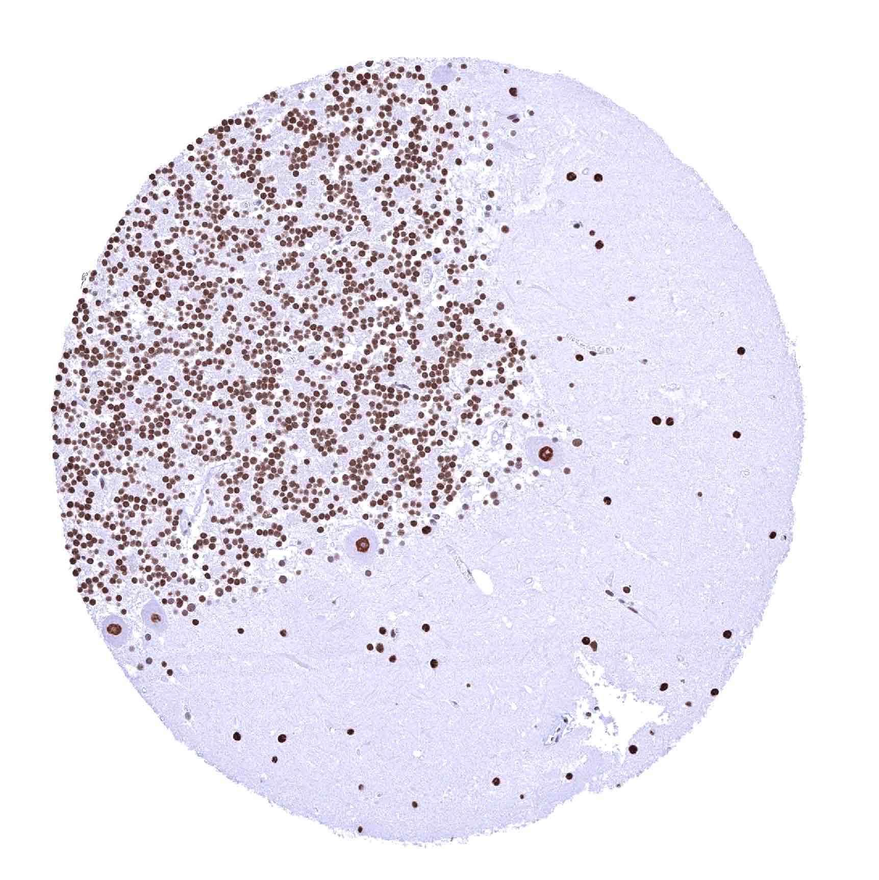
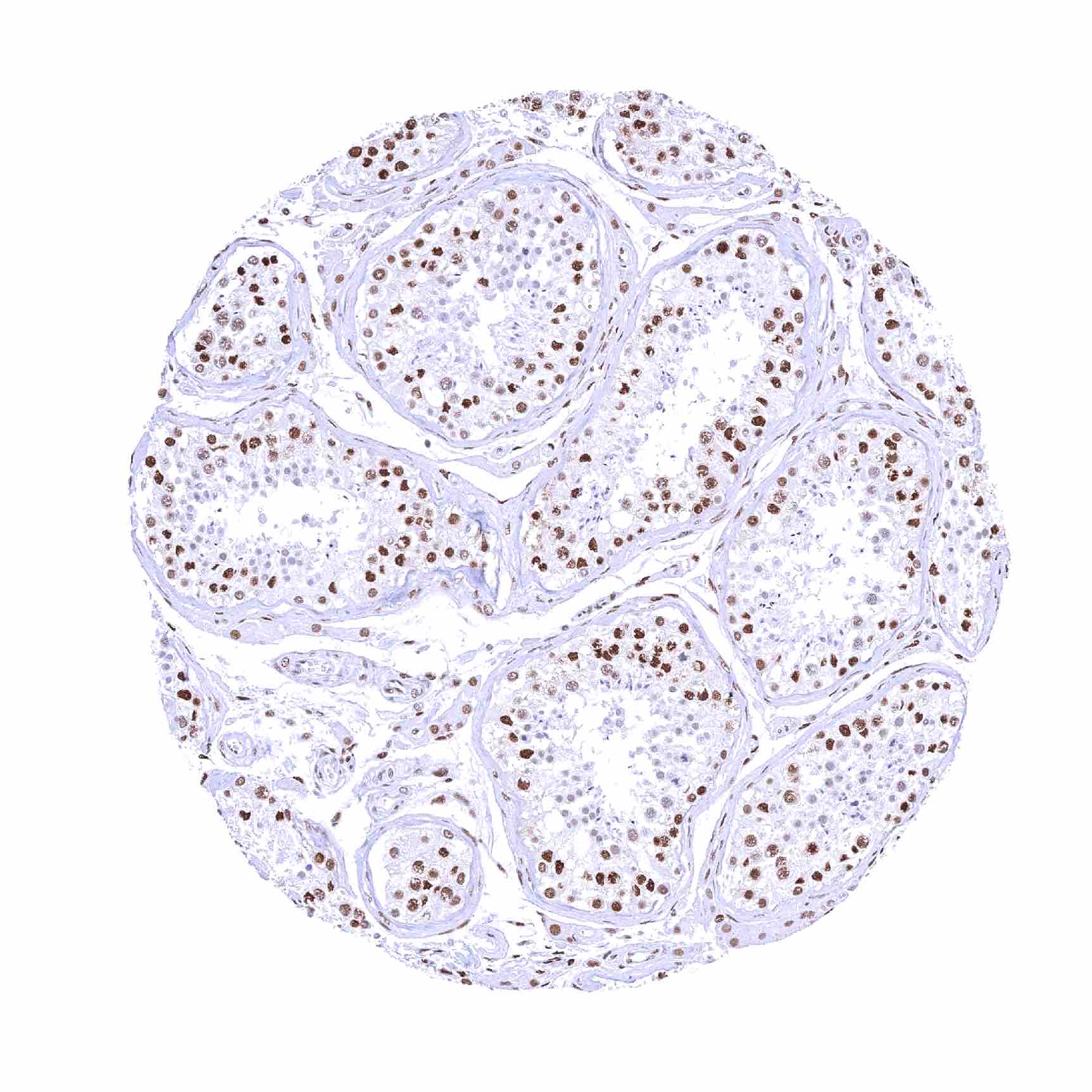
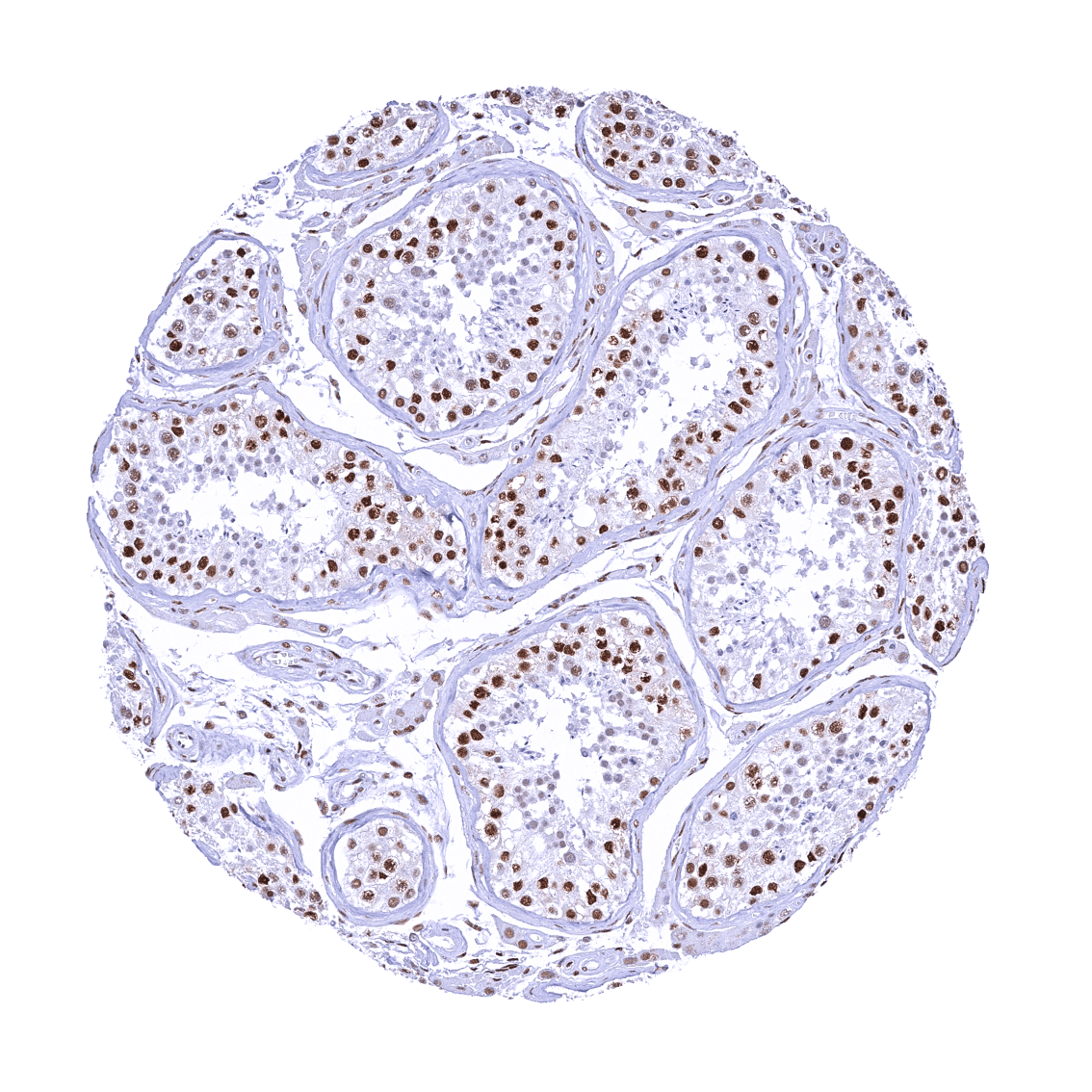
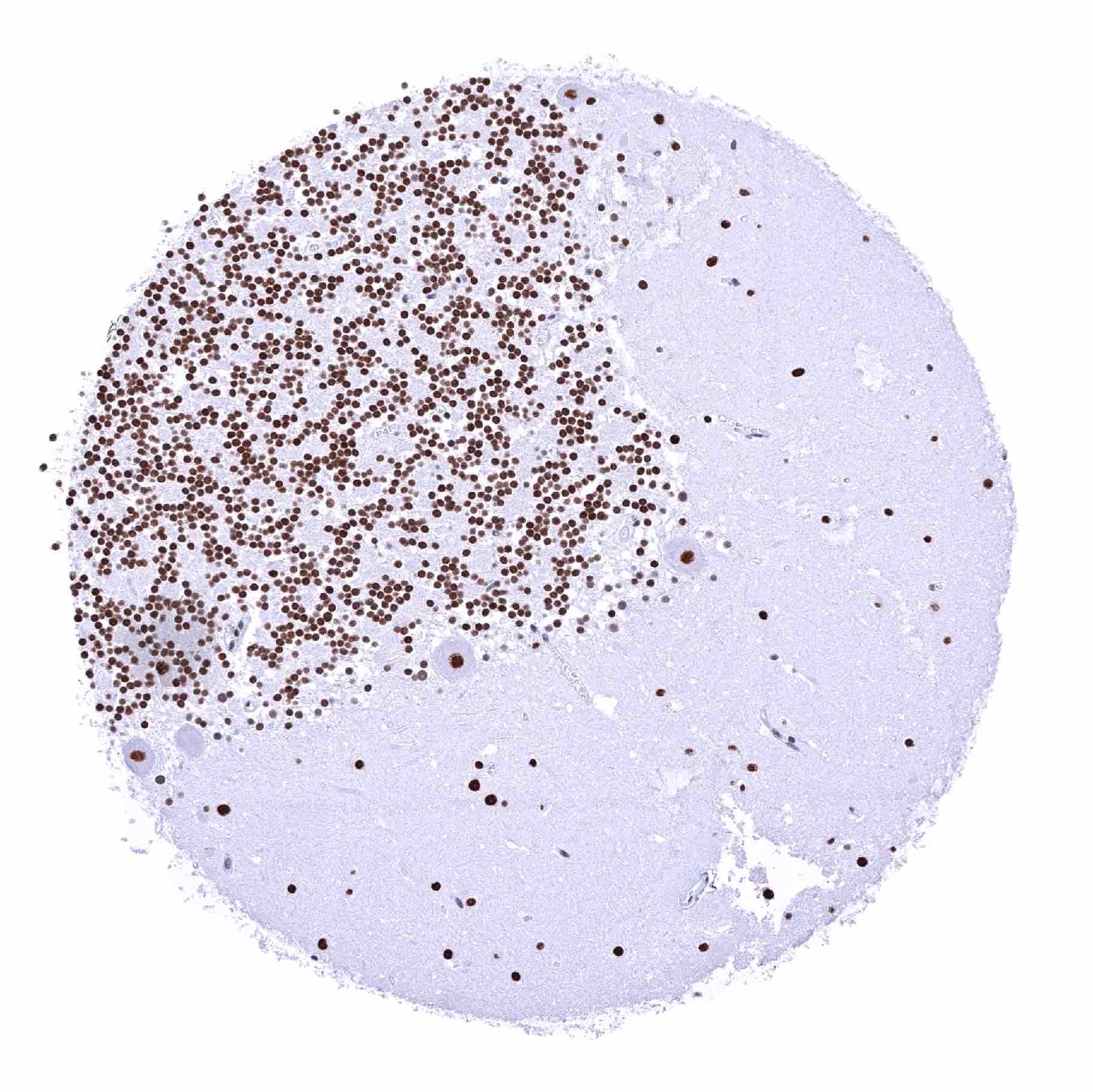
Positive control = Testis: 53BP1 staining should be strong in spermatogonia but decrease with maturation of germ cells.
Negative control = Testis: 53BP1 staining should be absent in mature spermatocytes and spermatids.
Cellular localization = Nucleus
Reactivity = Human
Application = Immunohistochemistry
Dilution = 1:100 – 1:200
Intended Use = Research Use Only
Relevance of Antibody
53BP1 is a DNA damage checkpoint critical for DNA repair pathway choice.
Biology Behind
p53-binding protein 1 (53BP1) is a scaffold protein without enzymatic activity which is encoded by the TP53BP1 gene at chromosome 15q15.3. 53BP1 participates in DNA damage checkpoint control and DNA repair, and acts as a key determinant of DNA double-strand break (DSB) repair pathway choice. The main mechanisms for DSB repair include the homologous recombinational repair (HR) pathway (requiring the sister chromatid as a template) and the non-homologous end-joining (NHEJ) pathway which rejoins the broken ends without the use of extensive homology. NHEJ, although faster than HR, is less accurate. The early divergent step between these two pathways is end resection which is regulated by numerous factors including BRCA1 and 53BP1. In response to DSBs, 53BP1 rapidly accumulates on the chromatin surrounding the DNA damage site and recruits the DNA break-responsive effectors that promote NHEJ-mediated DSB repair while inhibiting homologous recombination (HR) signaling. Reduced function of 53BP1 may promote the even more error-prone microhomology-mediated end joining (MMEJ), also known as alternative nonhomologous end-joining (Alt-NHEJ) which normally is responsible for less than 10% of DSB repair and plays a major role in aging, genomic instability and cancer. Loss of p53BP1 results in resistance to PARP inhibition treatment in Brca1-deficient tumors.
Staining Pattern in Normal Tissues
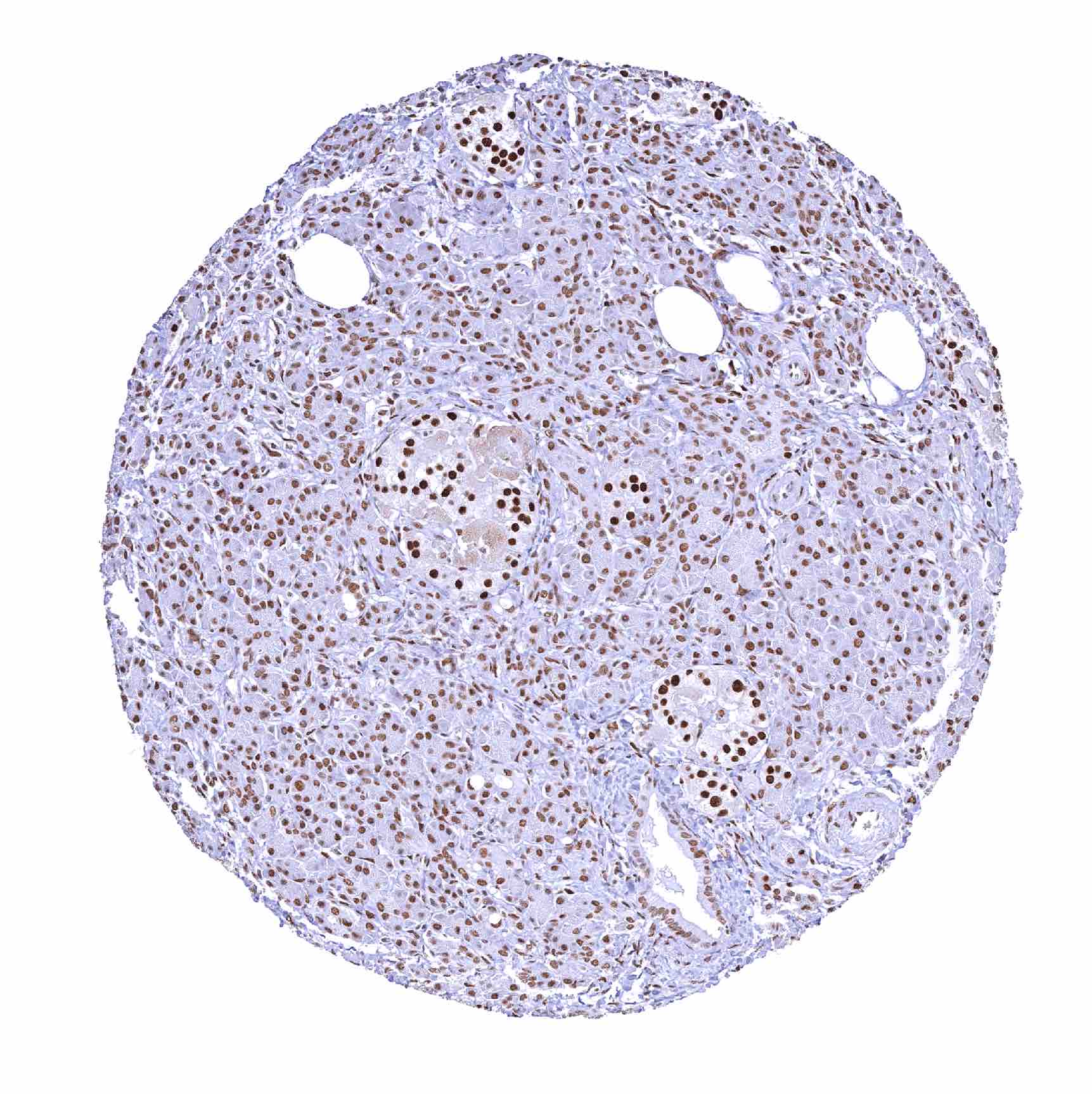
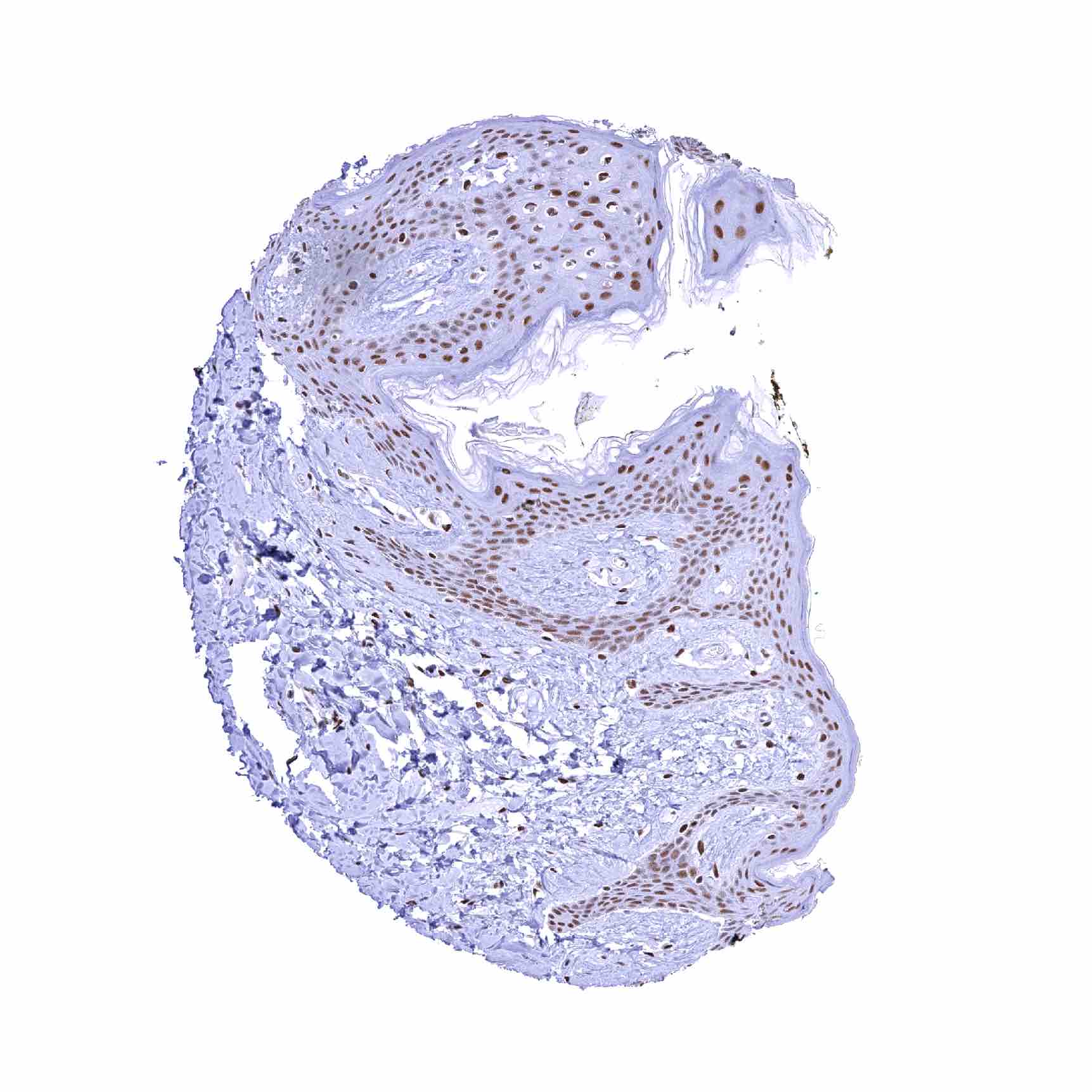
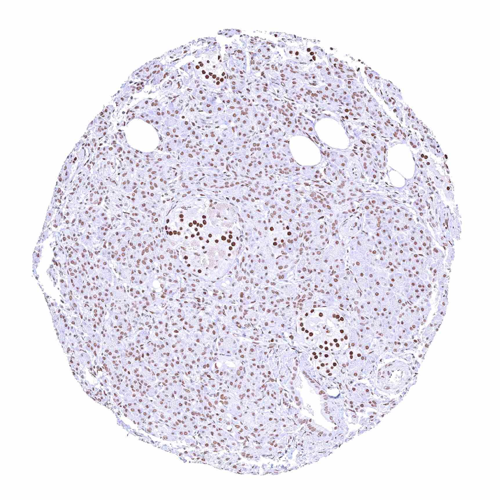
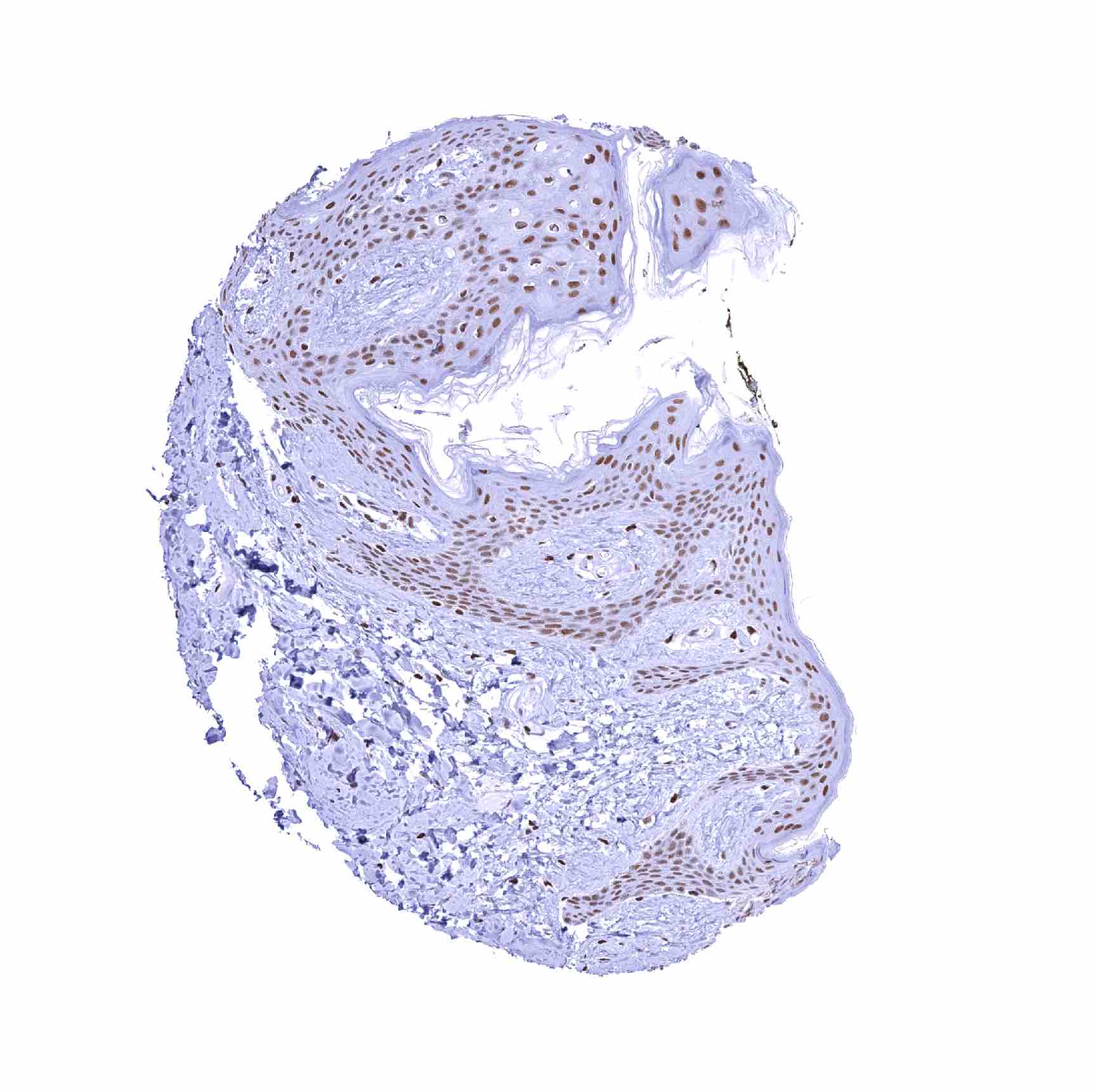
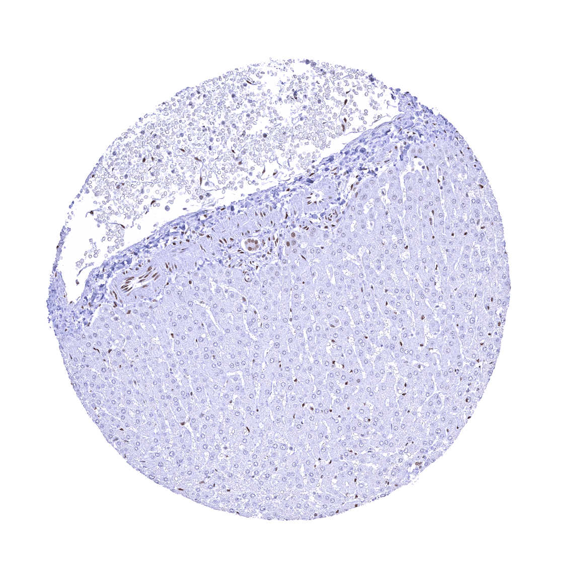
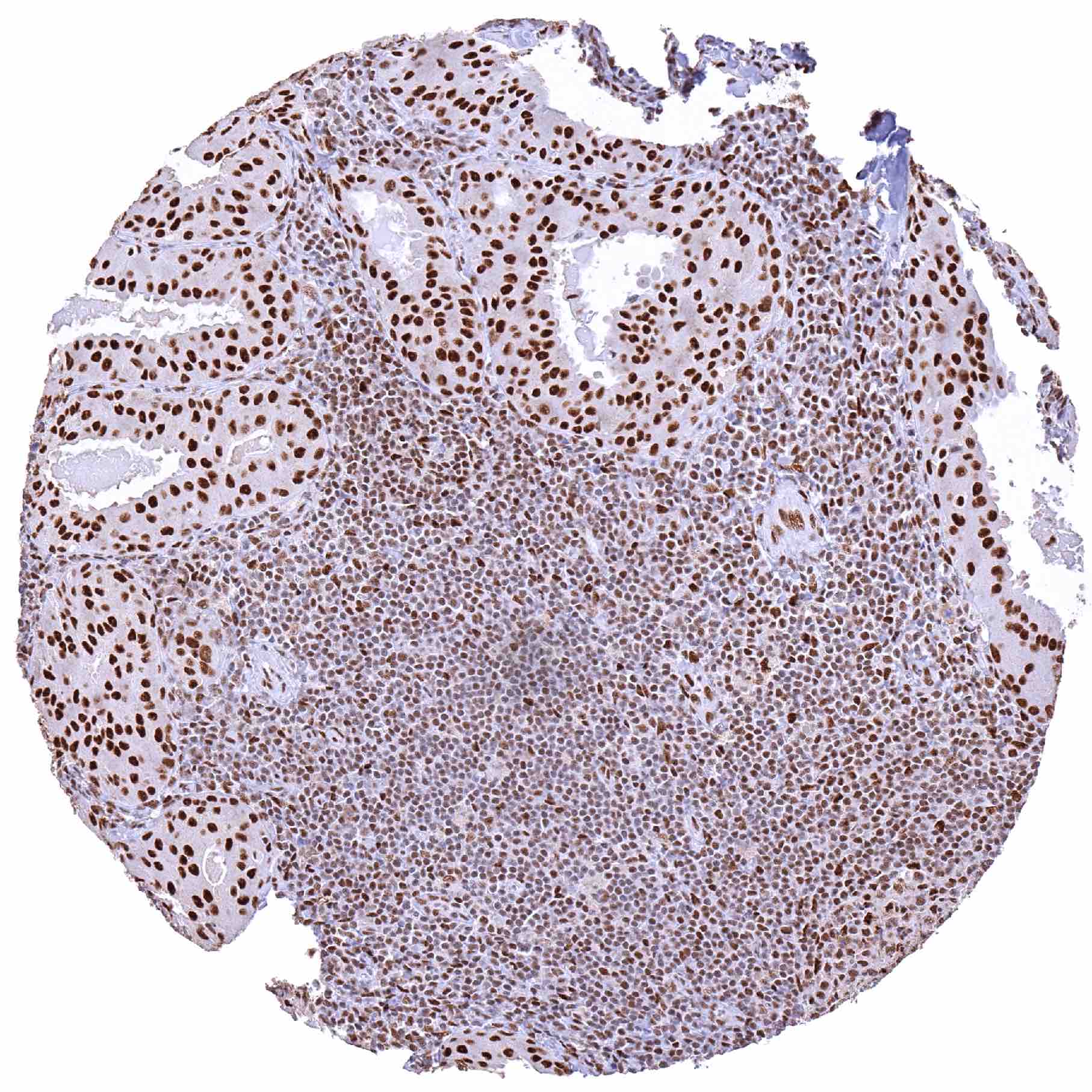
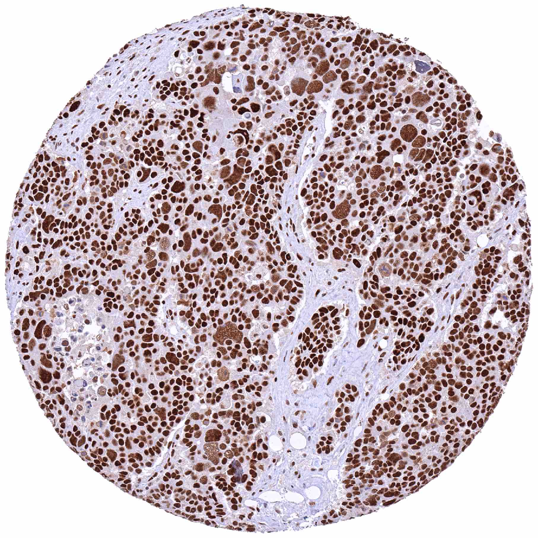
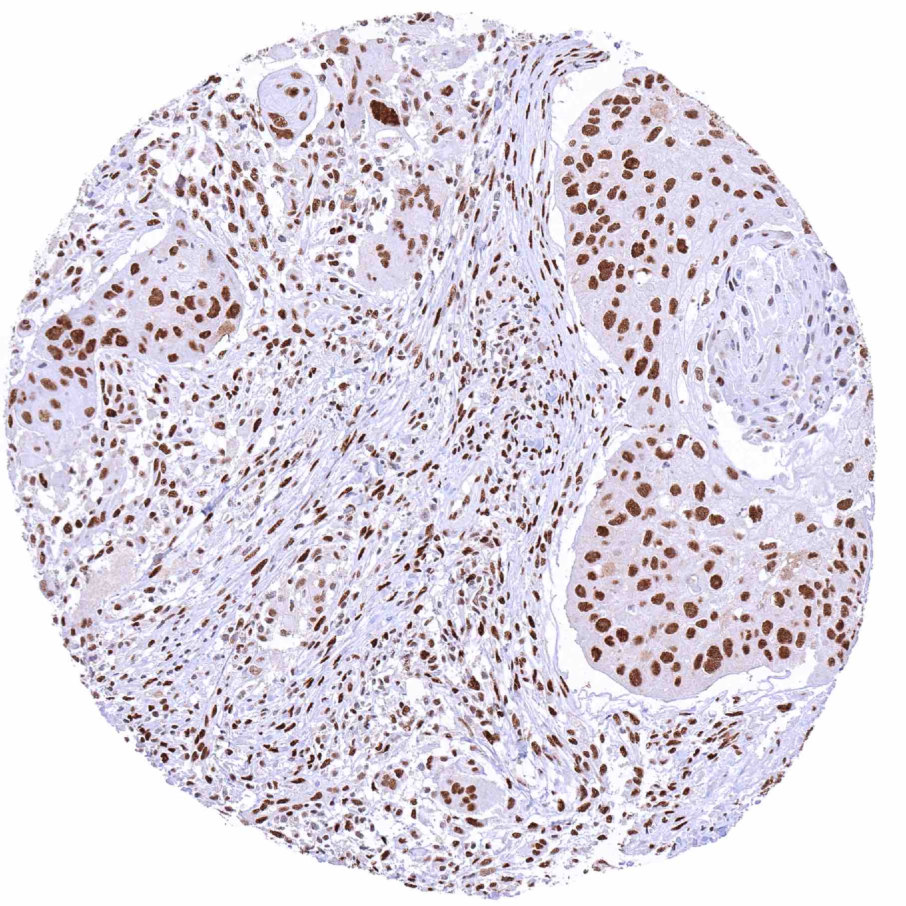
A nuclear 53BP1 staining occurs in virtually all tissues and cell types. The level of 53BP1 expression varies between tissues/cell types to some extent. It is particularly low or absent in hepatocytes. In the testis, the 53BP1 staining is strong in spermatogonia but staining intensity gradually decreases with maturation of germ cells. Mature spermatocytes and spermatids are 53BP1 negative. Images describing the 53BP1 staining pattern in normal tissues obtained by the antibody HMV324 are shown in our “Normal Tissue Gallery”.
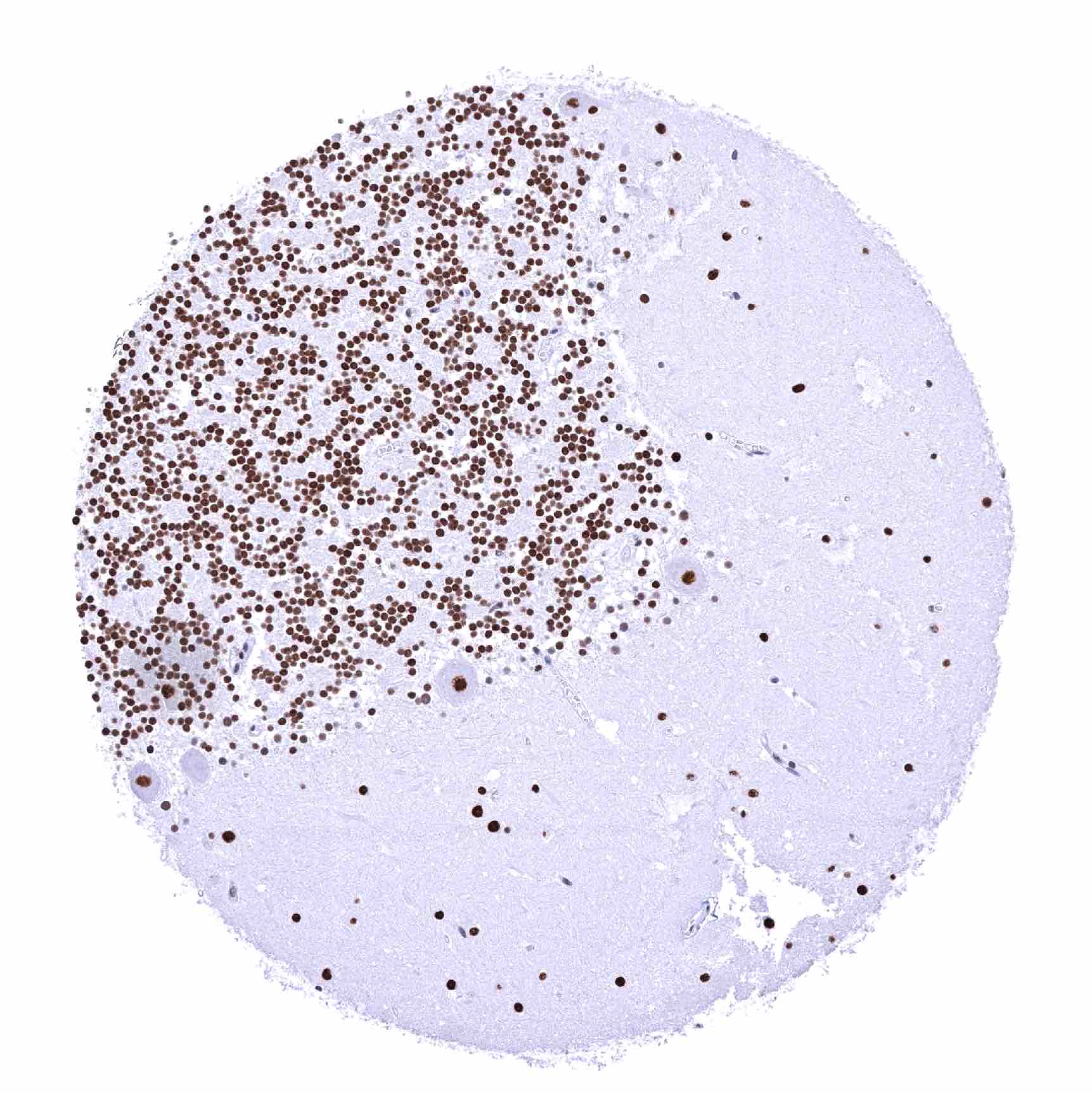
| Brain | Cerebrum | Strong 53BP1 positivity of all cells. |
| Cerebellum | Strong 53BP1 positivity of all cells. | |
| Endocrine Tissues | Thyroid | Strong 53BP1 positivity of all cells. |
| Parathyroid | Strong 53BP1 positivity of all cells. | |
| Adrenal gland | Strong 53BP1 positivity of all cells. | |
| Pituitary gland | Strong 53BP1 positivity of all cells. | |
| Respiratory system | Respiratory epithelium | Strong 53BP1 positivity of all cells. |
| Lung | Strong 53BP1 positivity of all cells. | |
| Gastrointestinal Tract | Salivary glands | Strong 53BP1 positivity of all cells. |
| Esophagus | Strong 53BP1 positivity of all cells. | |
| Stomach | Distinct 53BP1 positivity of all cells. | |
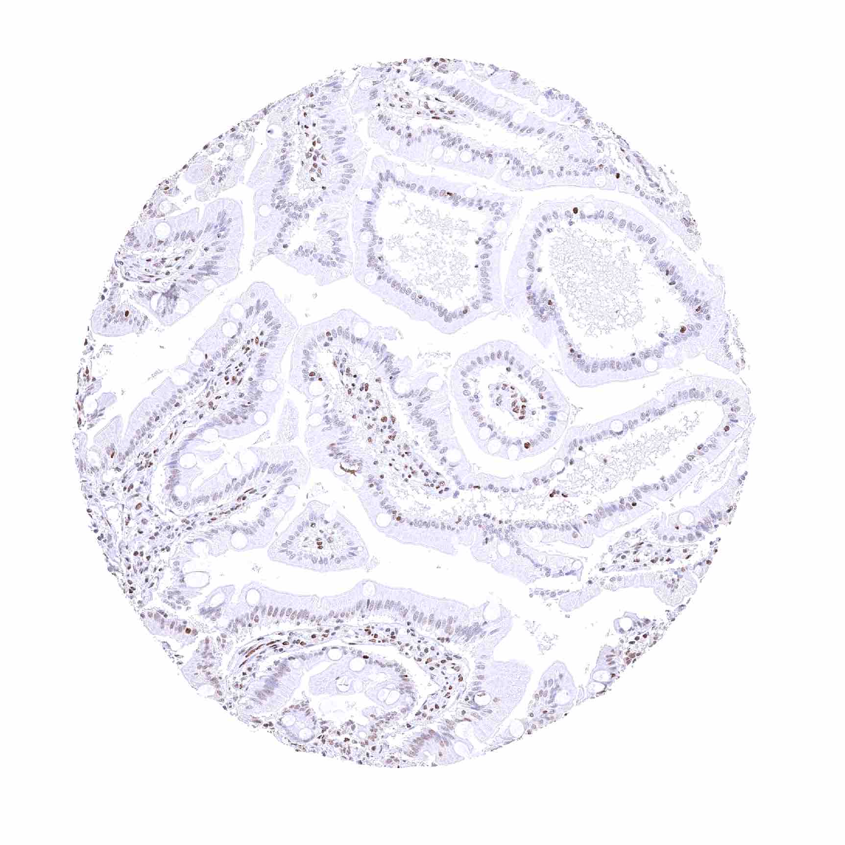
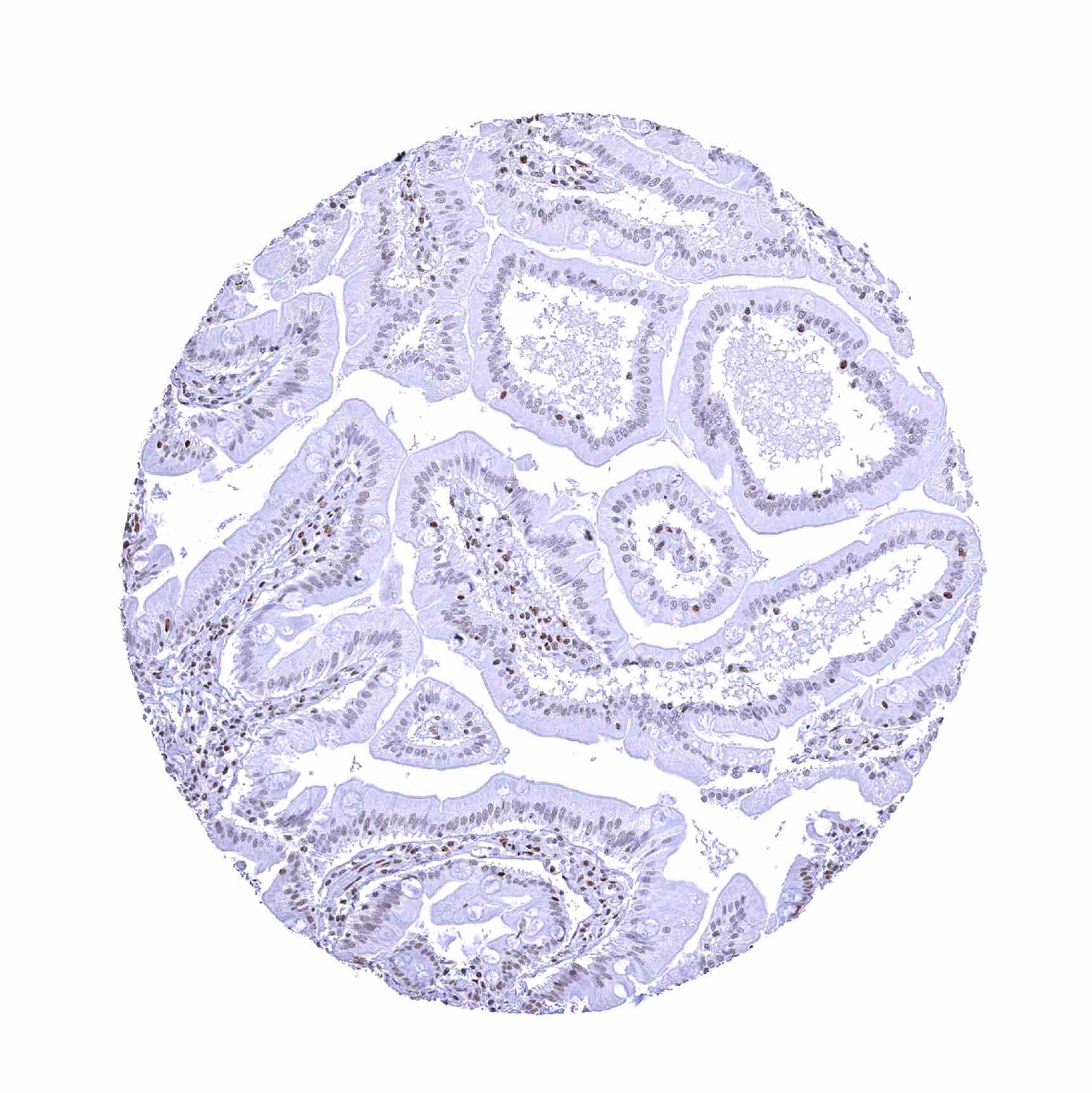
| Duodenum | Nuclear 53BP1 staining is weak or absent in epithelial cells of the villi. | |
| Small intestine | Nuclear 53BP1 staining is weak or absent in epithelial cells of the villi. | |
| Appendix | Strong 53BP1 positivity of all cells. | |
| Colon | Strong 53BP1 positivity of all cells. | |
| Rectum | Strong 53BP1 positivity of all cells. | |
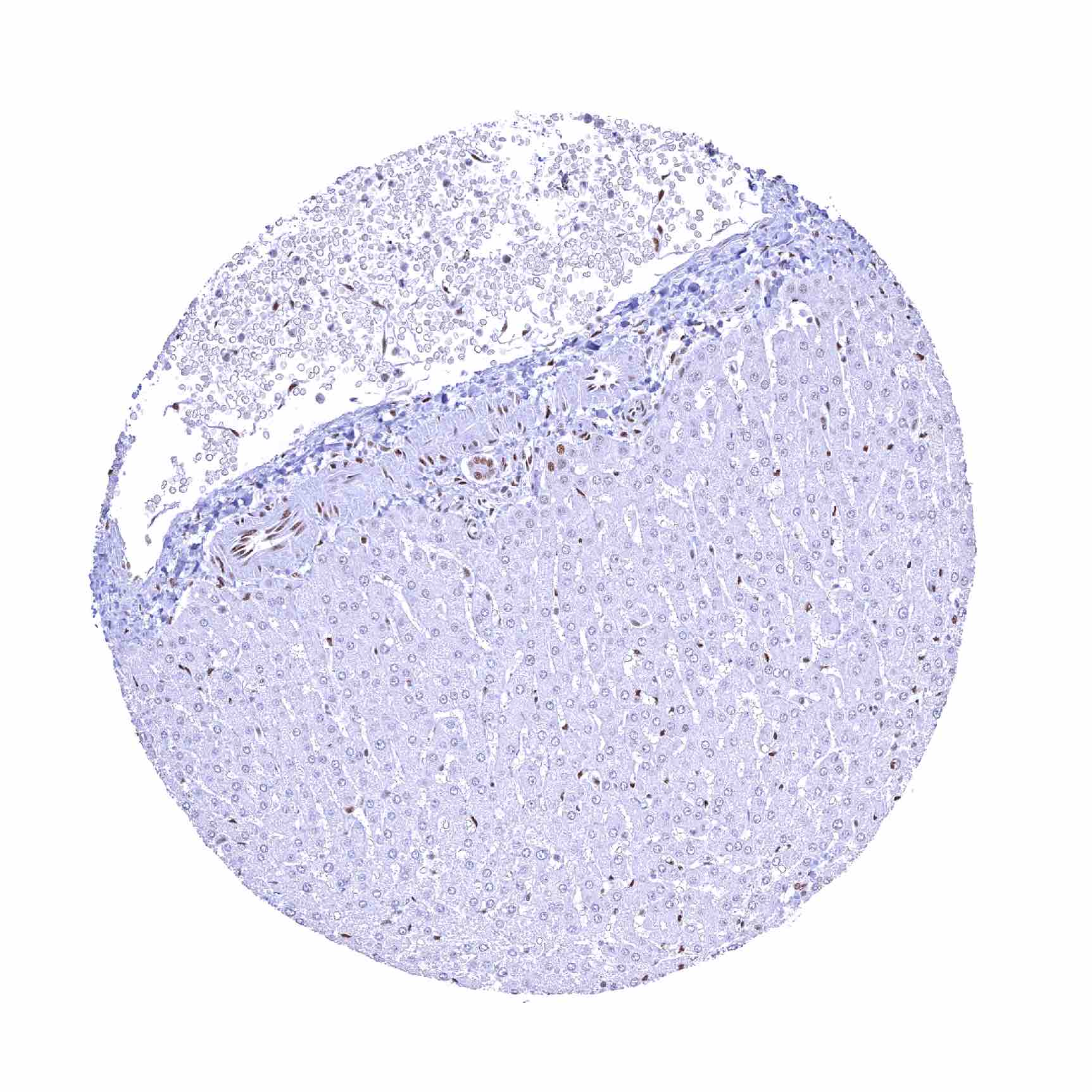
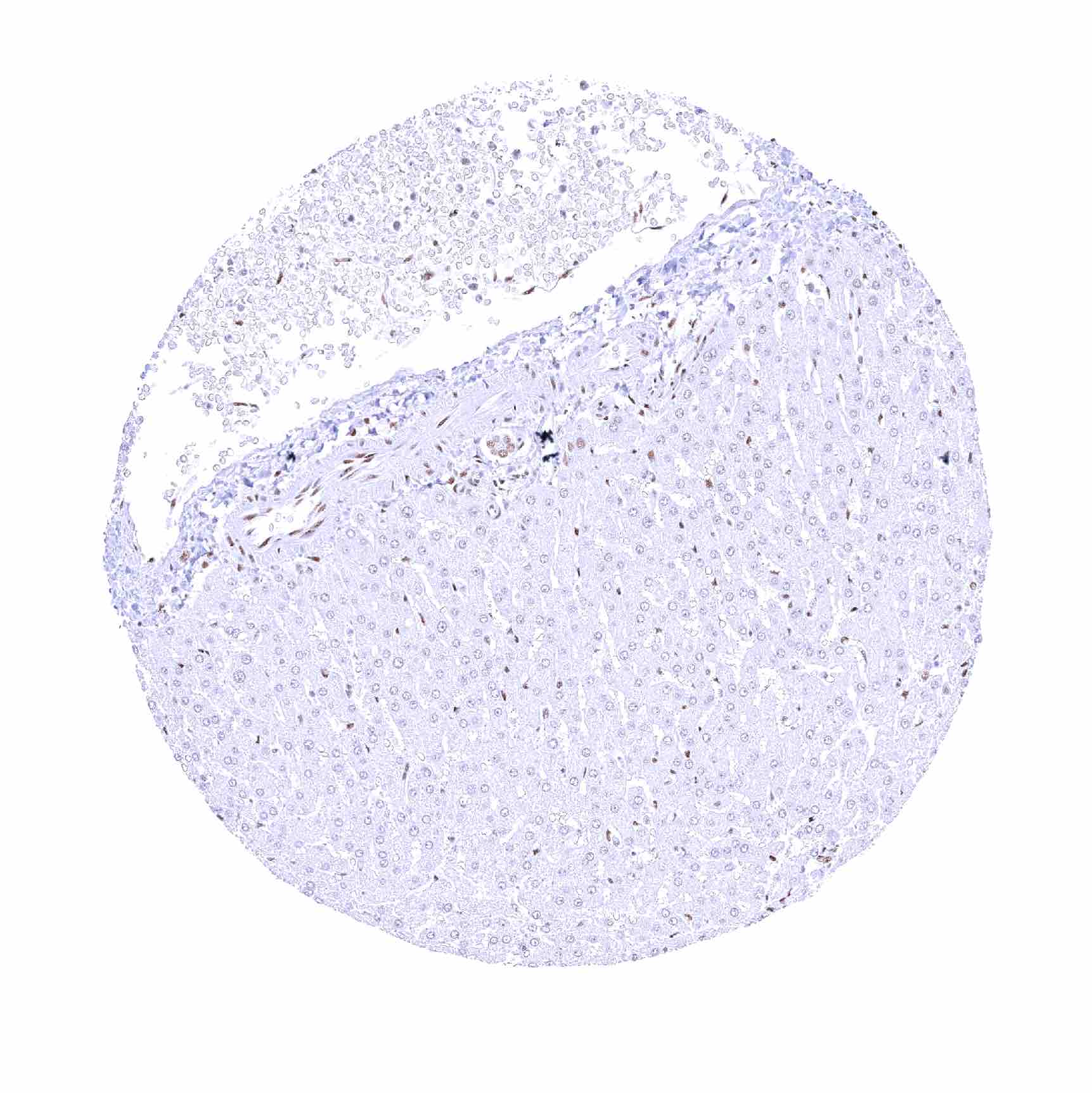
| Liver | Nuclear 53BP1 staining is absent in hepatocytes. | |
| Gallbladder | Strong 53BP1 positivity of all cells. | |
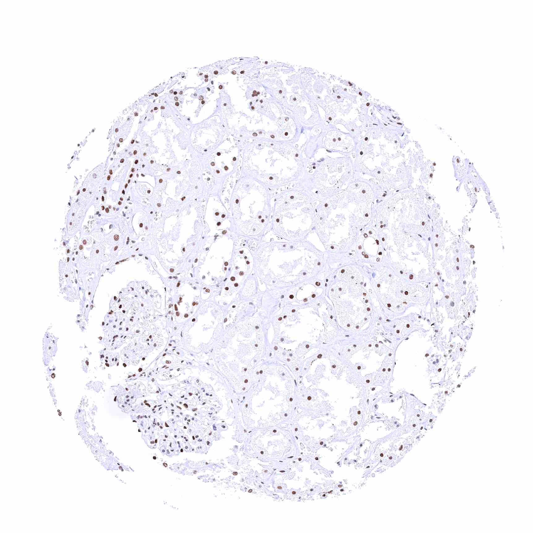
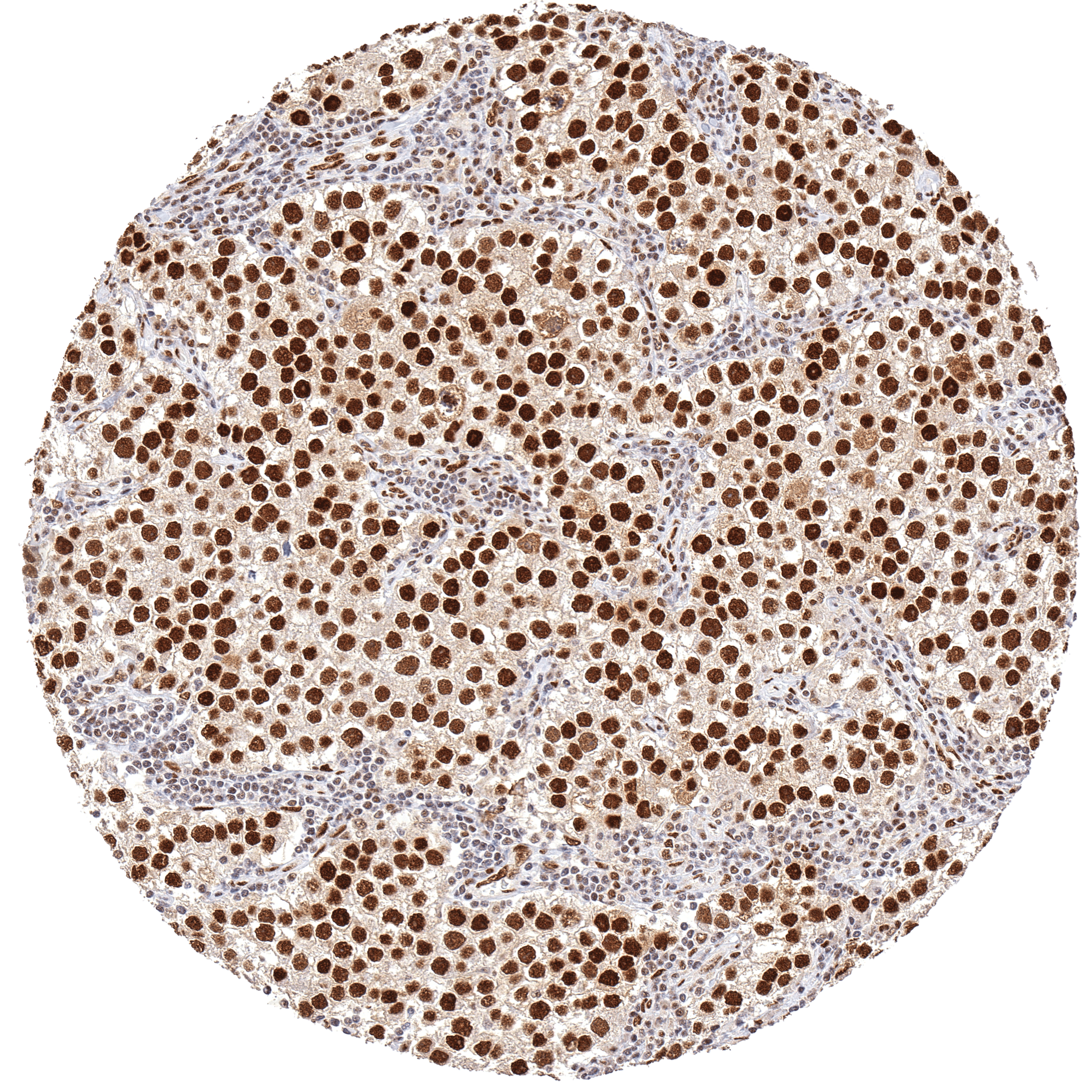
| Pancreas | Nuclear 53BP1 staining is particularly strong in islet cells. | |
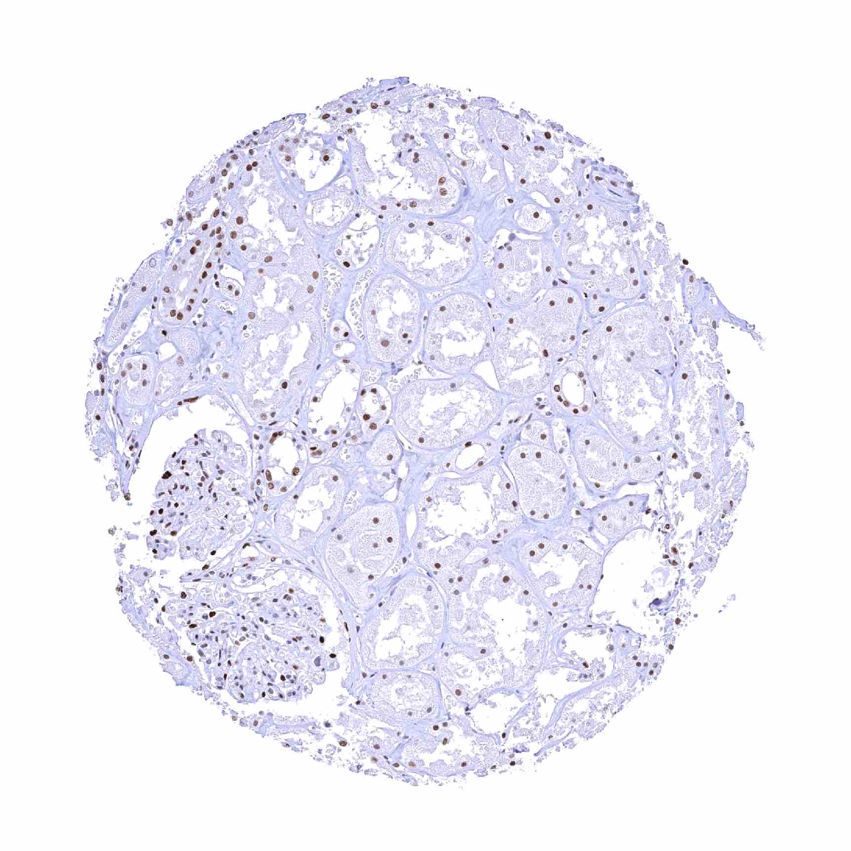
| Genitourinary | Kidney | Nuclear 53BP1 staining is least intense in proximal tubuli. |
| Urothelium | Strong 53BP1 positivity of all cells. | |
| Male genital | Prostate | Strong 53BP1 positivity of all cells. |
| Seminal vesicles | Strong 53BP1 positivity of all cells. | |
| Testis | Spermatogonia show strong 53BP1 staining but staining intensity decreases with maturation of germ cells. Mature spermatocytes and spermatids are 53BP1 negative. | |
| Epididymis | Strong 53BP1 positivity of all cells. | |
| Female genital | Breast | Strong 53BP1 positivity of all cells. |
| Uterus, myometrium | Strong 53BP1 positivity of all cells. | |
| Uterus, ectocervix | Strong 53BP1 positivity of all cells. | |
| Uterus endocervix | Strong 53BP1 positivity of all cells. | |
| Uterus, endometrium | Strong 53BP1 positivity of all cells. | |
| Fallopian Tube | Strong 53BP1 positivity of all cells. | |
| Ovary | Strong 53BP1 positivity of all cells. | |
| Placenta early | Strong 53BP1 positivity of all cells. | |
| Placenta mature | Strong 53BP1 positivity of all cells. | |
| Amnion | Strong 53BP1 positivity of all cells. | |
| Chorion | Strong 53BP1 positivity of all cells. | |
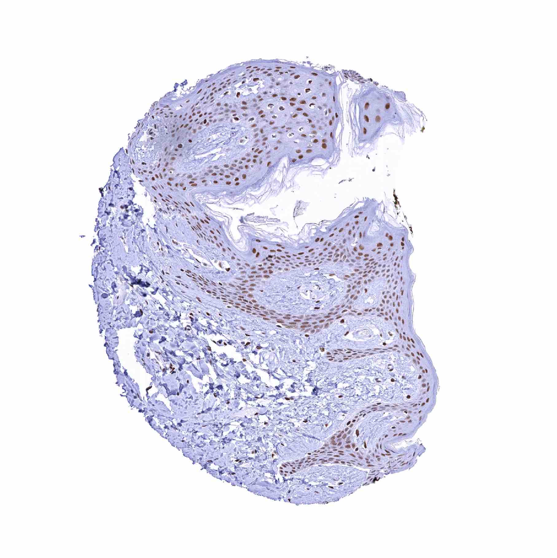
| Skin | Epidermis | Strong 53BP1 positivity of all cells. |
| Sebaceous glands | Strong 53BP1 positivity of all cells. | |
| Muscle/connective tissue | Heart muscle | Strong 53BP1 positivity of all cells. |
| Skeletal muscle | Strong 53BP1 positivity of all cells. | |
| Smooth muscle | Strong 53BP1 positivity of all cells. | |
| Vessel walls | Strong 53BP1 positivity of all cells. | |
| Fat | Strong 53BP1 positivity of all cells. | |
| Stroma | Strong 53BP1 positivity of all cells. | |
| Endothelium | Strong 53BP1 positivity of all cells. | |
| Bone marrow/ lymphoid tissue | Bone marrow | Strong 53BP1 positivity of all cells. |
| Lymph node | Strong 53BP1 positivity of all cells. | |
| Spleen | Strong 53BP1 positivity of all cells. | |
| Thymus | Strong 53BP1 positivity of all cells. | |
| Tonsil | Strong 53BP1 positivity of all cells. | |
| Remarks | A distinct/strong nuclear 53BP1 staining is seen in virtually all cell types. |
These findings are largely comparable to the RNA and protein data described in the Human Protein Atlas (Tissue expression 53BP1).
Positive control = Testis: 53BP1 staining should be strong in spermatogonia but decrease with maturation of germ cells.
Negative control = Testis: 53BP1 staining should be absent in mature spermatocytes and spermatids.
Staining Pattern in Relevant Tumor Types
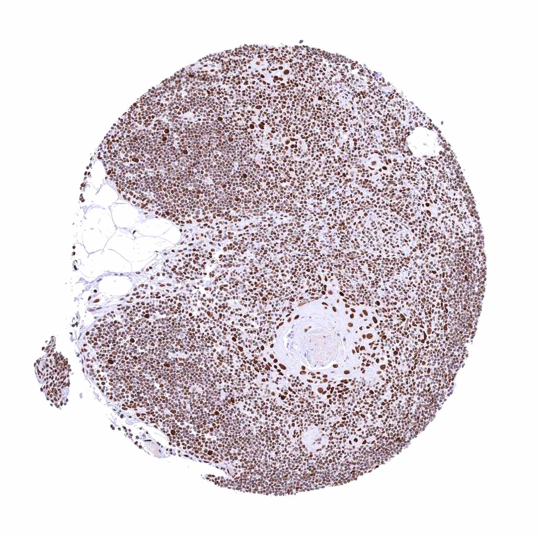
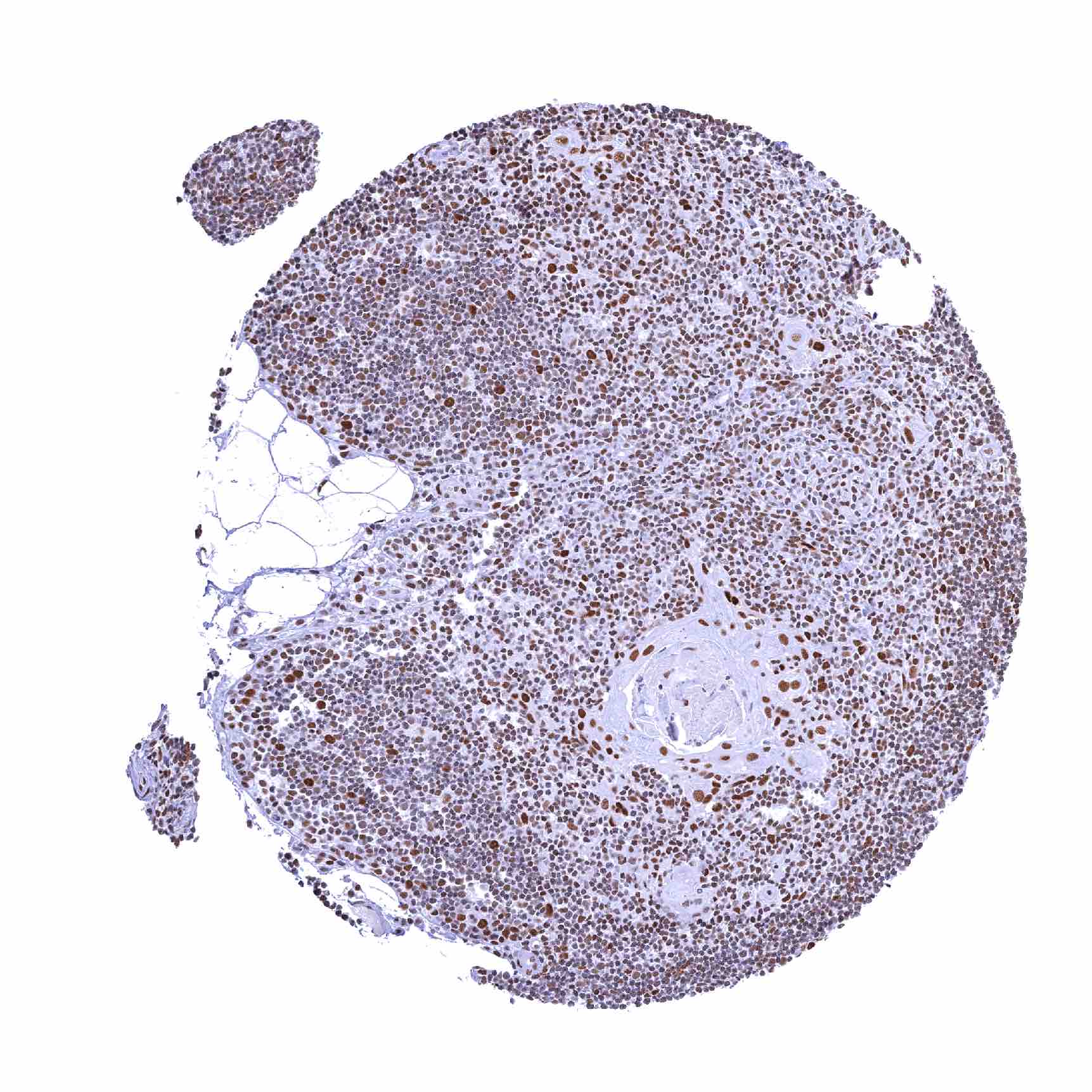
A positive 53BP1 immunostaining (intensity may vary) is usually seen in tumor cells of virtually all cancer types as well as in tumor associated stromal and inflammatory cells.
The TCGA findings on 53BP1 RNA expression in different tumor categories have been summarized in the Human Protein Atlas.
Compatibility of Antibodies
No data available at the moment
Protocol Recommendations
IHC users have different preferences on how the stains should look like. Some prefer high staining intensity of the target stain and even accept some background. Others favor absolute specificity and lighter target stains. Factors that invariably lead to more intense staining include higher concentration of the antibody and visualization tools, longer incubation time, higher temperature during incubation, higher temperature and longer duration of the heat induced epitope retrieval (slide pretreatment). The impact of the pH during slide pretreatment has variable effects and depends on the antibody and the target protein.
All images and data shown here and in our image galleries are obtained by the manual protocol described below. Other protocols resulting in equivalent staining are described as well.
Manual protocol
Freshly cut sections should be used (less than 10 days between cutting and staining). Heat-induced antigen retrieval for 5 minutes in an autoclave at 121°C in pH 7,8 Target Retrieval Solution buffer. Apply HMV324 at a dilution of 1:200 at 37°C for 60 minutes. Visualization of bound antibody by the EnVision Kit (Dako, Agilent) according to the manufacturer’s directions.
Potential Research Applications
- The role of 53BP1 in DNA repair needs to be further investigated.
- The prognostic and predictive role of 53BP1 expression levels in cancer is unclear.
Evidence for Antibody Specificity in IHC
There are two ways how the specificity of antibodies can be documented for immunohistochemistry on formalin fixed tissues. These are: 1. Comparison with a second independent method for target expression measurement across a large number of different tissue types (orthogonal strategy), and 2. Comparison with one or several independent antibodies for the same target and showing that all positive staining results are also seen with other antibodies for the same target (independent antibody strategy).
Orthogonal validation: For the antibody HMV324 specificity is in line with data from three independent RNA screening studies, including the Human Protein Atlas (HPA) RNA-seq tissue dataset, the FANTOM5 project, and the Genotype-Tissue Expression (GTEx) project, which are all summarized in the Human Protein Atlas (Tissue expression 53BP1). In agreement with HMV324 immunostaining data, 53BP1 RNA expression predominated in the brain and in lymphoid tissues and it was comparably low in the liver. However, orthogonal validation is not optimal for assessing ubiquitously expressed protein.
Comparison of antibodies: True expression of 53BP1 in all cell types with 53BP1 positivity seen by HMV324 is corroborated by an identical staining obtained by a commercially available independent second antibody (termed “validation antibody”). Most of all, both antibodies showed a complete lack of nuclear staining only in mature spermatocytes and spermatids of the testis, and in hepatocytes in the liver. Moreover, both antibodies showed a particularly low staining intensity of epithelial cells of villi of the small intestine, and a particularly strong staining of islet cells of the pancreas.