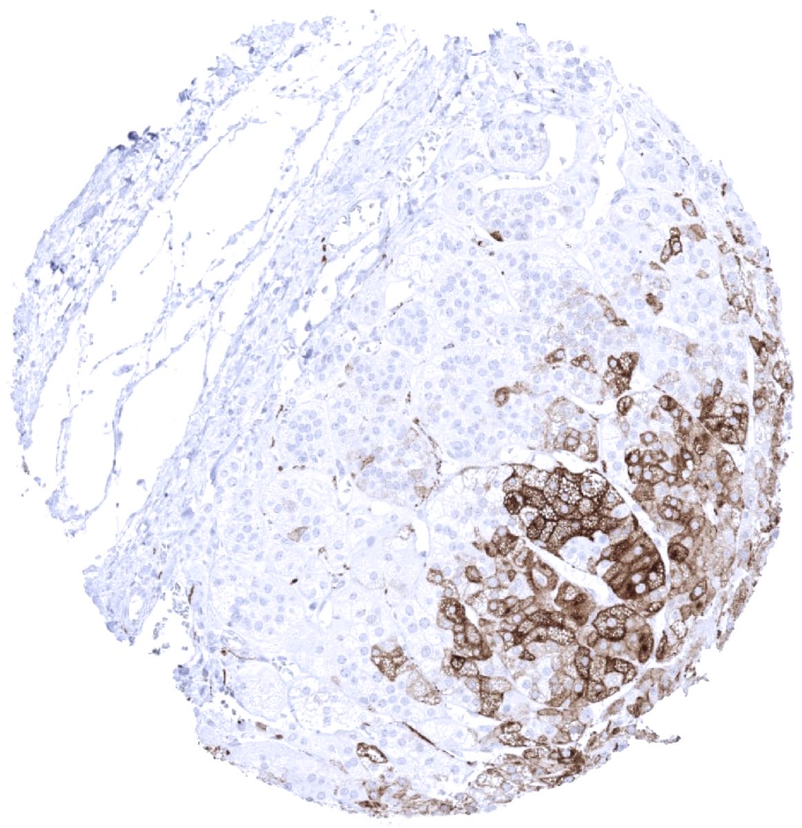
Adrenal gland - A strong synaptophysin immunostaining regularly occurs in the medulla of the adrenal gland. Some cortical cells may show weak to moderate staining.
Adrenal gland - A weak to moderate synaptophysin immunostaining can be found in a subset of cells of the adrenal cortex.

Aorta, media
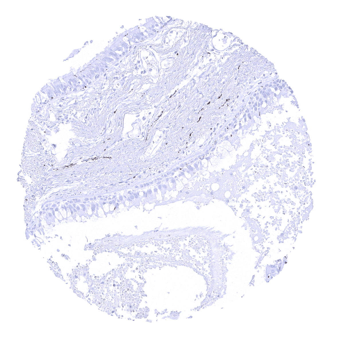
Appendix, mucosa - In the appendix, synaptophysin immunostaining is limited to neuroendocrine cells and nerve fibres.

Appendix, muscular wall

Bone marrow

Breast
Bronchus, mucosa

Cerebellum (molecular, granular and Purkinje layers; white matter) - Synaptophysin is strongly expressed in the cerebellum.
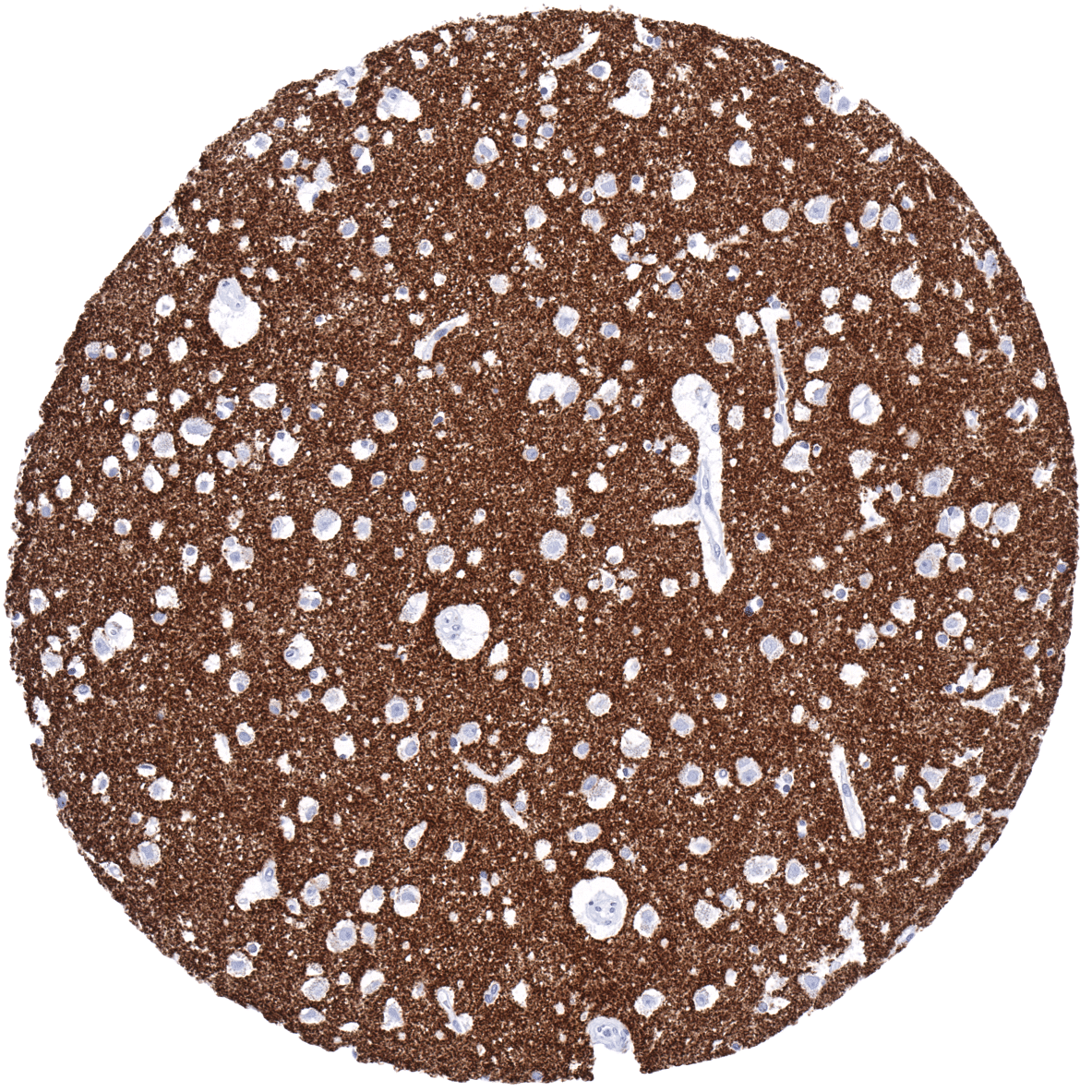
Cerebrum, grey matter - Synaptophysin is strongly expressed in the cerebrum.
Colon descendens, muscular wall
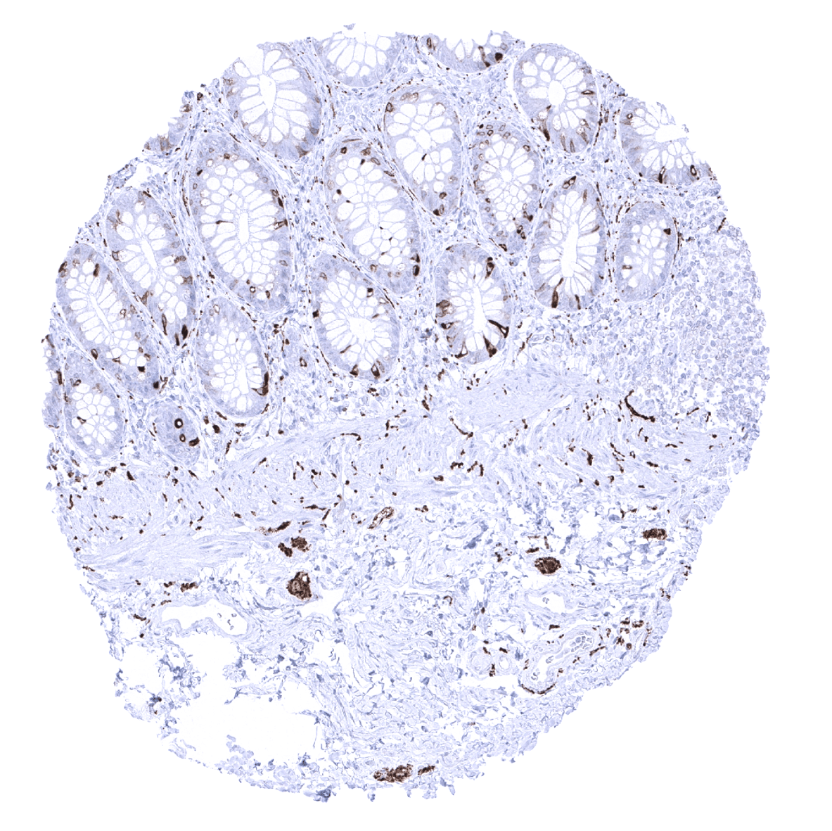
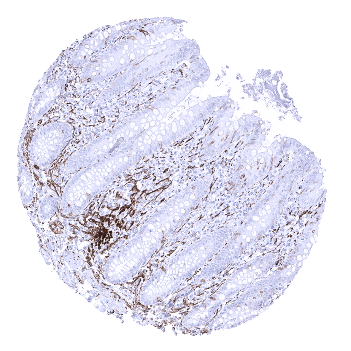
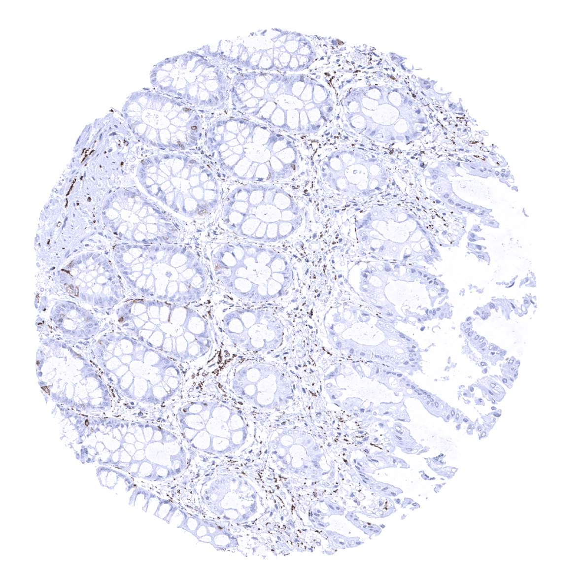
Colon descendes, mucosa - Strong synaptophysin immunostaining in neuroendocrine cells of the mucosa, nerve fibres and ganglion cells of the submucosa.

Duodenum, Brunner gland
Duodenum, mucosa - In the small intestine, synaptophysin immunostaining is seen in neuroendocrine cells and in goblet cells.

Epididymis

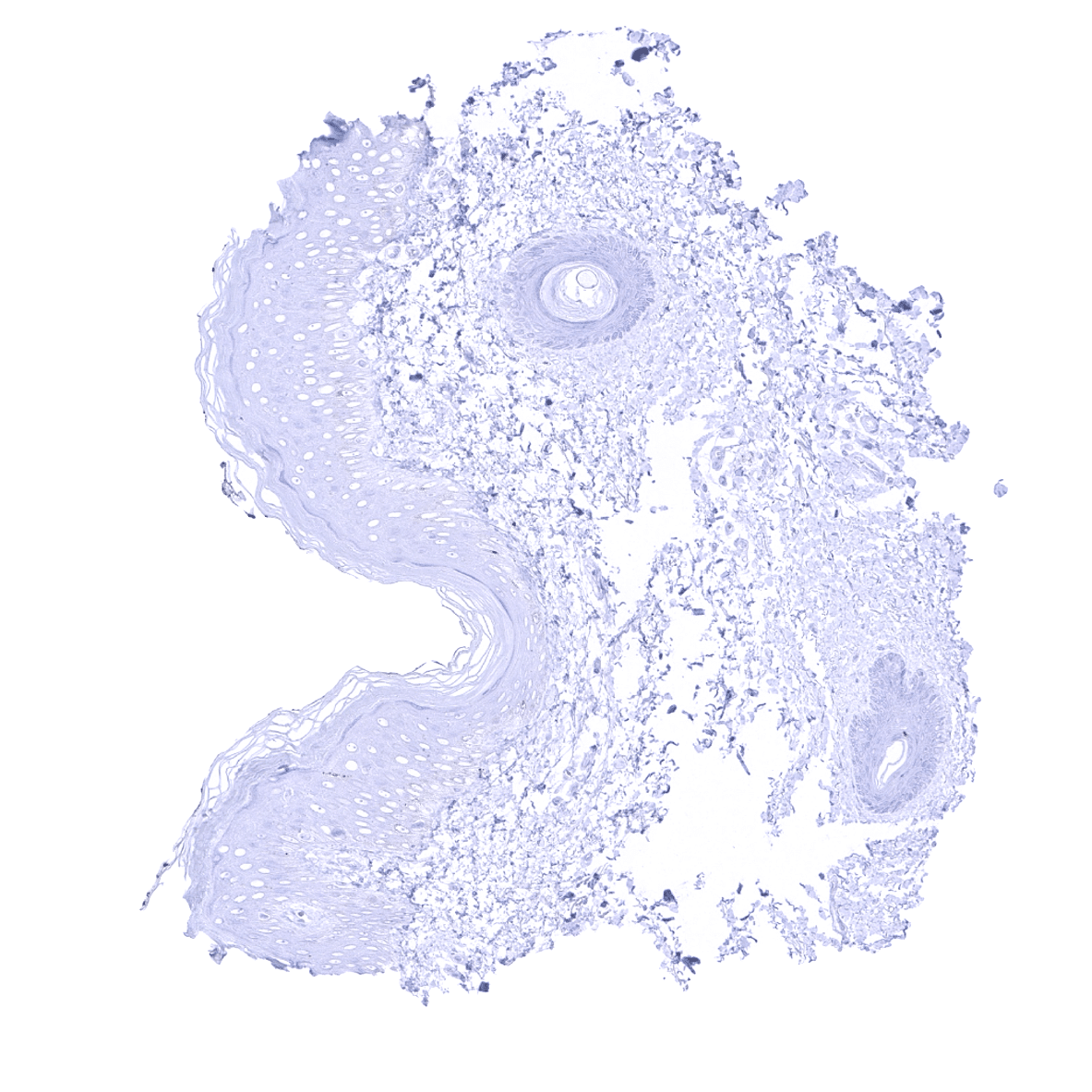
Esophagus, squamous epithelium
Fallopian tube, mucosa
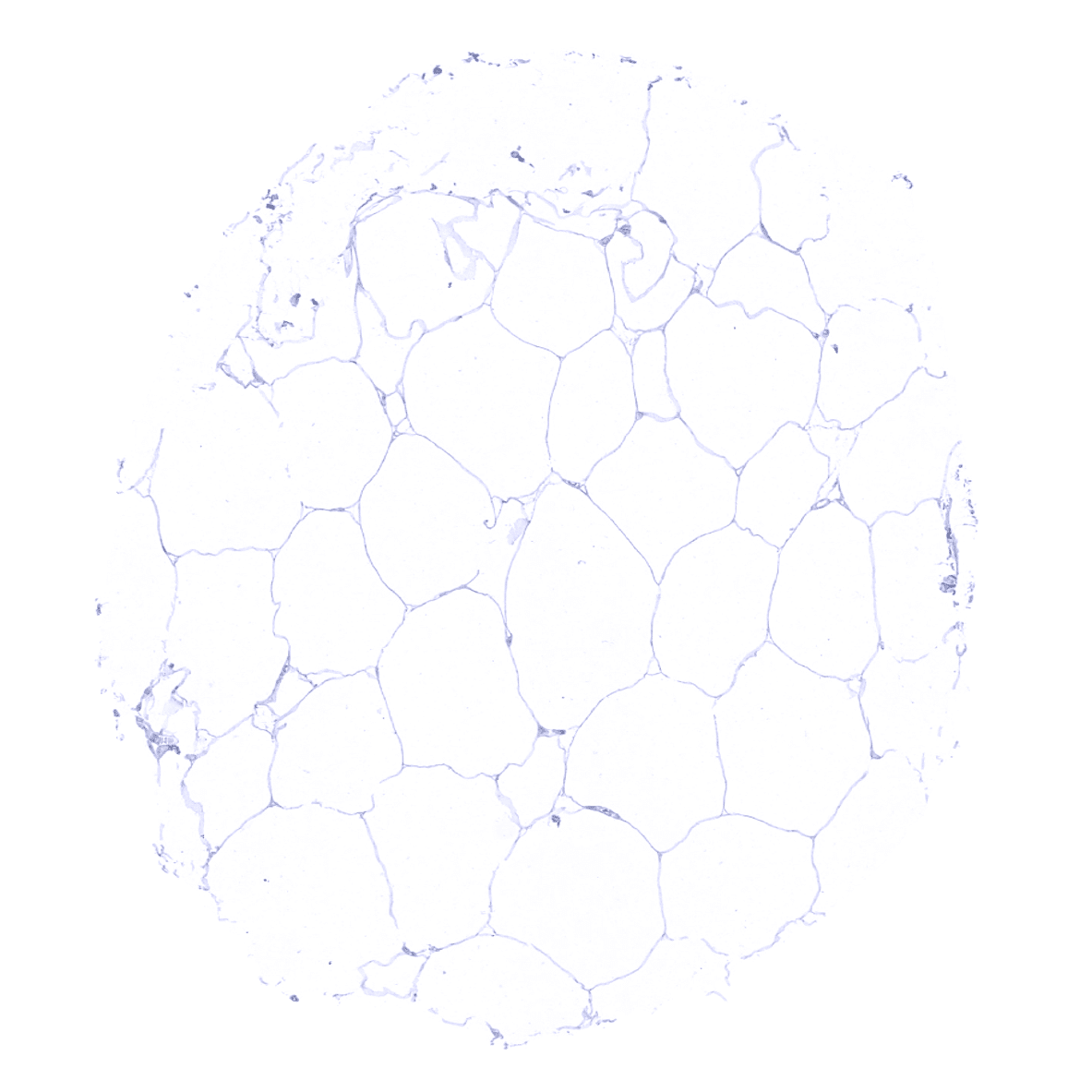
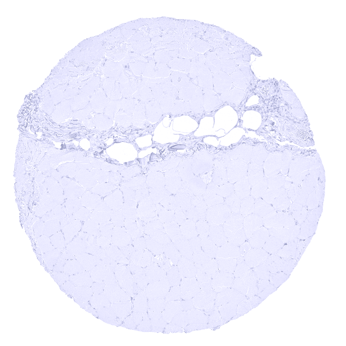
Fat
Gallbladder, epithelium
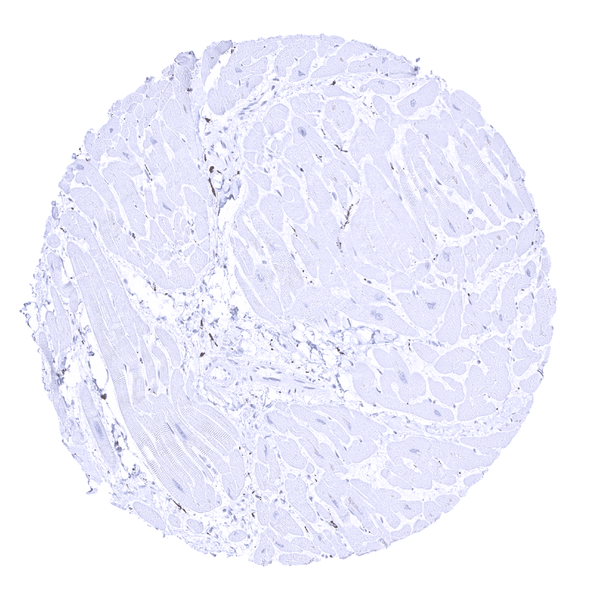
Heart muscle

Ileum, mucosa - In the small intestine, synaptophysin immunostaining is seen in neuroendocrine cells and in goblet cells.
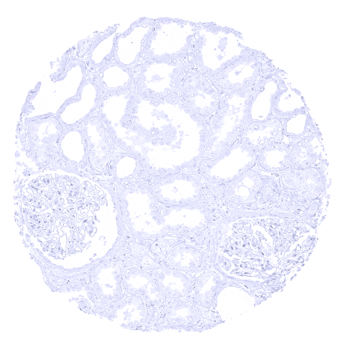
Kidney, cortex
Kidney, medulla
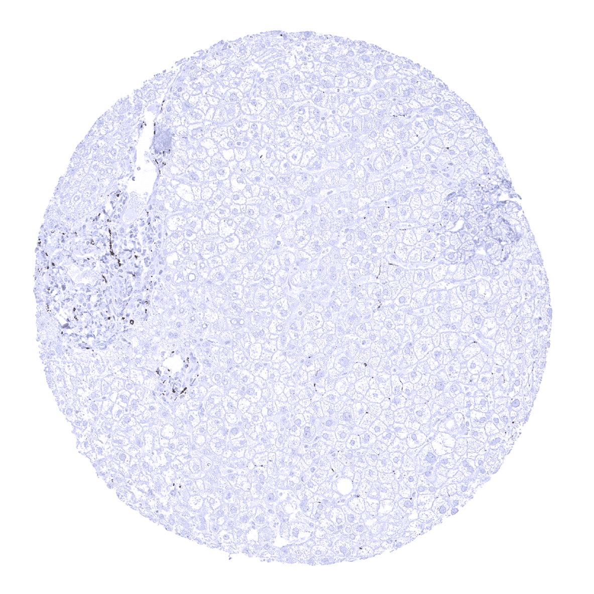
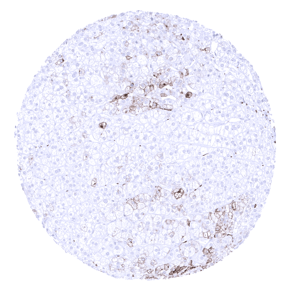
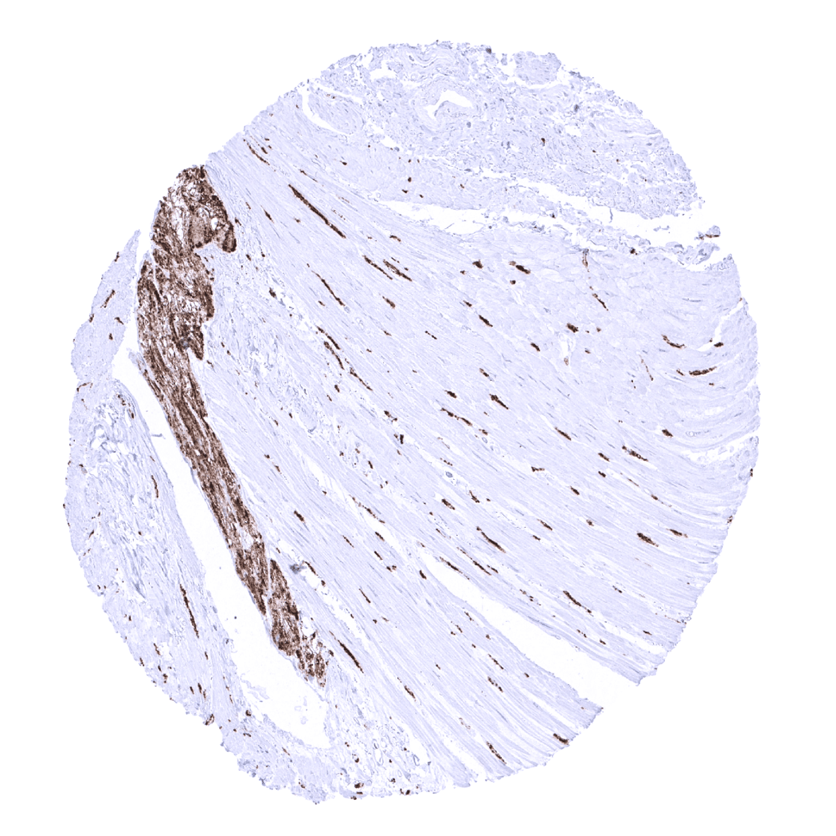
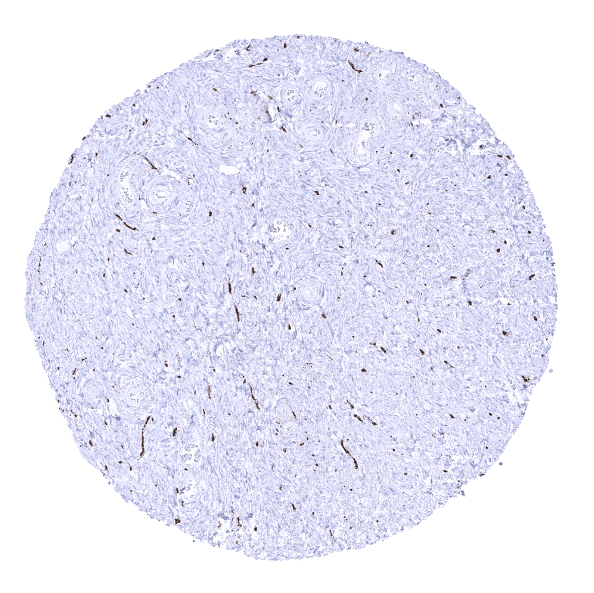
Liver - In the liver, synaptophysin positive nerve fibres are seen in portal fields.
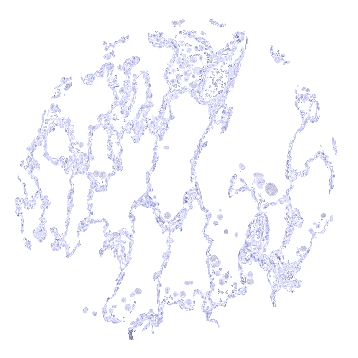
Lung
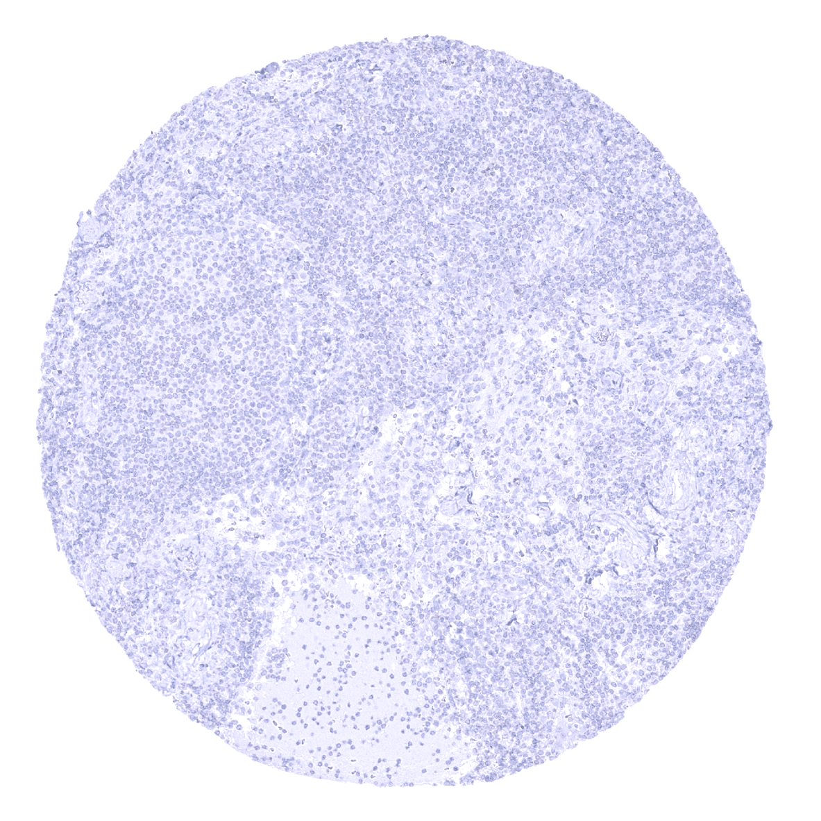
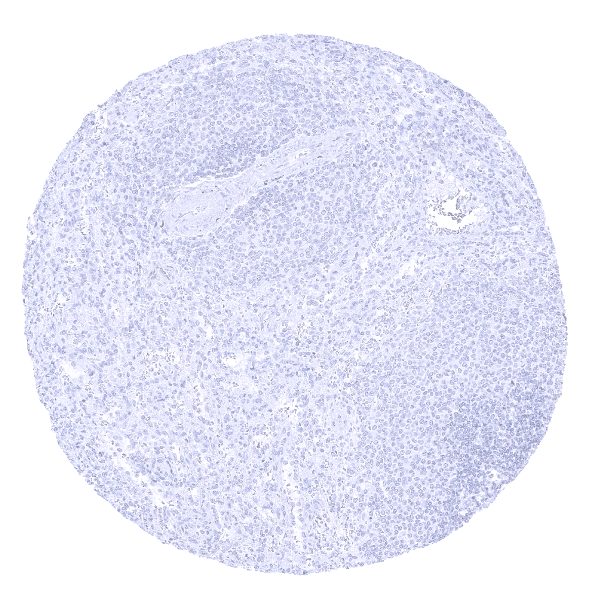
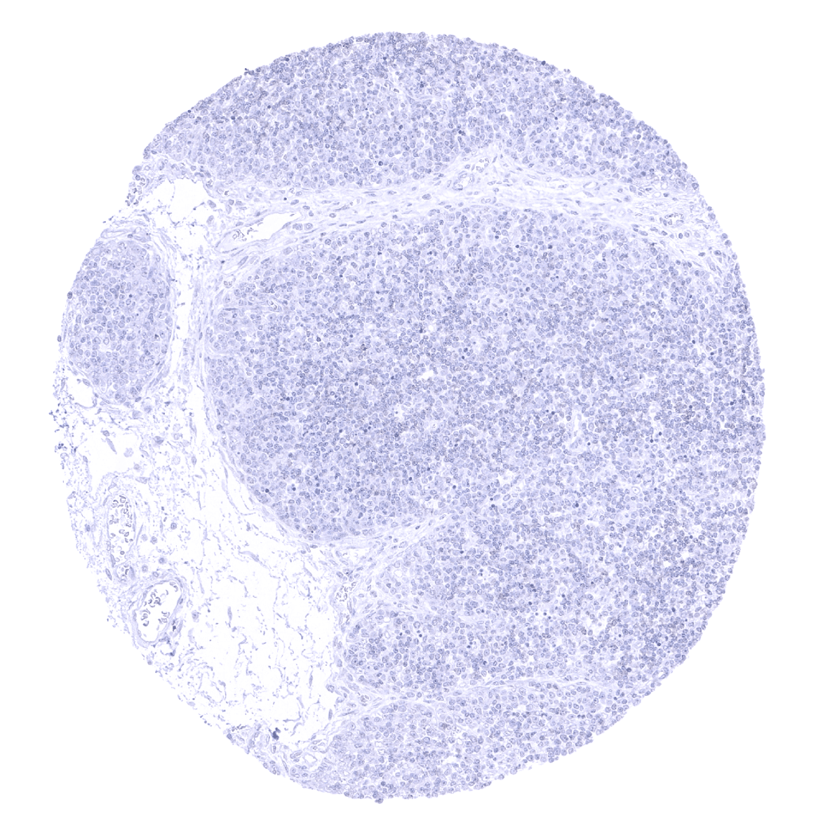
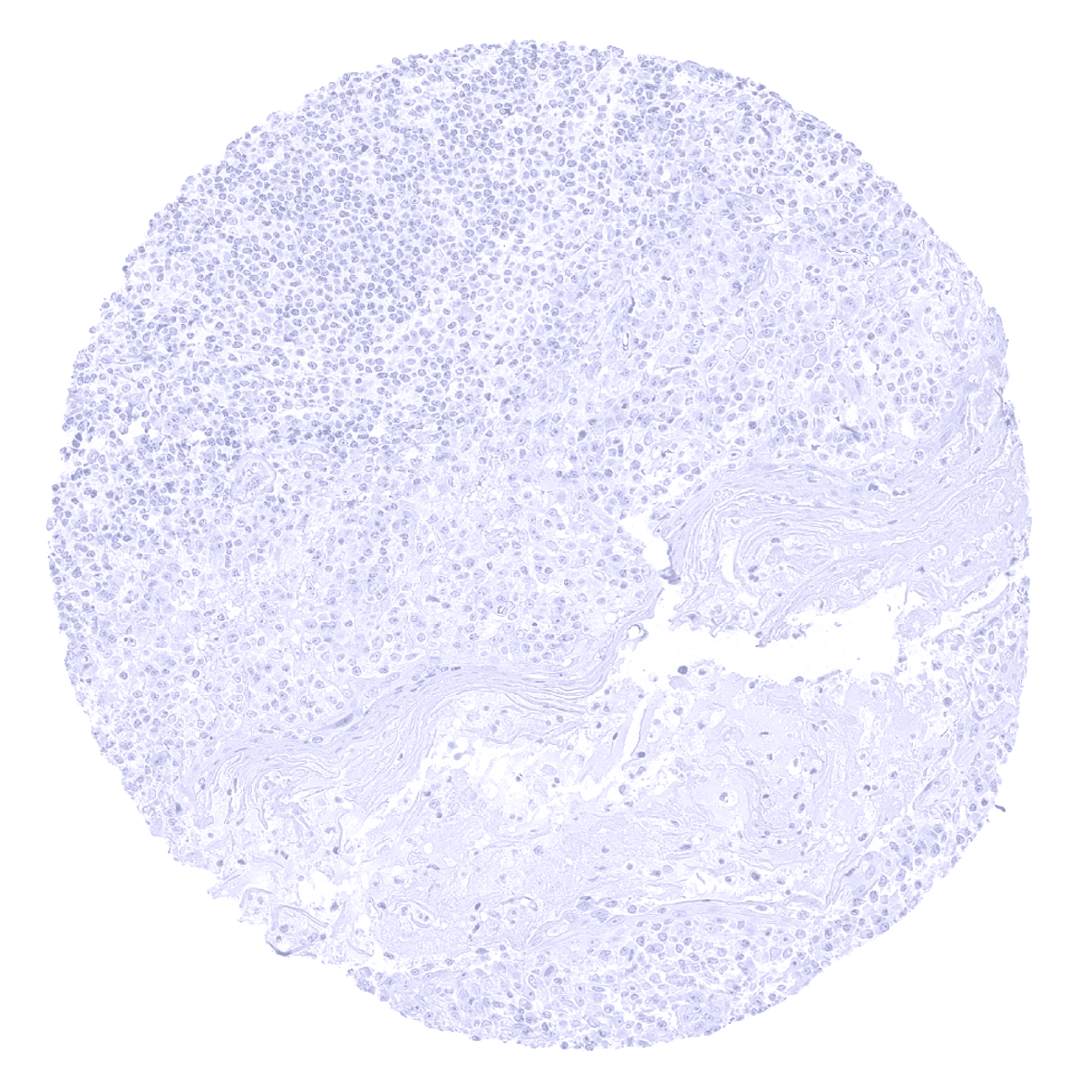
Lymph node
Ovary, stroma - A few nerve fibres occur in the ovarian stroma.
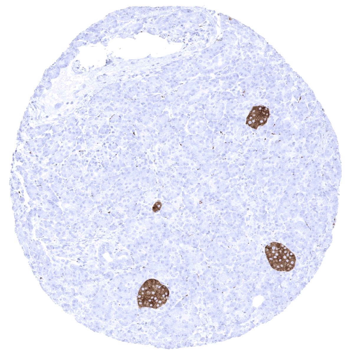
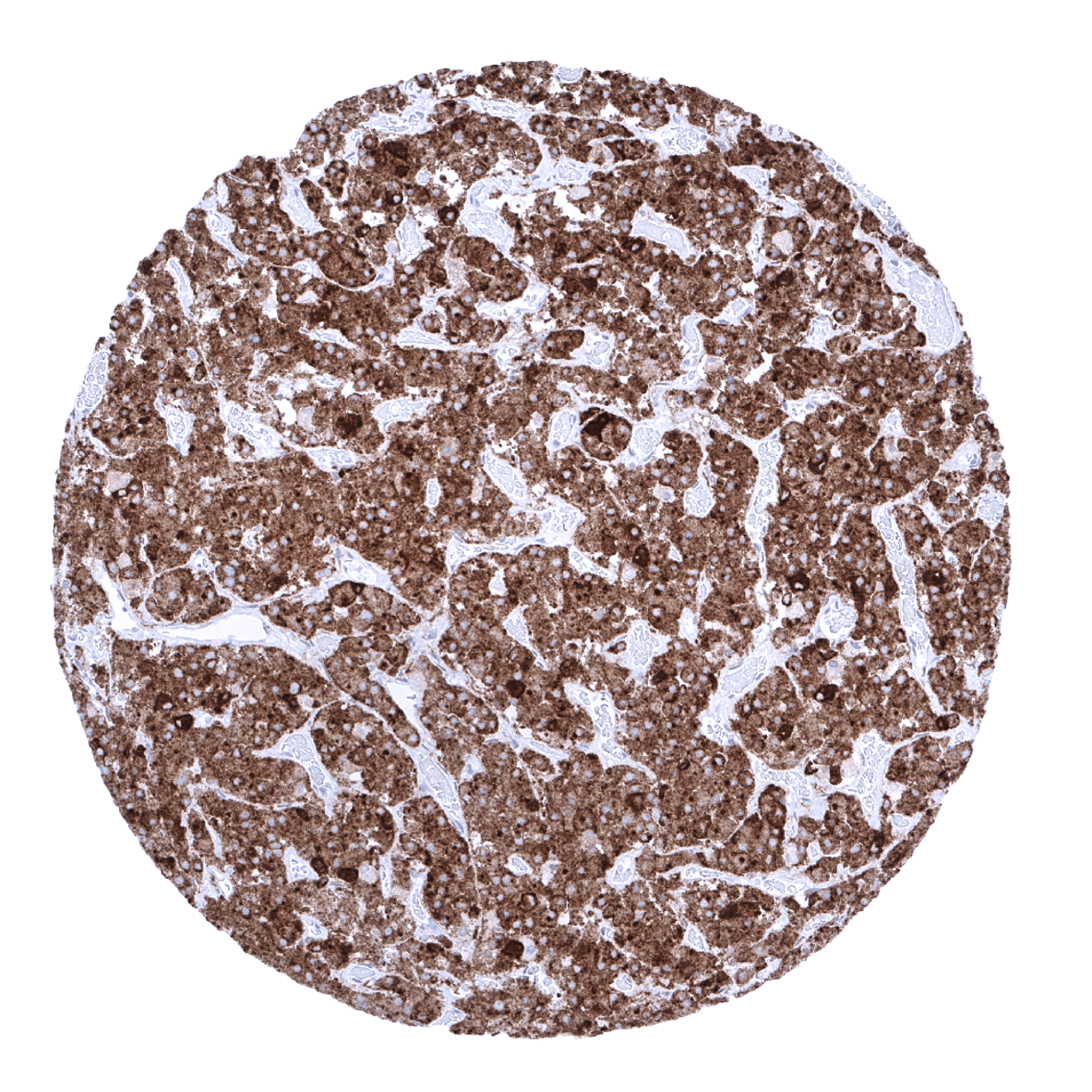
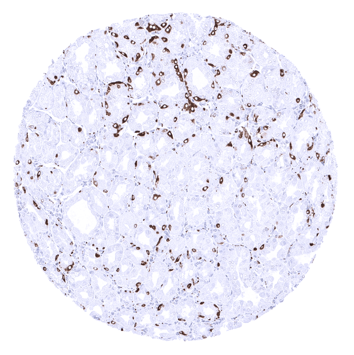
Pancreas - A strong synaptophysin immunostaining is seen in islet cells of Langerhans.
Parathyroid gland

Parotid gland - Small synaptophysin positive nerve fibres are present in the parotid gland.

Pituitary gland, posterior lobe - Strong synaptophysin immunostaining in the neurohypophysis.
Pituitary gland, anterior lobe - Strong synaptophysin immunostaining in all epithelial cells of the adenohypophysis.

Placenta (amnion and chorion)
Pregnant uterus (decidua)

Placenta (first trimenon)
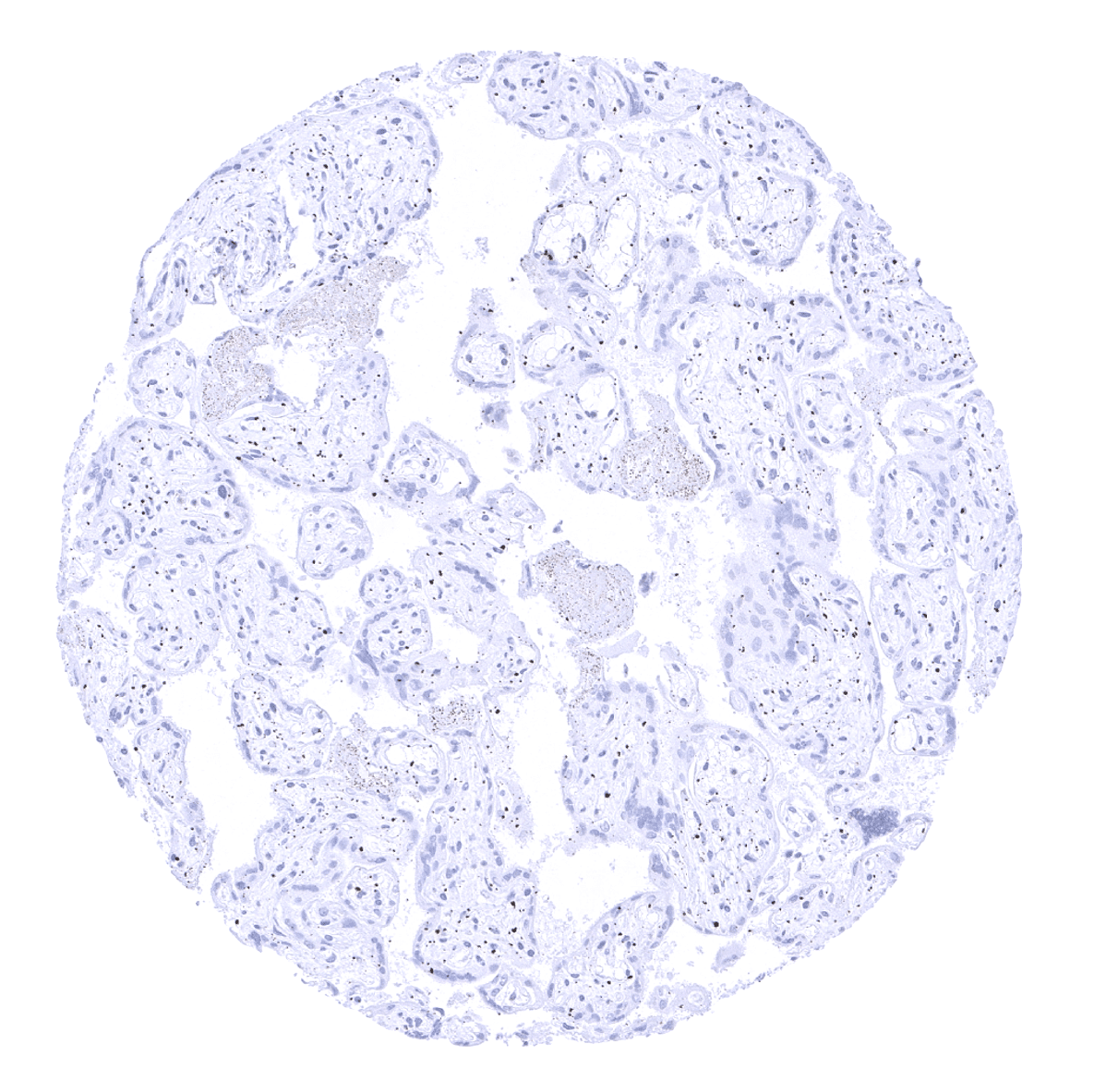
Placenta, mature
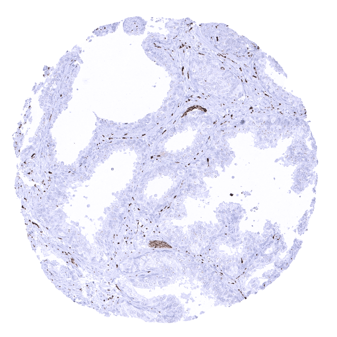
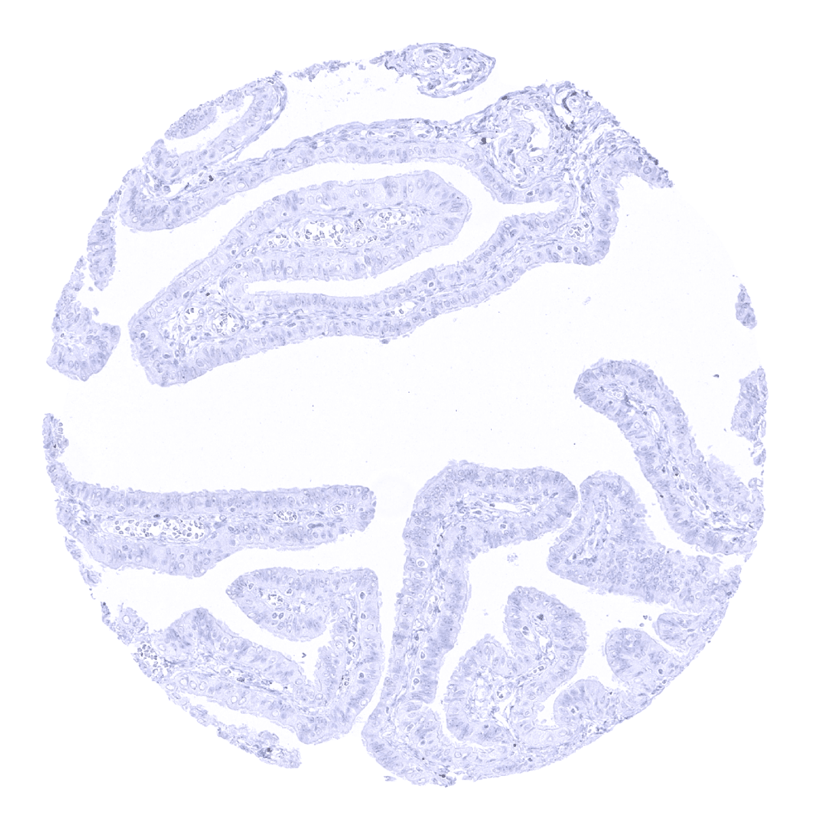
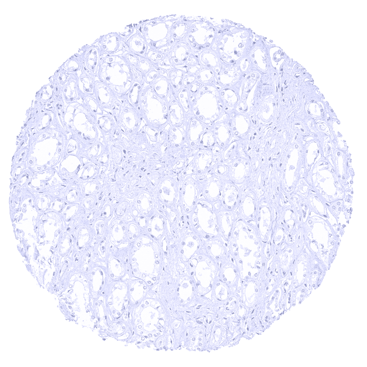
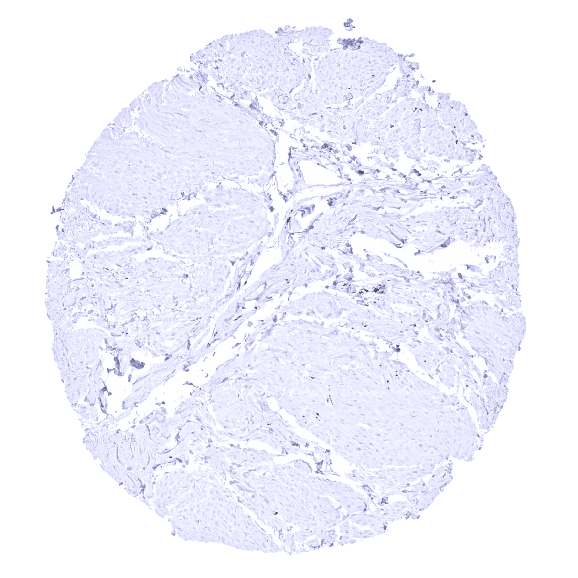
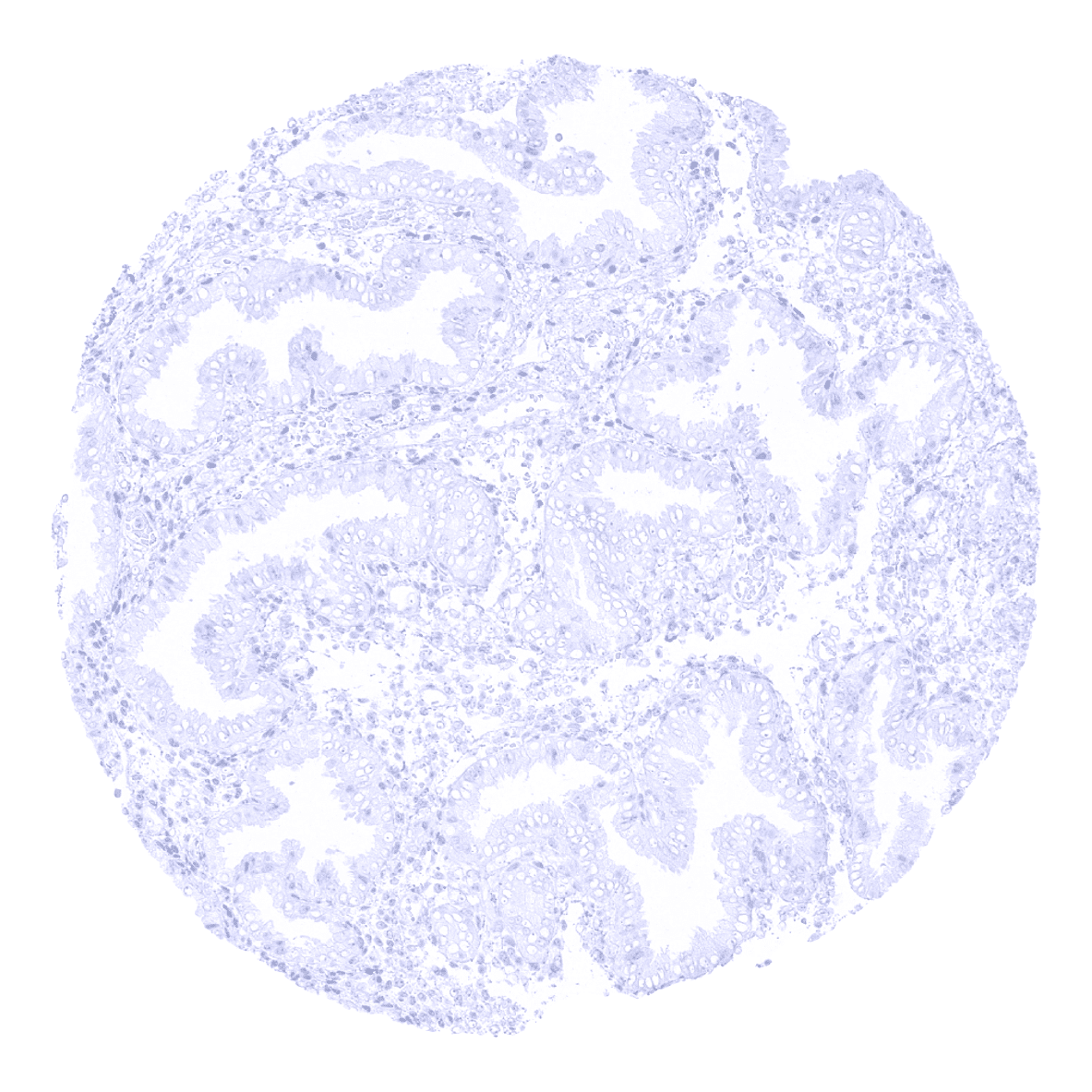
Prostate
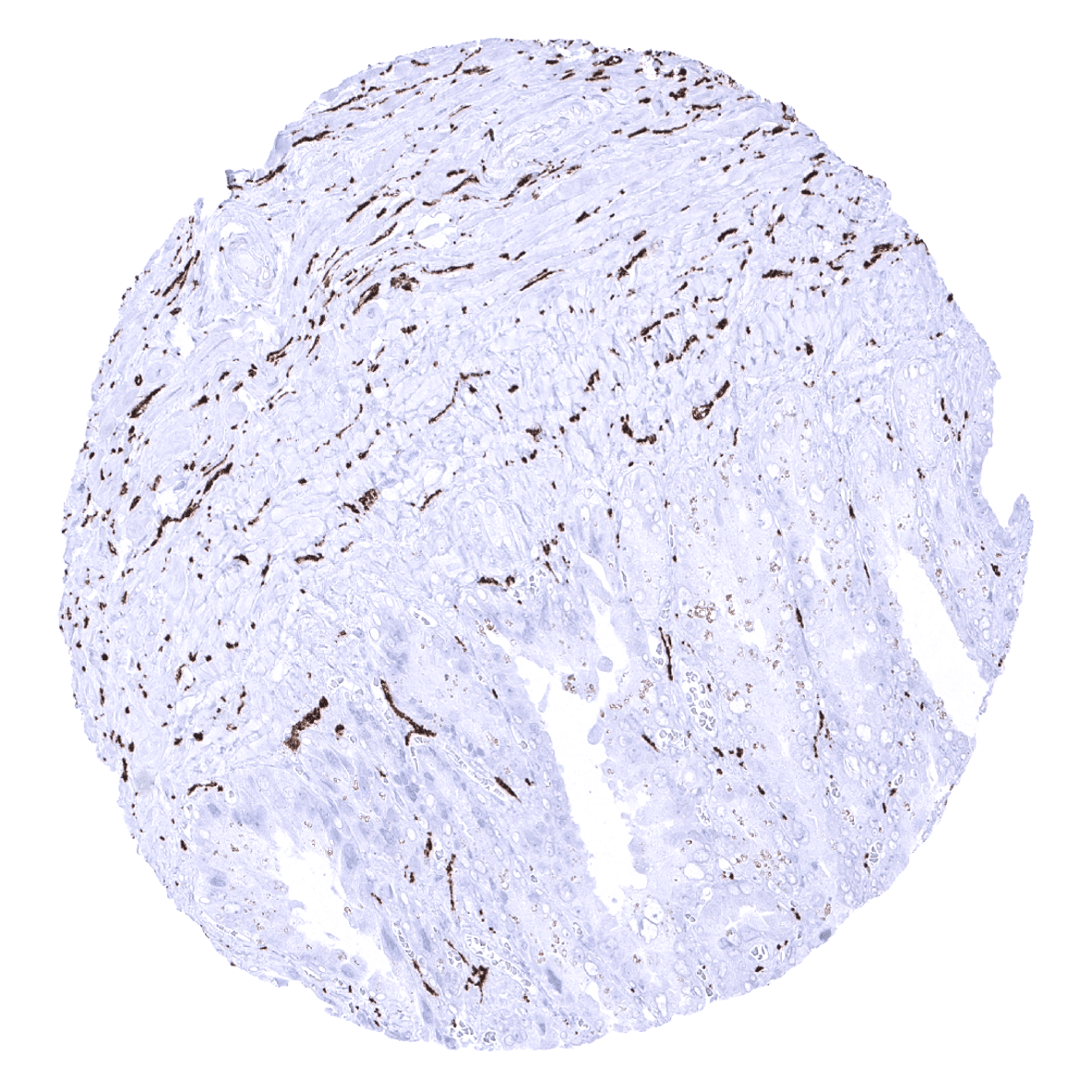
Rectum, mucosa - In the rectum mucosa, synaptophysin immunostaining is seen in neuroendocrine cells and nerve fibres.
Seminal vesicle
Sinus paranasales
Skeletal muscle
Skin
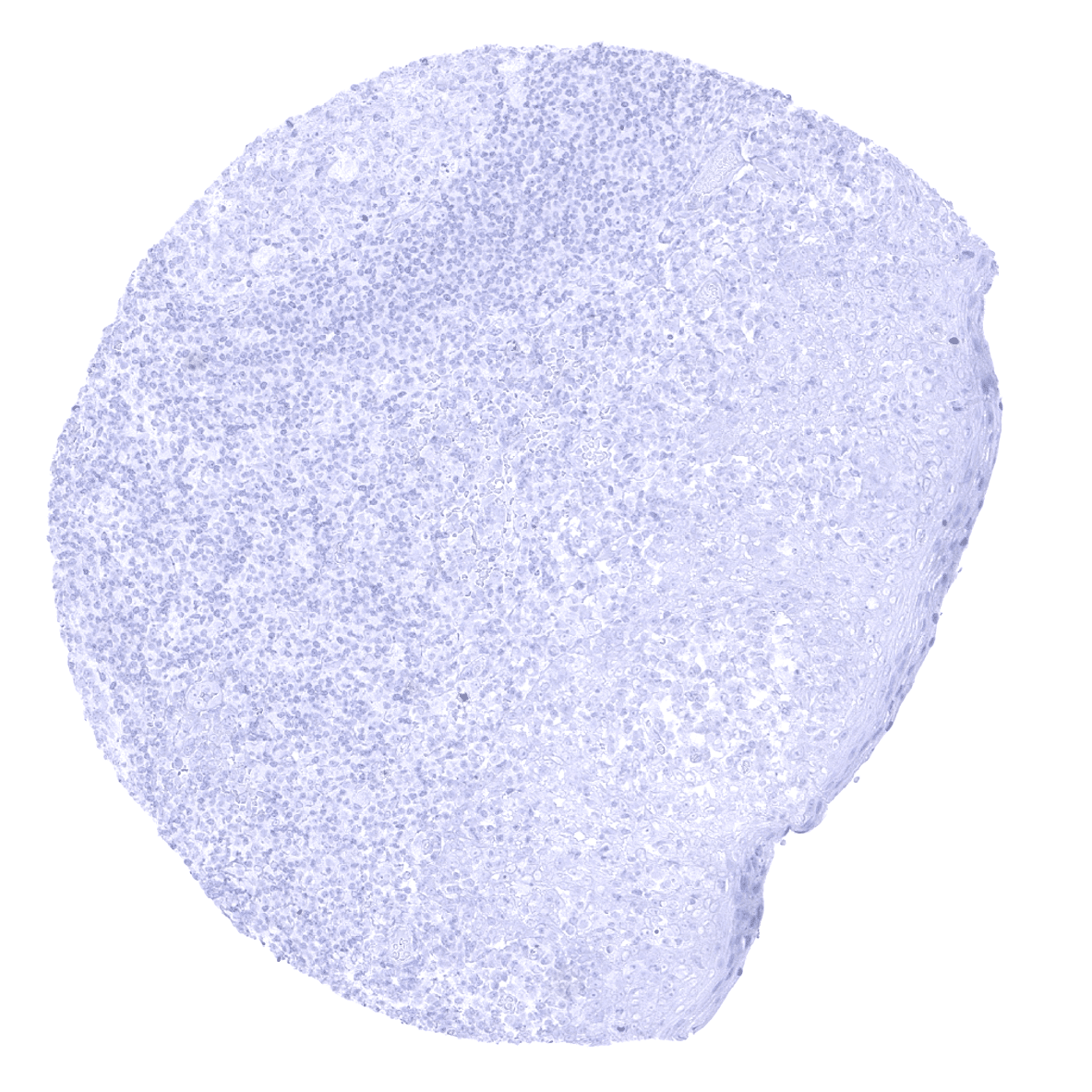
Spleen
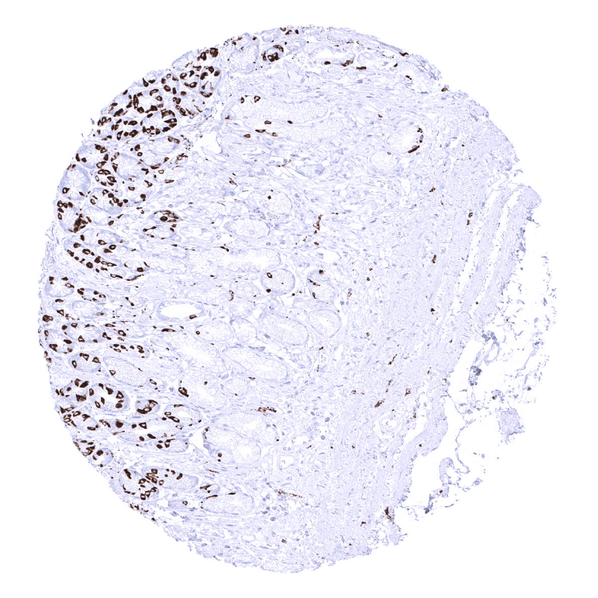
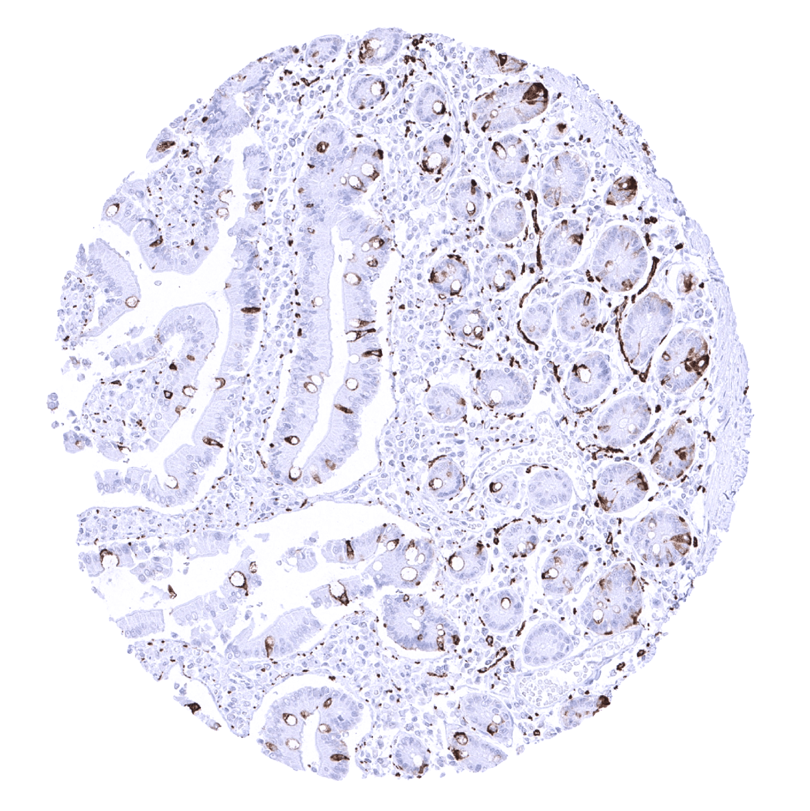
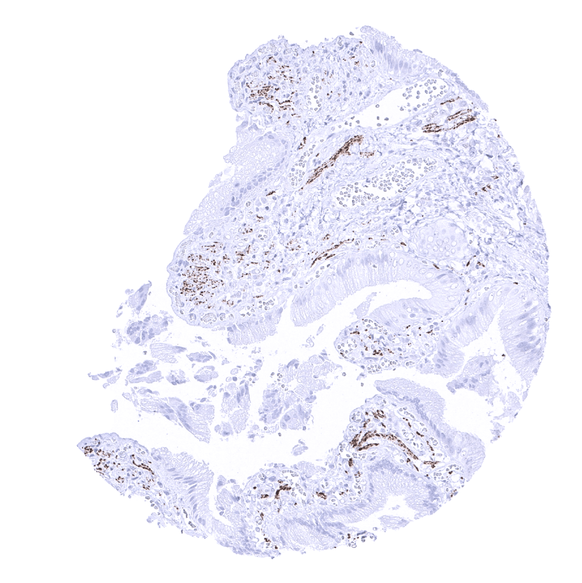
Stomach, antrum - Neuroendocrine cells show synaptophysin immunostaining in the stomach.
Stomach, corpus - Neuroendocrine cells show synaptophysin immunostaining in the stomach.
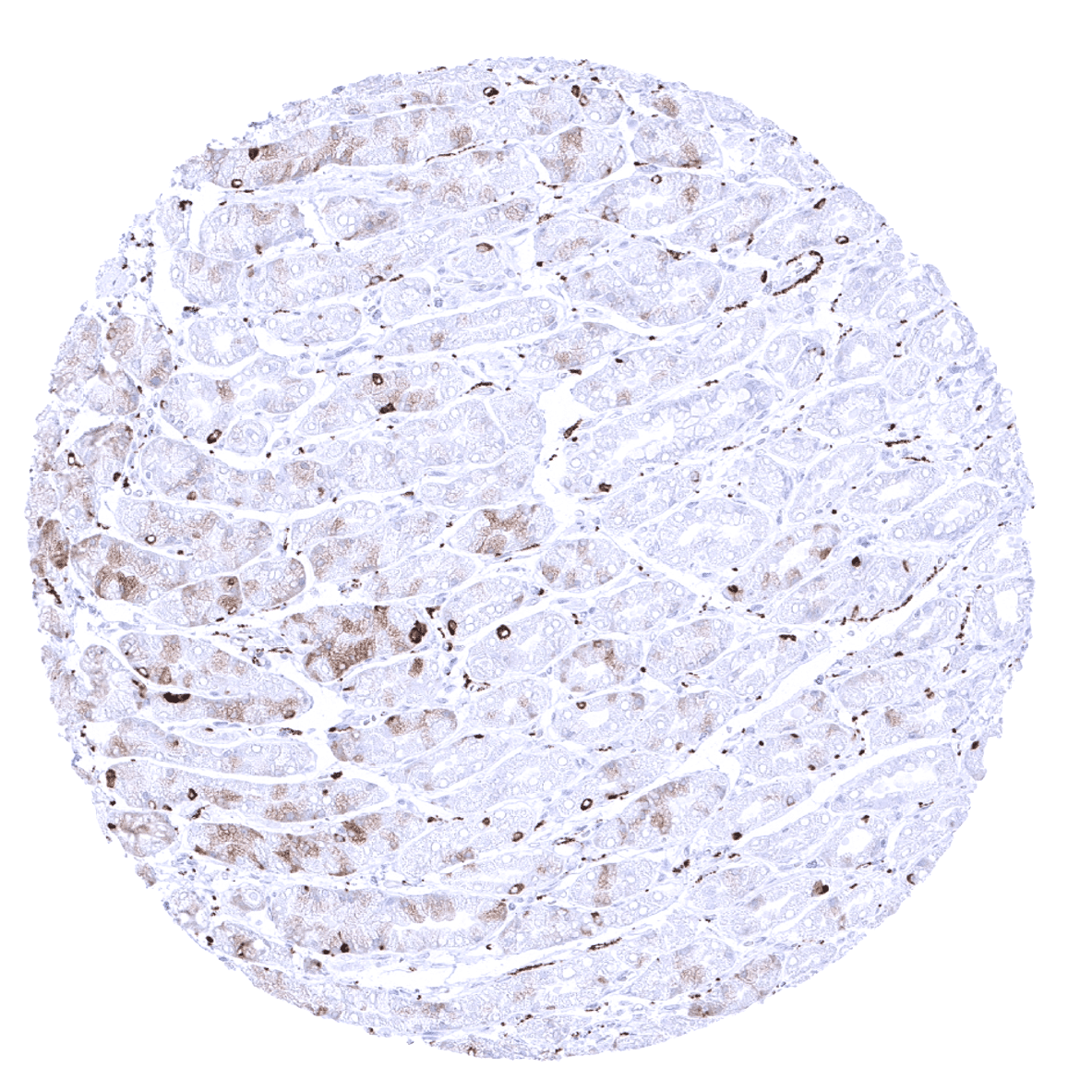
Stomach, corpus - Neuroendocrine cells show synaptophysin immunostaining in the stomach. In some samples, a fraction of glandular cells may show weak cytoplasmic staining.
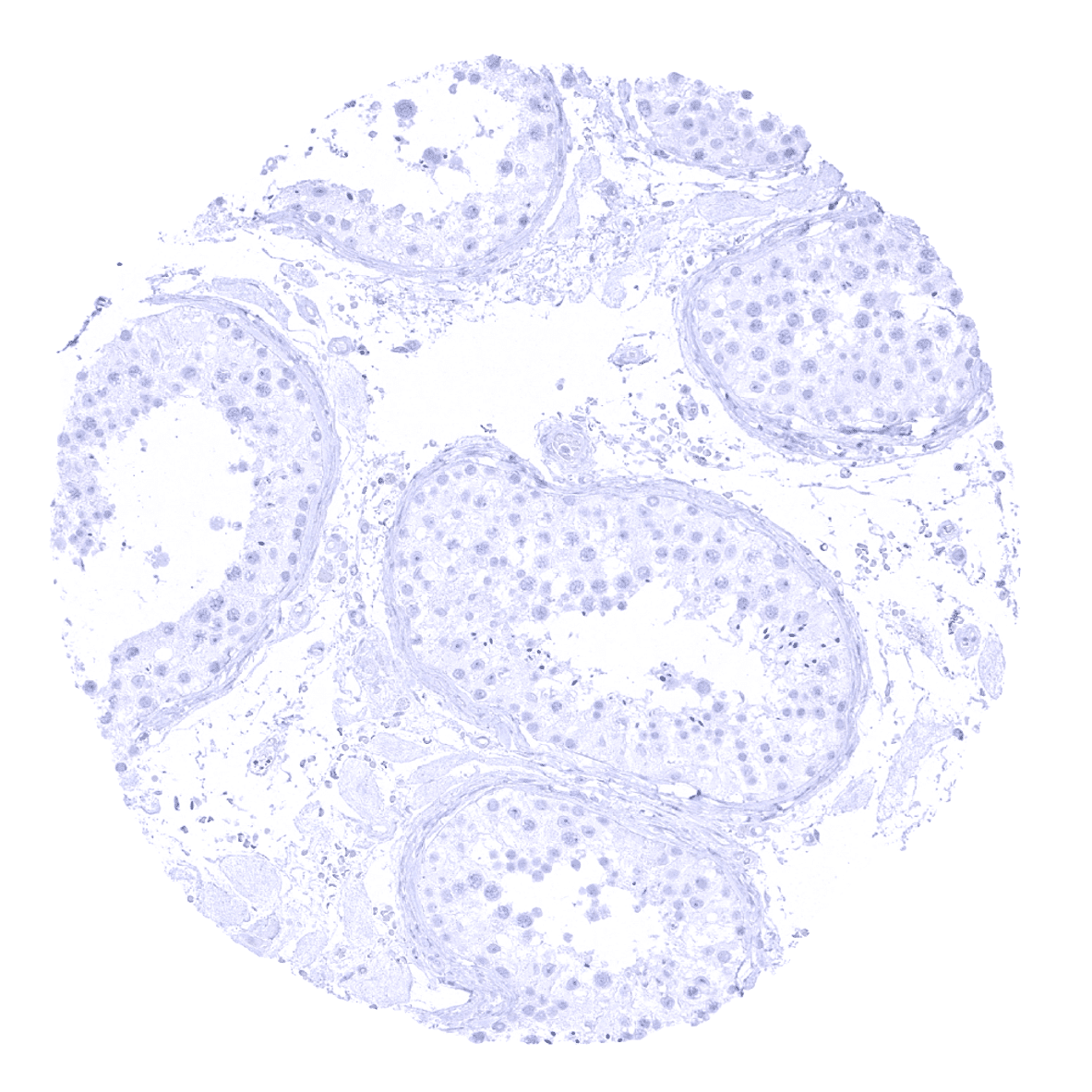
Testis
Thymus
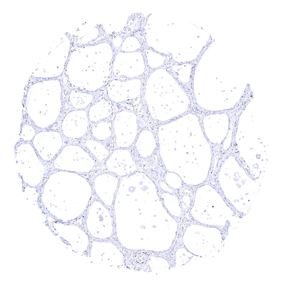
Thyroid gland - In the thyroid, synaptophysin immunostaining is limited to C-cells which are not seen on all tissue spots.
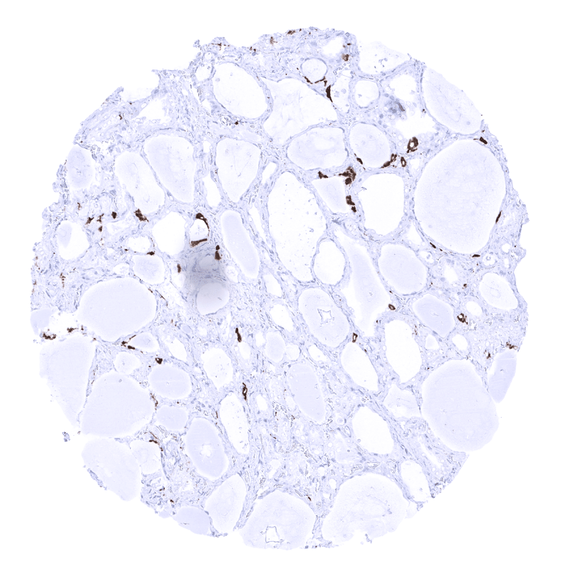
Thyroid gland - Strong synaptophysin immunostaining occurs in C-cells of the thyroid.
Tonsil, surface epithelium
Tonsil
Urinary bladder, muscular wall

Urinary bladder, urothelium
Uterus, ectocervix

Uterus, endocervix
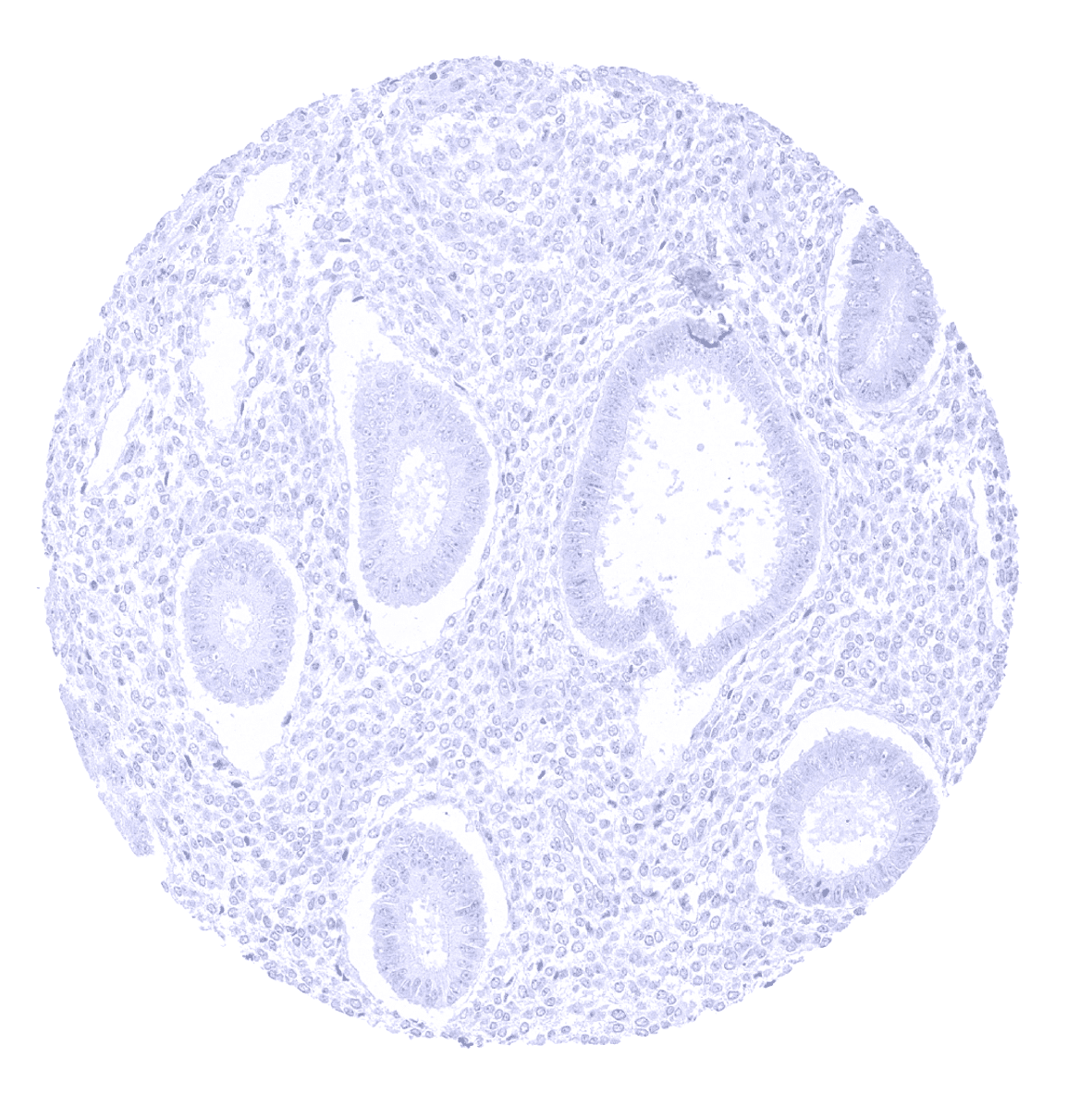
Uterus, endometrium (proliferation)
Uterus, endometrium (secretion)
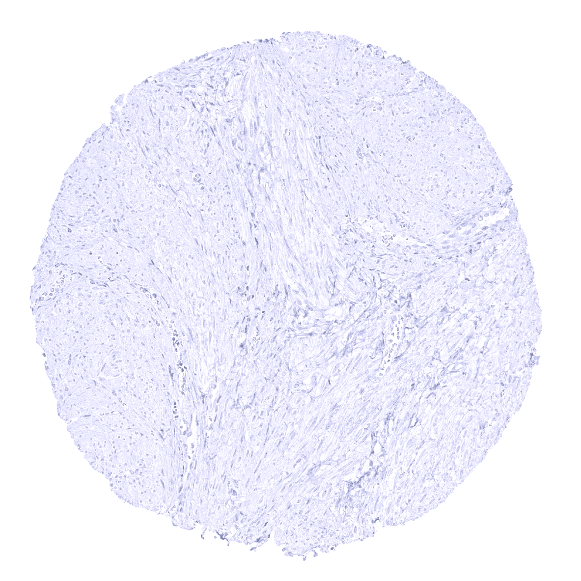
Uterus, myometrium