Adrenal gland - GS staining (variable intensity) in a fraction of cells
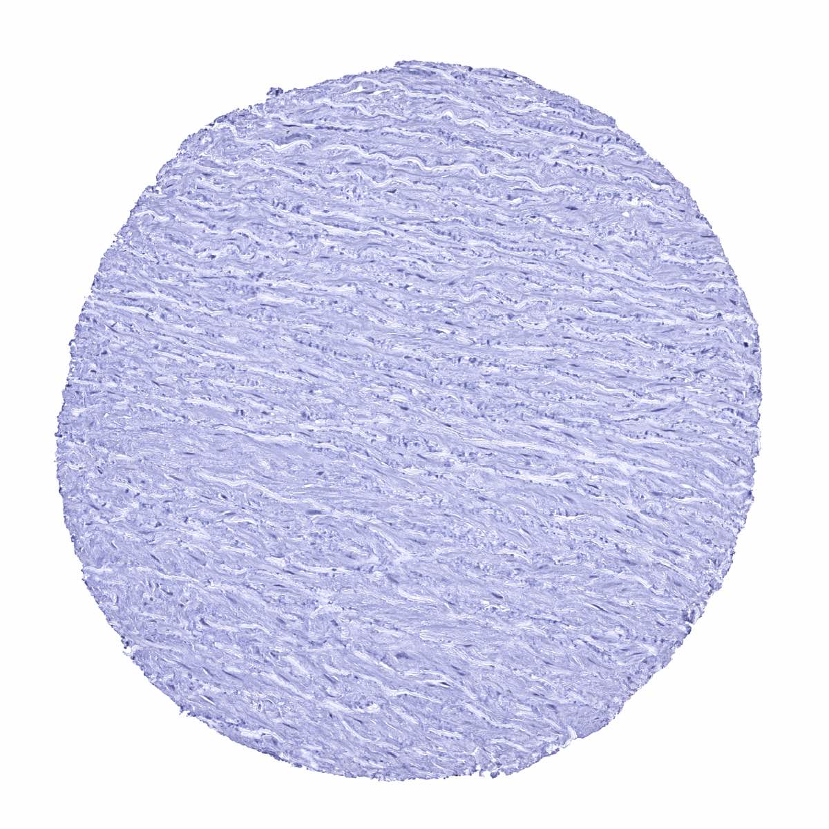
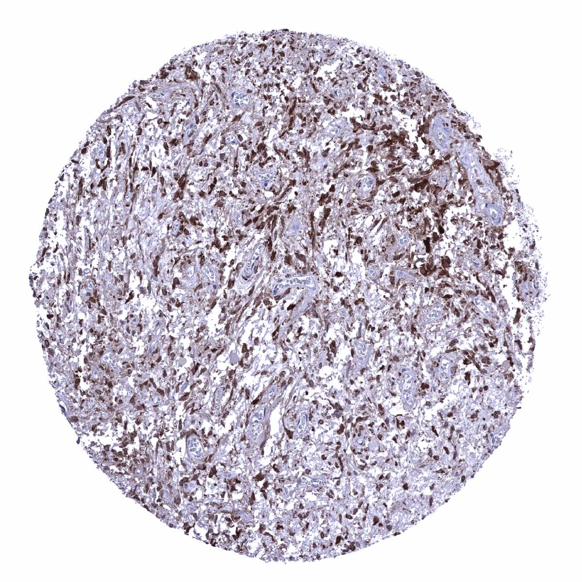
Aorta, media

Appendix, mucosa - GS staining predominates in macrophages and is only focal and mostly faint in epithelial cells
Appendix, mucosa - GS staining predominates in macrophages
Appendix, muscular wall
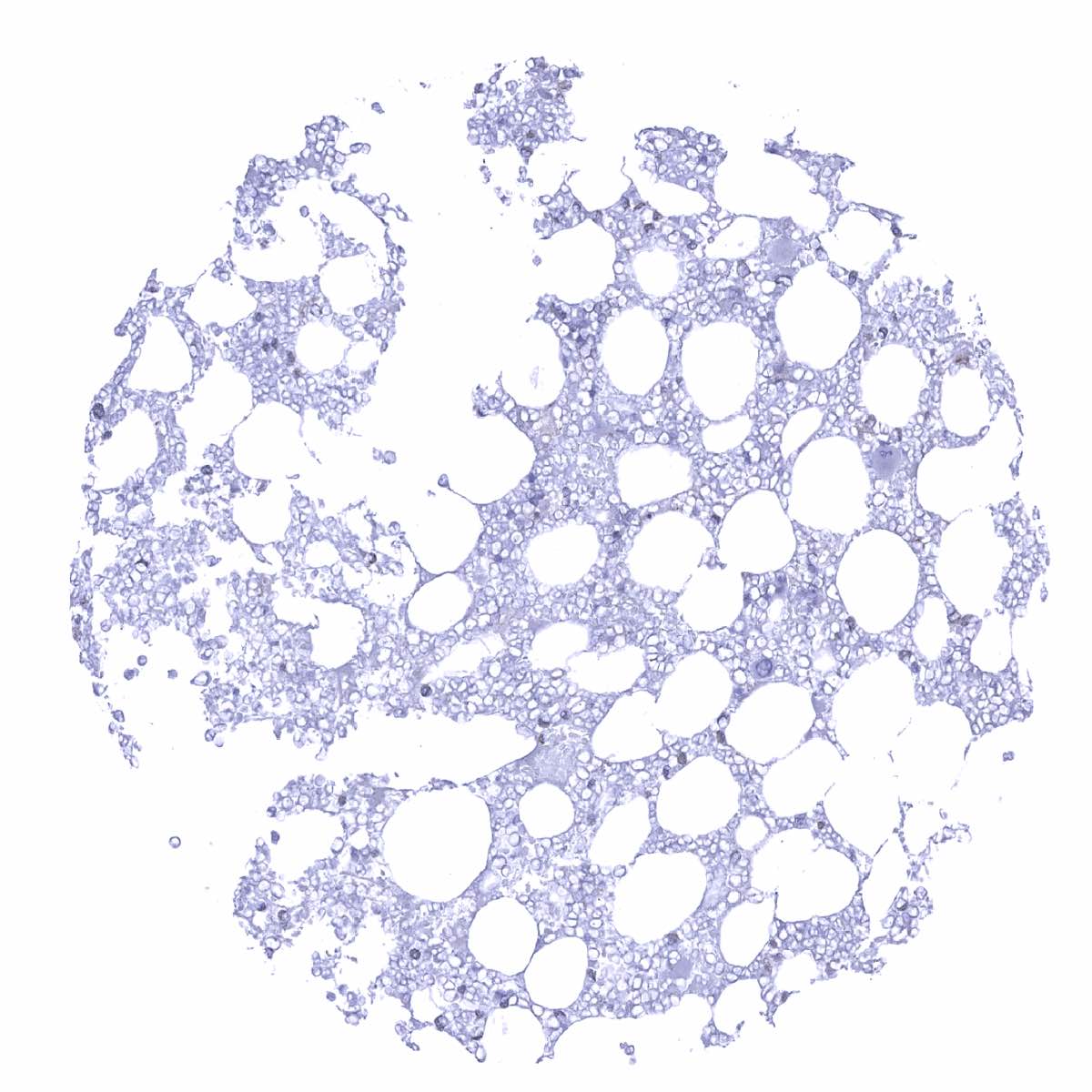
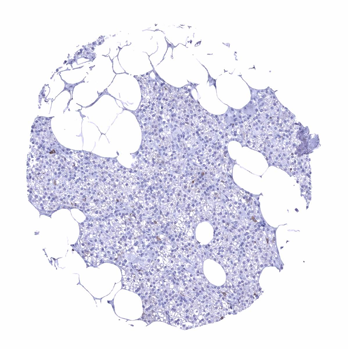
Bone marrow
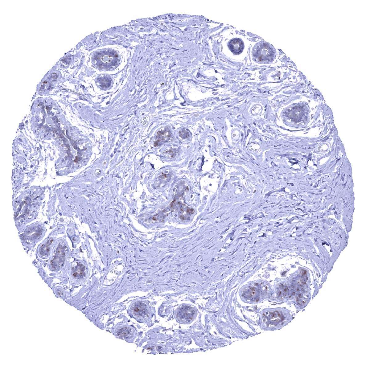
Breast - A faint GS staining occurs in acinar cells of the breast
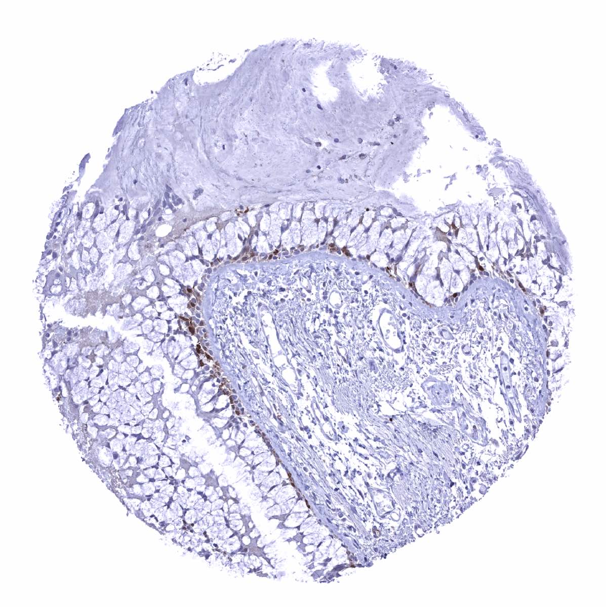
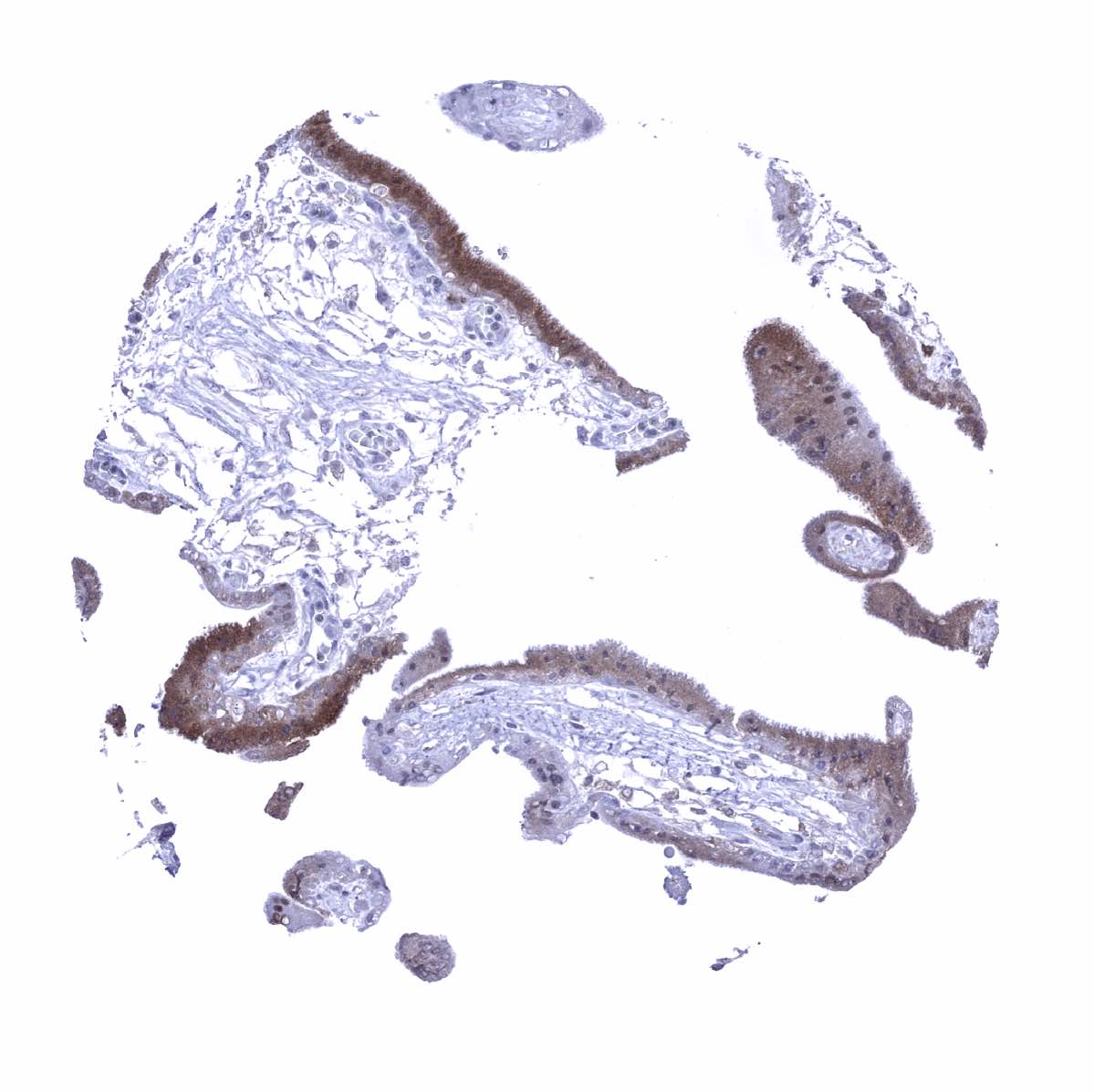
Bronchus, mucosa - A fraction of cells of the respiratory epithelium show a moderate GS staining
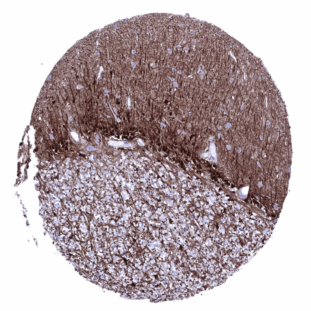
Cerebellum (molecular layer, Purkinje cell layer, granule cell layer, white matter) - An intense GS positivity occurs in glia cells and in associated fibres. Purkinje cells are GS negative.

Cerebellum (molecular layer, Purkinje cell layer, granule cell layer, white matter) - Intense GS positivity of glia cells
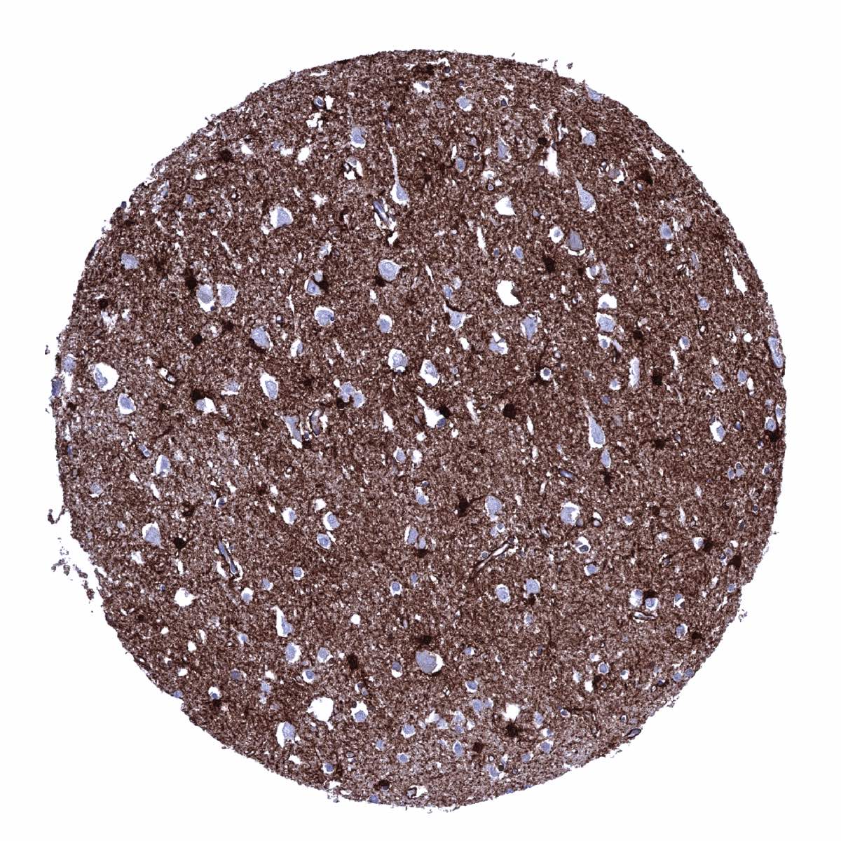
Cerebrum, grey matter - An intense GS positivity is seen in all glia cells and in associated fibres. Neurons are GS negative

Cerebrum, white matter - All glia cells show strong GS positivity
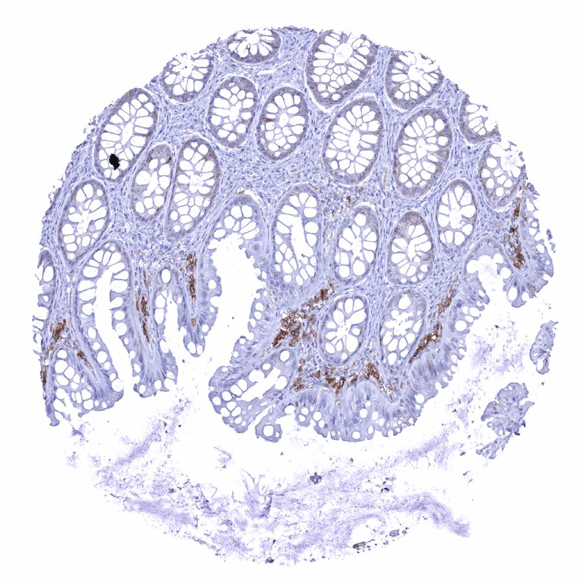
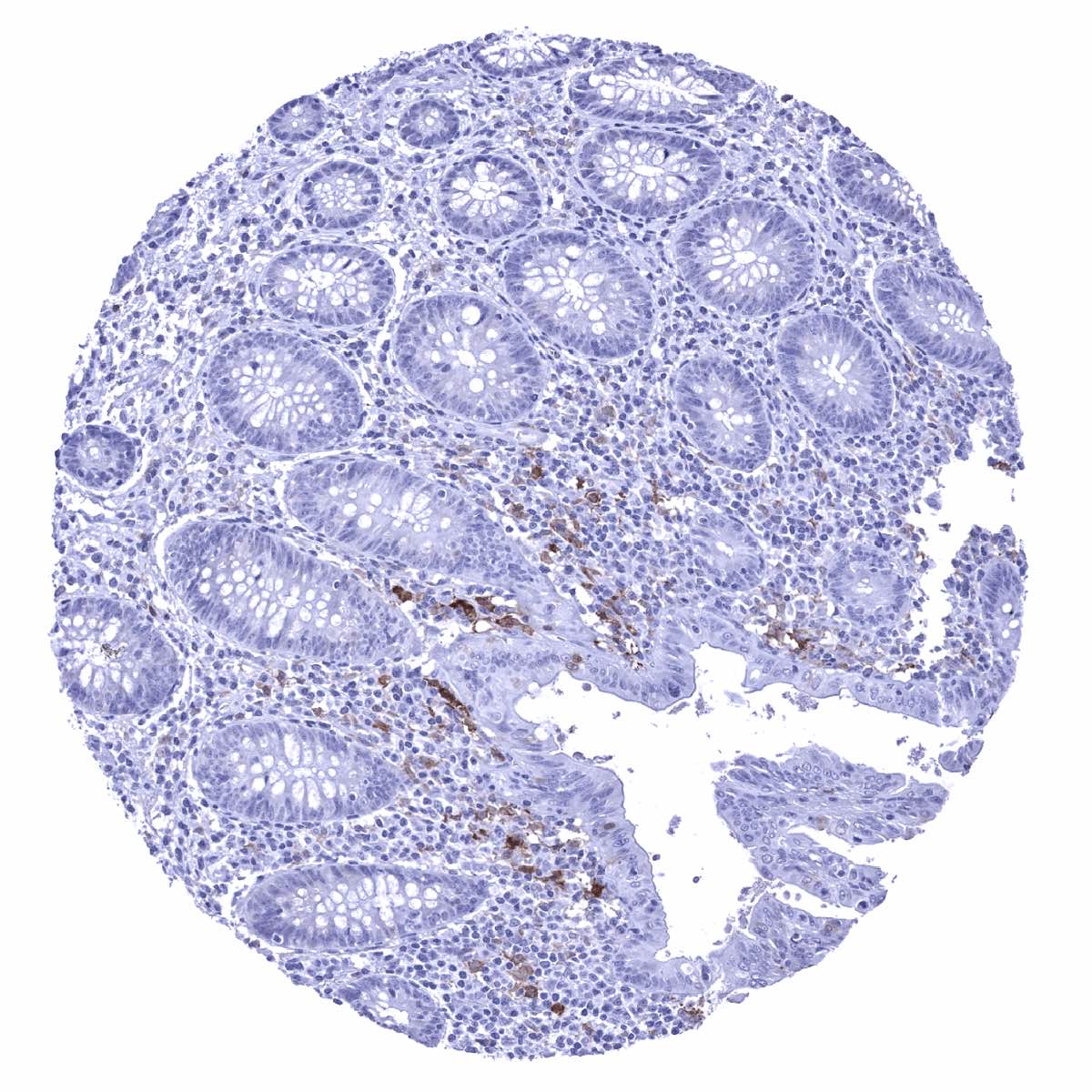
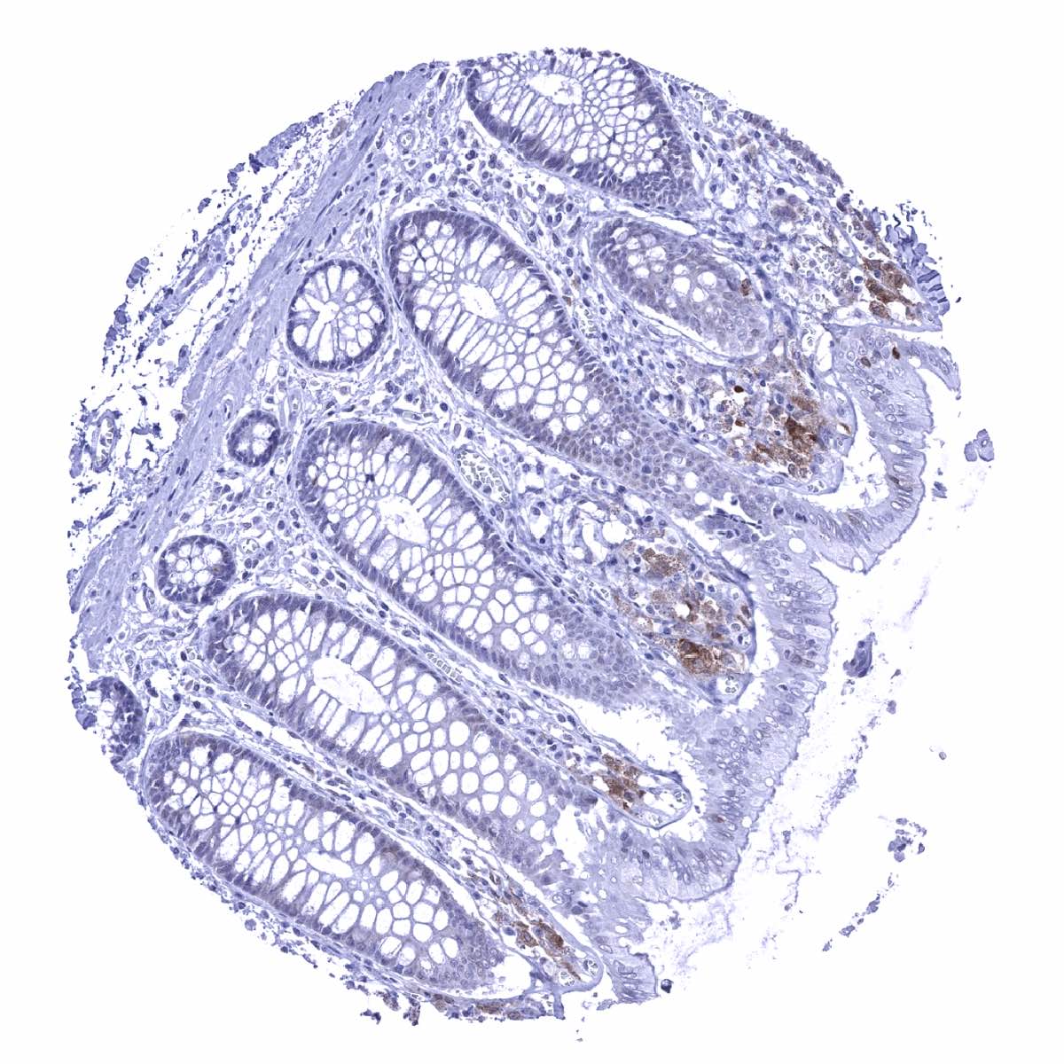
Colon descendens, mucosa - GS staining predominates in macrophages and is only faint in some epithelial cells

Colon descendens, muscular wall
Duodenum, Brunner gland
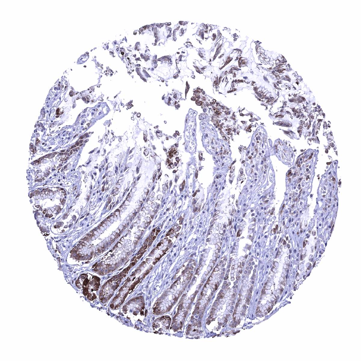
Duodenum, mucosa - GS staining predominates in macrophages and is only focal and faint in epithelial cells
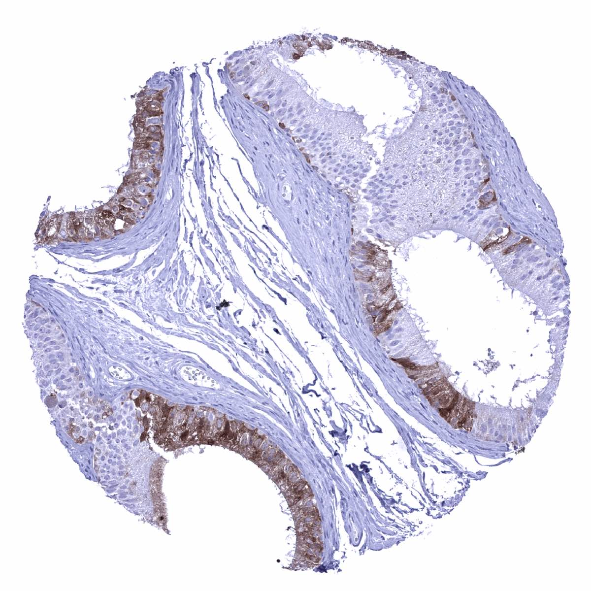
Epididymis - Focal GS staining of moderate intensity in the corpus
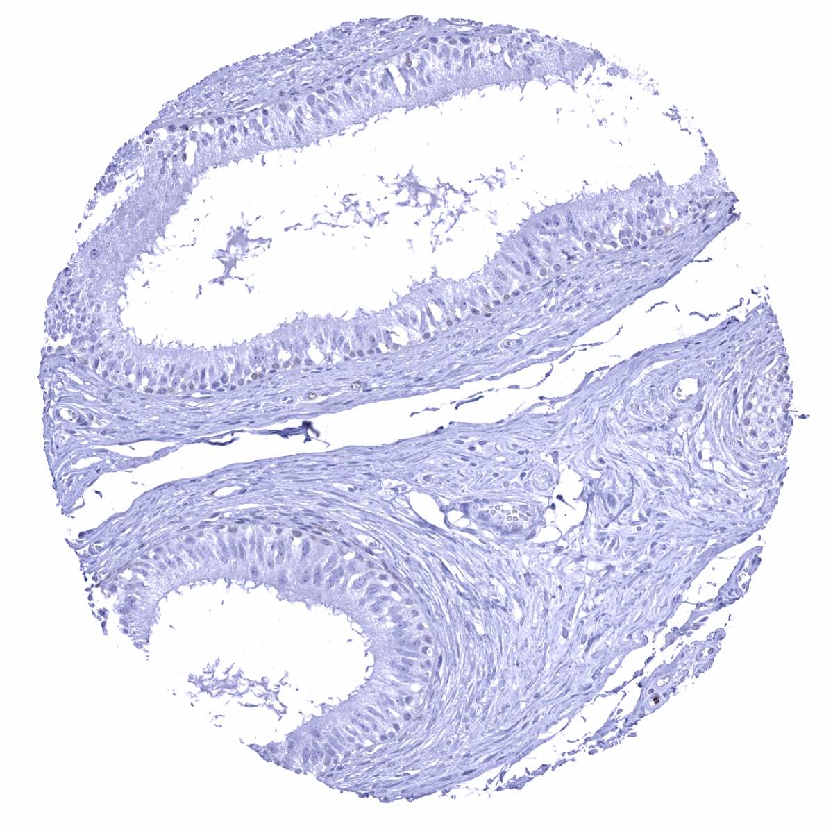
Epididymis - GS staining is absent in this sample of the corpus
Epididymis - Strong GS staining in most epithelial cells of the cauda epididymis
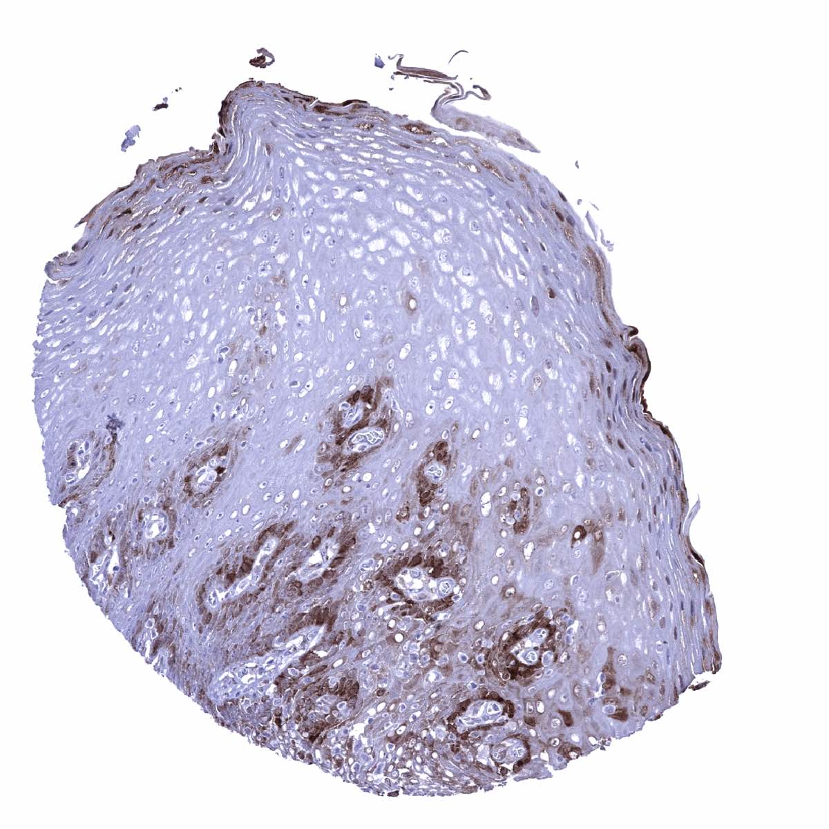
Esophagus, squamous epithelium - A moderate to strong nuclear and cytoplasmic GS staining occurs in the basal and suprabasal cell layers as well as in the top cell layers of the squamous epithelium.
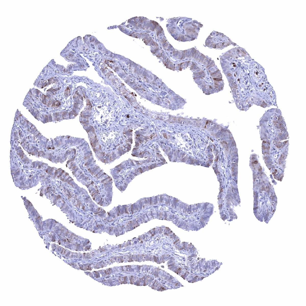
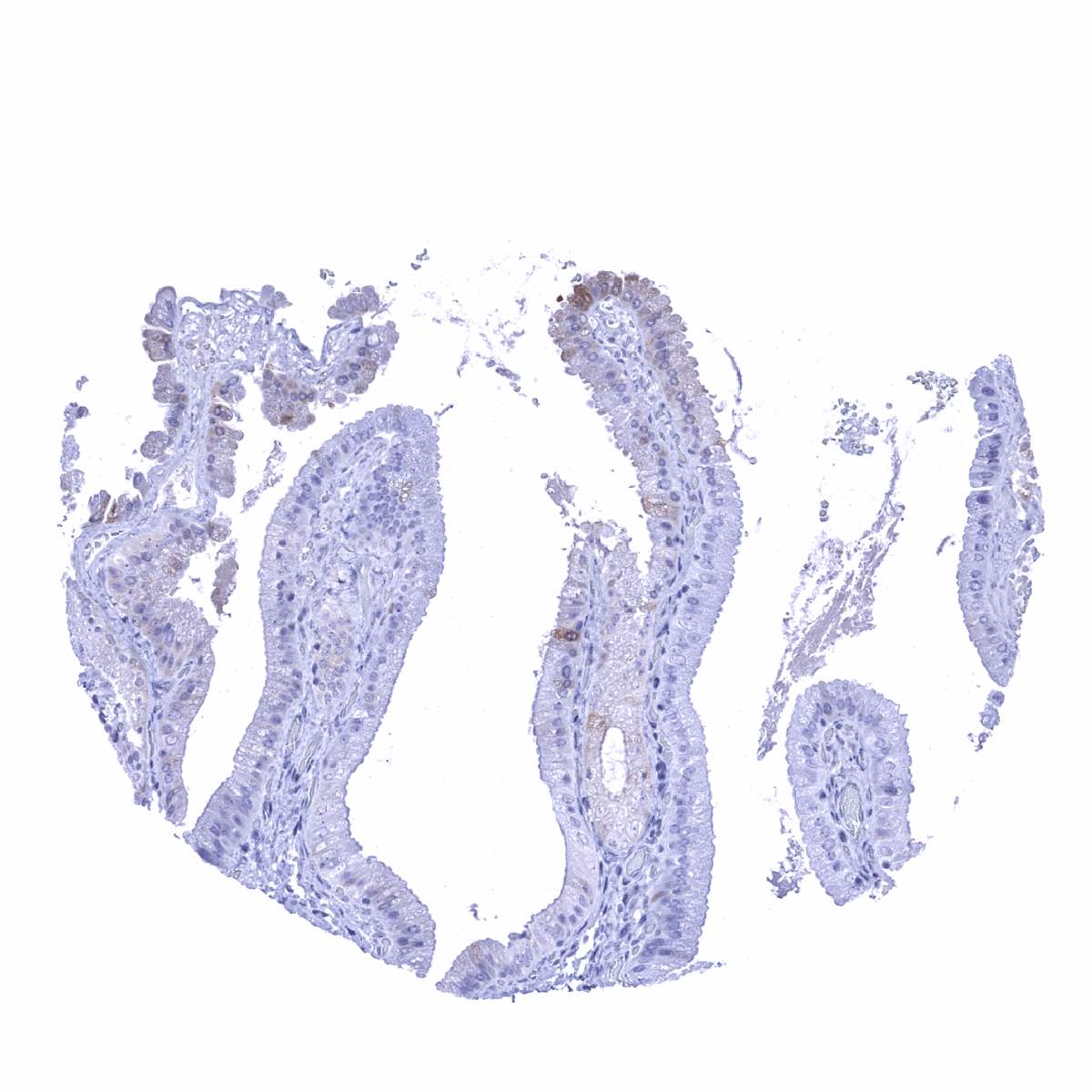
Fallopian tube, mucosa - A fraction of epithelial cells shows a weak to moderate staining

Fat
Gallbladder, epithelium - GS staining is focal and faint in epithelial cells
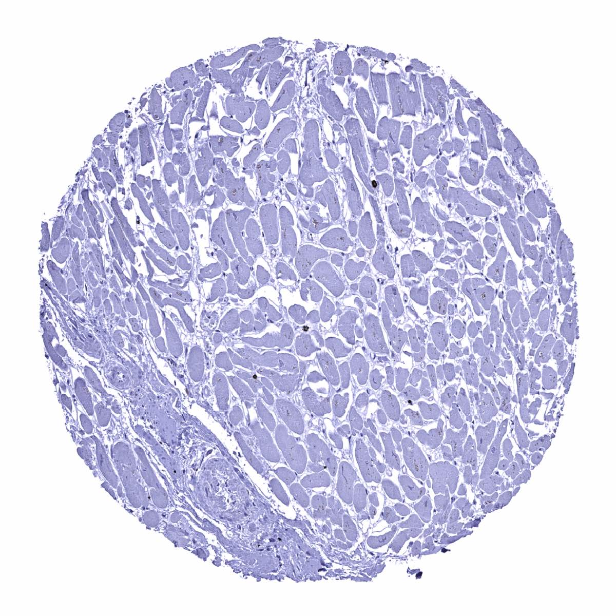
Heart muscle
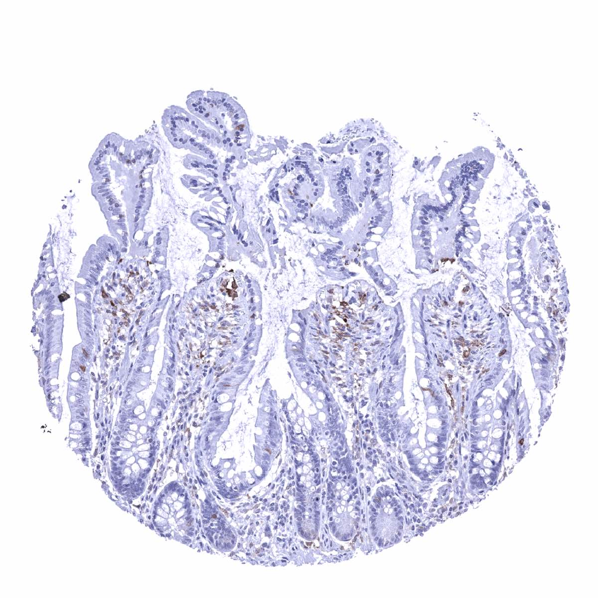
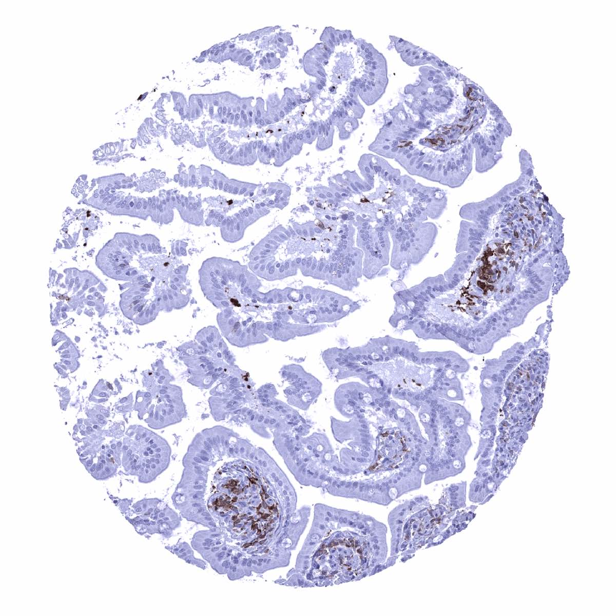
Ileum, mucosa - GS staining predominates in macrophages

Kidney pelvis (urothelium) - GS staining is largely absent in the urothelium
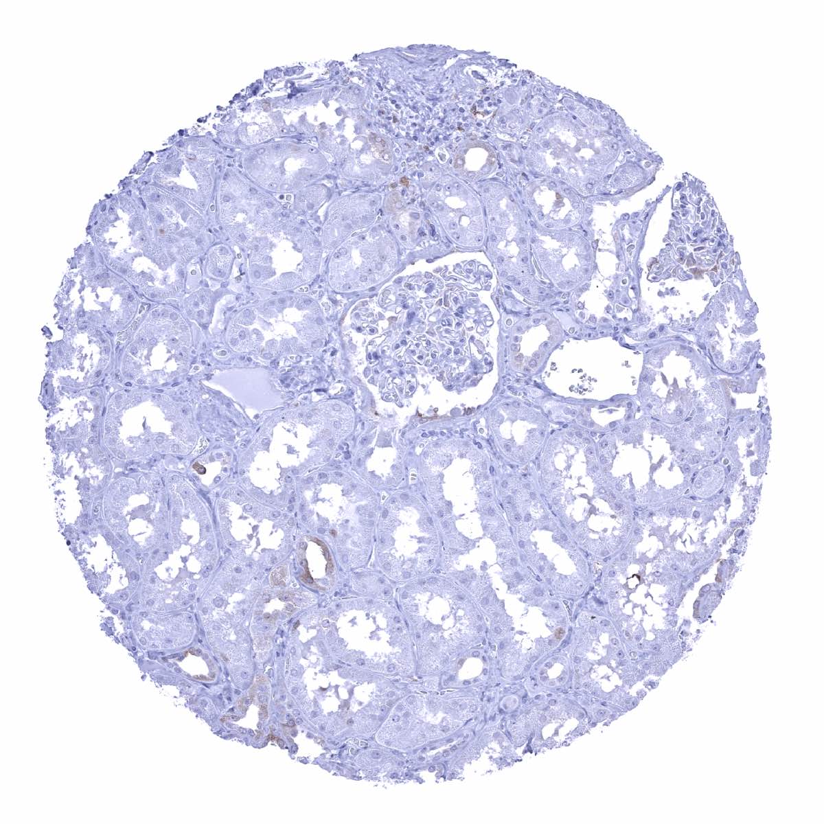
Kidney, cortex - Few distal tubuli or collecting ducts may show GS staining
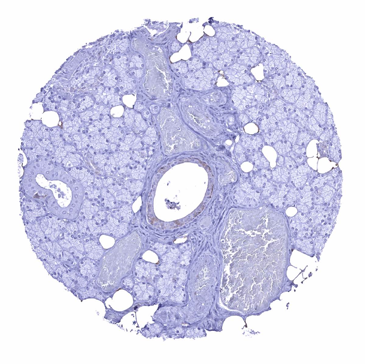
Kidney, medulla
Liver - A weak GS staining is seen in Kupffer cells
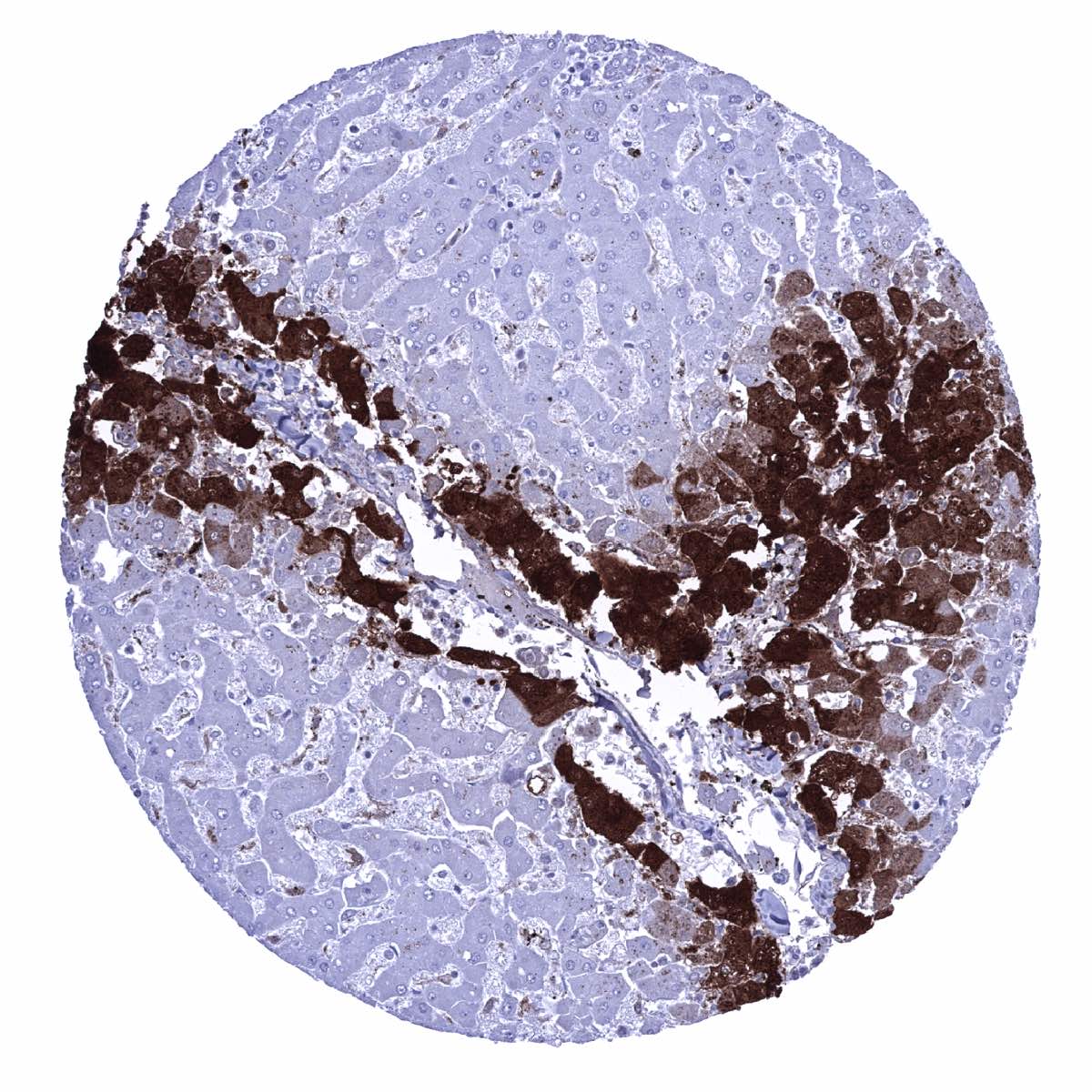
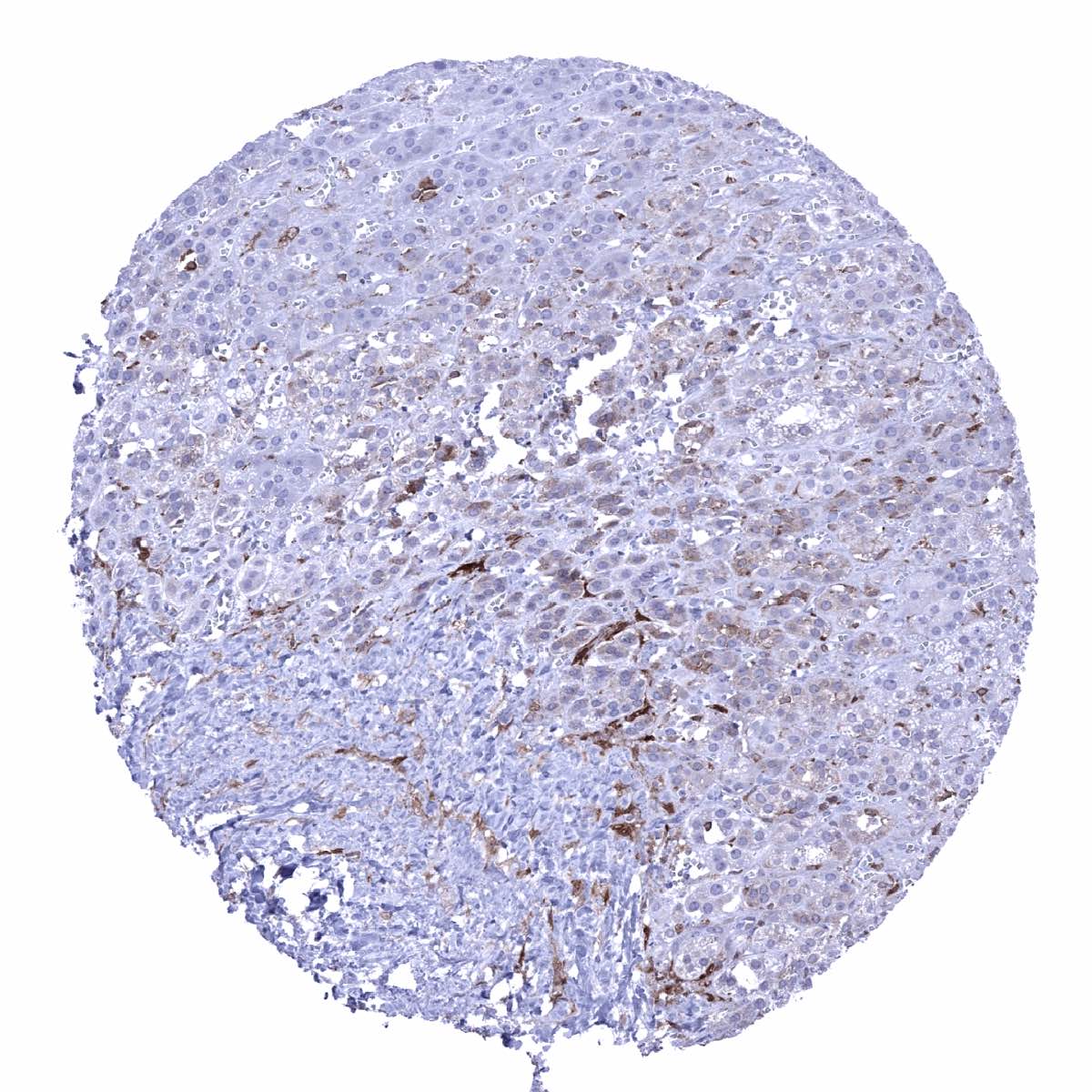
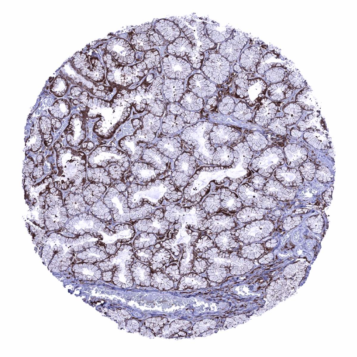
Liver - GS staining is strong in centrilobular hepatocytes, weak to moderate in Kupffer cells but absent in periportal hepatocytes
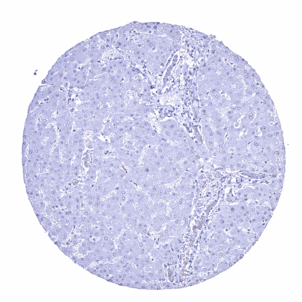
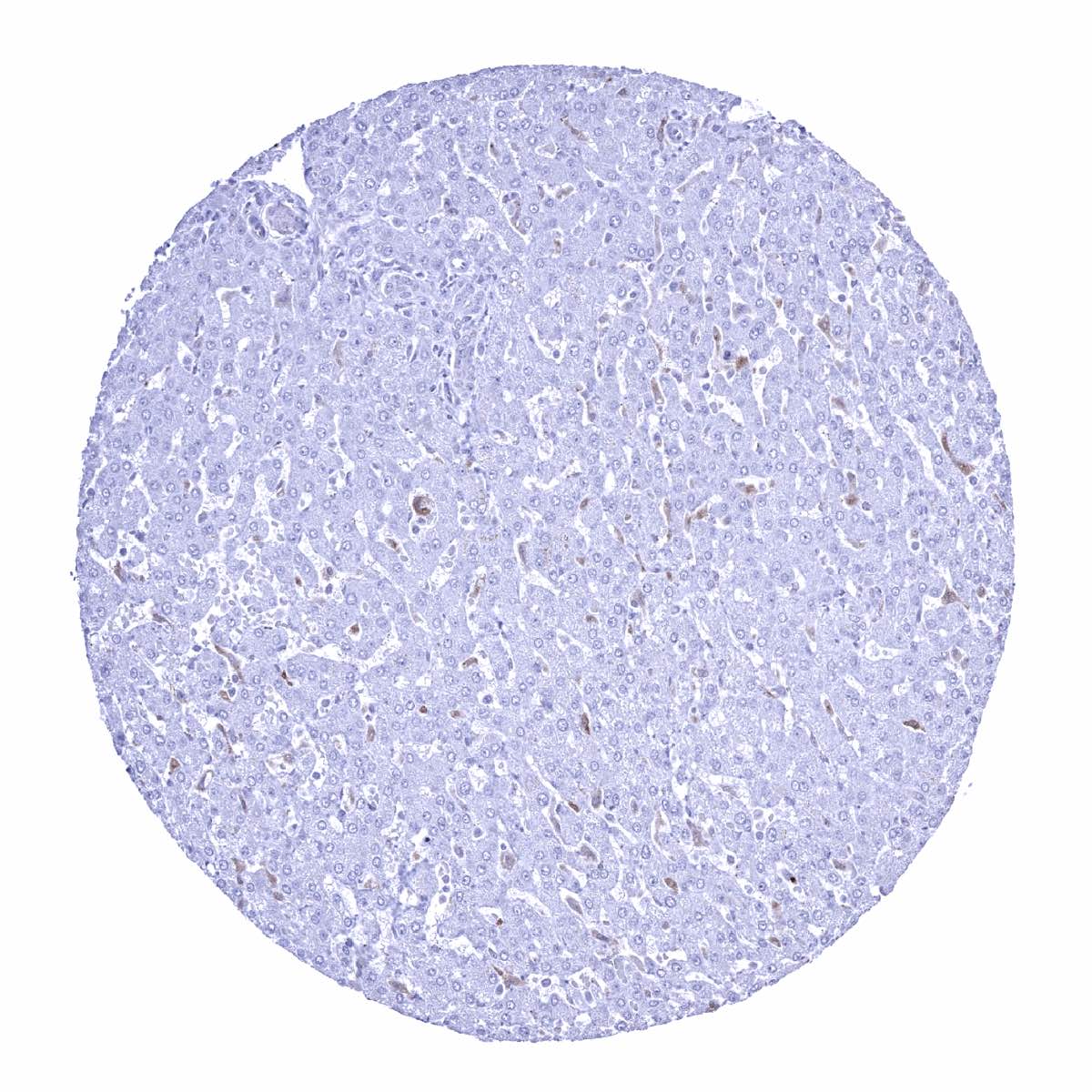
Liver - GS staining is weak in Kupffer cells but absent in periportal hepatocytes and bile ducts
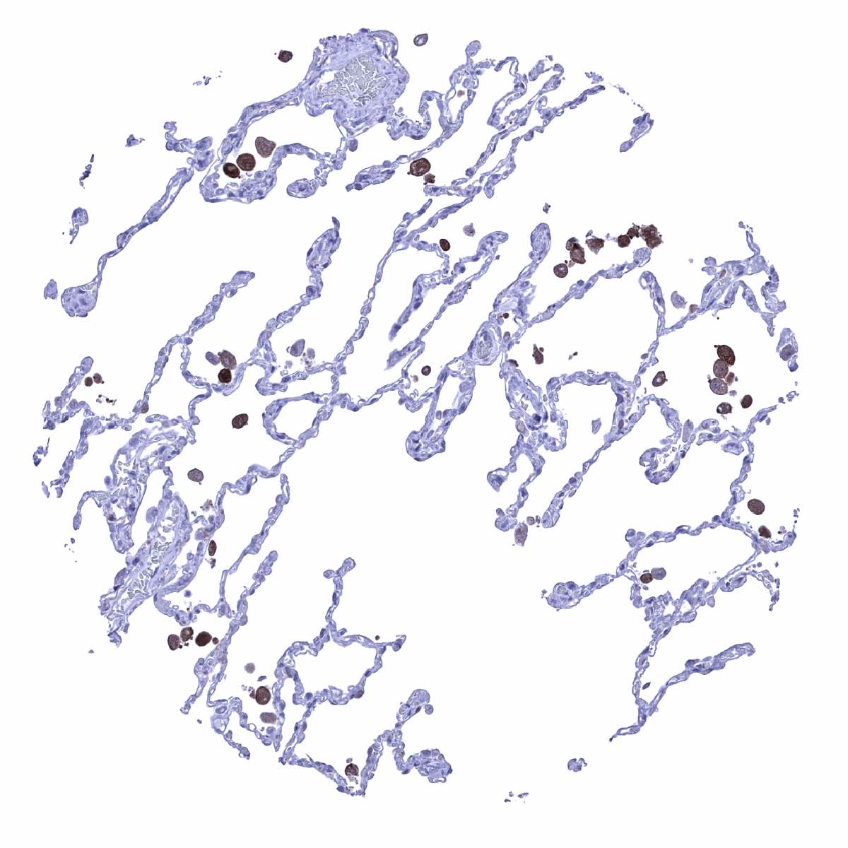
Lung - GS staining is limited to macrophages
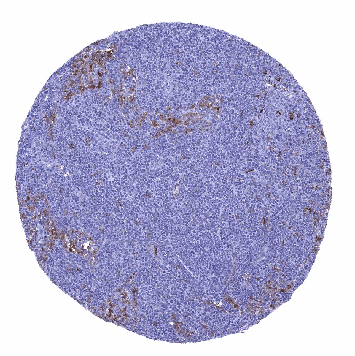
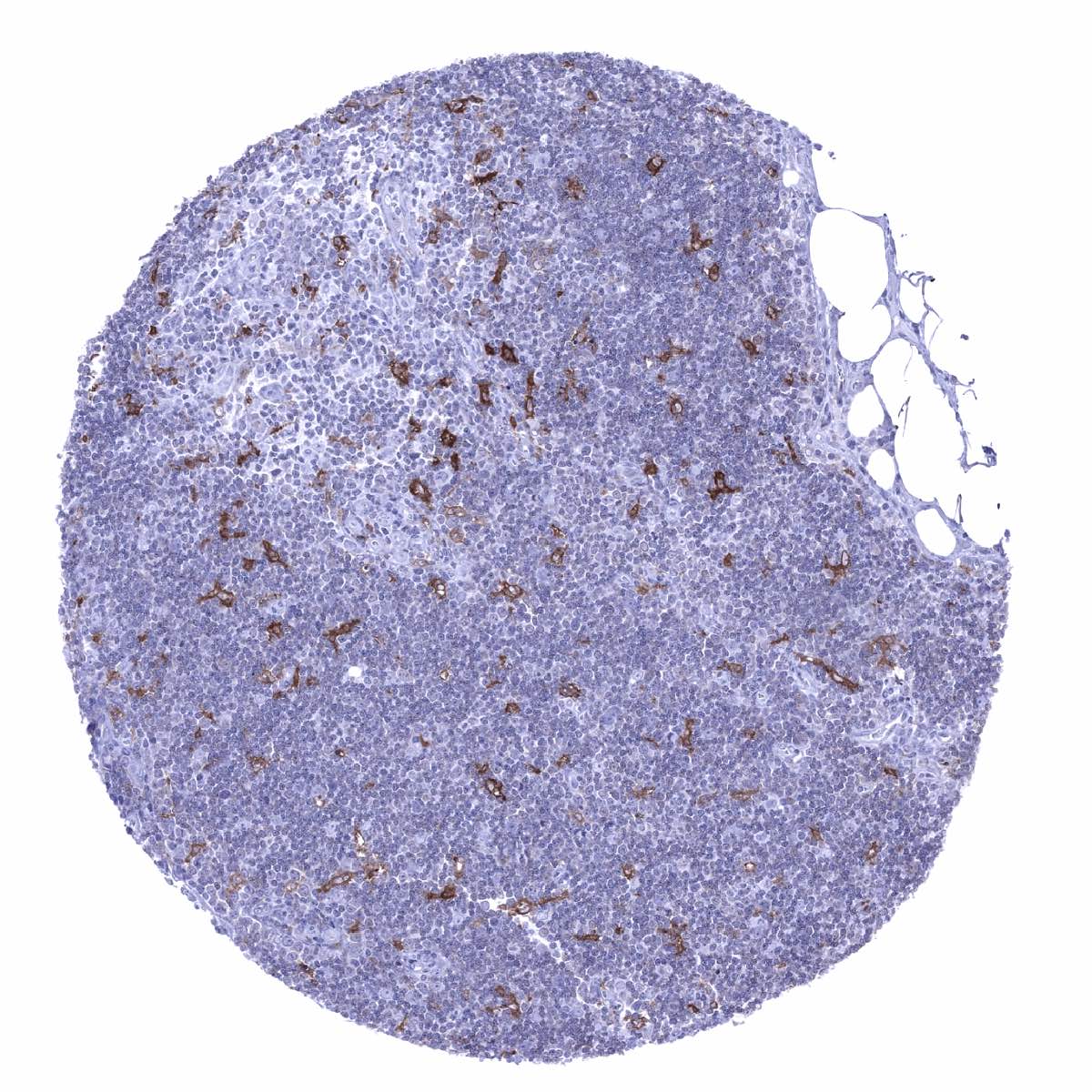
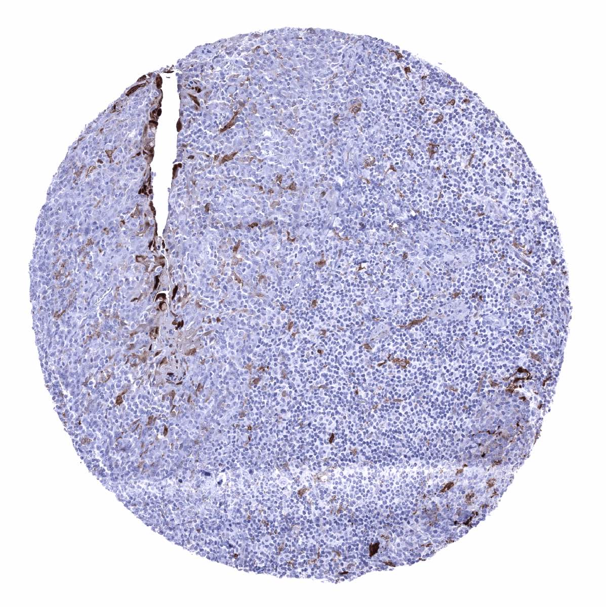
Lymph node - GS staining of weak to moderate intensity occurs in sinus macrophages, dendritic cells, and in endothelial cells of small capillaries
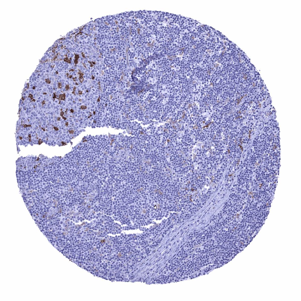
Lymph node - Strong GS staining of dendritic cells in a germinal centre

Ovary, corpus luteum - GS staining is variable and ranges from weak to strong (mosaic pattern)
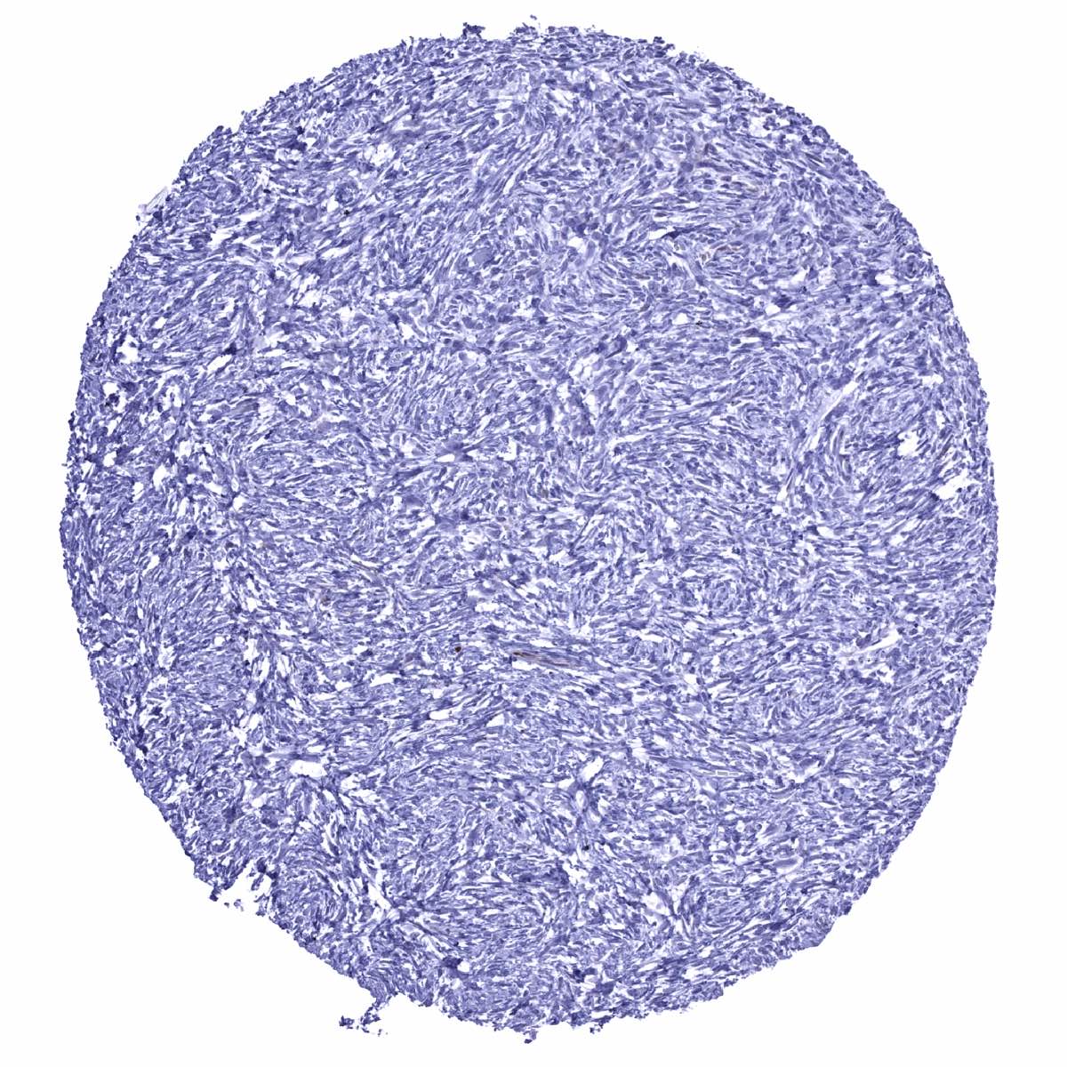
Ovary, stroma
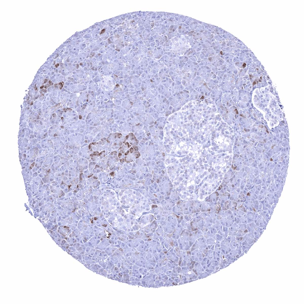
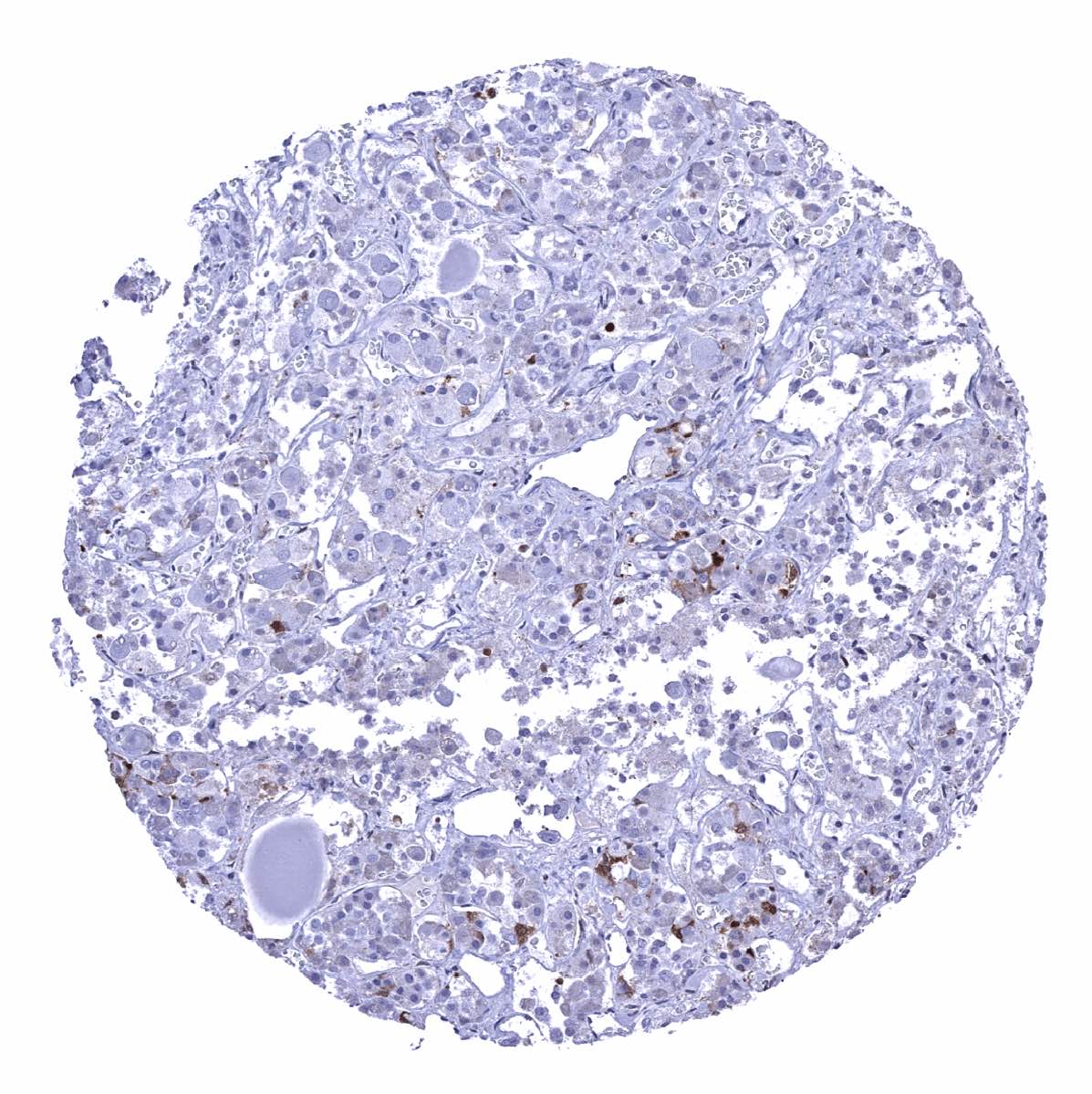
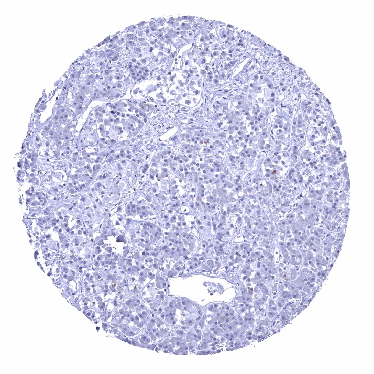
Pancreas - A weak to moderate GS staining is focally observed in acinar cells, intercalated ducts and excretory ducts while islet cells are negative
Parathyroid gland
Parotid gland - Weak GS staining of excretory ducts but moderate to strong positivity of fat cells
Pituitary gland, anterior lobe - A weak to moderate GS staining can occur in few cells of the adenohypophysis
Pituitary gland, anterior lobe - GS positive cells are not always seen in samples from the adenohypophysis
Pituitary gland, posterior lobe - Intense GS positivity of glia cells and associated fibres
Placenta (amnion and chorion) - Chorion and amnion cells are negative but some decidua cells show a weak to moderate GS staining
Placenta, early
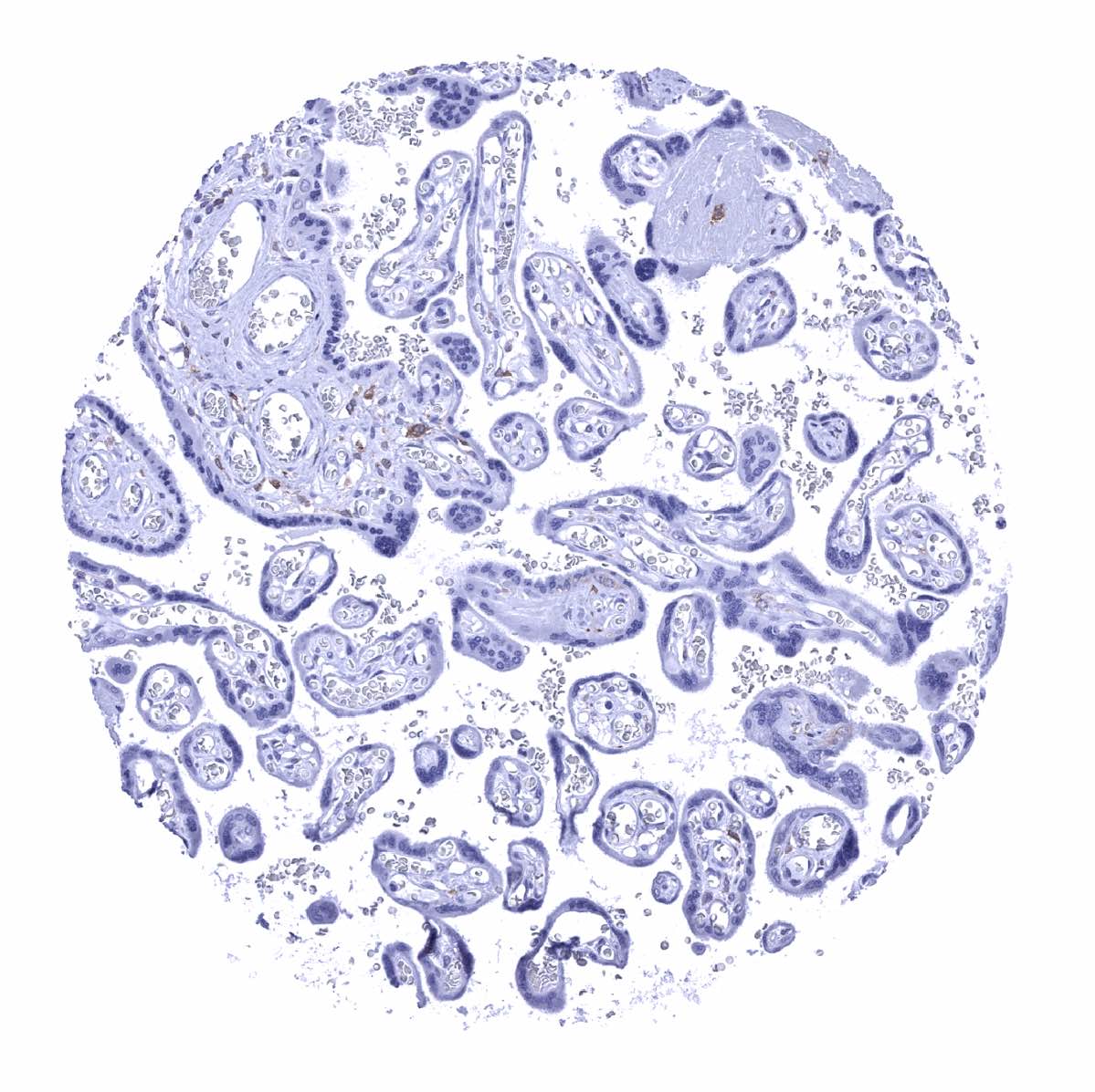
Placenta, mature
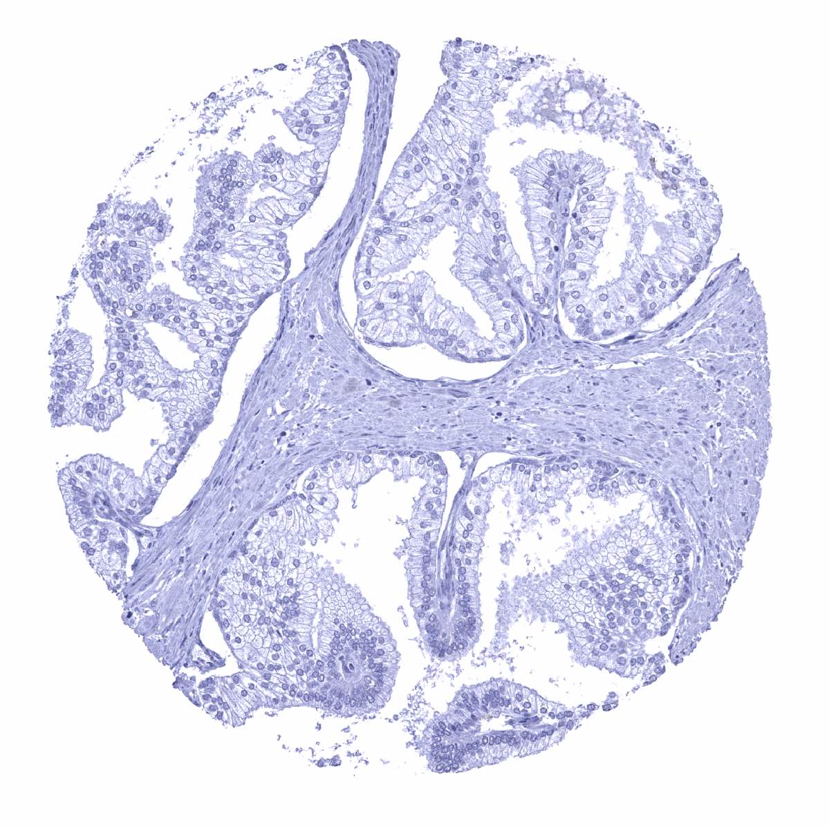
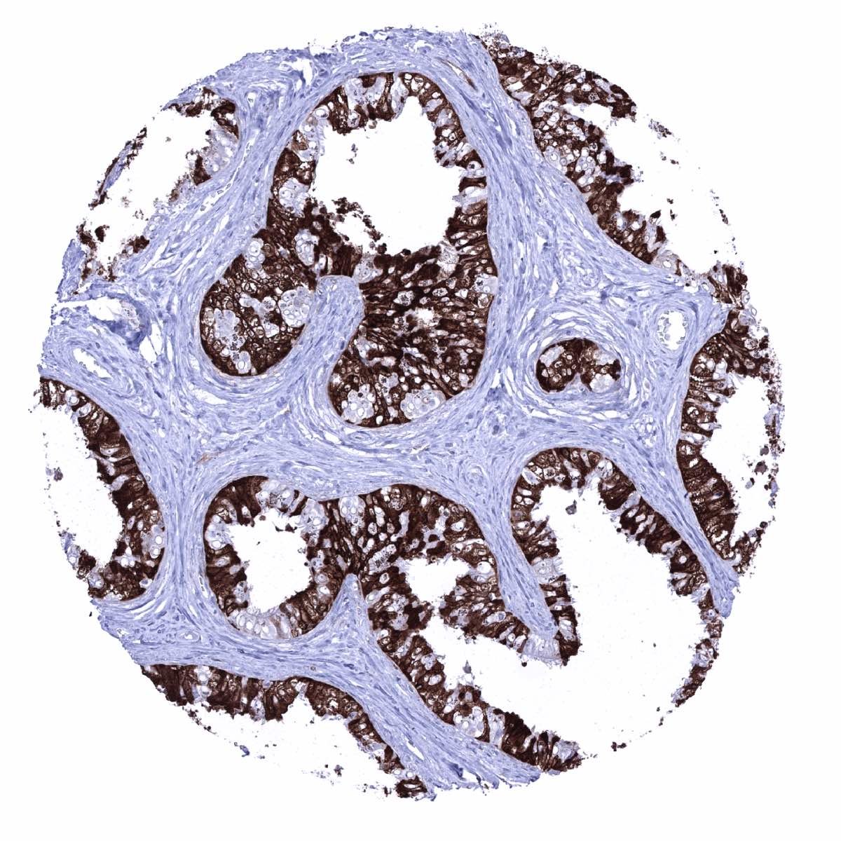
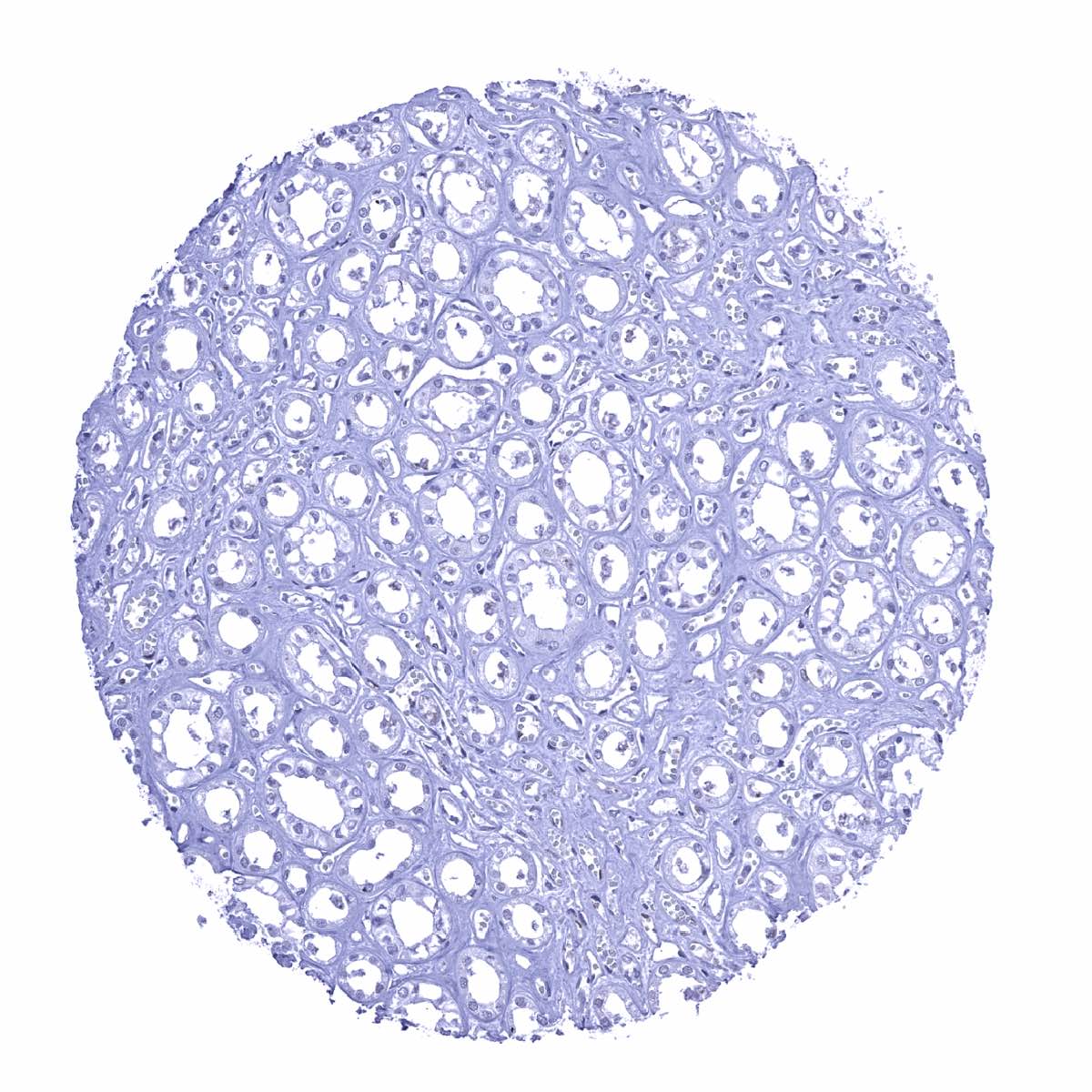
Prostate
Rectum, mucosa - GS staining of macrophages

Seminal vesicle - GS staining of variable intensity occurs in a fraction of epithelial cells
Sinus paranasales - Most cells of the respiratory epithelium and of associated glands show a moderate to strong GS staining
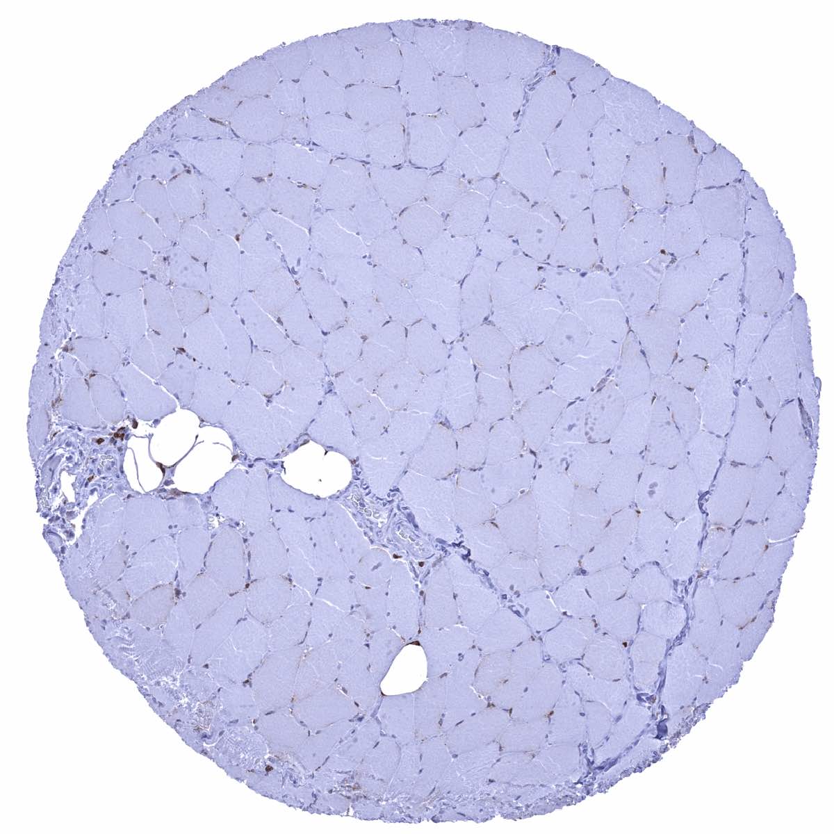
Skeletal muscle - A weak GS staining is seen in satellite cells of skeletal muscle
Skin - A nuclear and cytoplasmic GS staining can be seen in the lower third and in the top layers of the squamous epithelium
Skin (anal canal) - A nuclear and cytoplasmic GS staining is seen in the top third of the squamous epithelium
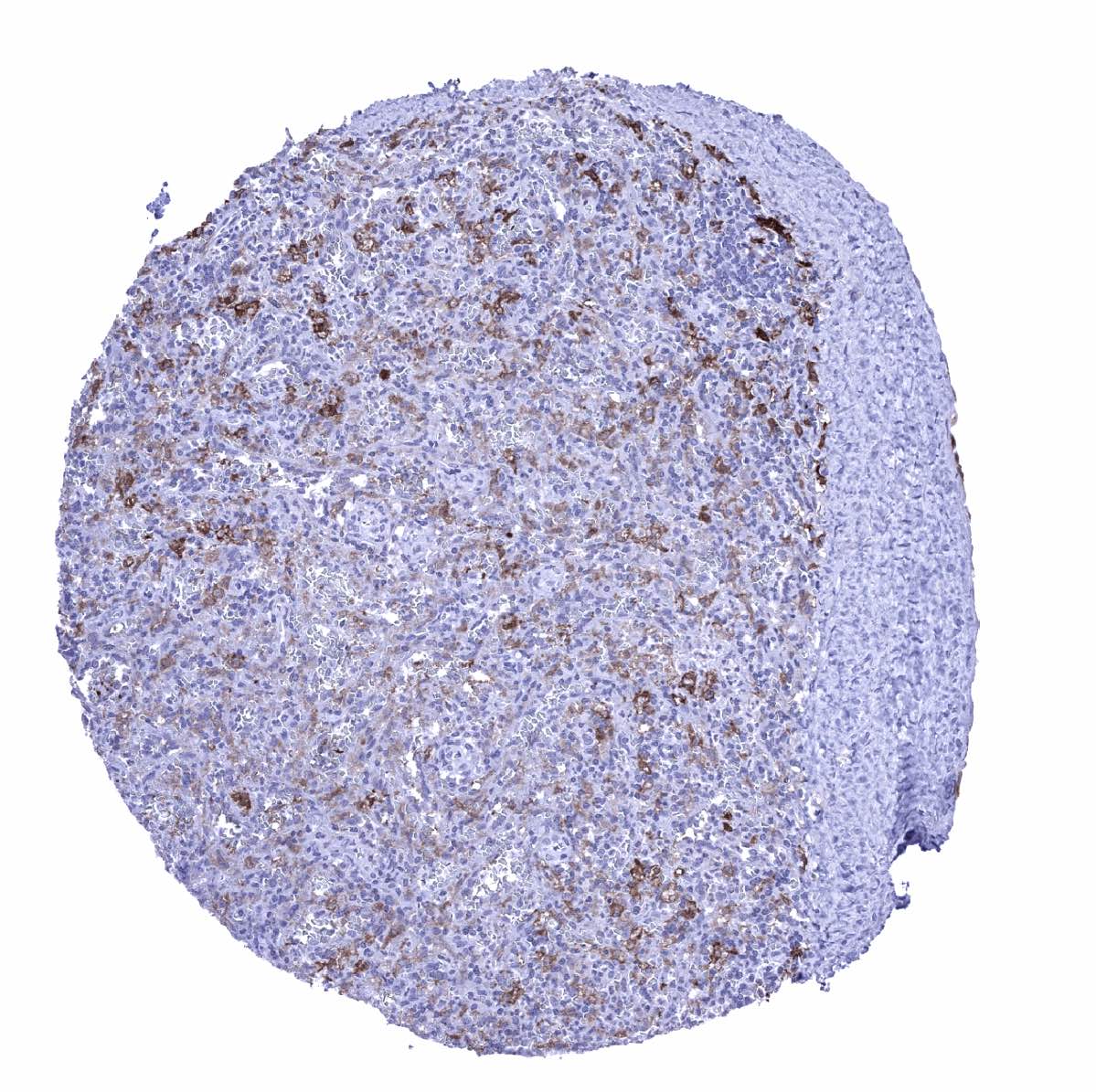
Spleen - Weak to moderate GS staining in histiocytic cells and granulocytes
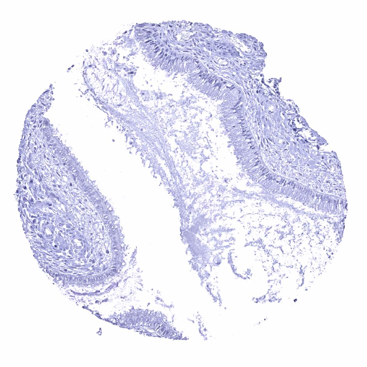
Stomach, antrum
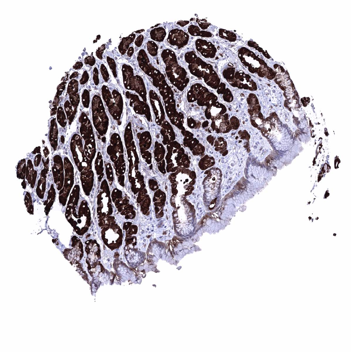
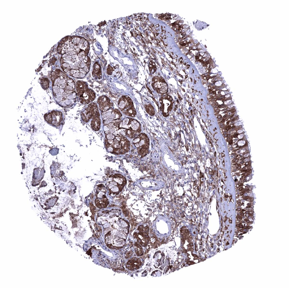
Stomach, corpus - A strong GS staining is seen in glandular cells while staining is only weak in surface epithelial cells

Sublingual gland - Strong GS staining of myoepithelial cells
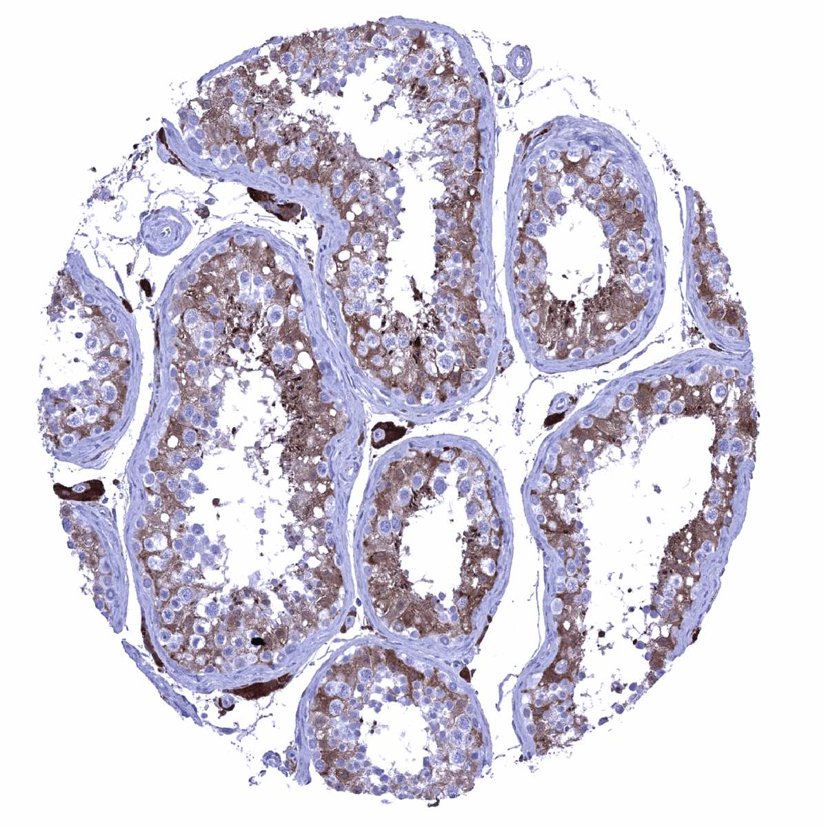
Testis - GS staining is weak to moderate in Sertoli cells and strong and Leydig cells
Thymus - Prominent GS staining of macrophages and dendritic cells
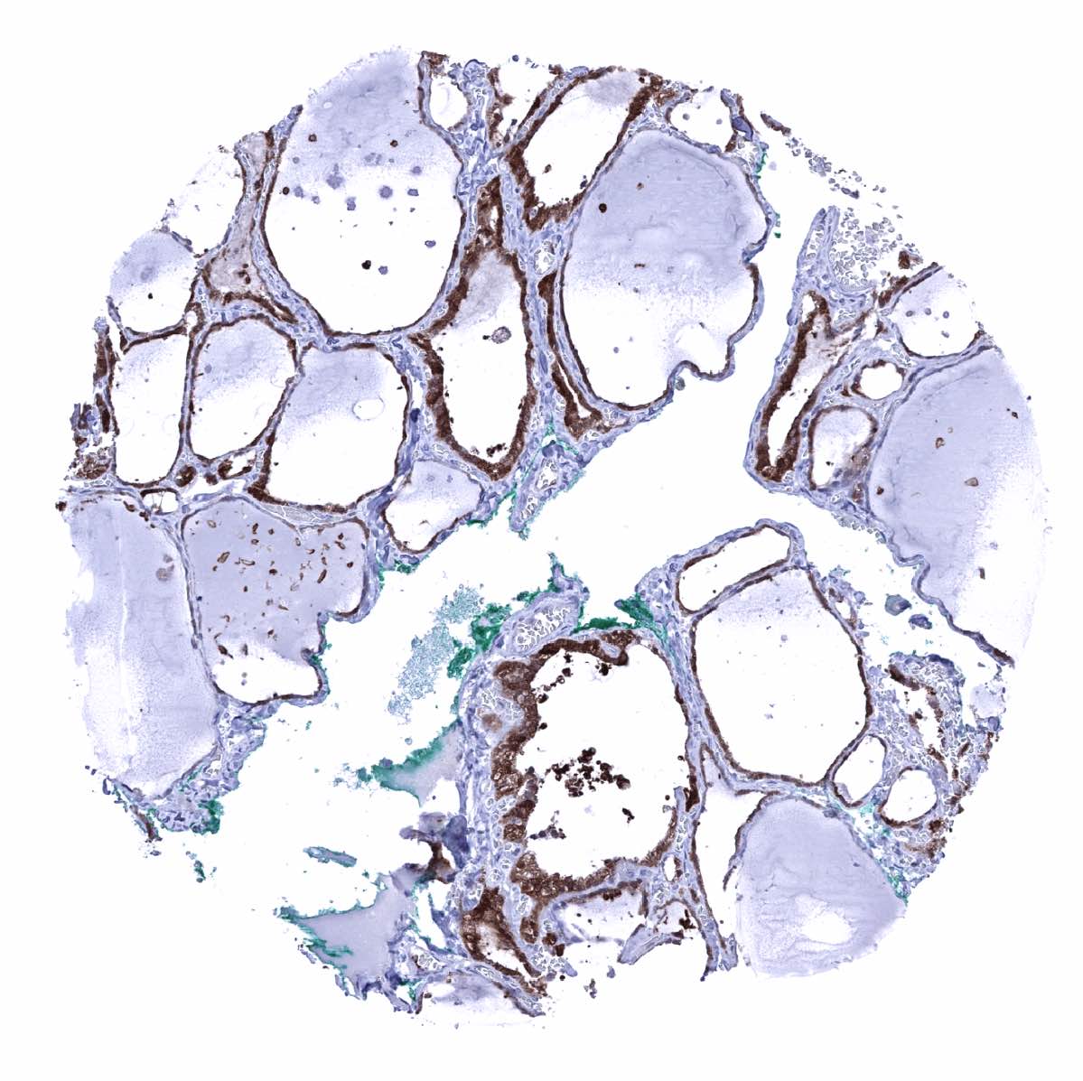
Thyroid gland - Strong GS staining in follicle cells of the thyroid
Tonsil - GS staining of macrophages and of a fraction of squamous epithelial cells of a crypt

Tonsil, surface epithelium - A nuclear and cytoplasmic GS staining is preferentially seen in the top third of the squamous epithelium. Some basal and intermediary cells do also stain.
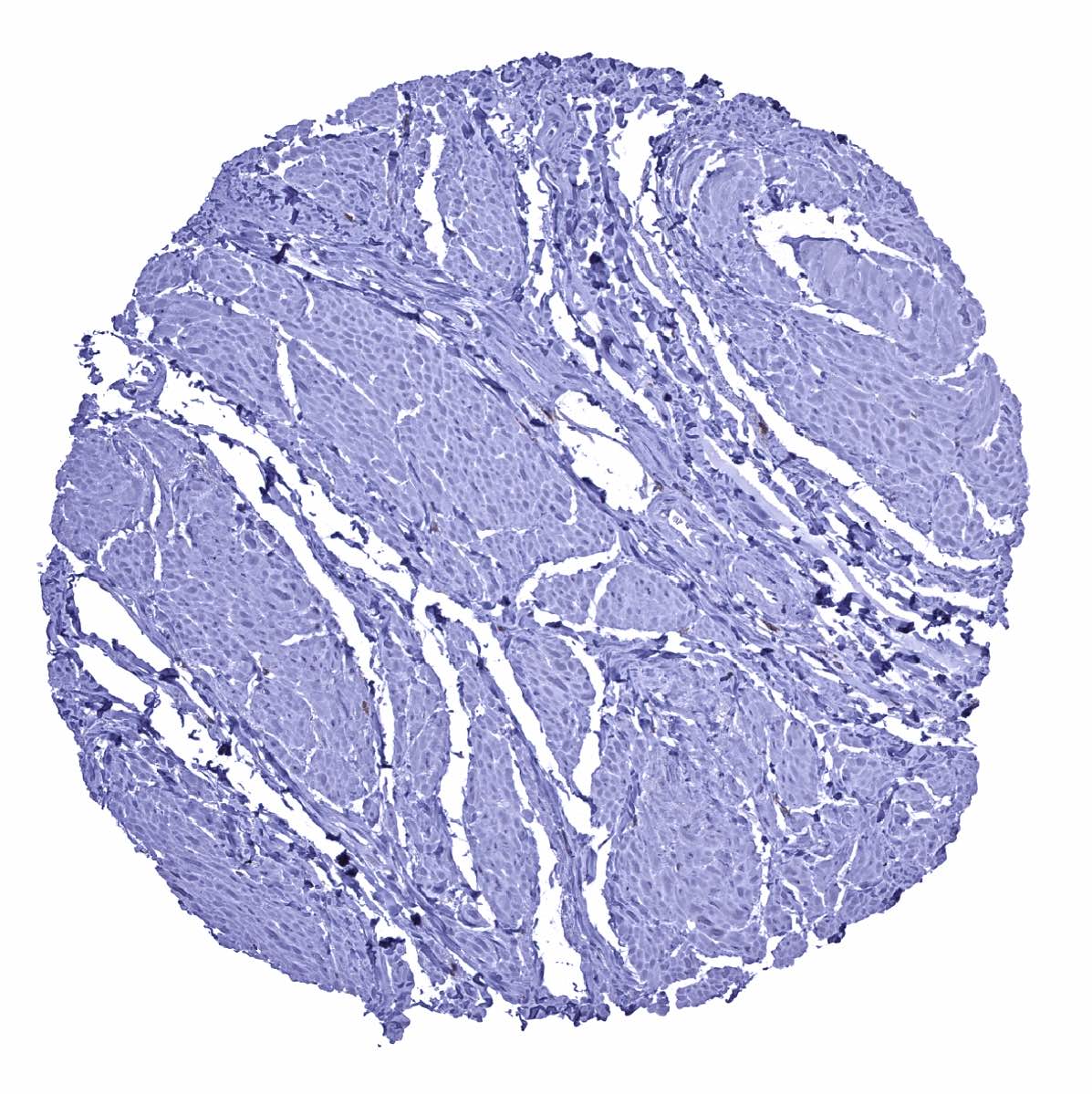
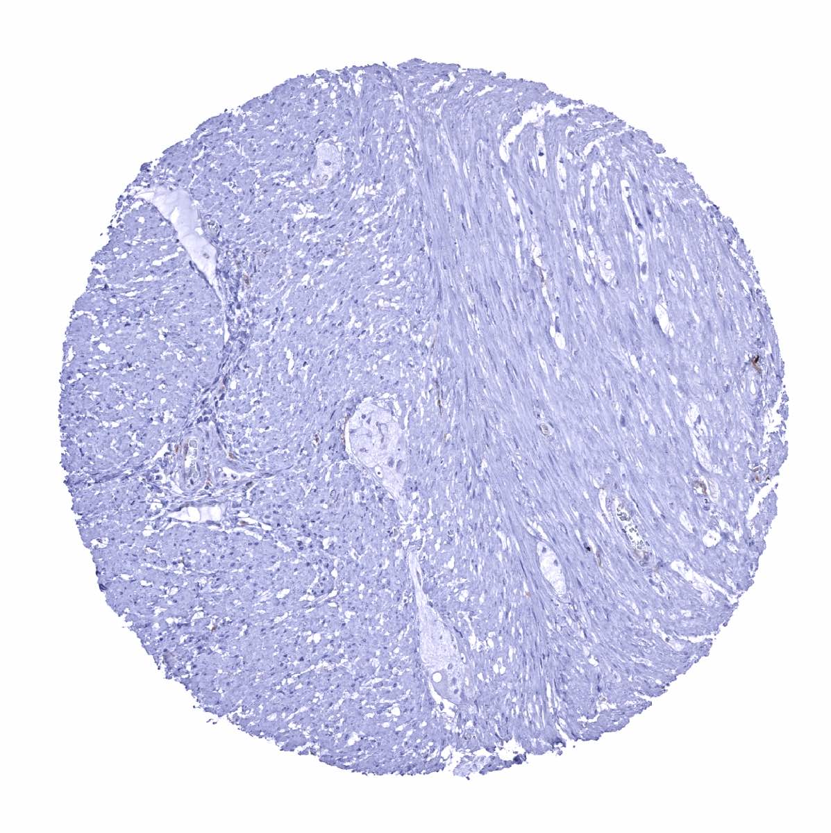
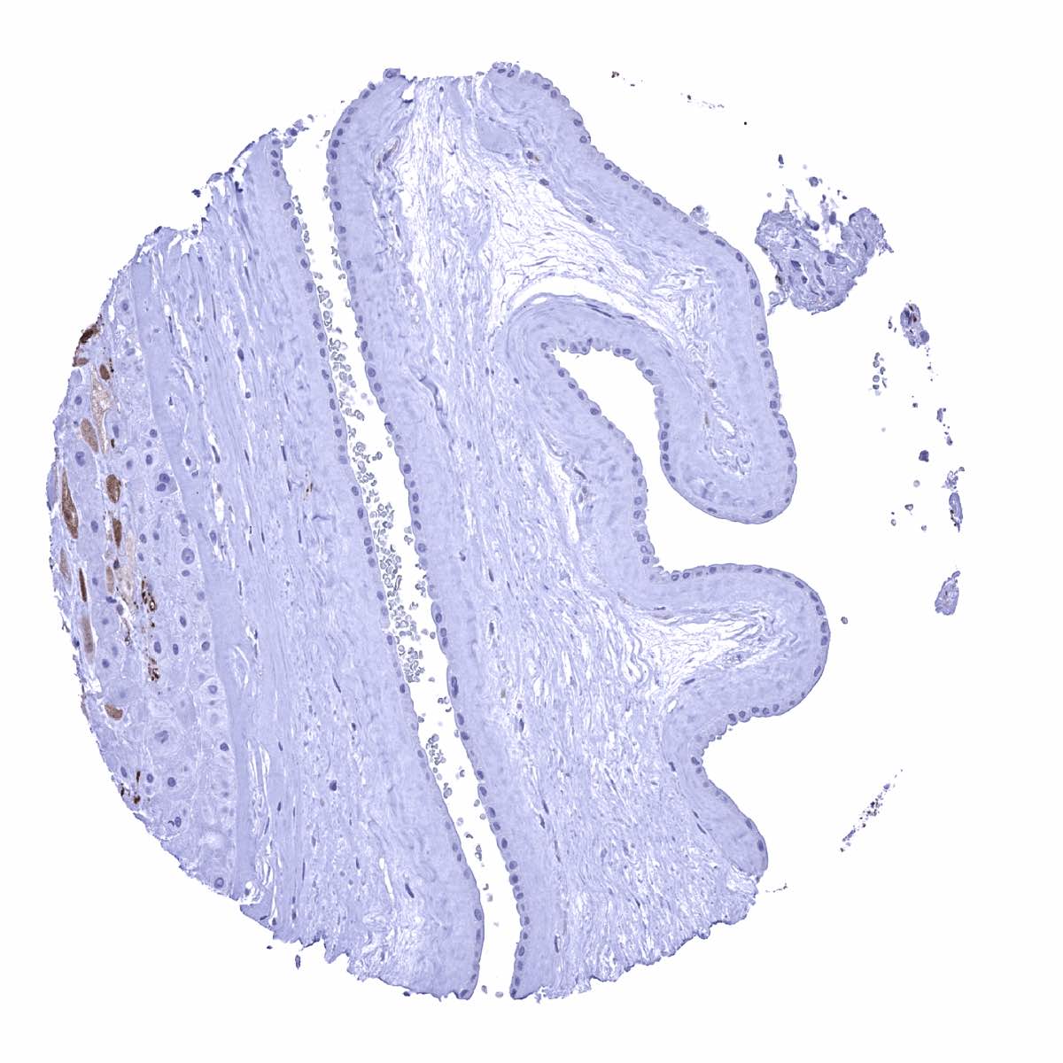
Urinary bladder, muscular wall
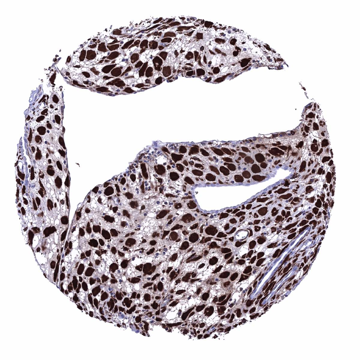
Urinary bladder, urothelium - A strong nuclear and cytoplasmic GS staining occurs in the urothelium. Staining is weakest in basal and umbrella cells
Uterus, ectocervix - A nuclear and cytoplasmic GS staining is seen in the top third of the squamous epithelium
Uterus, ectocervix - A nuclear and cytoplasmic GS staining occurs in the top third of the squamous epithelium
Uterus, endocervix
Uterus, endometrium (pregnancy) - GS staining is very intense in decidua cells
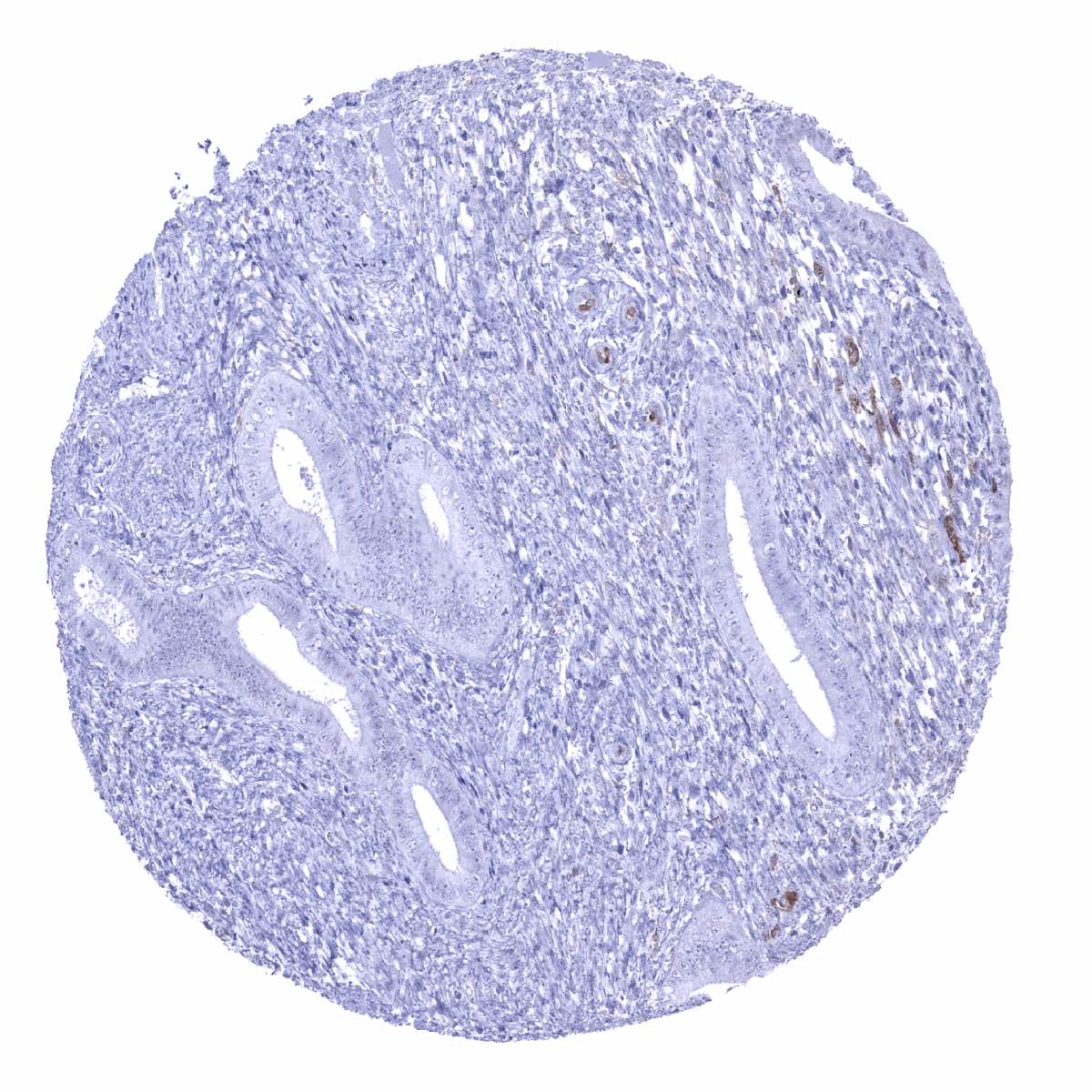
Uterus, endometrium (proliferation)
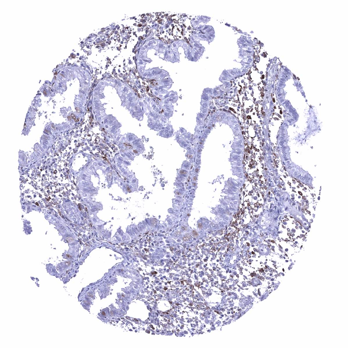
Uterus, endometrium (secretion) - GS staining predominates in stromal and inflammatory cells and is only focal and faint in epithelial cells
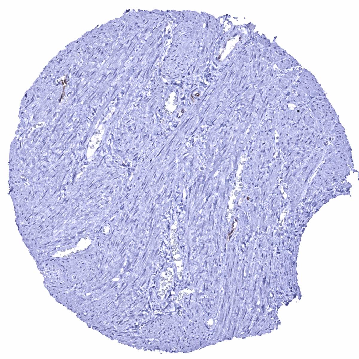
Uterus, myometrium - A weak GS staining is seen in few endothelial cells of small capillaries