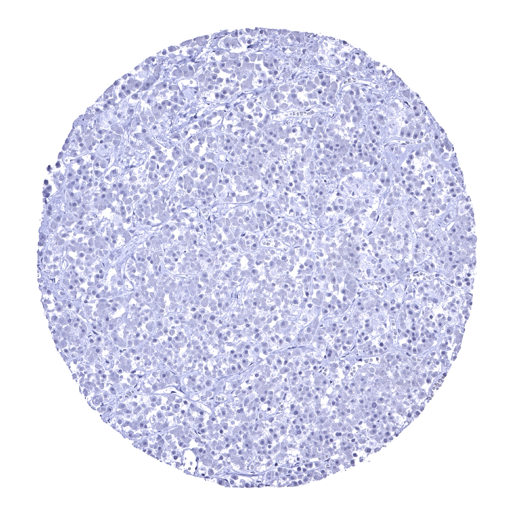
Adrenal gland

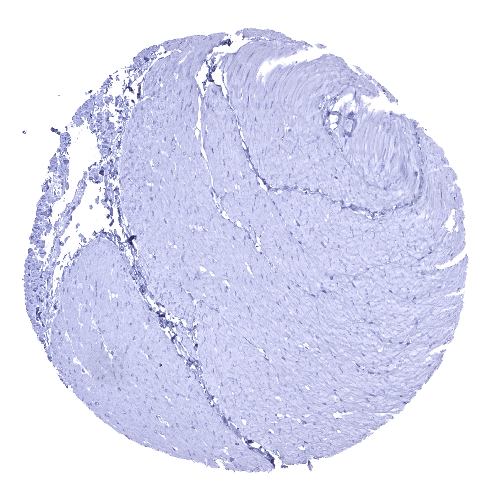
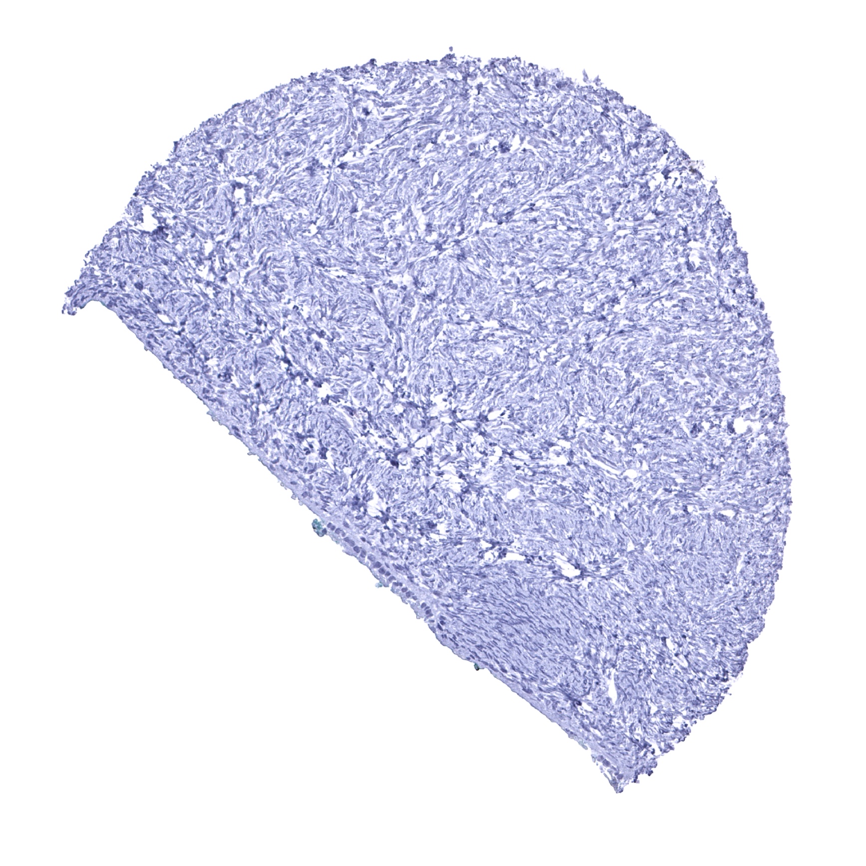
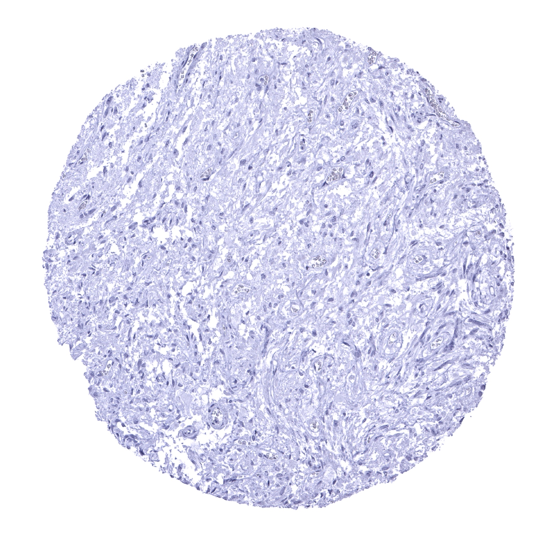
Aorta, media
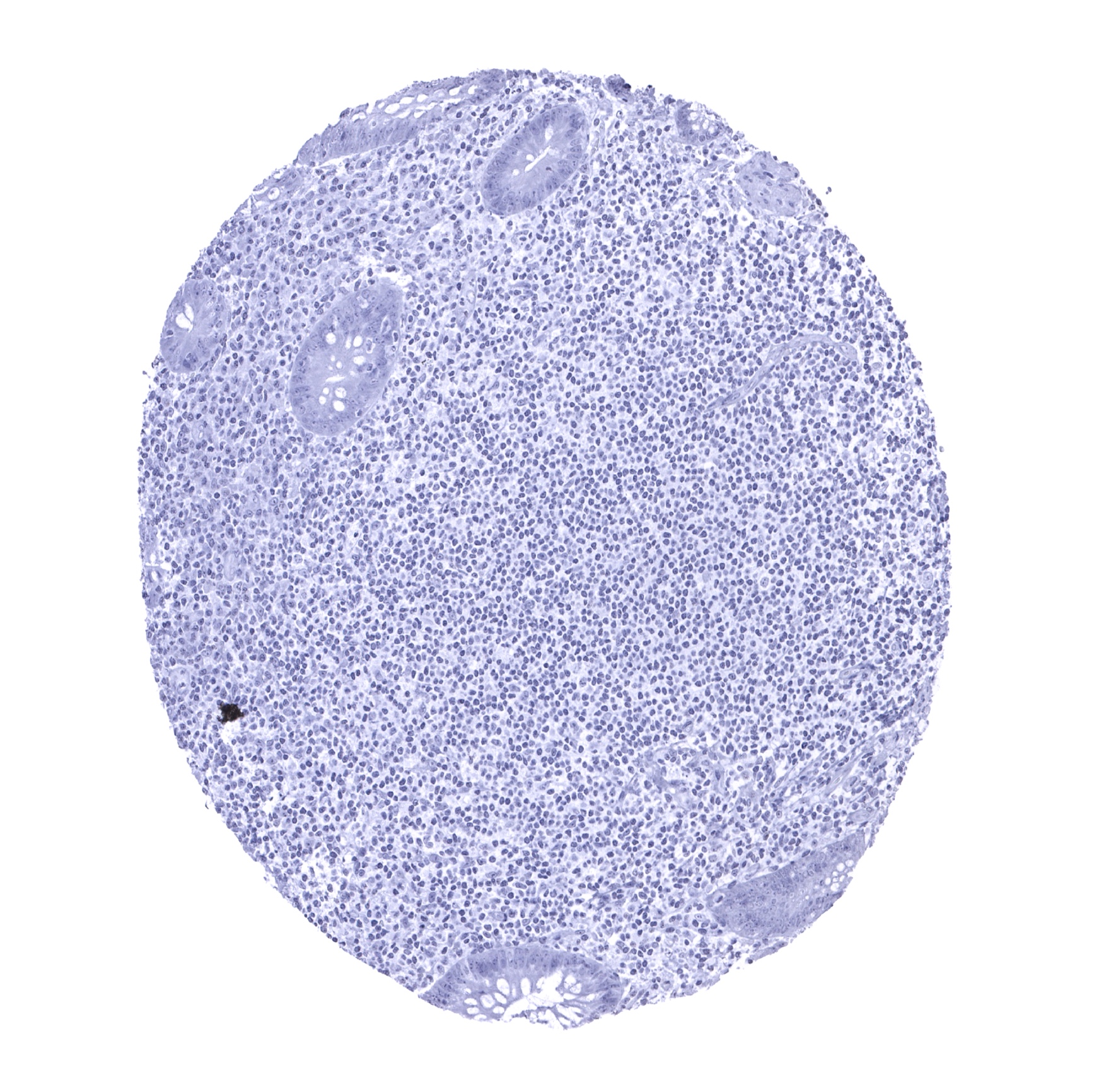
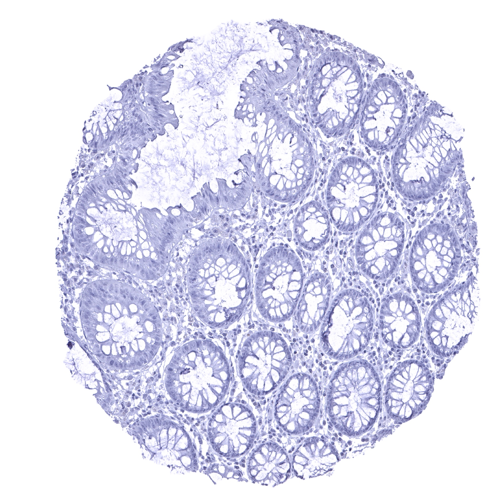
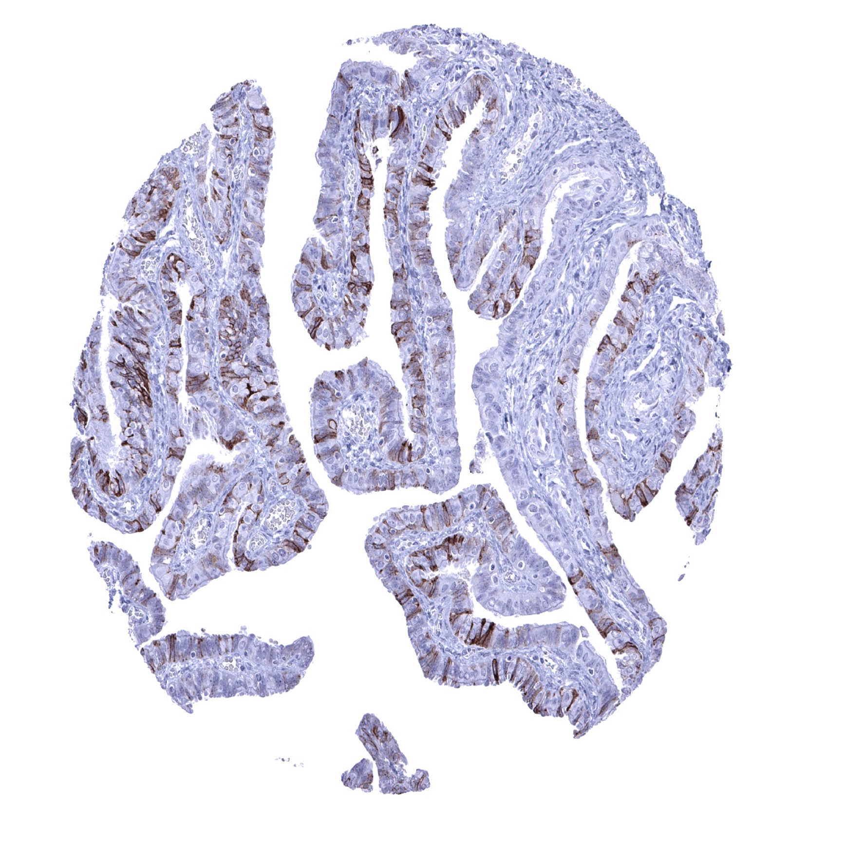
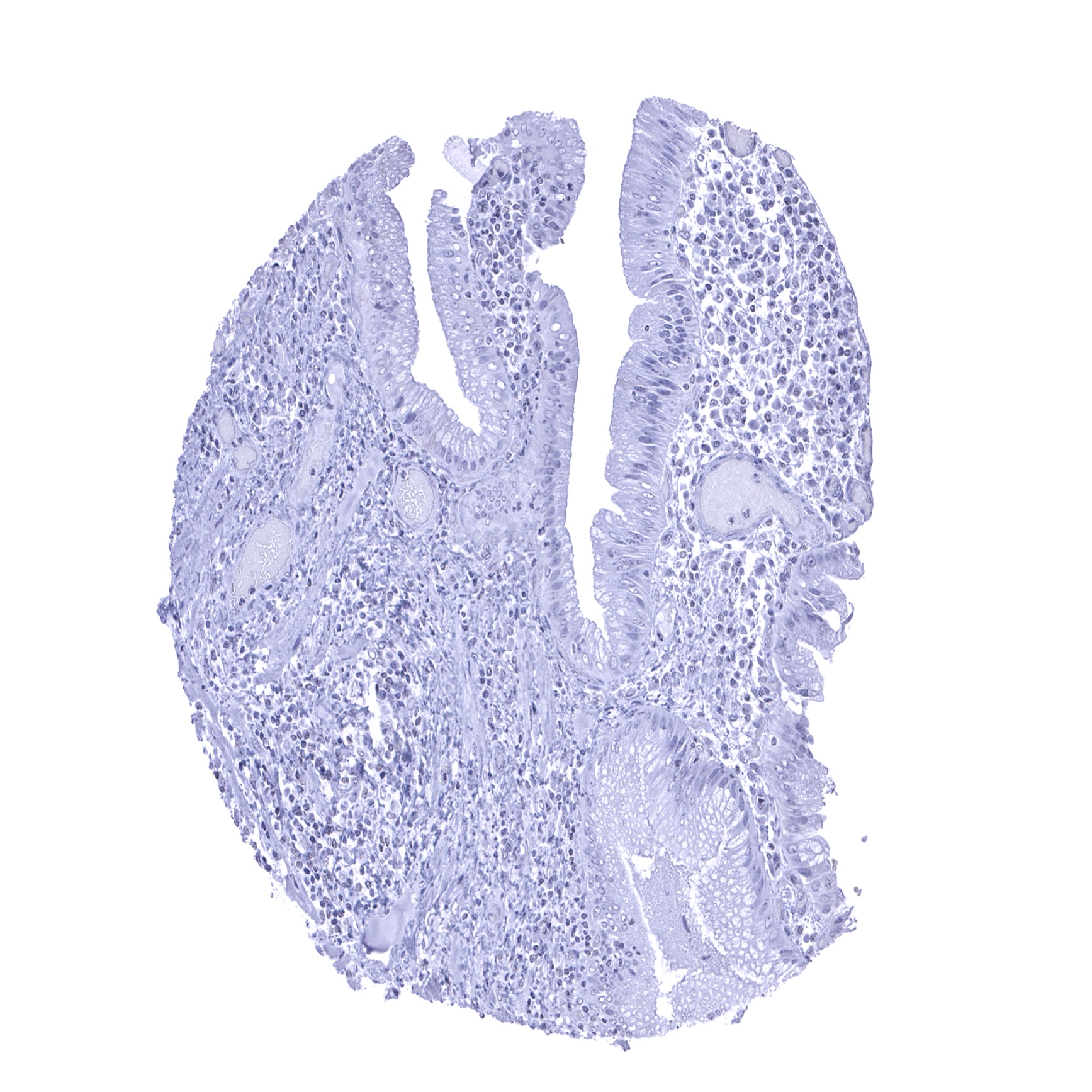
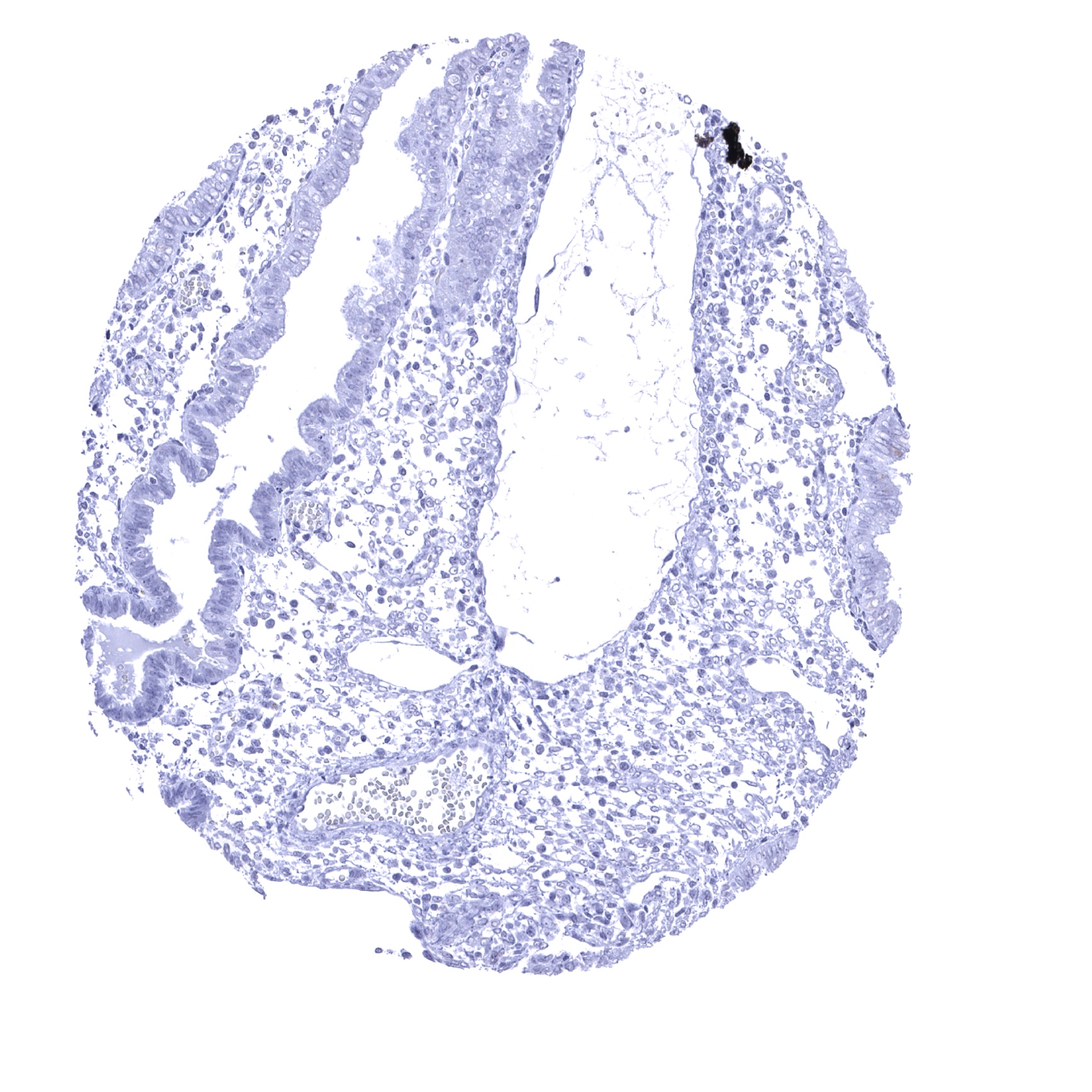
Appendix, mucosa
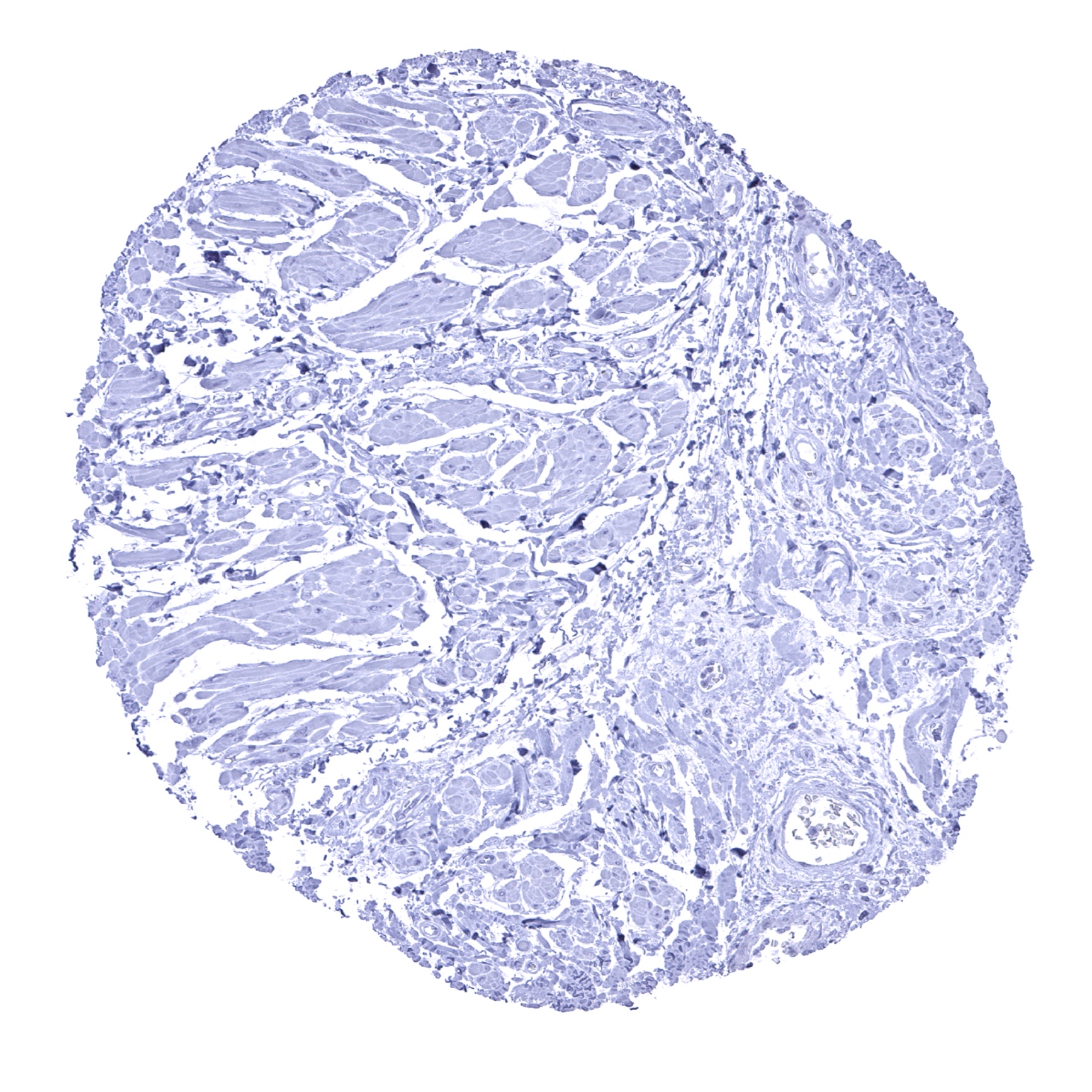
Appendix, muscular wall
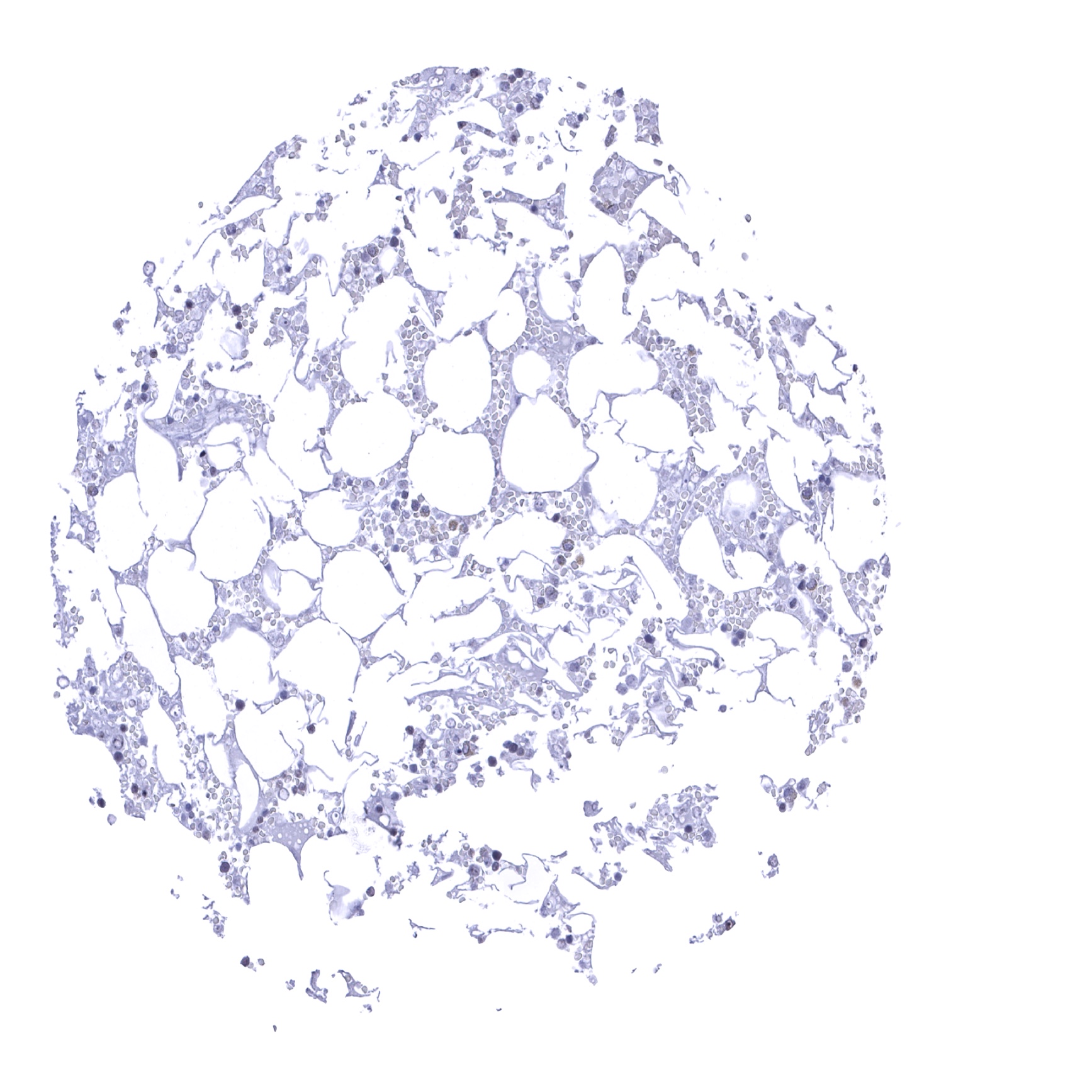
Bone marrow
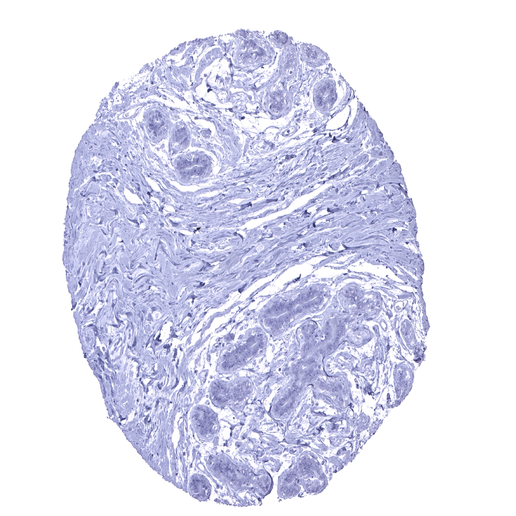
Breast

Bronchus, glands
Cerebellum (molecular layer, Purkinje cell layer, granule cell layer)
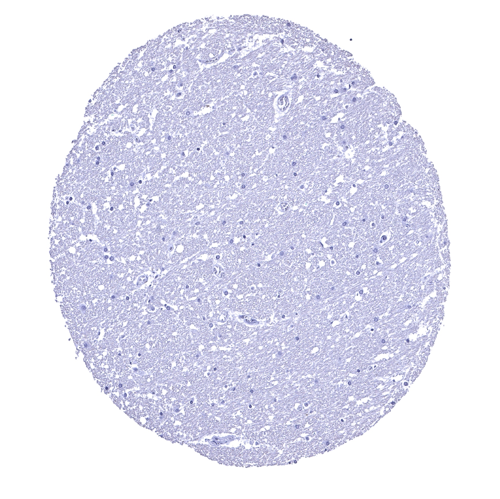
Cerebellum (white matter)
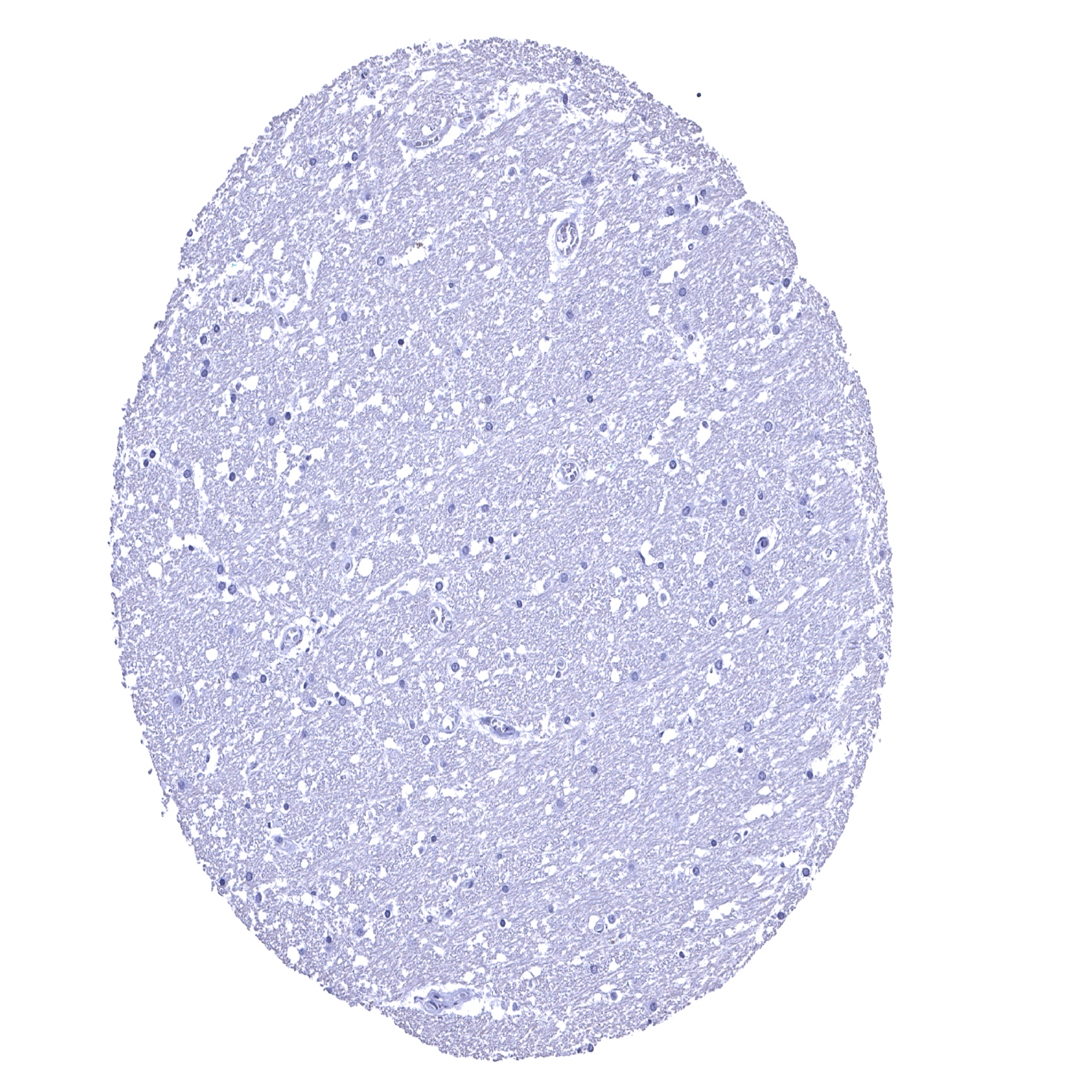
Cerebellum, grey (Stratum neuronorum)

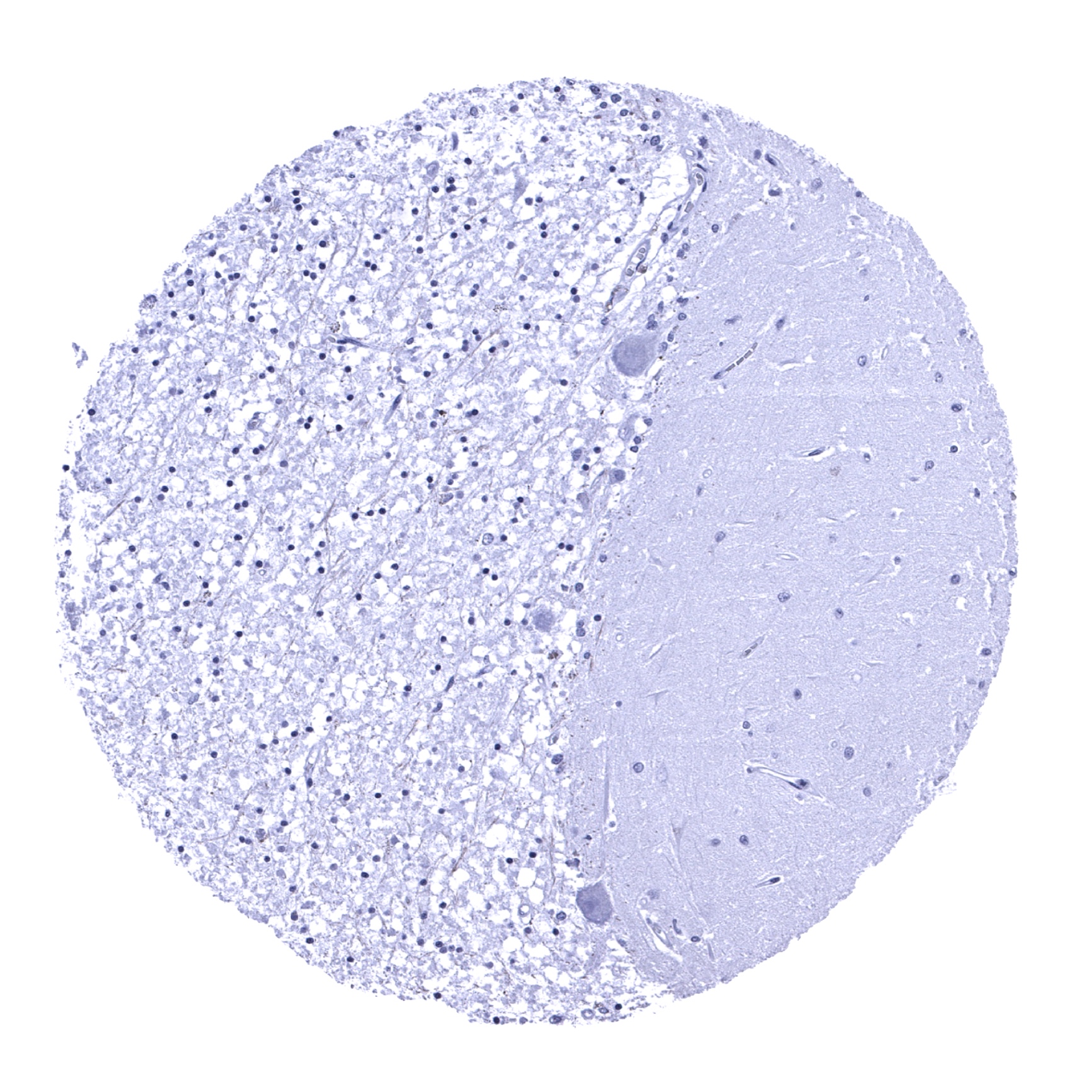
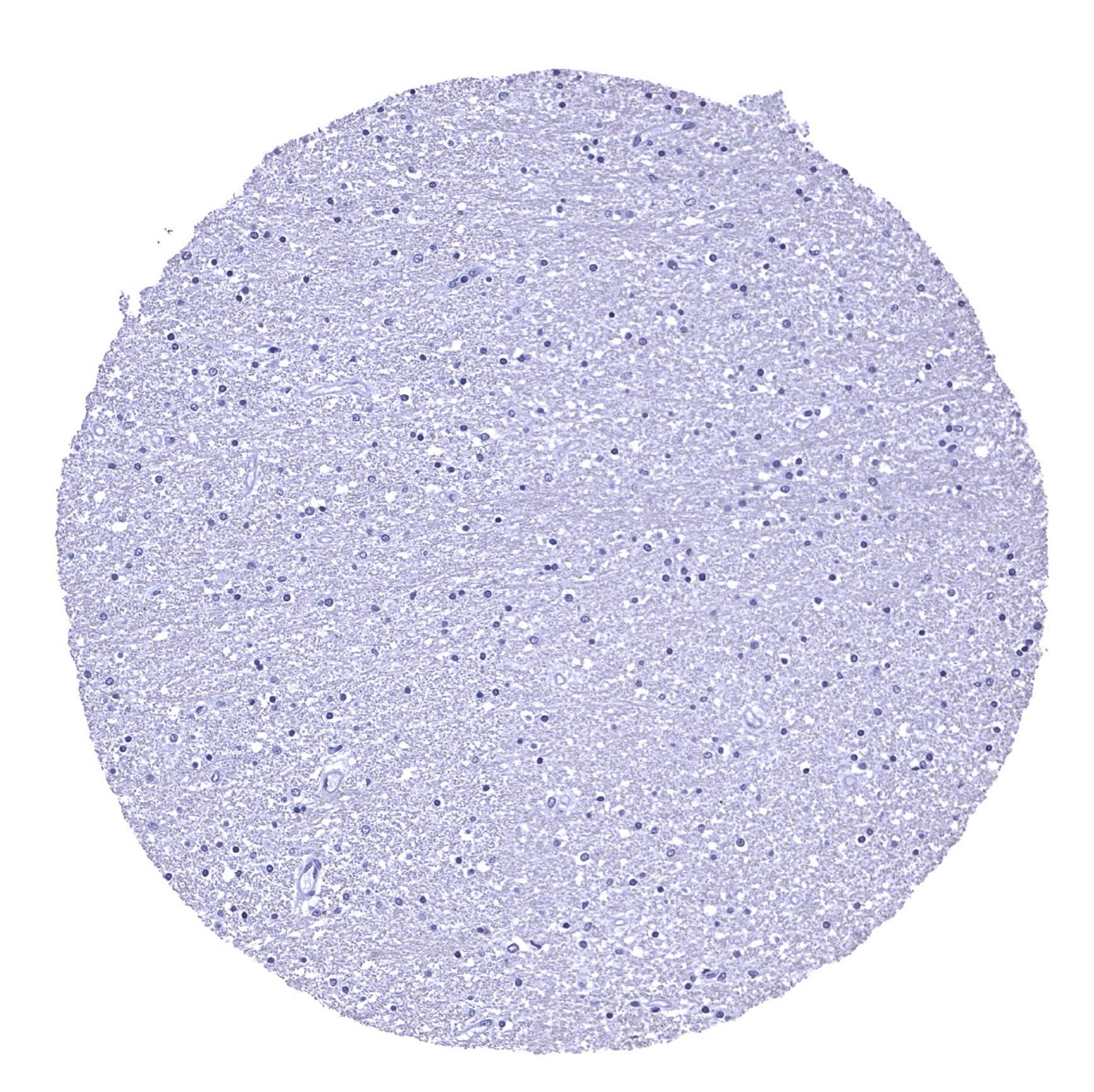
Cerebrum, grey matter
Cerebrum, white matter
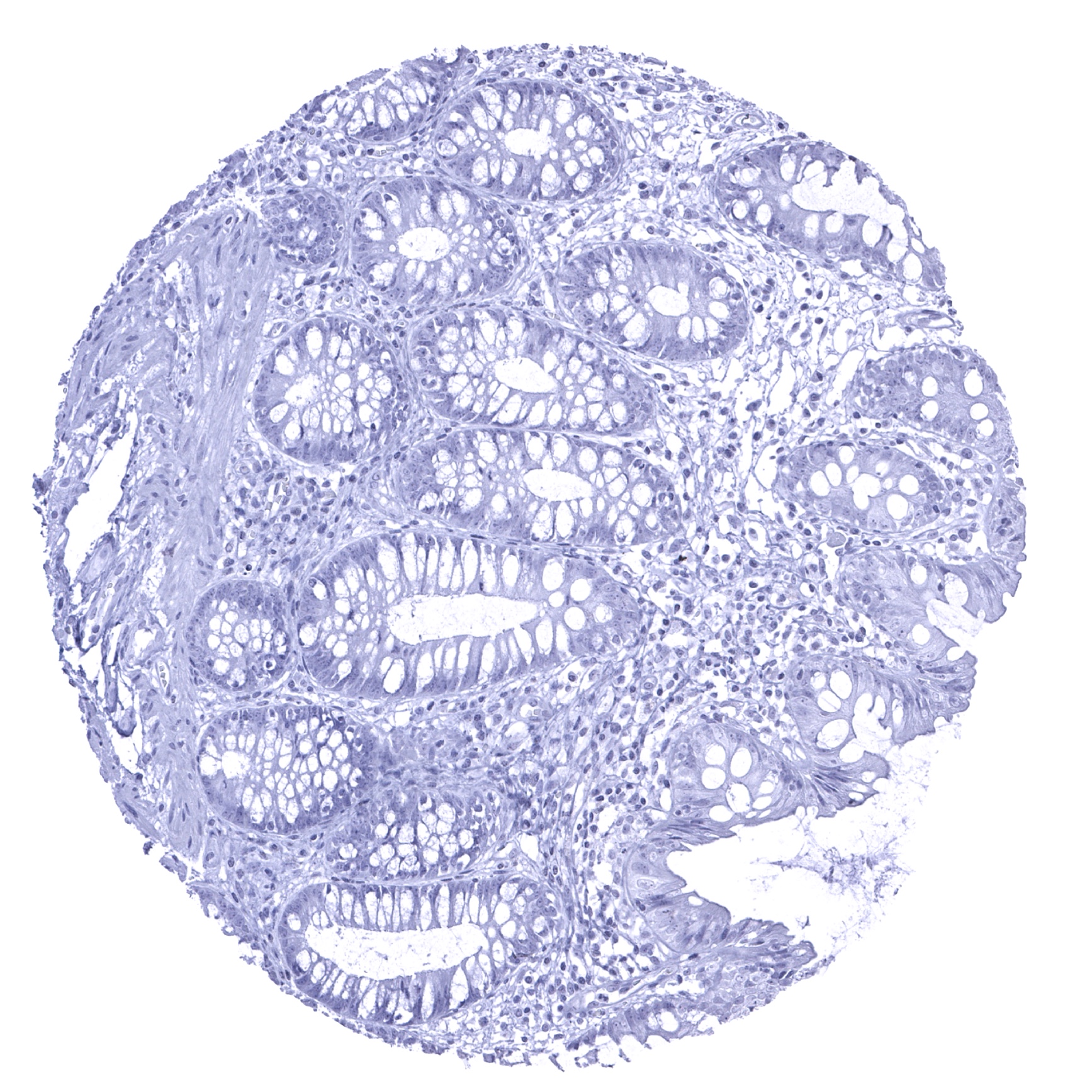
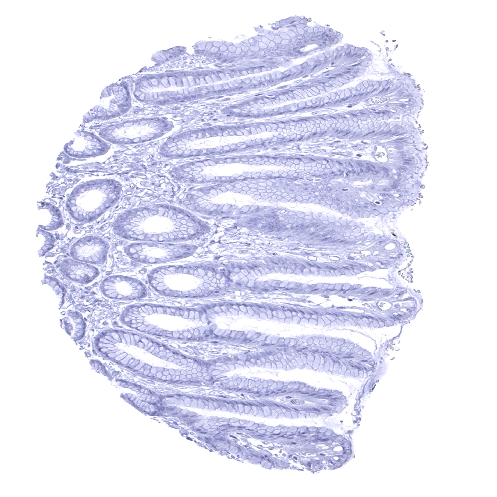
Colon descendens, mucosa
Colon descendens, muscular wall
Duodenum, Brunner gland

Duodenum, mucosa

Epididymis - CDH16 staining is absent in the caput epididymis.

Epididymis - Strong membranous CDH16 staining of epithelial cells in the cauda epididymis (CDH16 immunohistochemistry).
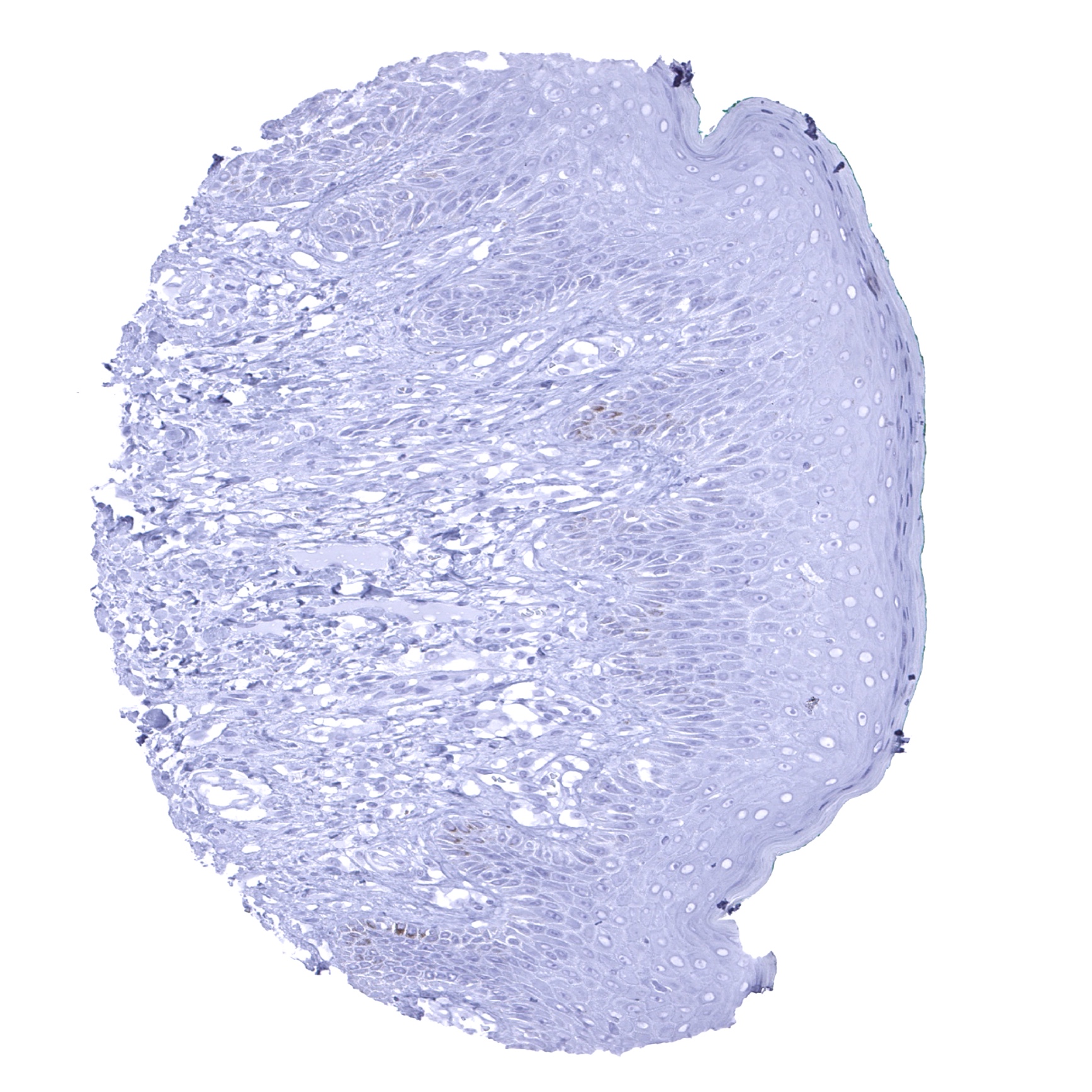
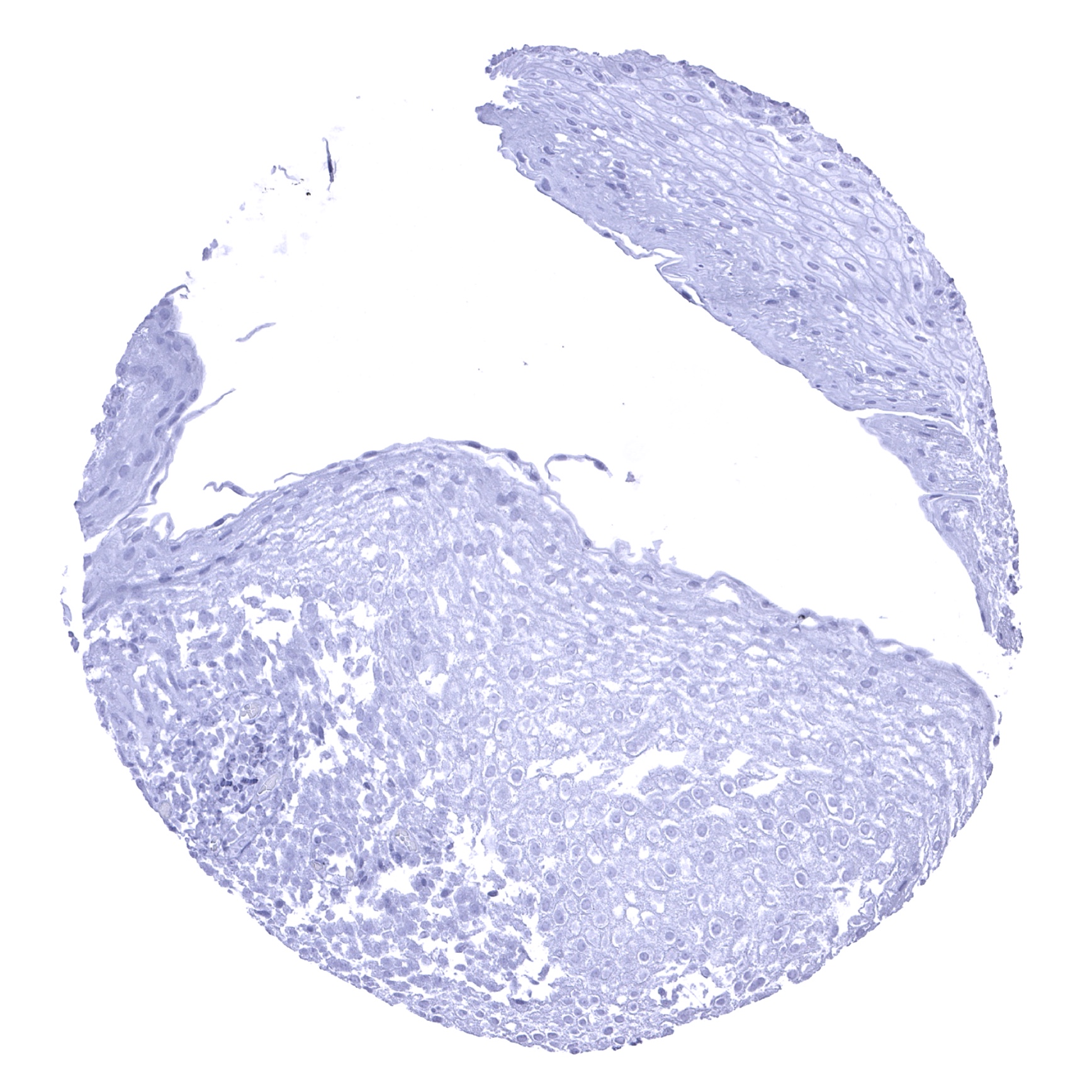
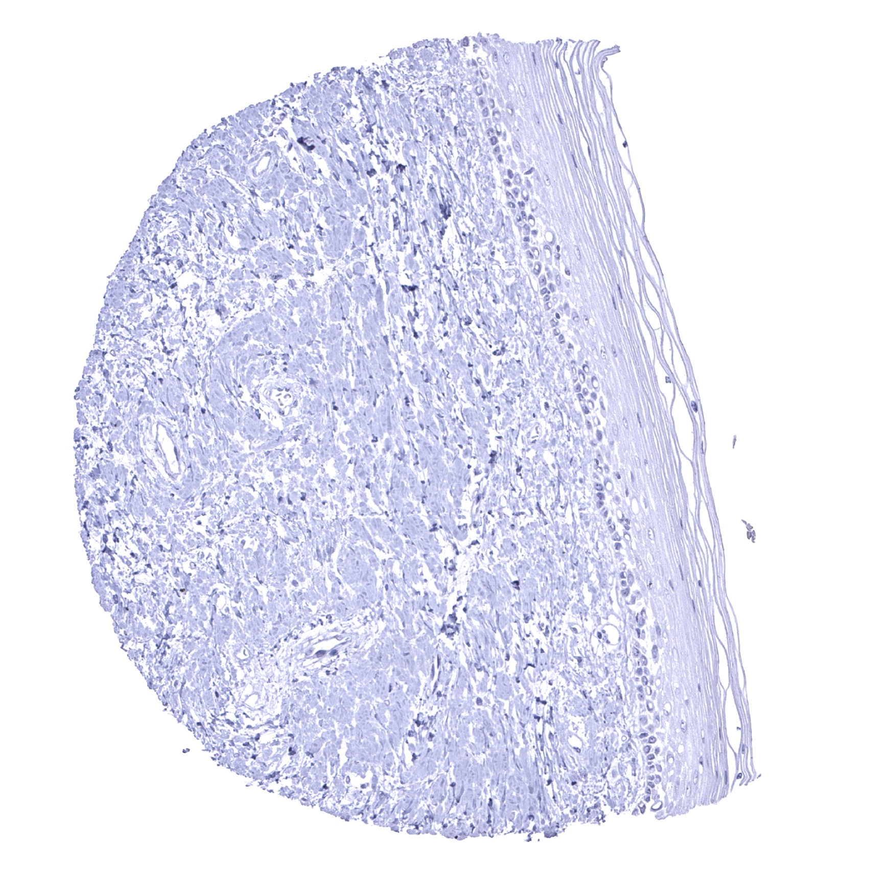
Esophagus, squamous epithelium
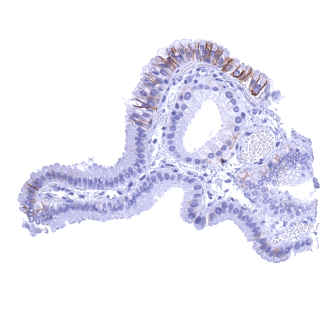
Fallopian tube, mucosa - A membranous CDH16 staining of a fraction of epithelial cells can be seen in the fallopian tube (CDH16 immunohistochemistry).
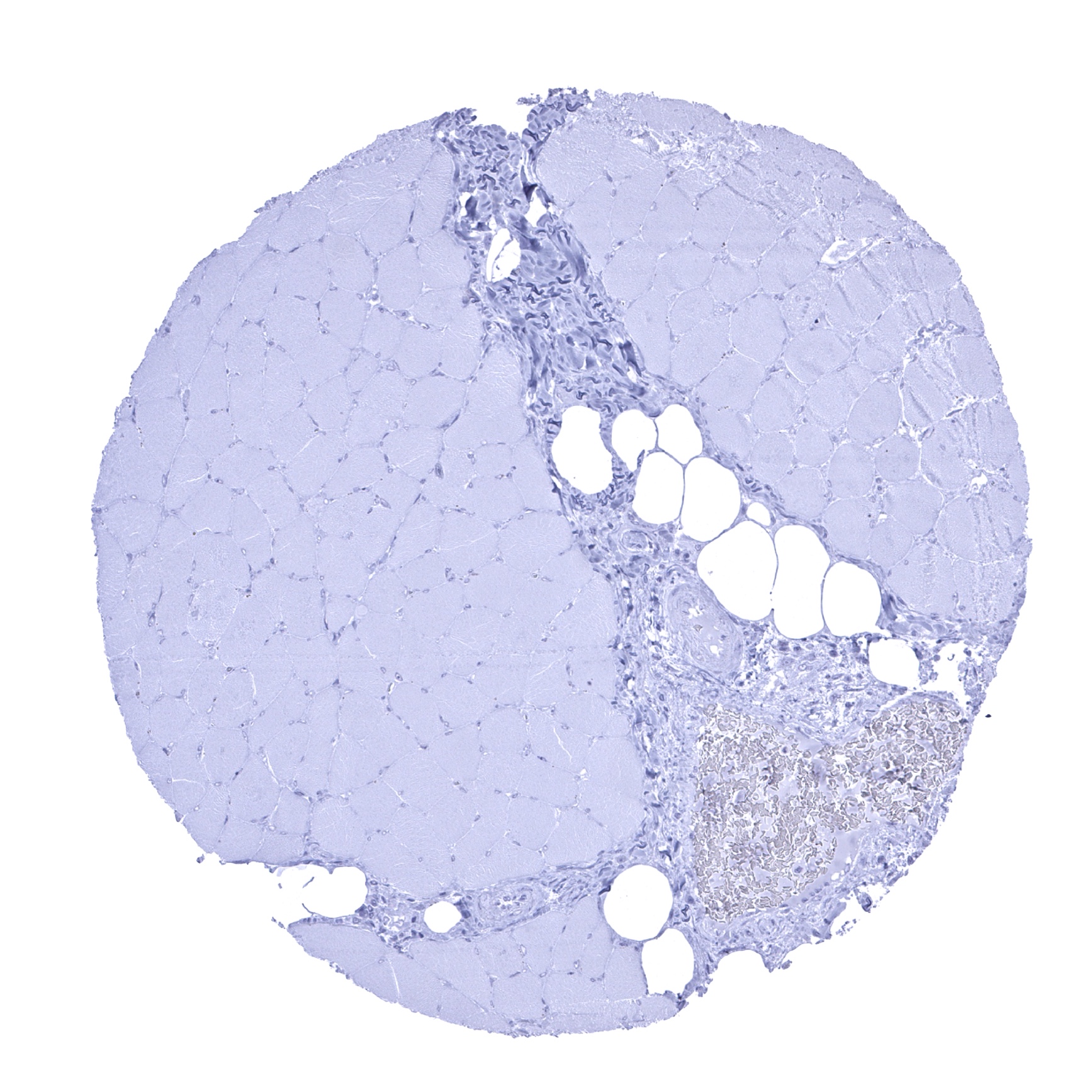
Fat
Gallbladder, epithelium - A focal CDH16 staining is seen in this gallbladder sample.
Gallbladder, epithelium
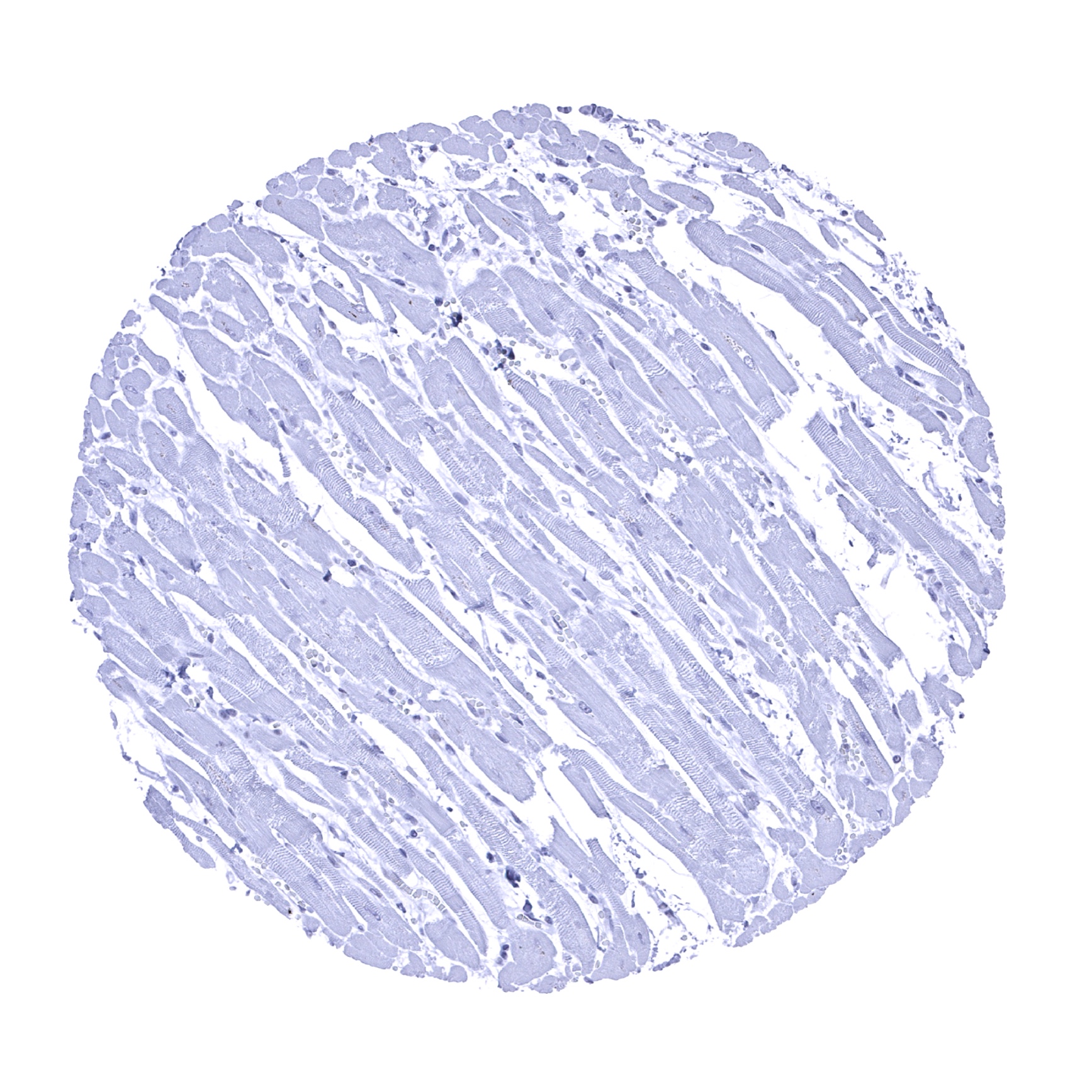
Heart muscle
Ileum, mucosa
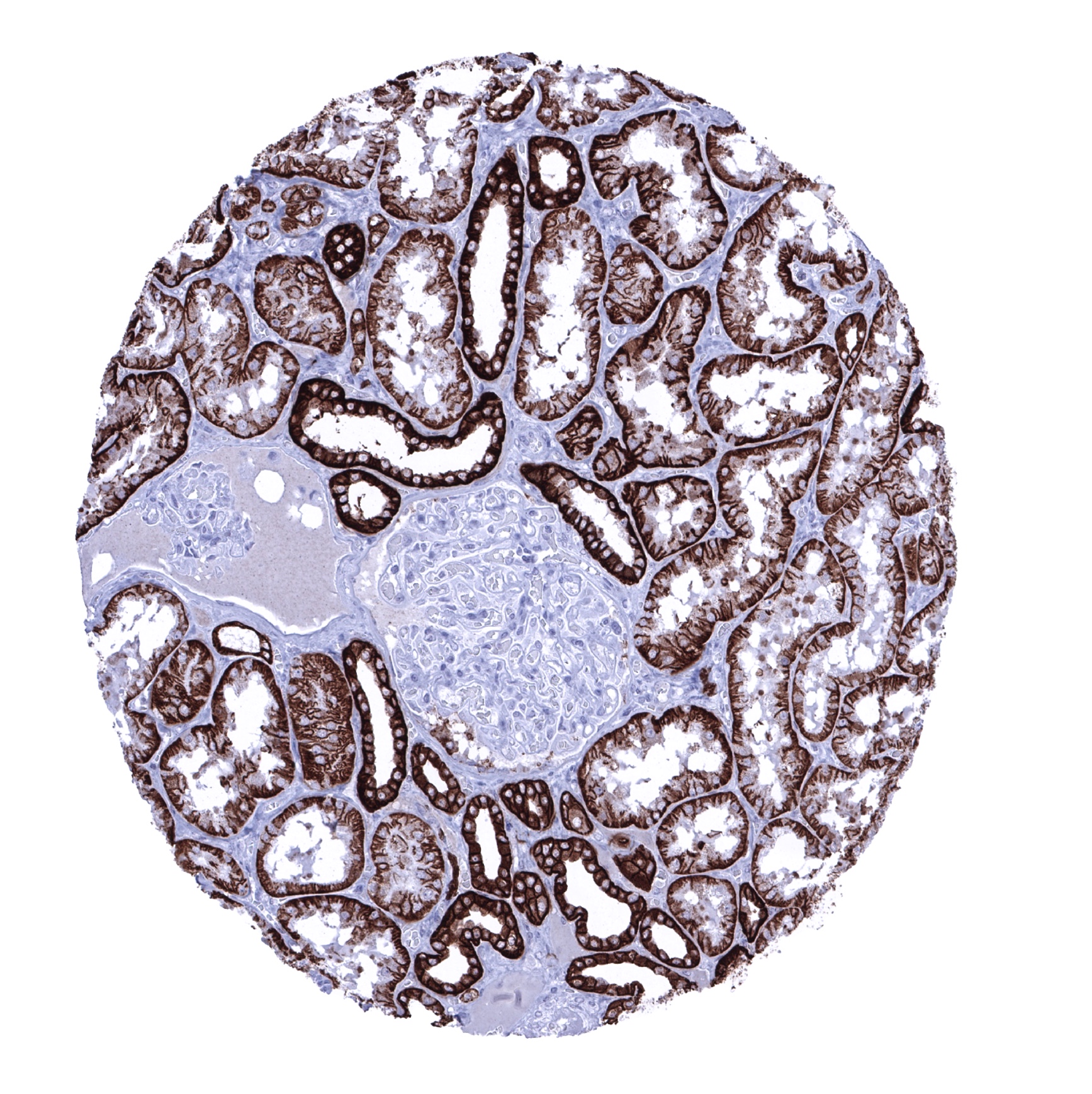
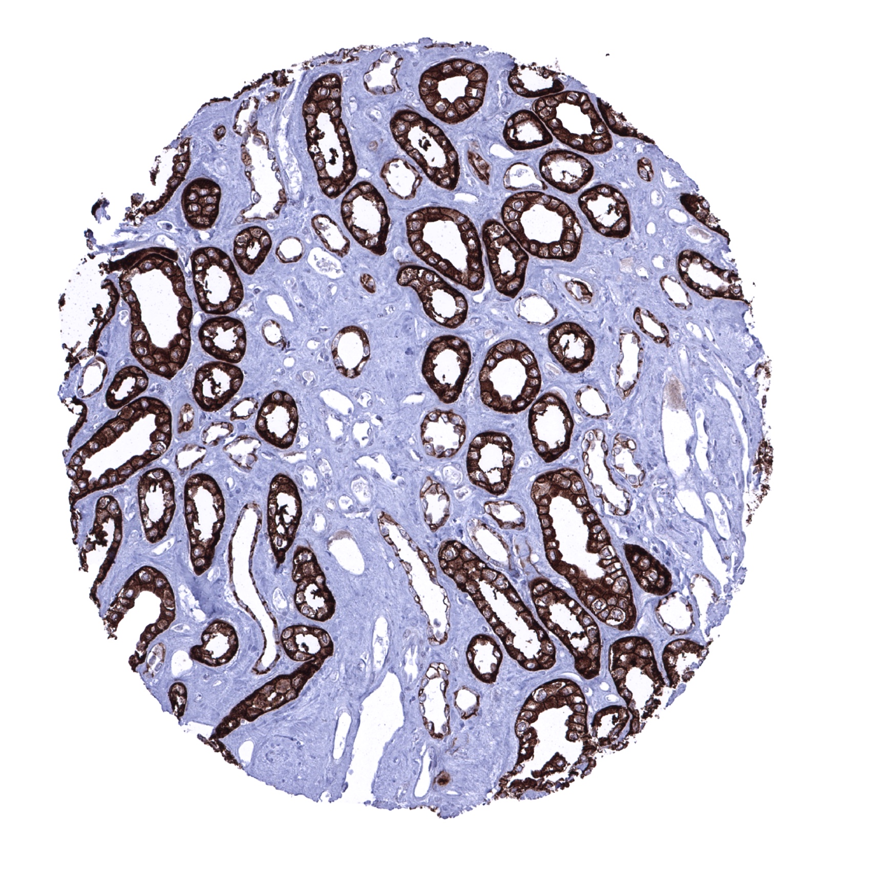
Kidney, cortex - CDH16 immunostaining is stronger in distal than in proximal tubuli. The staining pattern is membranous (predominantly basolateral) and also cytoplasmic (CDH16 immunohistochemistry).
Kidney, medulla - Strong and predominantly membranous CDH16 immunostaining of collecting duct cells (CDH16 immunohistochemistry).
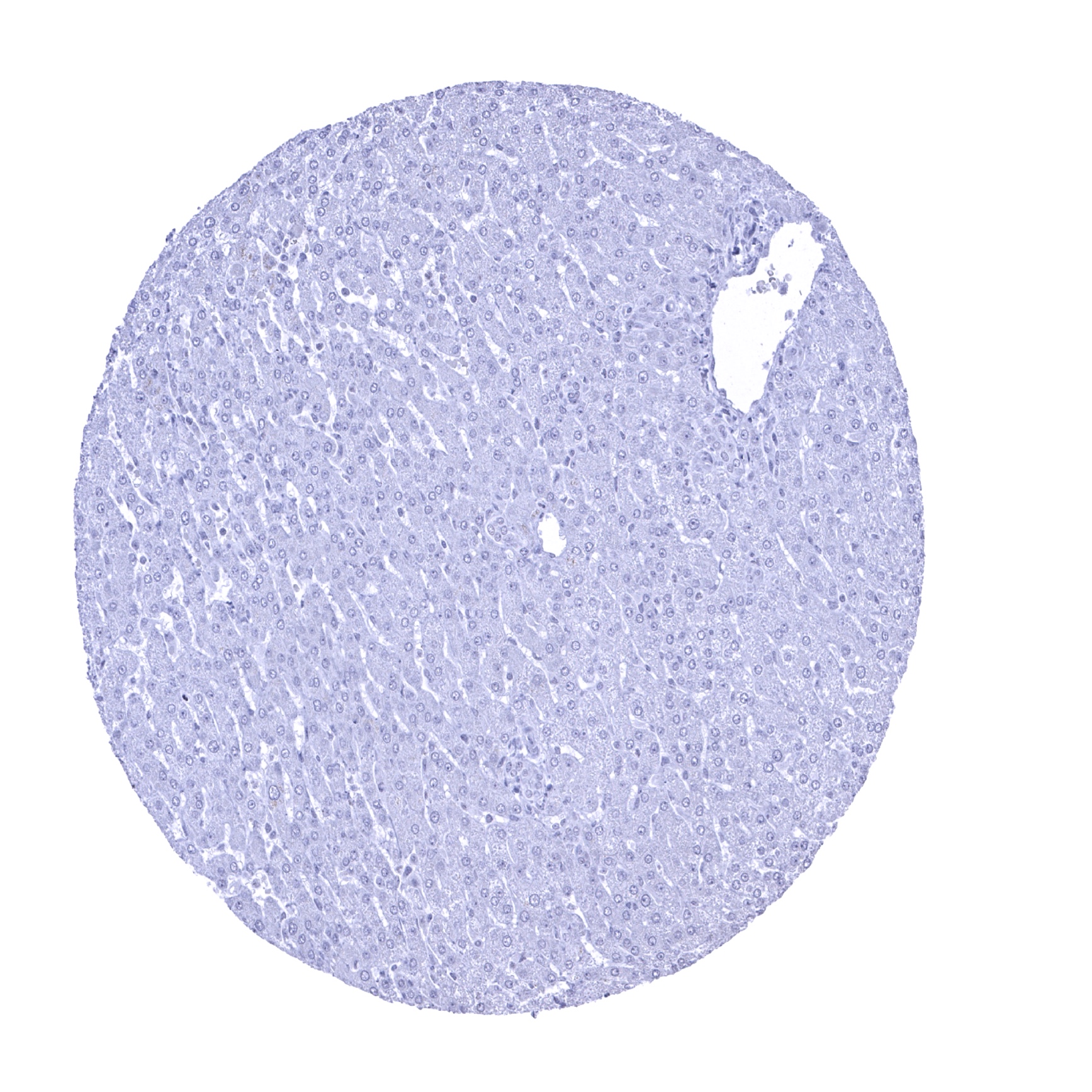
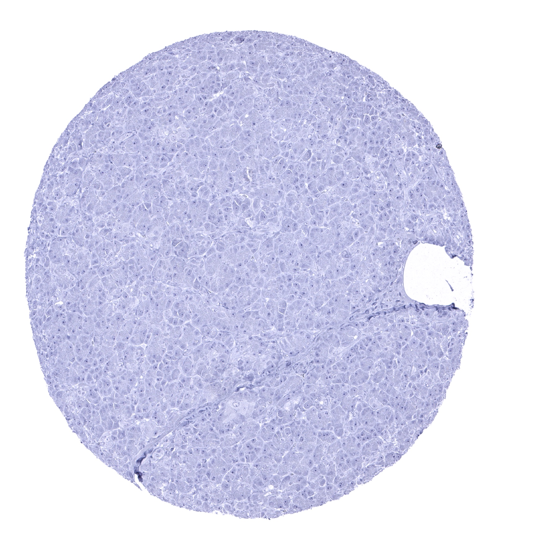
Liver
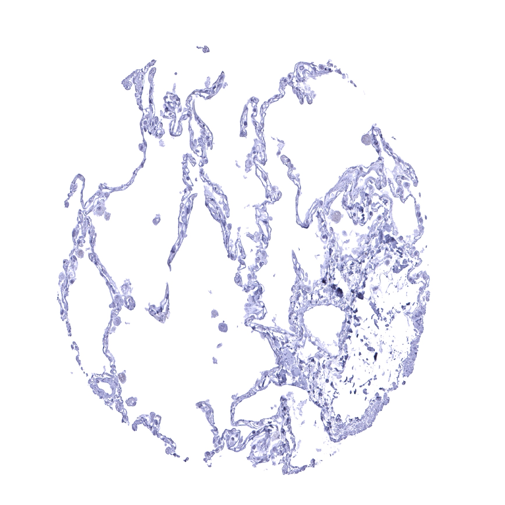
Lung
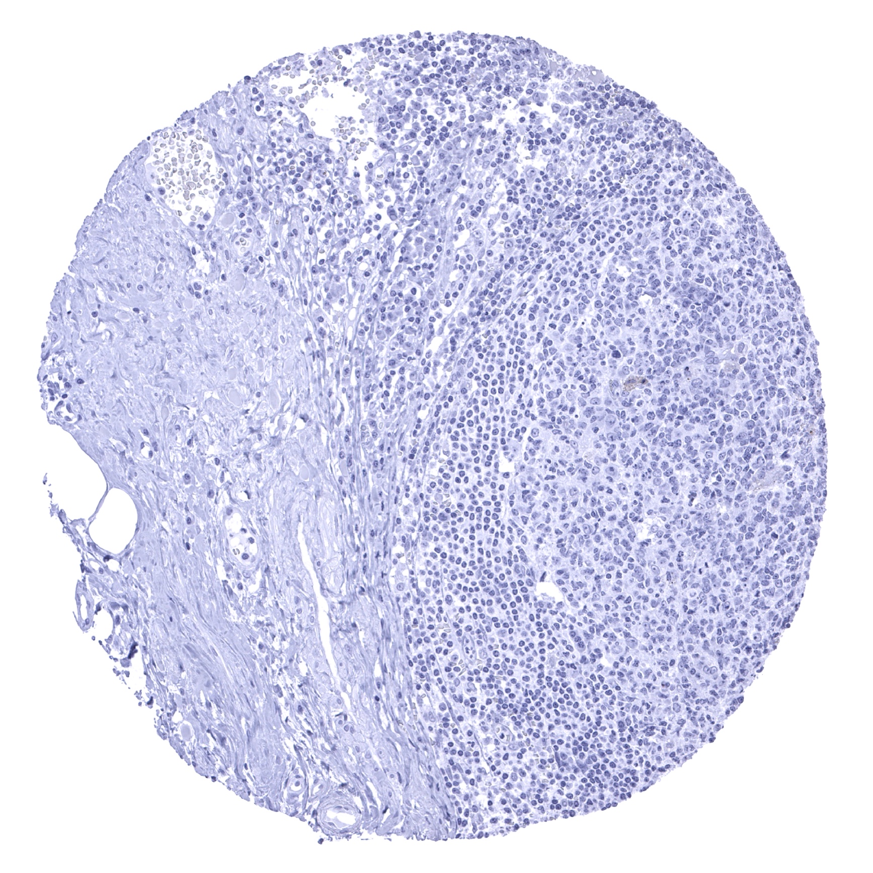
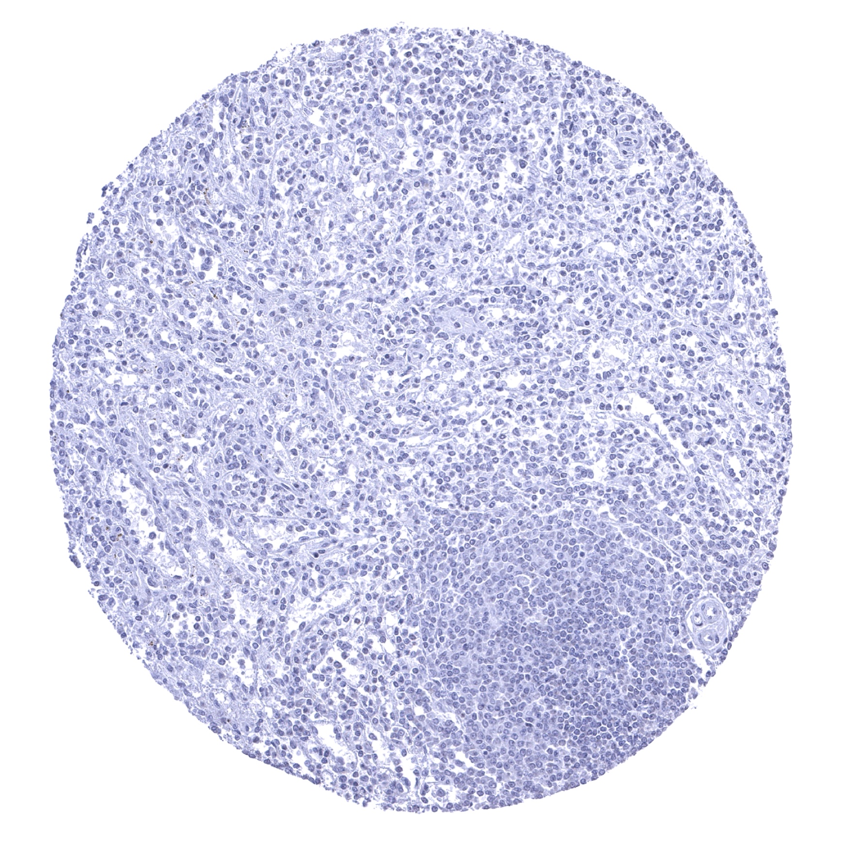
Lymph node
Ovary, stroma
Pancreas
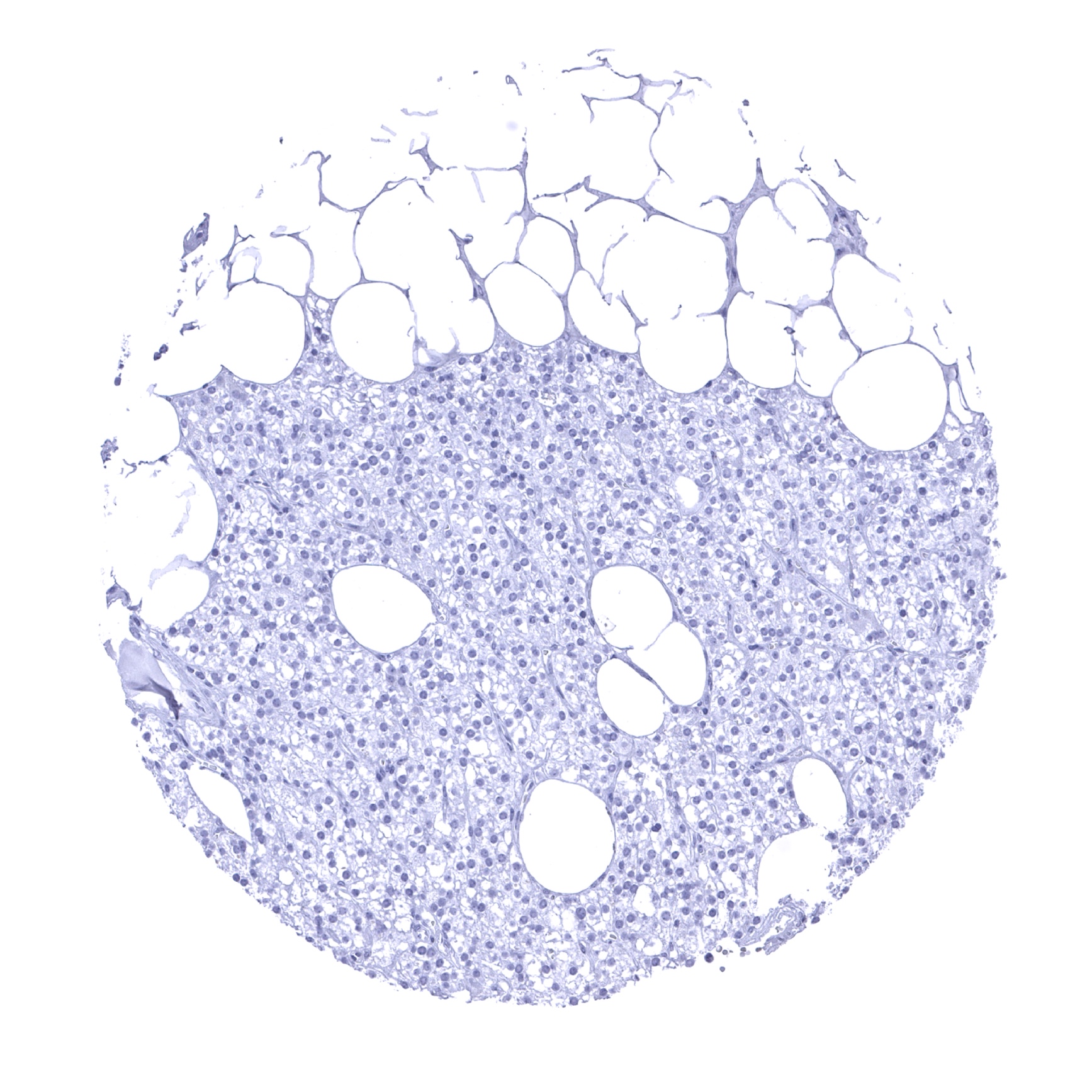
Parathyroid gland
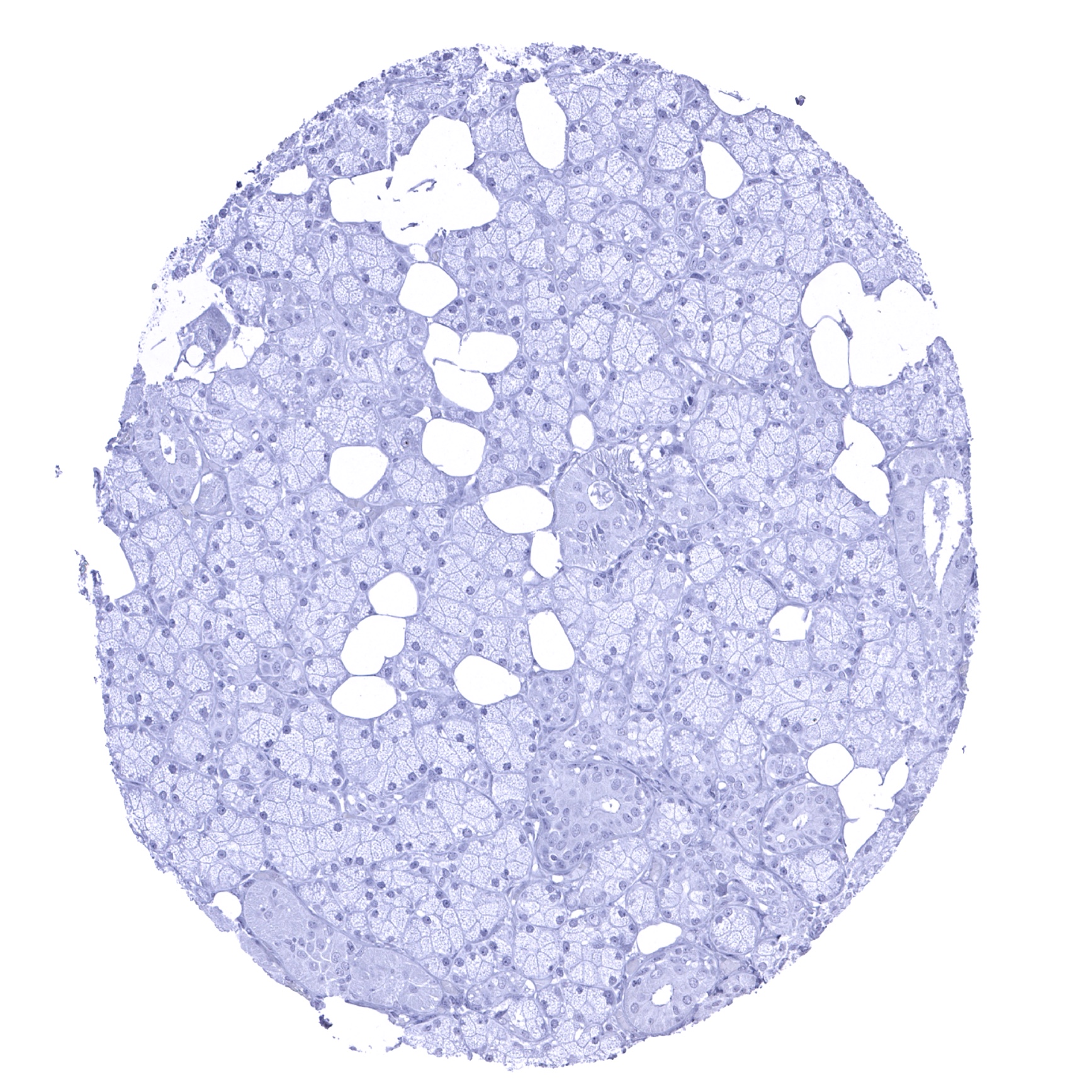
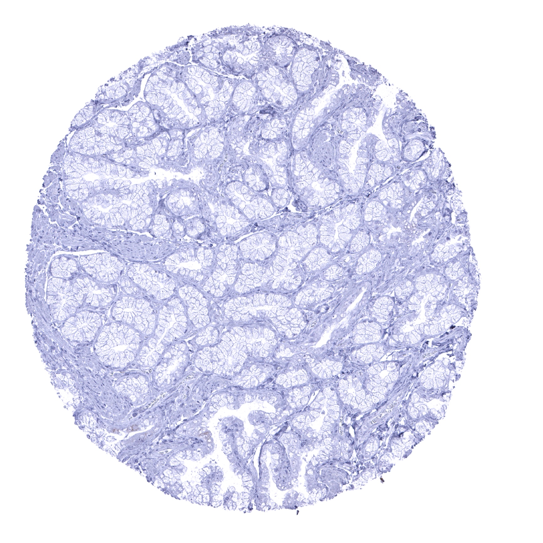
Parotid gland
Pituitary gland, anterior lobe
Pituitary gland, posterior lobe

Placenta (amnion and chorion)
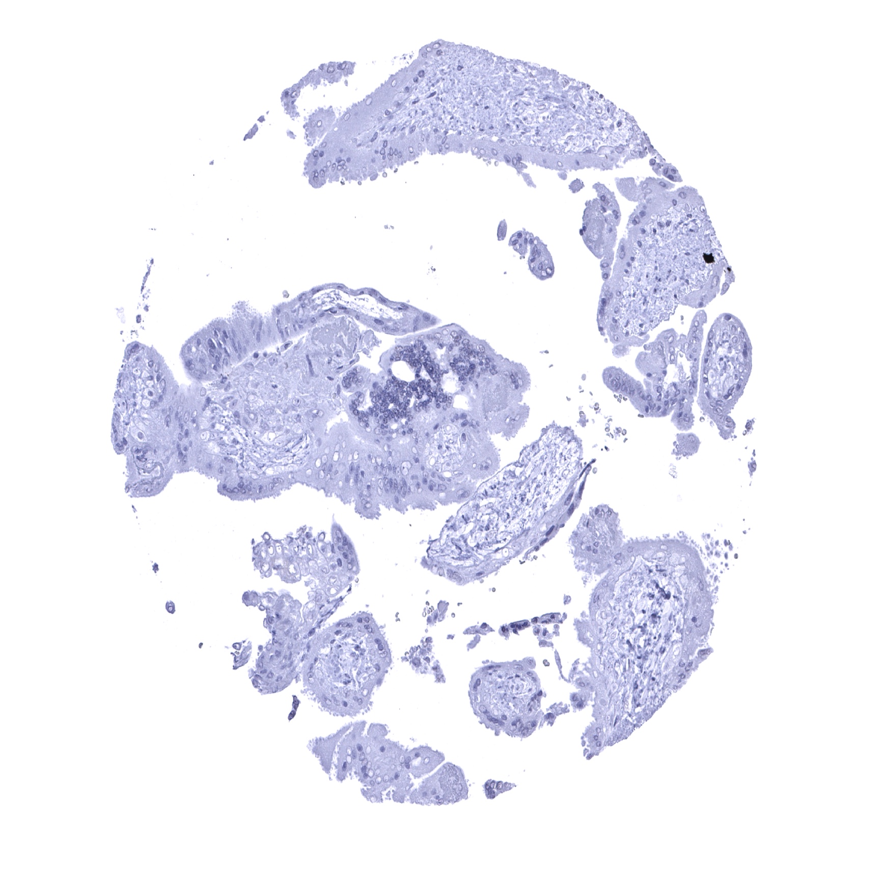
Placenta, early
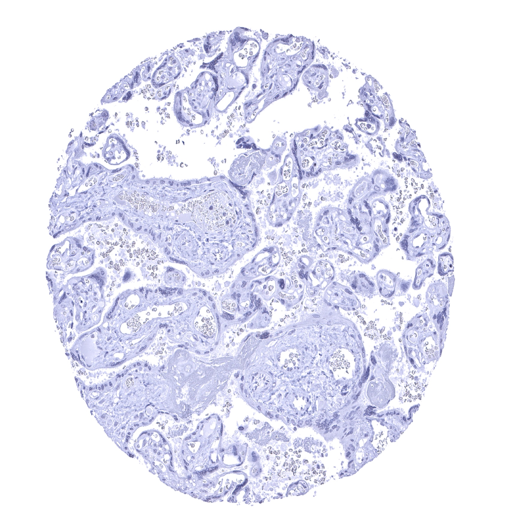
Placenta, mature
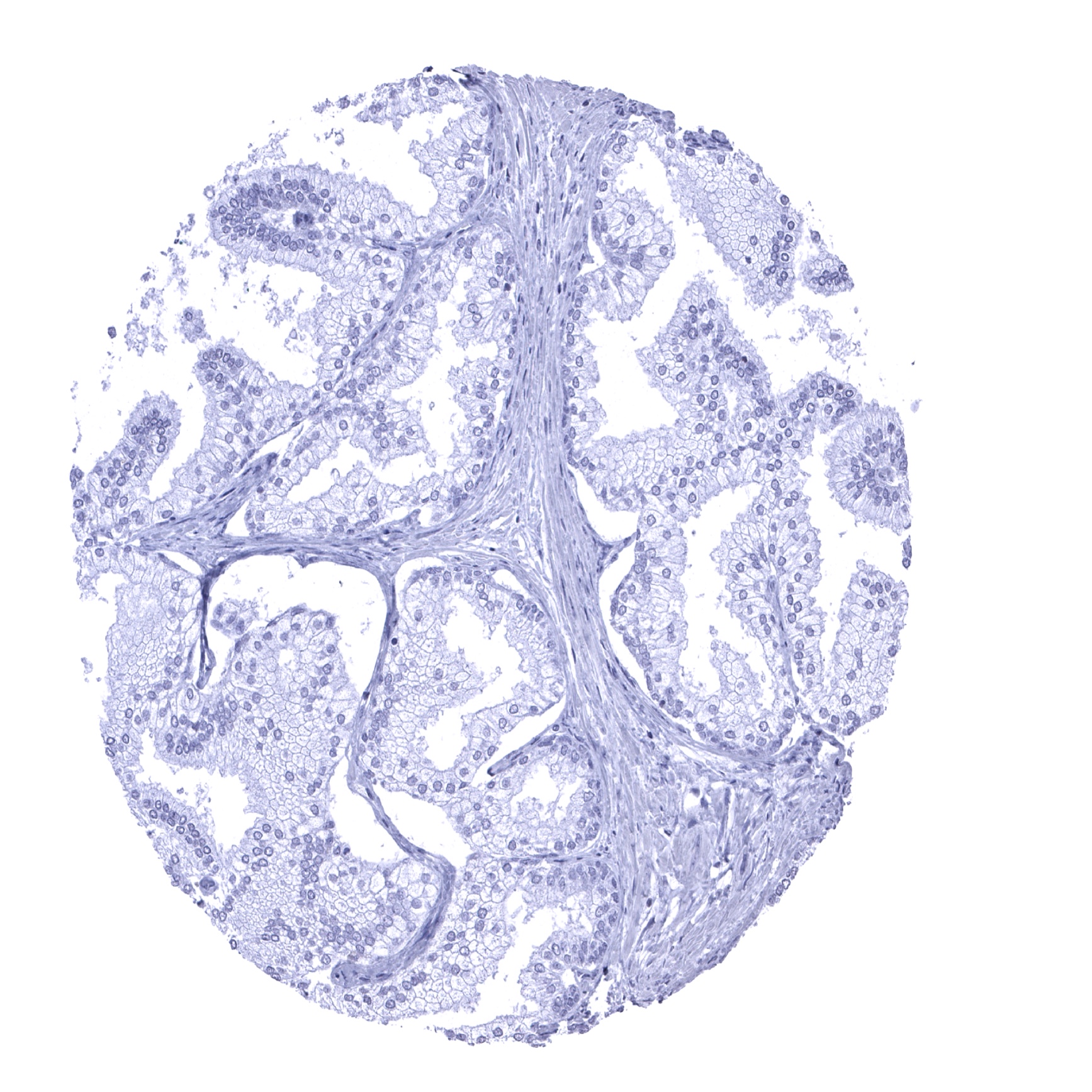
Prostate
Rectum, mucosa
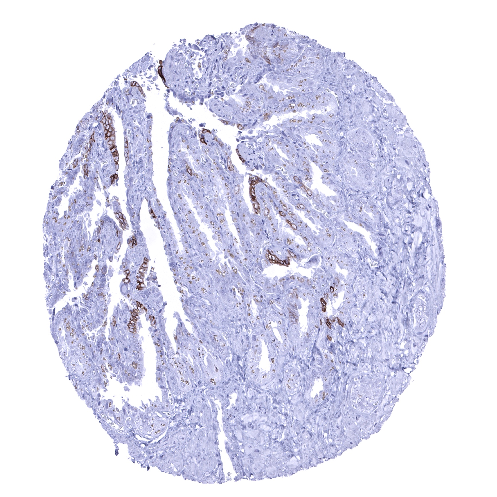
Seminal vesicle - A small fraction of epithelial cells, often arranged in groups, shows a moderate to strong membranous CDH16 staining (CDH16 immunohistochemistry).
Skeletal muscle
Skin
Spleen
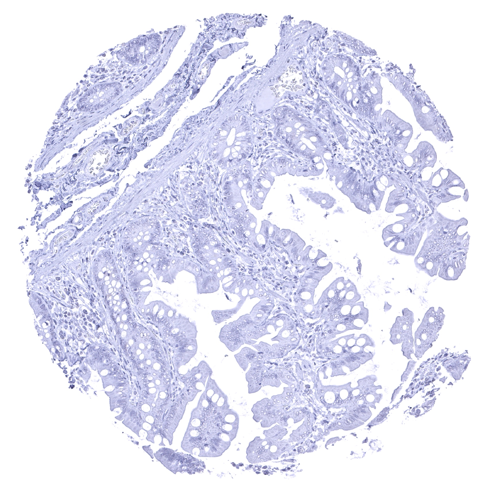
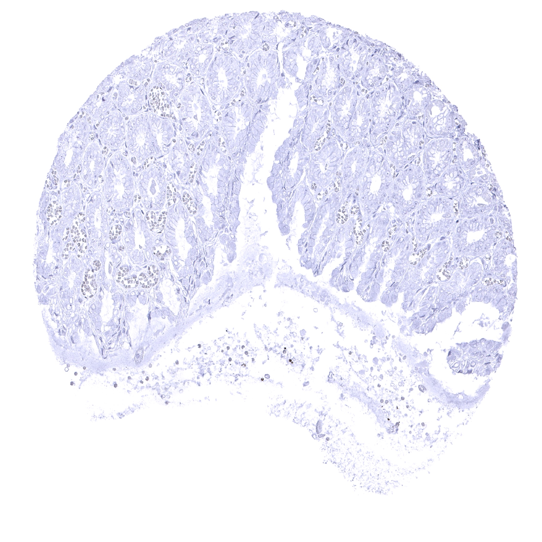
Stomach, antrum
Stomach, corpus
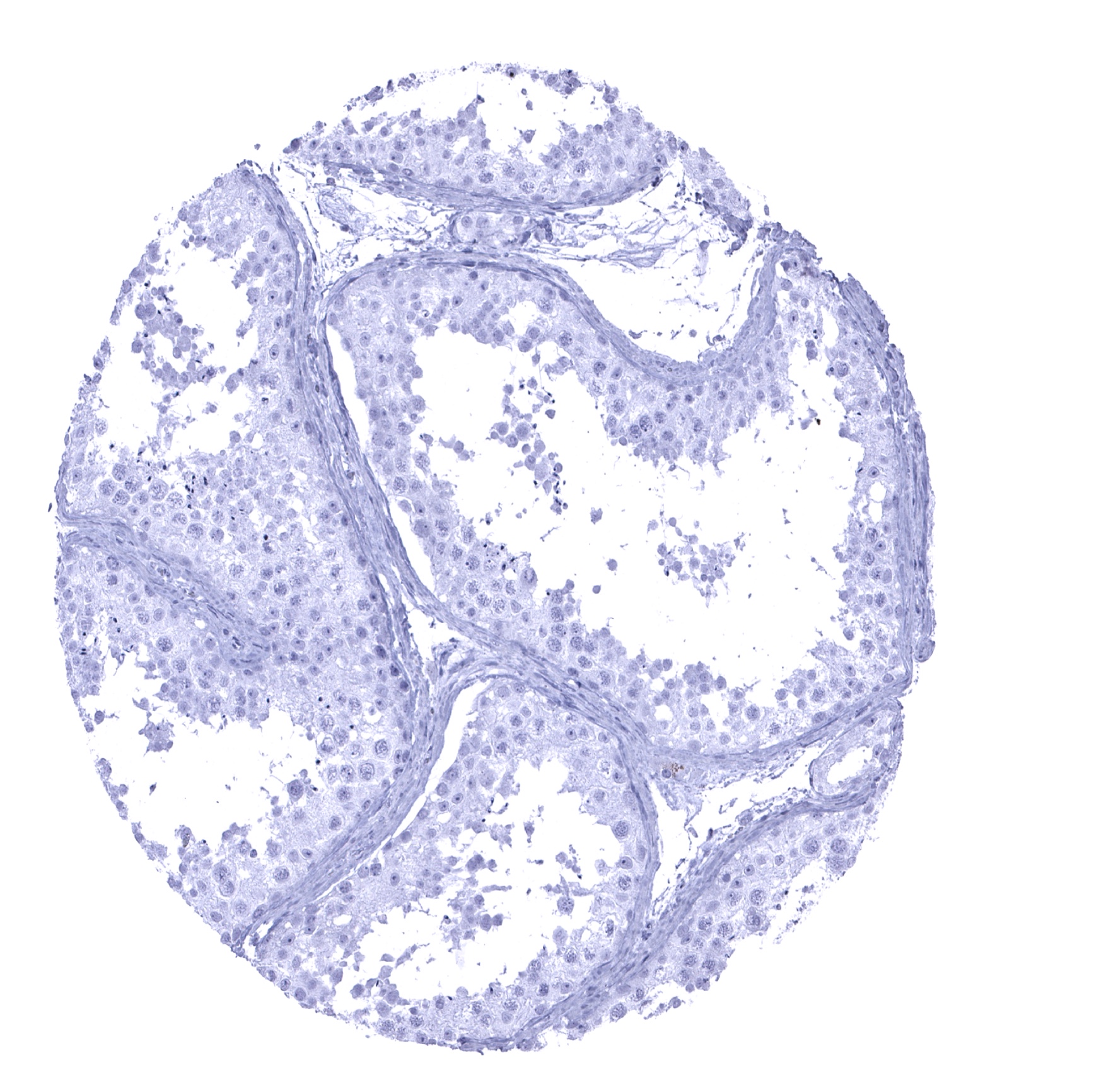
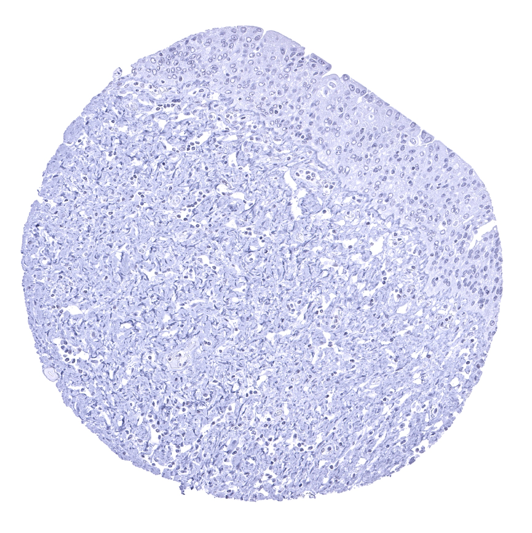
Testis

Thymus
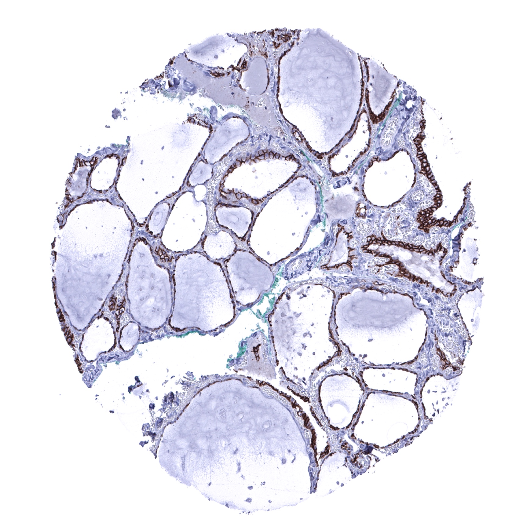
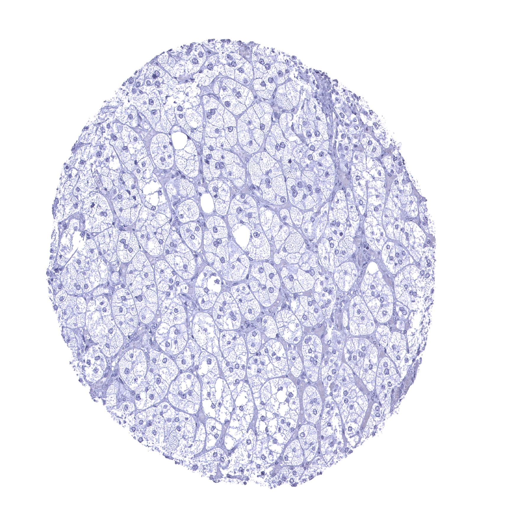
Thyroid gland - Strong membranous CDH16 staining of follicle cells (CDH16 immunohistochemistry).
Tonsil, surface epithelium
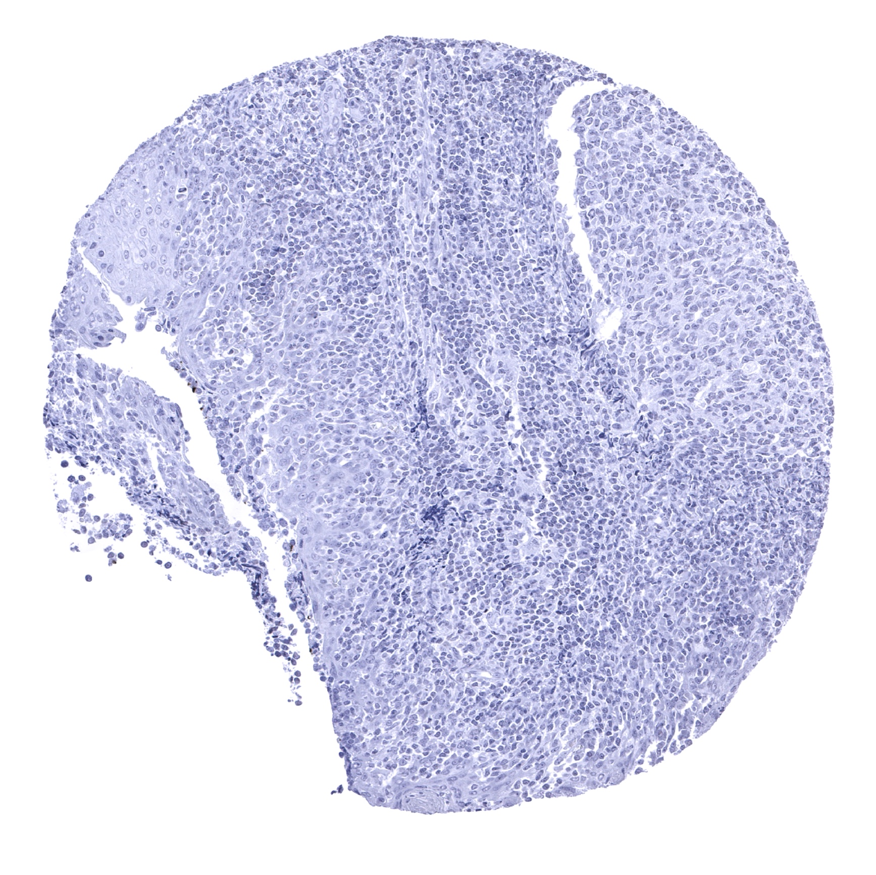
Tonsil
Urinary bladder, muscular wall
Urinary bladder, urothelium
Uterus, ectocervix

Uterus, endocervix

Uterus, endometrium (pregnancy)
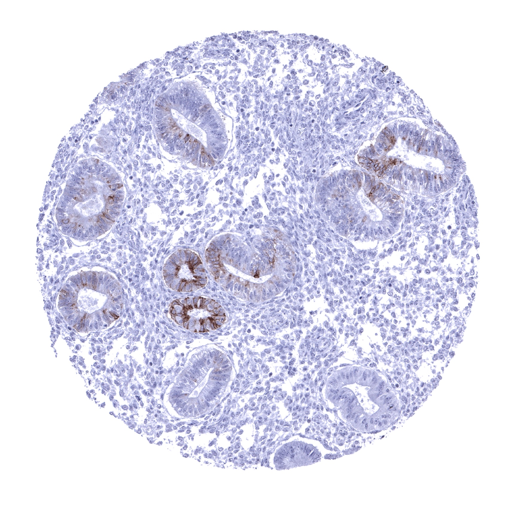
Uterus, endometrium (proliferation) -A focal membranous CDH16 staining of a fraction of glands can be seen in the endometrium (CDH16 immunohistochemistry).
Uterus, endometrium (secretion)

Uterus, myometrium