Login / Register
MS Validated Antibodies - Logo
  • About us
  • Products
    • Primary antibodies
    • Gallery
  • Distributors
  • Validation
    • Tissue Microarrays
  • Publications
    • Large Scale Studies
    • Further Publications
    • Events
    • Quiz
  • Contact
  • About us
  • Products
    • Primary antibodies
    • Gallery
  • Distributors
  • Validation
    • Tissue Microarrays
  • Publications
    • Large Scale Studies
    • Further Publications
    • Events
    • Quiz
  • Contact
×

Cancer Tissue Gallery PMEL / gp100 / Melanosome

Path Created with Sketch.
Go to PMEL / gp100 / Melanosome Normal Tissue GalleryGo back to Product Page
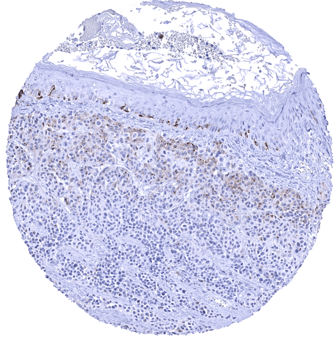
Skin - Benign nevus showing weak PMEL immunostaining in the superficial tumor layers while PMEL staining is lost in more mature cells of deeper tumor cell nests. Note: Strong PMEL positivity of melanocytes in the normal skin.
Skin – Benign nevus showing weak PMEL immunostaining in the superficial tumor layers while PMEL staining is lost in more mature cells of deeper tumor cell nests. Note: Strong PMEL positivity of melanocytes in the normal skin.
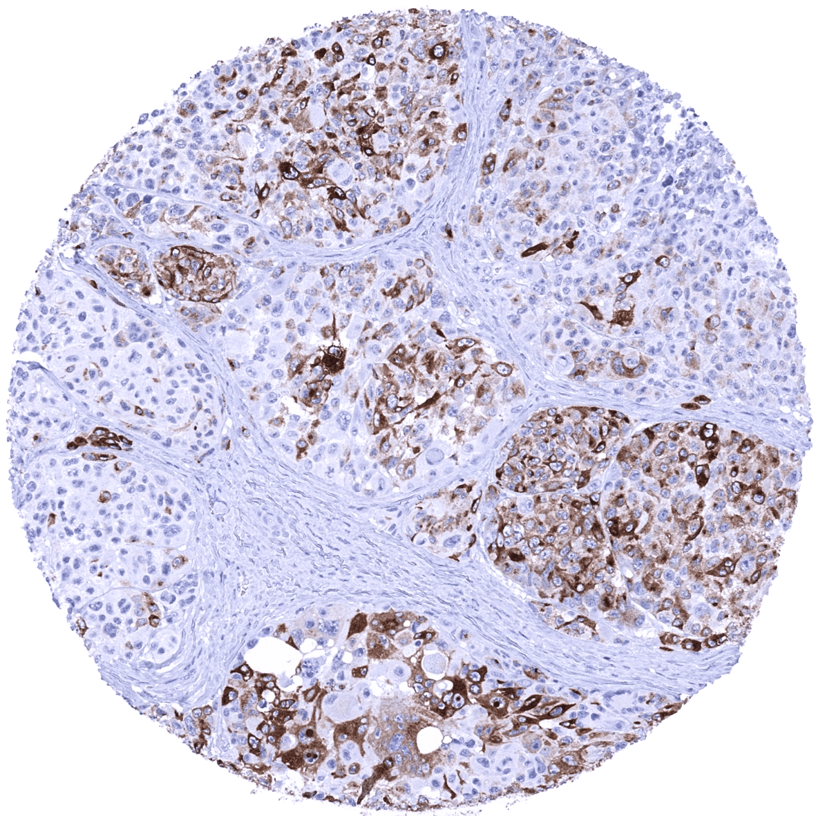
Skin - Malignant melanoma showing moderate to strong PMEL immunostaining in a fraction of tumor cells.
Skin – Malignant melanoma showing moderate to strong PMEL immunostaining in a fraction of tumor cells.
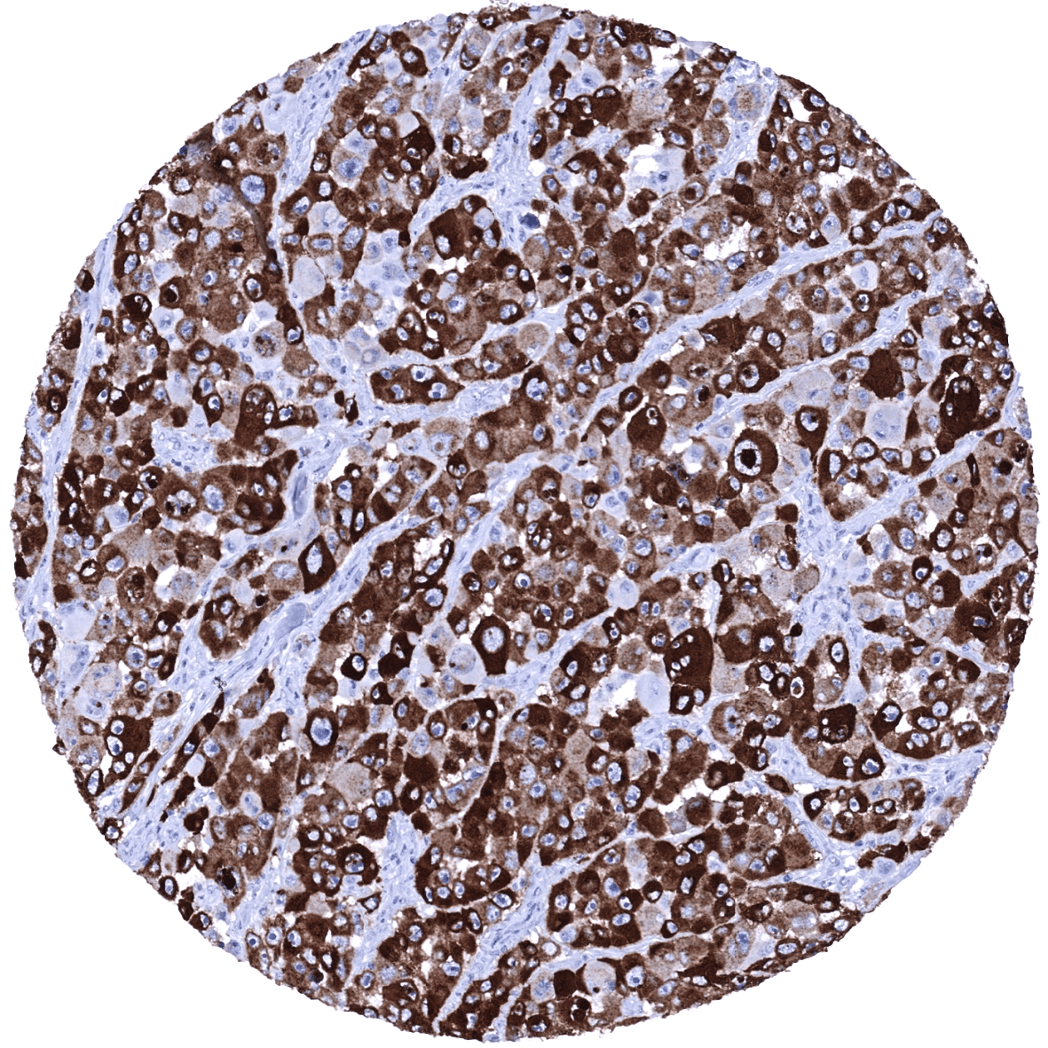
Skin - Malignant melanoma showing strong PMEL immunostaining in all tumor cells.
Skin – Malignant melanoma showing strong PMEL immunostaining in all tumor cells.
Skin - Malignant melanoma showing strong PMEL immunostaining in virtually all tumor cells.
Skin – Malignant melanoma showing strong PMEL immunostaining in virtually all tumor cells.
Skin - Malignant melanoma showing weak to moderate PMEL immunostaining in few tumor cells.
Skin – Malignant melanoma showing weak to moderate PMEL immunostaining in few tumor cells.
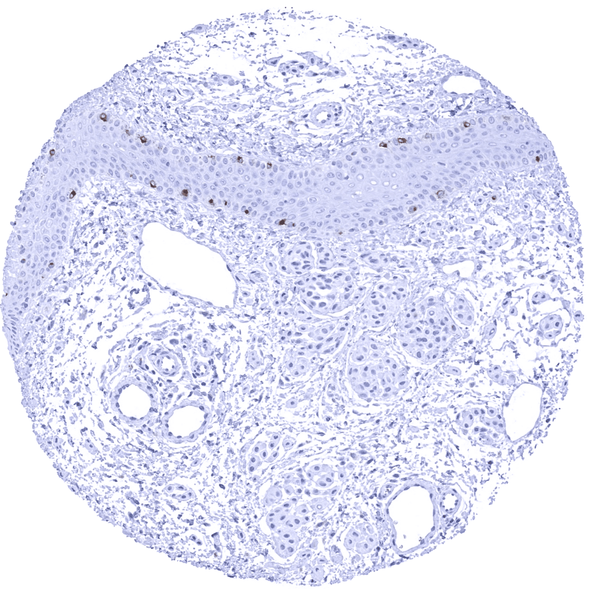
Skin - PMEL negative basal cell carcinoma containing few (non-neoplastic) PMEL positive melanocytes.
Skin – PMEL negative basal cell carcinoma containing few (non-neoplastic) PMEL positive melanocytes.
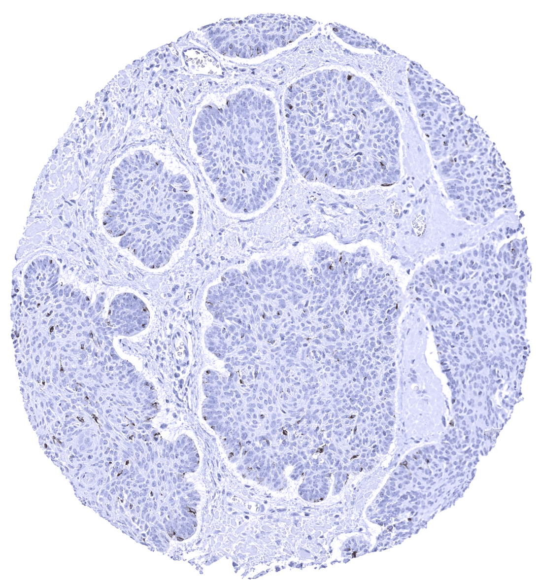
Skin - PMEL negative basal cell carcinoma containing numerous PMEL positive (non-neoplastic) melanocytes.
Skin – PMEL negative basal cell carcinoma containing numerous PMEL positive (non-neoplastic) melanocytes.
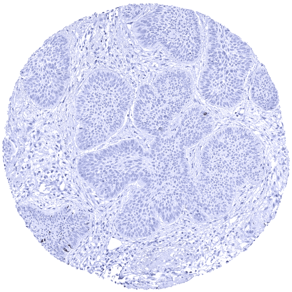
Skin - PMEL negative basal cell carcinoma.
Skin – PMEL negative basal cell carcinoma.
Skin - PMEL negative benign nevus.
Skin – PMEL negative benign nevus.
Skin - PMEL negative benign nevus. Note: Strong PMEL positivity of melanocytes in the normal skin.
Skin – PMEL negative benign nevus. Note- Strong PMEL positivity of melanocytes in the normal skin.
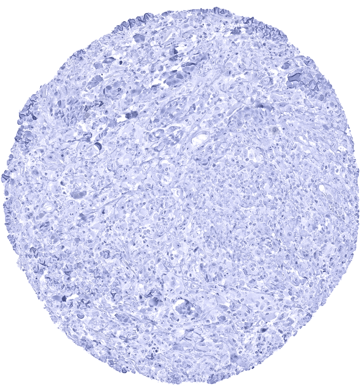
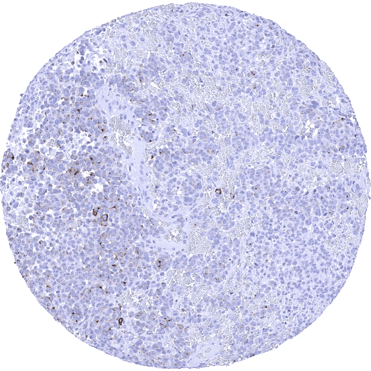
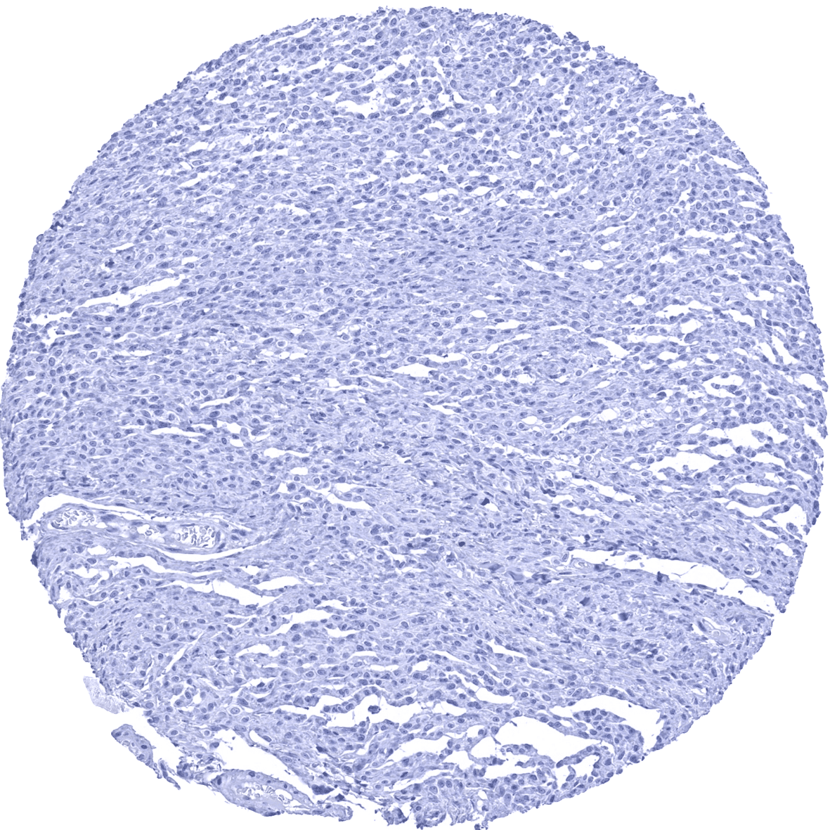
Skin - PMEL negative malignant melanoma.
Skin – PMEL negative malignant melanoma.
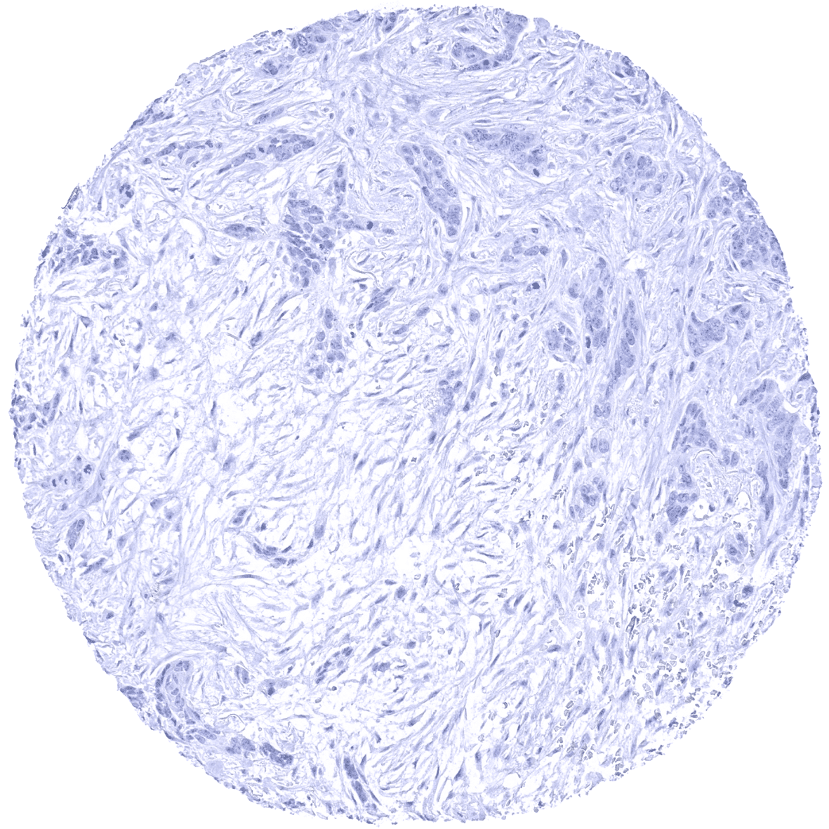
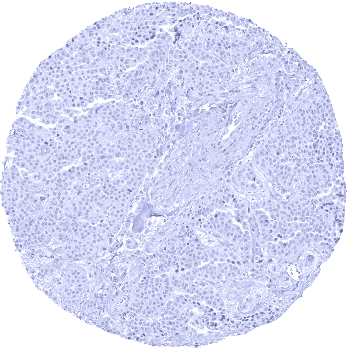
Skin - PMEL negative squamous cell carcinoma.
Skin – PMEL negative squamous cell carcinoma.
Thyroid - PMEL negative adenoma.
Thyroid – PMEL negative adenoma.
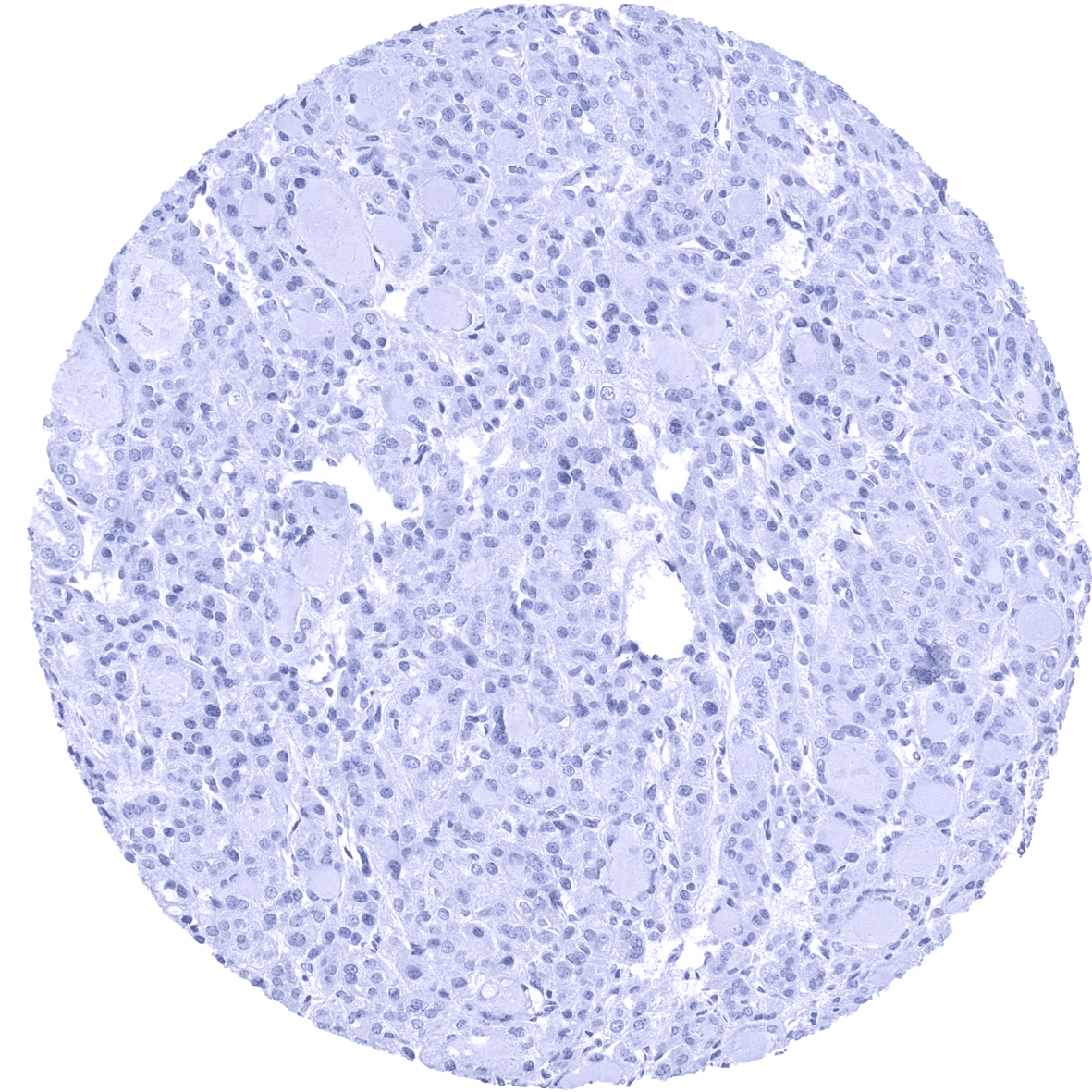
Thyroid - PMEL negative anaplastic carcinoma.
Thyroid – PMEL negative anaplastic carcinoma.
Thyroid - PMEL negative follicular carcinoma.
Thyroid – PMEL negative follicular carcinoma.
Thyroid - PMEL negative medullary carcinoma.
Thyroid – PMEL negative medullary carcinoma.
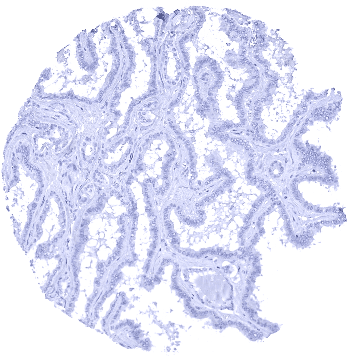
Thyroid - PMEL negative papillary carcinoma.
Thyroid- PMEL negative papillary carcinoma.
  • Linkedin
  • My Account
  • Search
    ×
  • Cart 0
© 2025 MS Validated Antibodies
  • Imprint
  • Privacy Policy
  • Distributors