Login / Register
MS Validated Antibodies - Logo
  • About us
  • Products
    • Primary antibodies
    • Gallery
  • Distributors
  • Validation
    • Tissue Microarrays
  • Publications
    • Large Scale Studies
    • Further Publications
    • Events
    • Quiz
  • Contact
  • About us
  • Products
    • Primary antibodies
    • Gallery
  • Distributors
  • Validation
    • Tissue Microarrays
  • Publications
    • Large Scale Studies
    • Further Publications
    • Events
    • Quiz
  • Contact
×

Cancer Tissue Gallery CD1a

Path Created with Sketch.
Go to CD1a Normal Tissue GalleryGo back to Product Page
Cervival lymph node with a diffuse large B-cell lymphoma showing CD1a positive dendritic cells.
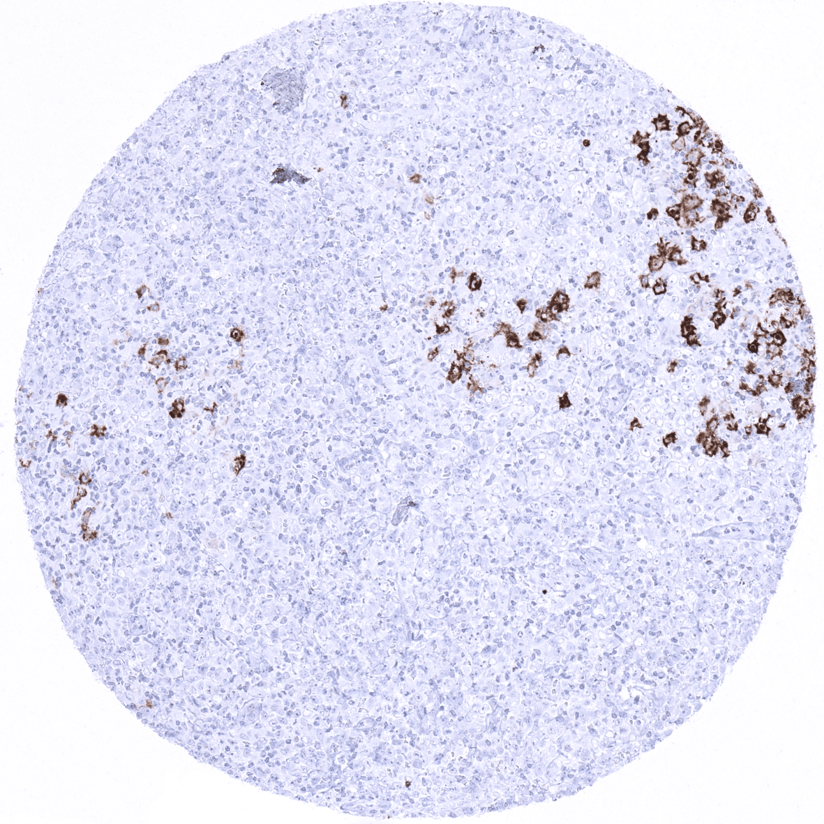
Diffuse large B-cell lymphoma (axillary lymph node) showing groups of CD1a positive dendritic cells.
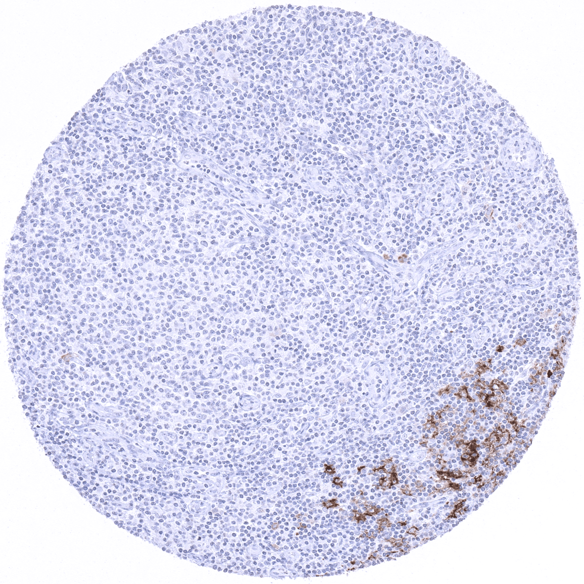
Follicular B-cell lymphoma in an inguinal lymph node showing scattered CD1a positive dendritic cells.
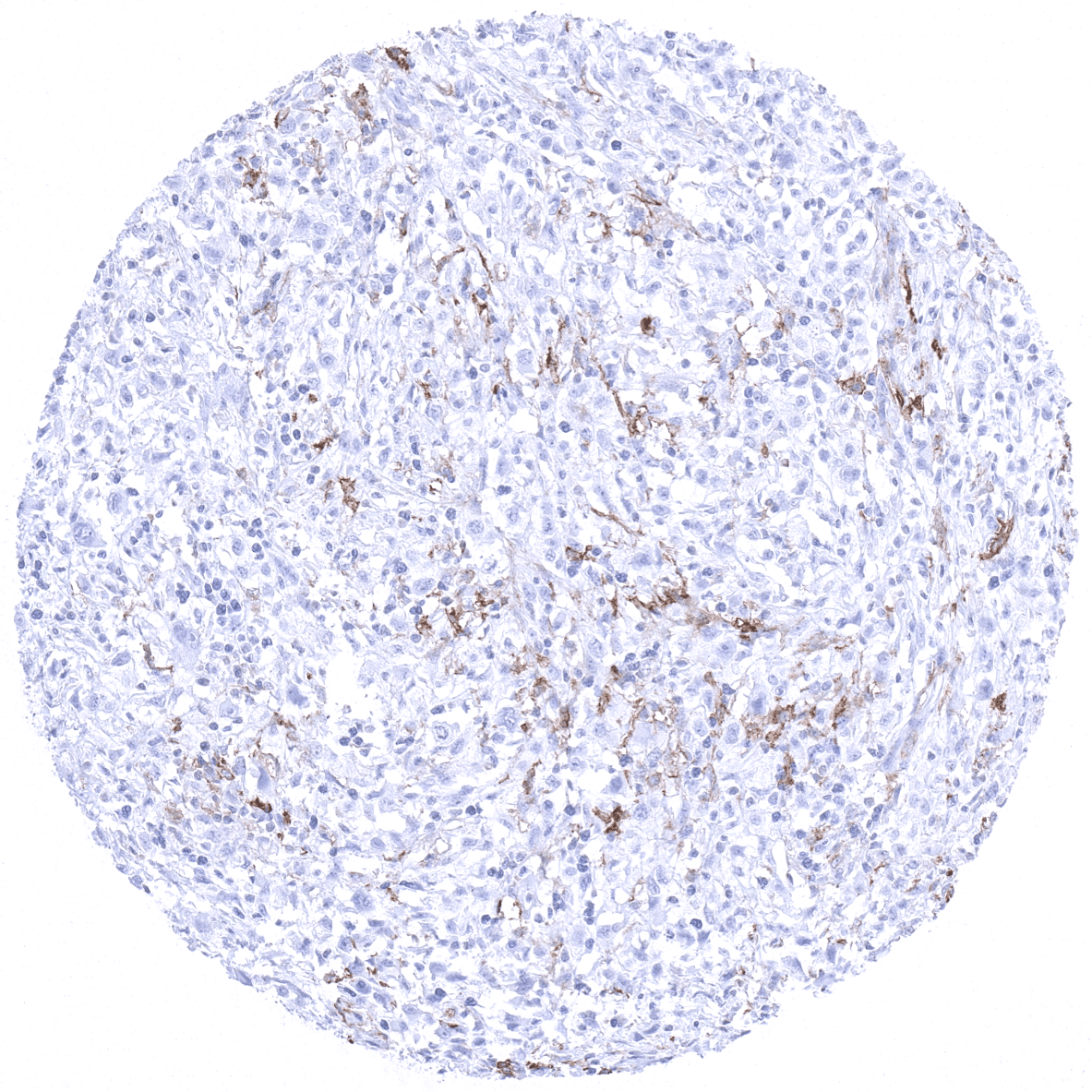
Hodgkin‘s lymphoma in a supraclavicular lymph node showing scattered CD1a positive dendritic cells.
Hodgkin‘s lymphoma in an inguinal lymph node showing scattered CD1a positive dendritic cells.
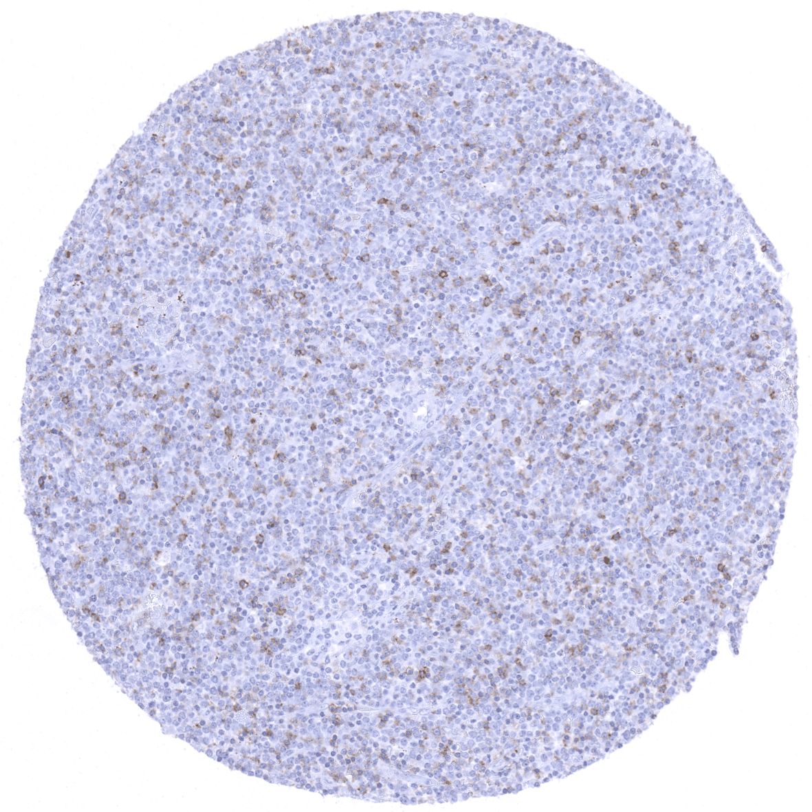
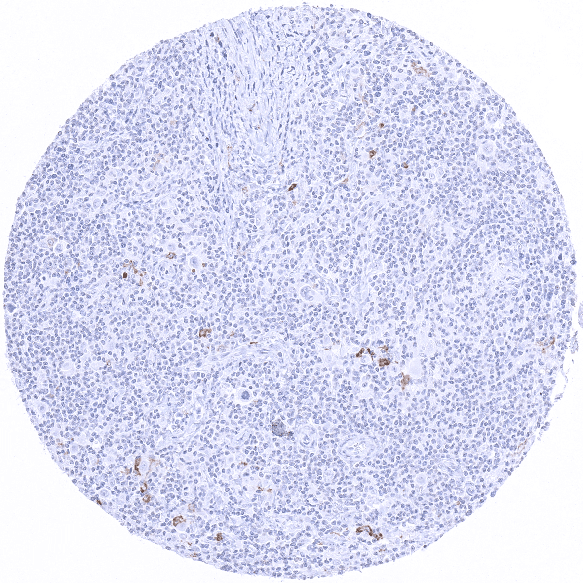
Lymphoblastic T-cell lymphoma showing weak to moderate CD1a immunostaining in a fraction of cells.
  • Linkedin
  • My Account
  • Search
    ×
  • Cart 0
© 2025 MS Validated Antibodies
  • Imprint
  • Privacy Policy
  • Distributors