195,00 € – 695,00 €
Product details
Synonyms = ENO2; ENOG; Enolase 2 gamma neuronal; Enolase2; Gamma-enolase; Neural enolase; Neuron specific gamma enolase; Neuron-specific enolase; NSE ; -phospho-D-glycerate hydrolyase
Antibody type = Mouse monoclonal / IgG1, kappa
Clone = MSVA-451M
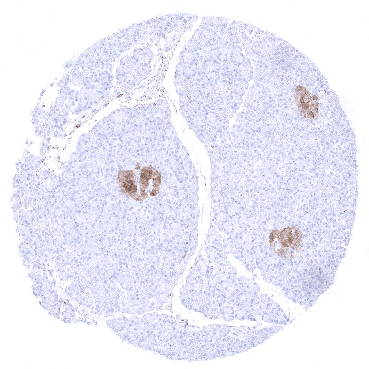
Positive control = In the colon, axons and ganglion cells in lamina propria and muscular wall must show at least a moderate NSE staining, while epithelial and lymphatic cells remain negative.
Negative control =In the colon, epithelial and lymphatic cells do not show NSE staining.
Cellular localization = Cytoplasmic
Reactivity = Human
Application = Immunohistochemistry
Dilution = 1:100 – 1:200
Intended Use = Research Use Only
Relevance of Antibody
Biology Behind
Neuron specific enolase (NSE), is a 78 kDa phosphopyruvate hydratase encoded by the ENO2 (enolase 2) gene at 12p13.31. NSE is a glycolytic enzyme involved in the energy-generating process of the cell. It catalyzes the conversion of 2-phosphoglycerate to phosphoenolpyruvate Gamma-enolase. NSE gamma is largely tissue specific. Ontogenetically, NSE gamma appears in the final stages of neuronal differentiation and can thus be utilized as a marker for nerve cell maturation. Immunohistochemical detection of NSE with antibodies can be used to identify neuronal cells and cells with neuroendocrine differentiation. NSE is also a commonly used serum marker for the monitoring of cancer patients. NSE is being non-specifically discharged from tumor cells to the blood as a consequence of cell death. Its blood level is elevated in various NSE expressing cancers.
Staining Pattern in Normal Tissues
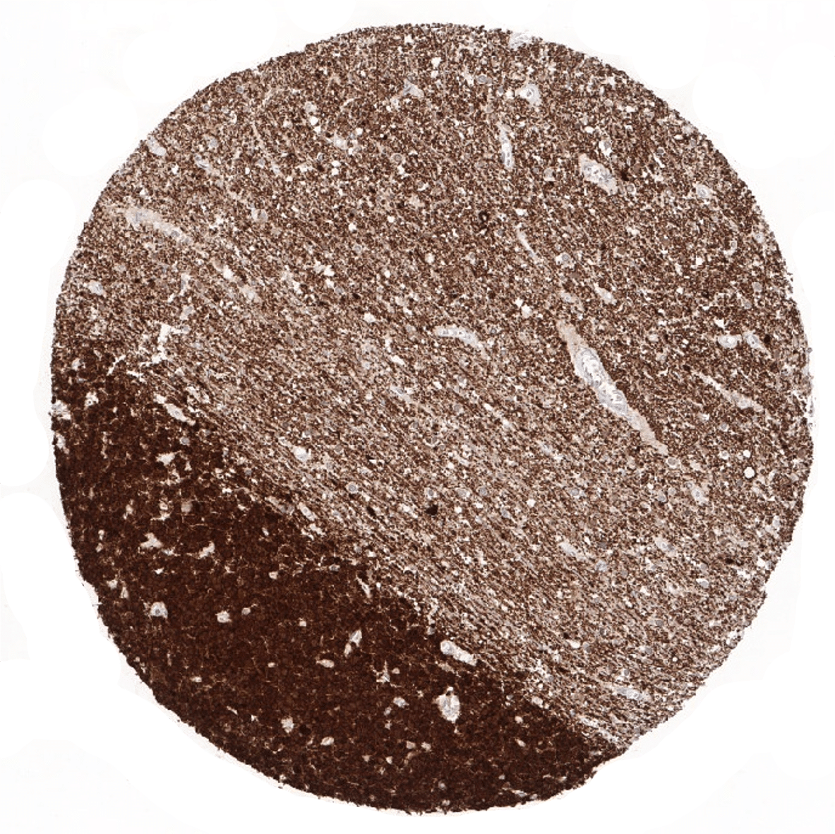
NSE is most strongly expressed in neuronal cells of the brain where it is also seen in all nerve fibres. An at least weak to moderate immunostaining is seen in axons and ganglion cells of the peripheral nerves which are particularly frequent in the gastrointestinal wall and in the seminal vesicle. NSE shows moderate to strong expression in the medulla but not the cortex of the adrenal gland. NSE is only rarely detectable in a small fraction of cells of the diffuse neuroendocrine system. Using MSVA-451M, NSE immunostaining is also seen in chorion cells of the placenta (moderate), few Sertoli cells (weak), some endothelial cells in the stomach (weak), and in few cells of the adenohypophysis (moderate).
These findings are largely consistent with RNA and protein data summarized in the Human Protein Atlas (Tissue expression NSE gamma).
Suggested positive tissue control: In the colon, axons and ganglion cells in lamina propria and muscular wall must show at least a moderate NSE staining, while epithelial and lymphatic cells remain negative.
Suggested negative tissue control: In the colon, epithelial and lymphatic cells do not show NSE staining.


Staining Pattern in Relevant Tumor Types
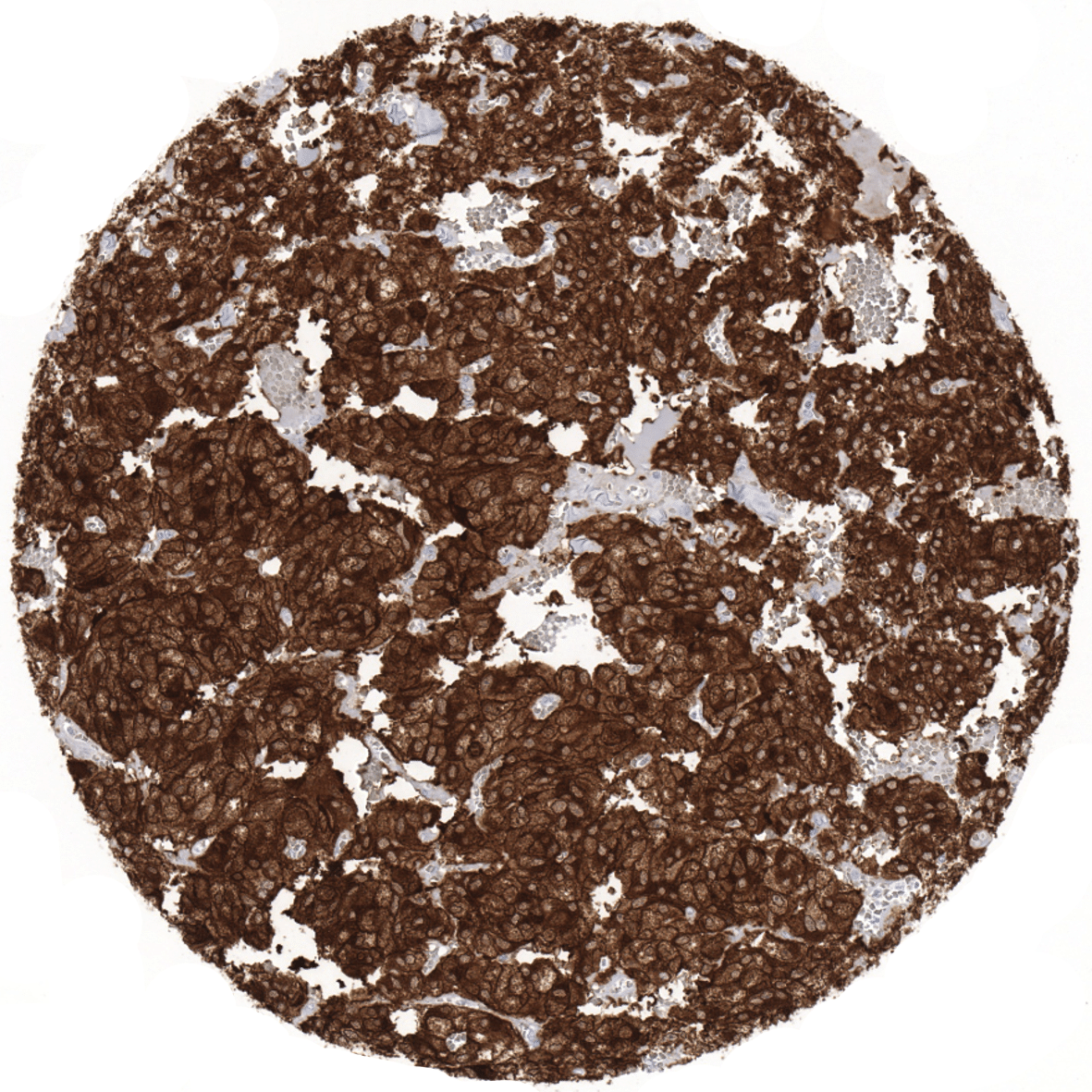
NSE is expressed in a large fraction of neuroendocrine tumors and carcinomas including small cell neuroendocrine tumors irrespective of their origins, Merkel cell carcinoma, medullary thyroid cancer, pheochromocytoma and granular cell tumors. Renal clear cell carcinoma is the most commonly NSE positive non-neuroendocrine cancer, followed by seminoma. Many other tumor types can show NSE immunostaining in some instances, however.
The TCGA findings on NSE gamma RNA expression in different tumor categories have been summarized in the Human Protein Atlas.


Compatibility of Antibodies
No data available at the moment
Protocol Recommendations
IHC users have different preferences on how the stains should look like. Some prefer high staining intensity of the target stain and even accept some background. Others favor absolute specificity and lighter target stains. Factors that invariably lead to more intense staining include higher concentration of the antibody and visualization tools, longer incubation time, higher temperature during incubation, higher temperature and longer duration of the heat induced epitope retrieval (slide pretreatment). The impact of the pH during slide pretreatment has variable effects and depends on the antibody and the target protein. Accordingly, multiple different protocols can generate identical staining results.
All images and data shown here and in our knowledge center are obtained by the manual protocol described below. Other protocols resulting in equivalent staining are described as well.
-Manual protocol
Freshly cut sections should be used (less than 10 days between cutting and staining). Heat-induced antigen retrieval for 5 minutes in an autoclave at 121°C in pH 9 Target Retrieval Solution buffer. Apply MSVA-451M at a dilution of 1:150 at 37°C for 60 minutes.
-Impact of pH


The strongest NSE gamma staining by MSVA-451M is obtained at a pH 9,0. However, pH 7,8 results in only a slight reduction of the staining intensity as compared to pH9. We thus consider pH7,8 as optimal for manual staining because of the better tissue preservation at pH7,8 than at pH 9,0.
Potential Research Applications
- A comprehensive study analyzing NSE expression in various different tumor entities would be helpful to assess the diagnostic significance of NSE IHC.
- The prognostic and clinical role of NSE expression should be further evaluated.
- The pattern of NSE immunostaining may be instrumental for postmortem assessment of brain damage.
Evidence for Antibody Specificity in IHC
Specificity of MSVA-451M is documented by strong positive staining in cell types that are well documented to express NSE such brain and medullary adrenal gland cells as well as neuronal structures in the gastrointestinal tract and absence of staining in all tissues known to not express NSE including tissues notorious for non-specific IHC background such as kidney, colonic mucosa epithelium, and epidermis.
