295,00 € – 995,00 €
Product details
Synonyms = Transcription factor SOX-10, DOM; PCWH; SOX10; SRY (sex determining region Y) box 10; SRY box containing gene 10; SRY related HMG box gene 10; Waardenburg syndrome type 2E(WS2E); WS4; Waardenburg syndrome type 4C (WS4C)
Antibody type = Recombinant Rabbit monoclonal / IgG
Clone = MSVA-710R
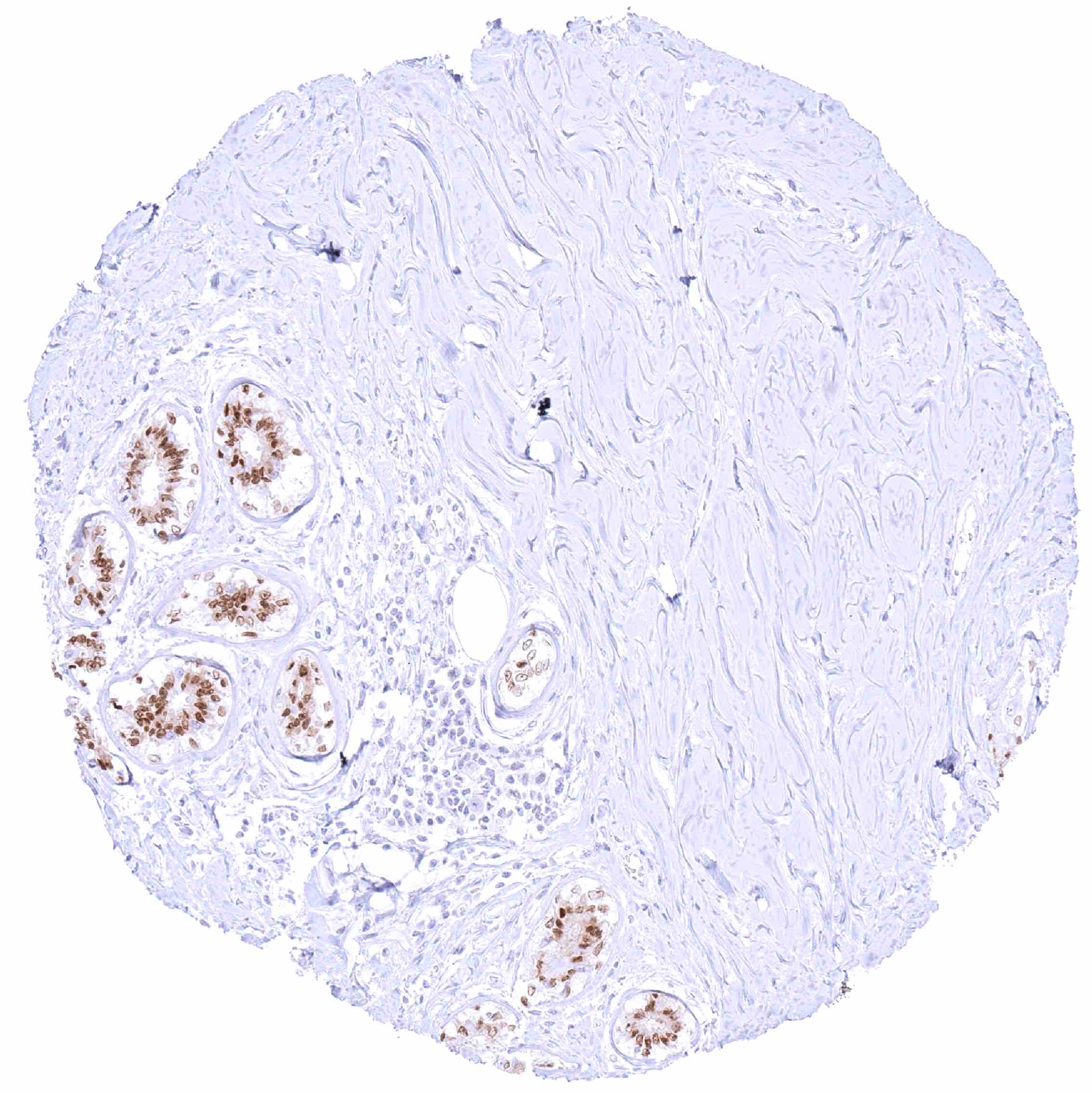
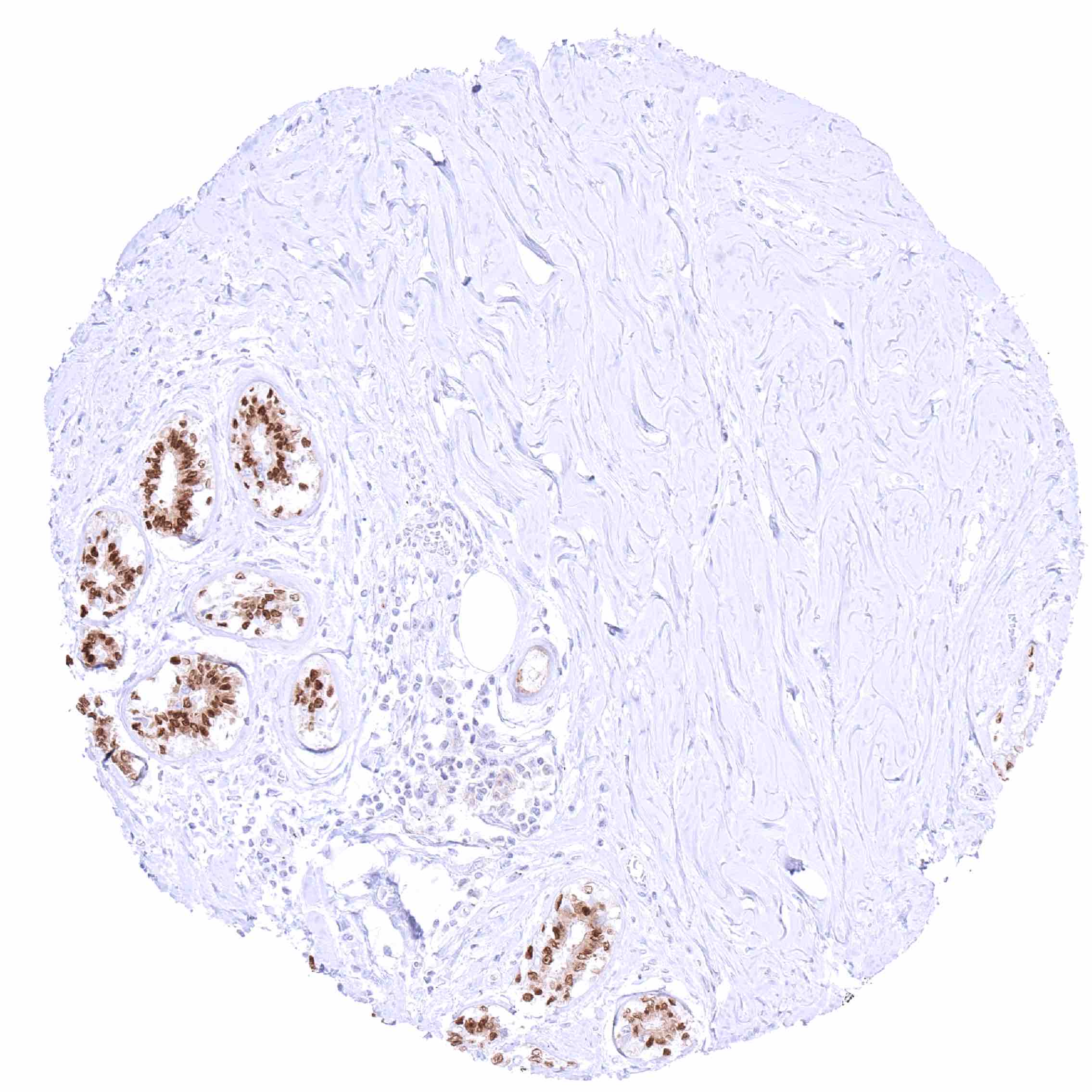
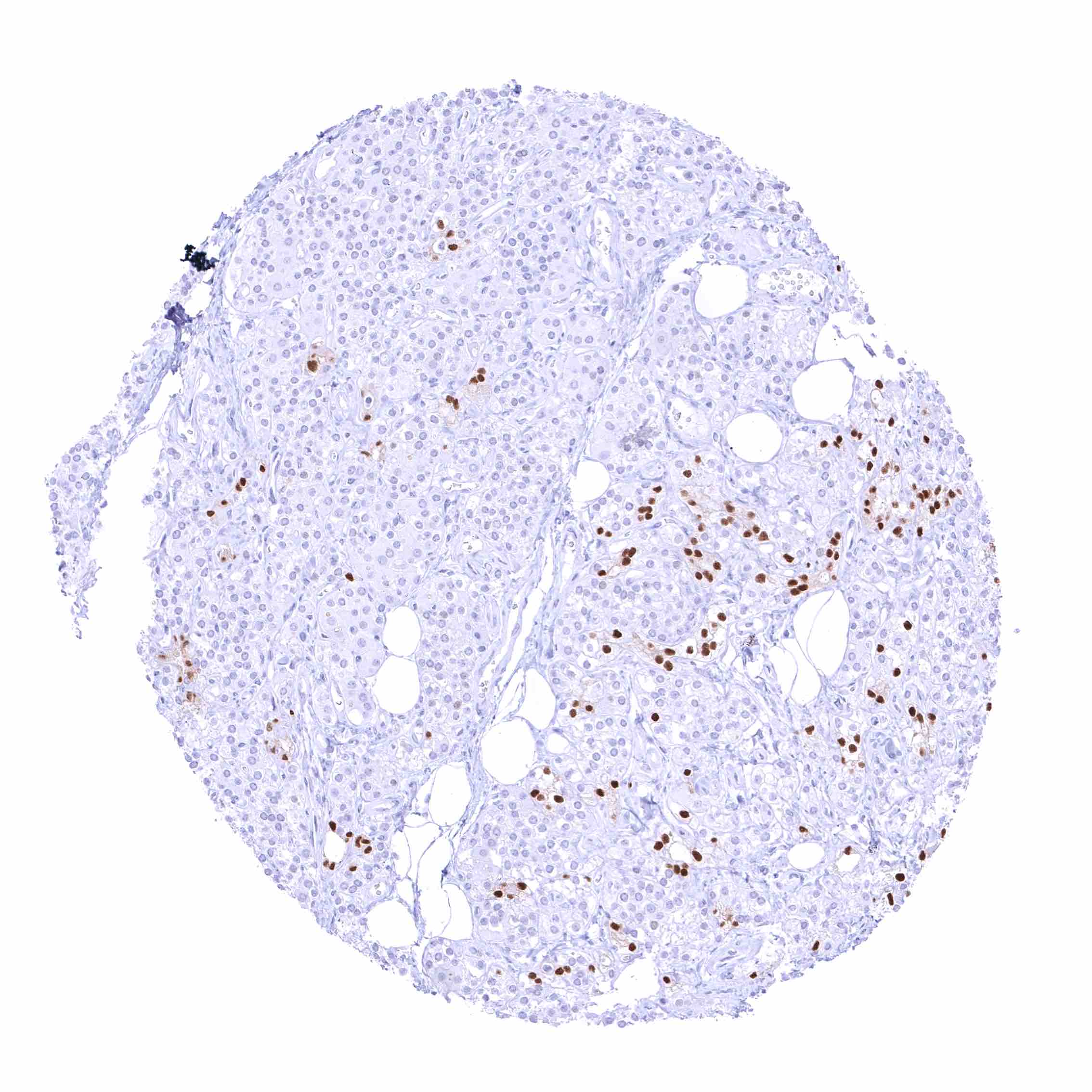
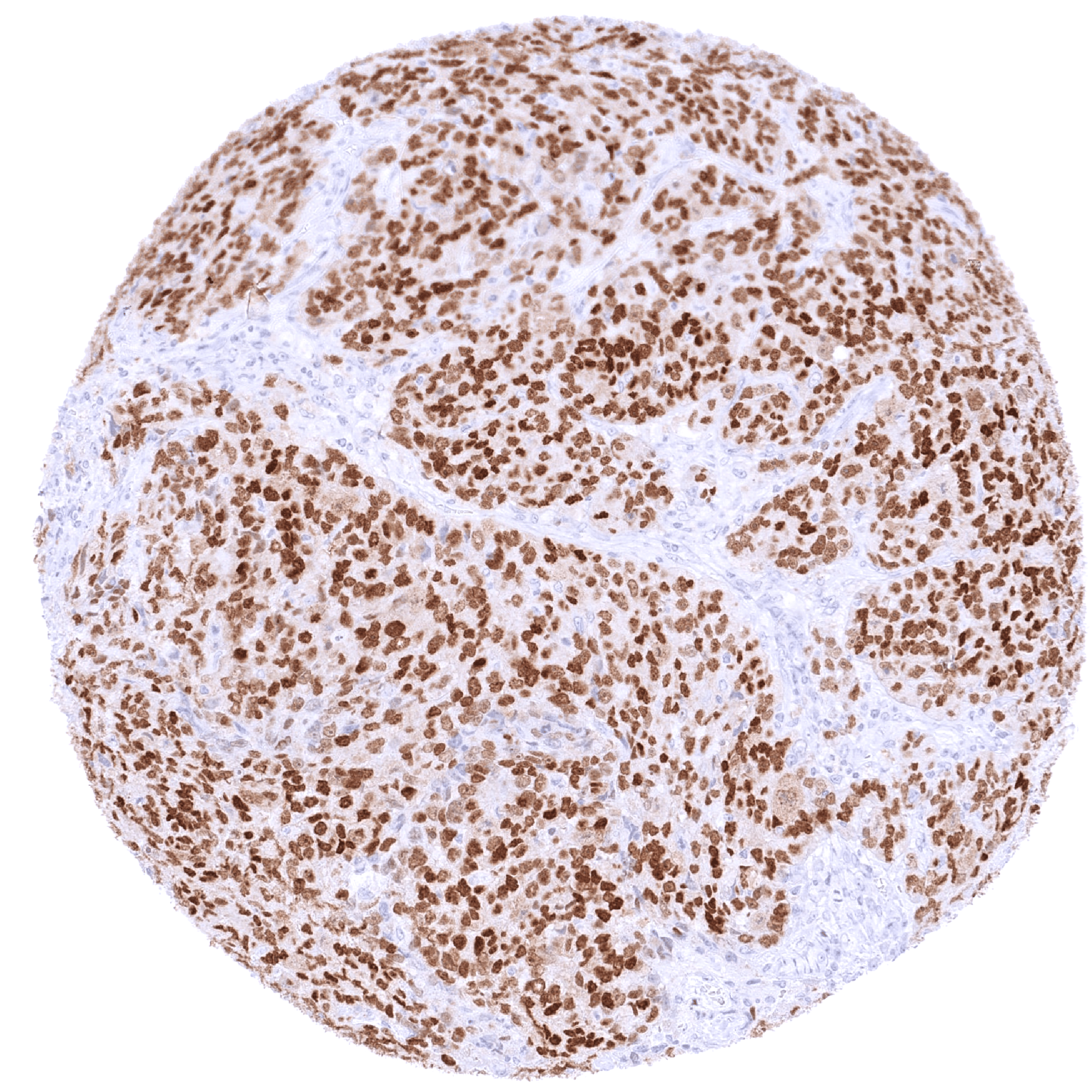
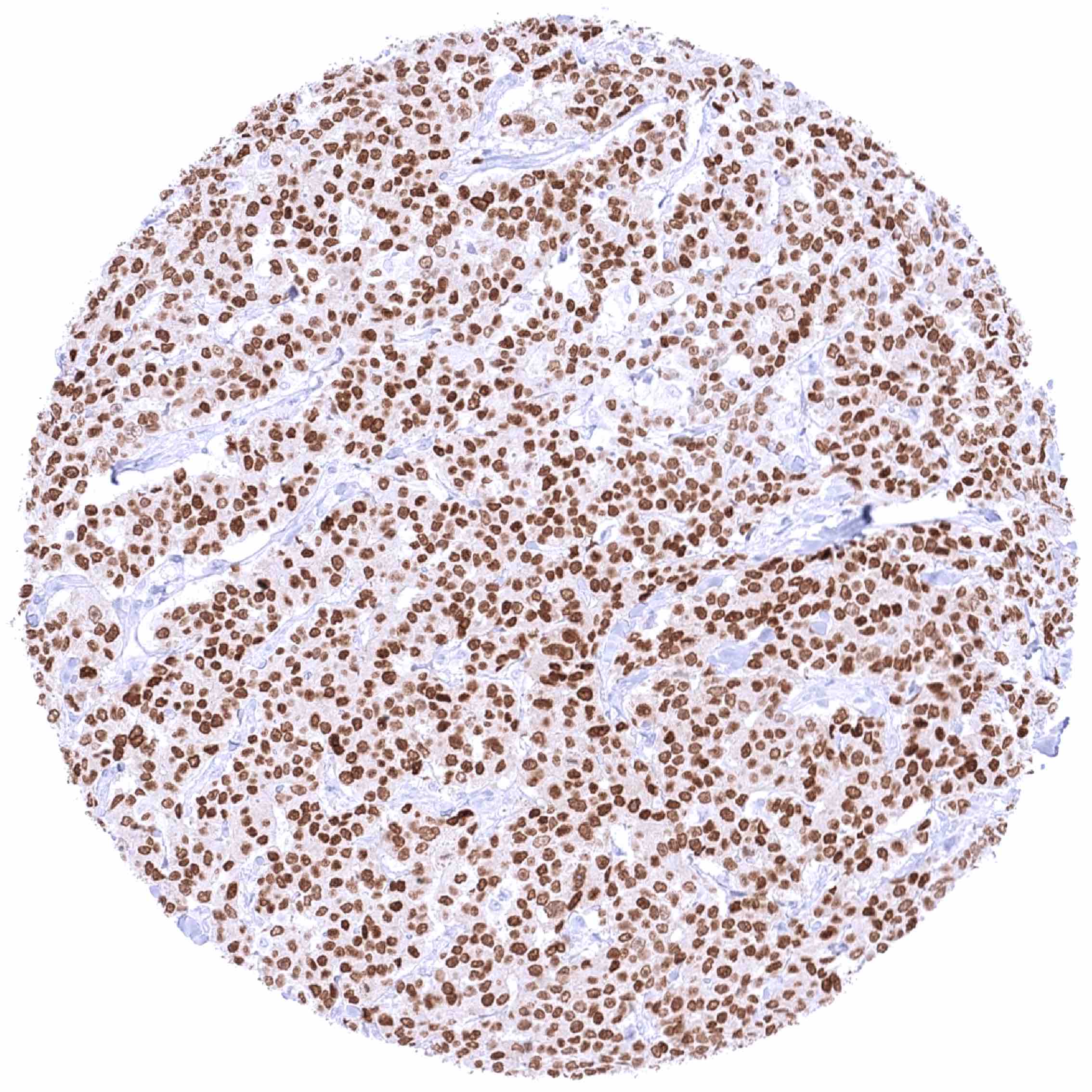
Positive control = Breast: A moderate to strong nuclear SOX10 staining should be seen in breast epithelial cells.
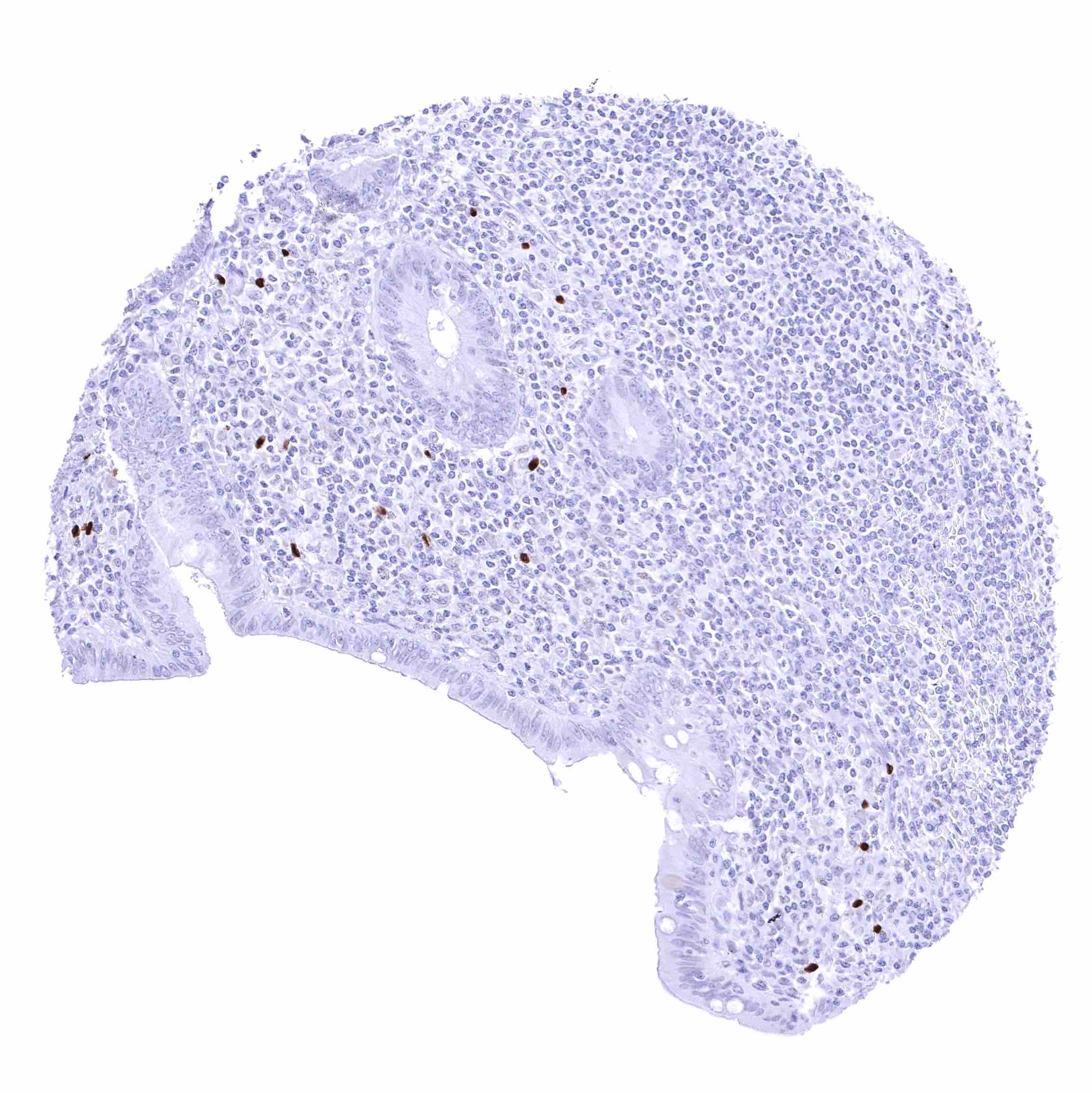
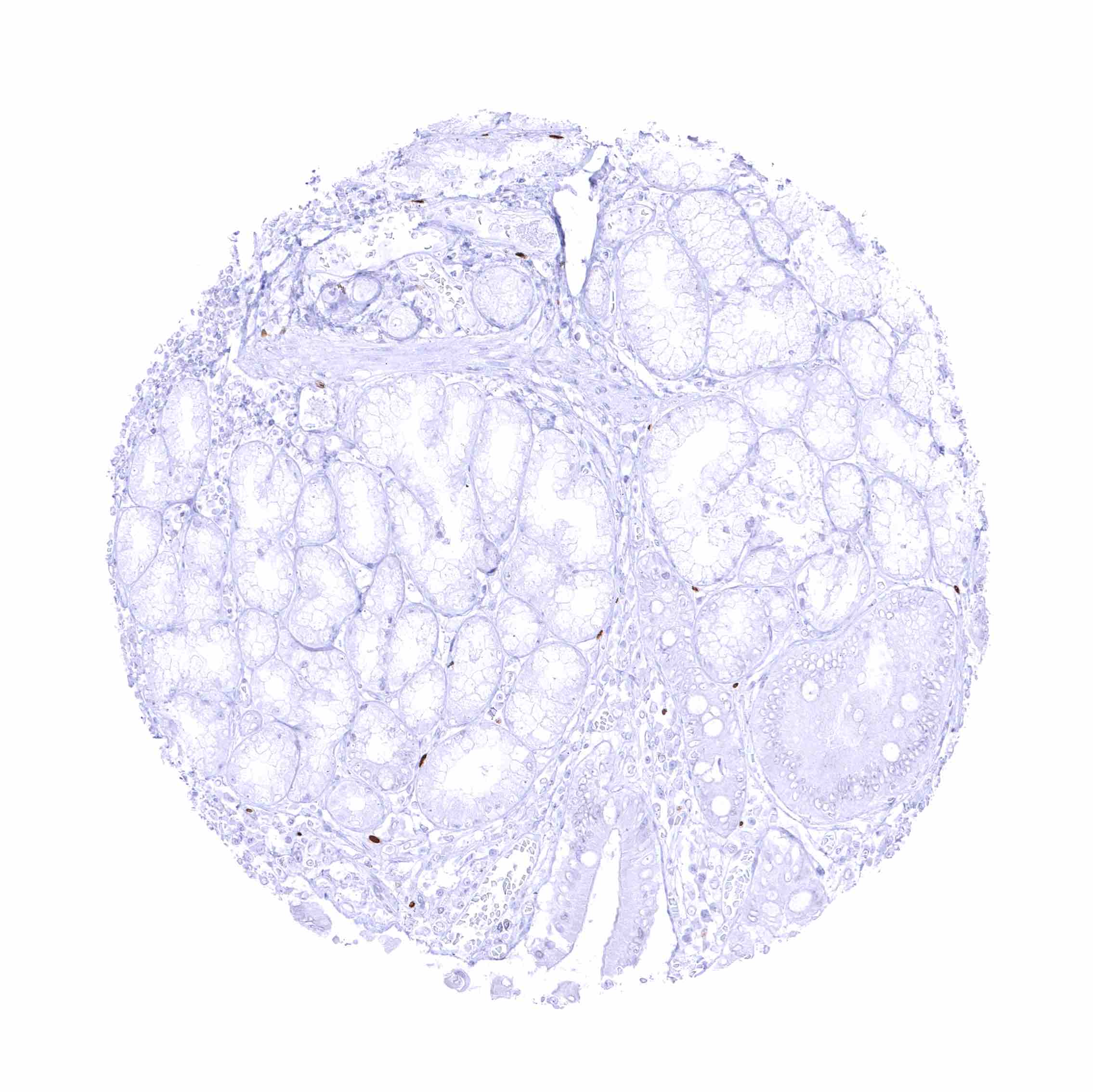
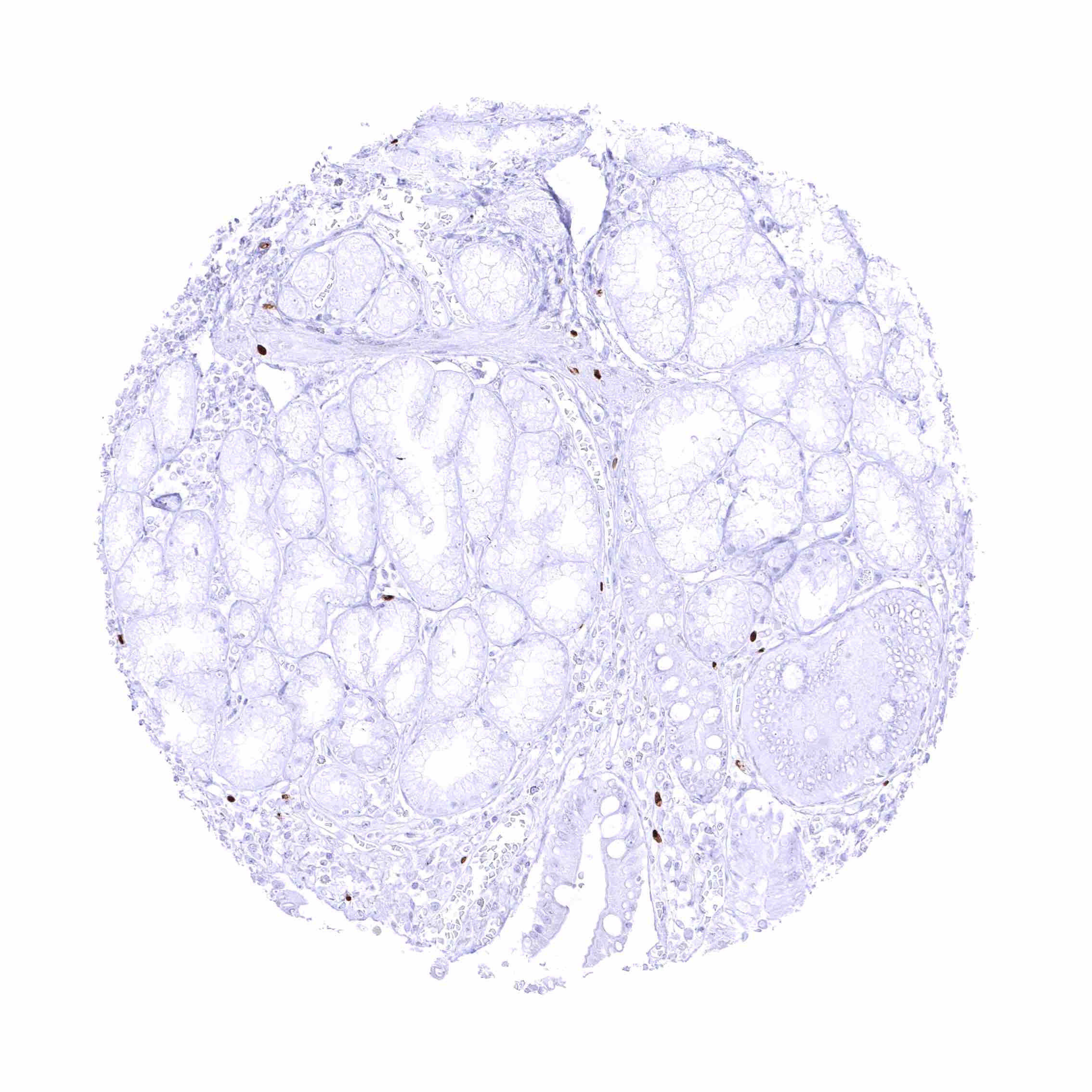
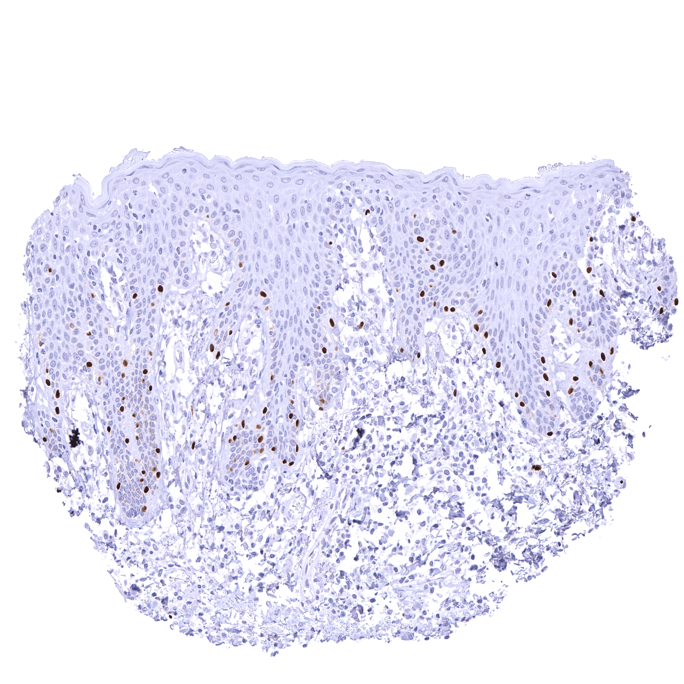
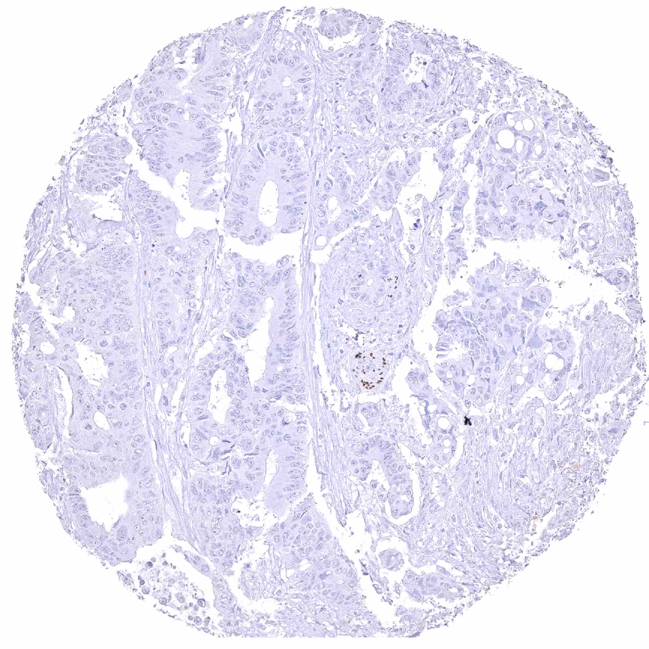
Negative control = Colon: SOX10 staining must be completely absent in epithelial cells.
Cellular localization = Intracellular
Reactivity = Human
Application = Immunohistochemistry
Dilution = 1:50 – 1:100
Intended Use = Research Use Only
Relevance of Antibody
SOX10 is a epigenetic regulator and therapeutic target protein.
Biology Behind
SOX10 is one of at least 20 members of the family of SRY (sex-determining region Y)-related high mobility group box-containing (SOX) proteins most of which have a role in embryogenesis and cell differentiation. SOX10 is coded by the SOX10 gene on chromosome 22q13.1. It is critical for the development of neural crest cells, glial cells, Schwann cells, neurons, osteoblasts, smooth muscle cells and melanocytes. SOX10 protein acts as a transcriptional activator after forming a protein complex with other proteins. In melanocytic cells, SOX10 expression is regulated by MITF. Heterozygous germline mutations occurring within and around SOX10 can cause various clinical syndroms characterized by pigment abnormalities, disorders of gastrointestinal motility, loss of smell, and hearing loss. Depending on the type of mutations, Waardenburg syndrome type 4 (auditory pigmentary abnormalities and Hirschsprung disease), Waardenburg syndrome type 2 (Waardenburg syndrome without Hirschsprung disease), PCWH (peripheral demyelinating neuropathy, central dysmyelination, Waardenburg syndrome, with or without Hirschsprung disease), uveal melanoma, or other disorders can occur.
Staining Pattern in Normal Tissues
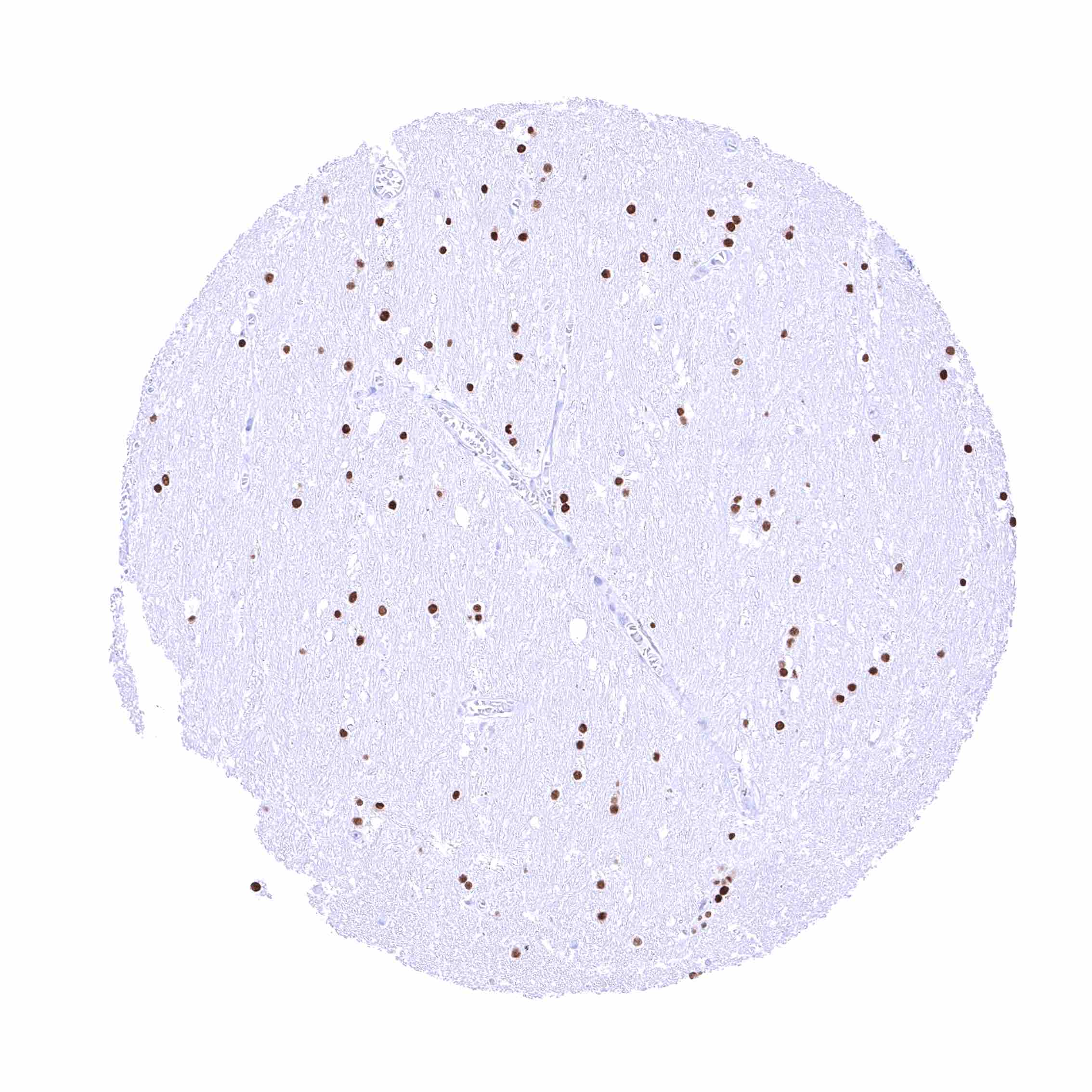
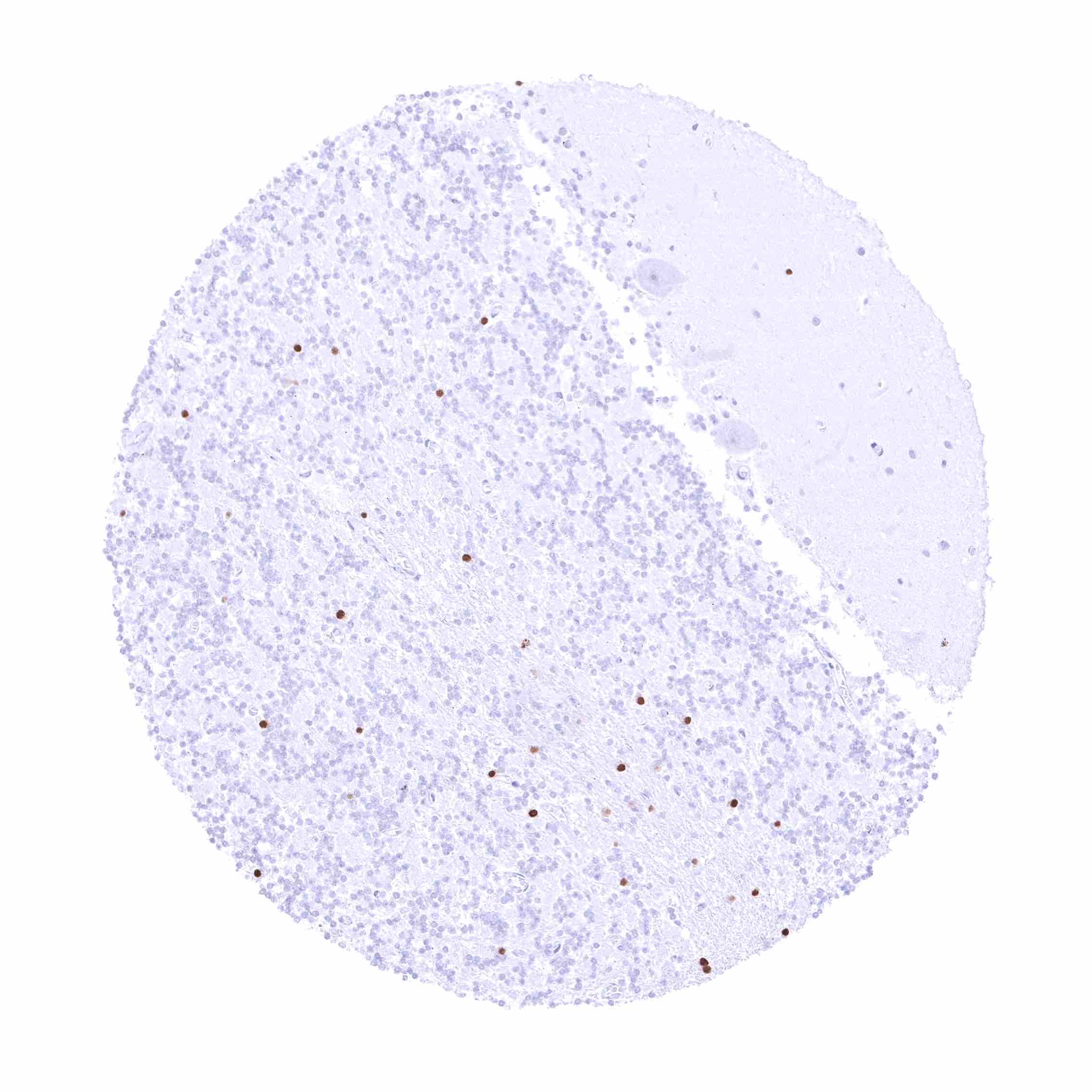
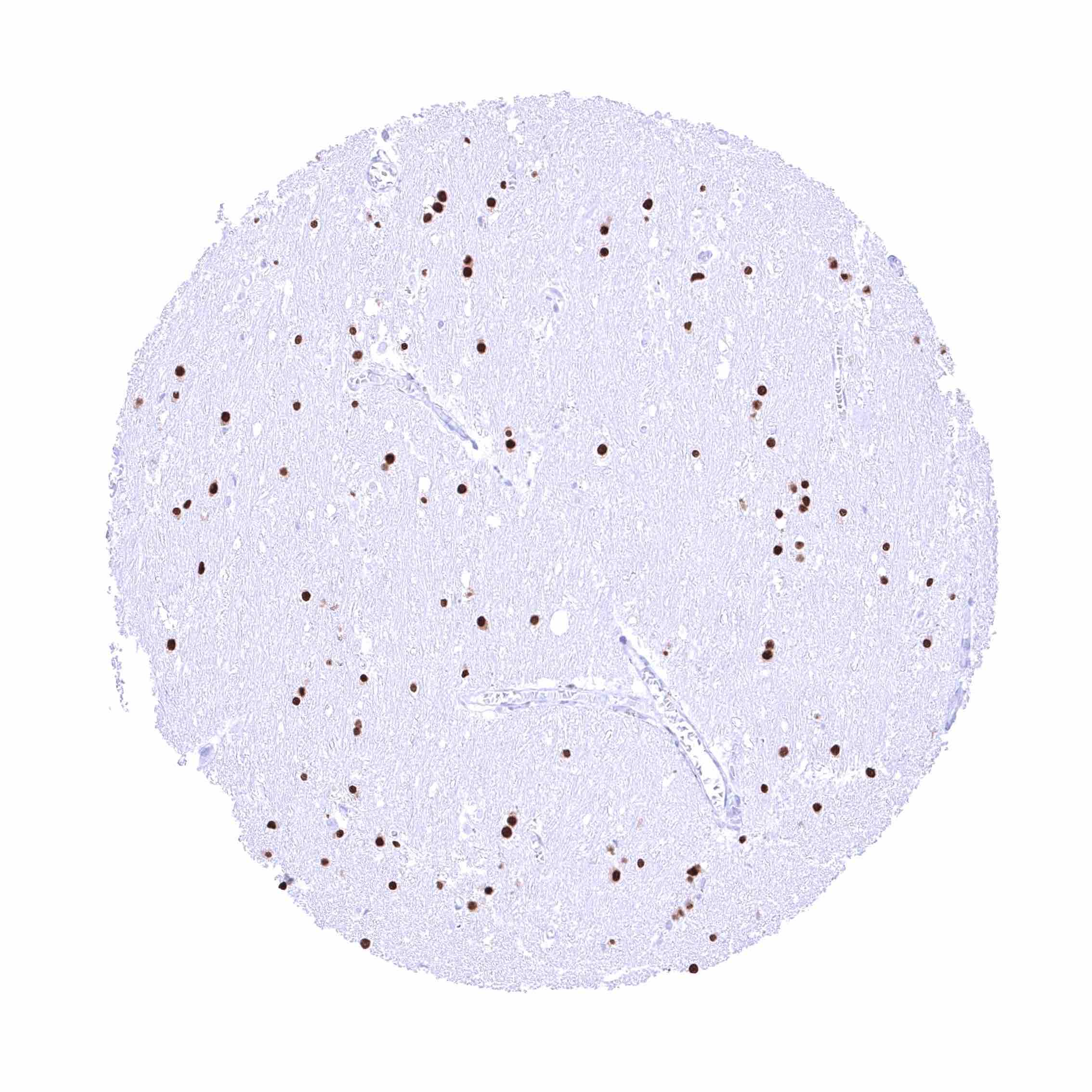
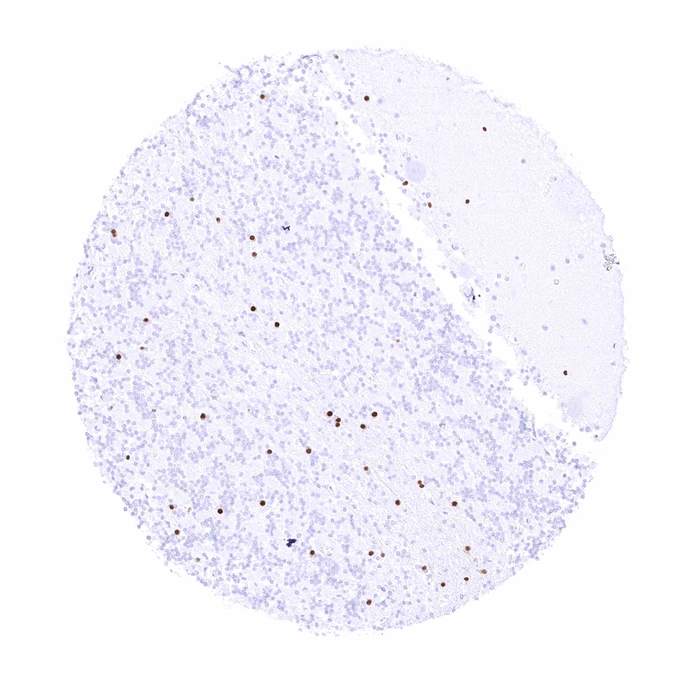
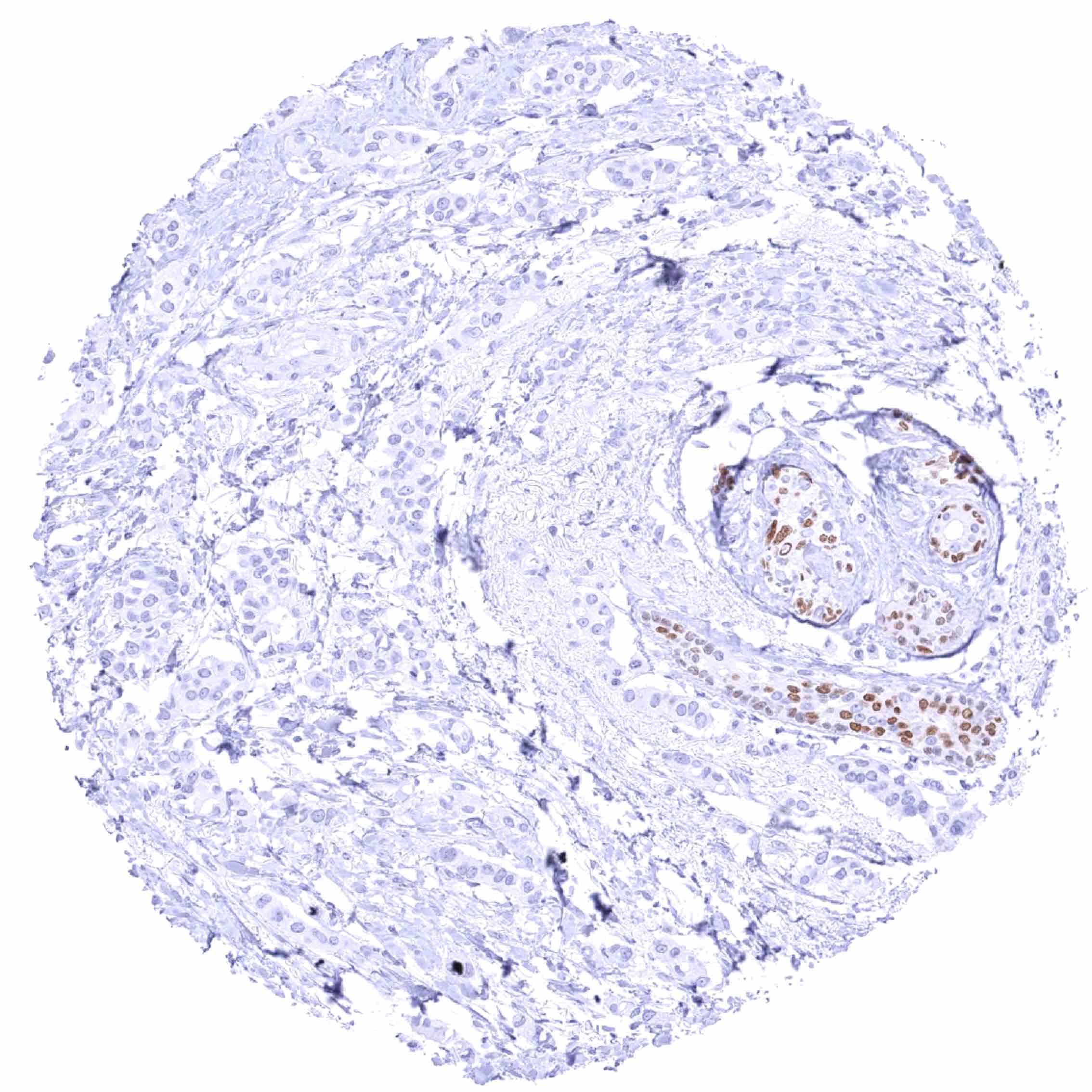
SOX10 is a nuclear protein which is preferentially expressed in melanocytes of the skin, glia cells of the cerebrum and the cerebellum, eccrine skin glands, salivary glands, and the breast. A strong nuclear SOX10 staining of scattered spindle shaped stroma cells can be found in various tissues Images describing the SOX10 staining pattern in normal tissues obtained by the antibody MSVA-710R are shown in our “Normal Tissue Gallery”.
| Brain | Cerebrum | Strong nuclear SOX10 positivity of most glia cells. |
| Cerebellum | Strong nuclear SOX10 positivity of most glia cells. | |
| Endocrine Tissues | Thyroid | Negative. |
| Parathyroid | Numerous scattered spindle shaped SOX10 positive cells between glandular cells. | |
| Adrenal gland | Scattered spindle shaped SOX10 positive cells between glandular cells. | |
| Pituitary gland | Negative. | |
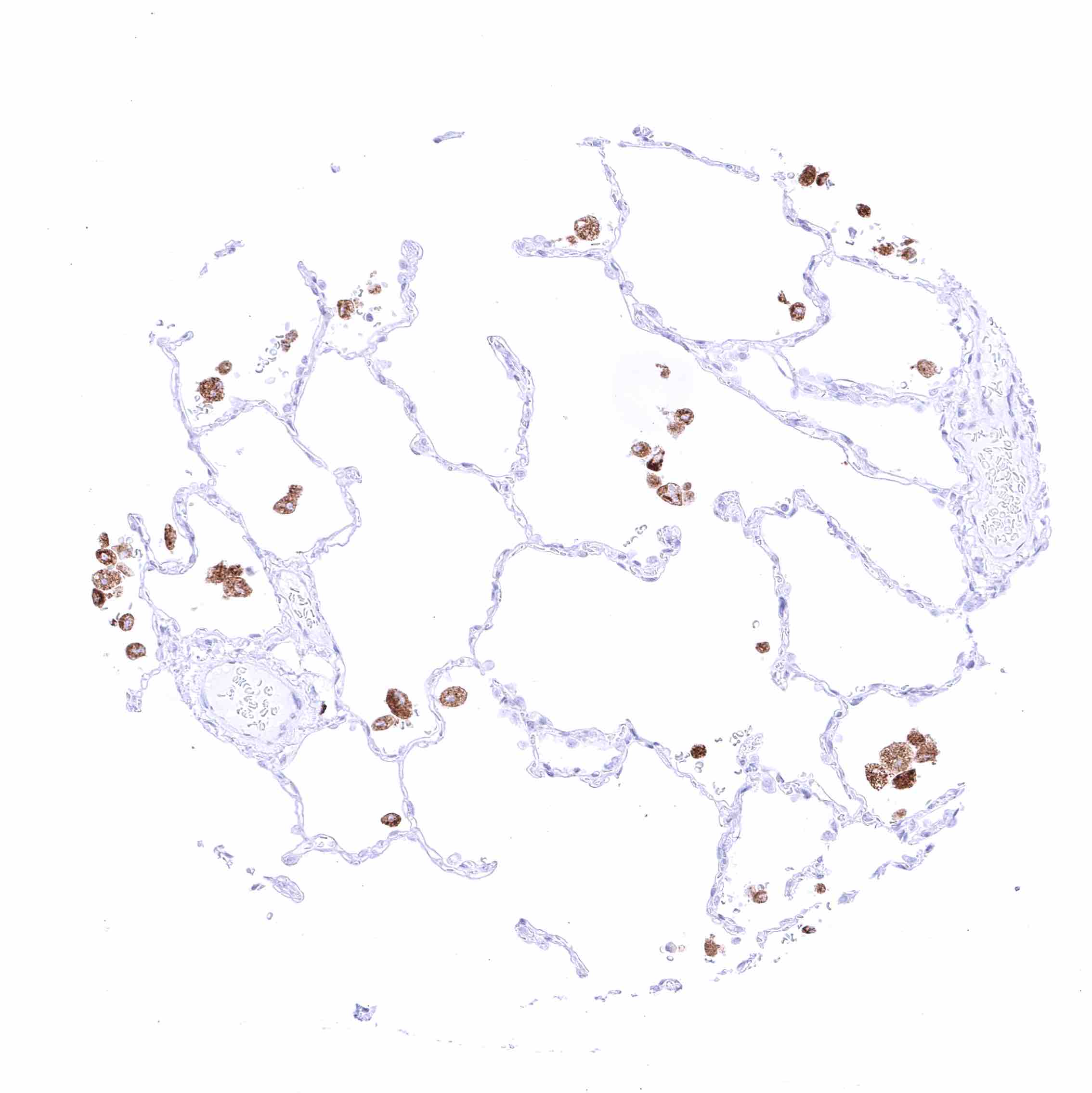
| Respiratory system | Respiratory epithelium | Respiratory epithelium is SOX10 negative. Bronchial glands with a distinct nuclear SOX10 positivity. Scattered spindle shaped SOX10 positive cells in the stroma. |
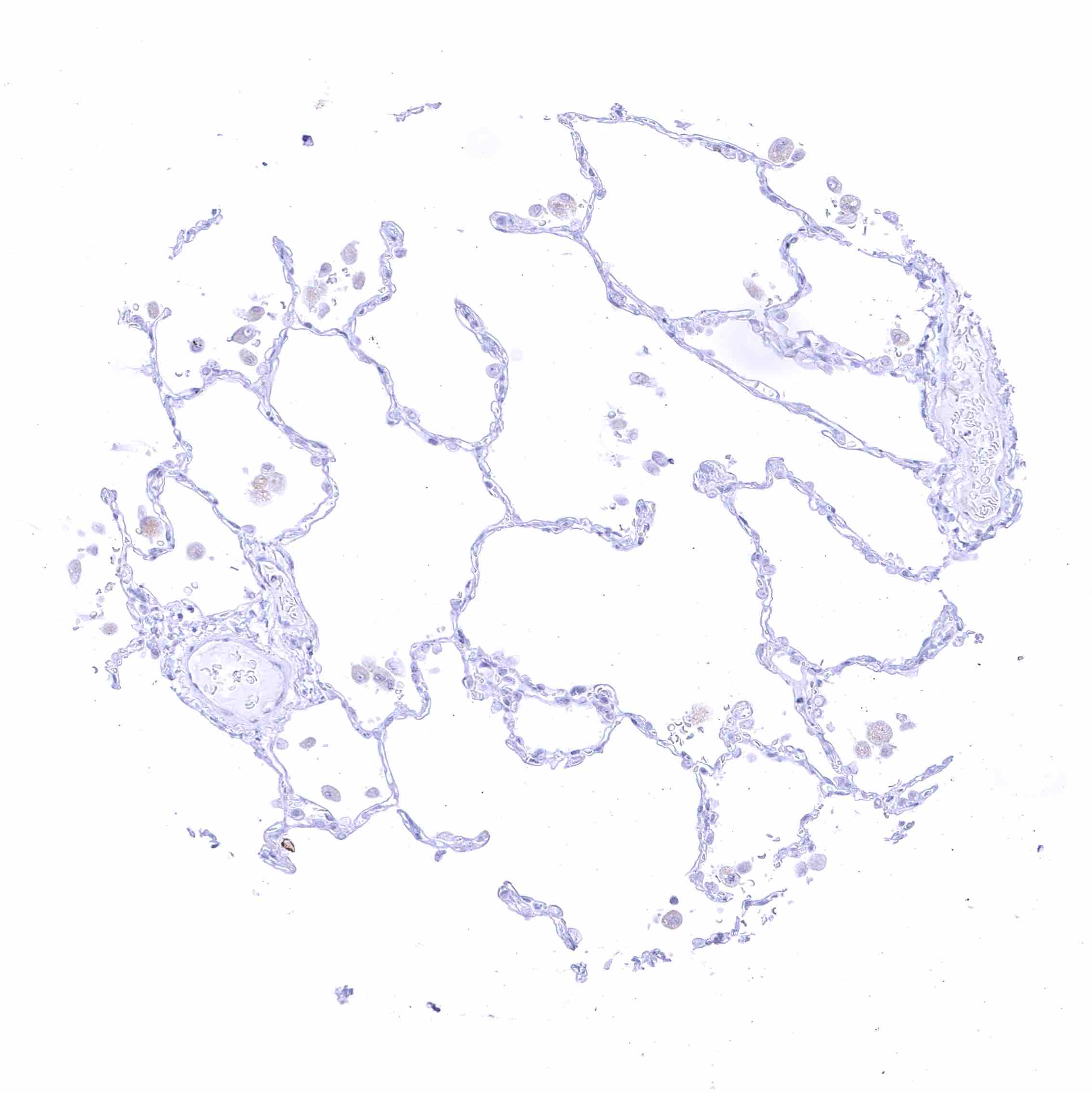
| Lung | Negative. | |
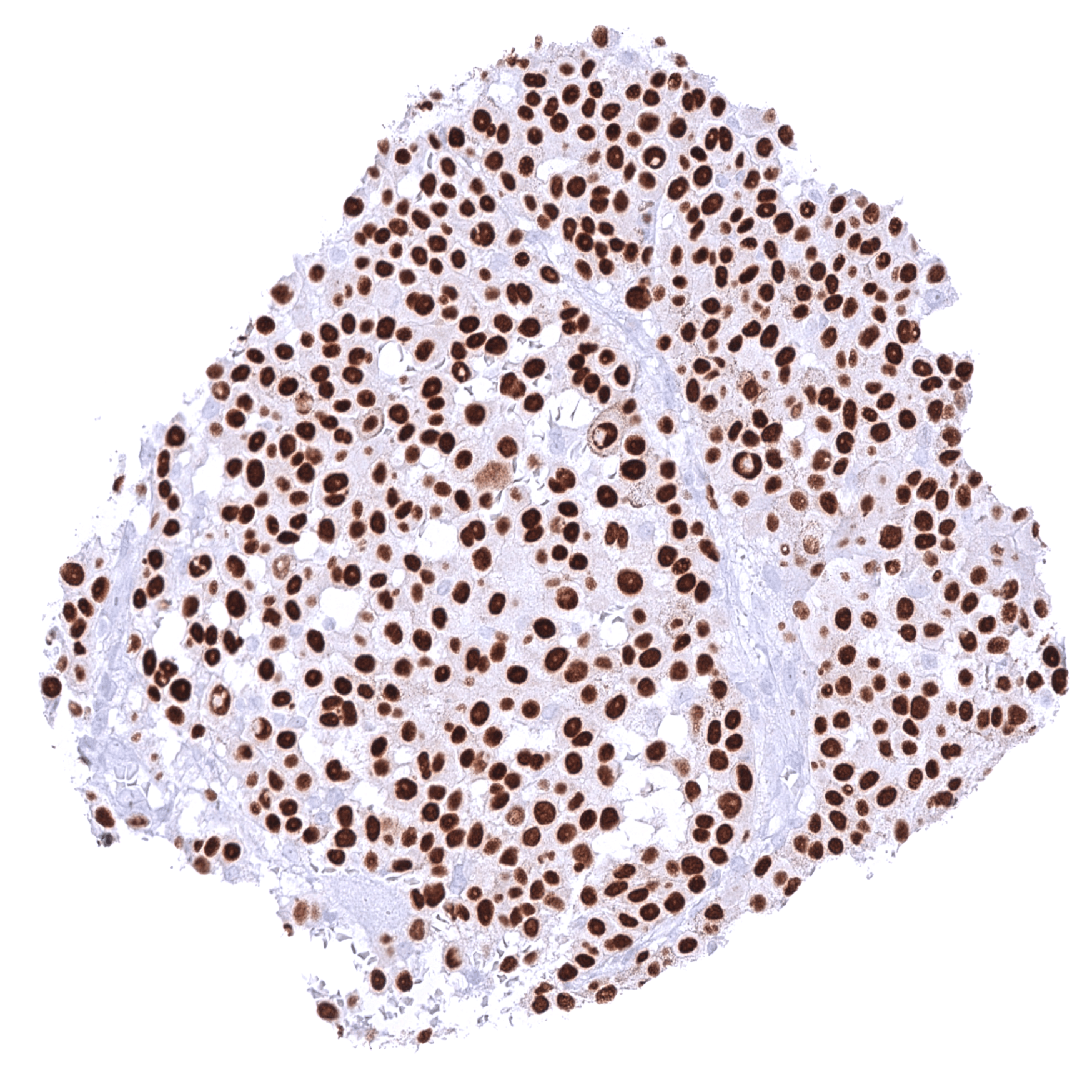
| Gastrointestinal Tract | Salivary glands | Strong nuclear SOX10 positivity of glandular cells while ductal cells are SOX10 negative. |
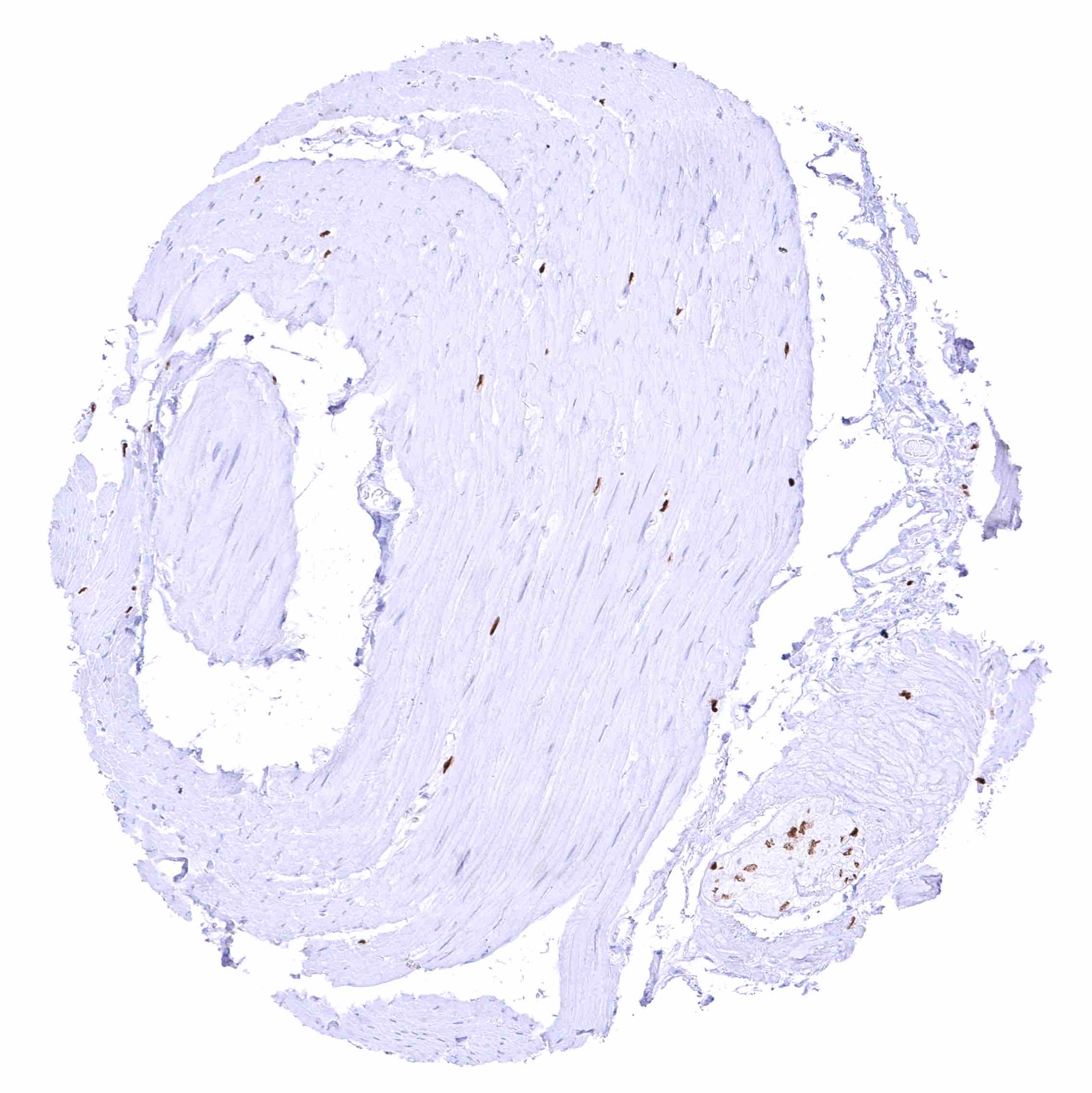
| Esophagus | A good number of scattered spindle shaped SOX10 positive cells between smooth muscle cells, ganglion cells, and nerves in the muscular wall. | |
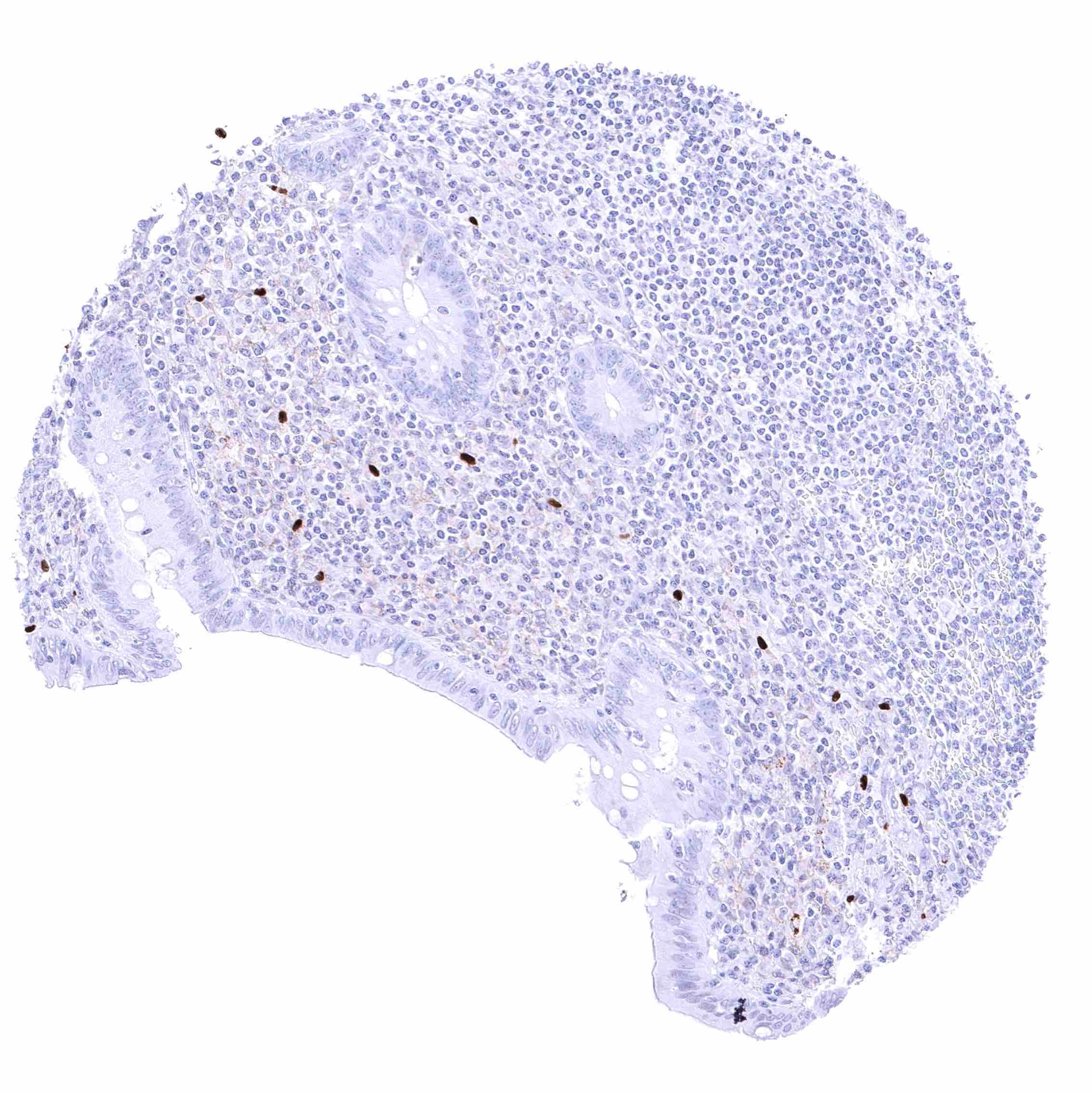
| Stomach | Few scattered spindle shaped SOX10 positive cells along gastric glands. A good number of scattered spindle shaped SOX10 positive cells between smooth muscle cells, ganglion cells, and nerves in the muscular wall. | |
| Duodenum | Few scattered spindle shaped SOX10 positive cells in the mucosa and adjacent to Brunner glands. Brunner glands are SOX10 negative. | |
| Small intestine | Few scattered spindle shaped SOX10 positive cells in the mucosa. A good number of scattered spindle shaped SOX10 positive cells between smooth muscle cells, ganglion cells, and nerves in the muscular wall. | |
| Appendix | Few scattered spindle shaped SOX10 positive cells in the mucosa. A good number of scattered spindle shaped SOX10 positive cells between smooth muscle cells, ganglion cells, and nerves in the muscular wall. | |
| Colon | A good number of scattered spindle shaped SOX10 positive cells between smooth muscle cells, ganglion cells, and nerves in the muscular wall. | |
| Rectum | A good number of scattered spindle shaped SOX10 positive cells between smooth muscle cells, ganglion cells, and nerves in the muscular wall. | |
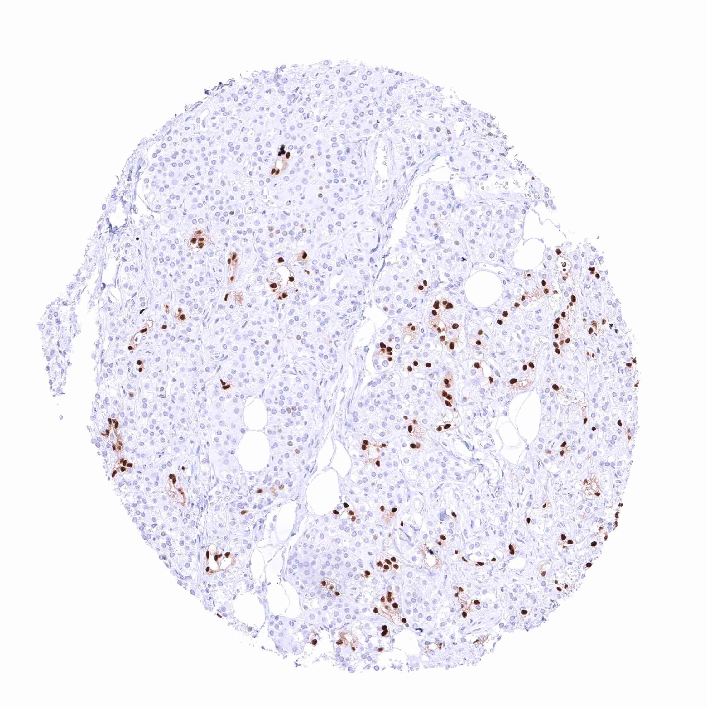
| Liver | Few scattered spindle shaped SOX10 positive cells, especially in portal fields. | |
| Gallbladder | Few scattered spindle shaped SOX10 positive cells in the mucosa. | |
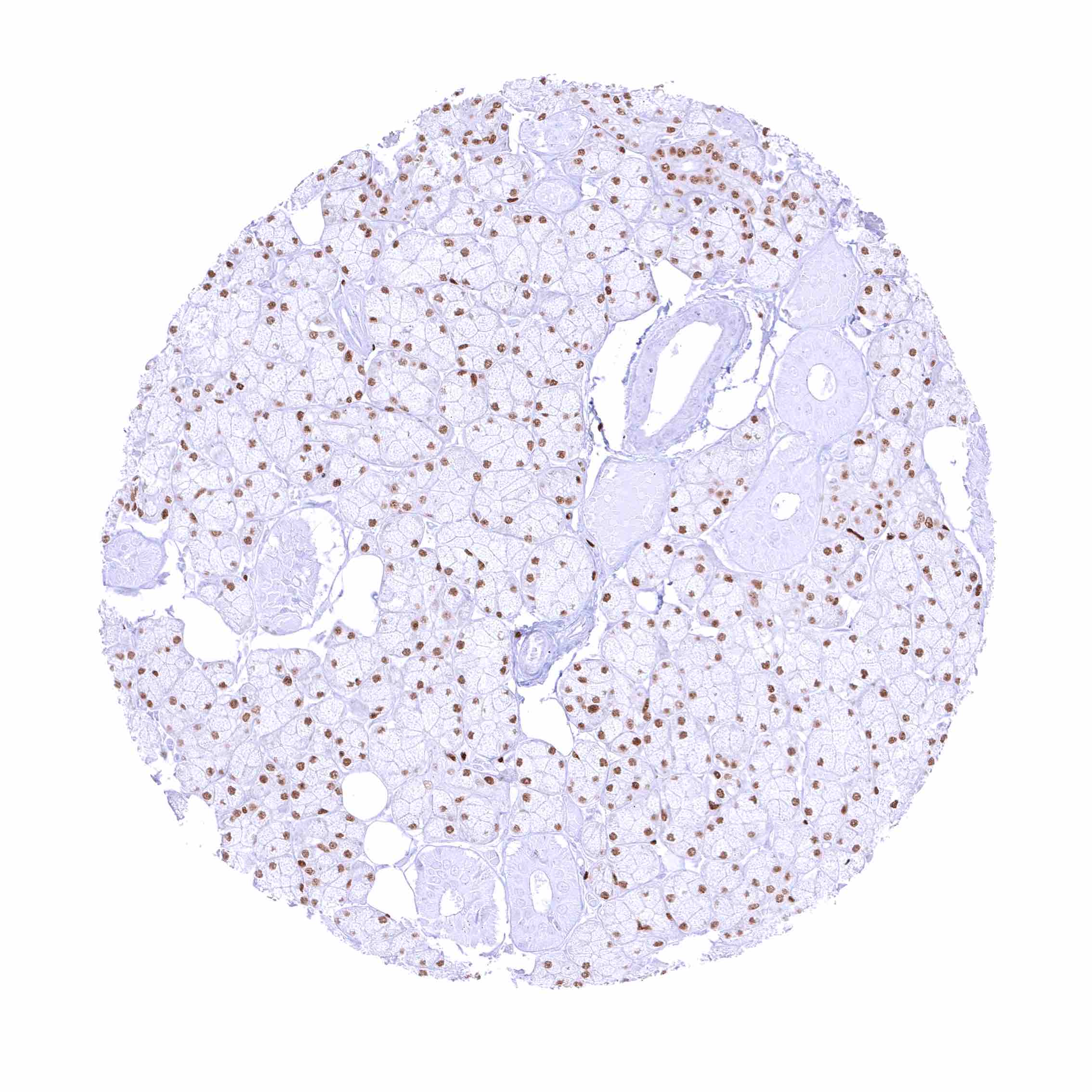
| Pancreas | Acinar cells, islet cells, and duct cells are SOX10 negative. Few scattered SOX10 positive spindle shaped cells in the stroma. | |
| Genitourinary | Kidney | Few scattered spindle shaped SOX10 positive cells adjacent to tubuli. Very few scattered spindle shaped SOX10 positive cells between smooth muscle cells in the muscular wall of the pyelon. |
| Urothelium | Very few scattered spindle shaped SOX10 positive cells between smooth muscle cells in the muscular wall of the bladder. | |
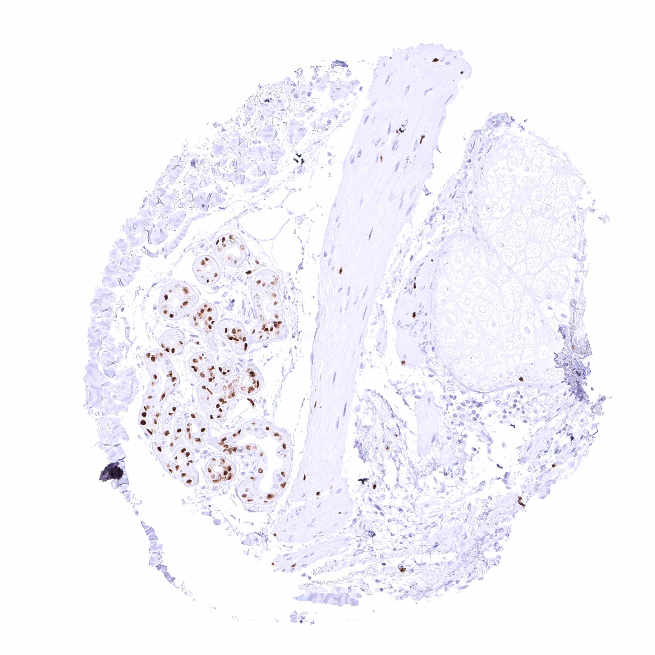
| Male genital | Prostate | Scattered spindle shaped SOX10 positive cells in the stroma. |
| Seminal vesicles | Scattered spindle shaped SOX10 positive cells in the stroma. | |
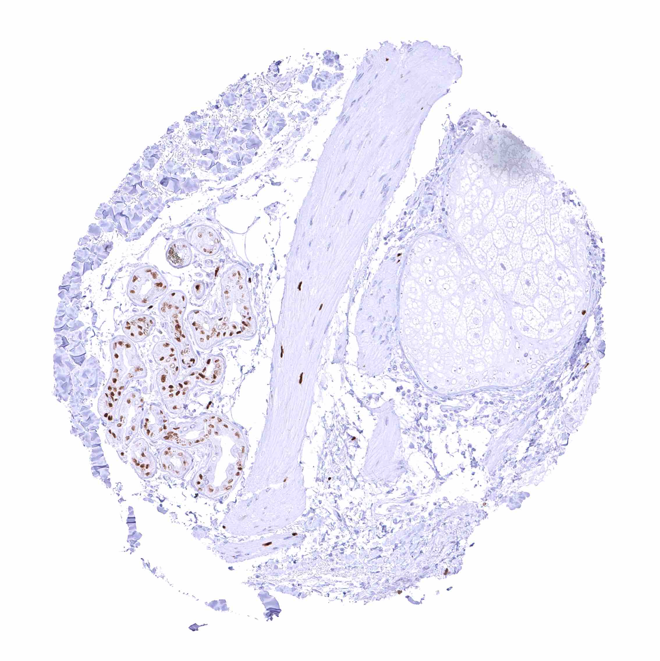
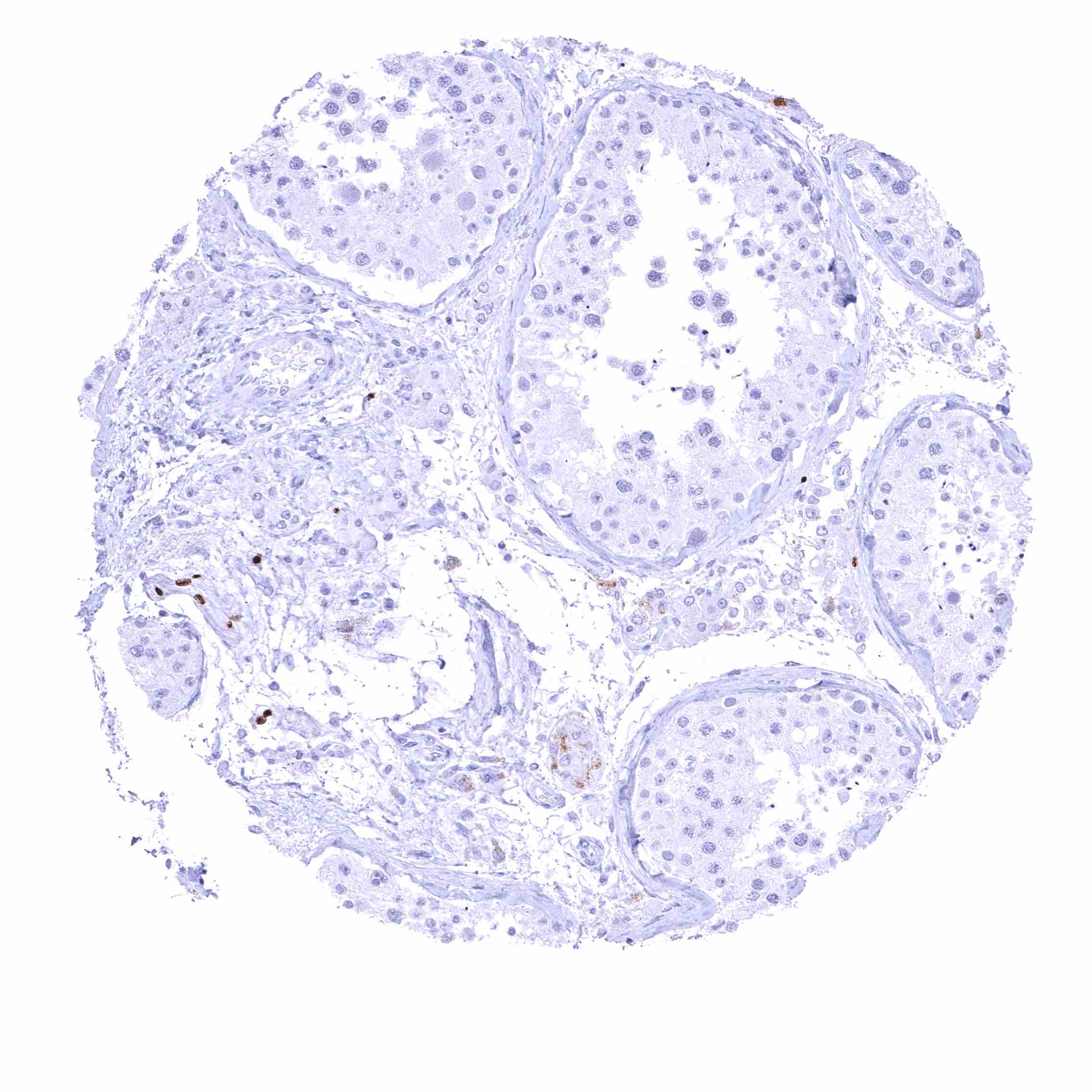
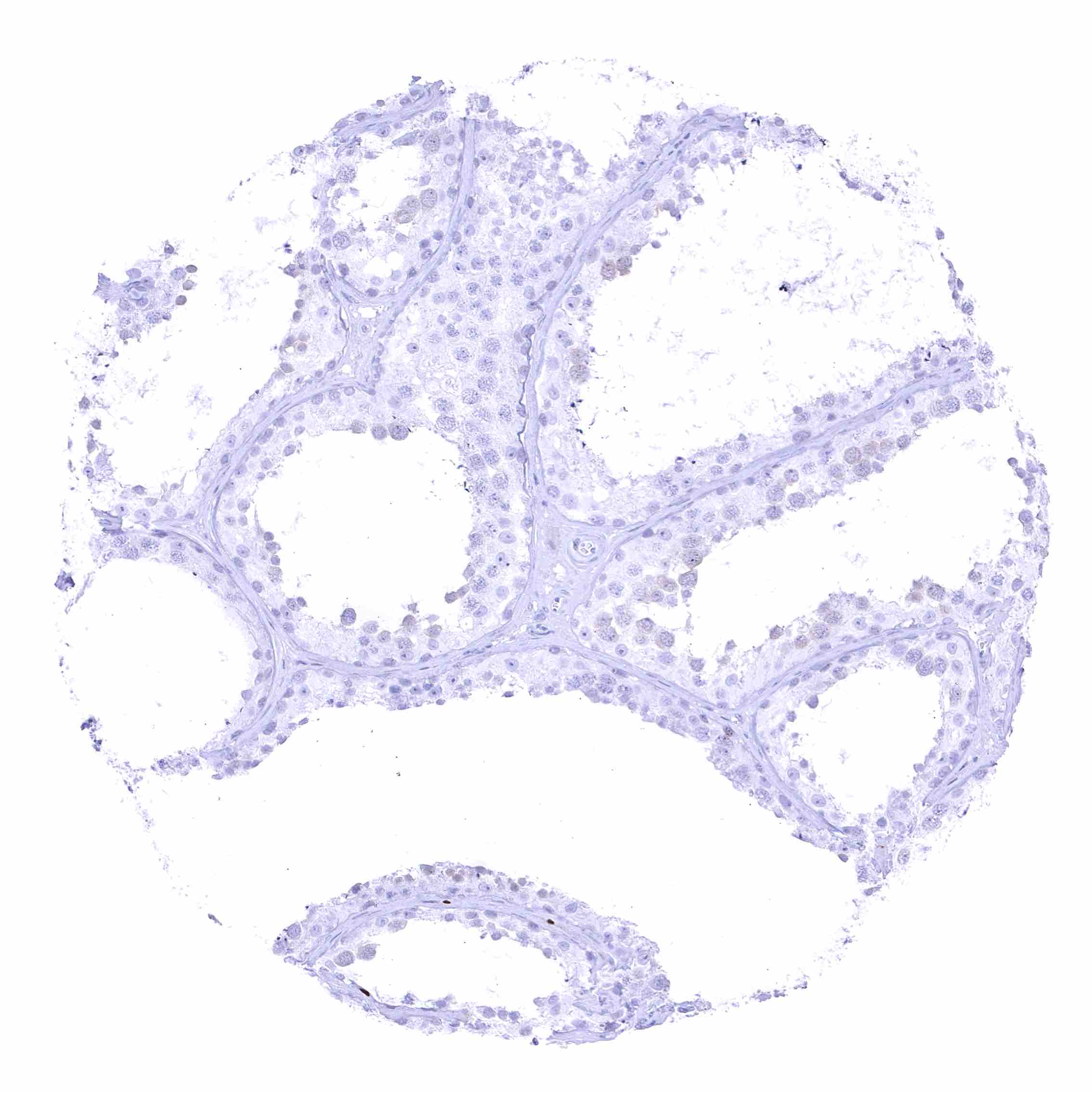
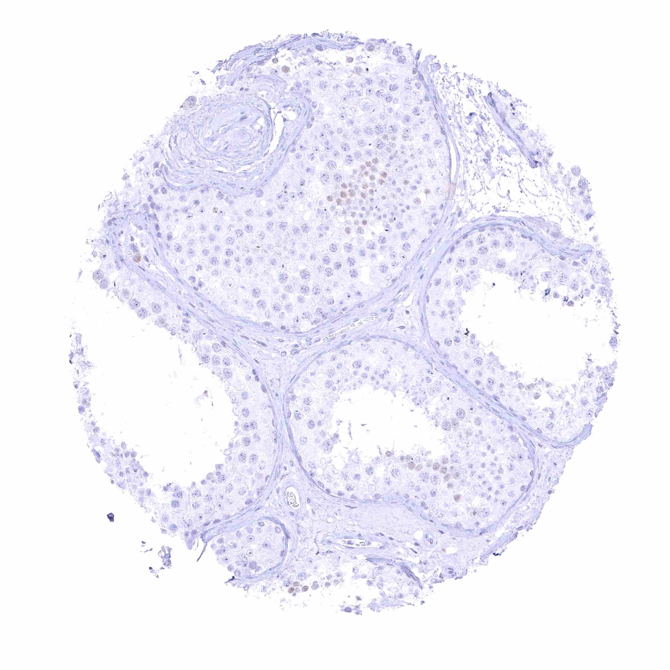
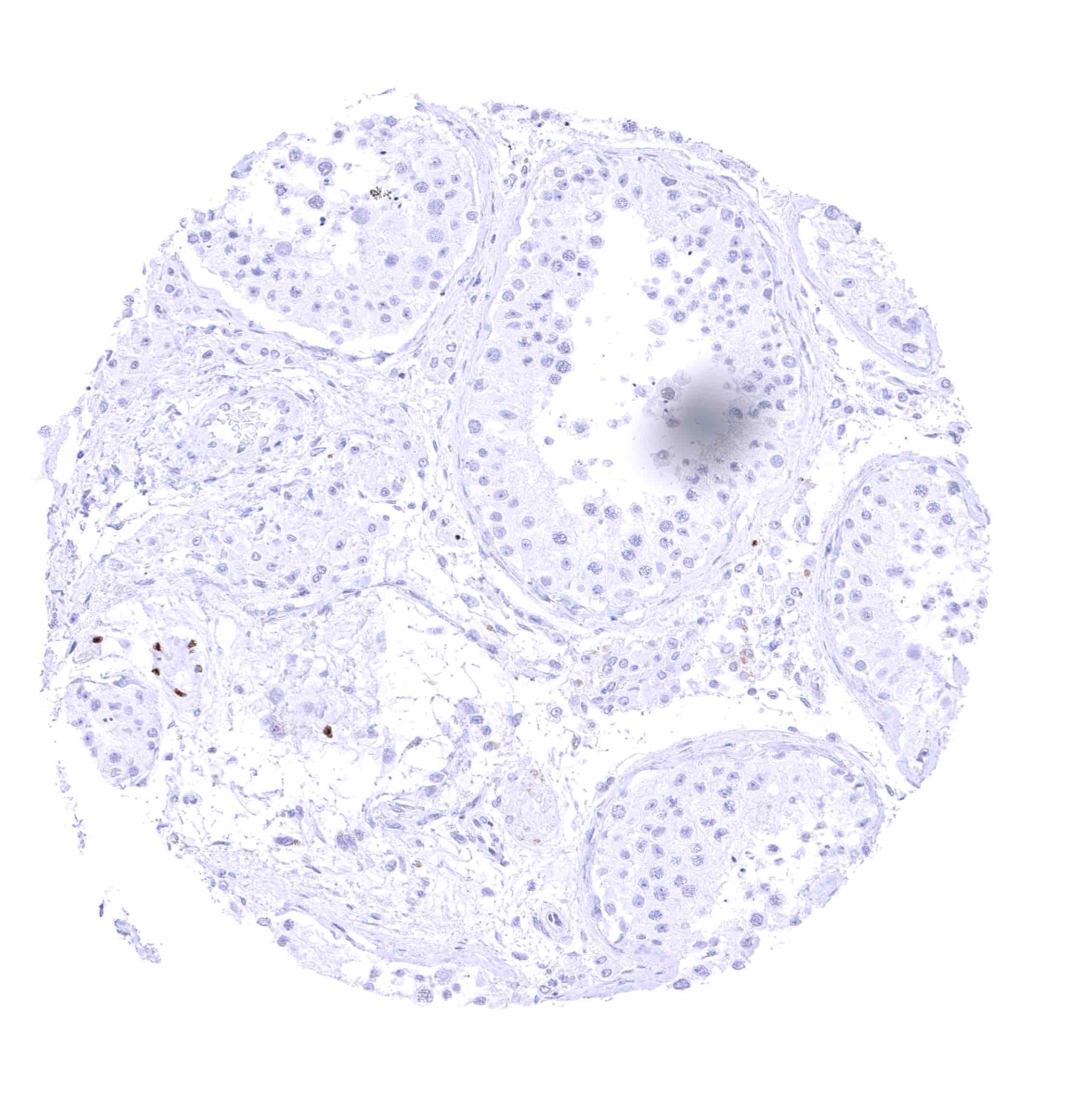
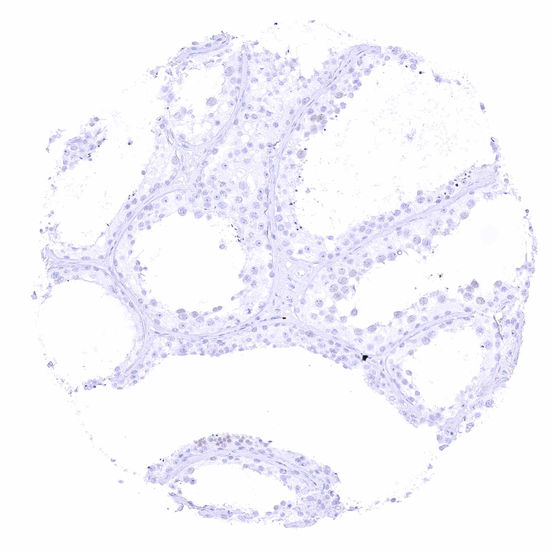
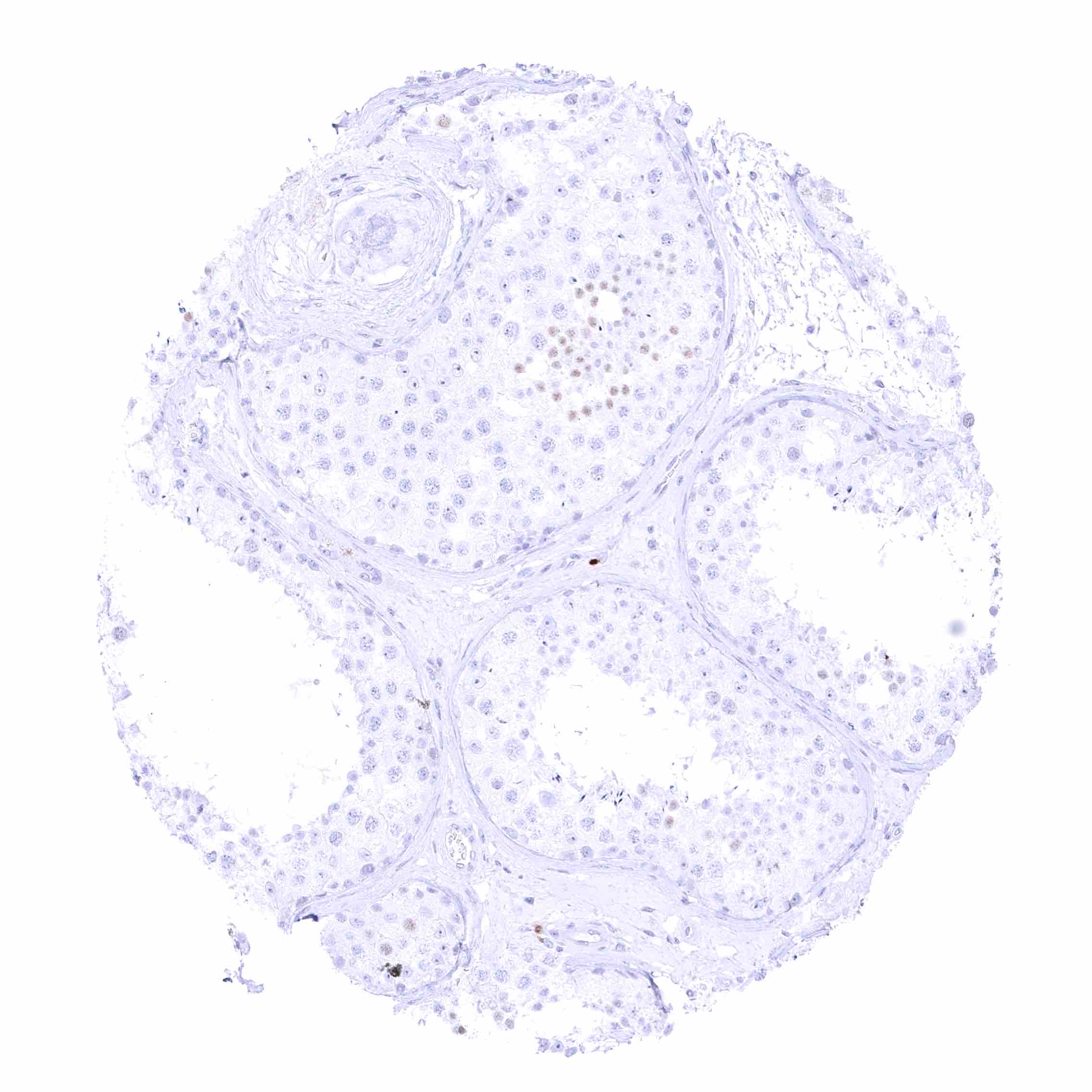
| Testis | Few spindle shaped SOX10 positive cells can be along small blood vessels. | |
| Epididymis | Negative. Few scattered spindle shaped SOX10 positive cells in the stroma.. | |
| Female genital | Breast | Moderate to strong nuclear (and nuclear) SOX10 positivity of glandular cells (staining is somewhat weaker in myoepithelial cells). |
| Uterus, myometrium | Few scattered spindle shaped SOX10 positive cells. | |
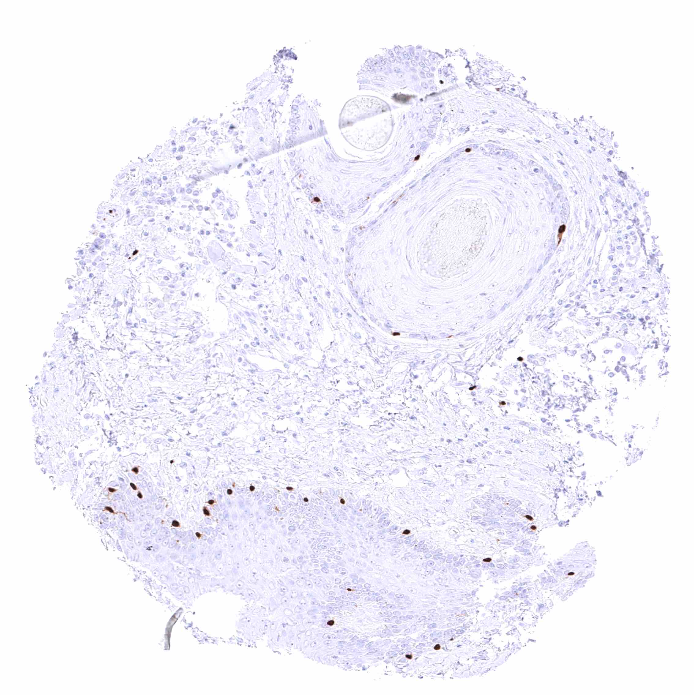
| Uterus, ectocervix | Negative. | |
| Uterus endocervix | Negative. | |
| Uterus, endometrium | Negative. | |
| Fallopian Tube | Negative. | |
| Ovary | Stroma cells are generally negative but scattered spindle shaped SOX10 positive cells occur. | |
| Placenta early | Negative. | |
| Placenta mature | Negative. | |
| Amnion | ||
| Chorion | Negative. | |
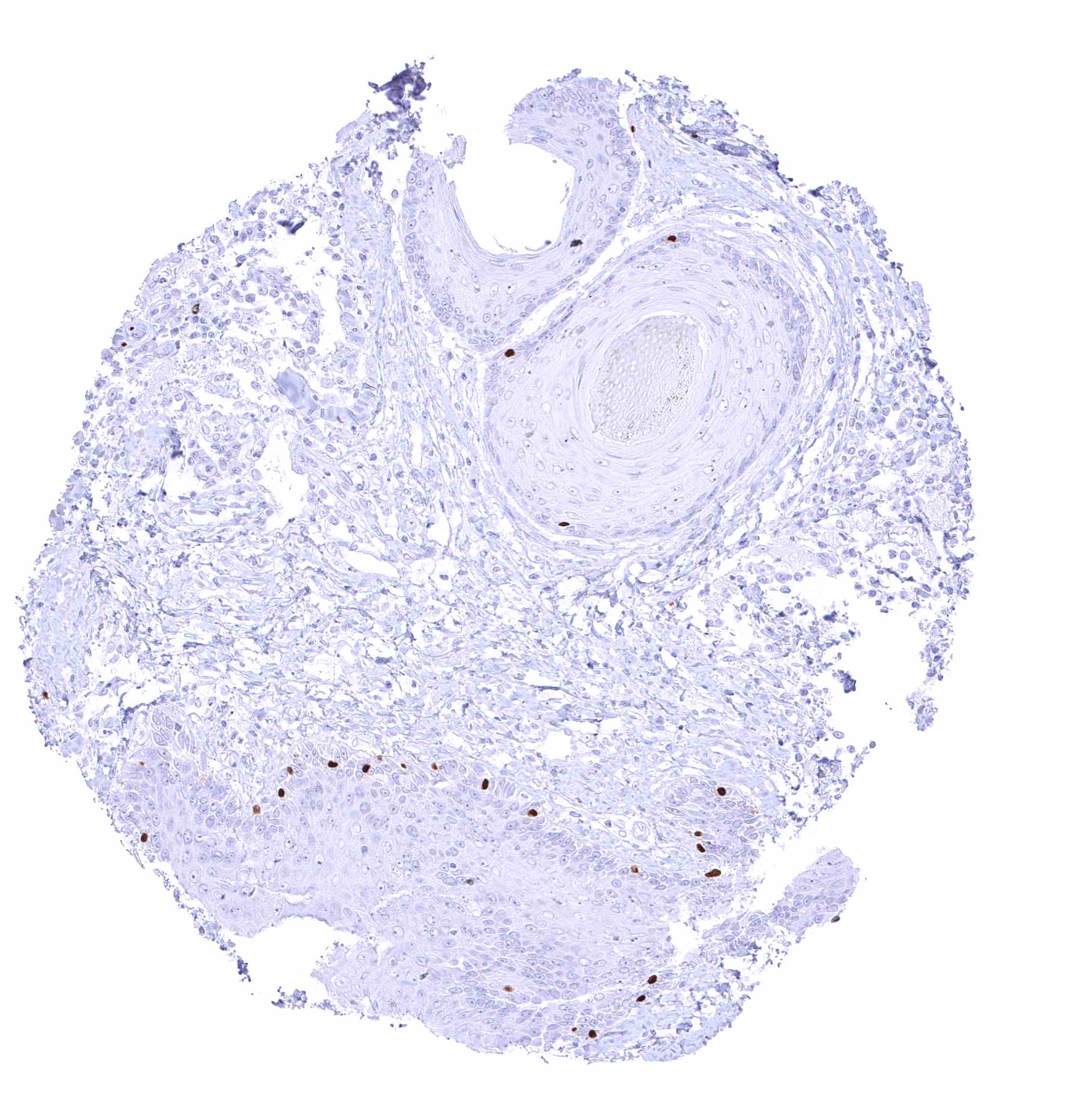
| Skin | Epidermis | Strong nuclear SOX10 staining of melanocytes. |
| Sebaceous glands | Negative. Scattered spindle shaped SOX10 positive cells around the glands. Eccrineglands of the skin are SOX10 positive. | |
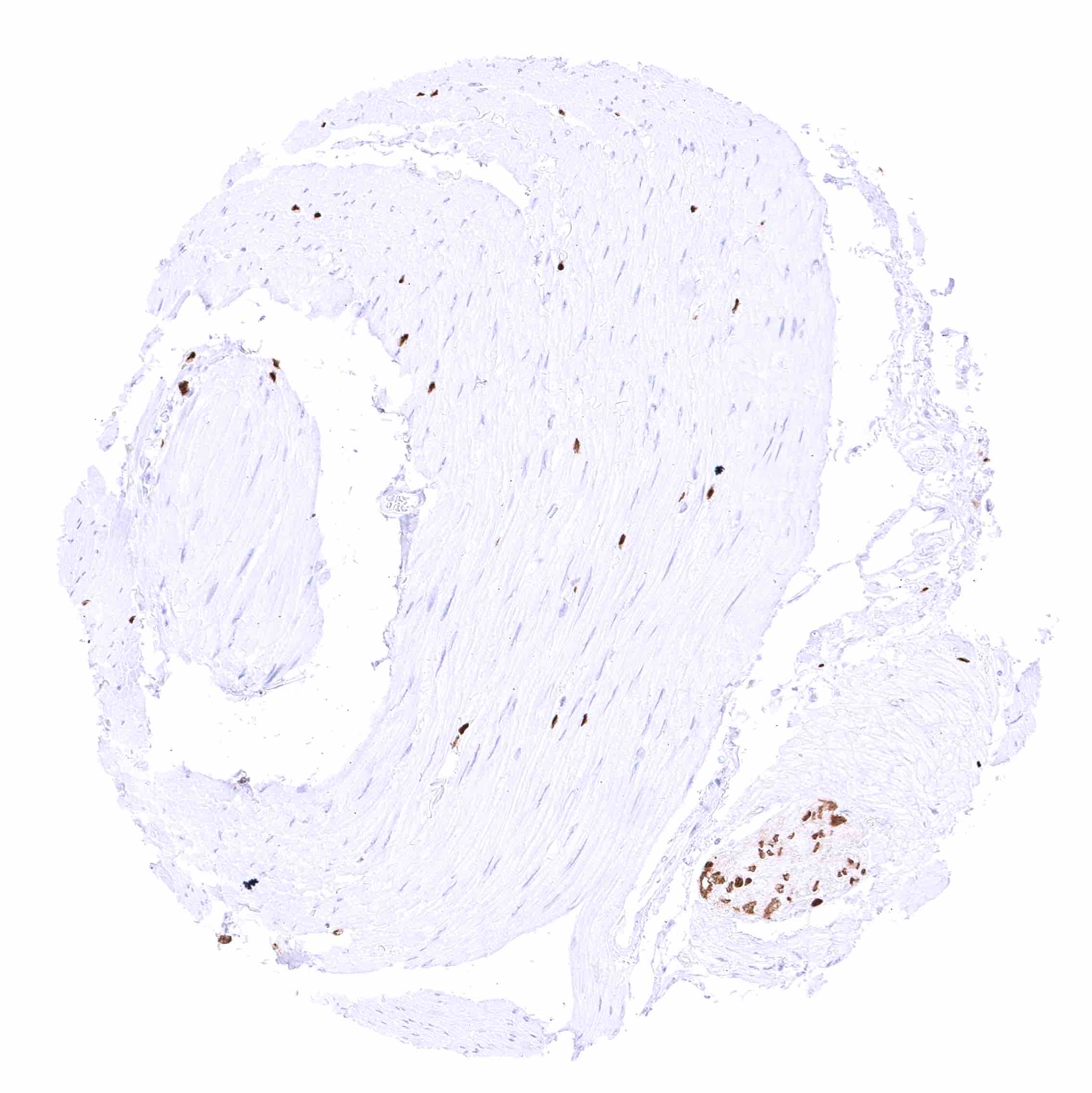
| Muscle/connective tissue | Heart muscle | Scattered spindle shaped SOX10 positive cells in the stroma. |
| Skeletal muscle | Scattered spindle shaped SOX10 positive cells in the stroma. | |
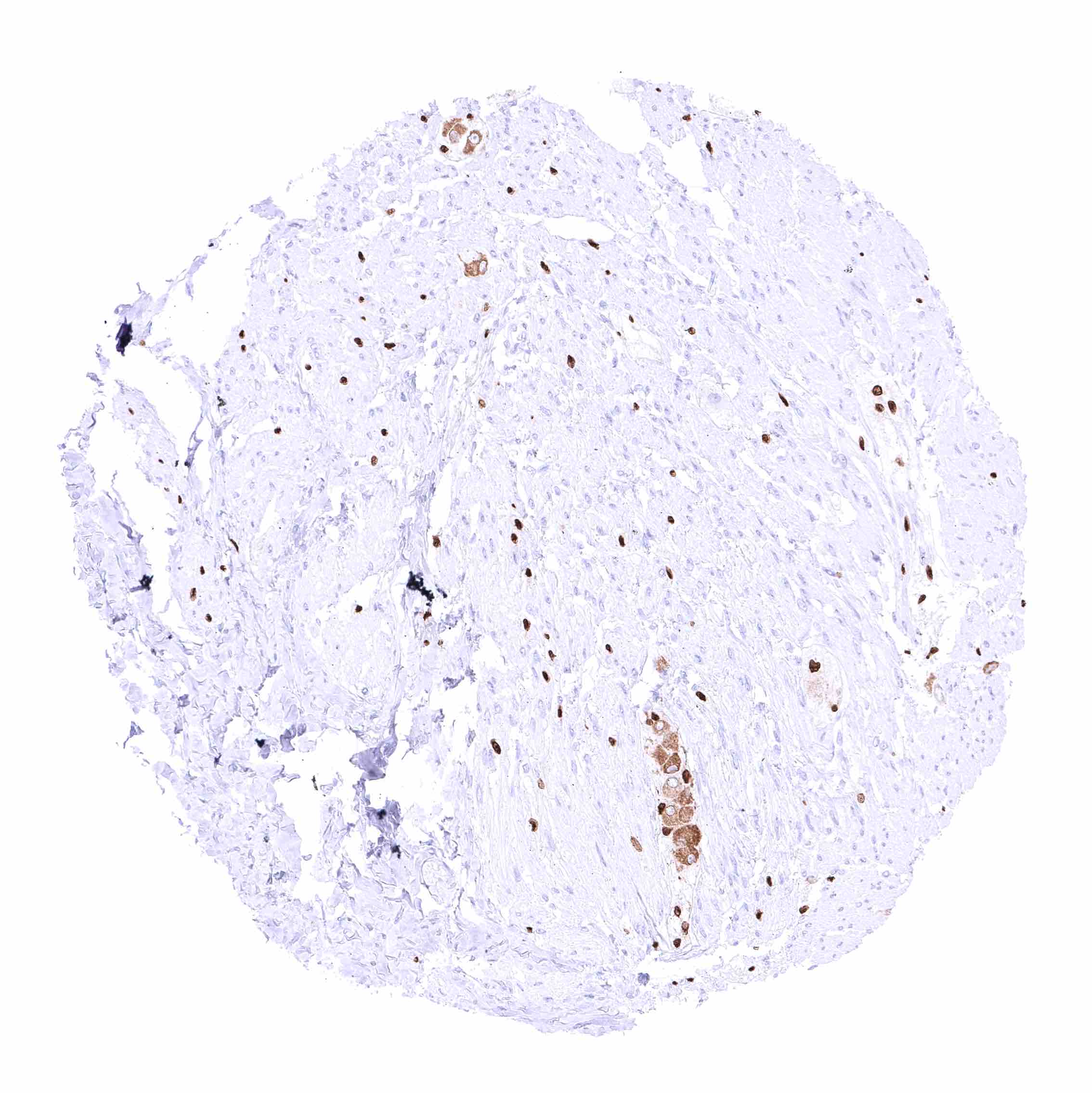
| Smooth muscle | A good number of scattered spindle shaped SOX10 positive cells occur between smooth muscle cells and ganglion cells in many different organs. This phenomenon appears to be particularly prominent in the gastrointestinal muscular wall. | |
| Vessel walls | Negative, but scattered spindle shaped SOX10 positive cells can occur around vessels. | |
| Fat | Negative. | |
| Stroma | Scattered spindle shaped SOX10 positive cells occur in many tissues. | |
| Endothelium | Negative. | |
| Bone marrow/ lymphoid tissue | Bone marrow | Negative. |
| Lymph node | Lymphocytes are SOX10 negative. Scattered spindle shaped SOX10 positive cells can occur along small blood vessels. | |
| Spleen | Negative. | |
| Thymus | Lymphocytes are SOX10 negative. | |
| Tonsil | Lymphocytes are SOX10 negative. | |
| Remarks | Scattered spindle shaped SOX10 positive cells occur in many tissues. |
A membranous CD38 expression occurs in various subtypes of inflammatory cells, endothelial cells at selected locations as well as in the epithelium of the prostate and of seminal vesicles.
RNA and protein expression data of SOX10 findings are also described in the Human Protein Atlas (Tissue expression SOX10).
Positive control = Breast: A moderate to strong nuclear SOX10 staining should be seen in breast epithelial cells.
Negative control = Colon: SOX10 staining must be completely absent in epithelial cells.
Staining Pattern in Relevant Tumor Types
SOX10 expression preferentially occurs in gliomas, neoplasms of melanocytes and nerve sheaths, as well as of cancers derived from salivary, breast, and eccrine skin glands.
The TCGA findings on SOX10 RNA expression in different tumor categories have been summarized in the Human Protein Atlas.
Compatibility of Antibodies
No data available at the moment
Protocol Recommendations
IHC users have different preferences on how the stains should look like. Some prefer high staining intensity of the target stain and even accept some background. Others favor absolute specificity and lighter target stains. Factors that invariably lead to more intense staining include higher concentration of the antibody and visualization tools, longer incubation time, higher temperature during incubation, higher temperature and longer duration of the heat induced epitope retrieval (slide pretreatment). The impact of the pH during slide pretreatment has variable effects and depends on the antibody and the target protein.
All images and data shown here and in our image galleries are obtained by the manual protocol described below. Other protocols resulting in equivalent staining are described as well.
Manual protocol
Freshly cut sections should be used (less than 10 days between cutting and staining). Heat-induced antigen retrieval for 5 minutes in an autoclave at 121°C in pH 7,8 Target Retrieval Solution buffer. Apply MSVA-710R at a dilution of 1:50 at 37°C for 60 minutes. Visualization of bound antibody by the EnVision Kit (Dako, Agilent) according to the manufacturer’s directions.
Potential Research Applications
- The prevalence and role of SOX10 expression in different tumor types needs to be further clarified.
- The function of SOX10 and its role in various diseases needs to be further investigated.
- The suitability of SOX10 as a therapeutic target needs to be tested.
Evidence for Antibody Specificity in IHC
There are two ways how the specificity of antibodies can be documented for immunohistochemistry on formalin fixed tissues. These are: 1. Comparison with a second independent method for target expression measurement across a large number of different tissue types (orthogonal strategy), and 2. Comparison with one or several independent antibodies for the same target and showing that all positive staining results are also seen with other antibodies for the same target (independent antibody strategy).
Orthogonal validation: For the antibody MSVA-710R specificity is supported by the good concordance of the immunostaining data with data from three independent RNA screening studies, including the Human Protein Atlas (HPA) RNA-seq tissue dataset, the FANTOM5 project, and the Genotype-Tissue Expression (GTEx) project, which are all summarized in the Human Protein Atlas (Tissue expression SOX10). SOX10 staining by MSVA-710R was preferably detected in salivary glands, the brain and in breast glands which were the tissues for which the highest levels of RNA expression had been recorded. A weaker SOX10 RNA expression in many other tissues is in line with the occurrence of a variable number of SOX10 positive cells in almost all tissues.
Comparison of antibodies: True expression of SOX10 in cell types with documented SOX10 immunostaining by MSVA-710R is further validated by identical staining patterns obtained by a second, independent SOX10 antibody, termed “validation antibody”. The additional cytoplasmic staining of alveolar macrophages and of ganglion cells which were only seen when the validation antibody was used, were considered antibody specific cross-reactivities of the validation antibody.