295,00 € – 995,00 €
Product details
Synonyms = cyclin dependent kinase inhibitor 1B , CDKN4 , KIP1 , MEN1B , MEN4 , P27KIP1
Antibody type = Recombinant Rabbit monoclonal / IgG
Clone = HMV3970
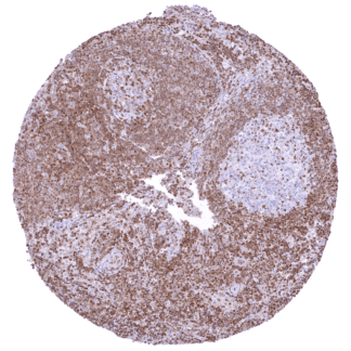
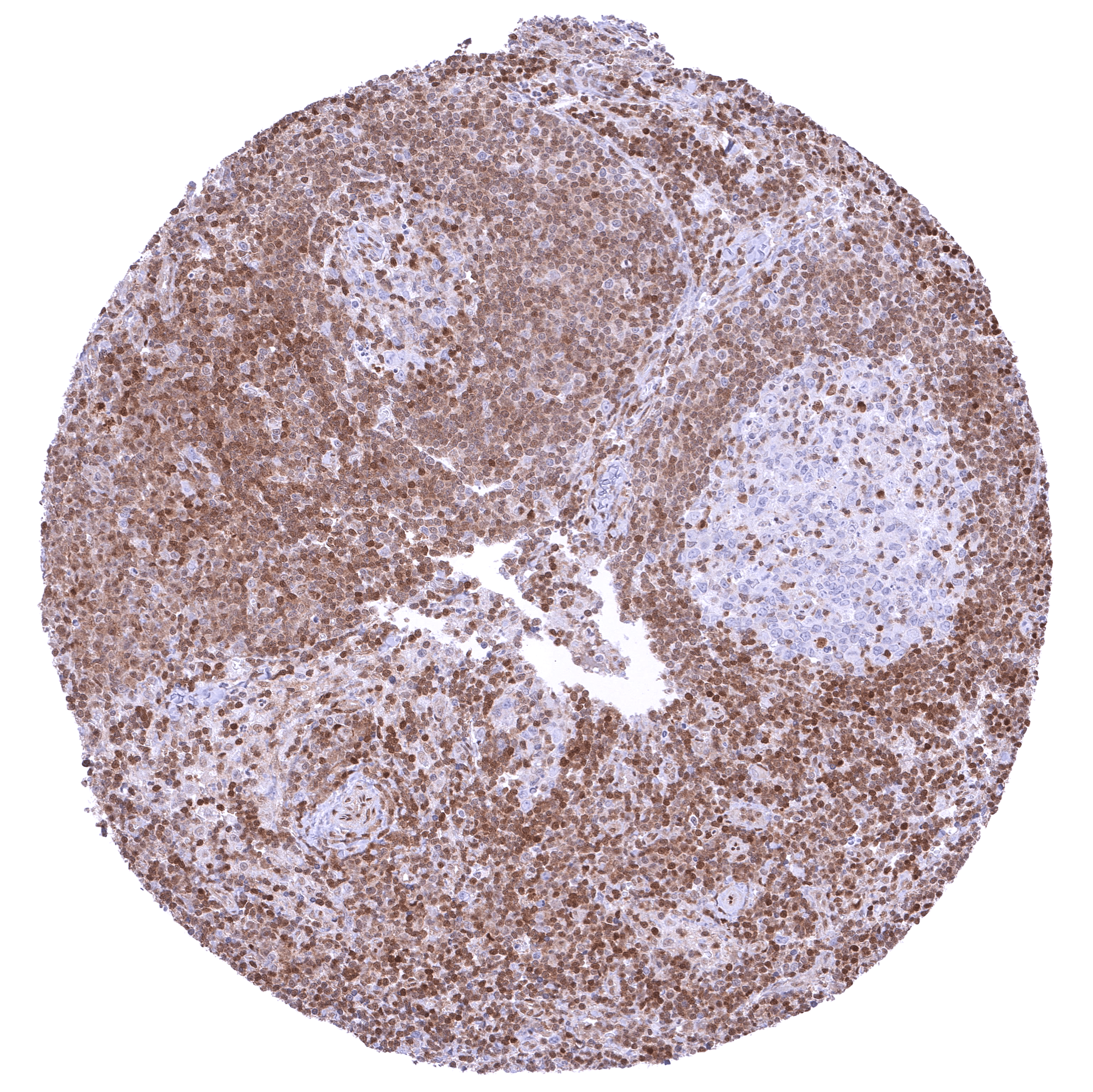
Positive control = Tonsil: A strong nuclear p27 staining should be seen in a small subset of germinal centre cells and in a large fraction of interfollicular lymphocytic cells.
Negative control = Tonsil: p27 staining should be completely absent in most germinal centre cells.
Cellular localization = Nuclear and cytoplasmic
Reactivity = Human
Application = Immunohistochemistry
Dilution = 1:100 – 1:200
Intended Use = Research Use Only
Relevance of Antibody
Biology Behind
p27/kip1 is a cyclin dependent kinase (Cdk) inhibitor which is coded by the CDKN1B gene at chromosome 12p13. The protein consists of 198 amino acids. Because of its lack of a stable tertiary structure it is classified as an intrinsically disordered protein. The best known role of p27 is its downregulation of cell proliferation through inhibition of most cyclin-Cdk complexes which are needed for cell cycle progression by direct binding to both cyclins and Cdks. p27 includes a large kinase inhibitory domain (KID) containing a cyclin-binding (D1), a Cdk-binding (D2), and a linker subdomain to connect D1 and D2, as well as a nuclear export signal (NES) at aa 32-46. P27 exerts its inhibiting role by inserting D2 into the catalytic center of the Cdks where it competes with ATP and blocks the transfer of phosphate to its substrates. Known transcription factors that activate the cellular production of p27 include KHR-L1, AFX, FOXO, Sp1, E2F1, and menin while c-myc, Id3 and Ap1 repress p27 production. Posttranslational processes which also determine the subcellular localization of p27 may be even more important for the regulation of p27 function than expression alone. They also determine the subcellular localization of p27. Phosphorylation and acetylation of p27 at specific sites can trigger its intranuclear degradation or export into the cytoplasm. Apart from cell cycle regulation, p27 has a role in the regulation of cytoskeleton reorganization cell migration, apoptosis, autophagy, and gene expression. p27 deregulation such as decreased nuclear levels or cytoplasmic localization are thought to play a role in several types of disease including cancer and neurodegeneration.
Staining Pattern in Normal Tissues
p27 staining of at least a subset of nuclei occurs in all organs. Nuclear p27 staining is especially abundant in muscle cells, ovarian stroma, lymphocytes, lung and the brain. In various tissues, p27 expression is more prominent in cell types with a low proliferative activity than in those with high rate of cell division. In lymph nodes, for example, germinal centres contain fewer p27 positive cells than the interfollicular space. Images describing the p27 staining pattern in normal tissues obtained by the antibody HMV3970 are shown in our “Normal Tissue Gallery”.
| Brain | Cerebrum | Neurons are mostly negative in the grey matter while there are many p27 positive cells in the white matter. |
| Cerebellum | Strong nuclear p27 positivity of most granular cells while Purkinje cells and most cells of the molecular layer are p27 negative. In the white matter there are many strongly p27 positive cells. | |
| Endocrine Tissues | Thyroid | Weak to moderate nuclear p27 positivity of most follicular cells. |
| Parathyroid | Weak to moderate nuclear p27 positivity of most epithelial cells. | |
| Adrenal gland | Distinct nuclear p27 positivity of a large fraction of cells. | |
| Pituitary gland | Weak to moderate nuclear p27 positivity of a fraction of epithelial cells in the adenohypophysis. Pituicytes are mostly p27 negative. | |
| Respiratory system | Respiratory epithelium | Strong nuclear p27 positivity of a large fraction of respiratory epithelial cells. |
| Lung | Strong nuclear p27 positivity of a large fraction of pneumocytes. | |
| Gastrointestinal Tract | Salivary glands | p27 staining is more common in glandular cells than in excretory ducts. |
| Esophagus | Nuclear p27 staining of squamous epithelial cells predominates in the more mature cell layers (top 50% of the epidermis). | |
| Stomach | Nuclear p27 positivity predominates in superficial epithelial cells and is rarely seen in neck cells. | |
| Duodenum | Nuclear p27 positivity of a fraction of epithelial cells. | |
| Small intestine | Nuclear p27 positivity of a fraction of epithelial cells. | |
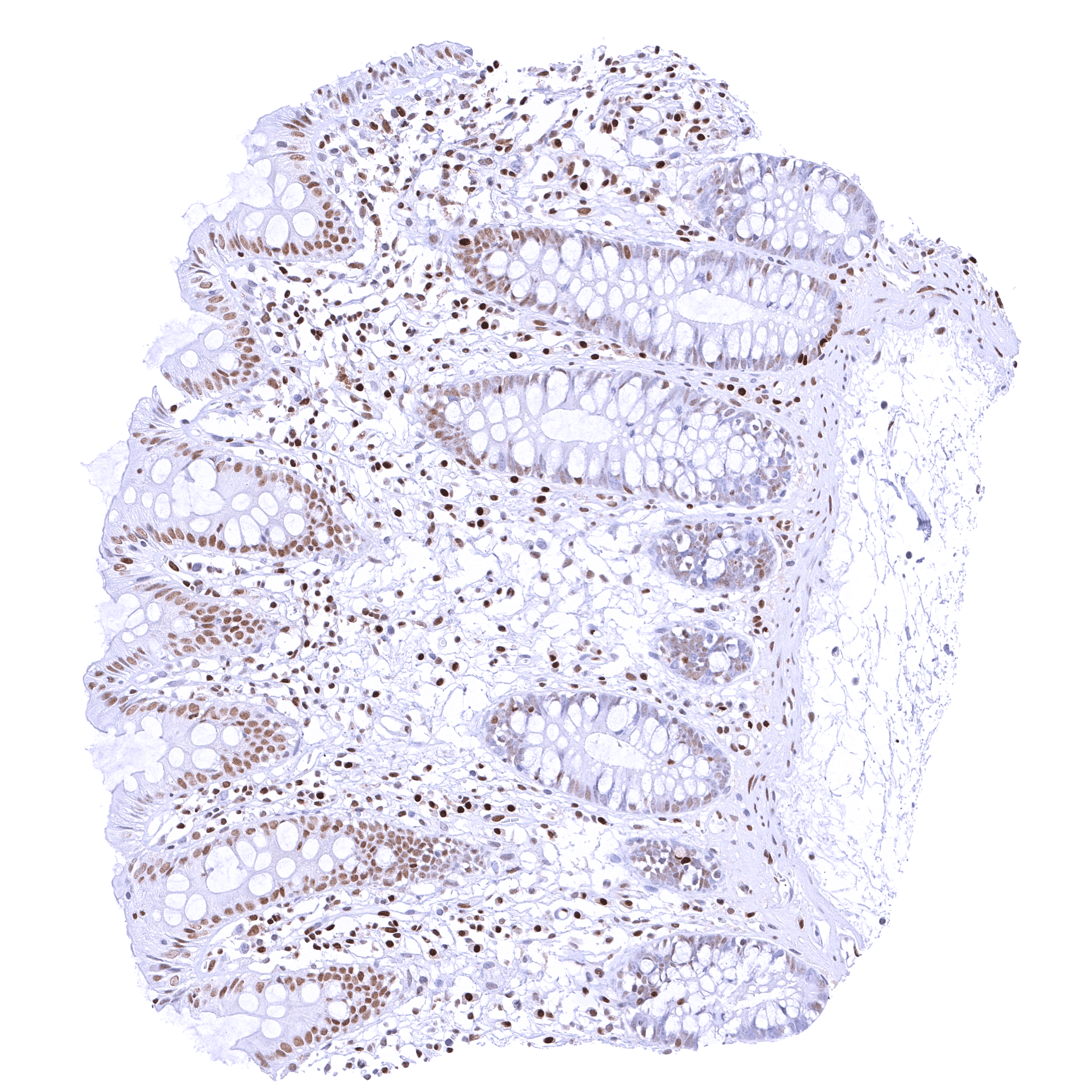
| Appendix | Among epithelial cells, nuclear p27 staining predominates in superficial cells layers and is rare in crypts. Most p27 staining occurs in lymphatic cells, however. | |
| Colon | Among epithelial cells, nuclear p27 staining predominates in superficial cells layers and is rare in crypts. | |
| Rectum | Among epithelial cells, nuclear p27 staining predominates in superficial cells layers and is rare in crypts. | |
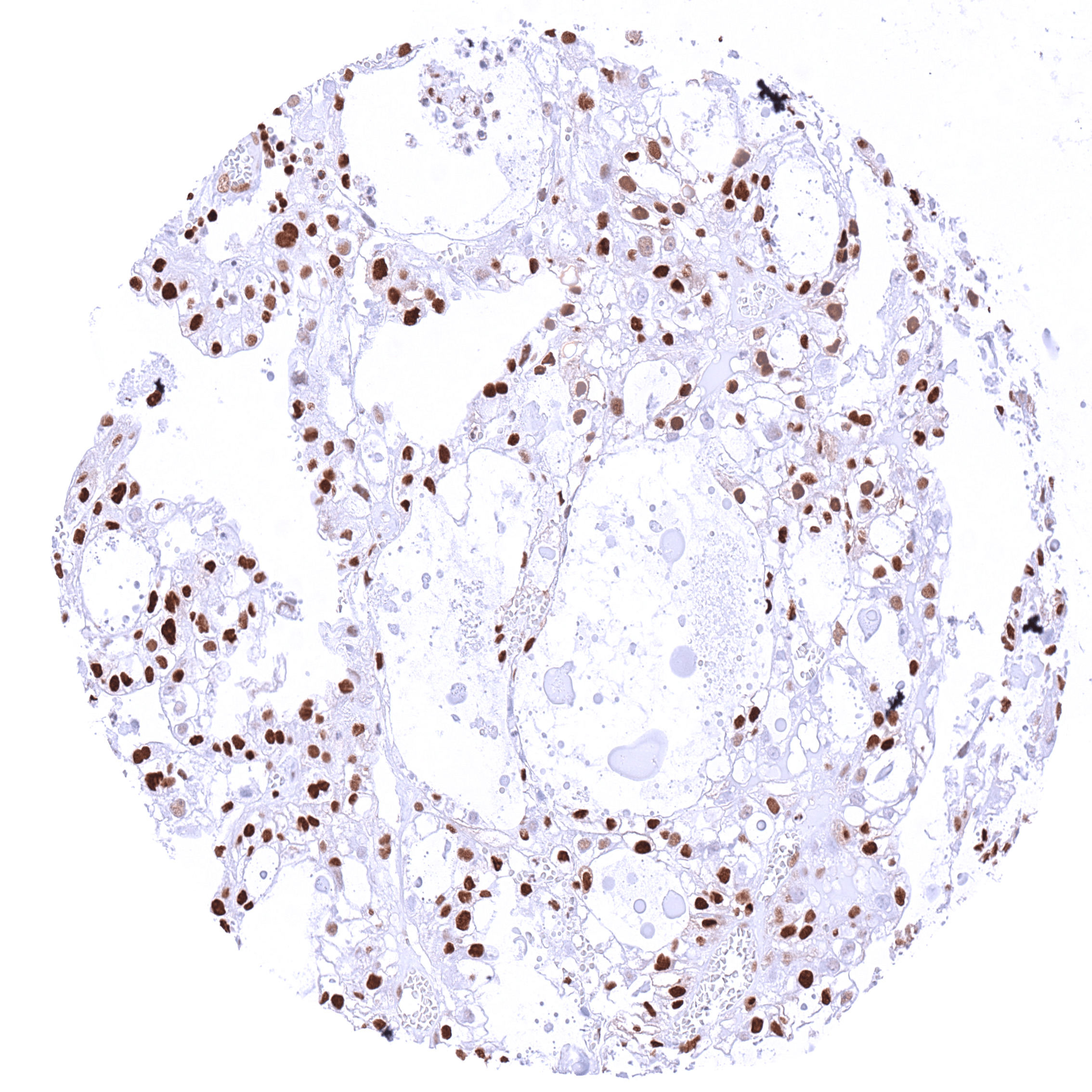
| Liver | p27 staining is rare or absent in hepatocytes. | |
| Gallbladder | ||
| Pancreas | p27 staining is more common in islet cells than in acinar cells. | |
| Genitourinary | Kidney | Tubuli are usually p27 negative. The strongest p27 staining occurs in glomeruli and in stroma cells including small vessels. |
| Urothelium | Nuclear p27 staining is mostly lacking in urothelial cells. If it occurs (only in few samples), it predominates in the more superficial cell layers. | |
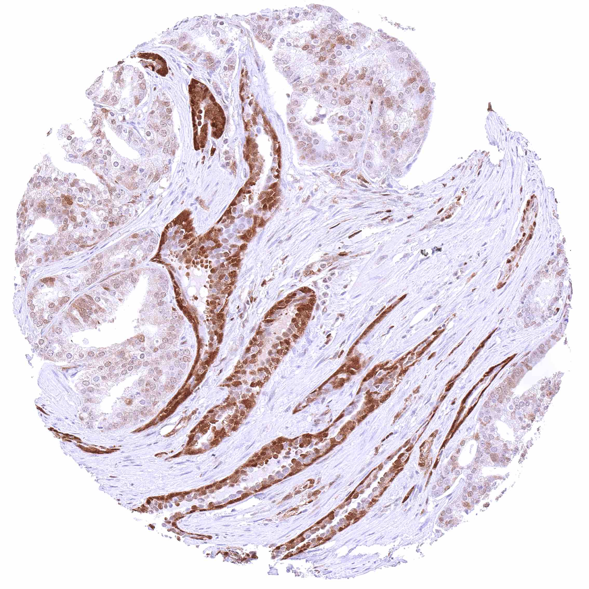
| Male genital | Prostate | Prostate epithelial cells are often p27 negative. |
| Seminal vesicles | Weak nuclear p27 staining in a subset of epithelial cells. | |
| Testis | Weak to moderate nuclear p27 staining of a subset of cells (predominantly Sertoli cells). | |
| Epididymis | Weak nuclear p27 staining of a subset of epithelial cells. | |
| Female genital | Breast | Moderate to strong nuclear p27 positivity of a large fraction of glandular epithelial cells. |
| Uterus, myometrium | Nuclear p27 staining of variable intensity in most muscle cells. | |
| Uterus, ectocervix | Nuclear staining of squamous epithelial cells predominates in the more mature cell layers (top 50% of the epidermis). | |
| Uterus endocervix | Nuclear p27 staining is highly variable in epithelial cells of the endocervix and ranges from negativity of virtually all cells to strong positivity of virtually all cells. | |
| Uterus, endometrium | Nuclear p27 staining is either weak or absent in normal endometrium. In case of pregnancy, the staining is distinct in both endometrium and decidua cells. | |
| Fallopian Tube | Strong nuclear p27 staining of most epithelial cells. | |
| Ovary | Strong nuclear p27 staining of most or all stroma cells. Weak to moderate nuclear p27 staining of all corpus luteum cells. | |
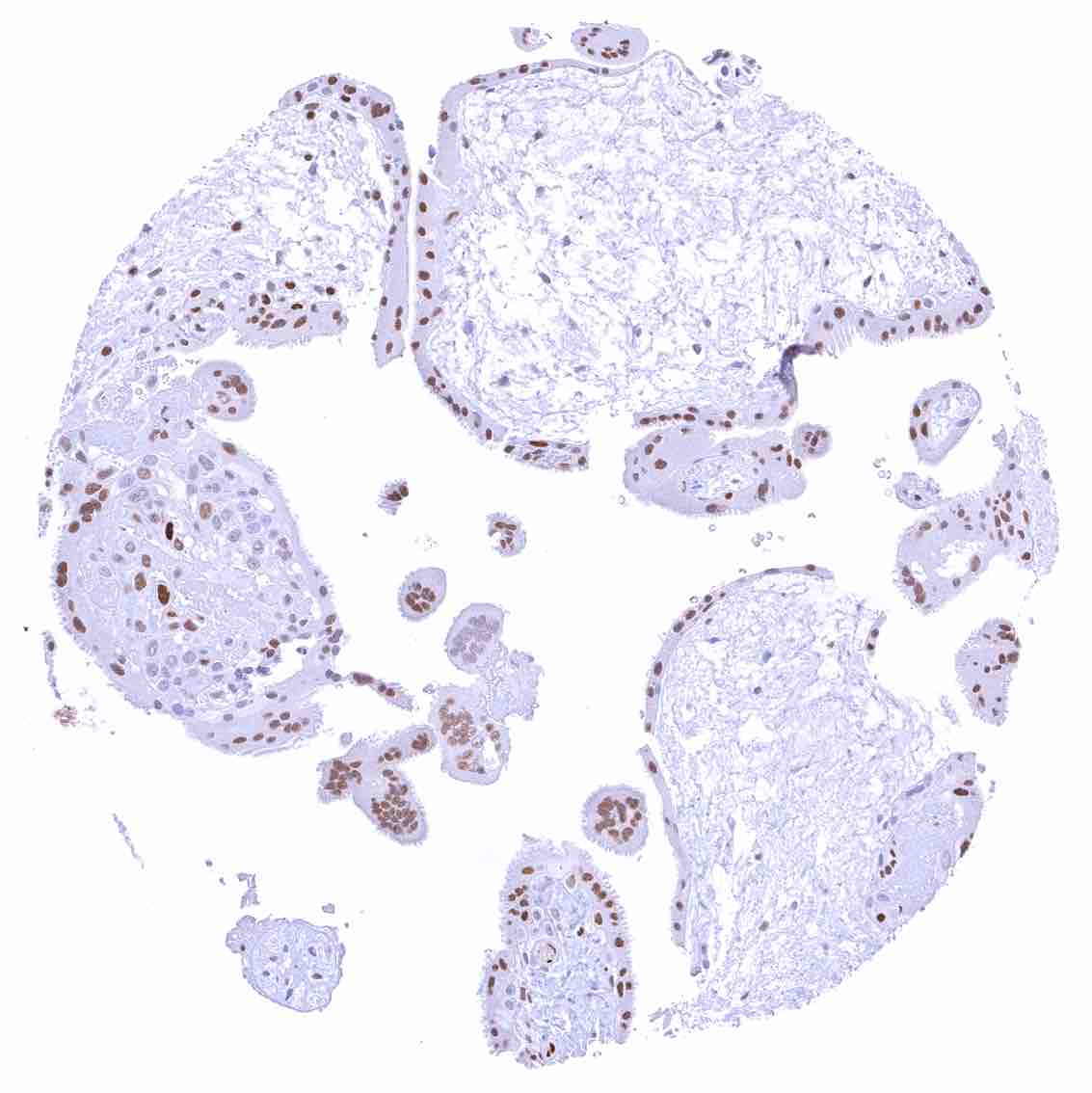
| Placenta early | Strong p27 staining of all nuclei of the syncytiotrophoblast. | |
| Placenta mature | Weak nuclear p27 staining of trophoblast and stroma cells. | |
| Amnion | A weak to moderate nuclear p27 staining occurs in a subset of amnion cells. | |
| Chorion | Strong nuclear p27 staining of most or all chorion cells. | |
| Skin | Epidermis | Moderate to strong nuclear p27 staining in a large fraction of squamous epithelial cells, predominantly in the more mature cell layers (top 50% of the epidermis). |
| Sebaceous glands | Moderate to strong nuclear p27 staining in most/all cells of sebaceous glands. | |
| Muscle/connective tissue | Heart muscle | Distinct nuclear p27 staining of stroma cells while myocytes are largely negative. |
| Skeletal muscle | Strong nuclear p27 staining of most but not all cells. | |
| Smooth muscle | Variable (intense – weak – absent) nuclear p27 staining of smooth muscle cells. | |
| Vessel walls | Strong nuclear p27 staining of muscle cells in the aorta. | |
| Fat | Variable nuclear p27 staining of fat cells. | |
| Stroma | Nuclear p27 staining can occur in various types of stroma cells. | |
| Endothelium | In some tissues a nuclear p27 positivity is seen in all endothelial cells. In other tissues, endothelial cells do not stain. | |
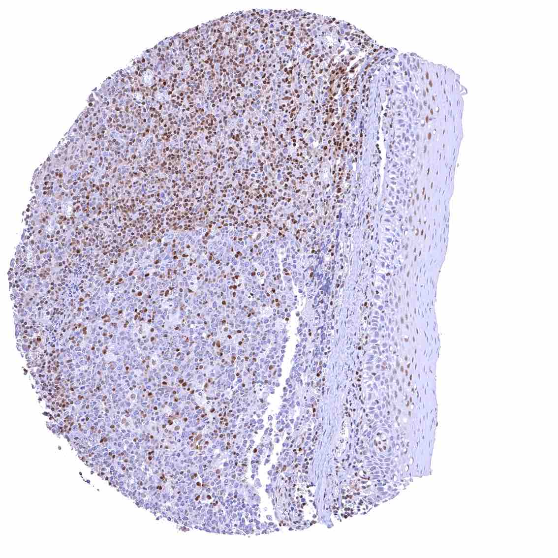
| Bone marrow/ lymphoid tissue | Bone marrow | The number of p27 positive cells is rather low. |
| Lymph node | Strong nuclear p27 staining of a significant subset of cells, especially in the interfollicular area. | |
| Spleen | Strong nuclear p27 staining of a large fraction of cells. | |
| Thymus | Strong nuclear p27 staining of a large fraction of cells. | |
| Tonsil | Strong nuclear p27 staining of a significant subset of cells, especially in the interfollicular area. Nuclear staining of squamous epithelial cells predominates in the more mature cell layers (top 50% of the epidermis). | |
| Remarks |
These findings are largely consistent with the RNA data described in the Human Protein Atlas (Tissue expression p27)
Positive control = Tonsil: A strong nuclear p27 staining should be seen in a small subset of germinal centre cells and in a large fraction of interfollicular lymphocytic cells.
Negative control = Tonsil: p27 staining should be completely absent in most germinal centre cells.
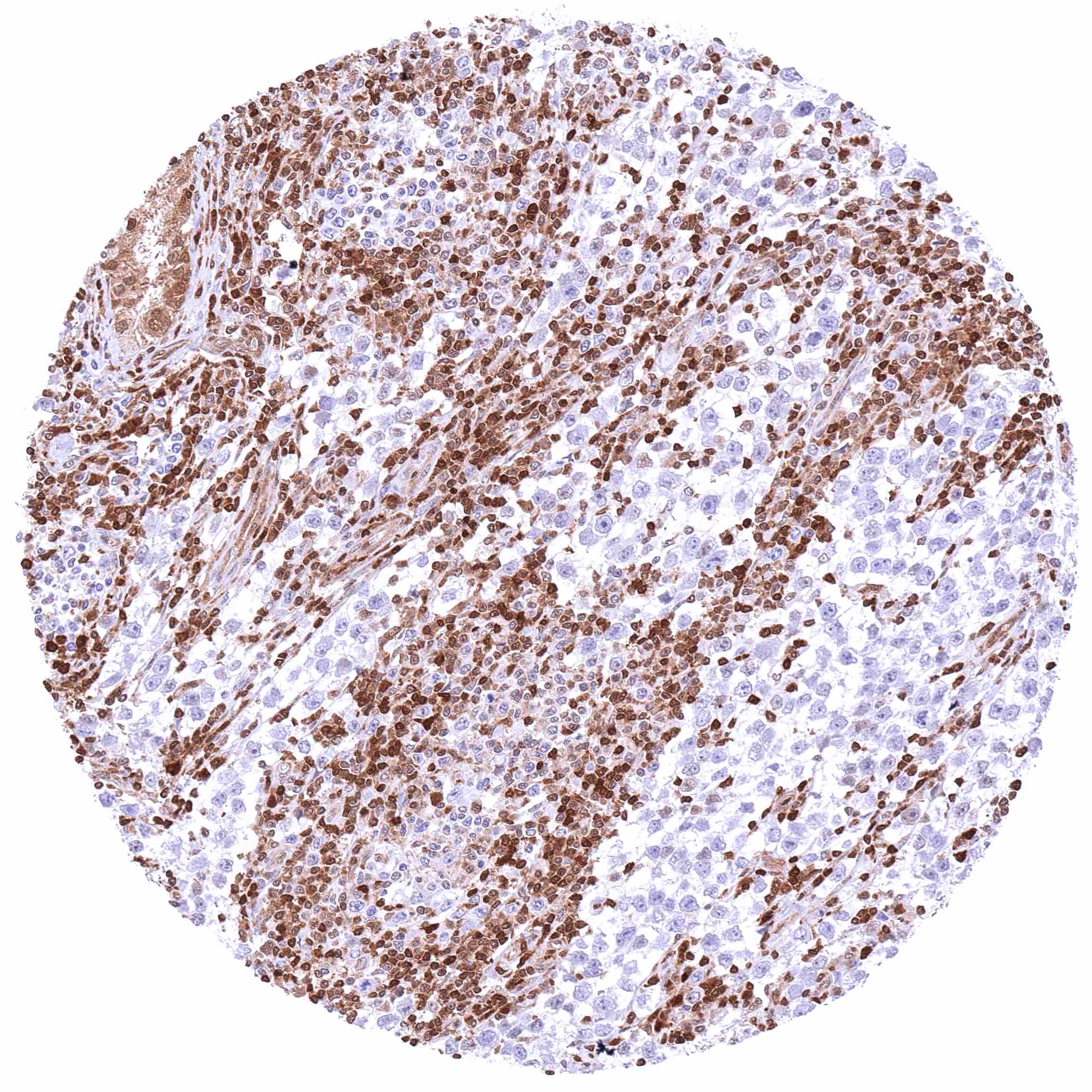
Staining Pattern in Relevant Tumor Types
p27 expression – at variable levels – has been described to occur in virtually all cancer types. Reduced expression has been linked to unfavorable tumor features or poor prognosis in many publications. The TCGA findings on p27 RNA expression in different tumor categories have been summarized in the Human Protein Atlas.
Protocol Recommendations
IHC users have different preferences on how the stains should look like. Some prefer high staining intensity of the target stain and even accept some background. Others favor absolute specificity and lighter target stains. Factors that invariably lead to more intense staining include higher concentration of the antibody and visualization tools, longer incubation time, higher temperature during incubation, higher temperature and longer duration of the heat induced epitope retrieval (slide pretreatment). The impact of the pH during slide pretreatment has variable effects and depends on the antibody and the target protein.
All images and data shown here and in on image galleries are obtained by the manual protocol described below. Other protocols resulting in equivalent staining are described as well.
Manual protocol
Freshly cut sections should be used (less than 10 days between cutting and staining). Heat-induced antigen retrieval for 5 minutes in an autoclave at 121°C in pH9 Target Retrieval Solution buffer. Apply HMV3970 at a dilution of 1:150 at 37°C for 60 minutes.
Potential Research Applications
- The prognostic role of p27 alterations needs to be further investigated in cancer.
- The role of p27 in cytoskeletal dynamics, cell migration, apoptosis, and autophagy needs to be further explored.
- The role of p27 as a transcriptional regulator awaits further exploration.
- The role of p27 in neurodegeneration has not been sufficiently clarified.
Evidence for Antibody Specificity in IHC
There are two ways how the specificity of antibodies can be documented for immunohistochemistry on formalin fixed tissues. These are: 1. Comparison with a second independent method for target expression measurement across a large number of different tissue types (orthogonal strategy), and 2. Comparison with one or several independent antibodies for the same target and showing that all positive staining results are also seen with other antibodies for the same target (independent antibody strategy).
Orthogonal validation: For the antibody HMV3970 specificity is in line with data from three independent RNA screening studies, including the Human Protein Atlas (HPA) RNA-seq tissue dataset, the FANTOM5 project, and the Genotype-Tissue Expression (GTEx) project, which are all summarized in the Human Protein Atlas (Tissue expression p27). In agreement with HMV3970 immunostaining data, high p27 RNA expression occurs in all normal tissues. However, it must be understood that orthogonal validation is not optimal for assessing ubiquitously expressed protein.
Comparison of antibodies: True expression of p27 in cell types with documented p27 immunostaining by HMV3970 is validated by an identical staining pattern obtained by a second, independent commercially available p27 antibody, termed “validation antibody” for virtually all normal tissues. An additional fibrillar staining in some areas of the brain was only observed by the validation antibody and therefore considered an antibody specific cross-reactivity of this validation antibody.