295,00 € – 995,00 €
Product details
Synonyms = MX dynamin like GTPase 1,IFI-78K,IFI78,MX,MxA
Antibody type = Recombinant Rabbit monoclonal / IgG
Clone = HMV316
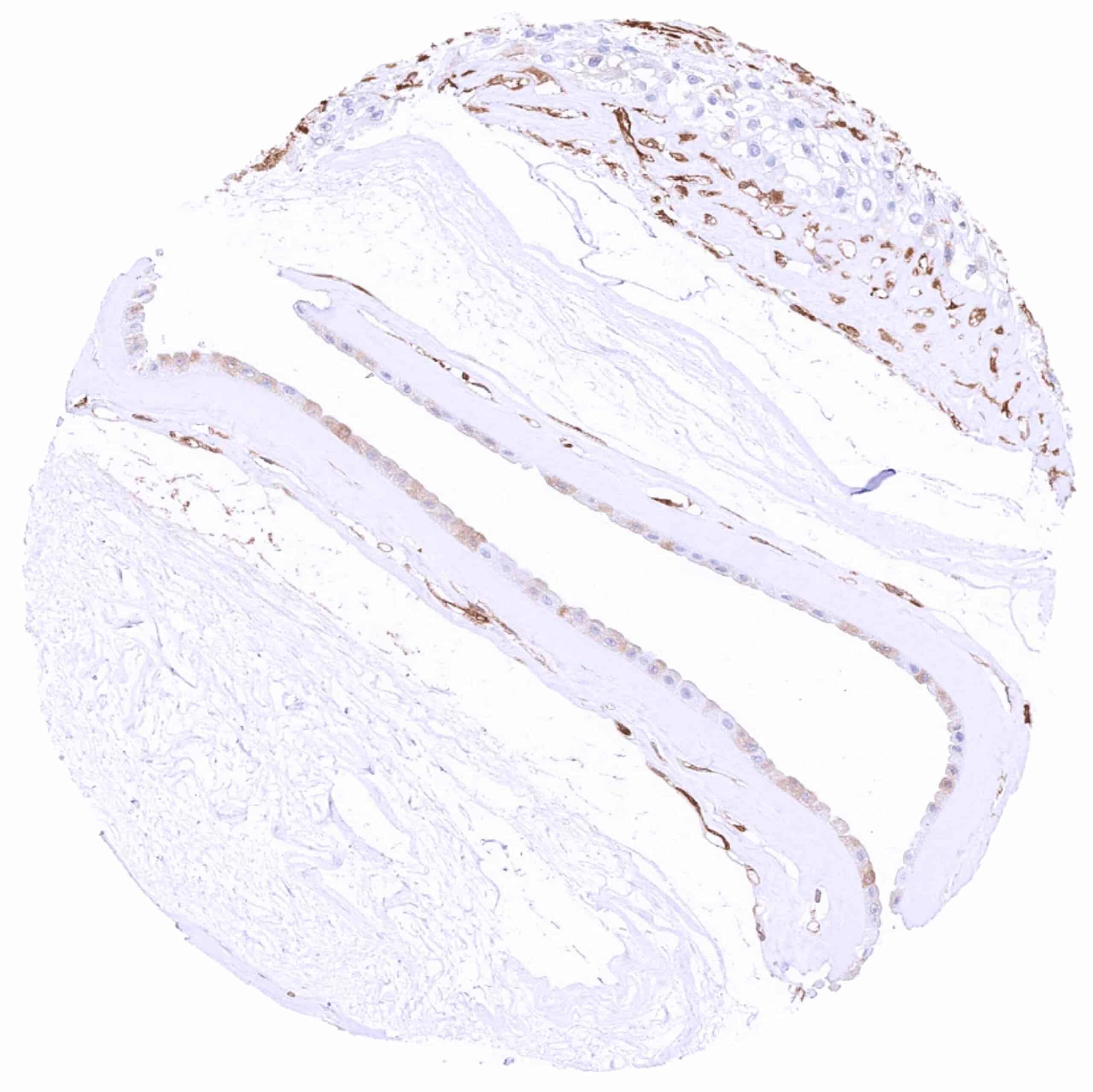
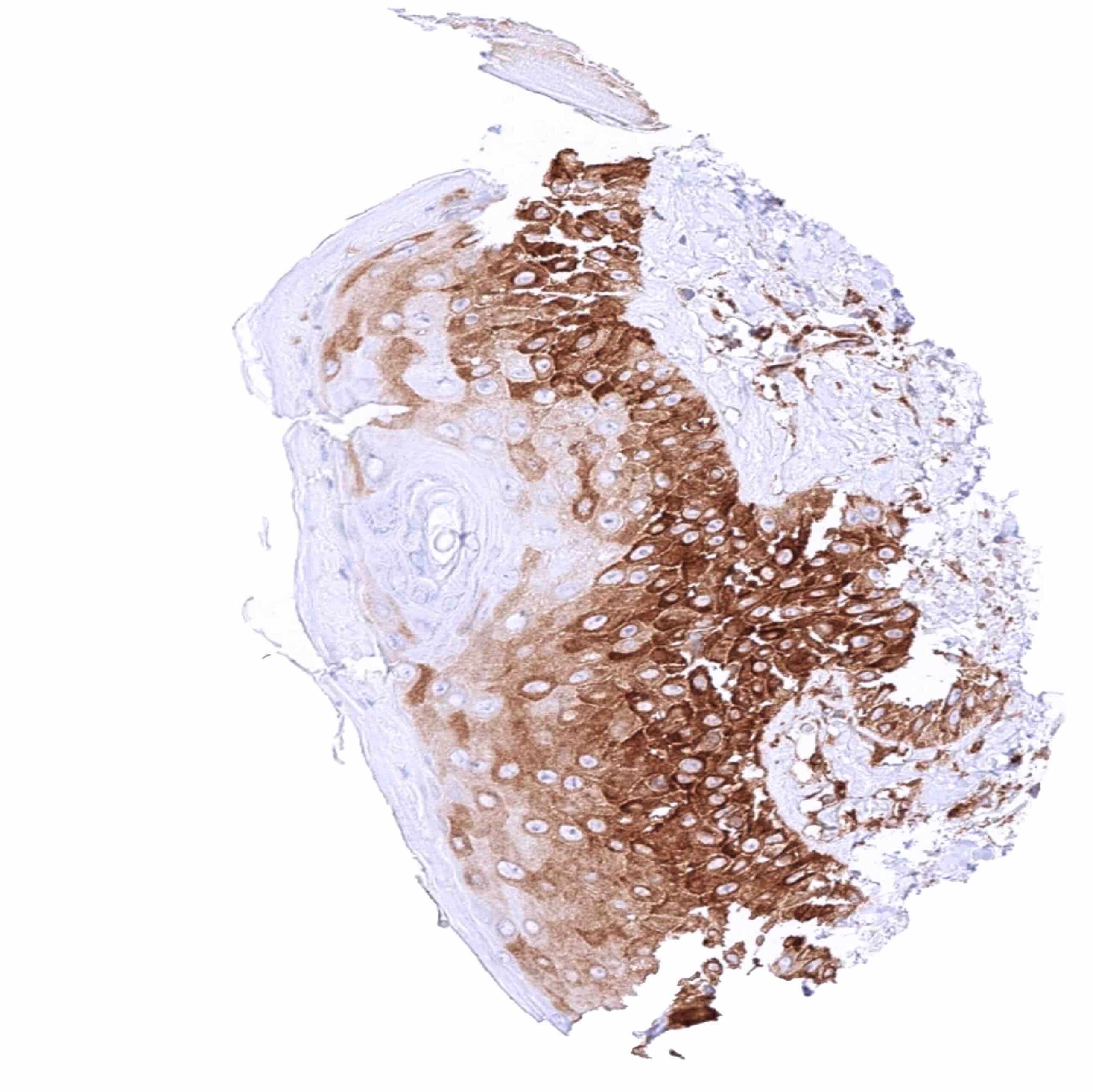
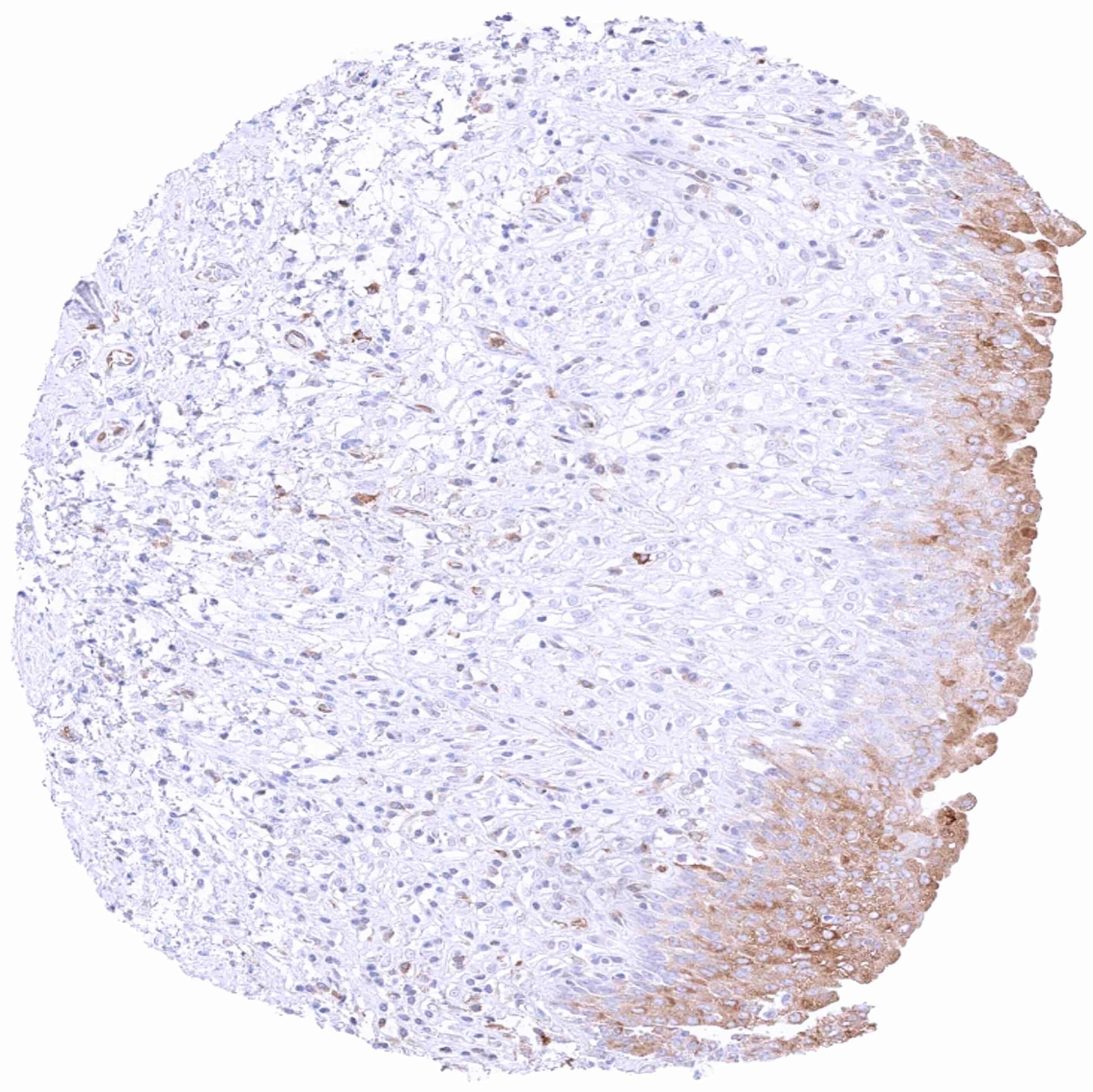
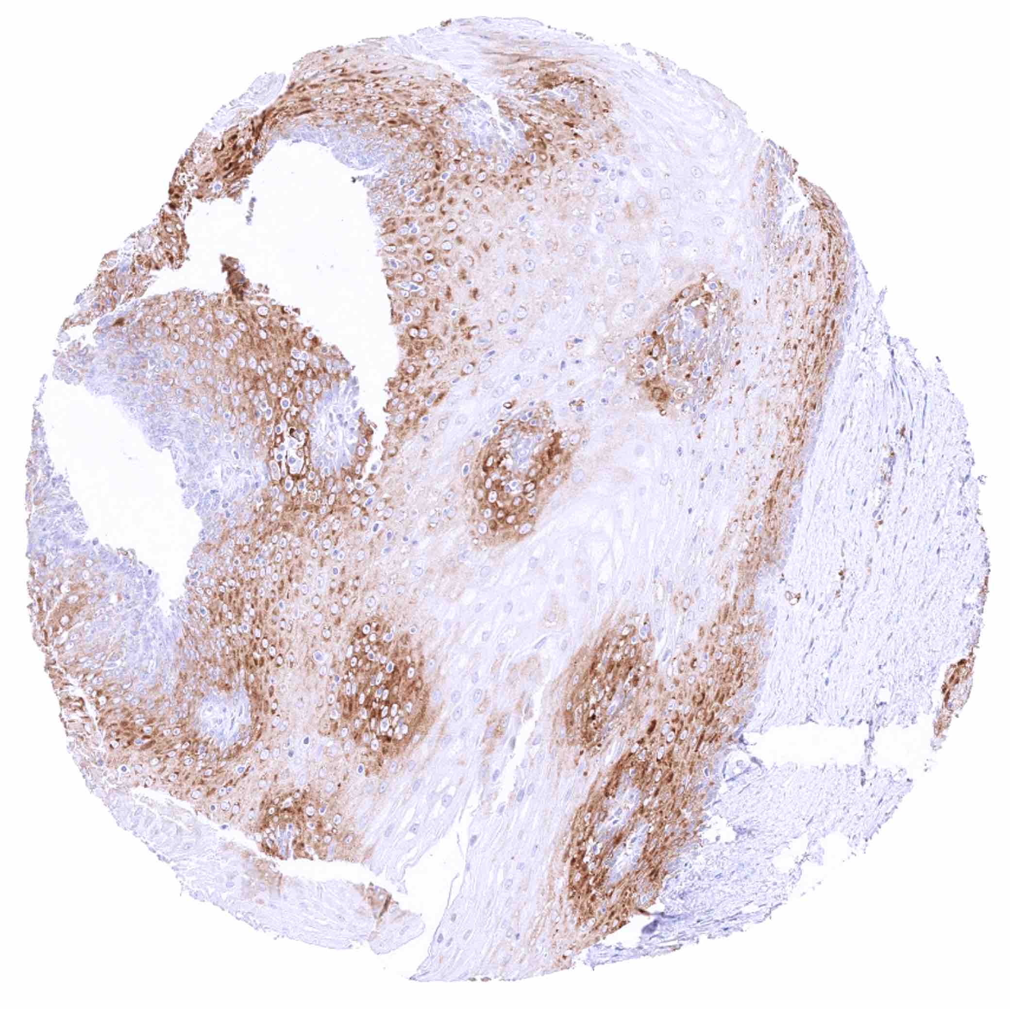
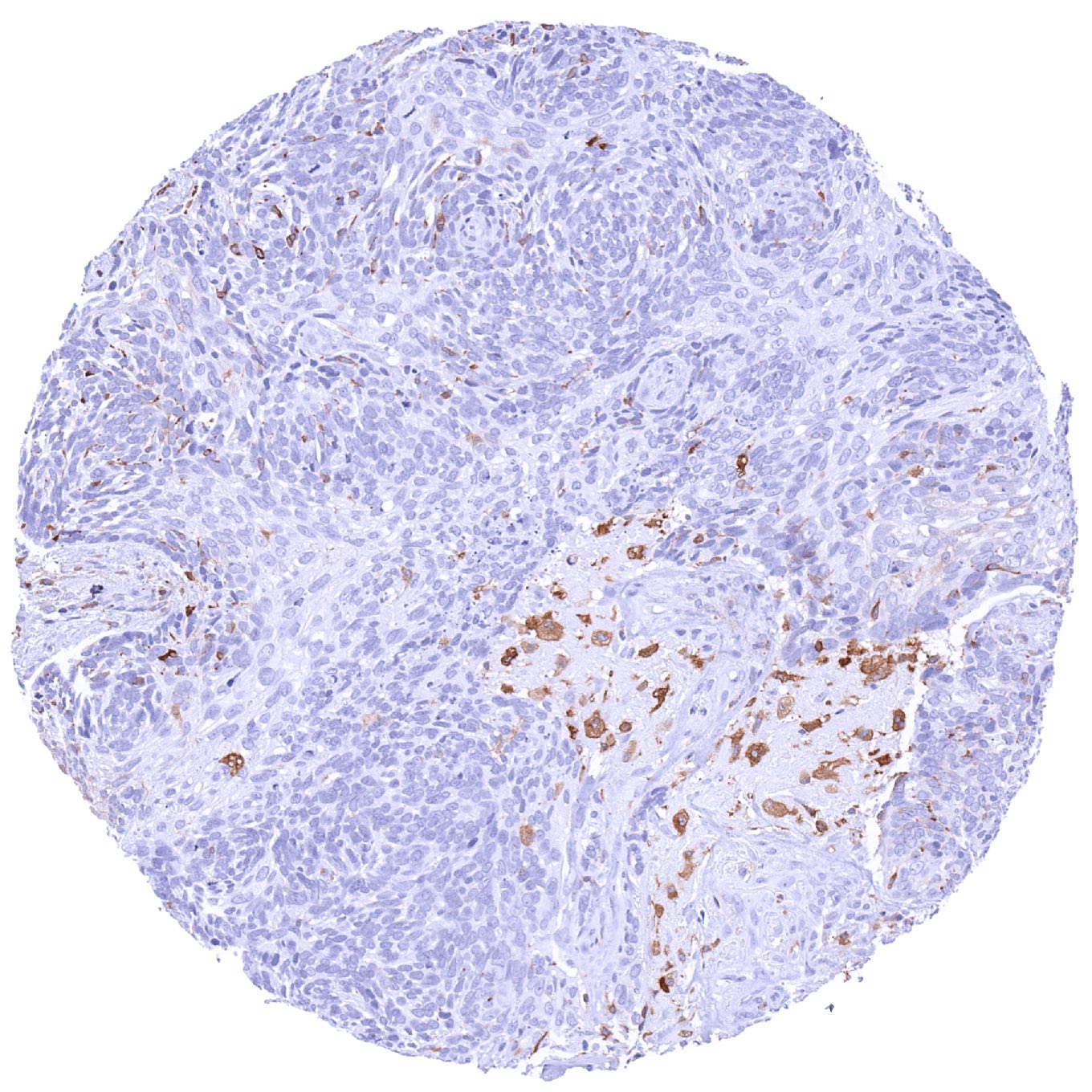
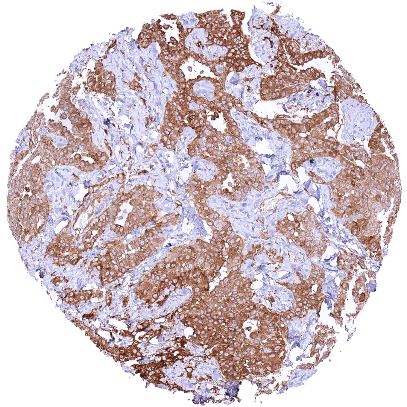
Positive control = Prostate: An at least weak to moderate MX1 staining should be seen in basal cells while acinar cells remain negative.
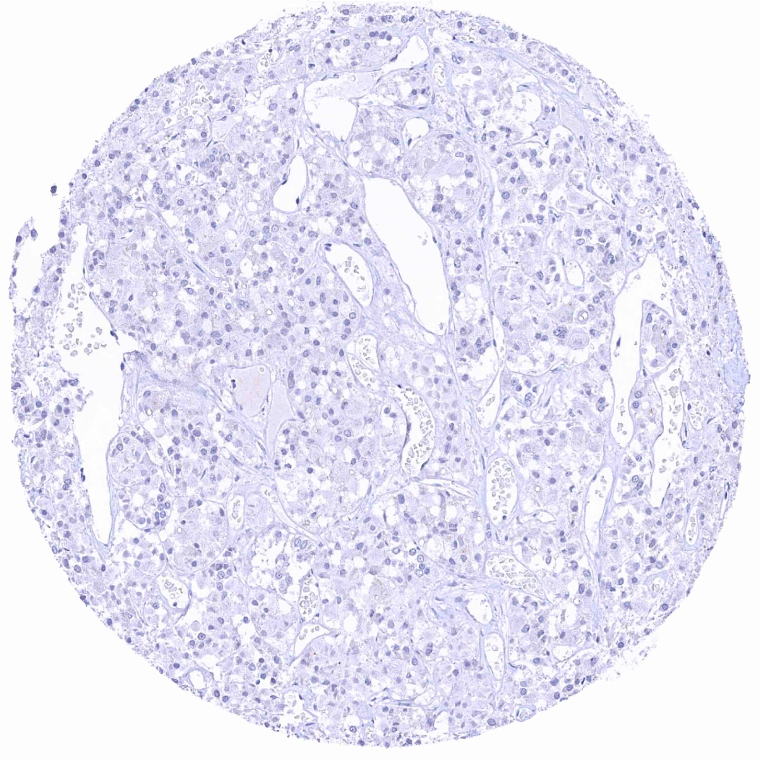
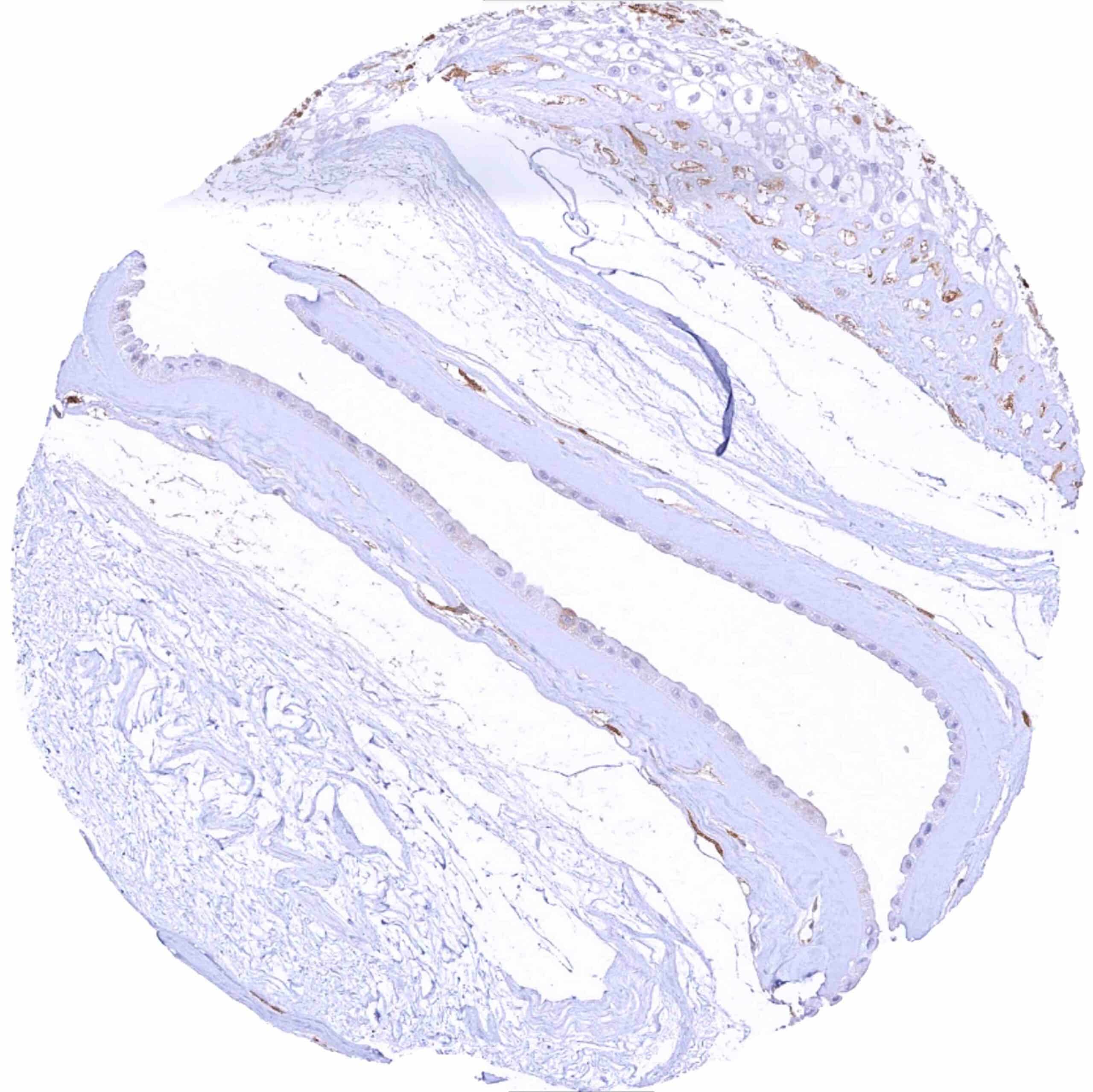
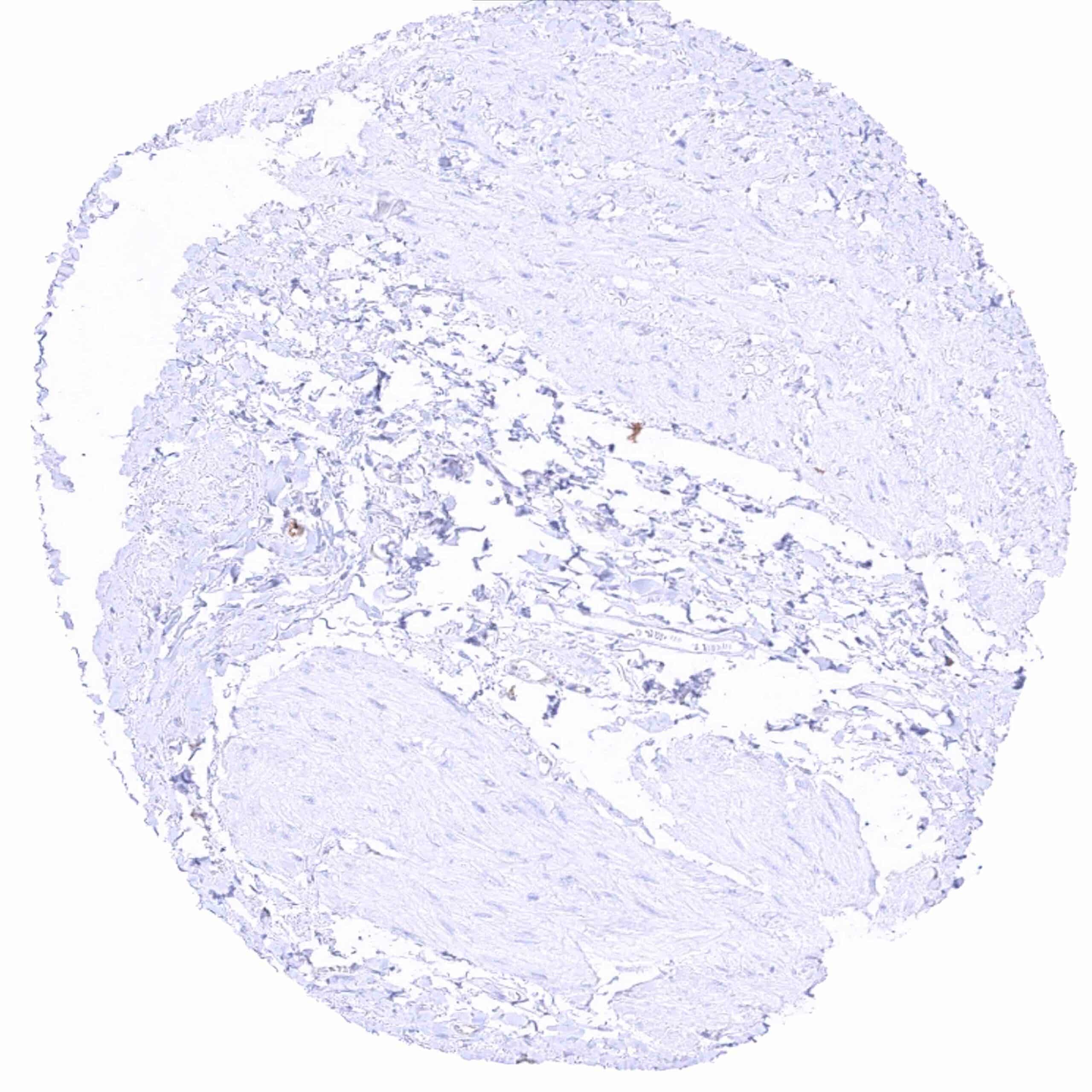
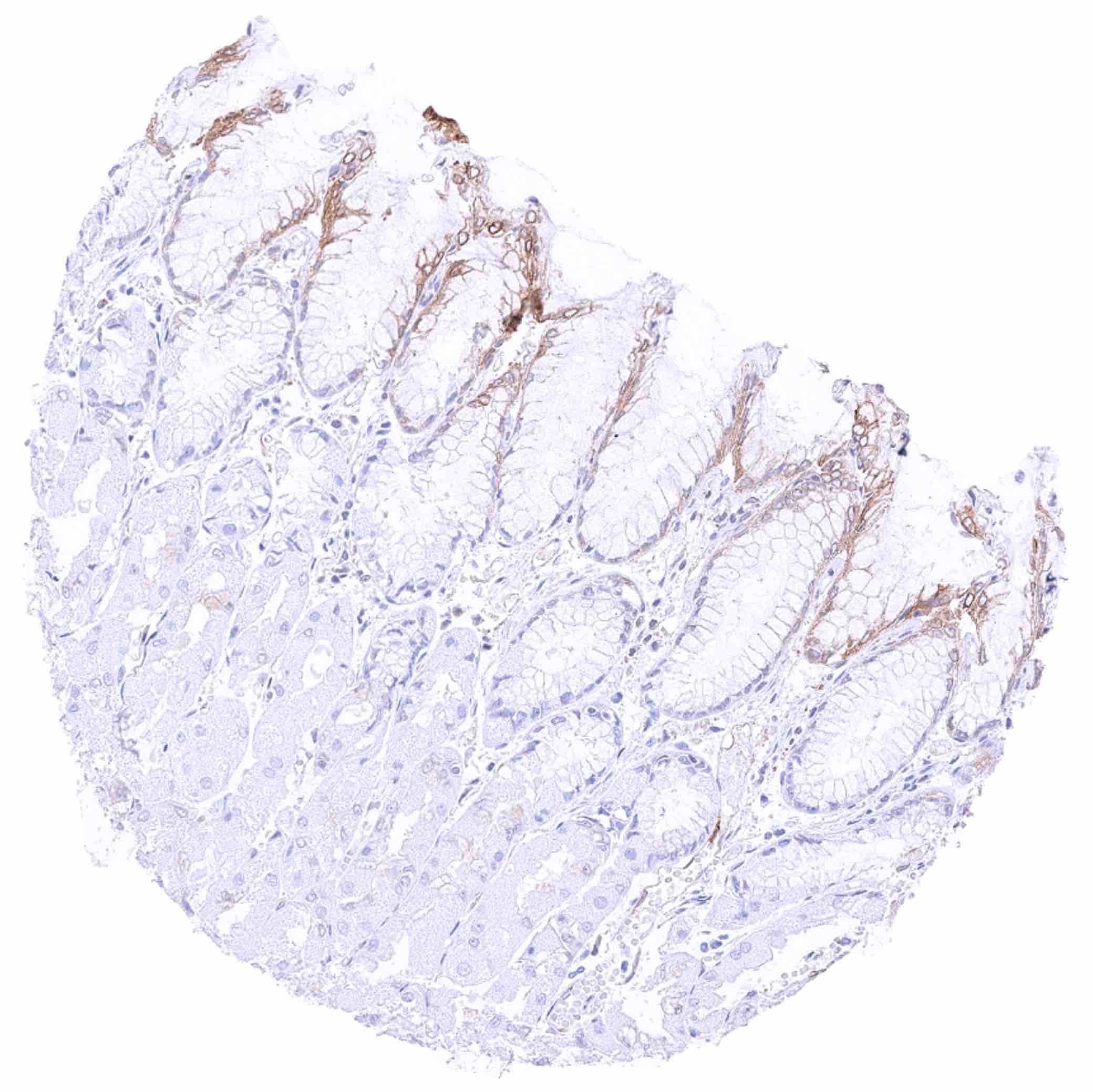
Negative control = Colon: Crypt epithelial cells should be MX1 negative.
Cellular localization = Intracellular
Reactivity = Human
Application = Immunohistochemistry
Dilution = 1:100 – 1:200
Intended Use = Research Use Only
Relevance of Antibody
MX1 is a GTP-binding protein with unknown biologic role.
Biology Behind
The interferon-induced GTP-binding protein MX1 (myxovirus resistance protein 1) is coded by the MX1 gene located at 21q22.3. The gene consists of 17 exons and extends over 33 kb. MX1 is a member of a family of large GTPases which belongs to the dynamin superfamily. MX1 is a mediator of the resistance mechanisms to infections by influenza and other viruses both in cell culture and in transgenic mice. Its role in human influenza virus resilience is not fully understood, however, as humans harboring heterozygous and homozygous combinations of allelic MX1 variants could be linked to neither increased susceptibility to influenza virus nor to increased likelihood of severe disease. RNA expression data suggest that MX1 is ubiquitously expressed in normal tissues with a predominance in lymphatic tissues. A role in cancer biology has also been proposed for MX1. MX1 was found to be overexpressed in various cancer entities and the level of expression has been found to be linked to parameters of cancer aggressiveness.
Staining Pattern in Normal Tissues
Images describing the MX1 staining pattern in normal tissues obtained by the antibody HMV316 are shown in our “Normal Tissue Gallery”.
| Brain | Cerebrum | MX1 staining of endothelial cells. |
| Cerebellum | MX1 staining of endothelial cells. | |
| Endocrine Tissues | Thyroid | Weak to moderate cytoplasmic MX1 staining of follicular cells. |
| Parathyroid | Negative. | |
| Adrenal gland | Moderate cytoplasmic MX1 staining of medullary cells. Adrenocortical cells are MX1 negative or (focally) show weak positivity. | |
| Pituitary gland | Negative. | |
| Respiratory system | Respiratory epithelium | Focal, weak to strong cytoplasmic MX1 staining in subsets of respiratory epithelial cells. |
| Lung | Pneumocytes are MX1 negative. Moderate to strong cytoplasmic MX1 staining of macrophages. | |
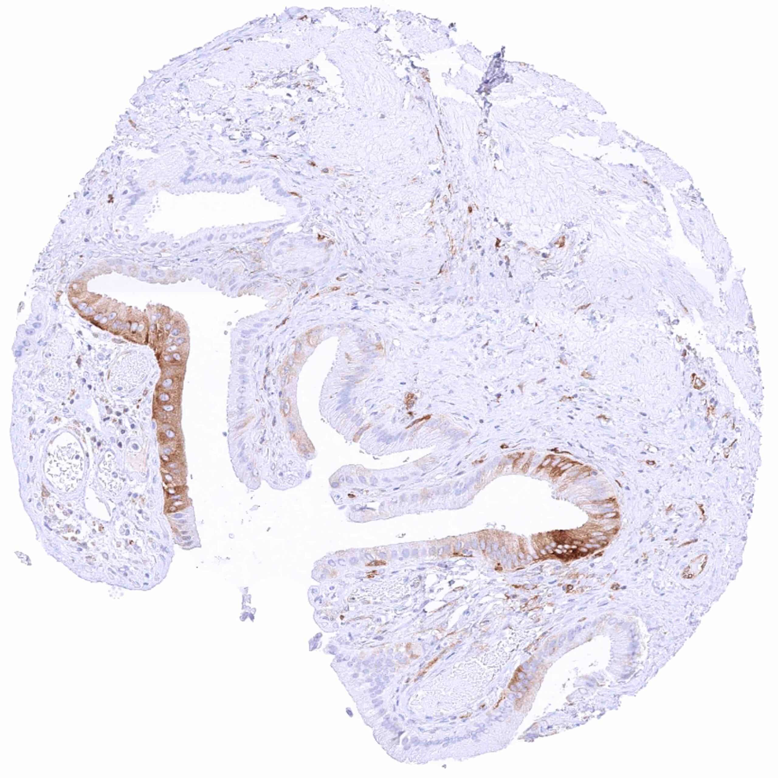
| Gastrointestinal Tract | Salivary glands | Moderate to strong cytoplasmic MX1 staining can be seen in some epithelial cell groups. |
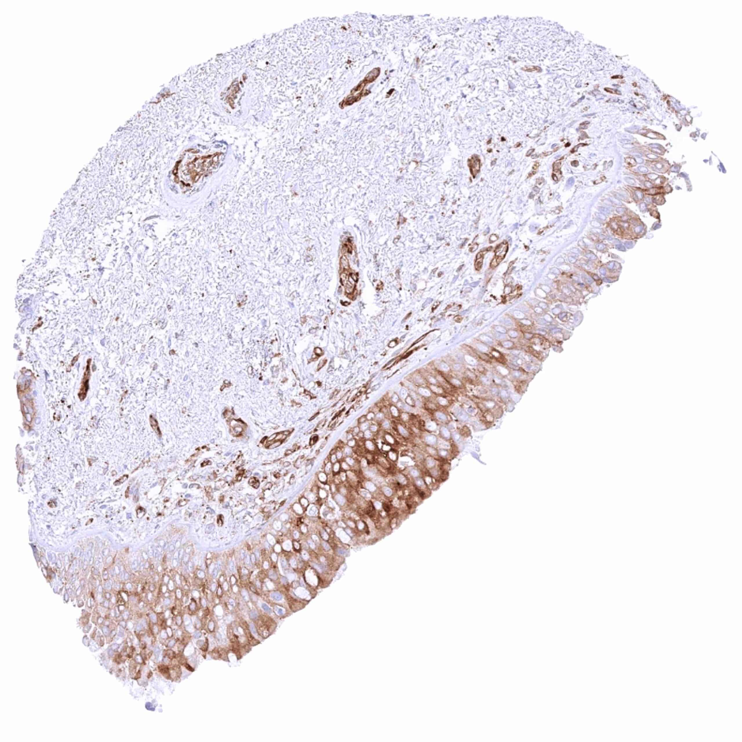
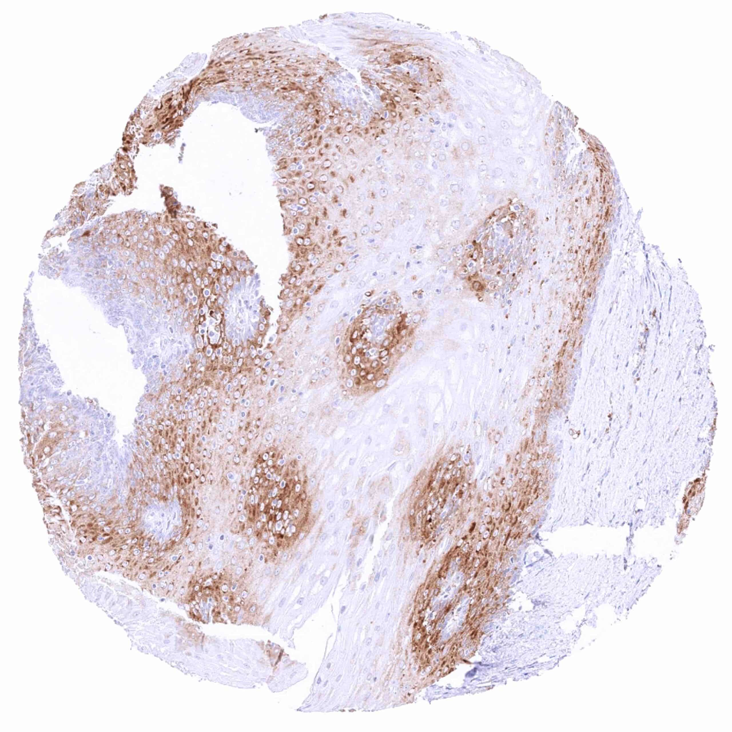
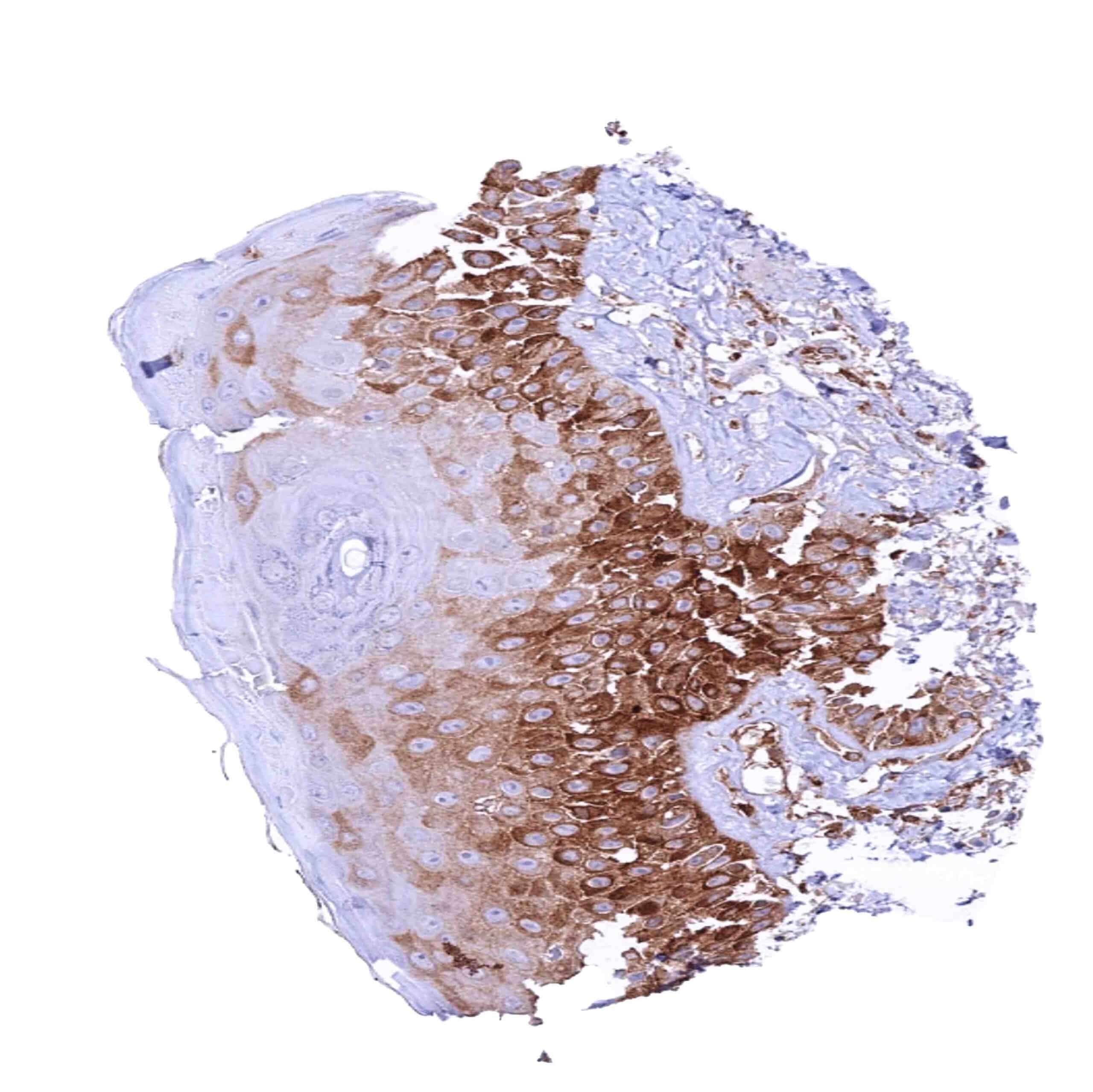
| Esophagus | Weak to strong cytoplasmic MX1 staining of suprabasal cell layers. | |
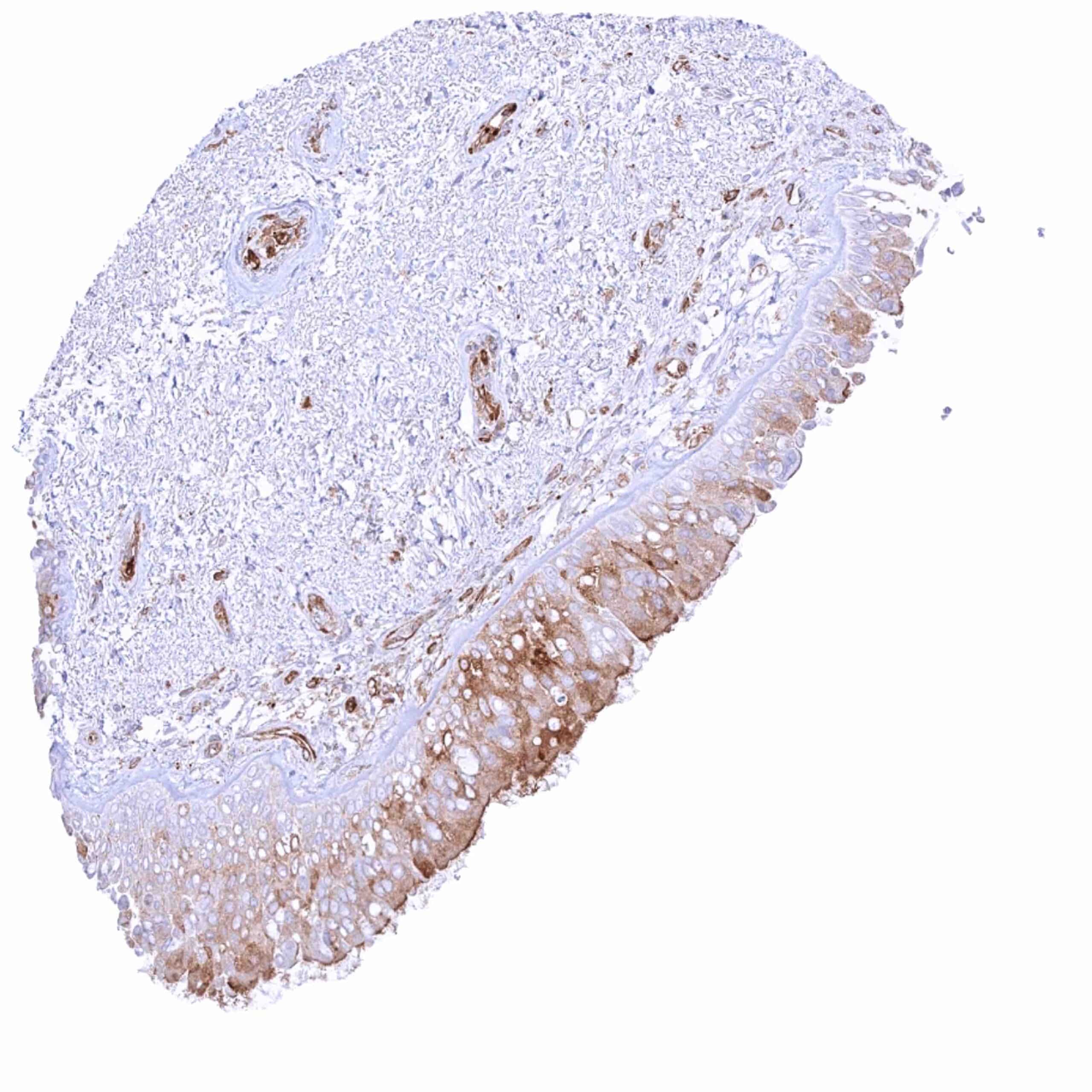
| Stomach | Weak to moderate cytoplasmic MX1 staining of surface epithelial cells. Staining is even more intense in a sample with chronic inflammation. | |
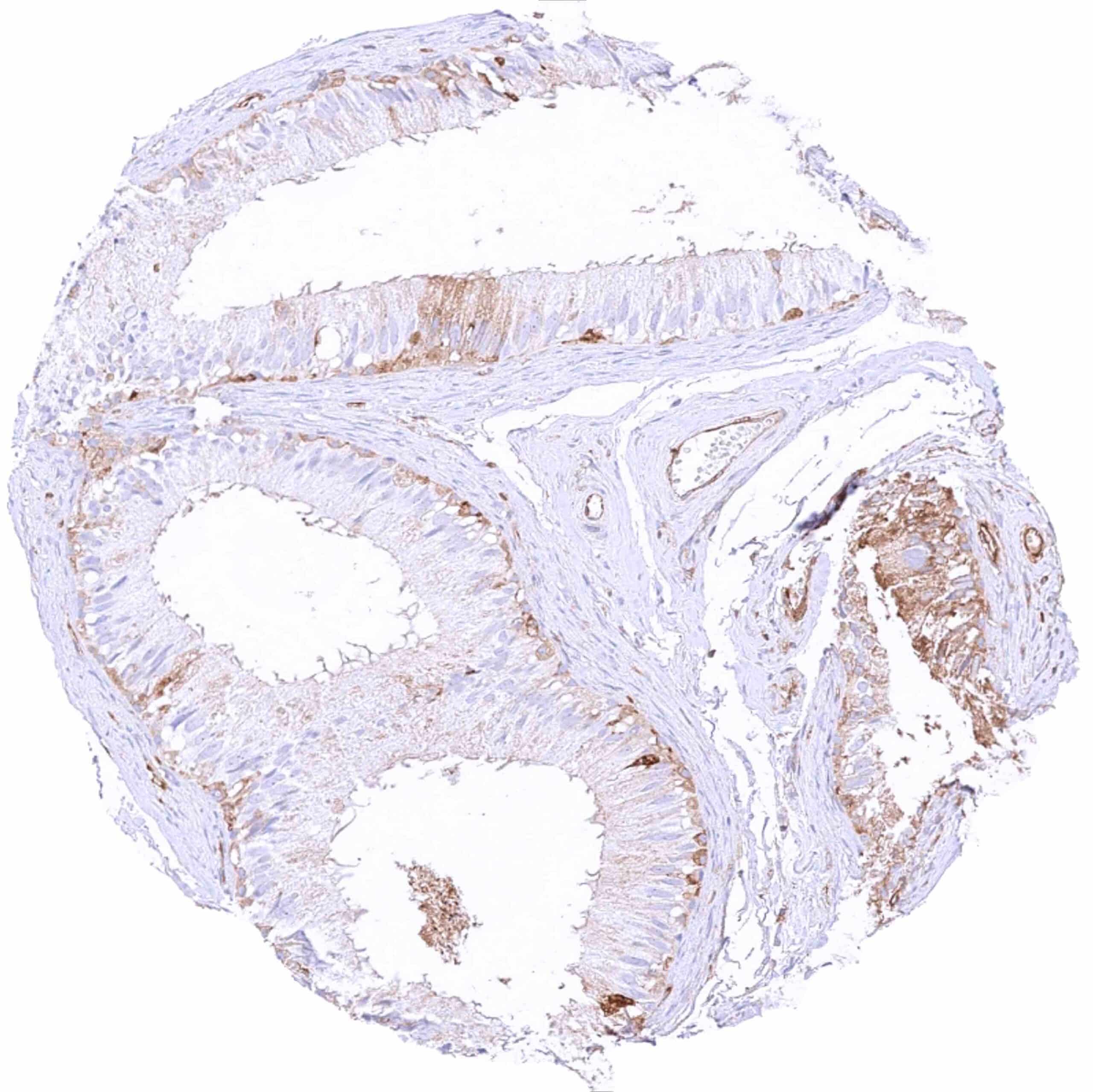
| Duodenum | Weak to moderate cytoplasmic MX1 staining of epithelial cells of the surface mucosa. Brunner gland cells are MX1 negative. | |
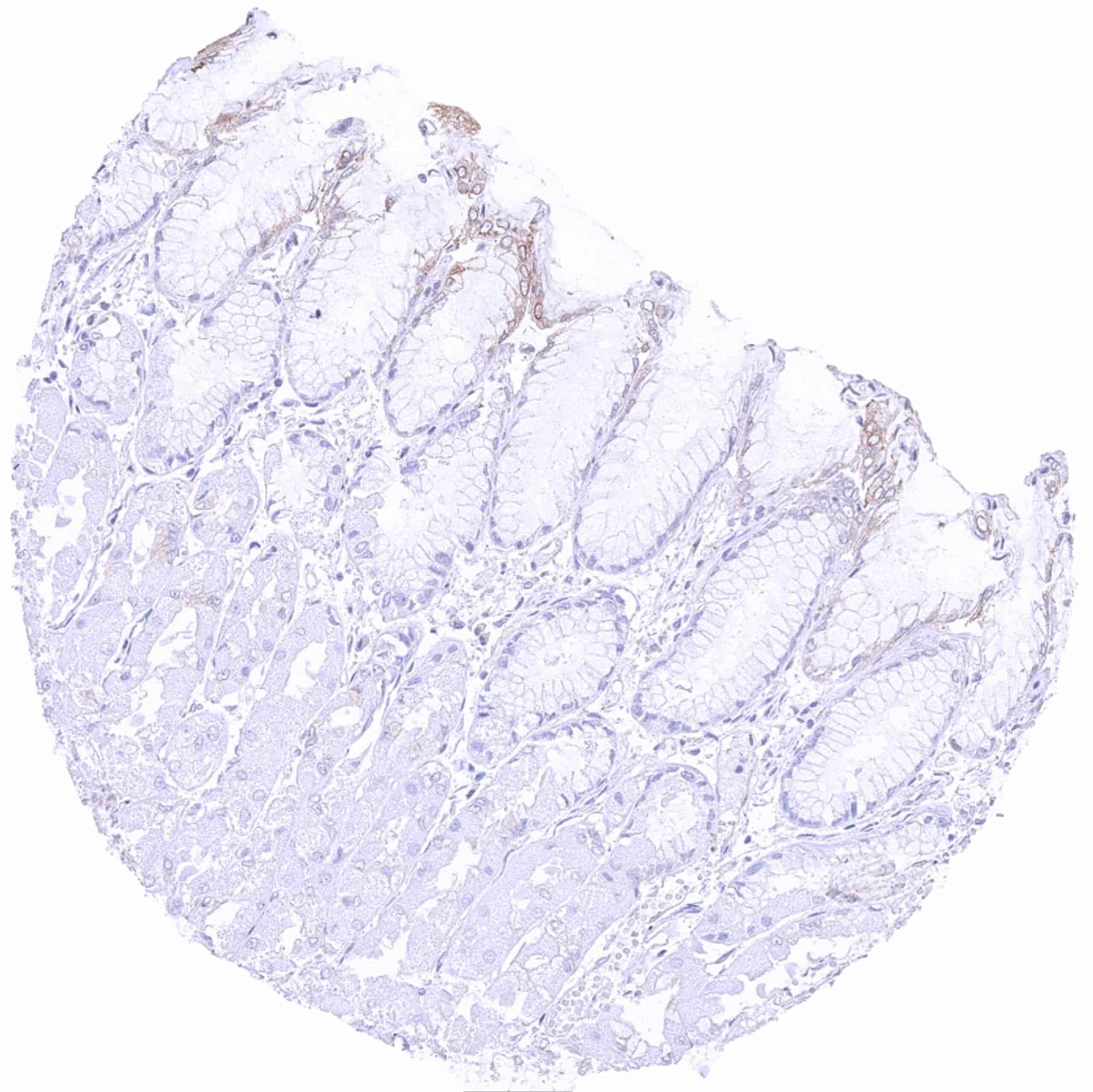
| Small intestine | Weak to moderate cytoplasmic MX1 staining of epithelial cells | |
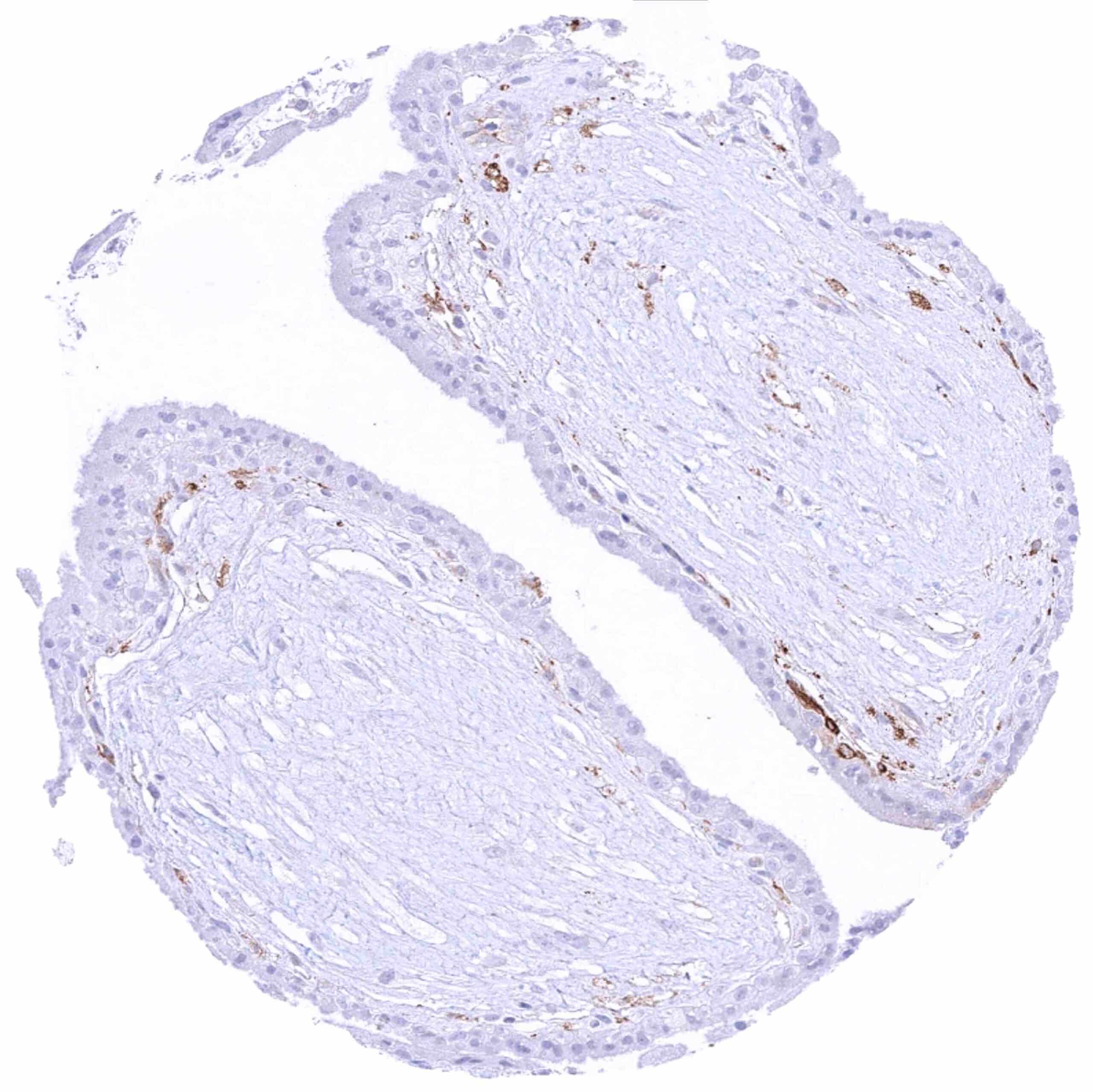
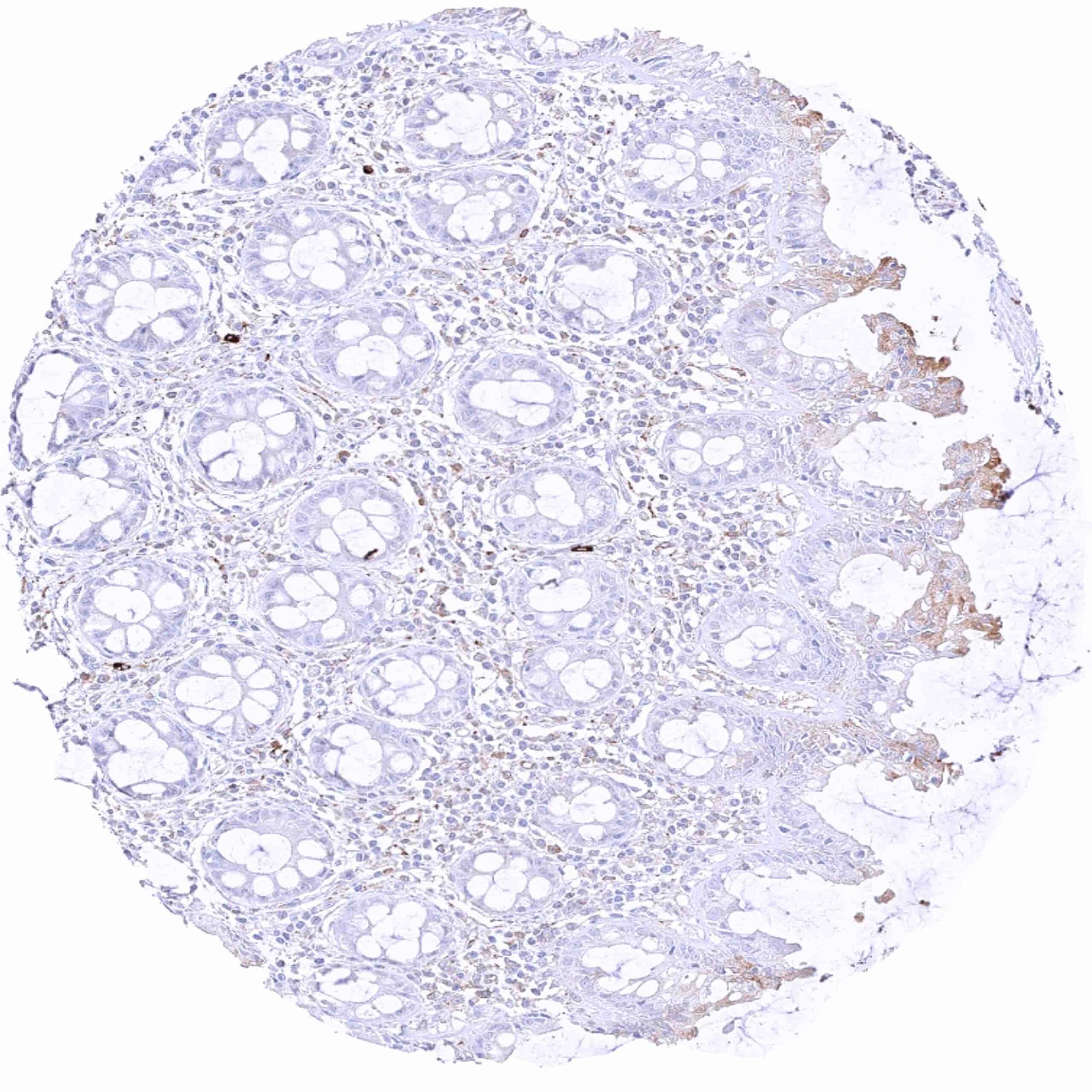
| Appendix | Cytoplasmic MX1 staining of epithelial cells is largely restricted to superficial cell layers. Crypt epithelial cells are largely MX1 negative.
Intense staining of lymphocytic cell types. |
|
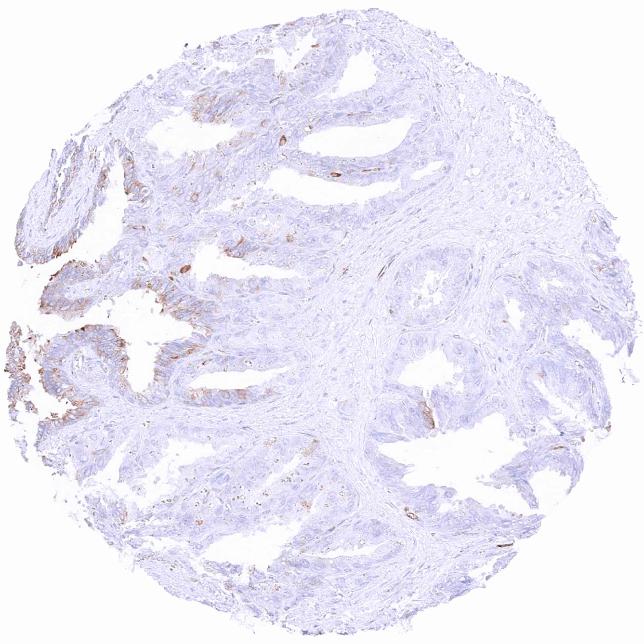
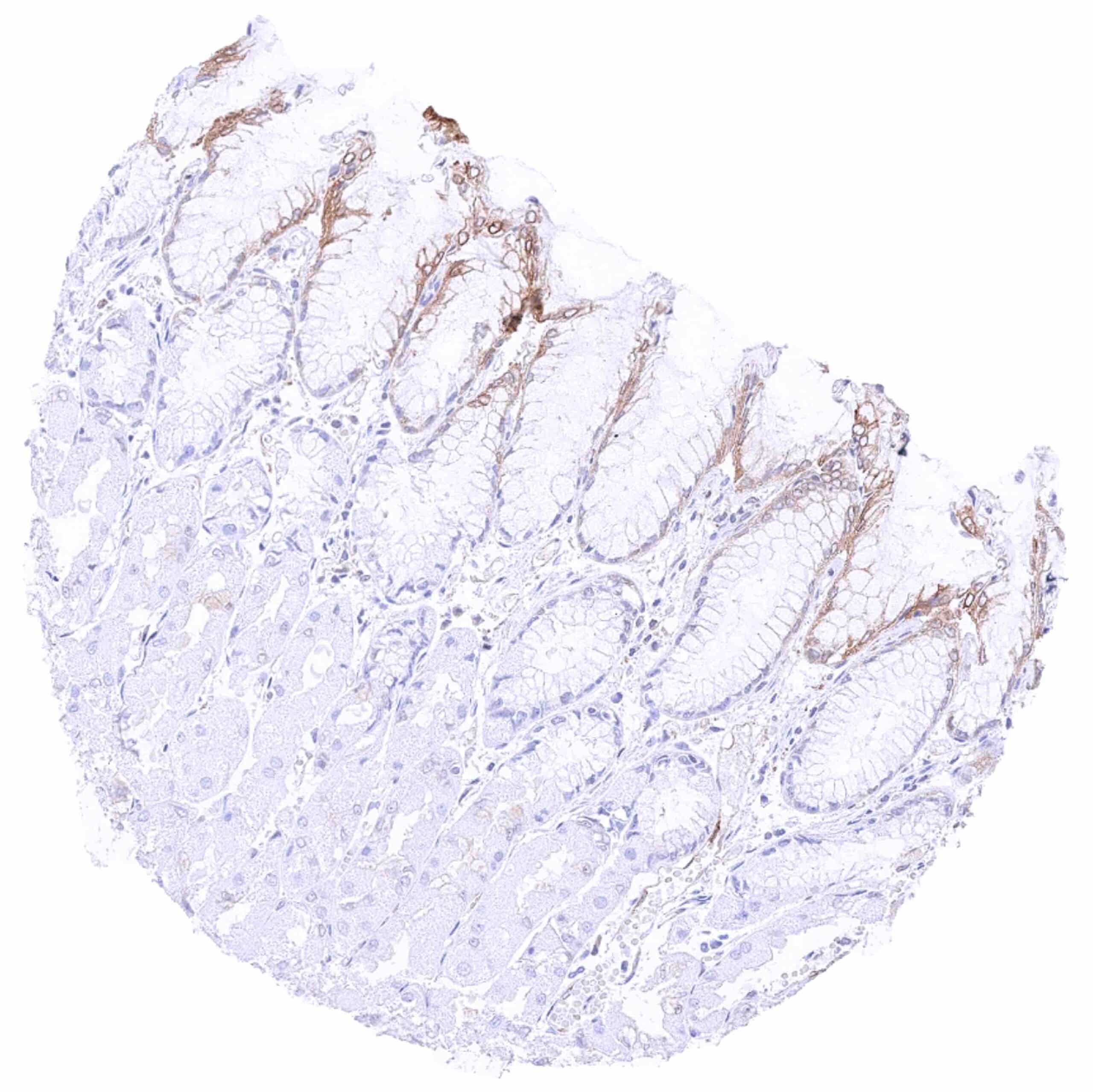
| Colon | Cytoplasmic MX1 staining of epithelial cells is largely restricted to superficial cell layers. Crypt epithelial cells are largely MX1 negative. | |
| Rectum | Cytoplasmic MX1 staining of epithelial cells is largely restricted to superficial cell layers. Crypt epithelial cells are largely MX1 negative. | |
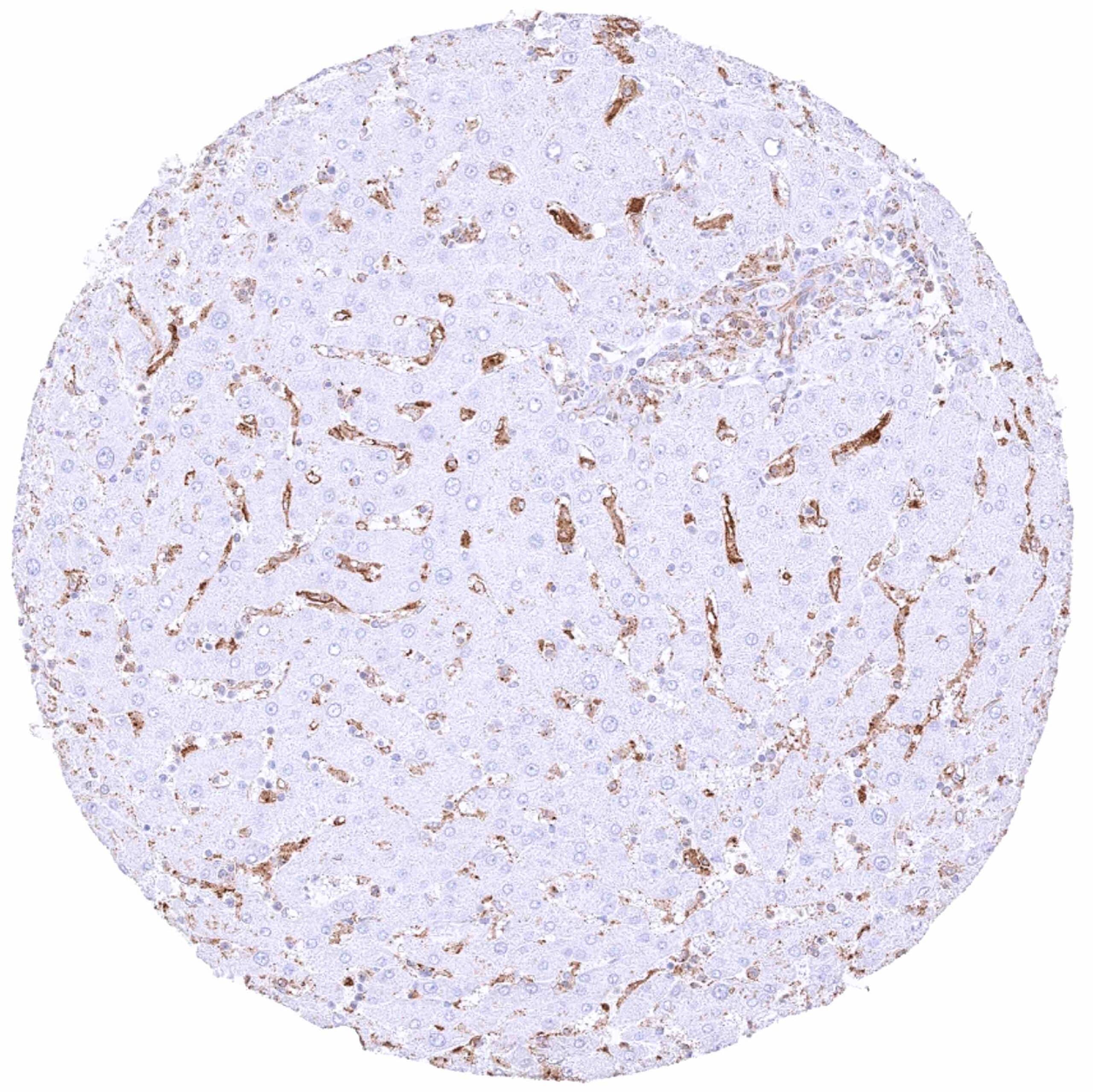
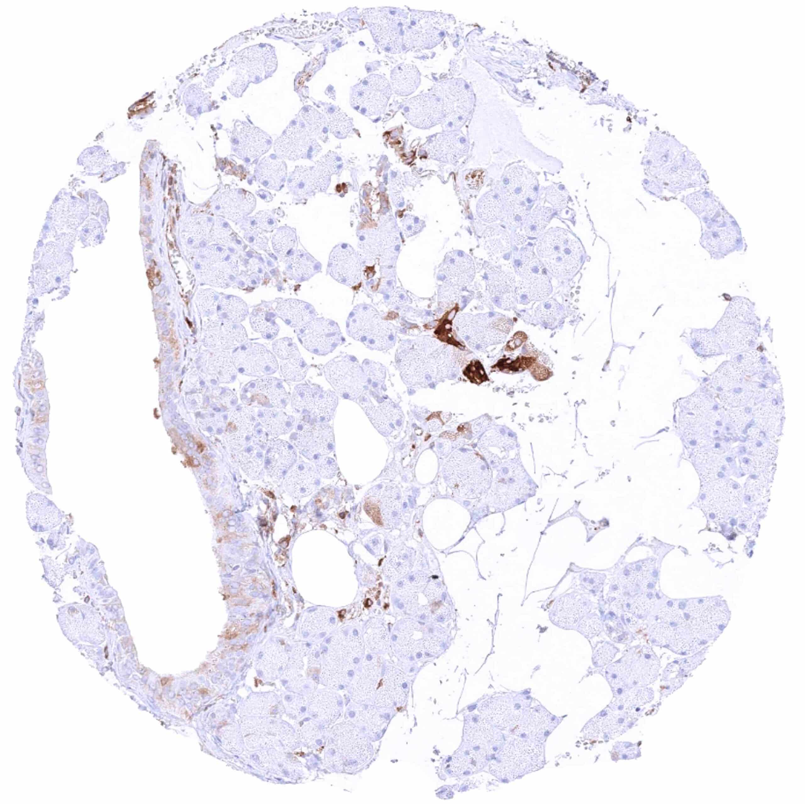
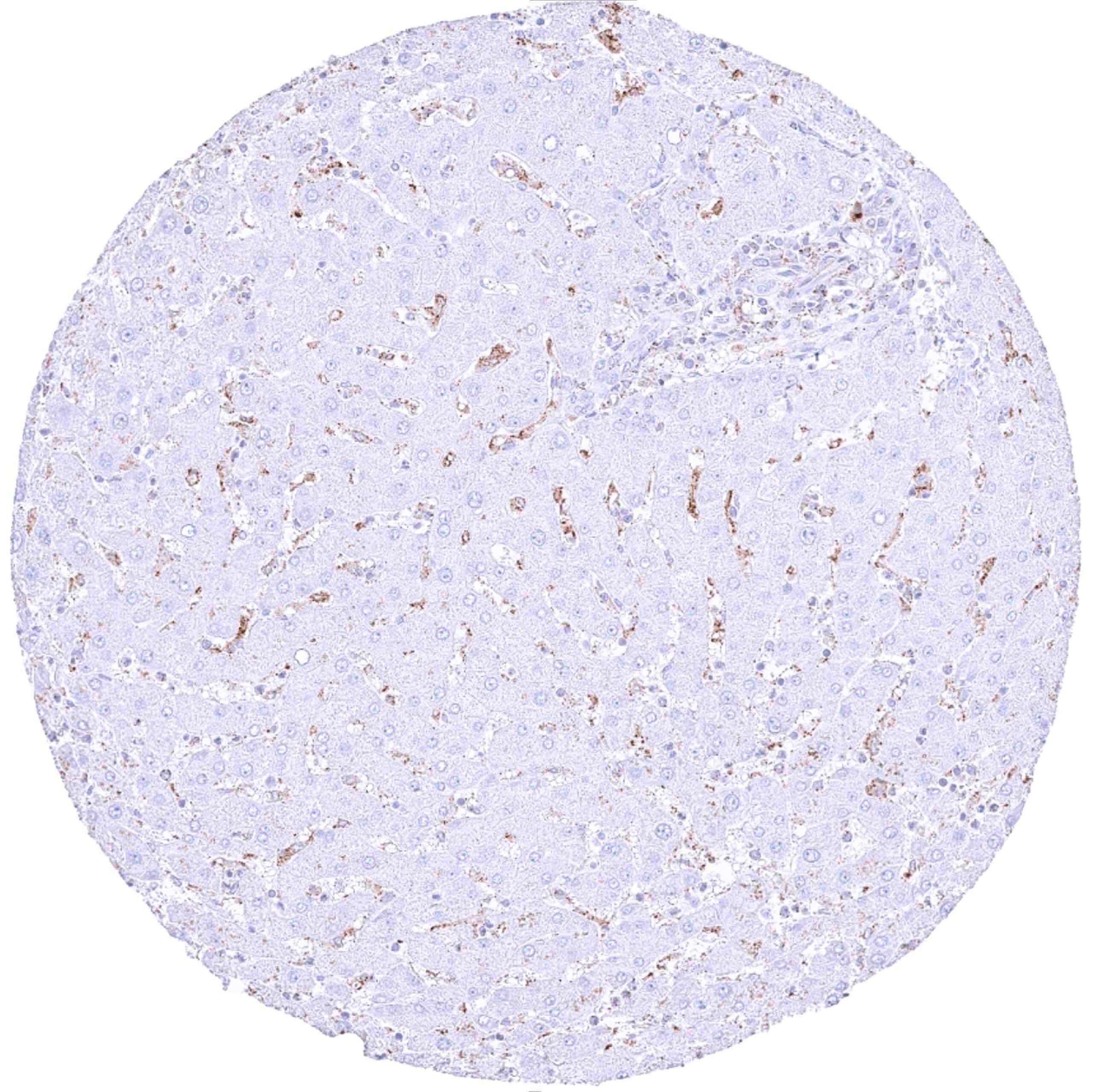
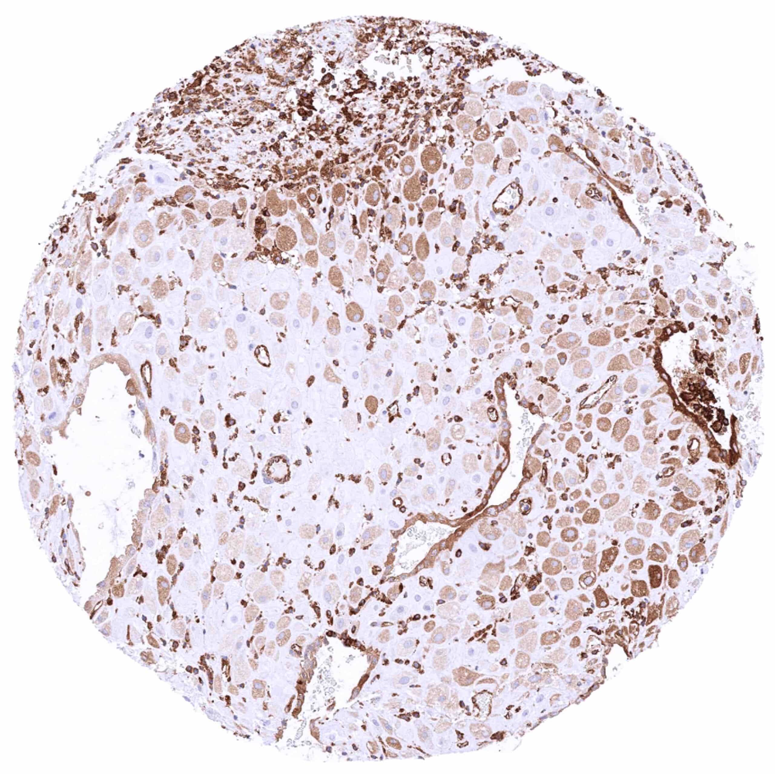
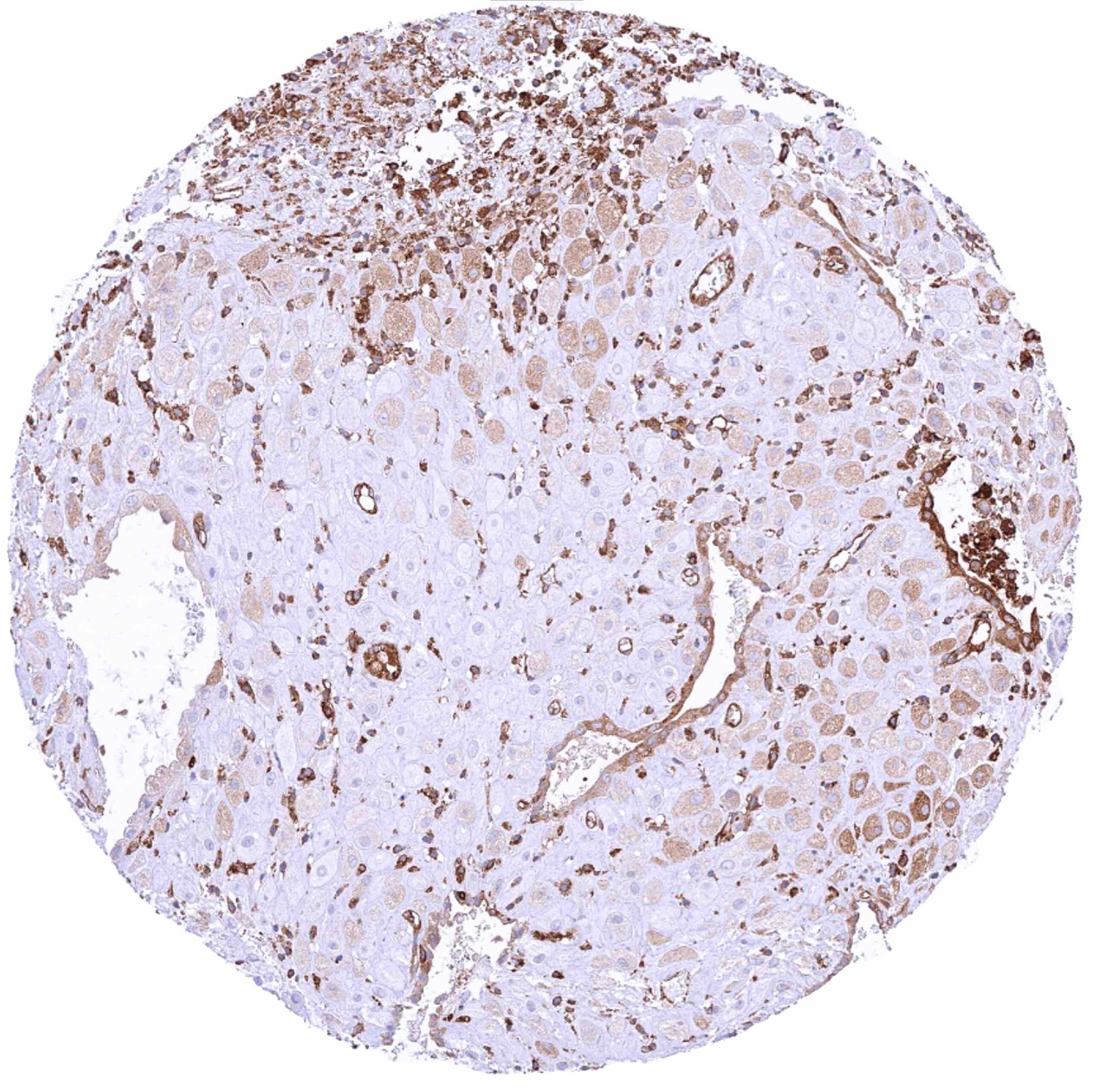
| Liver | Strong MX1 staining of sinus cells while hepatocytes are MX1 negative. | |
| Gallbladder | Strong focal MX1 staining can focally occur in epithelial cells while epithelial cells in other regions remain MX1 negative. | |
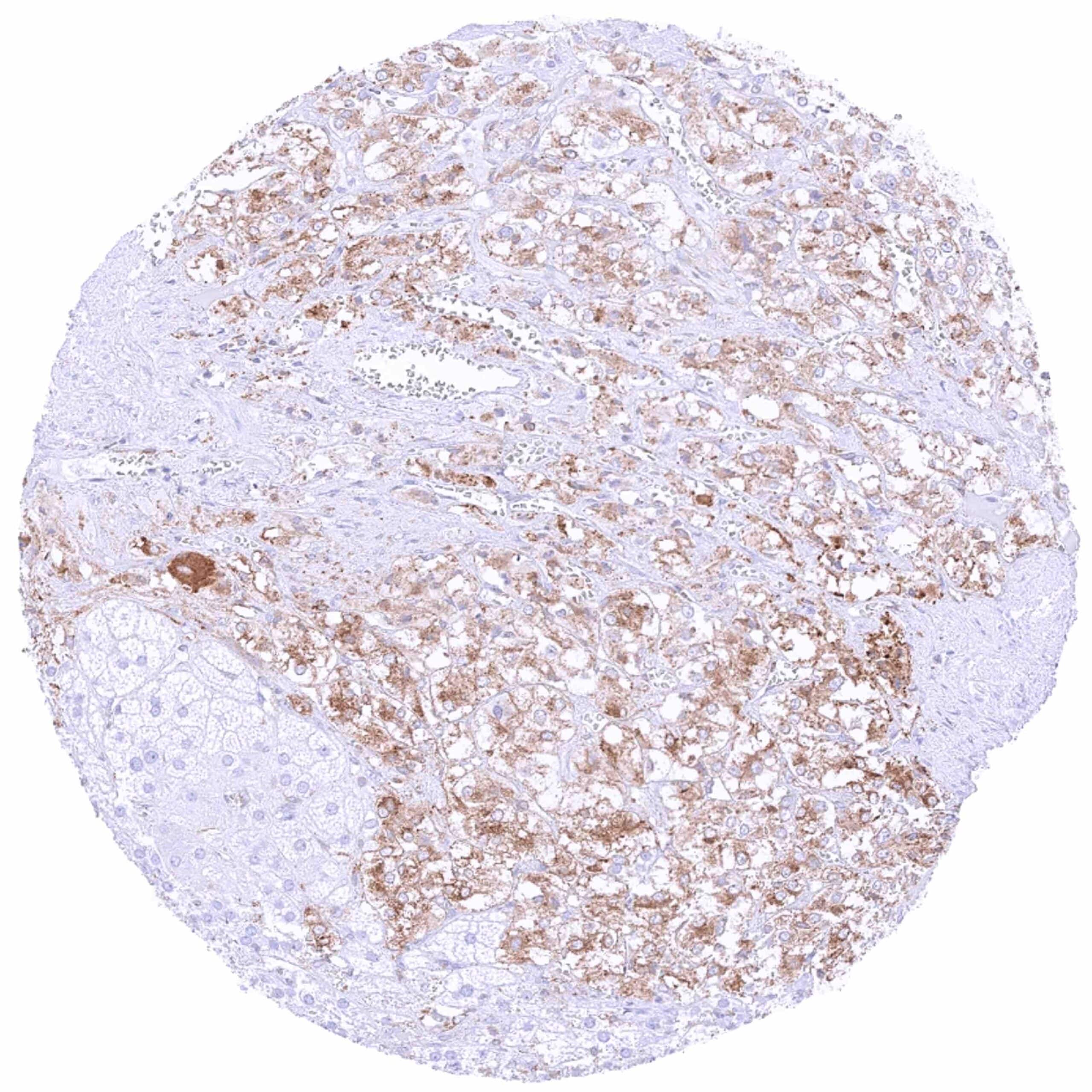
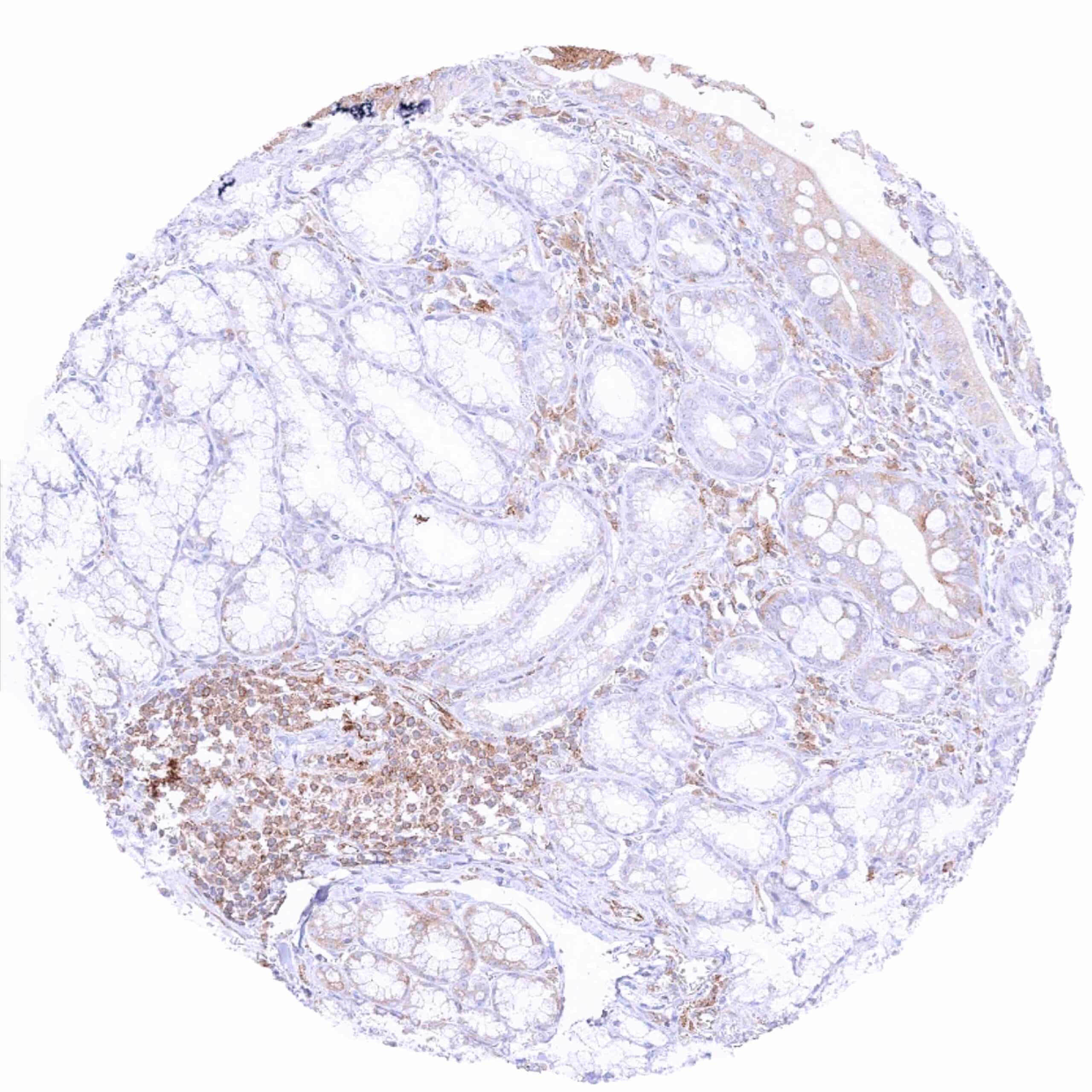
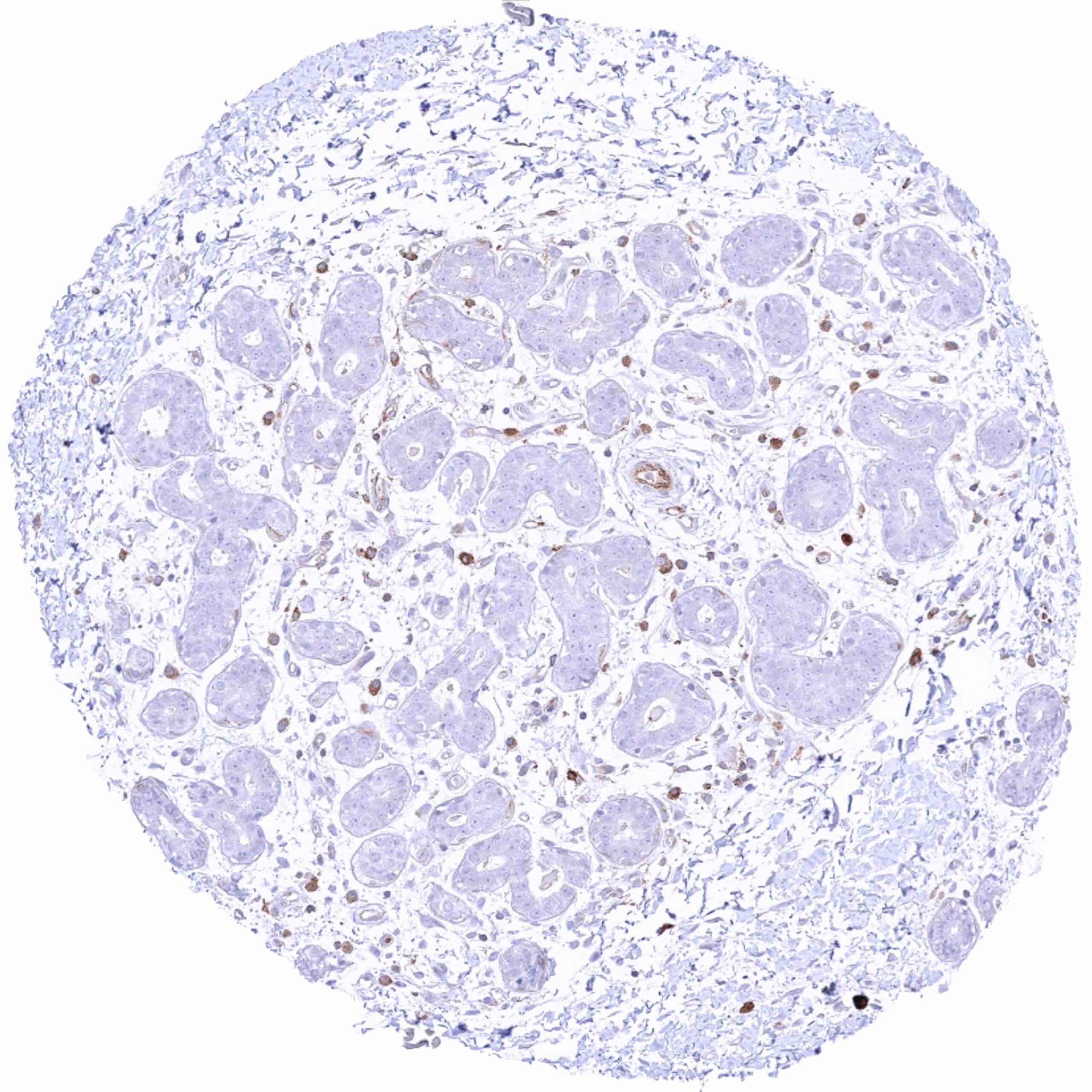
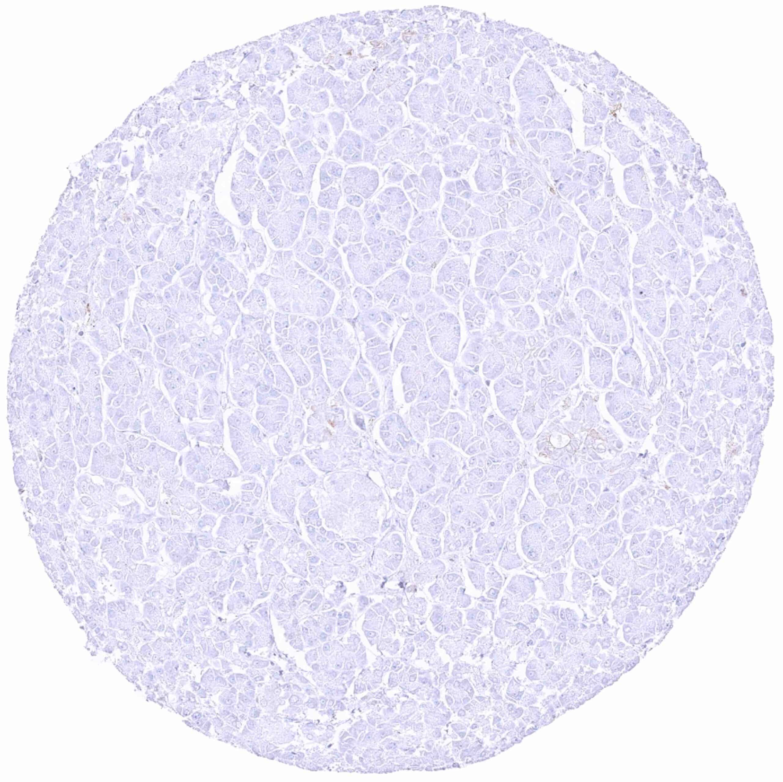
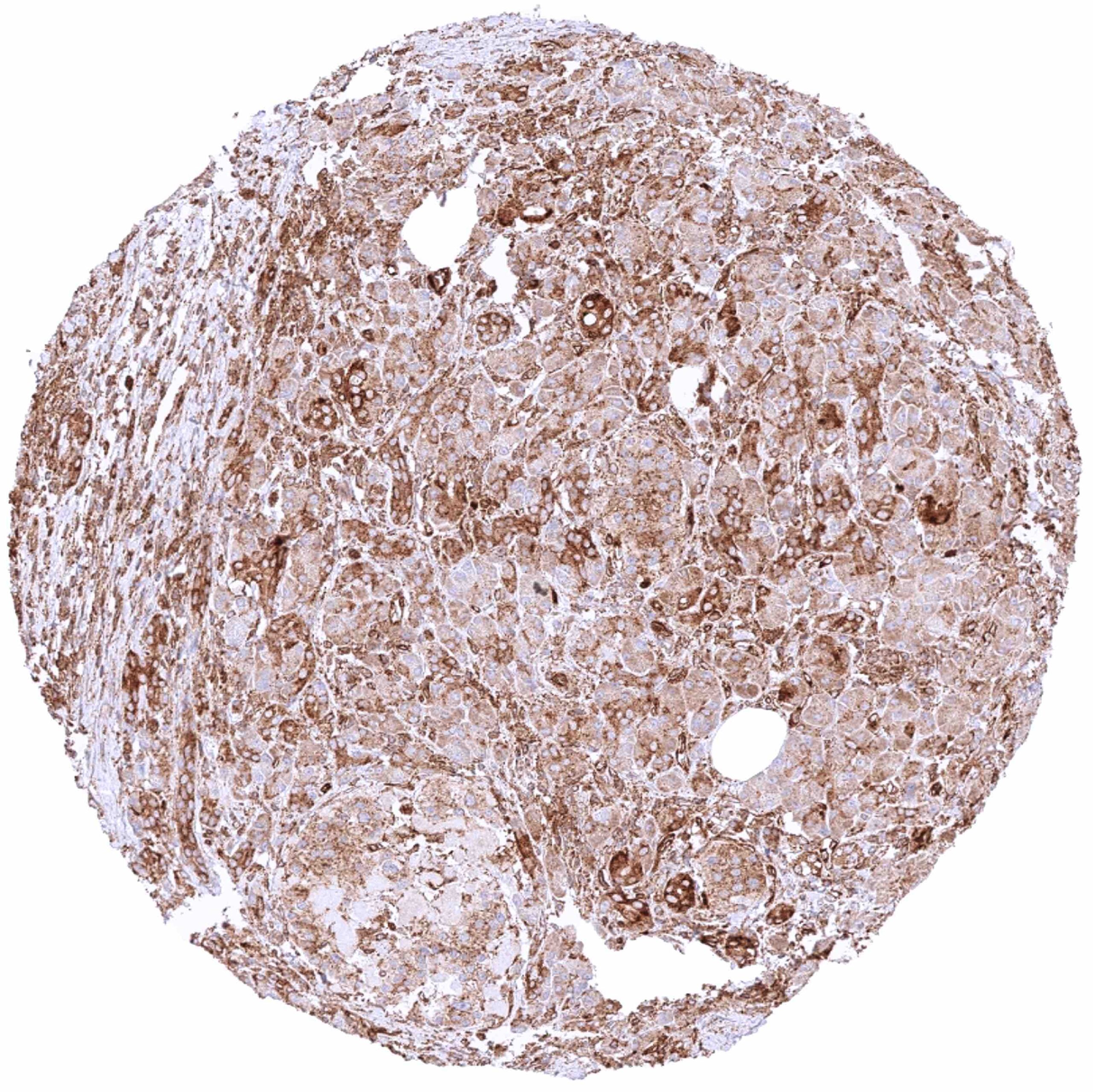
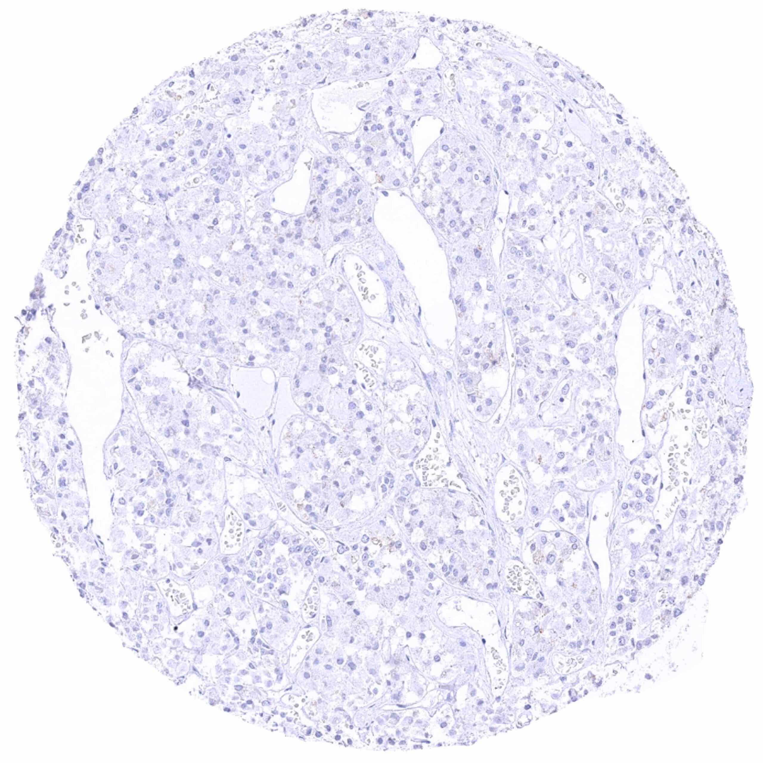
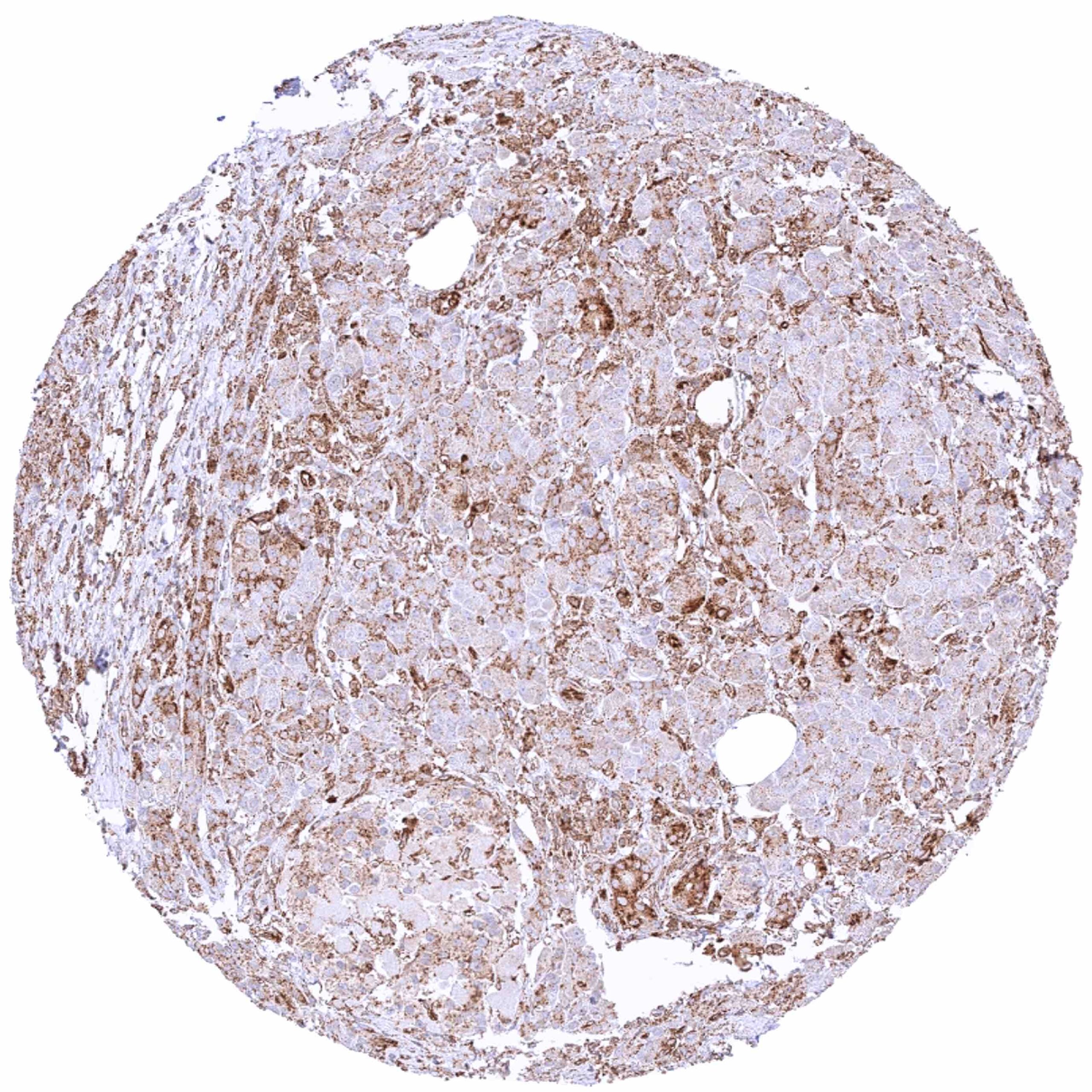
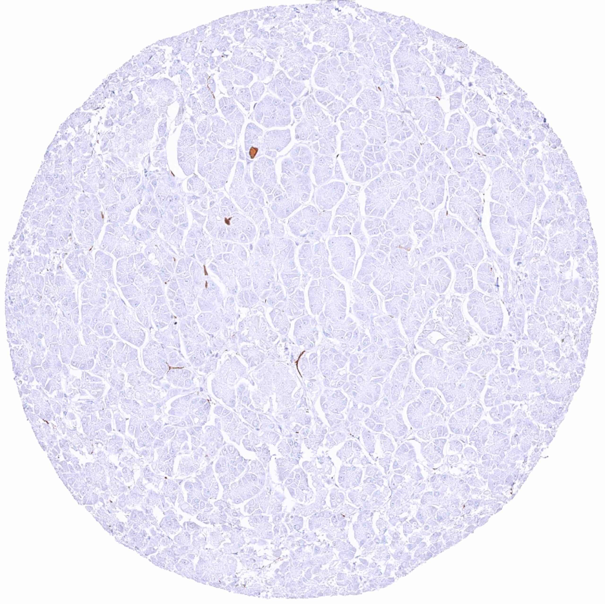
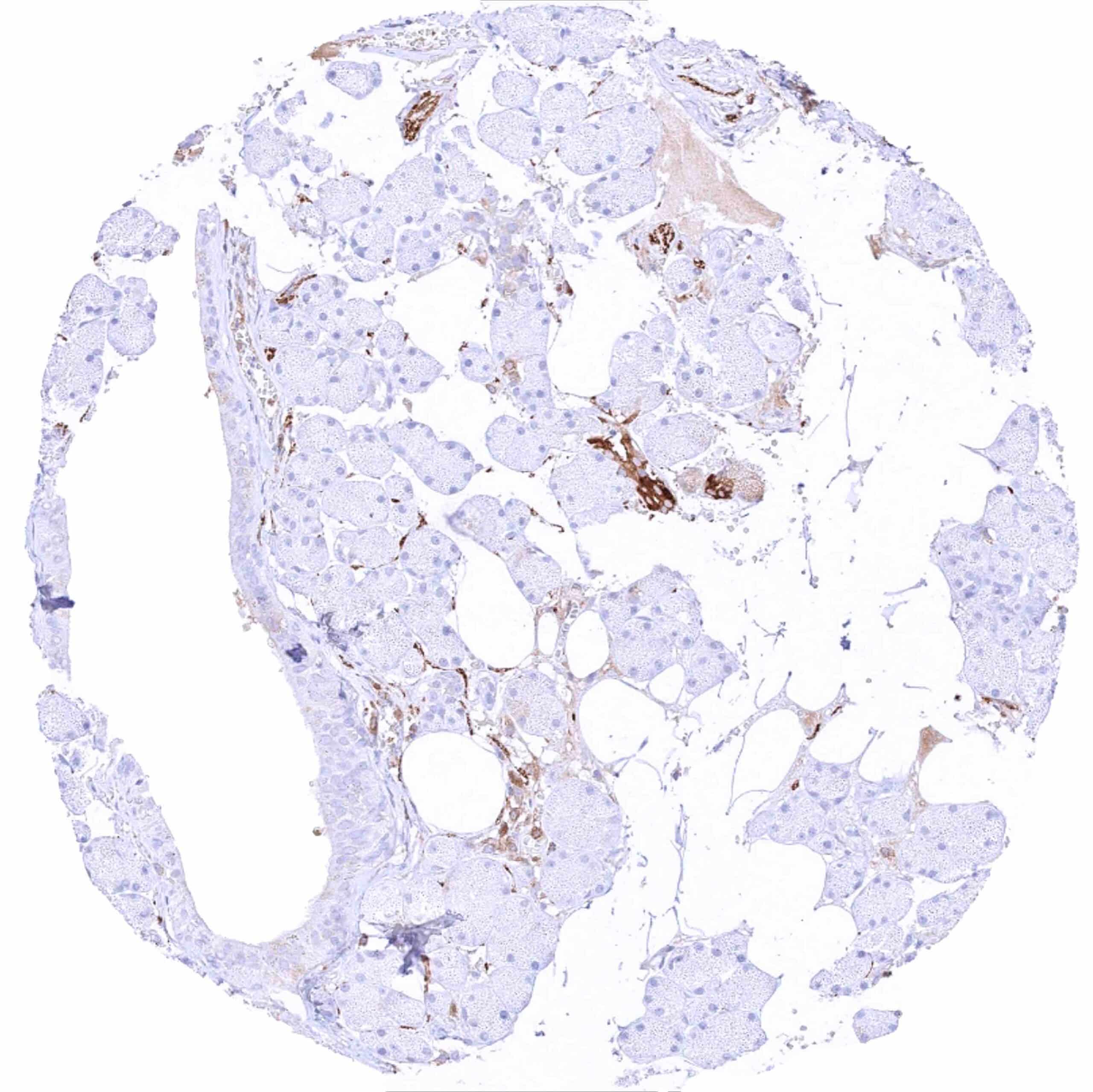
| Pancreas | Strong cytoplasmic MX1 staining of all epithelial cell types in one sample with inflammation and scar formation. MX1 staining is largely absent in other samples showing completely normal pancreatic tissues. | |
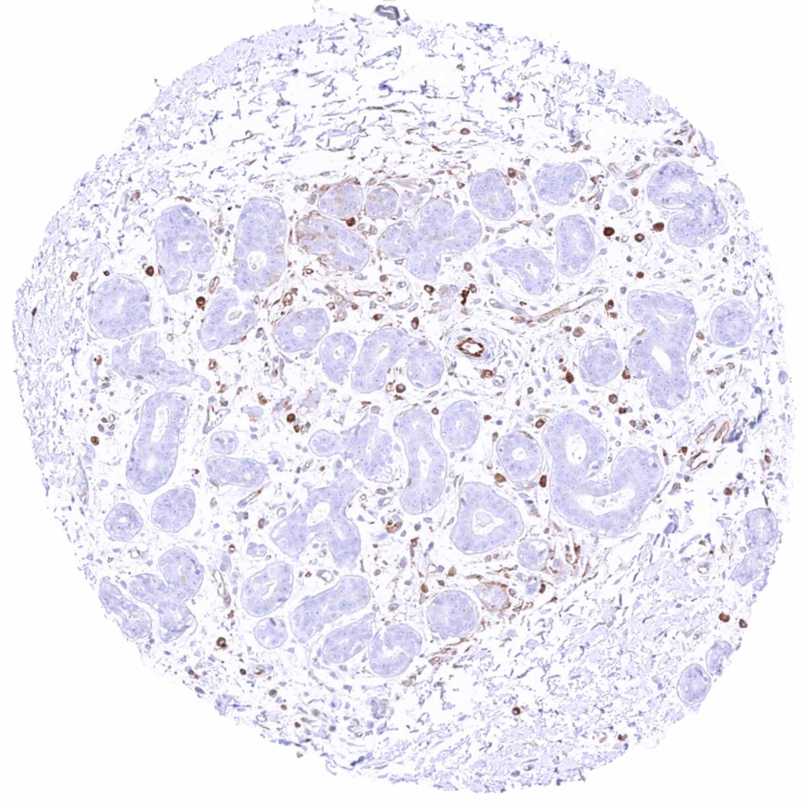
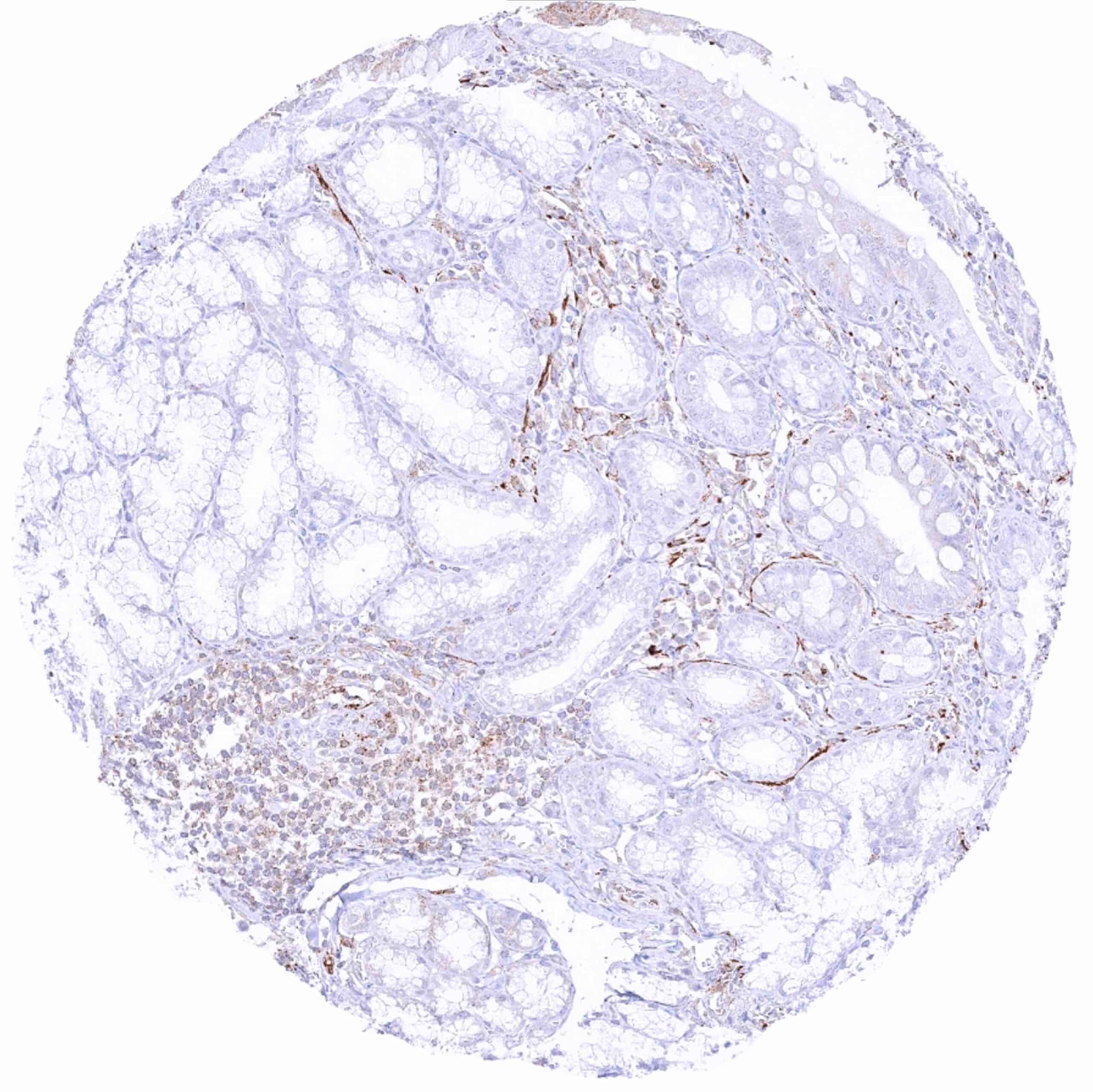
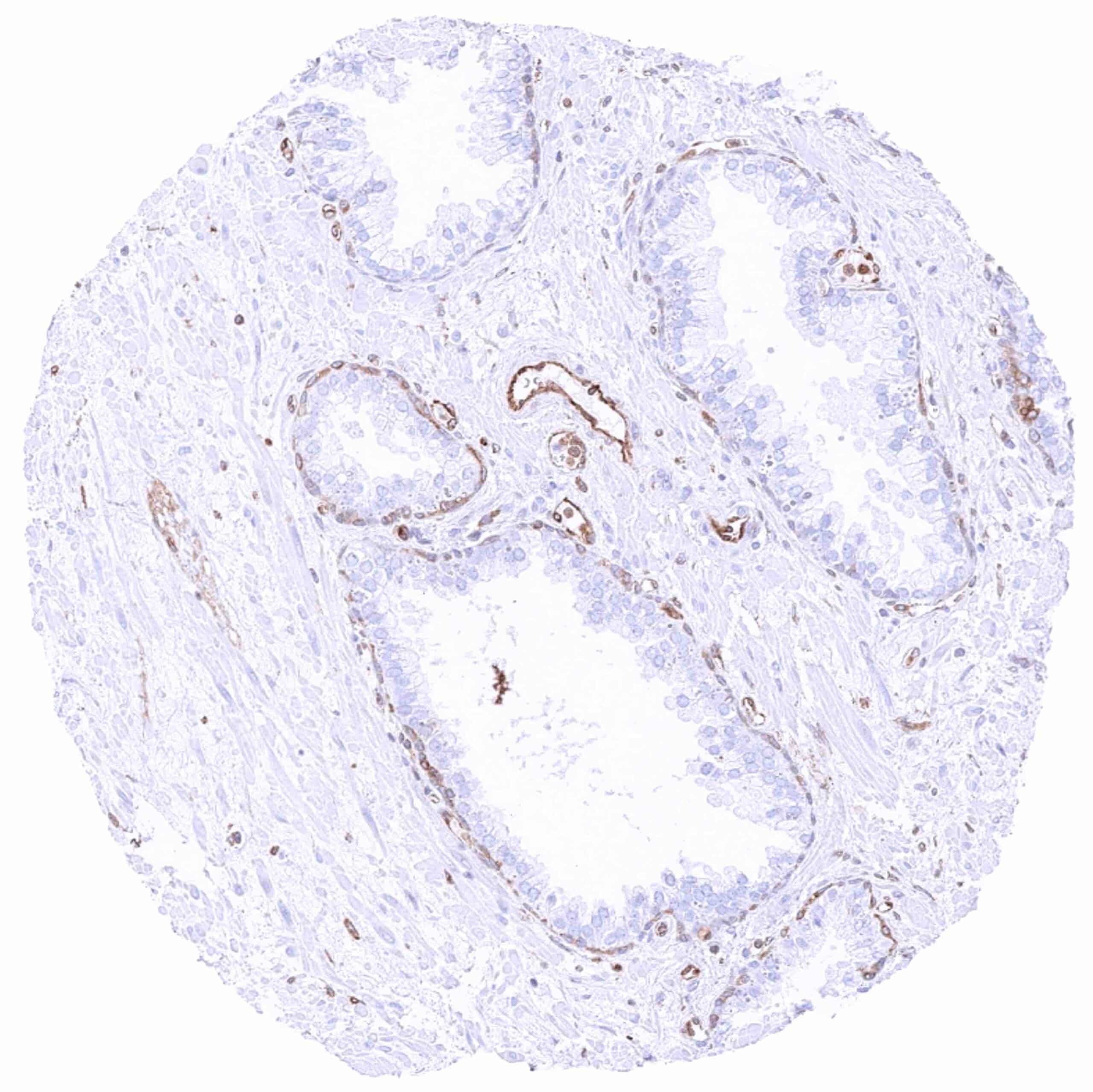
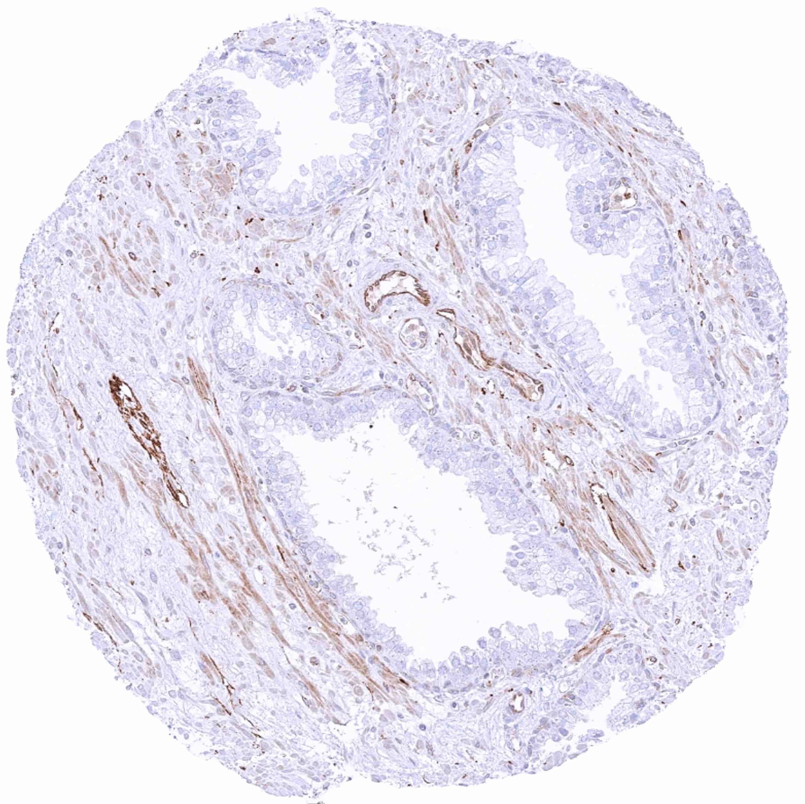
| Genitourinary | Kidney | Cytoplasmic MX1 staining mainly occurs in atrophic tubuli. It can also be seen in some collecting ducts (not in all samples). |
| Urothelium | Weak to moderate cytoplasmic MX1 staining can occur in the upper-half of urothelial cell layers. | |
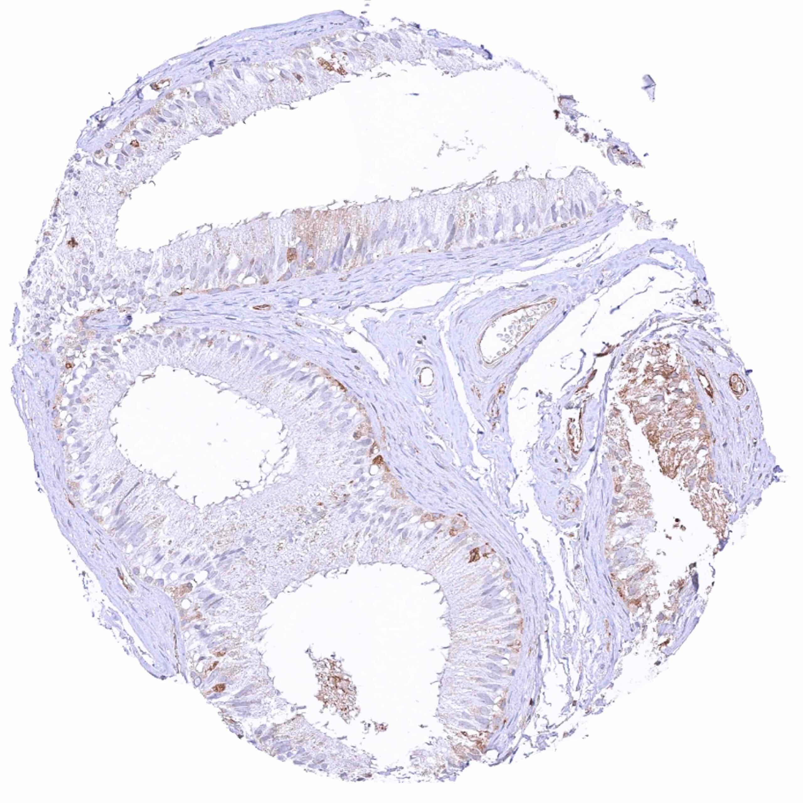
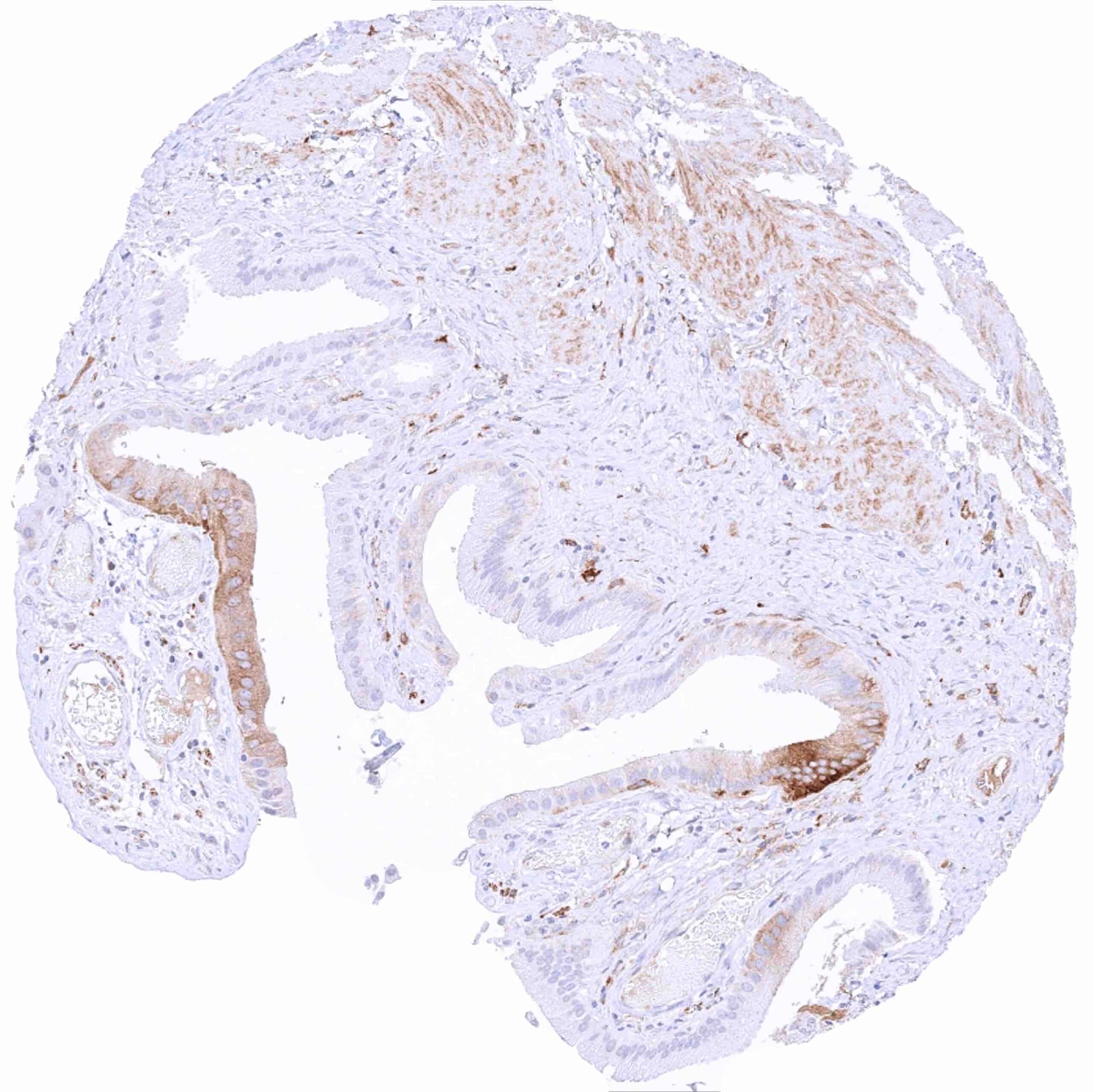
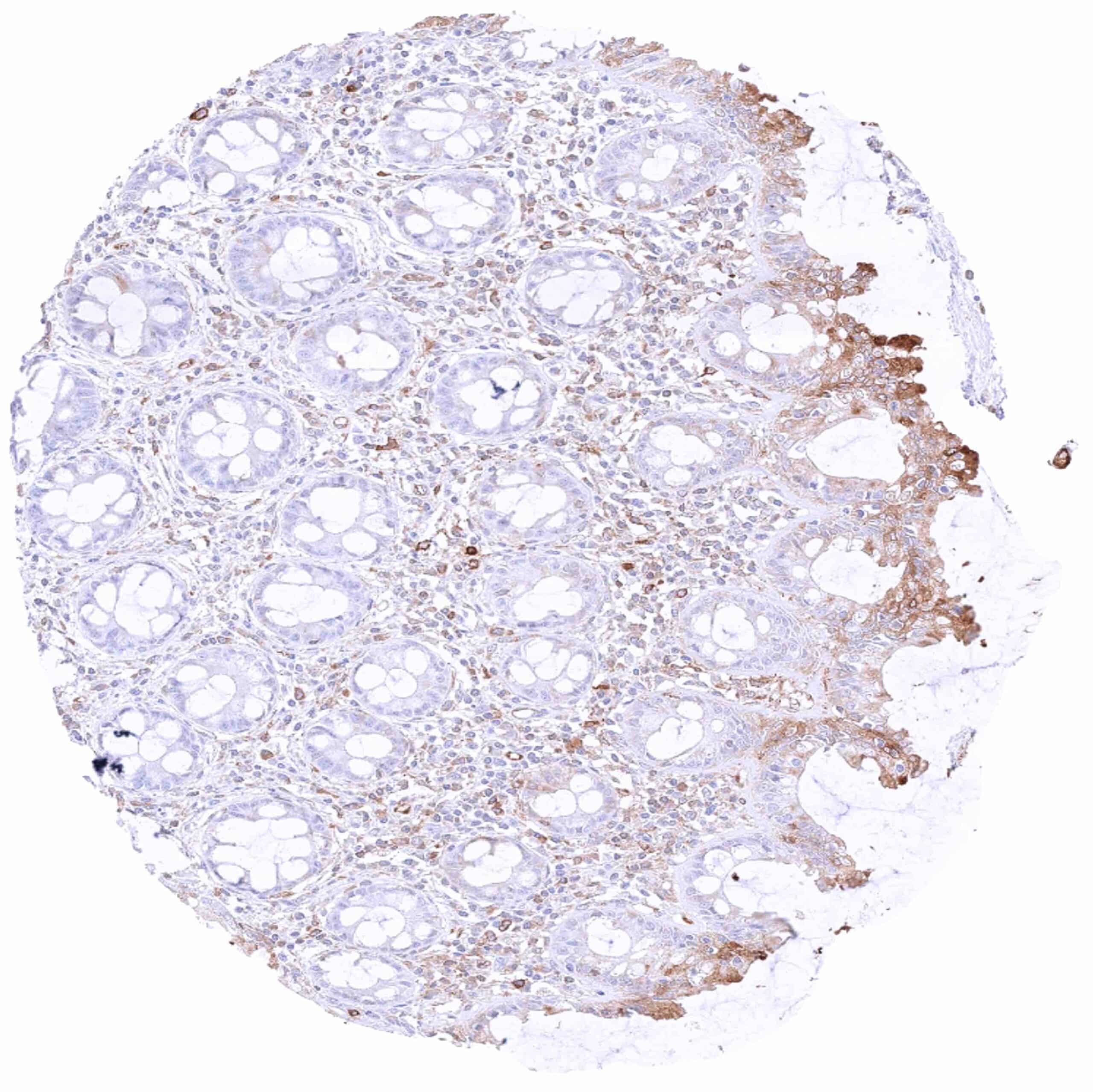
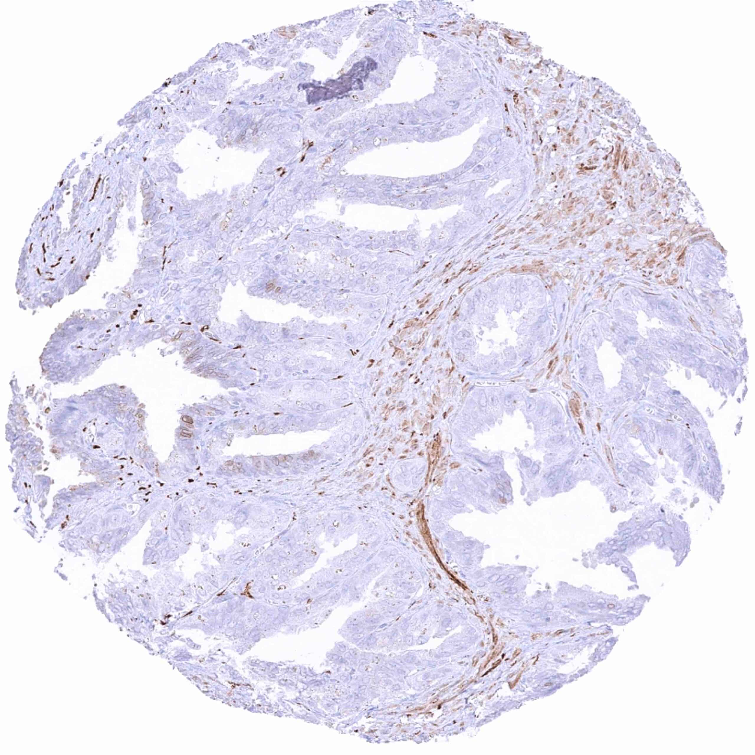
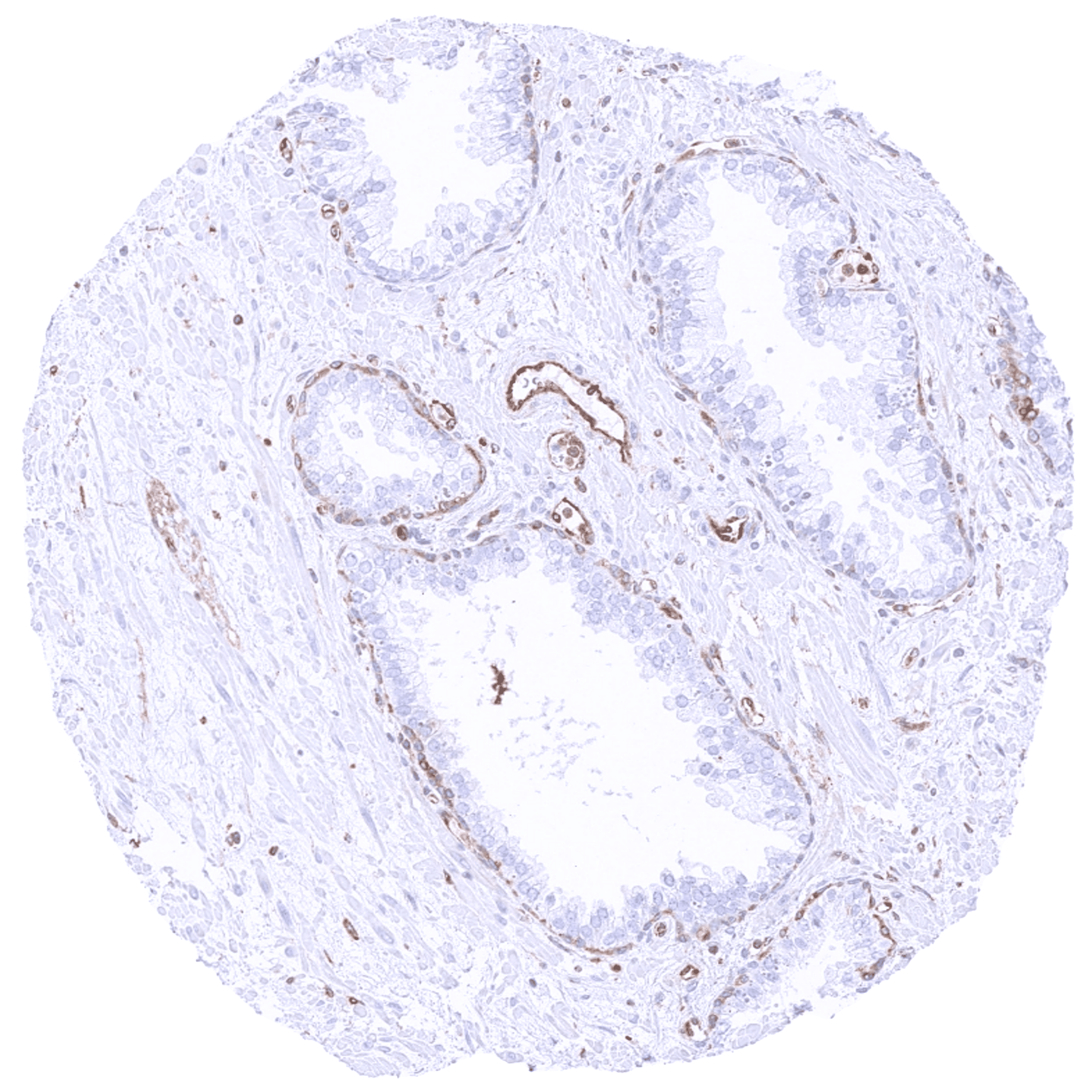
| Male genital | Prostate | Cytoplasmic MX1 staining of basal cells and of the endothelium of small vessels. |
| Seminal vesicles | Cytoplasmic MX1 staining of a subset of epithelial cells. | |
| Testis | Cytoplasmic MX1 staining is restricted to endothelial cells. | |
| Epididymis | In the caput epididymis, cytoplasmic MX1 staining mainly occurs in basal cells and in a subset of chief cells. | |
| Female genital | Breast | MX1 staining is limited to inflammatory and endothelial cells. |
| Uterus, myometrium | Negative. | |
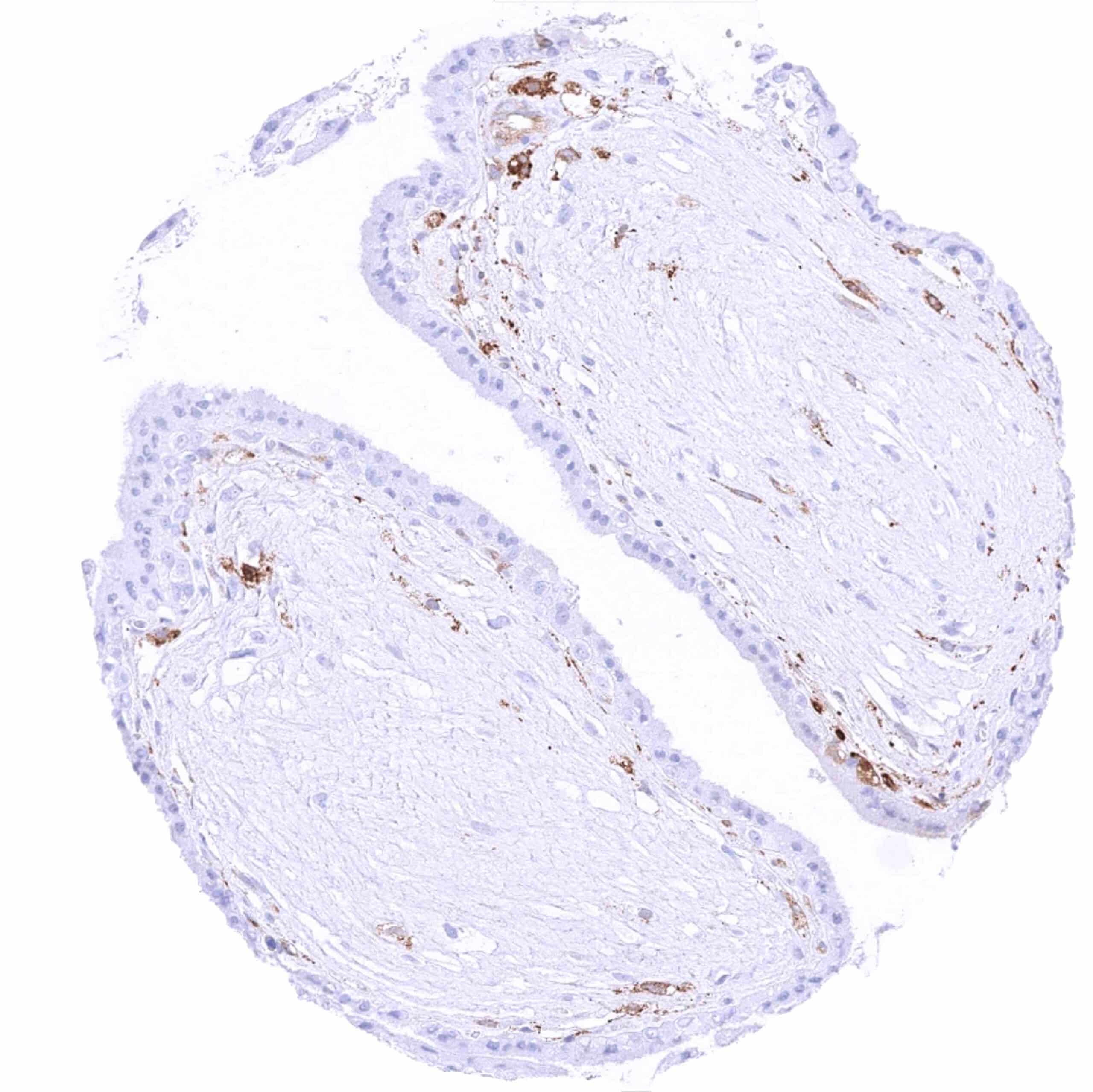
| Uterus, ectocervix | Weak to moderate cytoplasmic MX1 staining of suprabasal cell layers. | |
| Uterus endocervix | Absent or weak cytoplasmic MX1 staining of epithelial cells. | |
| Uterus, endometrium | Negative in non-pregnant uterus. Strong endometrium staining in case of pregnancy. | |
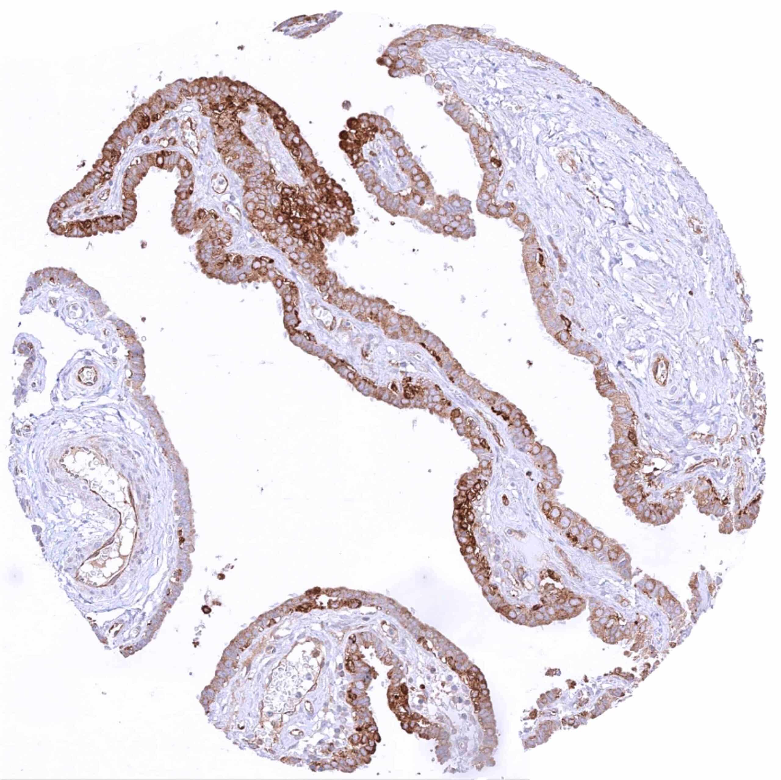
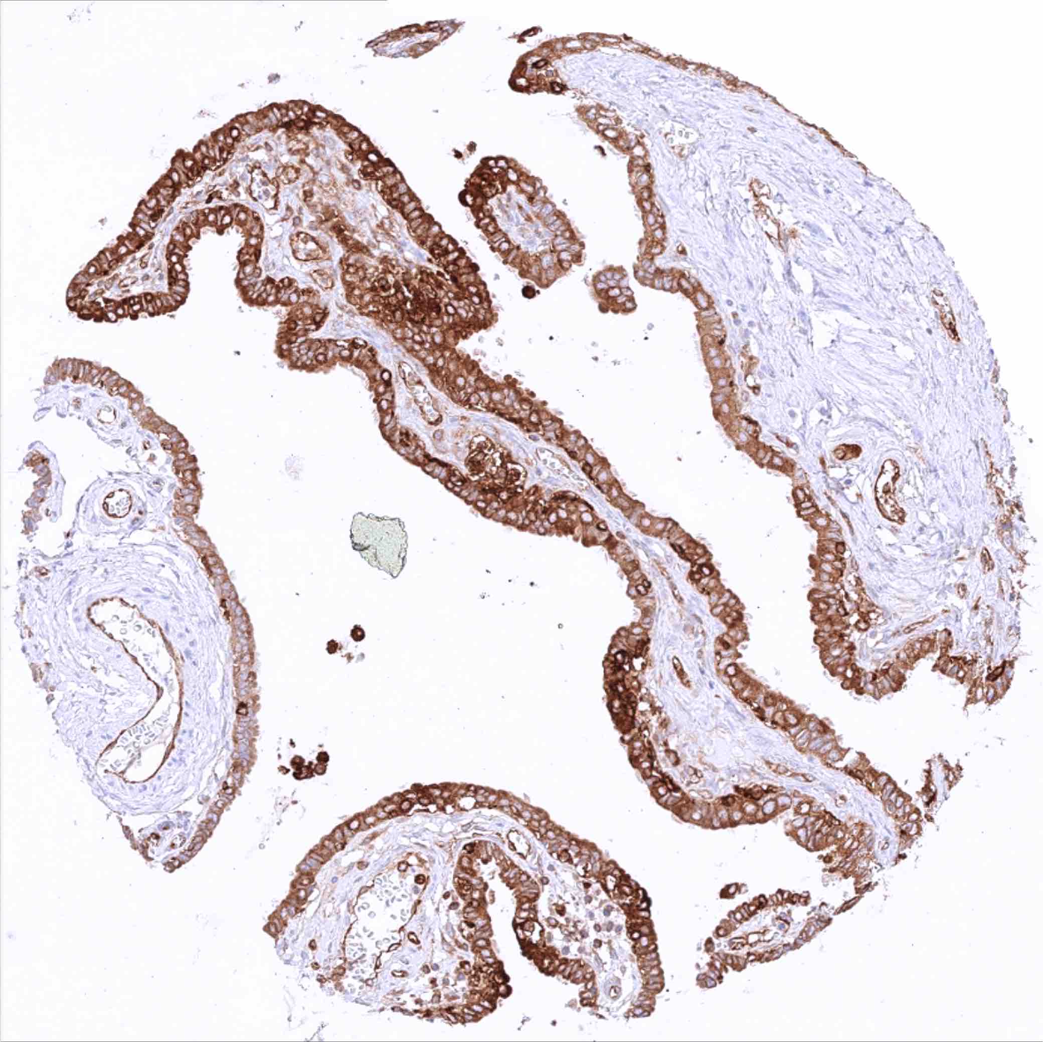
| Fallopian Tube | Moderate to strong MX1 staining of epithelial cells. | |
| Ovary | Stroma cells, granulosa cells, theca interna and externa cells as well as corpus luteum cells are MX1 negative. | |
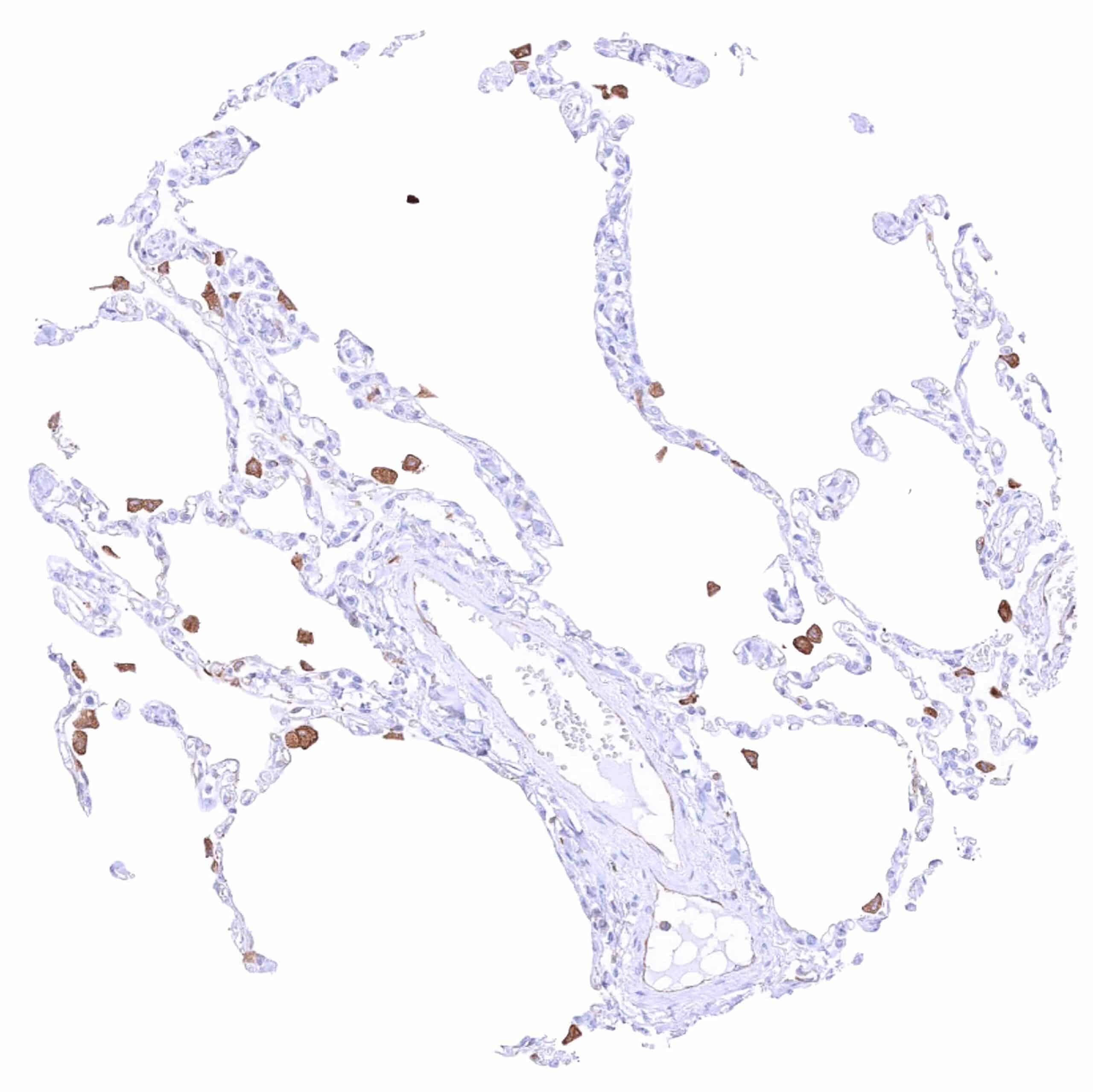
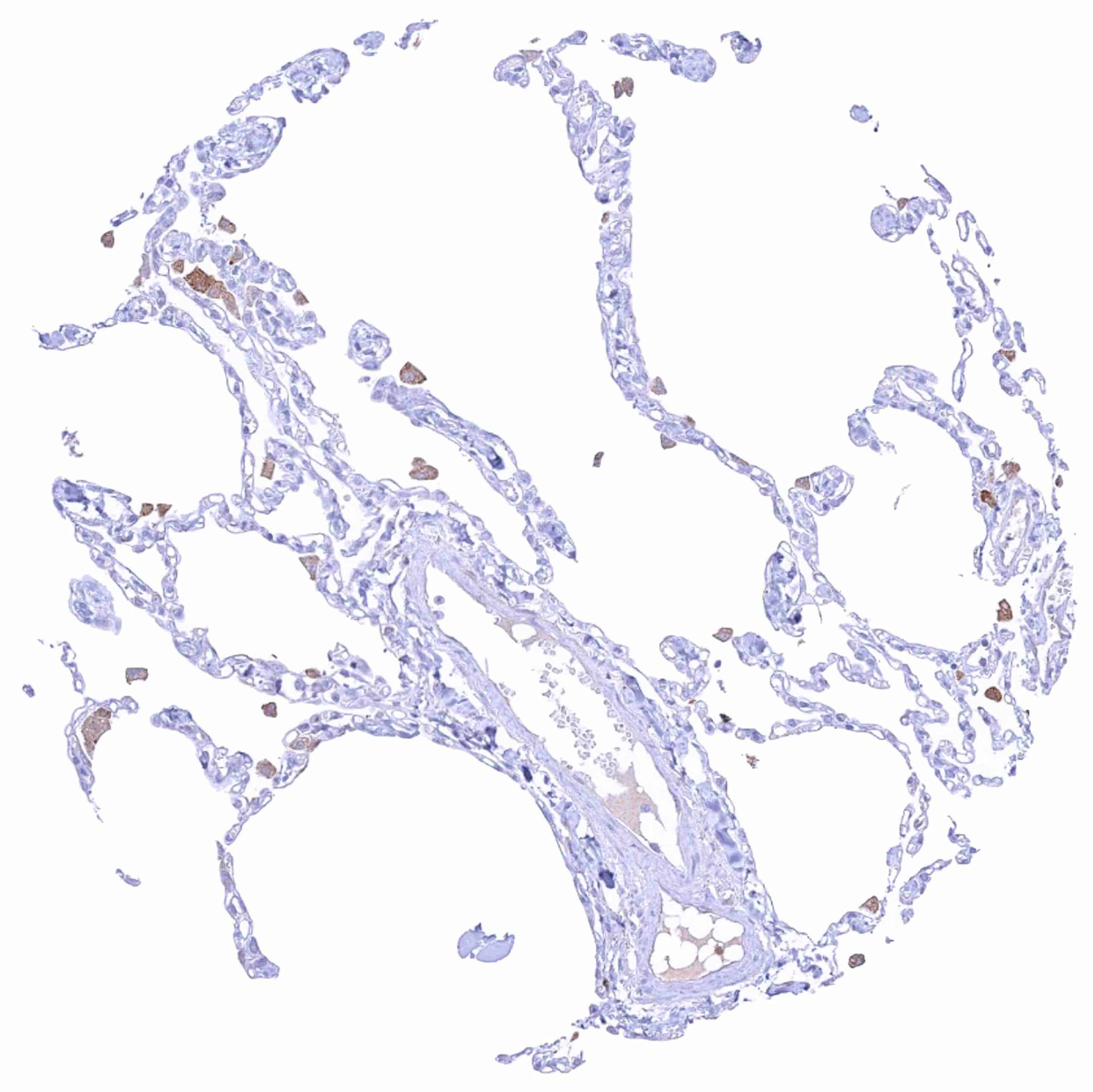
| Placenta early | Moderate to strong cytoplasmic MX1 positivity of decidua cells. | |
| Placenta mature | Trophoblast cells are MX1 negative. MX1 staining occurs in endothelial cells. | |
| Amnion | Weak cytoplasmic MX1 staining of amnion cells. | |
| Chorion | Negative. | |
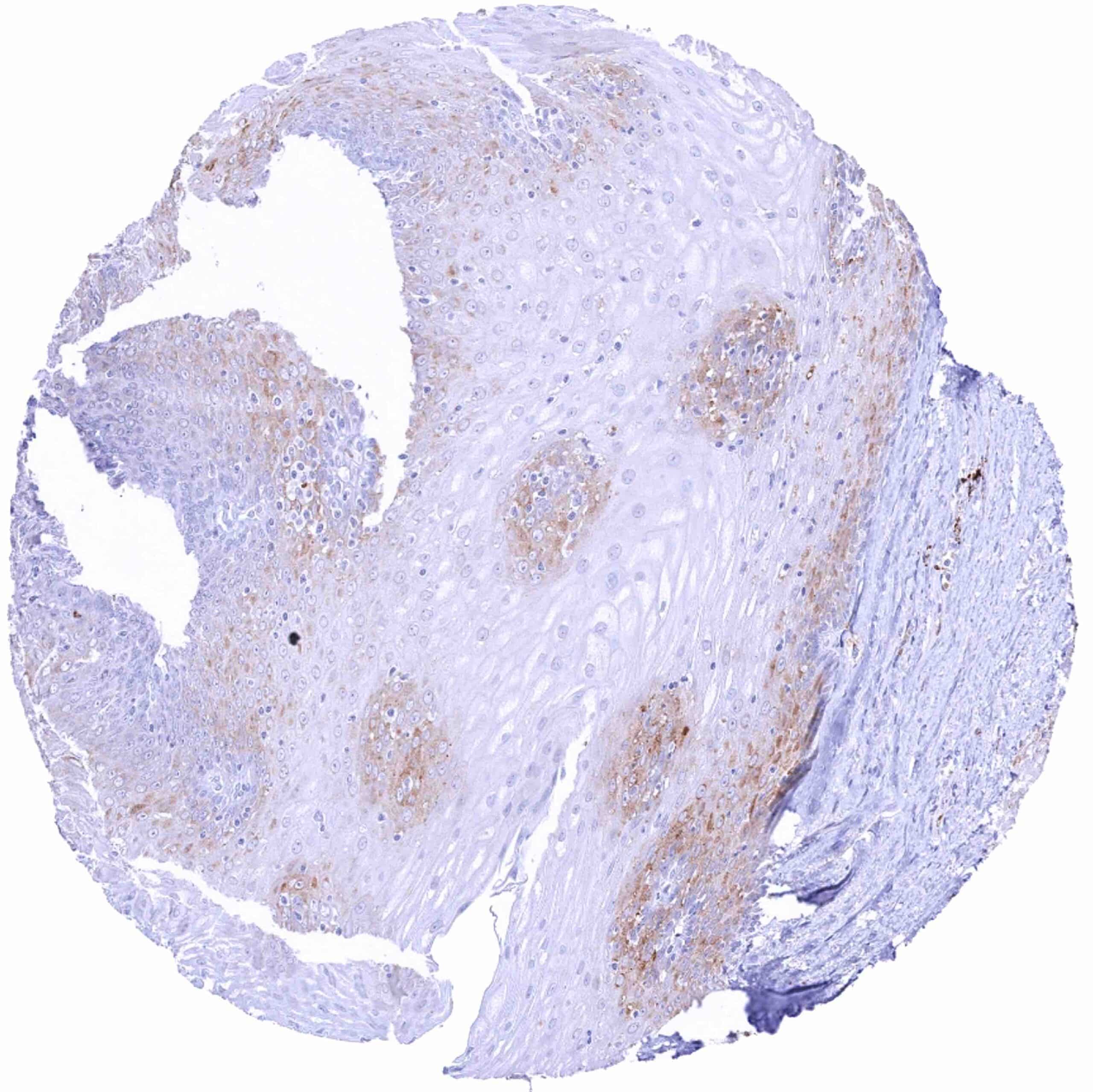
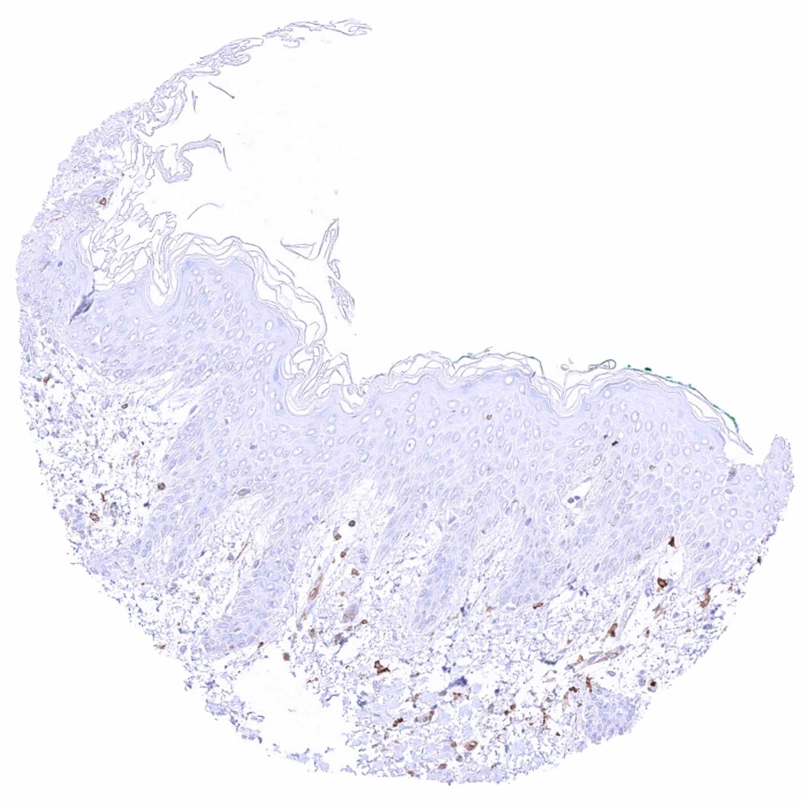
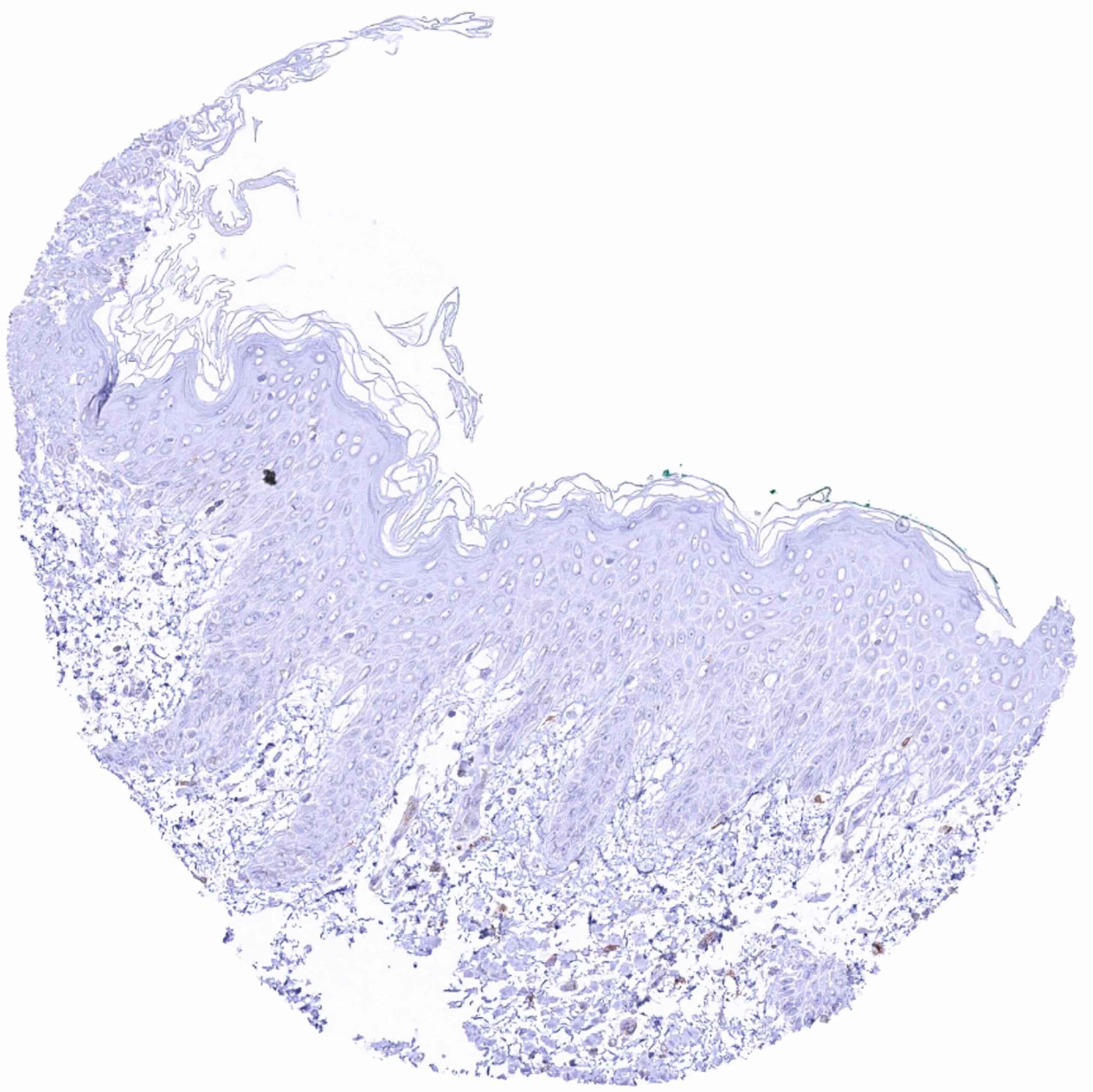
| Skin | Epidermis | Mostly negative. But individual samples show strong cytoplasmic MX1 positivity of bottom half of epithelial cells. |
| Sebaceous glands | ||
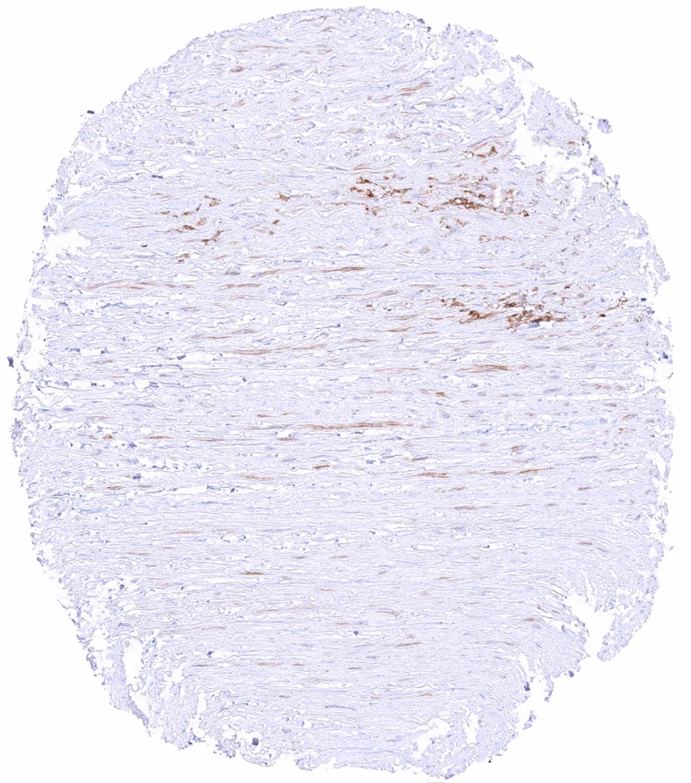
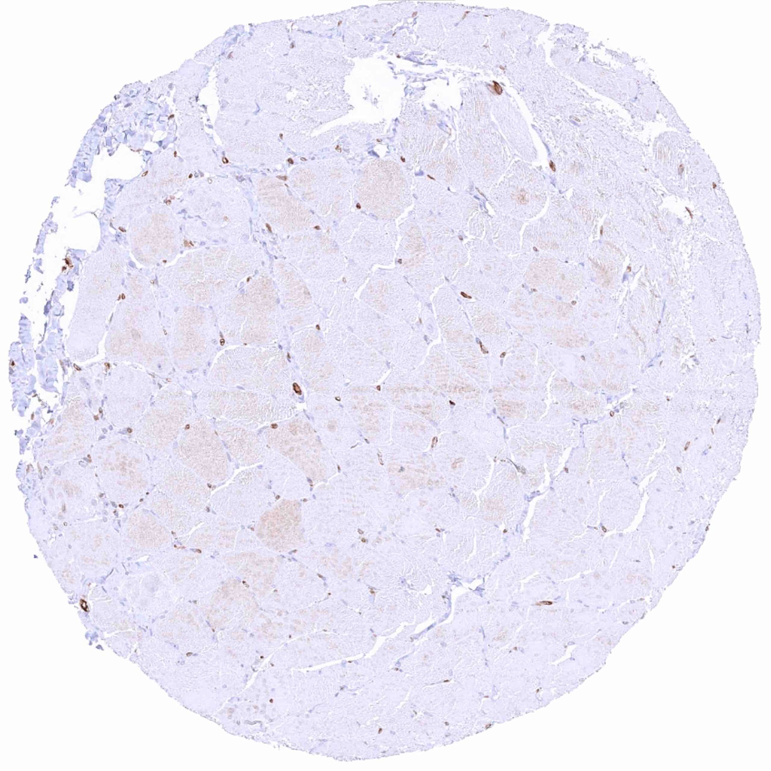
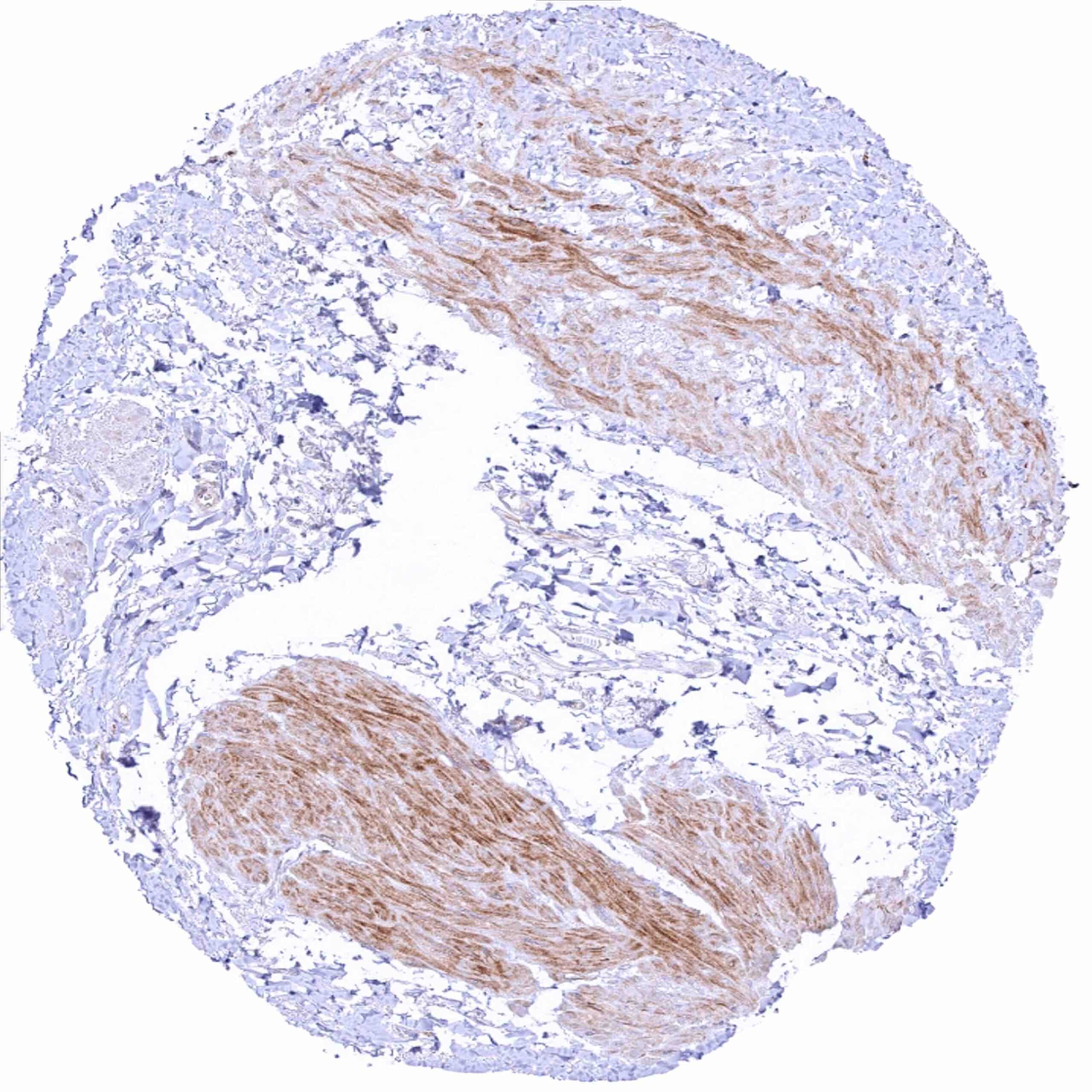
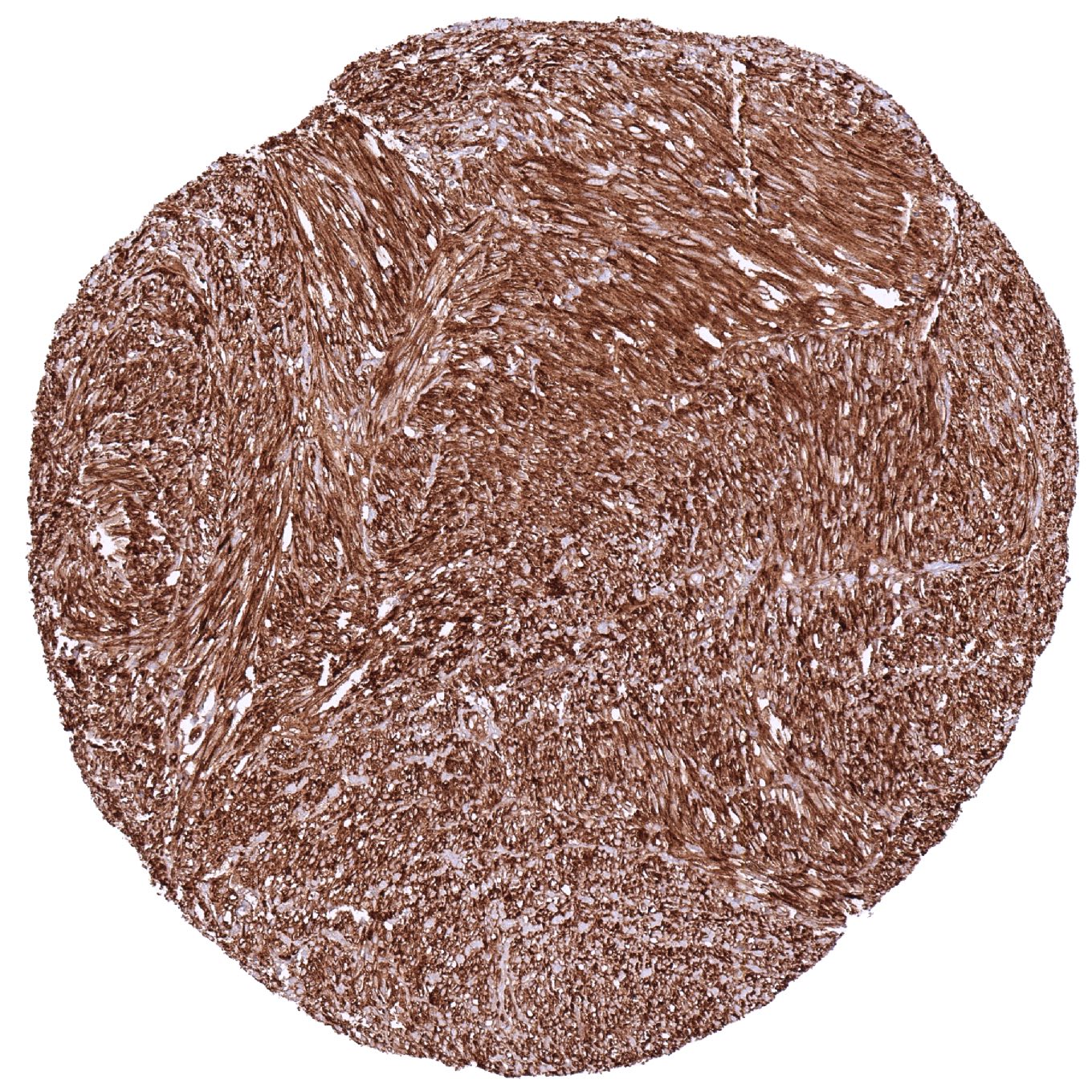
| Muscle/connective tissue | Heart muscle | MX1 staining predominates in the endothelium of small vessels. |
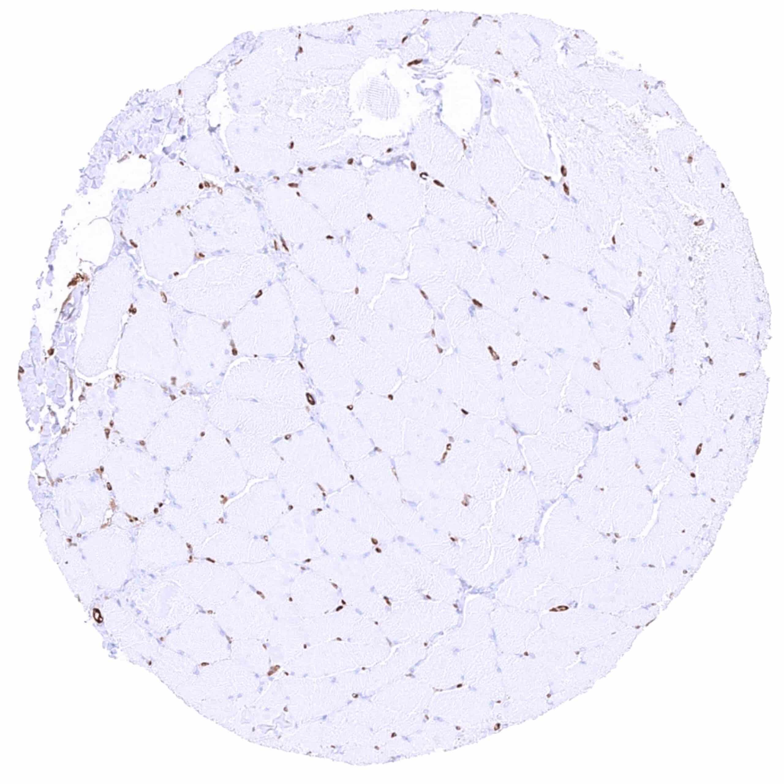
| Skeletal muscle | MX1 staining predominates in the endothelium of small vessels. | |
| Smooth muscle | Negative. | |
| Vessel walls | MX1 positive material can be found in the aortic wall (in some but not all samples). | |
| Fat | Negative. | |
| Stroma | ||
| Endothelium | Intense cytoplasmic MX1 staining of endothelial cells. | |
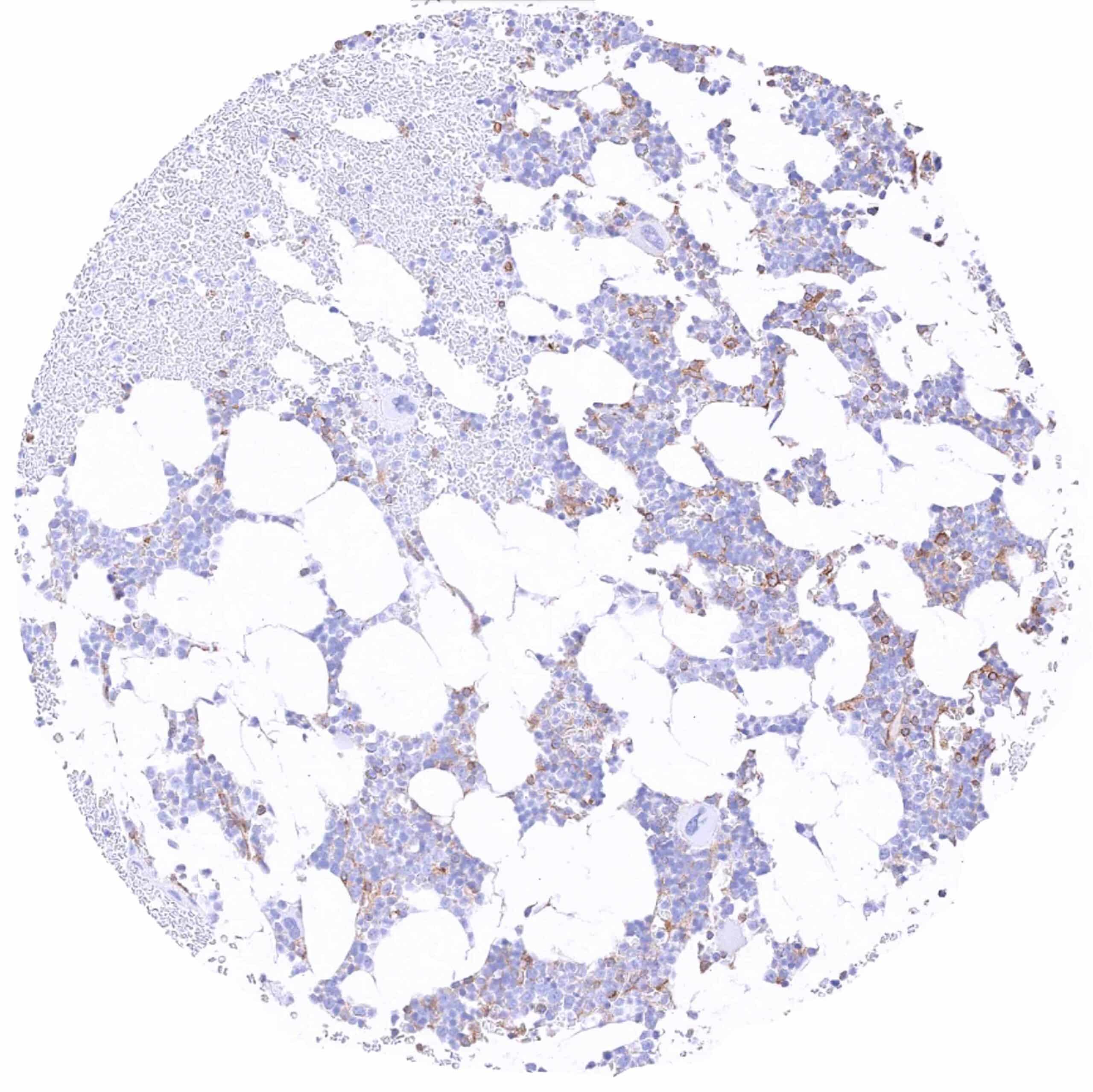
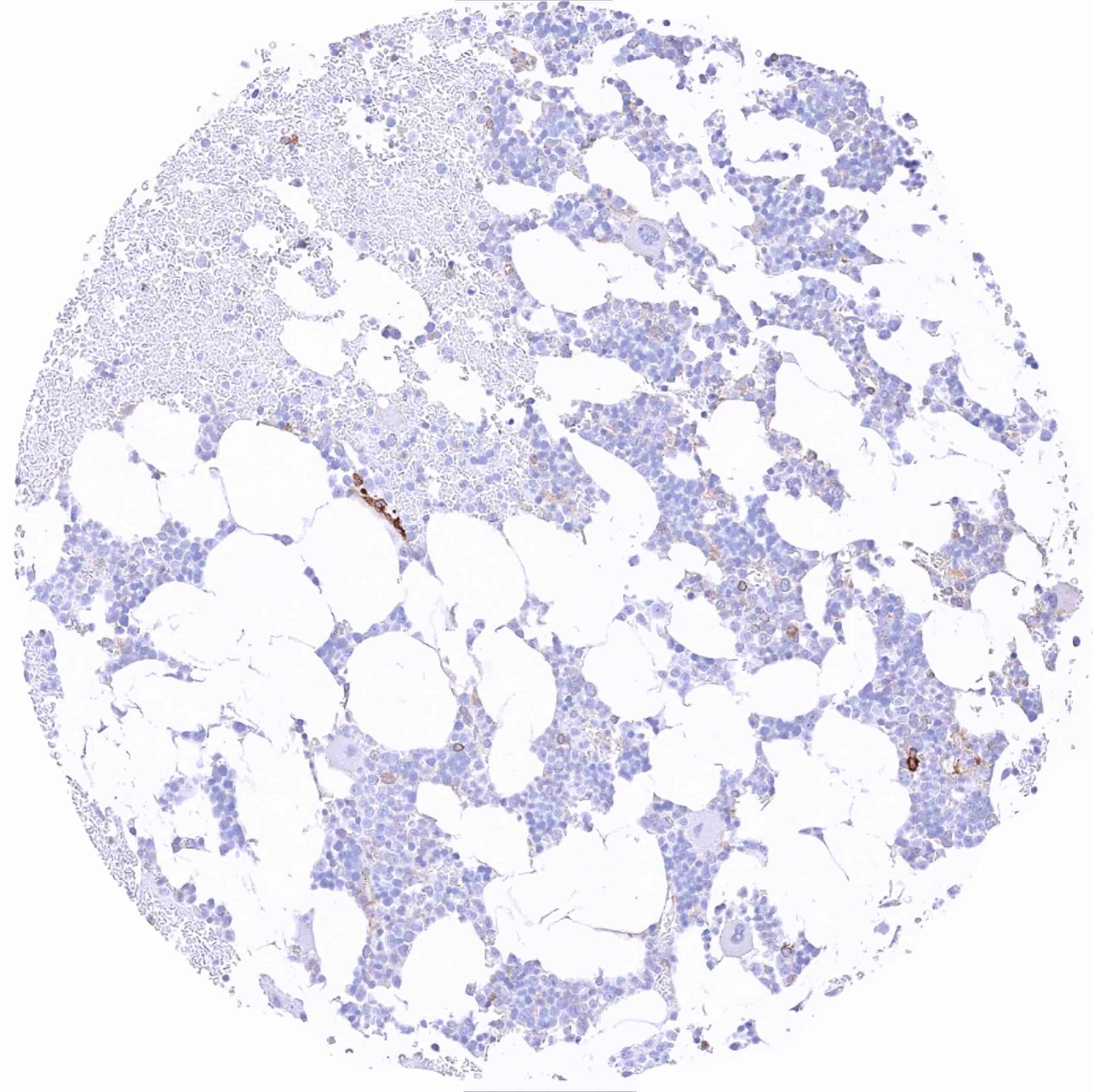
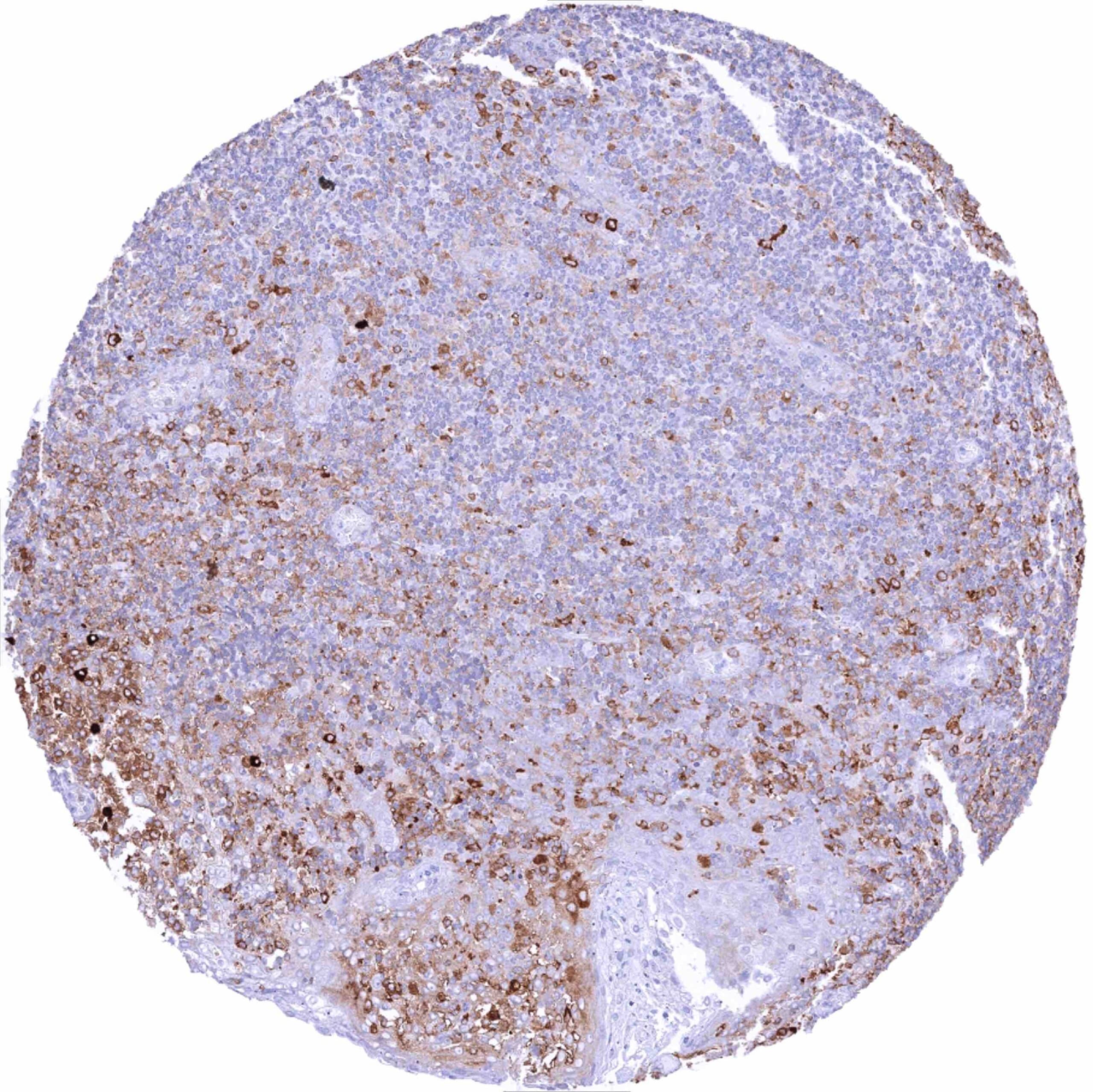
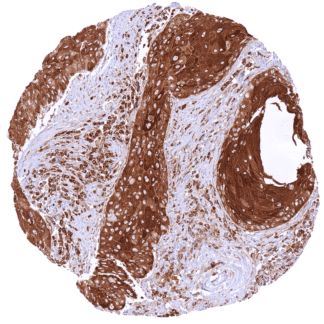
| Bone marrow/ lymphoid tissue | Bone marrow | Moderate MX1 staining of several hematopoetic cell types. |
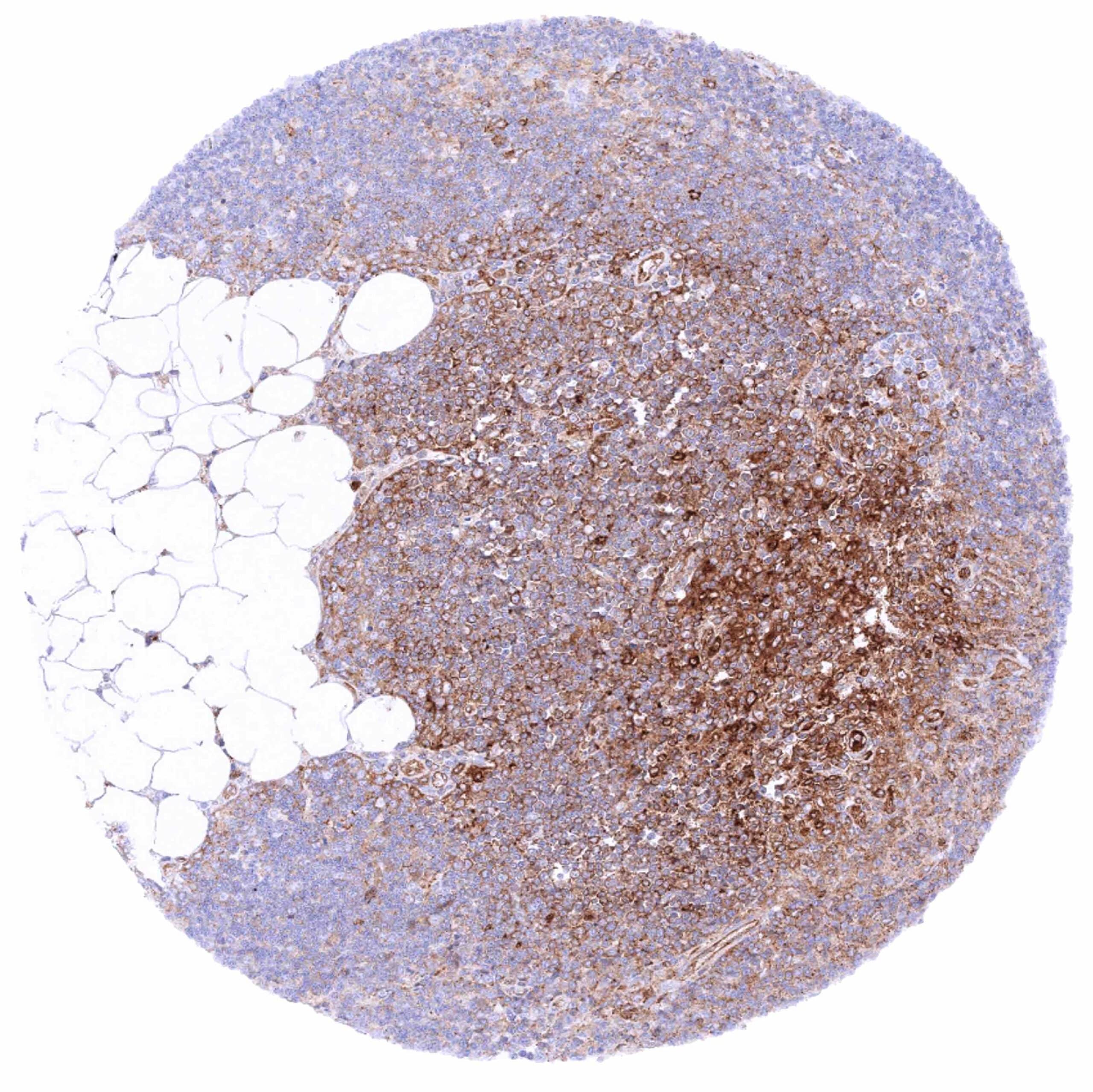
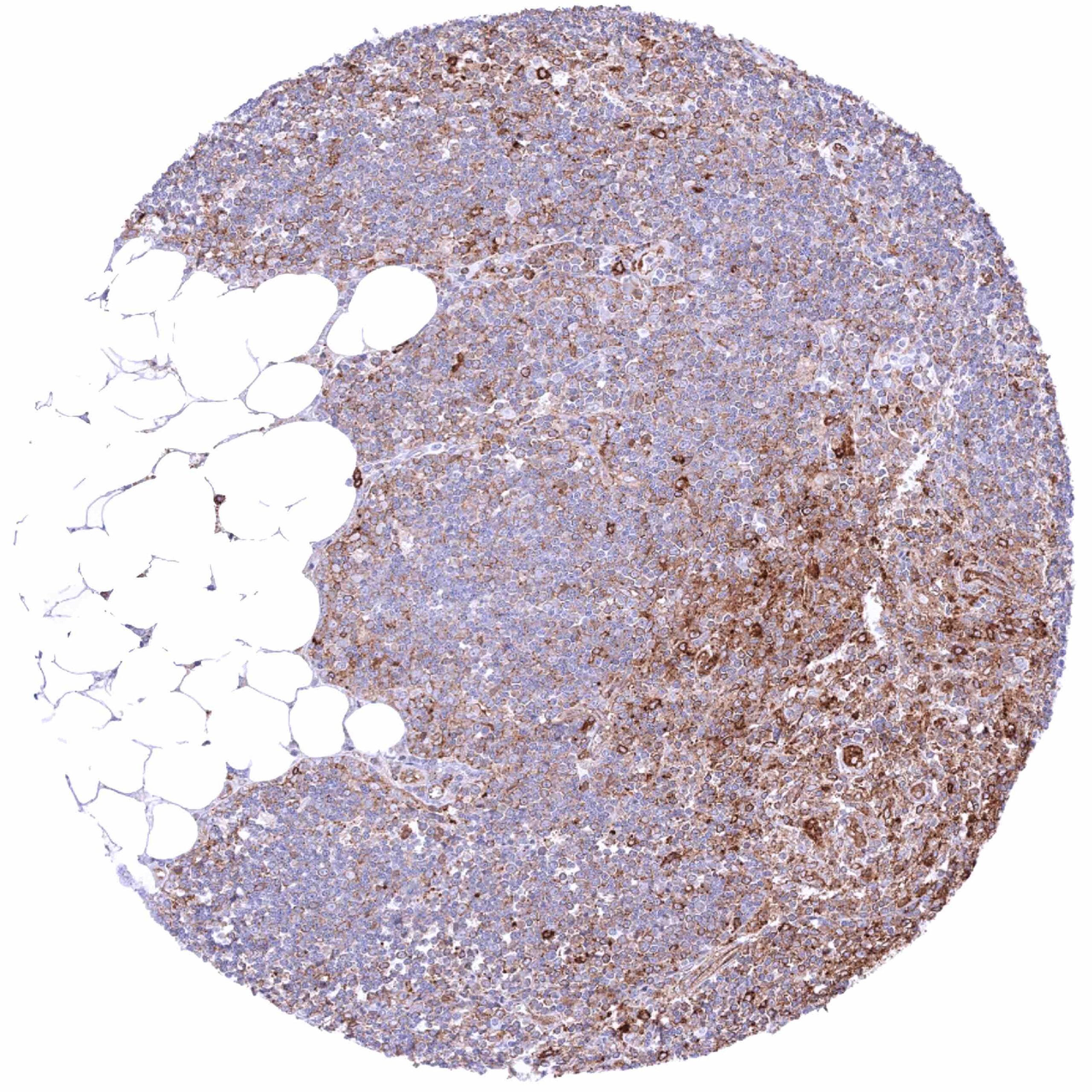
| Lymph node | Cytoplasmic MX1 staining of variable intensity of a large fraction of lymphatic cells. MX1 staining of lymphocytes is most intense in germinal centres. | |
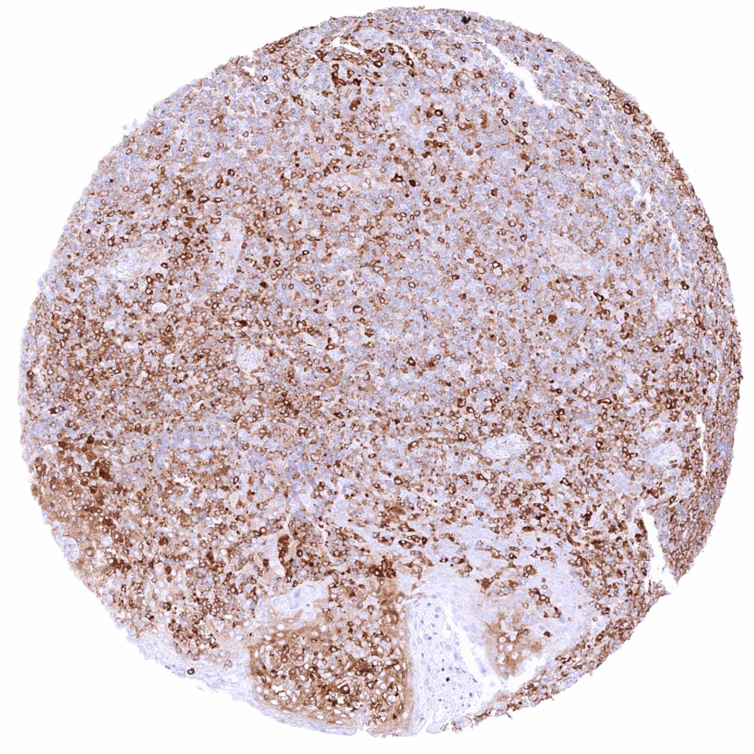
| Spleen | Cytoplasmic MX1 staining of variable intensity of a large fraction of cells. | |
| Thymus | Cytoplasmic MX1 staining of variable intensity of a large fraction of lymphocytic cells. | |
| Tonsil | Cytoplasmic MX1 staining of variable intensity of a large fraction of lymphatic cells. MX1 staining of lymphocytes is most intense in germinal centres. Epithelial cells are either negative or show only weak staining of basal cell layers. | |
| Remarks | MX1 is mainly localized in the cytoplasm and shows a granular staining pattern. The staining pattern within individual organs is not constant. Some evidence suggests increased expression in case of “tissue damage”. |
These findings are largely comparable to the RNA and protein data described in the Human Protein Atlas (Tissue expression MX1).
Positive control = Prostate: An at least weak to moderate MX1 staining should be seen in basal cells while acinar cells remain negative.
Negative control = Colon: Crypt epithelial cells should be MX1 negative.
Staining Pattern in Relevant Tumor Types
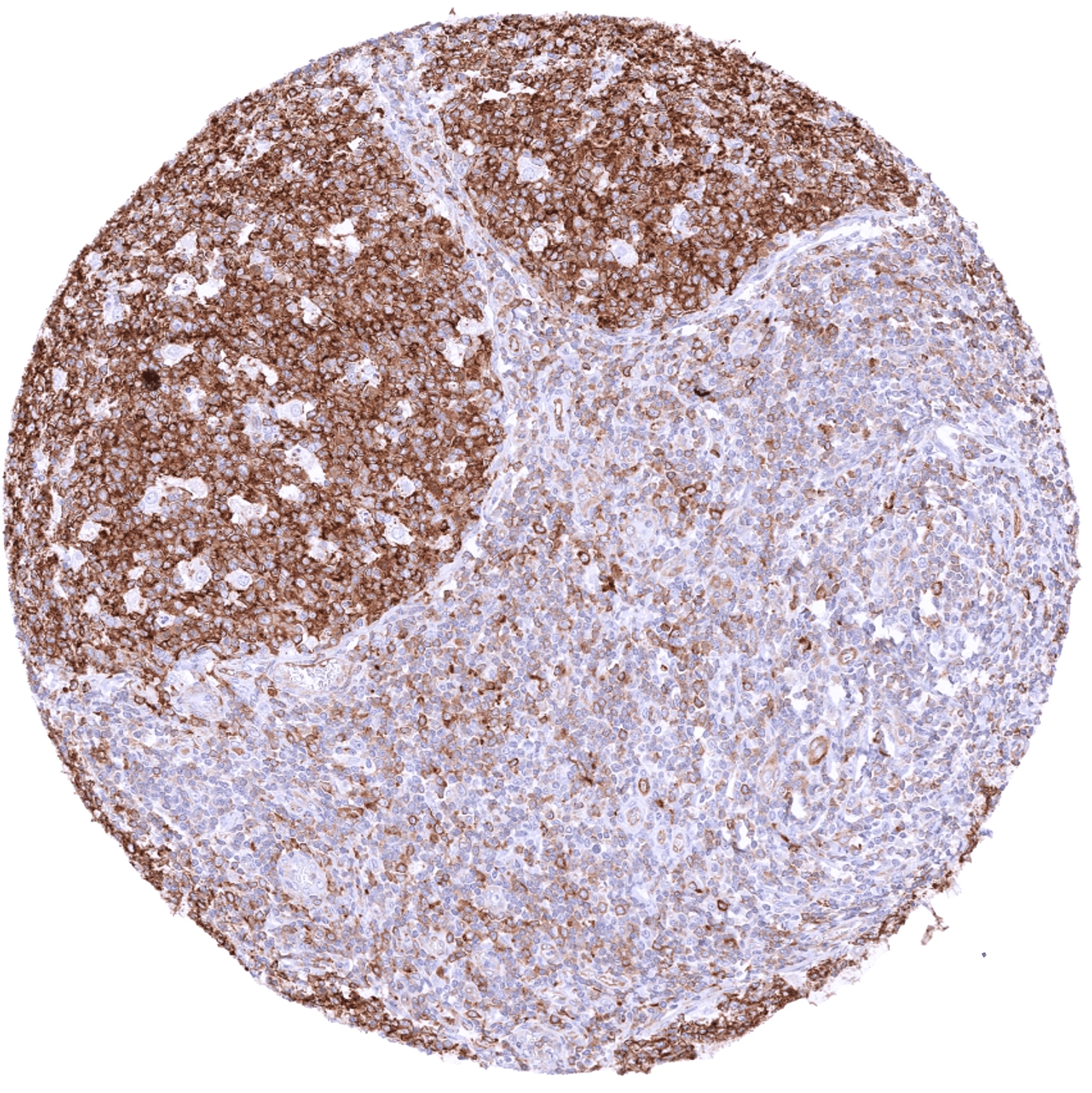
RNA data suggest that MX1 can be expressed in every tumor type. The clinical role is not clear yet – mostly because RNA data cannot discriminate the MX1 expression of tumor cells from stroma/inflammatory cells.
The TCGA findings on MX1 RNA expression in different tumor categories have been summarized in the Human Protein Atlas.
Compatibility of Antibodies
No data available at the moment
Protocol Recommendations
IHC users have different preferences on how the stains should look like. Some prefer high staining intensity of the target stain and even accept some background. Others favor absolute specificity and lighter target stains. Factors that invariably lead to more intense staining include higher concentration of the antibody and visualization tools, longer incubation time, higher temperature during incubation, higher temperature and longer duration of the heat induced epitope retrieval (slide pretreatment). The impact of the pH during slide pretreatment has variable effects and depends on the antibody and the target protein.
All images and data shown here and in our image galleries are obtained by the manual protocol described below. Other protocols resulting in equivalent staining are described as well.
Manual protocol
Freshly cut sections should be used (less than 10 days between cutting and staining). Heat-induced antigen retrieval for 5 minutes in an autoclave at 121°C in pH 7,8 Target Retrieval Solution buffer. Apply HMV316 at a dilution of 1:200 at 37°C for 60 minutes. Visualization of bound antibody by the EnVision Kit (Dako, Agilent) according to the manufacturer’s directions.
Potential Research Applications
- The diagnostic and prognostic relevance of MX1 expression in tumors and in preneoplastic disease needs to be investigated.
Evidence for Antibody Specificity in IHC
There are two ways how the specificity of antibodies can be documented for immunohistochemistry on formalin fixed tissues. These are: 1. Comparison with a second independent method for target expression measurement across a large number of different tissue types (orthogonal strategy), and 2. Comparison with one or several independent antibodies for the same target and showing that all positive staining results are also seen with other antibodies for the same target (independent antibody strategy).
Orthogonal validation: For proteins such as MX1 which are expressed in virtually all tissues but restricted to specific cell types and cell compartments, orthogonal validation is not suited.
For the antibody HMV316 specificity is, however, to some extent supported by the particularly high immunostaining level in lymphocytes and in cells of the hematopoesis. This is in agreement with RNA expression data from three independent RNA screening studies, including the Human Protein Atlas (HPA) RNA-seq tissue dataset, the FANTOM5 project, and the Genotype-Tissue Expression (GTEx) project, which are all summarized in the Human Protein Atlas (Tissue expression MX1).
Comparison of antibodies: True expression of MX1 in all cell types found to be MX1 positive by HMV316 is corroborated by identical staining patterns obtained by another commercially available independent antibody (termed “validation antibody”) although the staining by the validation antibody was always less intense. Independence of the “validation antibody” is supported by a diffuse staining of smooth muscle cells by the validation antibody but not by HMV316. These smooth muscle stainings represent an antbody specific cross-reactivity of the “validation antibody”.