395,00 € – 1.495,00 €
Product details
Synonyms = GATA3; GATA binding protein-3; GATA-binding factor 3; GATA3; HDR; HDRS; Transacting T-cell-specific transcription factor GATA-3
Antibody type = Rabbit monoclonal / IgG
Clone = MSVA-550R
Positive control
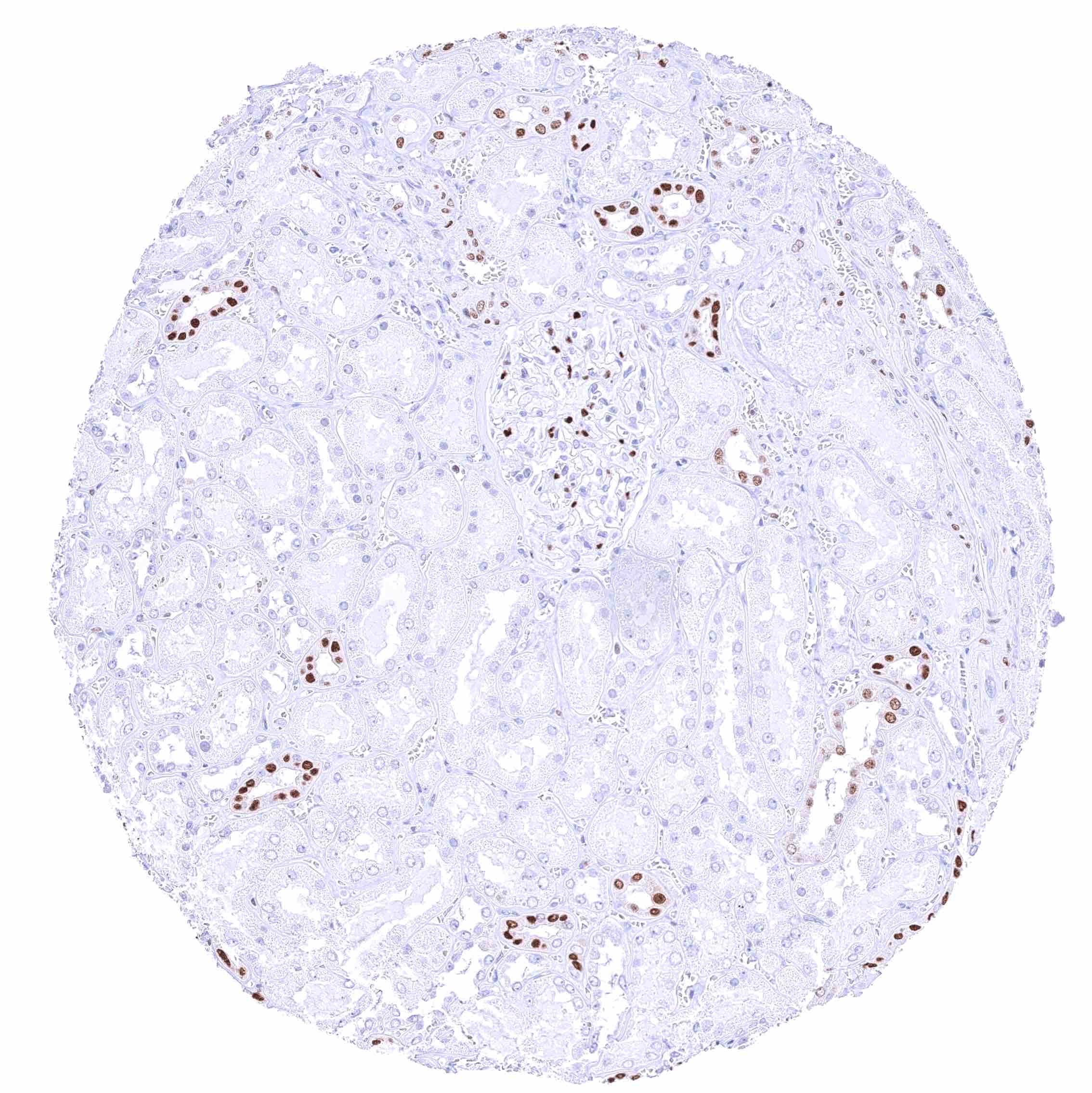
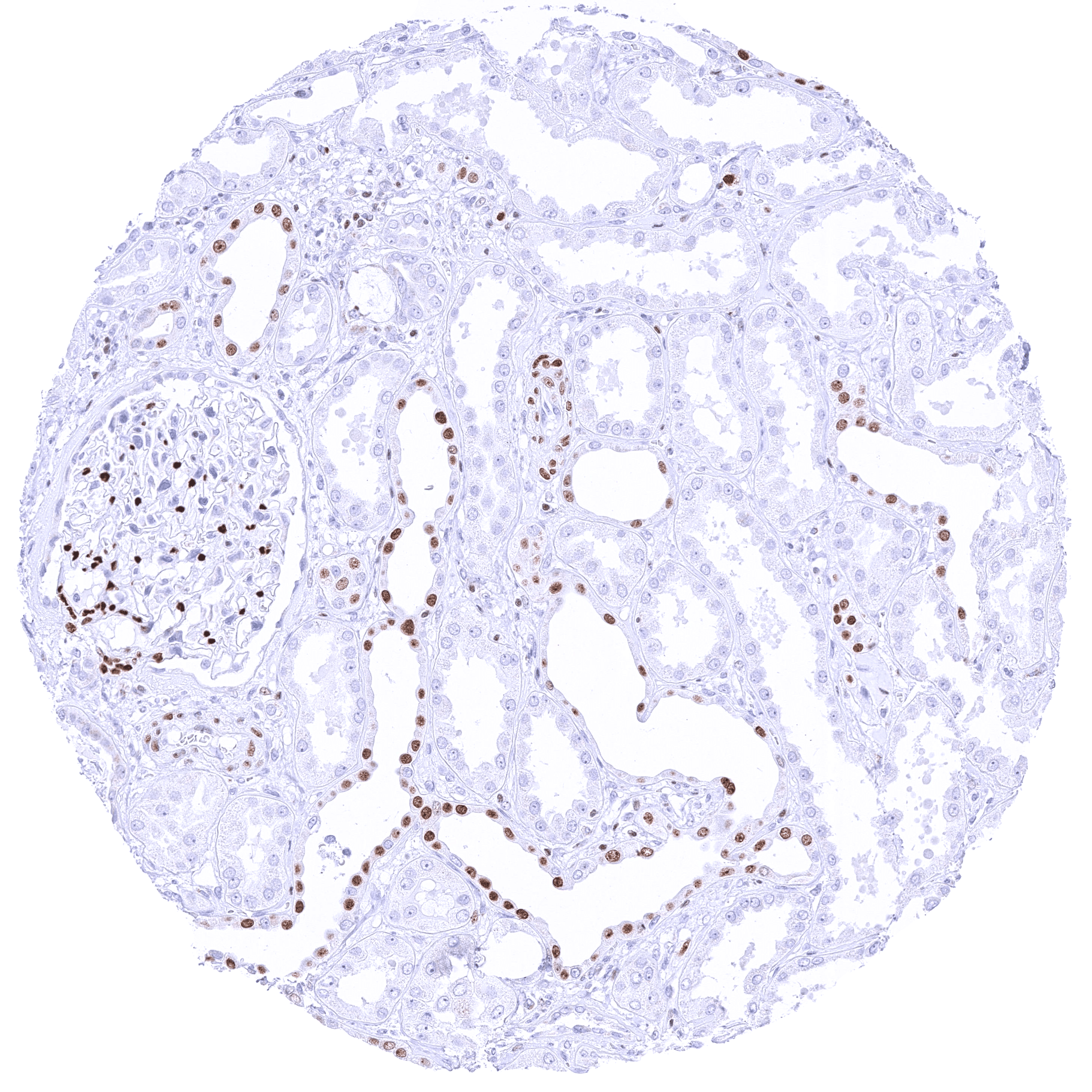
-Kidney: A moderate to strong nuclear staining reaction should be seen in a fraction of collecting duct cells and podocytes in glomeruli.
-Tonsil: The vast majority of T helper cells (Th2) in the T-zones should show a weak to moderate nuclear staining.
Negative control
-Kidney: Staining should be absent in proximal and distal tubuli as well as in blood vessels.
-Tonsil: Staining should be absent in B-cells.
Cellular localization = Nuclear
Reactivity = Human
Application = Immunohistochemistry
Dilution = 1:100 – 1:200
Intended Use = Research Use Only
Relevance of Antibody
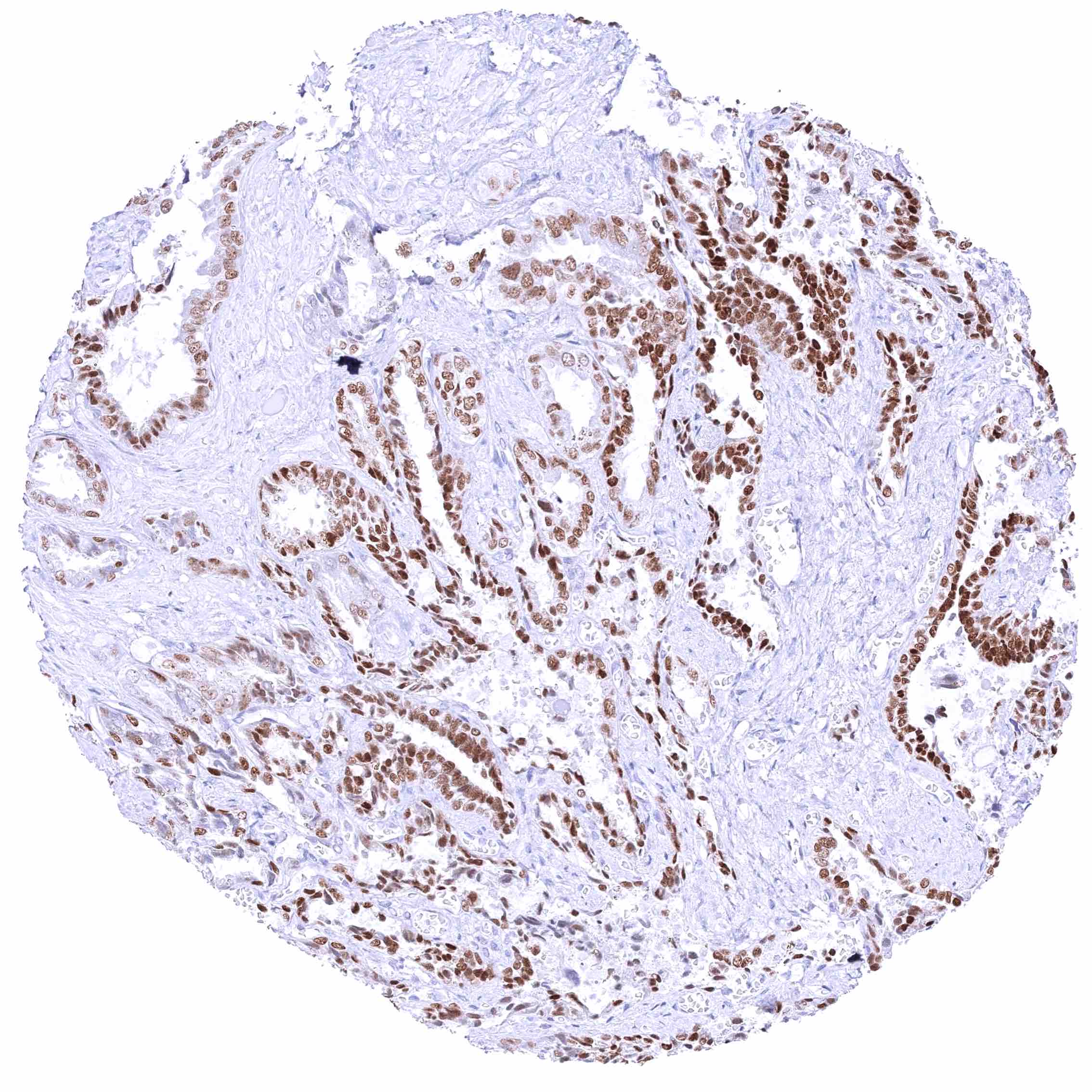
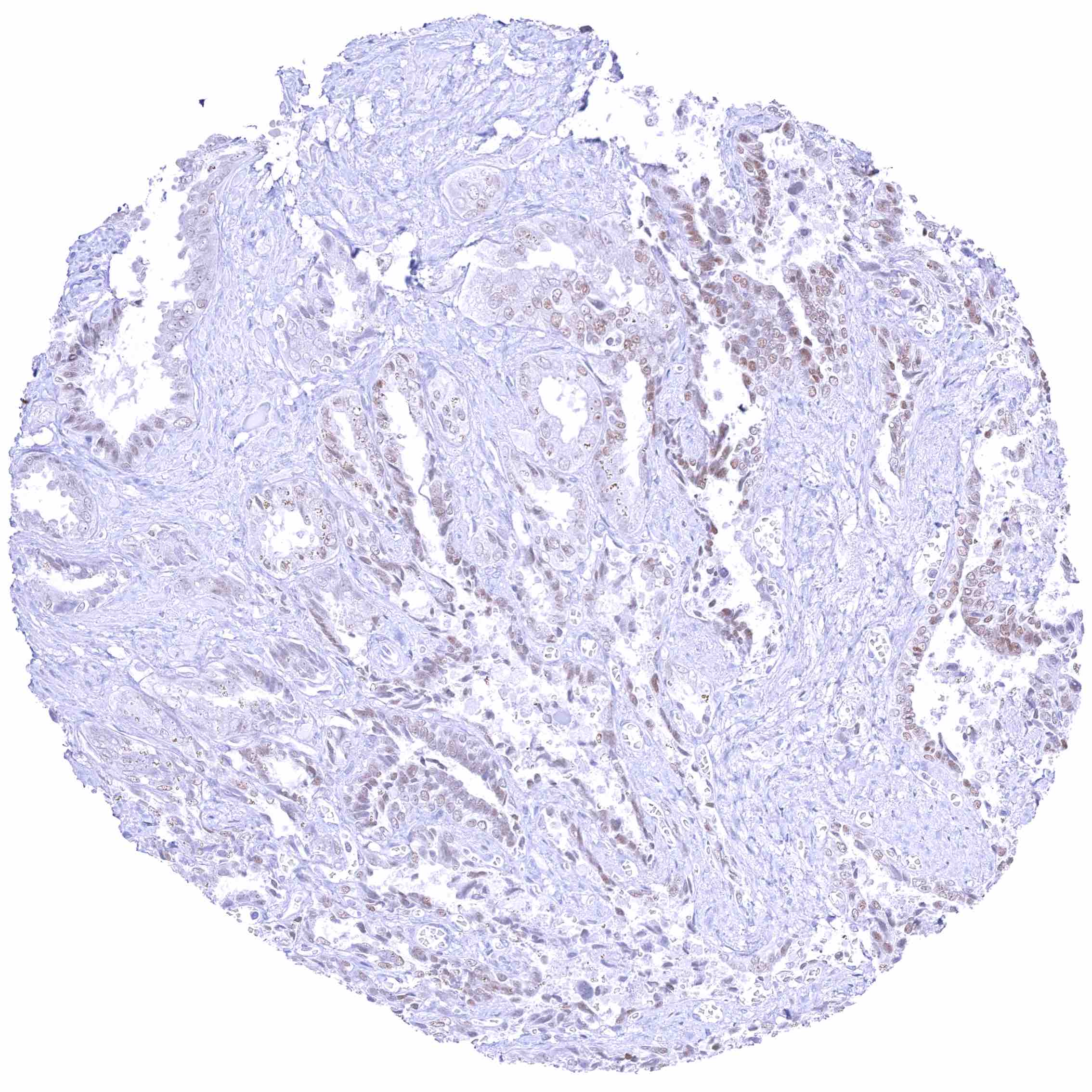
GATA3 is highly expressed in breast and urothelial carcinoma.
Biology Behind
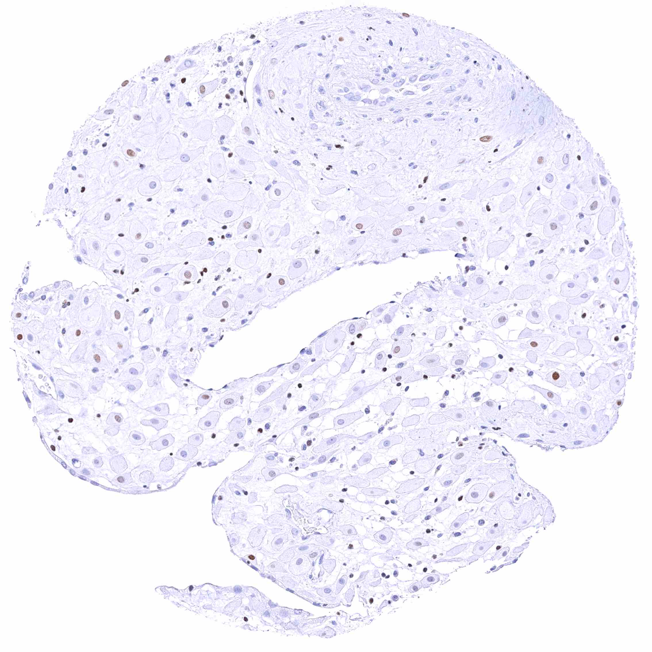
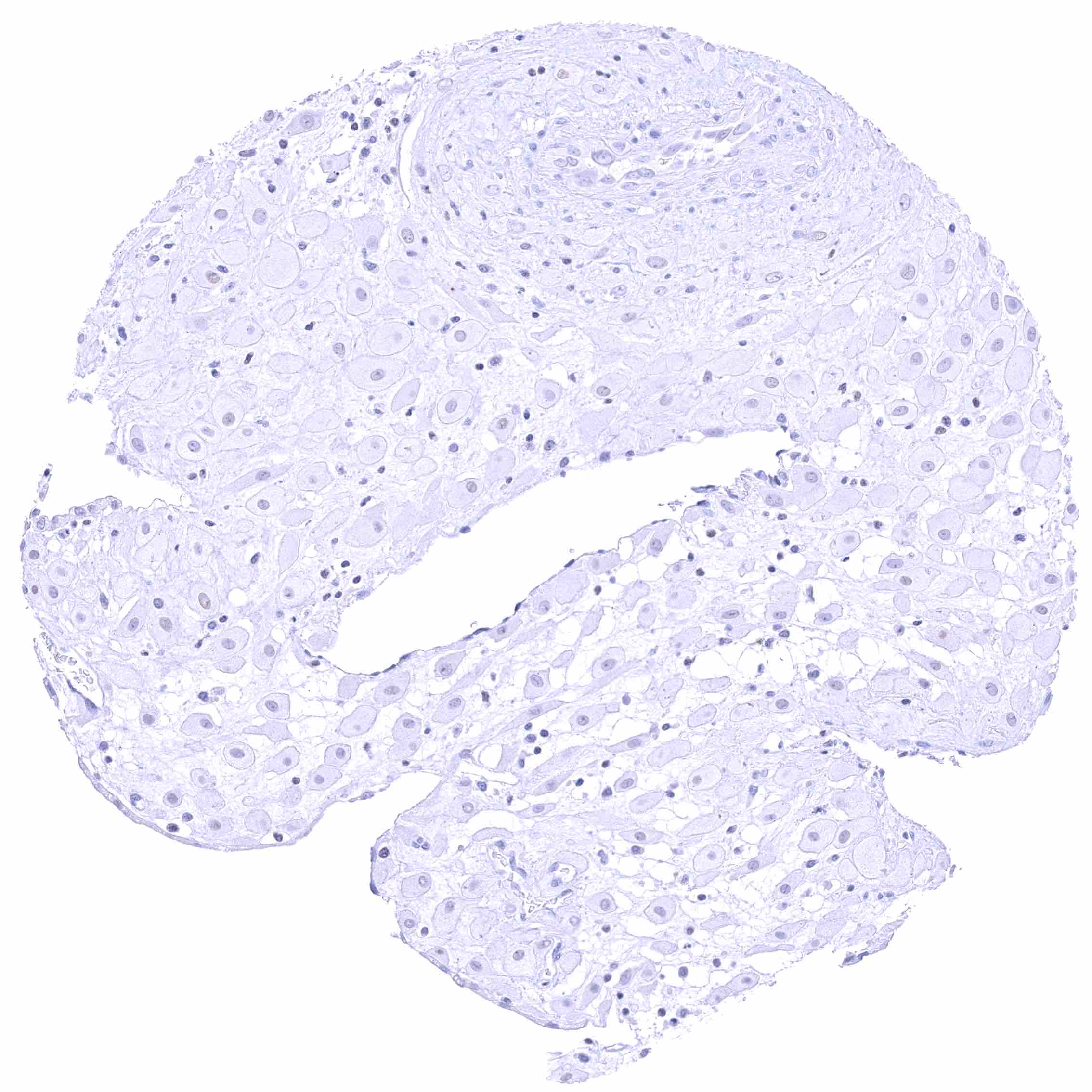
The GATA3 gene at 10p14 consists of 8 exons and codes for the GATA3 transcription factor which is critical for the embryonic development of various tissues including the parathyroid gland and the kidney. It plays a role in the luminal differentiation of breast epithelium, the development of the collecting system of the kidney and the urothelium, and in trophoblastic differentiation. In lymphoid cells, GATA3 regulates the expression of a wide range of biologically and clinically important genes. GATA3 is required for the formation of T helper (Th) cells, especially Th2 cells which are critical for the development of allergic and humoral immune responses. In normal tissues, nuclear GATA3 immunostaining is seen in various tissues and cell types. It is most intense in urothelium, squamous epithelium of the skin, parathyroid gland, trophoblastic cells and chorion cells of the placenta, collecting ducts and glomerular podocytes of the kidney, seminal vesicle epithelium, and a fraction of luminal cells GATA3 also plays a role in cancer biology. It is one of the most commonly mutated genes in breast cancer where it plays a role in estrogen and androgen receptor signaling. Among tumors, GATA3 expression is primarily seen in urothelial and breast cancers but it can also occur in other tumor entities. The antibody MSVA-550R is markedly more sensitive than MSVA-450M for detection of nuclear GATA3 but causes some non-specific cytoplasmic staining in several tissues due to cross-reactivity.
Staining Pattern in Normal Tissues
GATA3 staining pattern in Normal Tissues with antibody MSVA-550R (images are shown in our “Normal Tissue Gallery”)
| Brain | Cerebrum | Negative. |
| Cerebellum | Negative. | |
| Endocrine Tissues | Thyroid | Negative. |
| Parathyroid | Strong GATA3 positivity in all epithelial cells. | |
| Adrenal gland | Negative. | |
| Pituitary gland | Negative. | |
| Respiratory system | Respiratory epithelium | Negative. |
| Lung | Negative. | |
| Gastrointestinal Tract | Salivary glands | Negative. |
| Esophagus | Negative. | |
| Stomach | Negative. | |
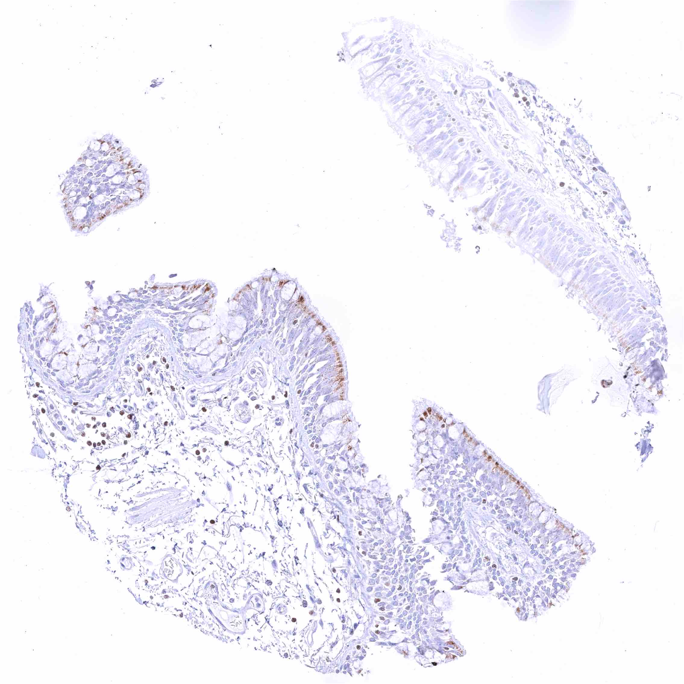
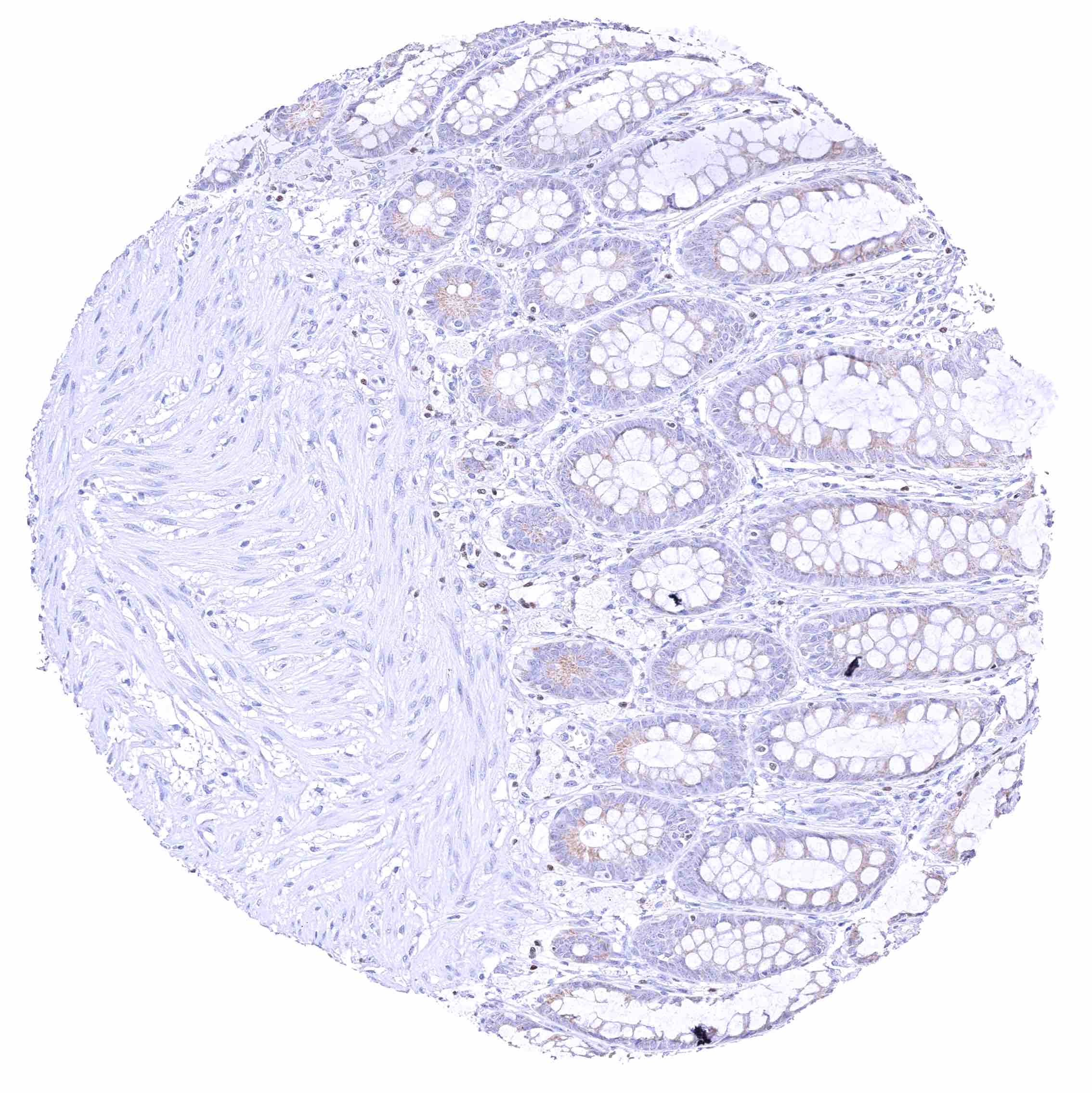
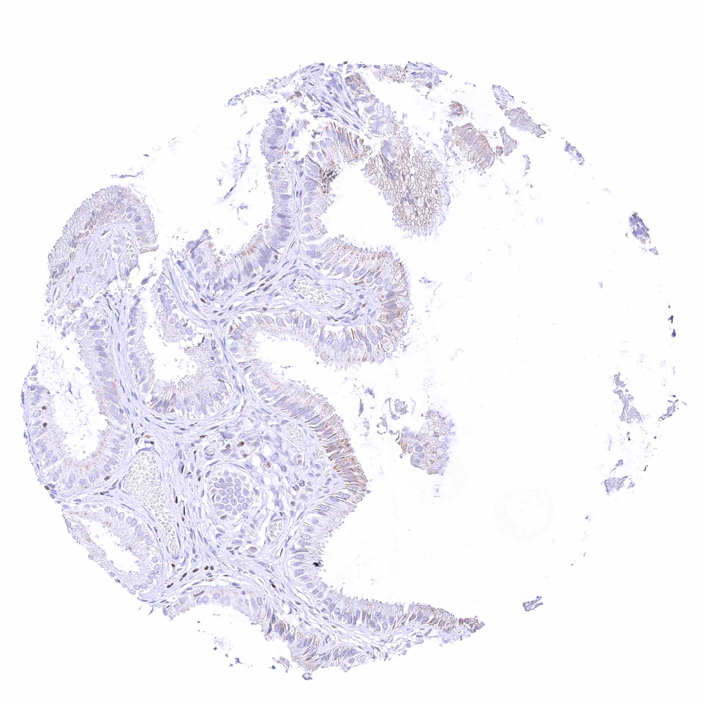
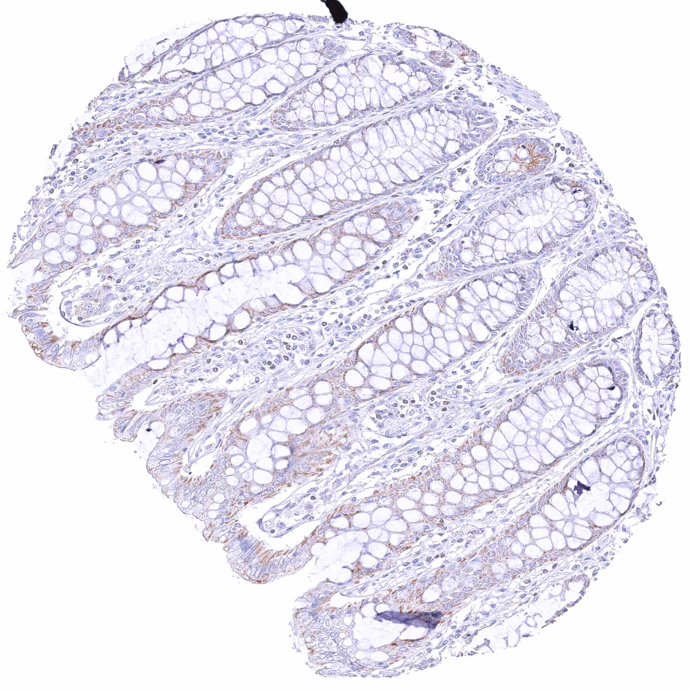
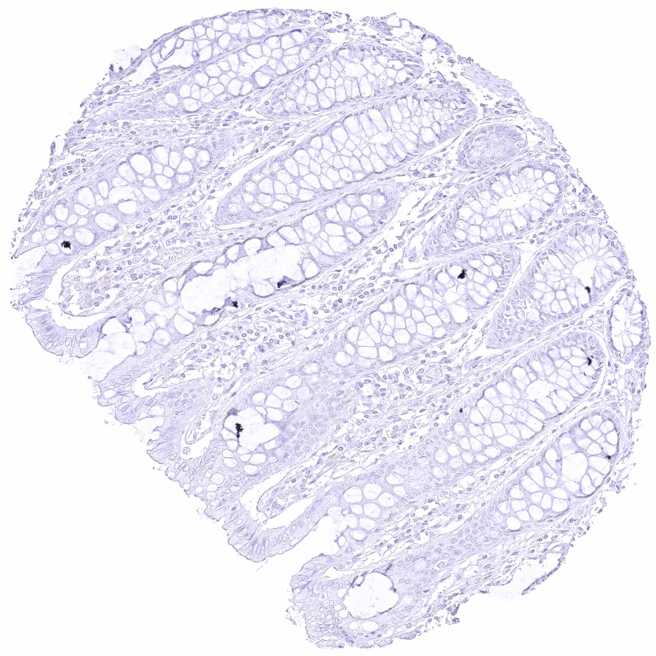
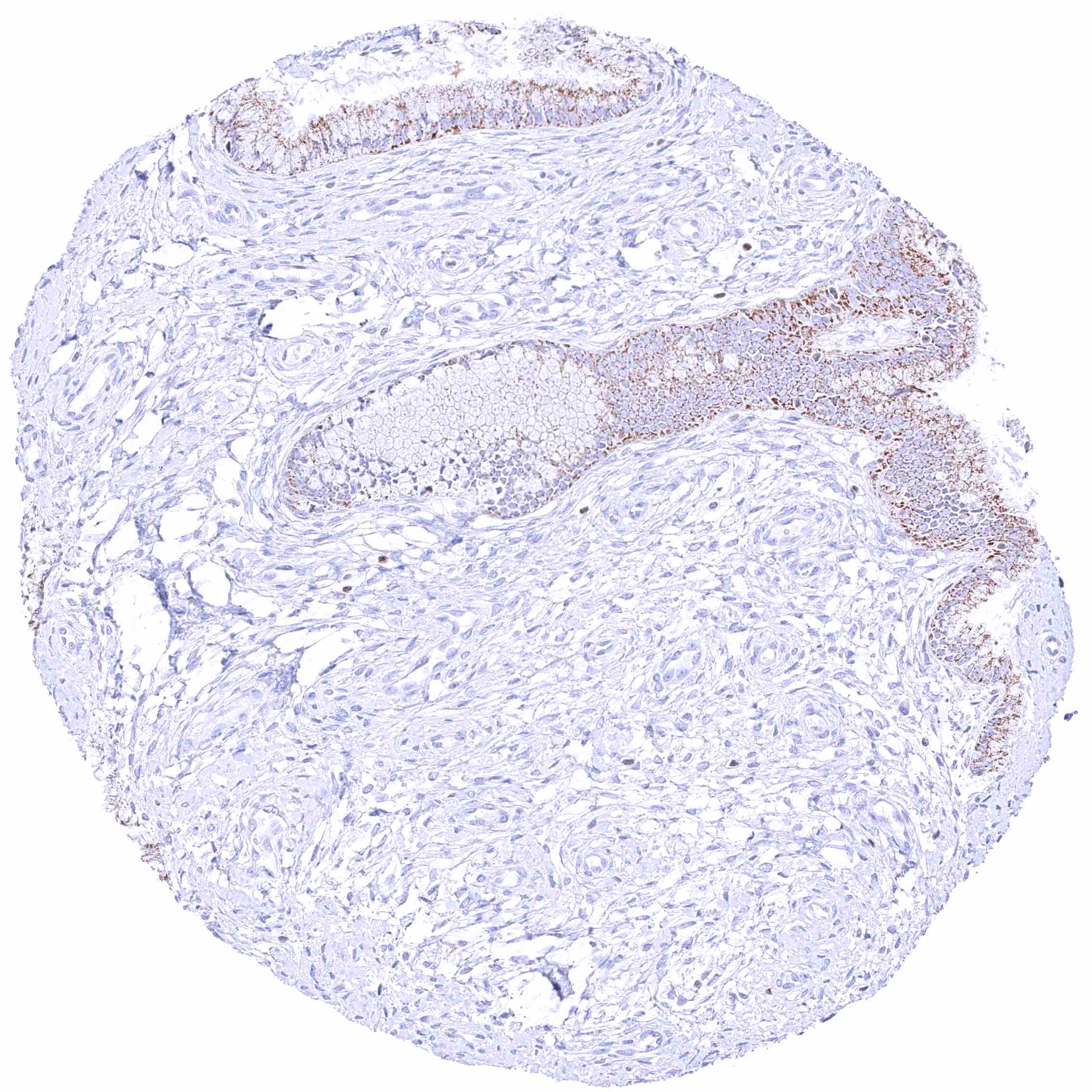
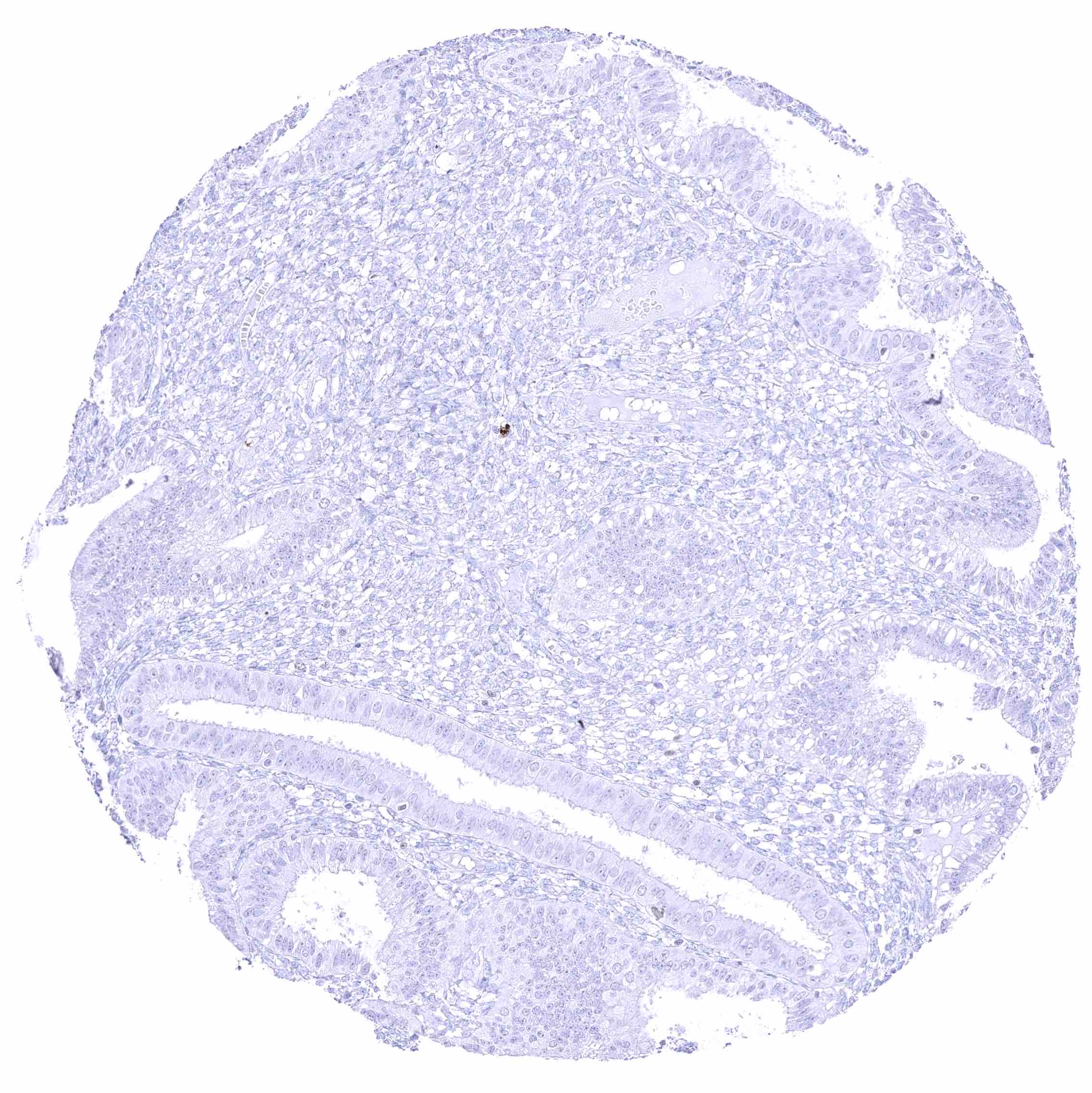
| Duodenum | Negative. A (cross-reactive) granular cytoplasmic GATA3 positivity of epithelial cells can occur. | |
| Small intestine | Negative. A (cross-reactive) granular cytoplasmic GATA3 positivity of epithelial cells can occur. | |
| Appendix | Negative. | |
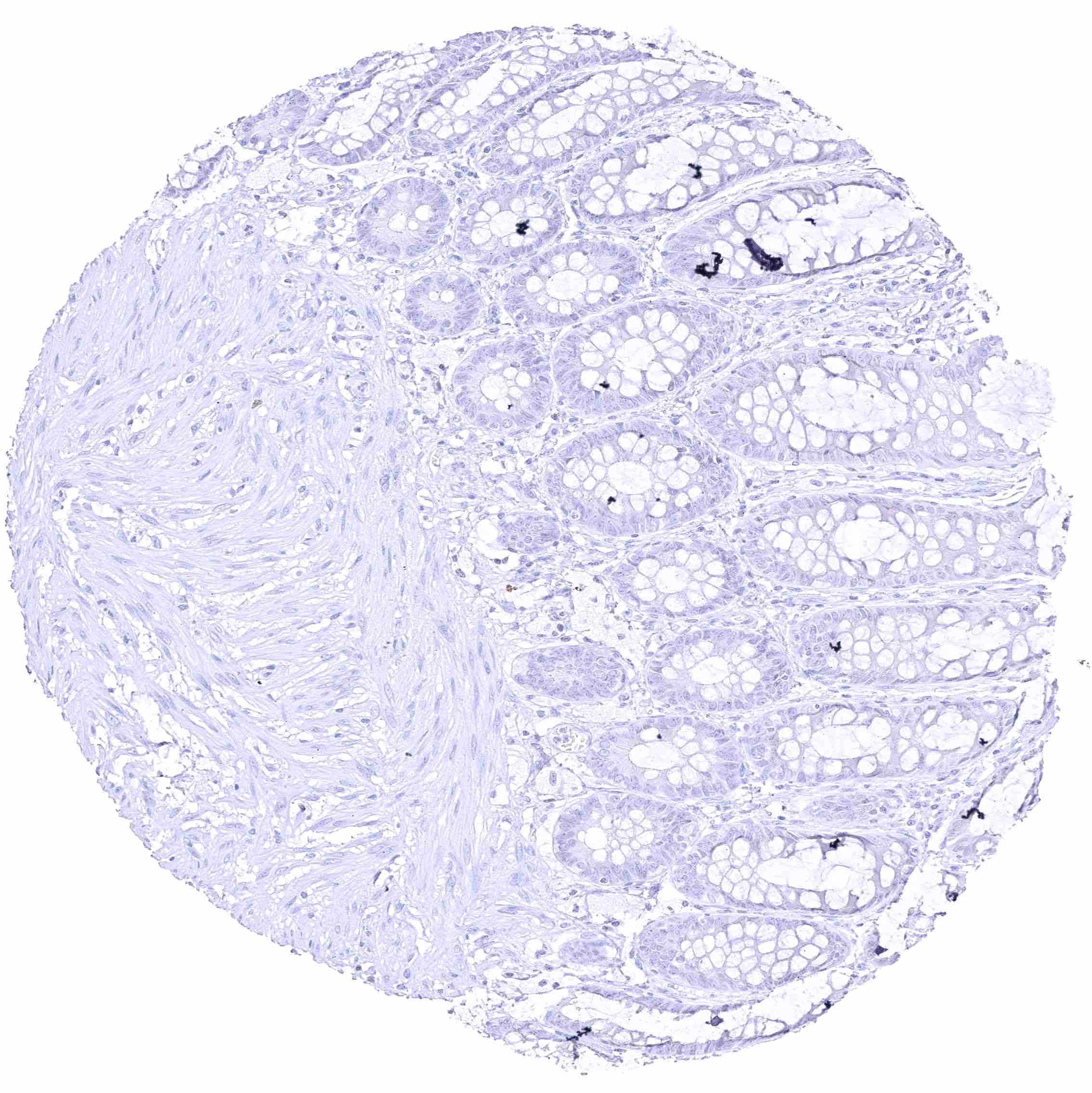
| Colon | Negative. A (cross-reactive) granular cytoplasmic GATA3 positivity of epithelial cells can occur. | |
| Rectum | Negative. A (cross-reactive) granular cytoplasmic GATA3 positivity of epithelial cells can occur. | |
| Liver | Negative. | |
| Gallbladder | Negative. | |
| Pancreas | Negative. | |
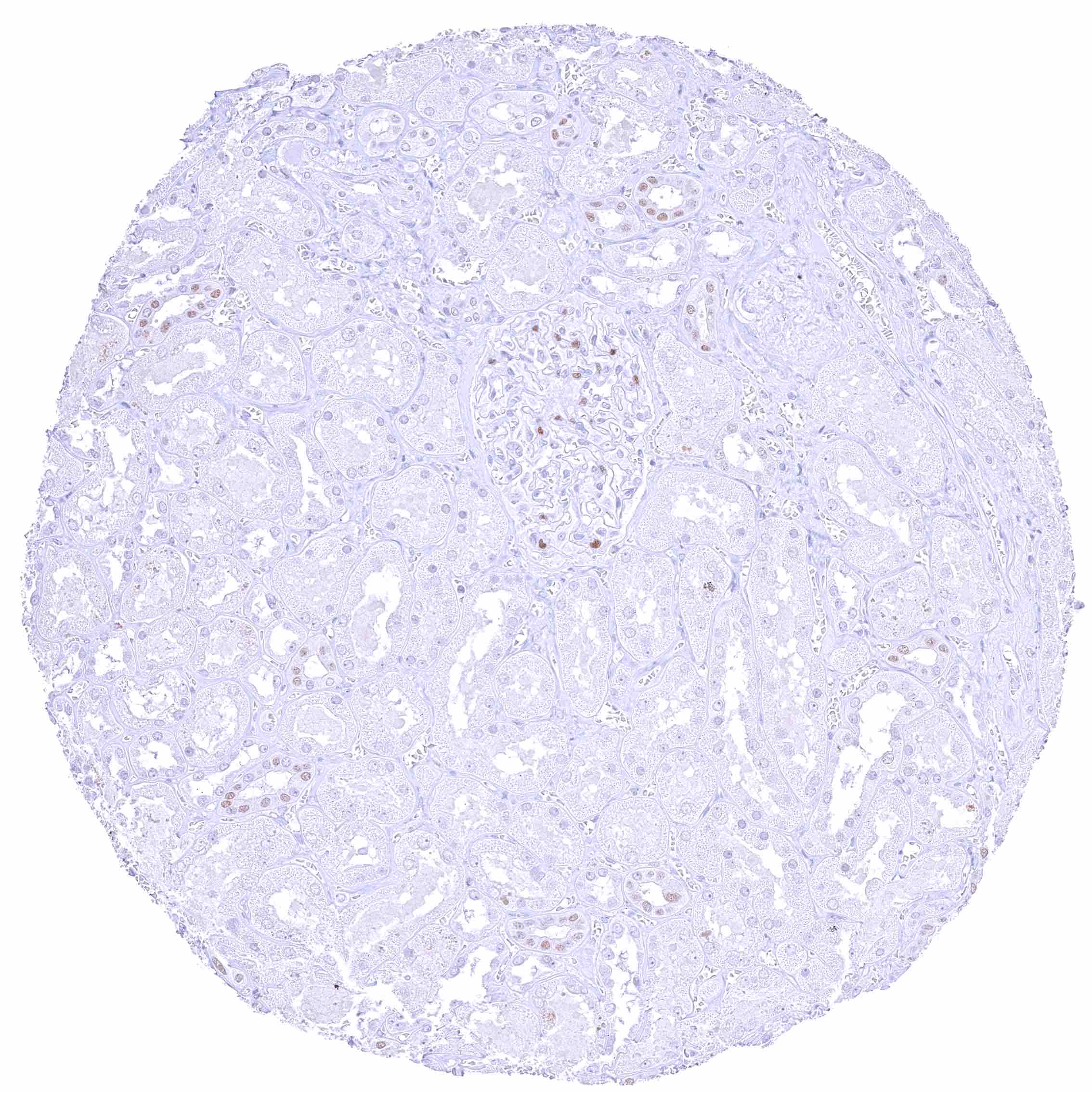
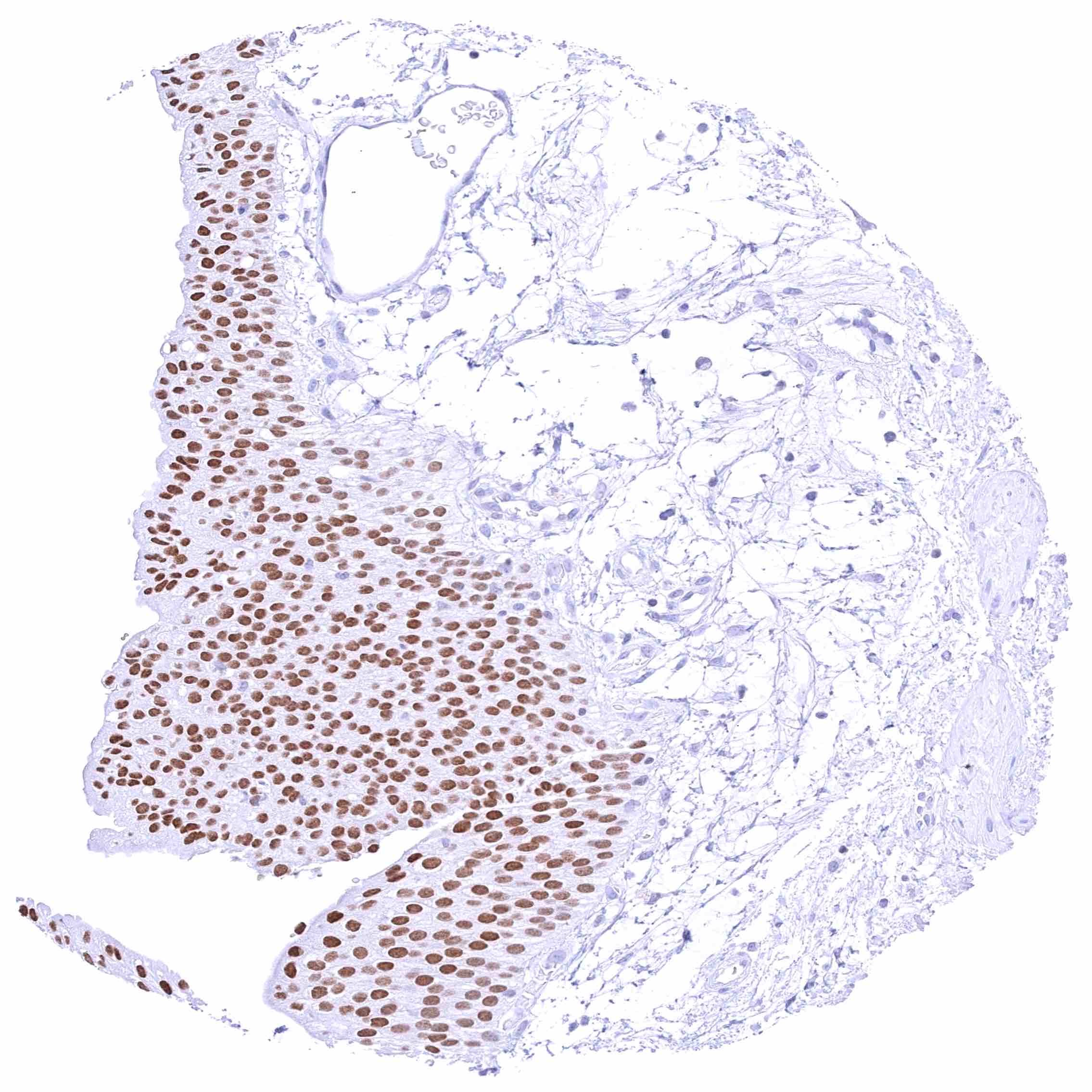
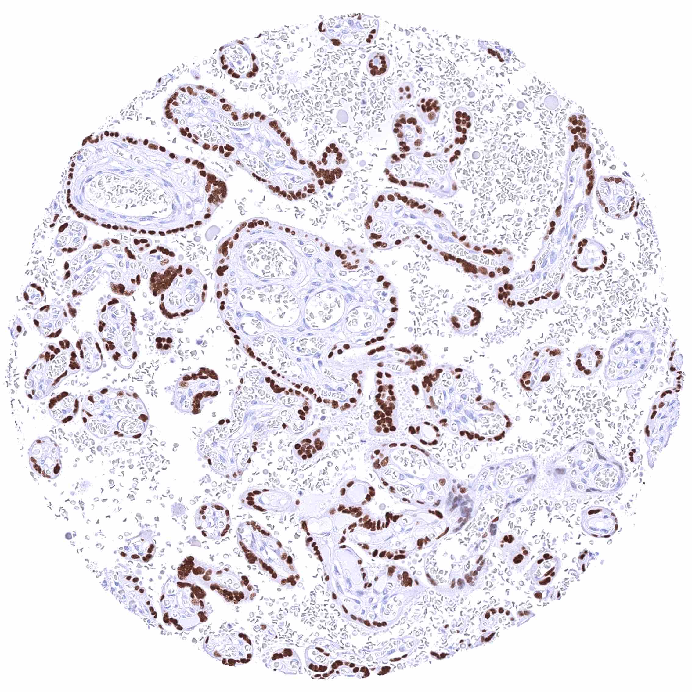
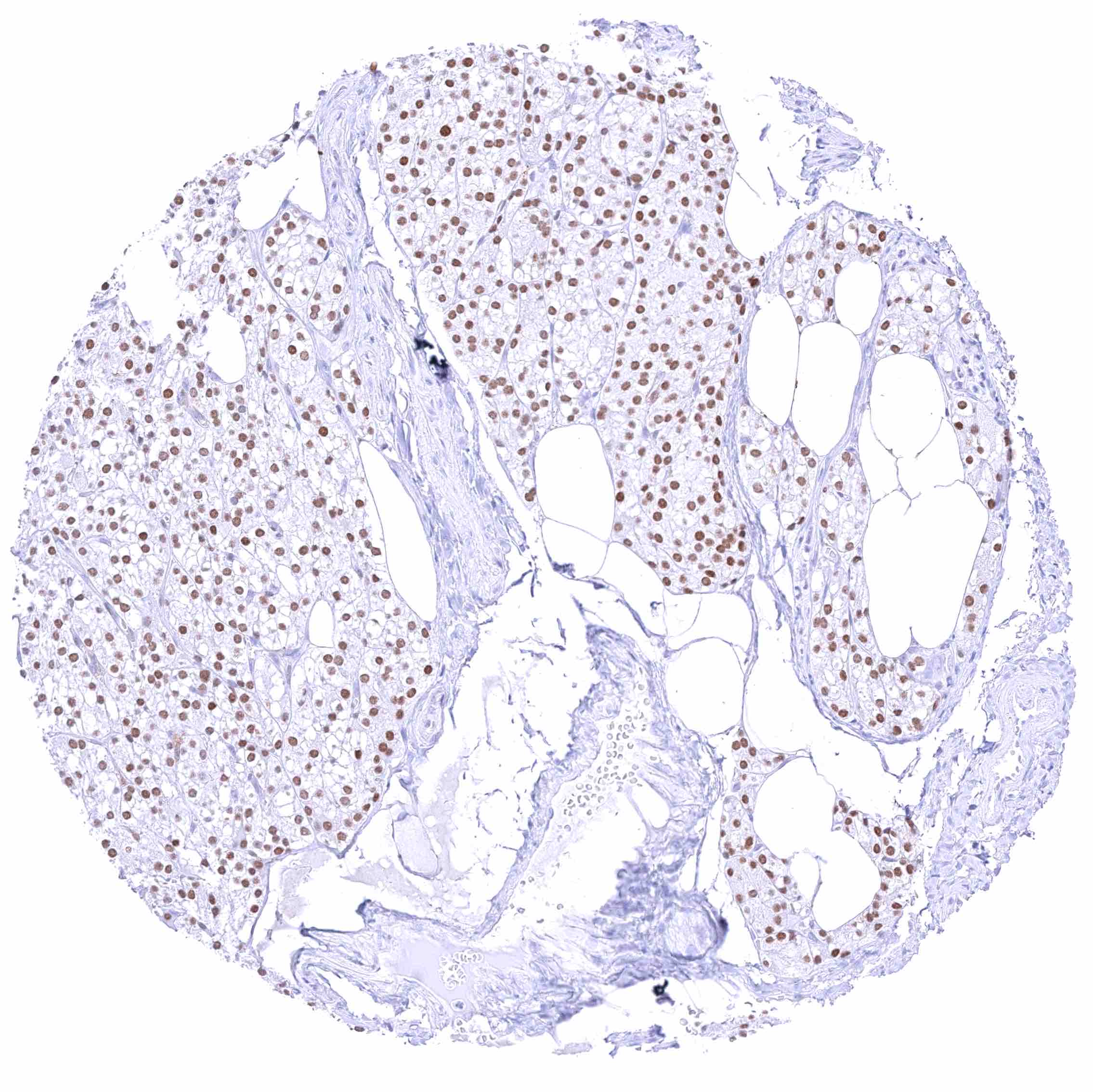
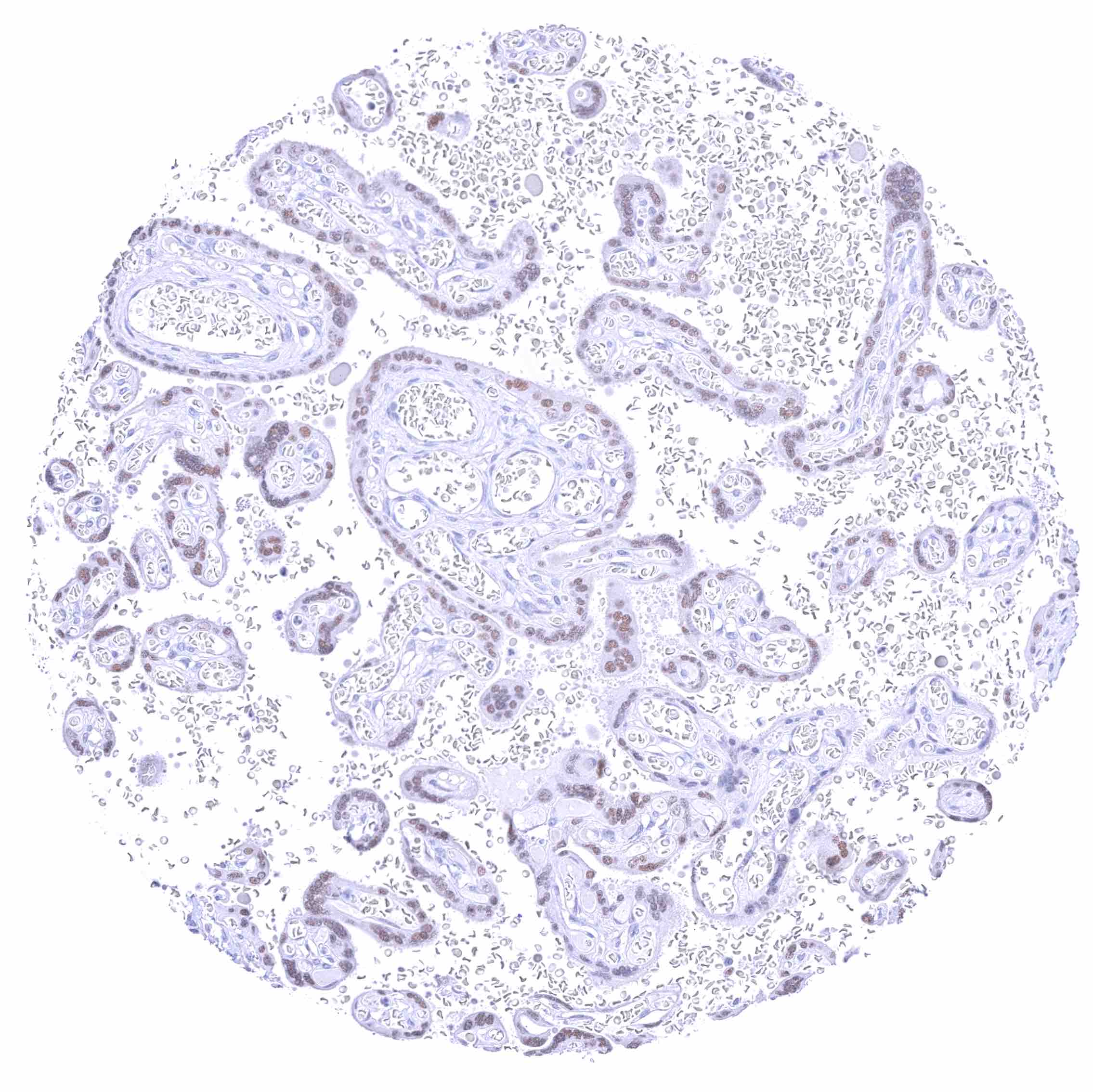
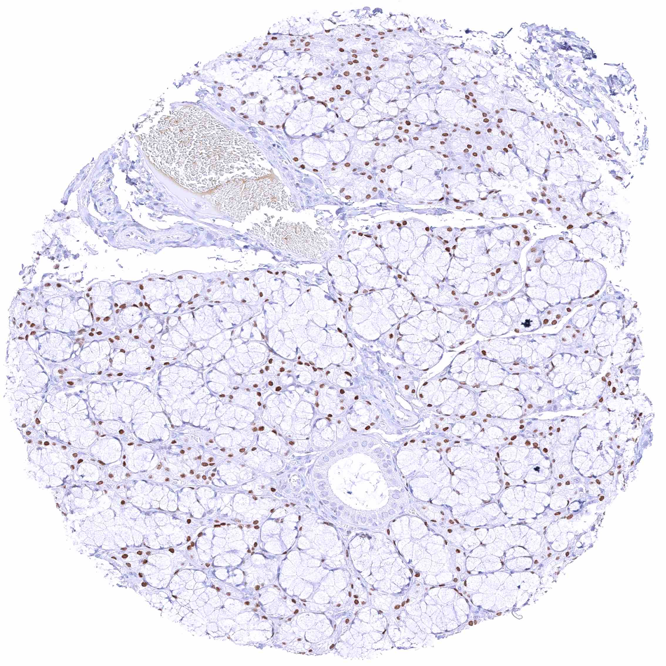
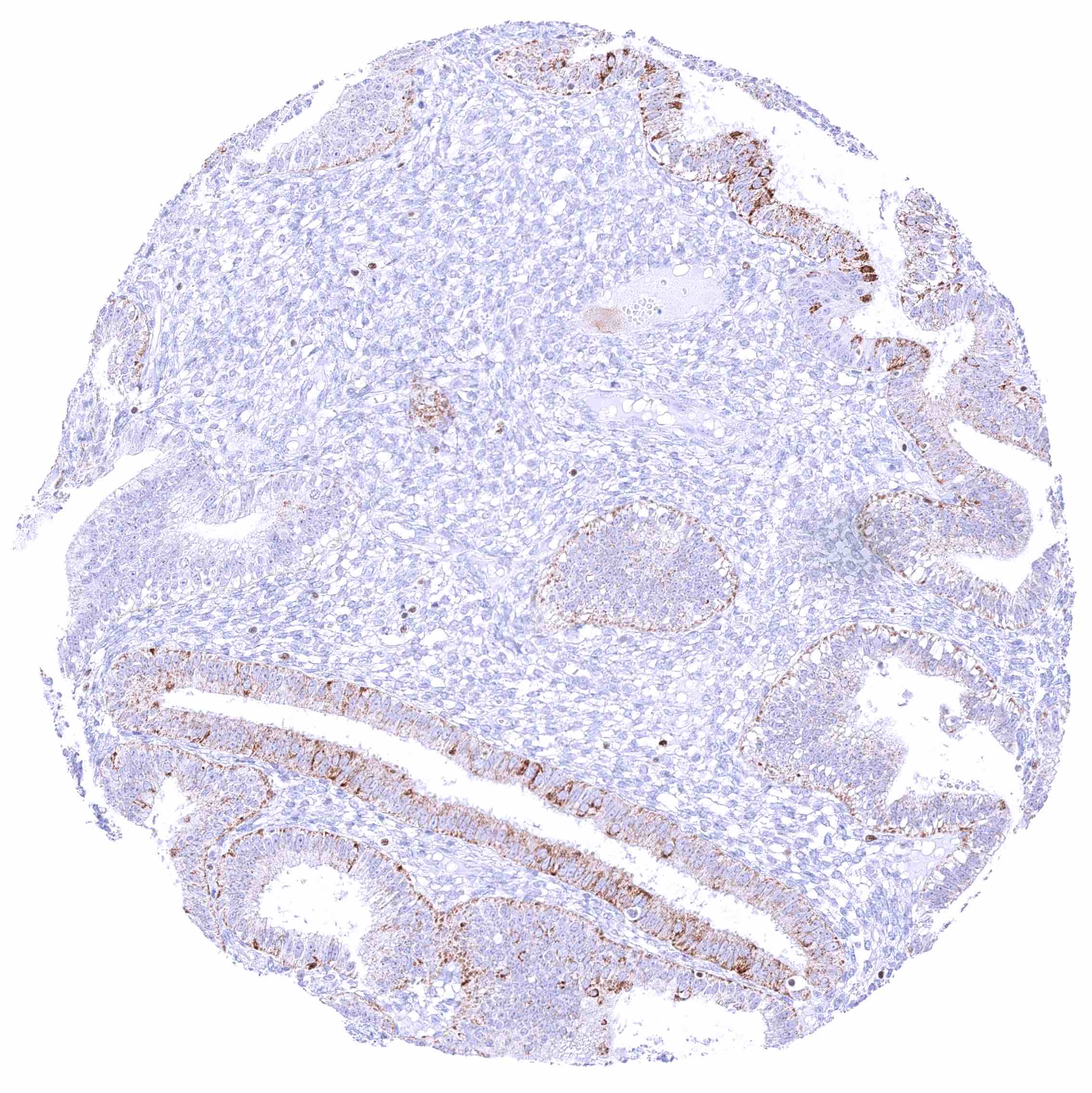
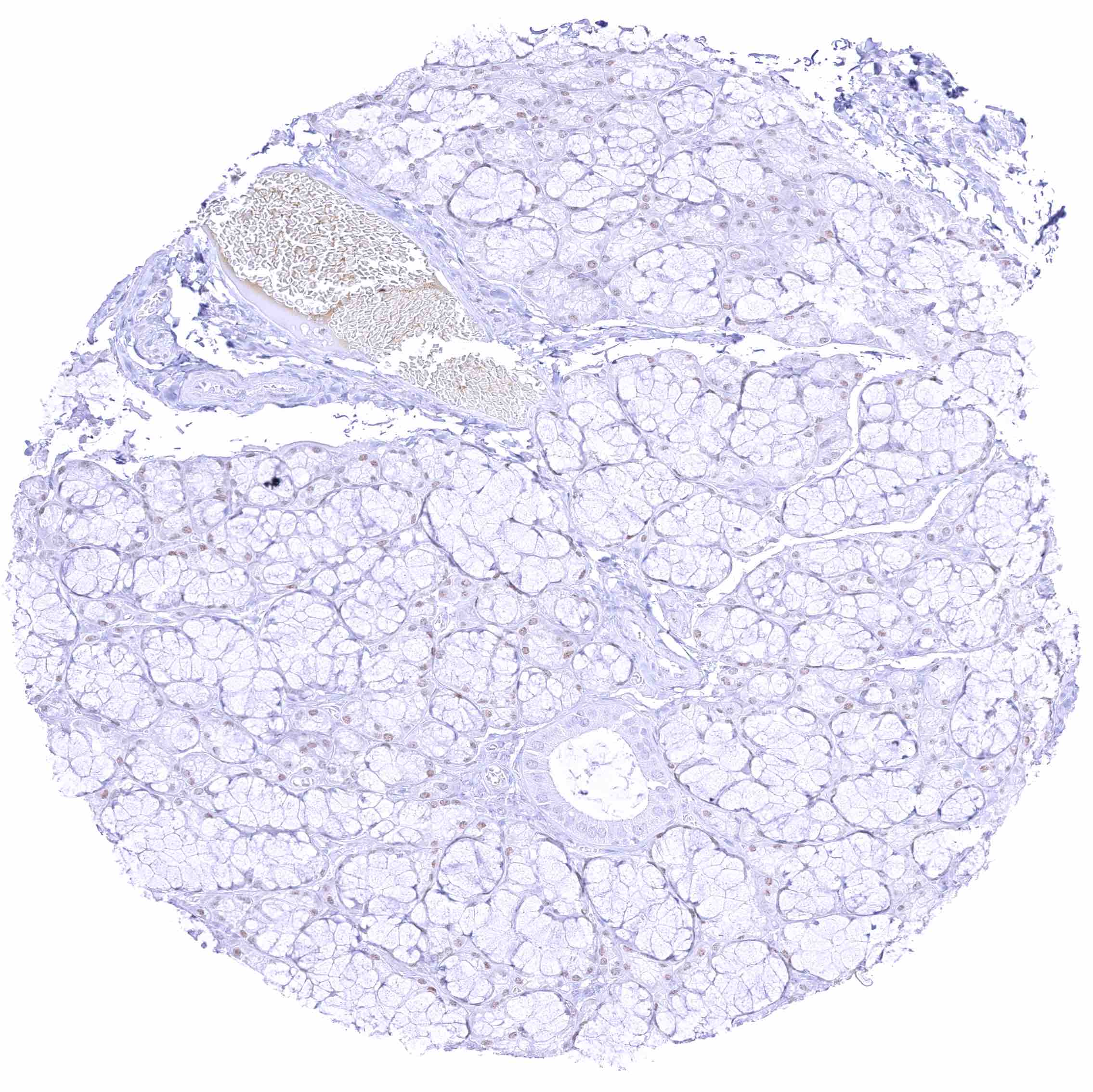
| Genitourinary | Kidney | Strong nuclear GATA3 positivity of collecting ducts and possibly also a fraction of distal tubuli. Moderate nuclear GATA3 staining of glomerular podocytes. |
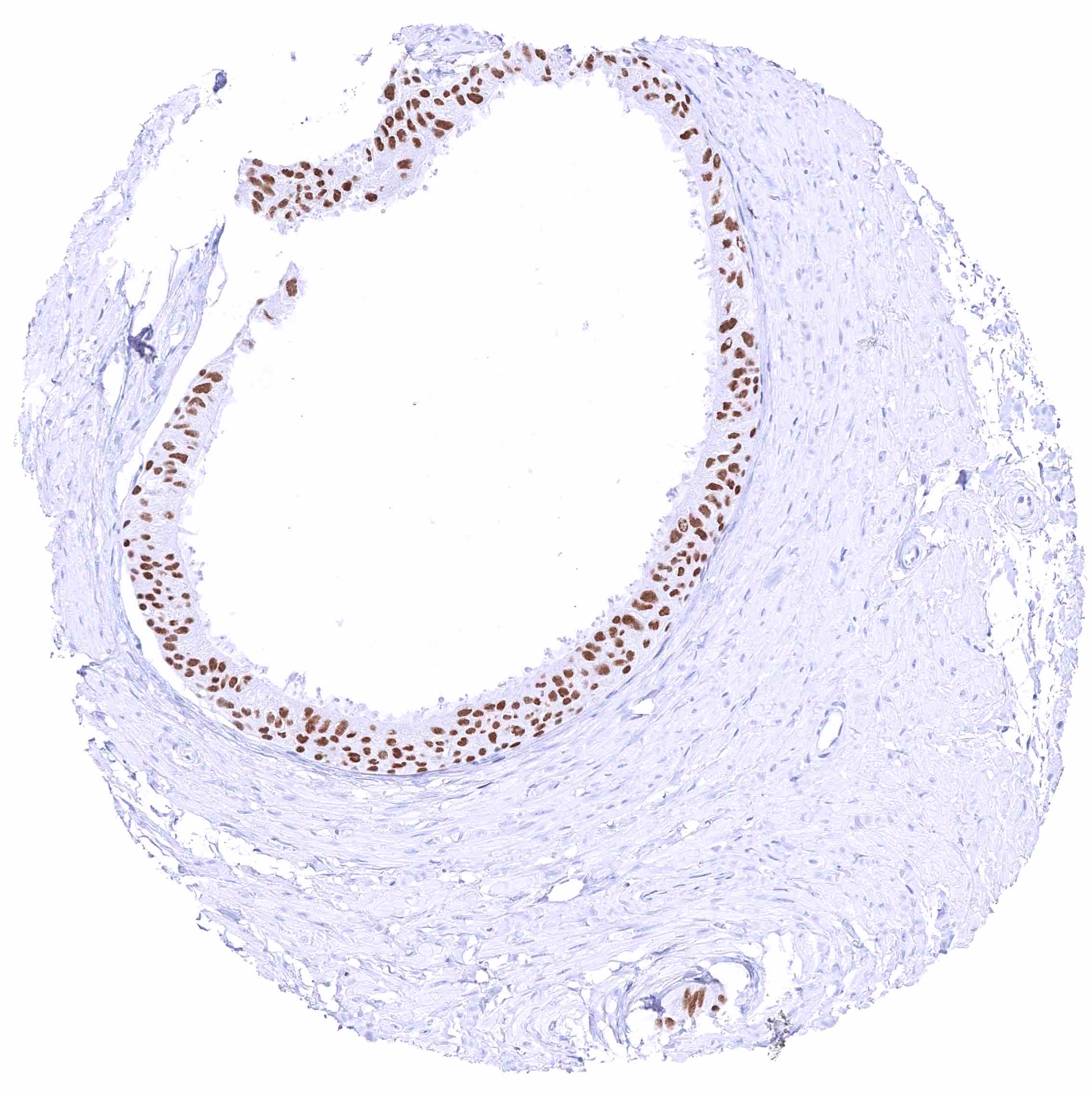
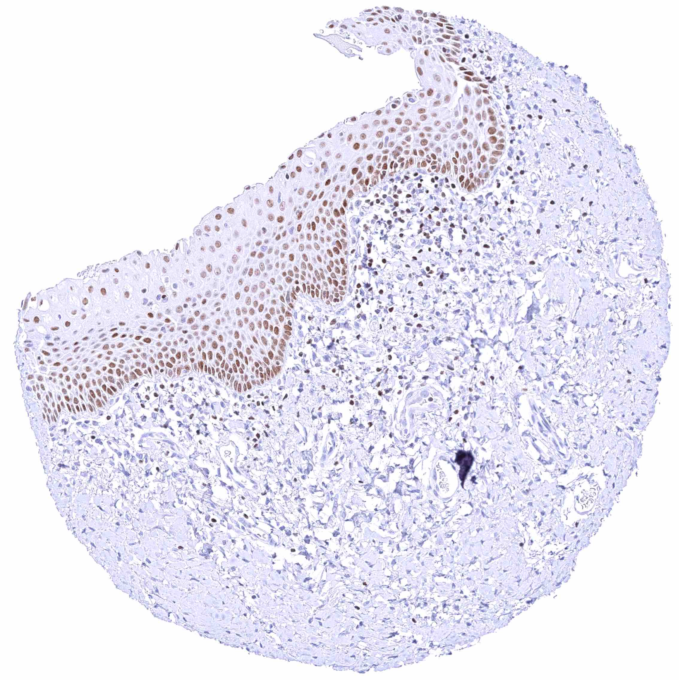
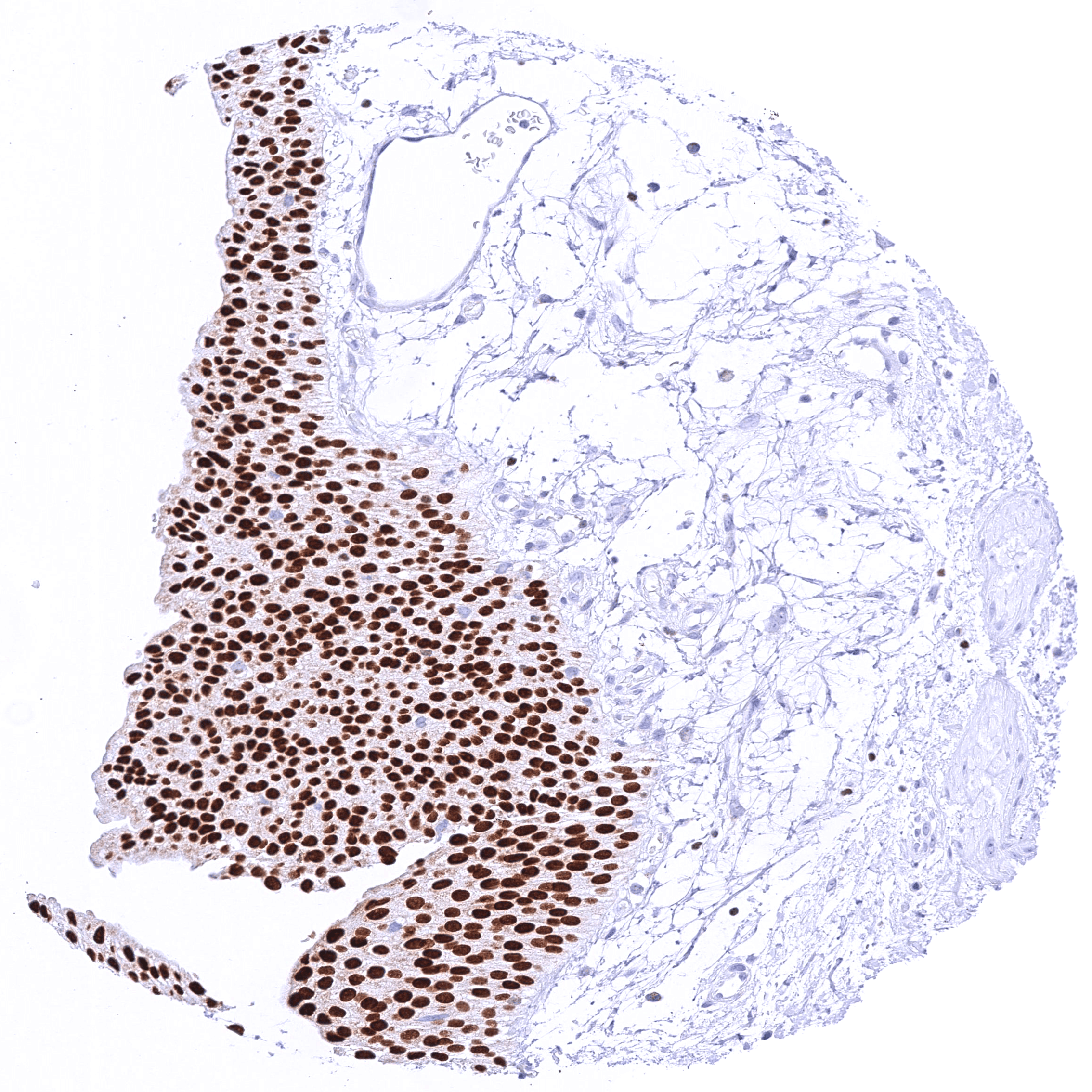
| Urothelium | Strong nuclear GATA3 positivity in all urothelial cells. | |
| Male genital | Prostate | Weak to moderate nuclear GATA3 positivity in a large fraction of basal cells. |
| Seminal vesicles | Strong GATA3 positivity of all epithelial cells. | |
| Testis | Negative. | |
| Epididymis | Moderate to strong staining of tall columnar cells and basal cells of the caput. | |
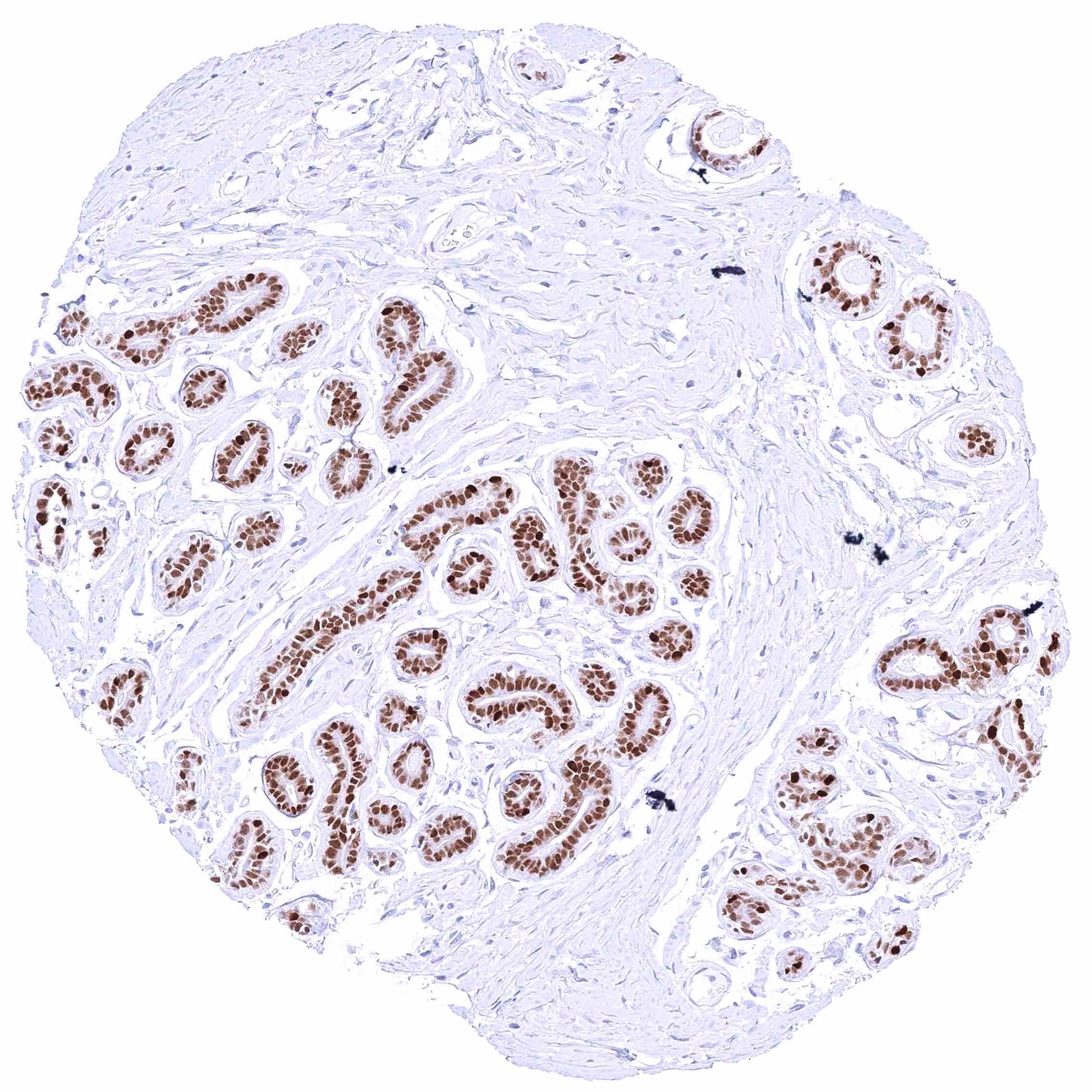
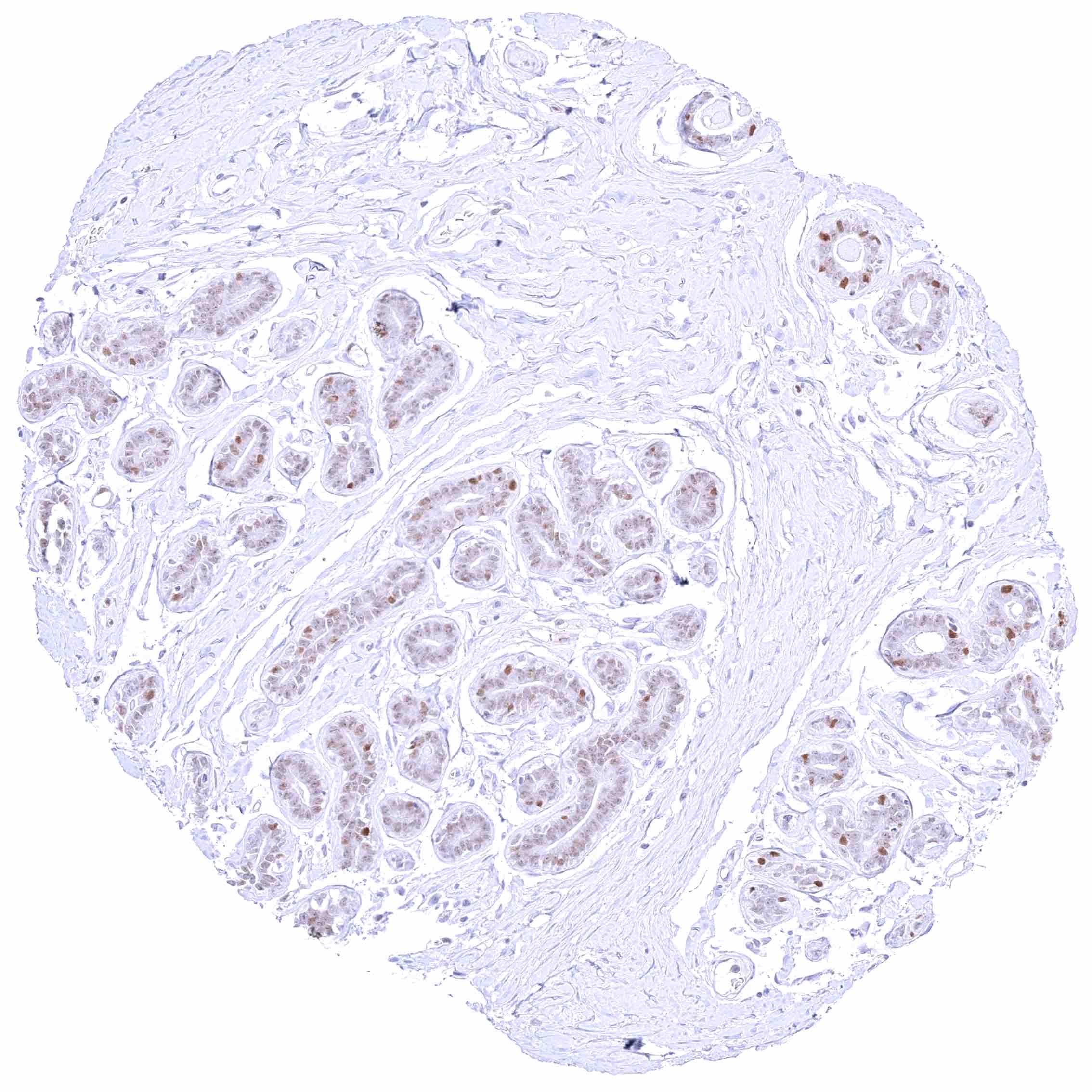
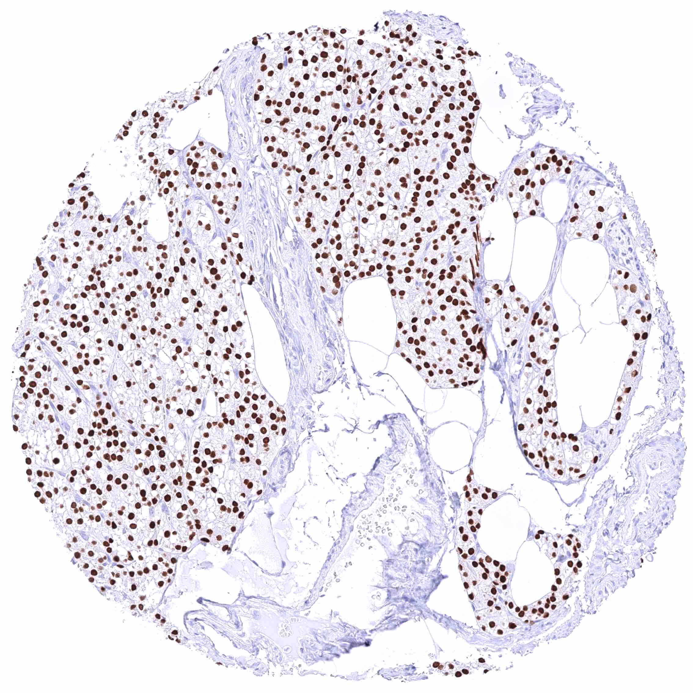
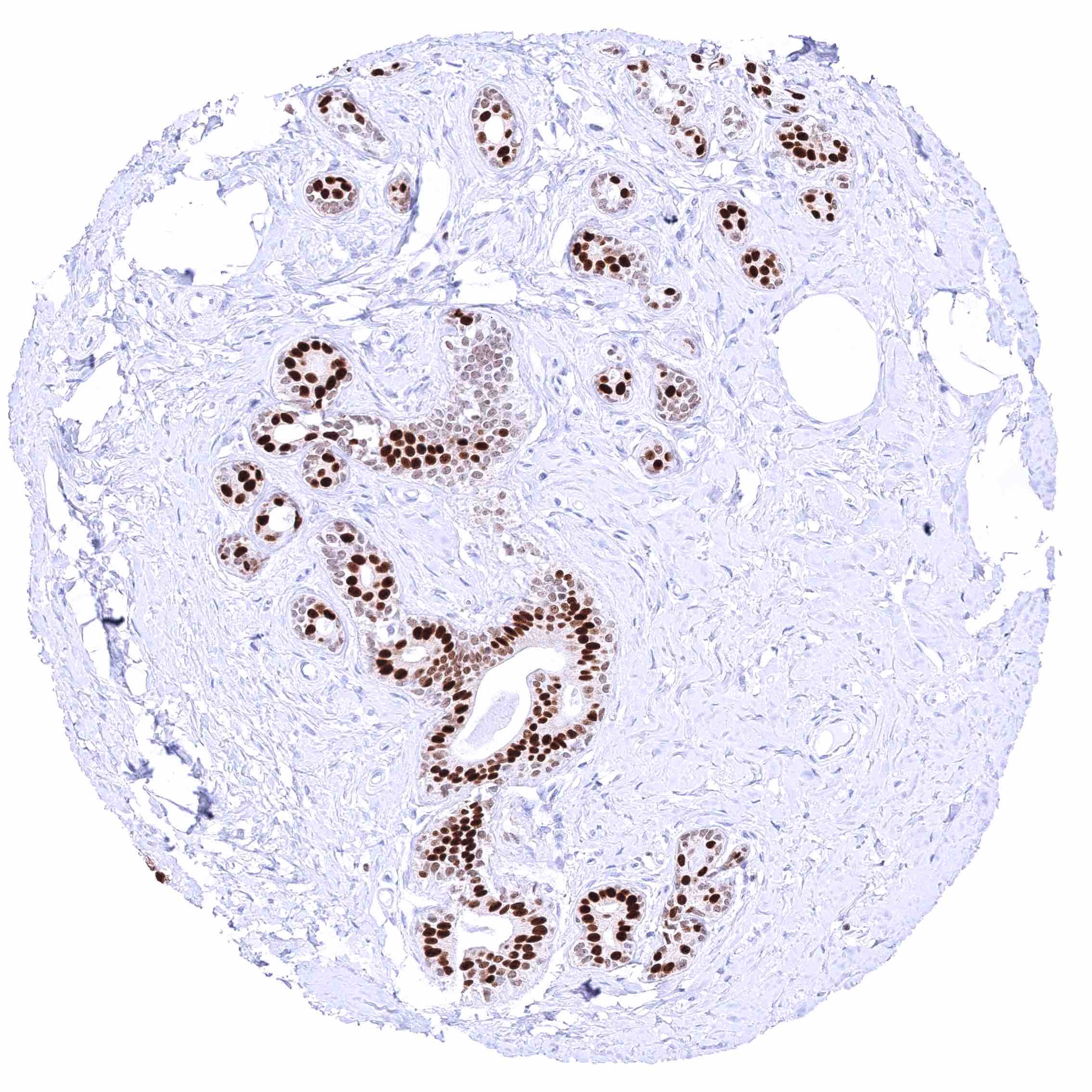
| Female genital | Breast | Moderate to strong GATA3 positivity of most or all fraction of luminal cells while myoepithelial cells remain GATA3 negative. |
| Uterus, myometrium | Negative. | |
| Uterus, ectocervix | Negative. | |
| Uterus endocervix | Negative. A (cross-reactive) granular cytoplasmic GATA3 positivity of epithelial cells can occur. | |
| Uterus, endometrium | Negative. A (cross-reactive) granular cytoplasmic GATA3 positivity of endometrial epithelial cells can occur. | |
| Fallopian Tube | Negative. | |
| Ovary | Negative. | |
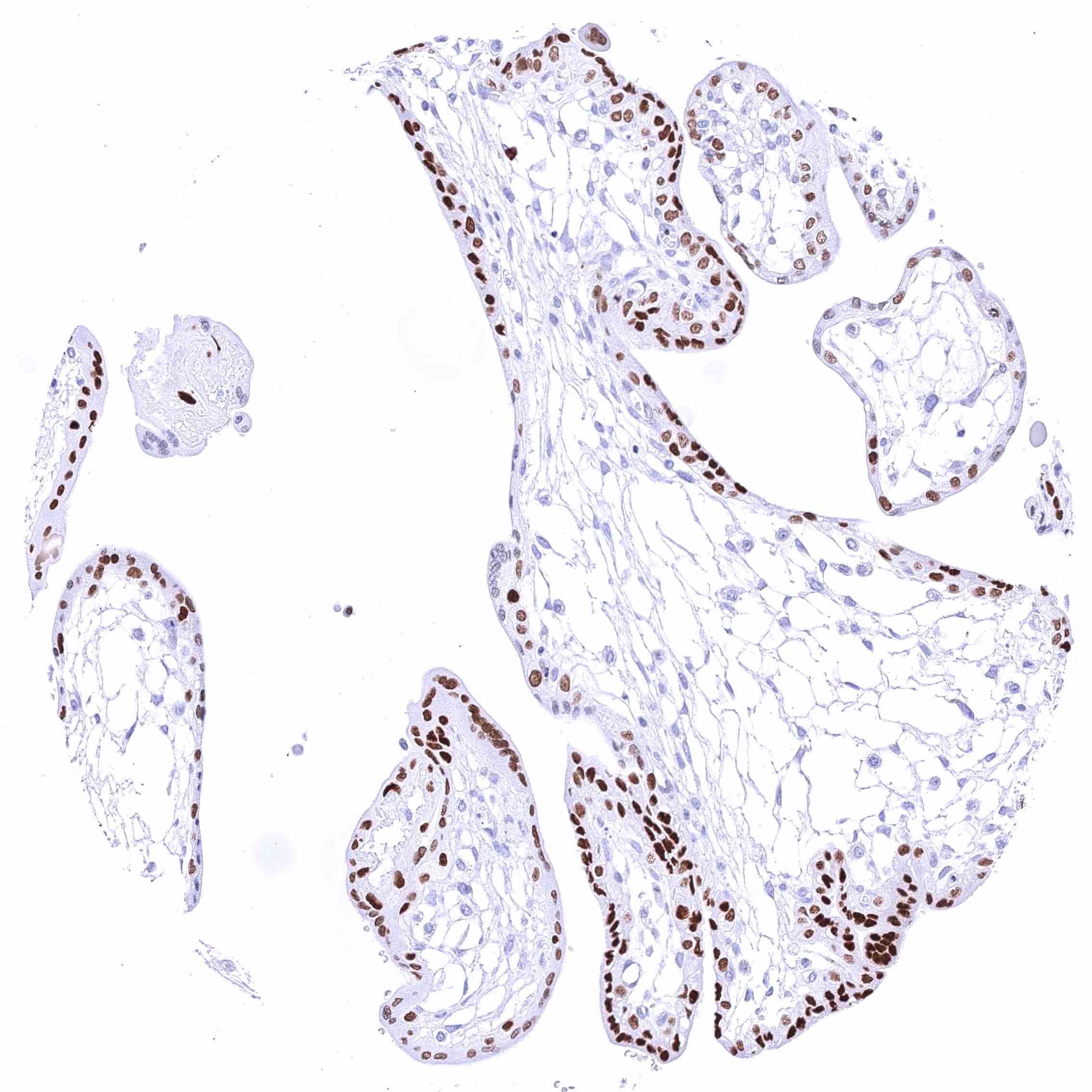
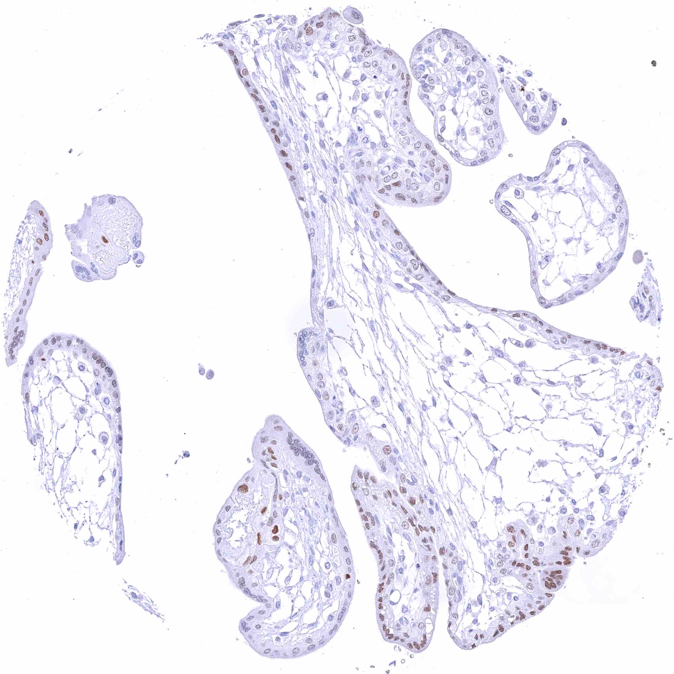
| Placenta early | Moderate to strong nuclear GATA3 positivity of trophoblast cells. | |
| Placenta mature | Moderate to strong nuclear GATA3 positivity of trophoblast cells. | |
| Amnion | Weak GATA3 positivity in amnion cells. | |
| Chorion | Strong GATA3 positivity in chorion cells. | |
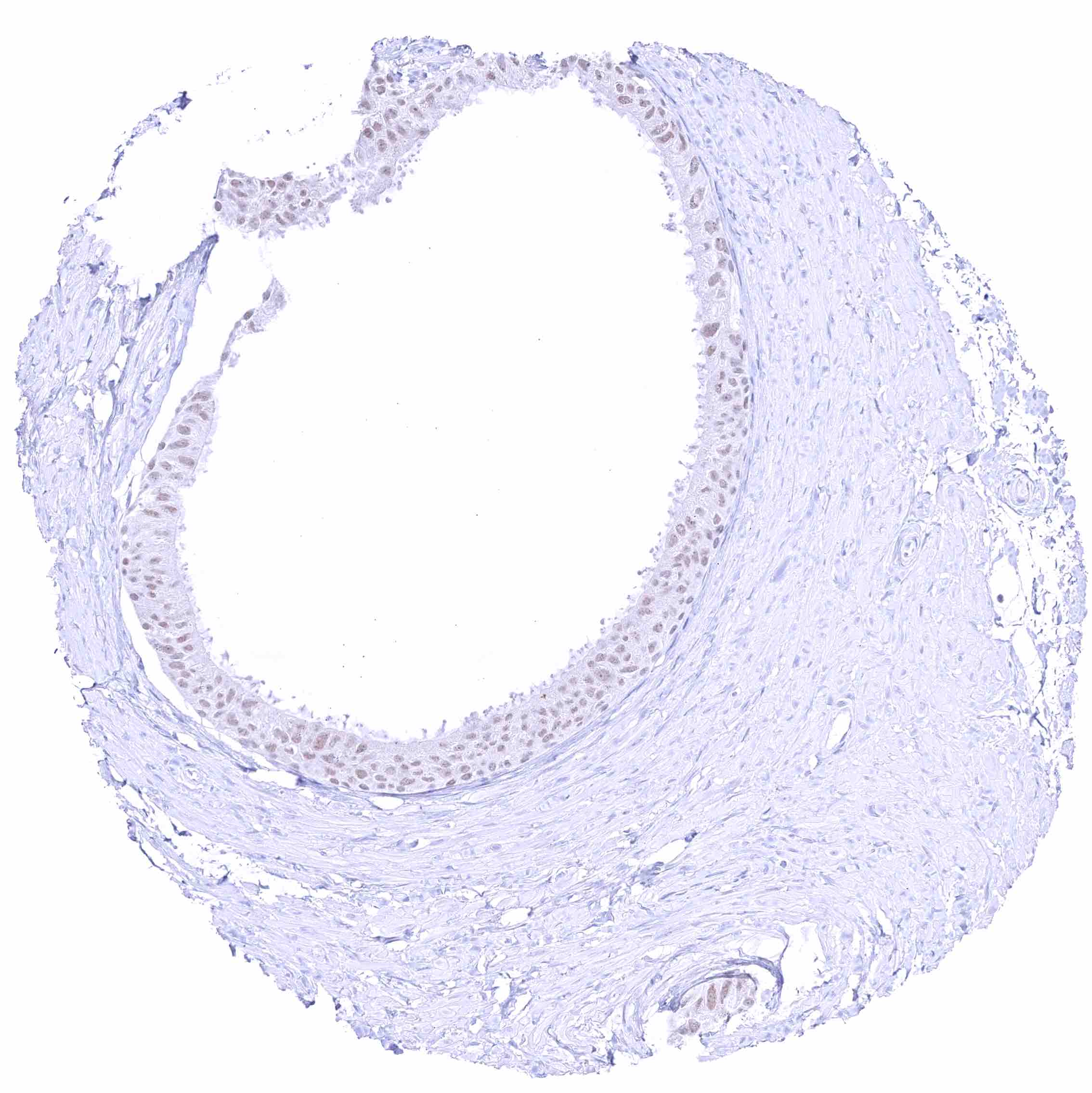
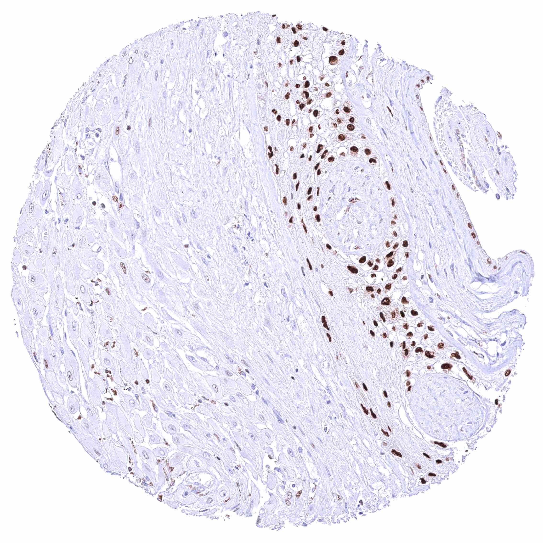
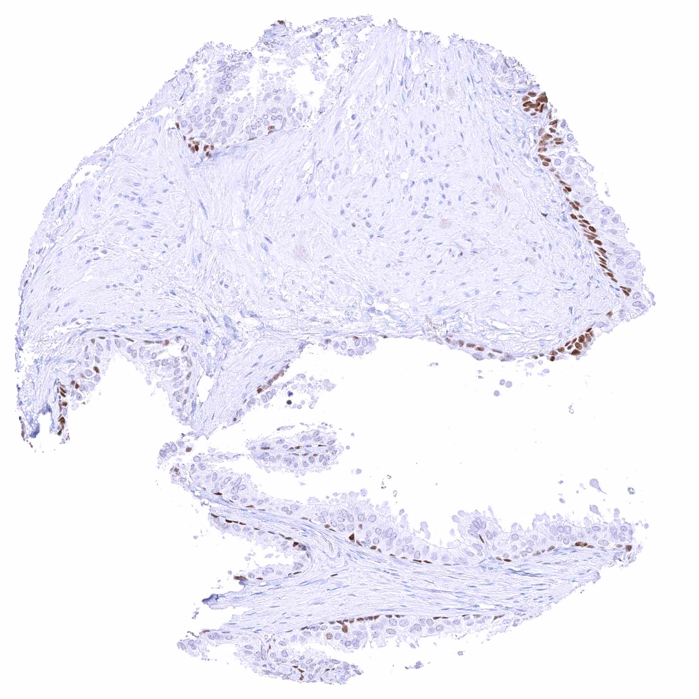
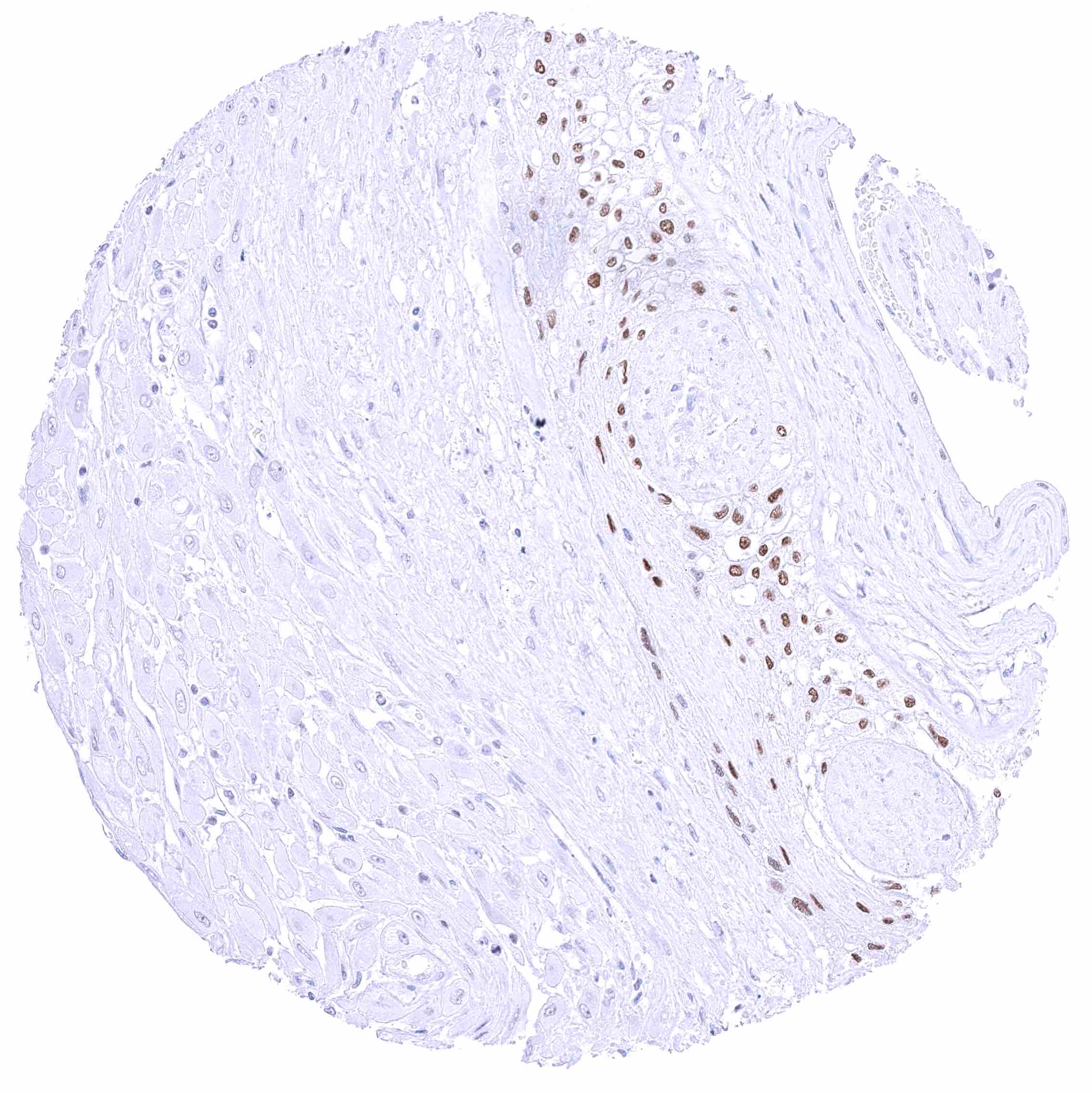
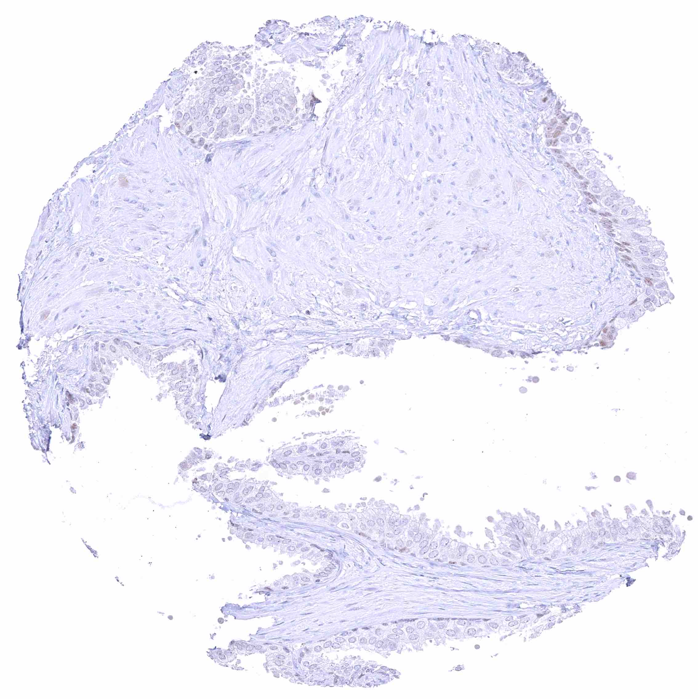
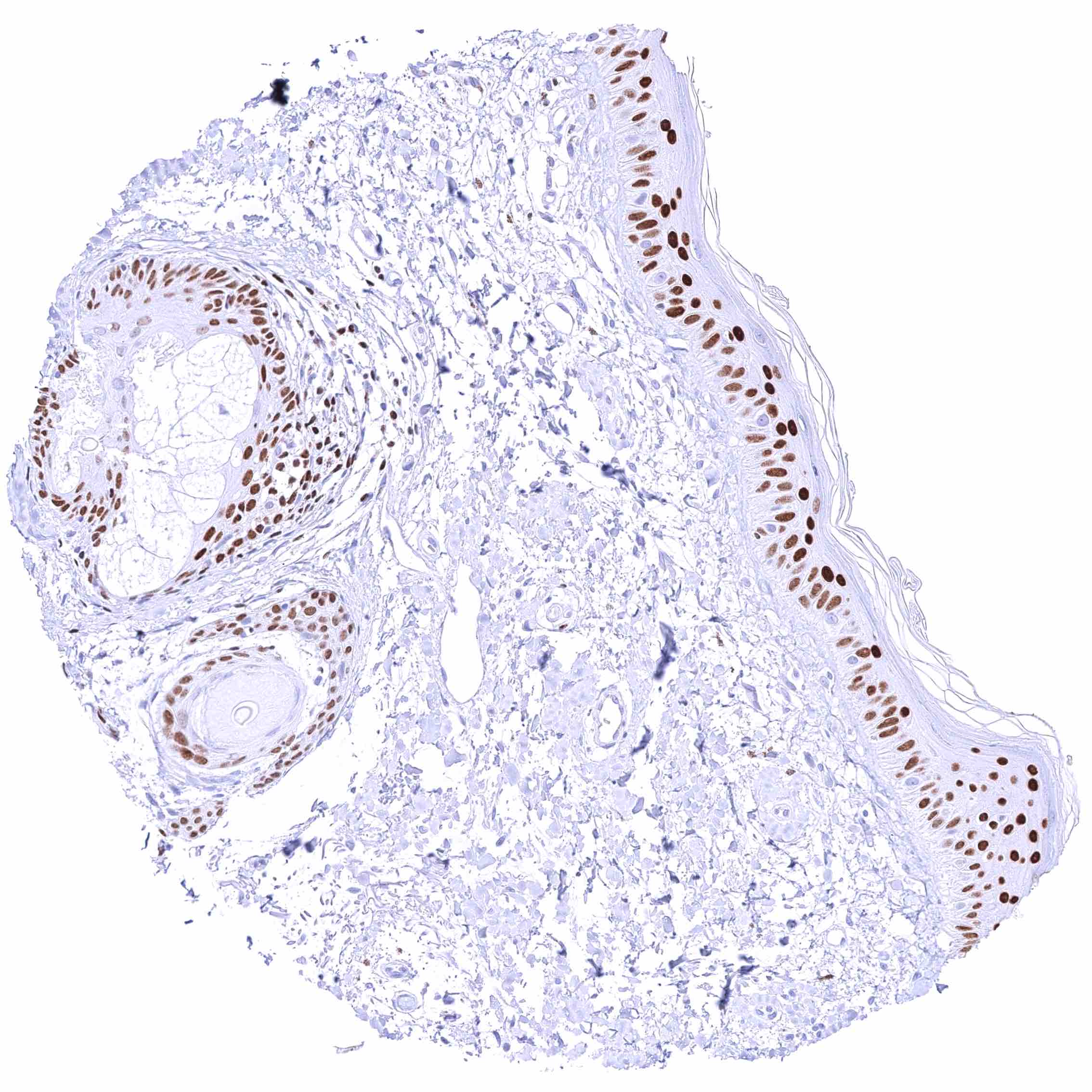
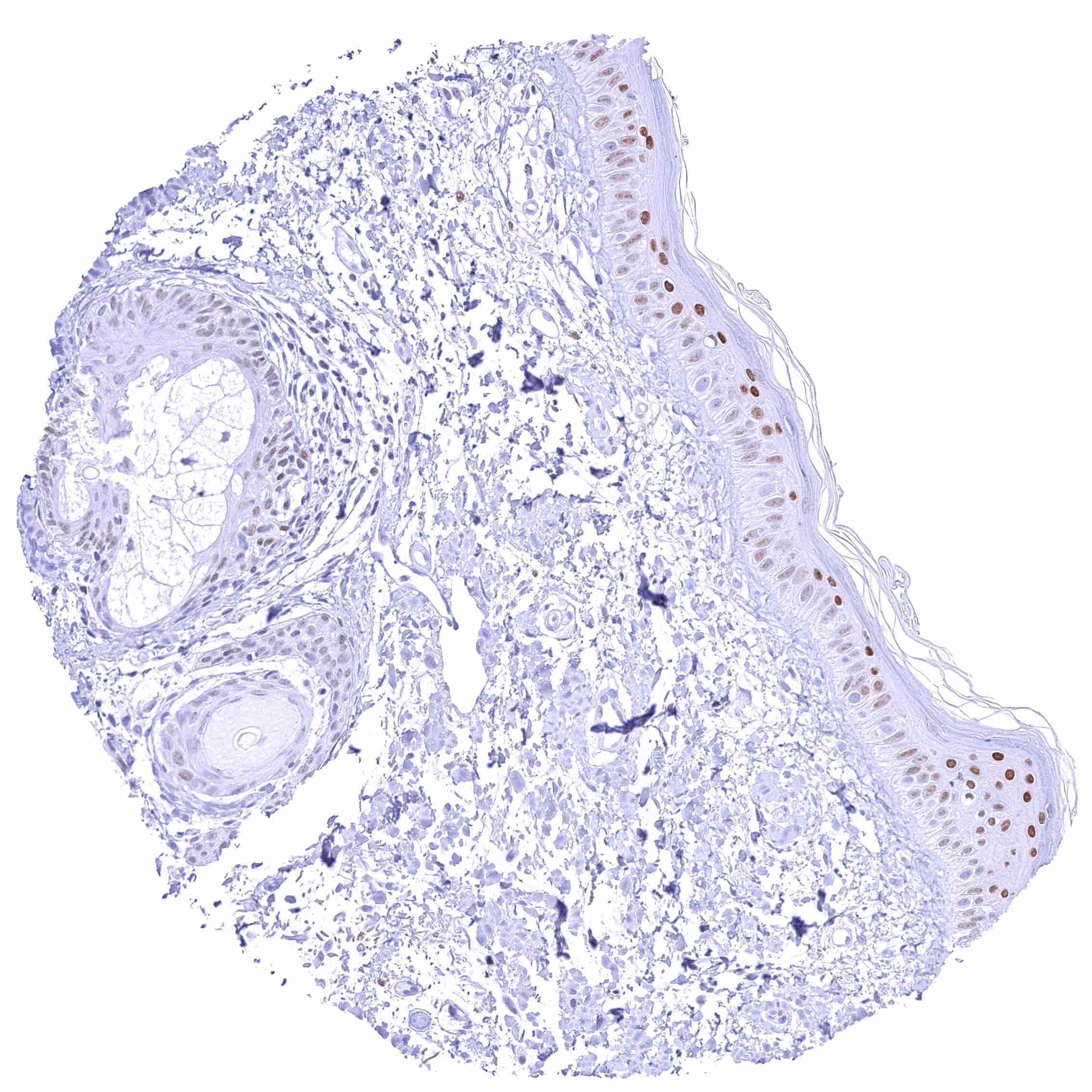
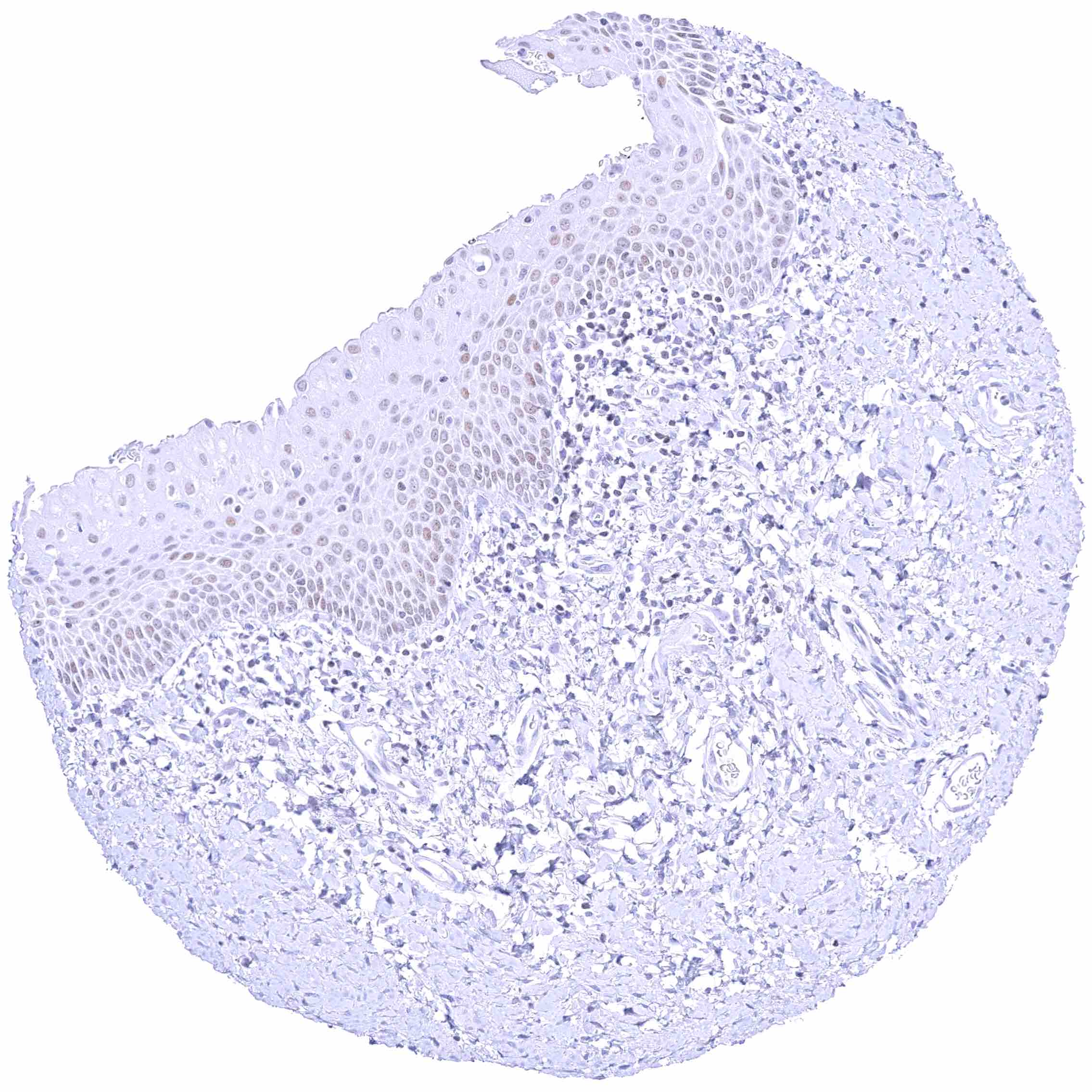
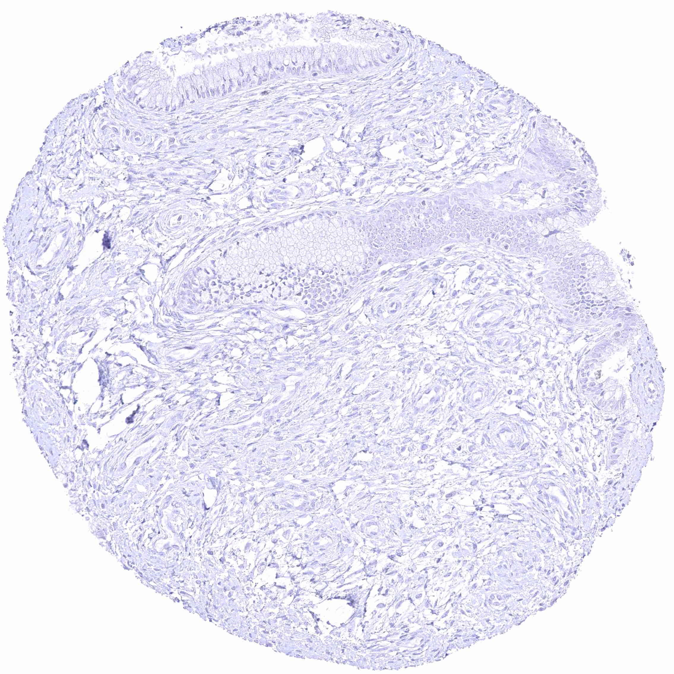
| Skin | Epidermis | Moderate to strong GATA3 positivity of squamous epithelial cells. Staining is often more intense in the lower half of the epidermis. |
| Sebaceous glands | Moderate to strong nuclear GATA3 positivity of peripheral germinative cells and of cells from including hair follicles. Sebaceous gland cells are only weakly positive or GAA3 negative. | |
| Muscle/connective tissue | Heart muscle | Negative. |
| Skeletal muscle | Negative. | |
| Smooth muscle | Negative. | |
| Vessel walls | Negative. | |
| Fat | Negative. | |
| Stroma | Negative. | |
| Endothelium | Negative. | |
| Bone marrow/ lymphoid tissue | Bone marrow | Negative. |
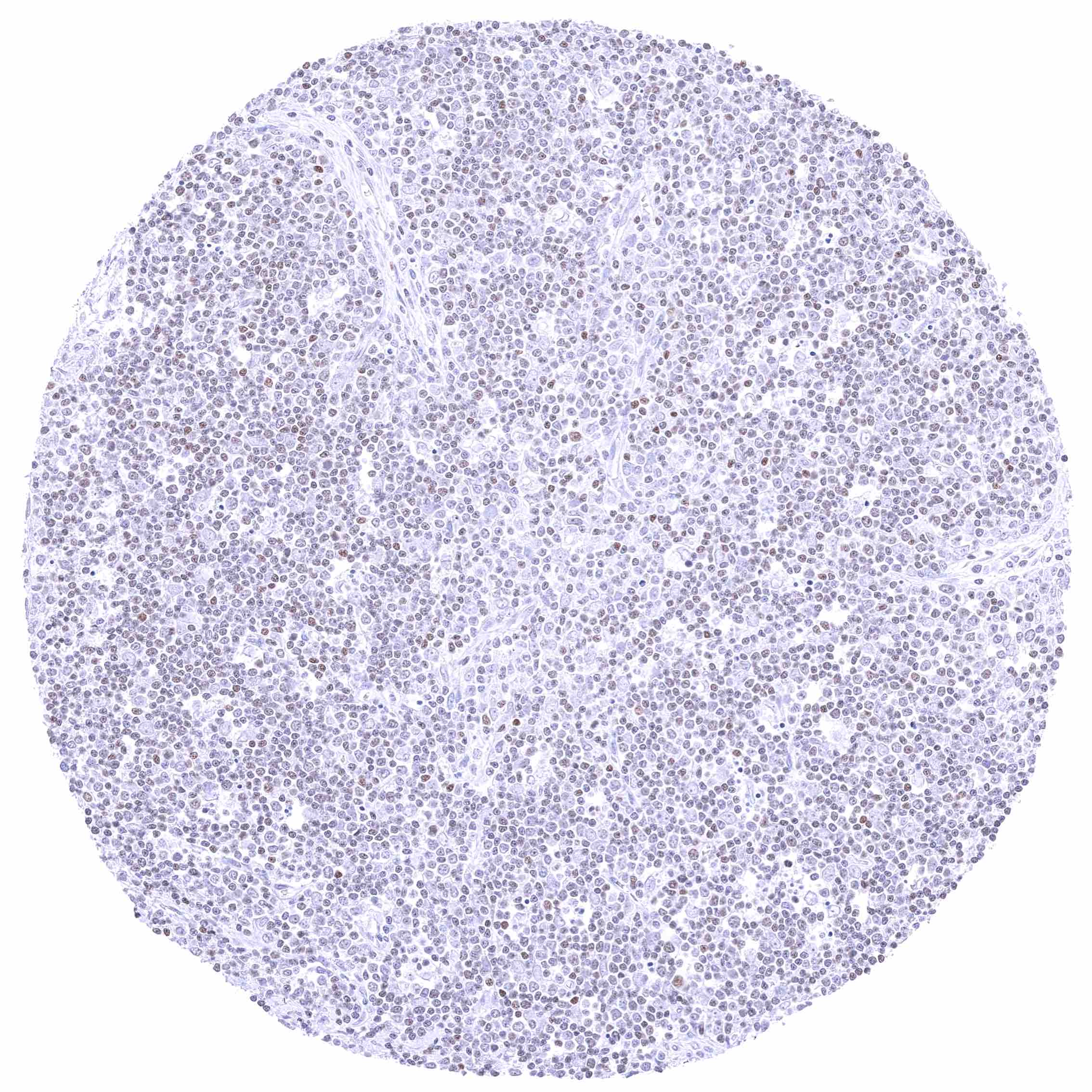
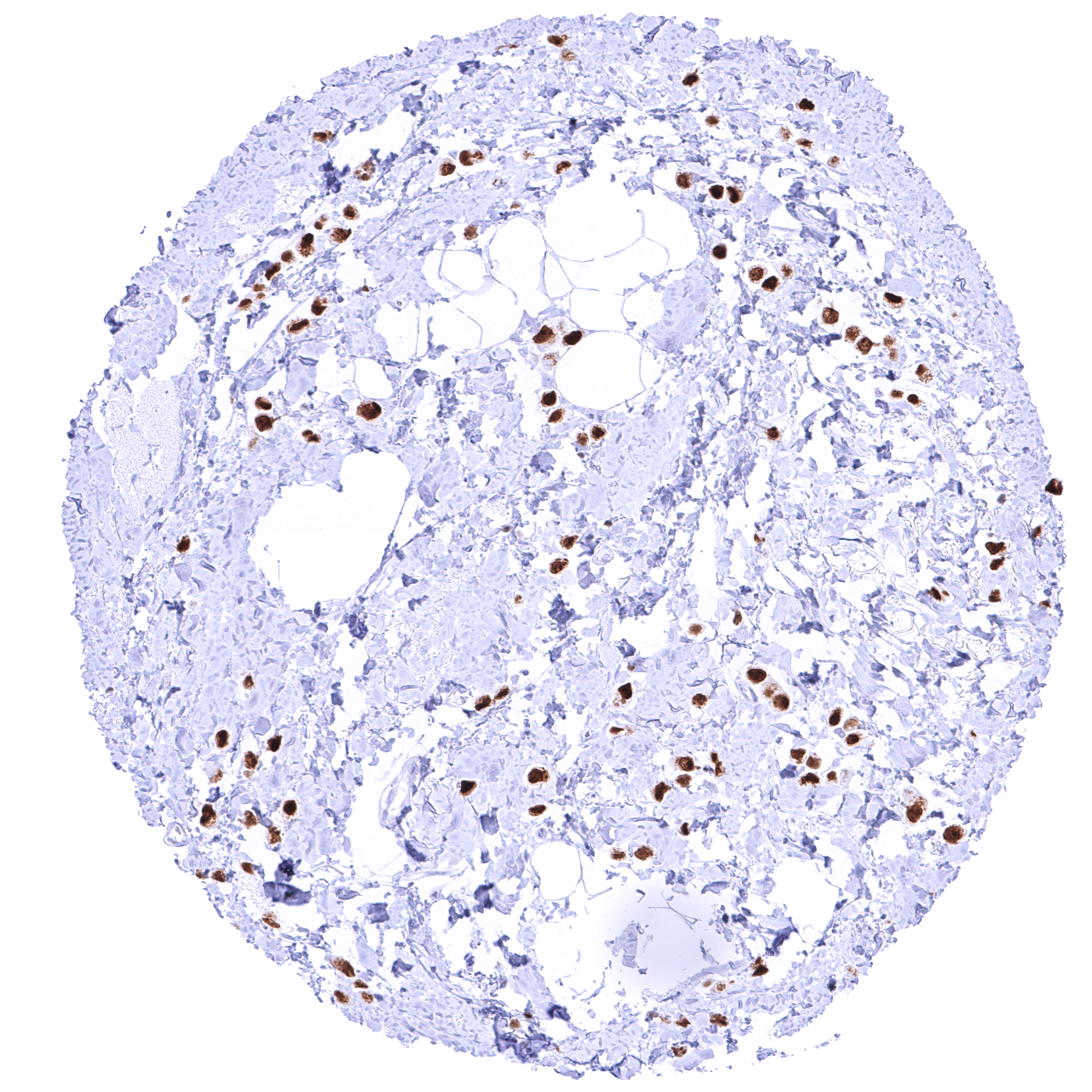
| Lymph node | Weak to moderate nuclear GATA3 positivity of a fraction of lymphocytes. | |
| Spleen | Few lymphocytes may show nuclear GATA3 positivity. | |
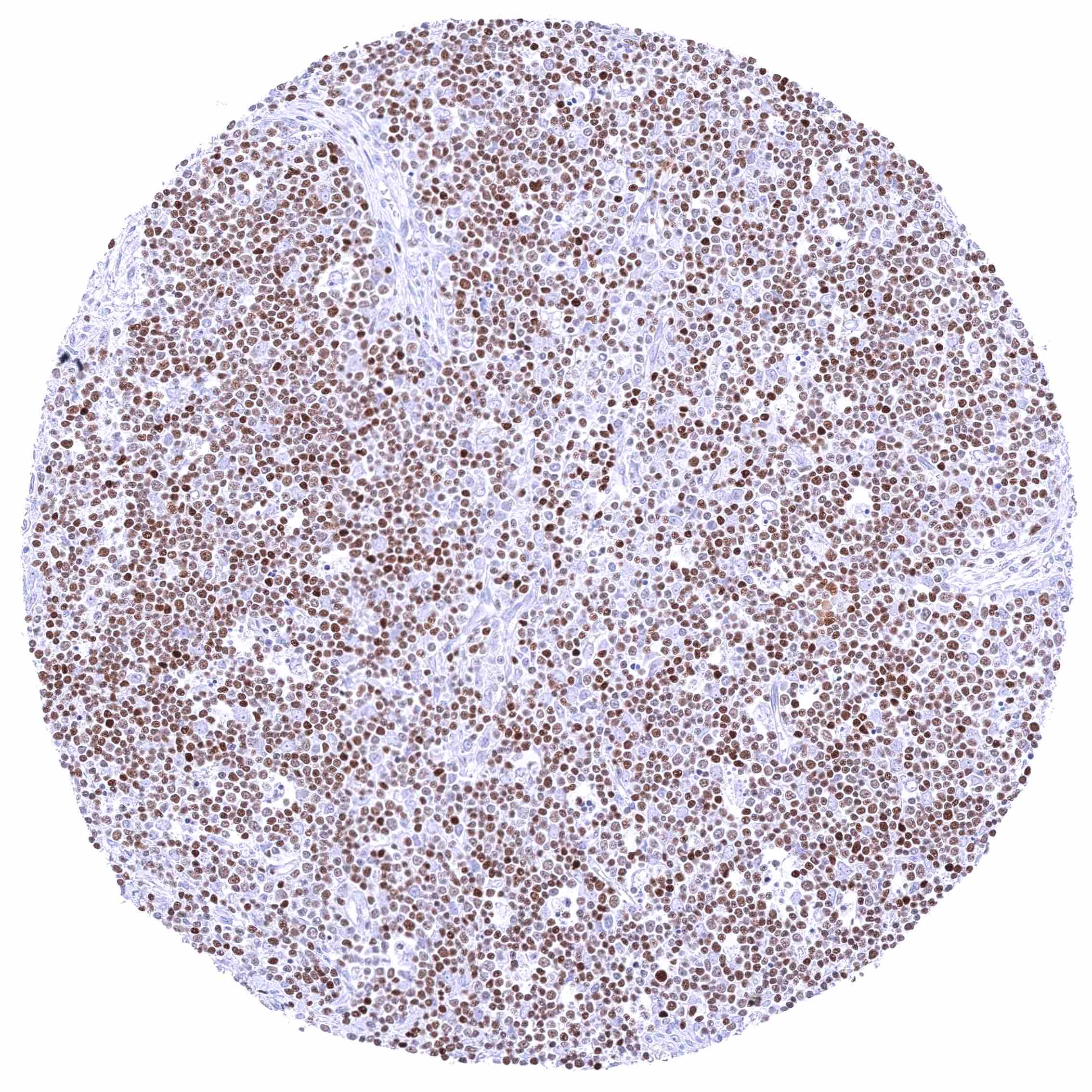
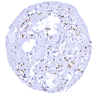
| Thymus | Moderate to strong nuclear GATA3 positivity of a large fraction of lymphocytes, especially in the cortex. | |
| Tonsil | Weak to moderate nuclear GATA3 positivity of a fraction of lymphocytes. | |
| Remarks | True GATA3 immunostaining is always nuclear. A purely cytoplasmic or membranous cross-reactivity occurs in various organs. |
The findings obtained by MSVA-550R almost perfectly match the RNA data summarized in the Human Protein Atlas (Tissue expression GATA3) and identify the cell types responsible for RNA expression. The only protein atlas findings which could not be corroborated was GATA3 expression in the vagina. The validation experiments performed with MSVA-550R had suggested that GATA3 expression occurs in all keratinized squamous epithelia but not in non-keratinized squamous epithelia.
Suggested positive tissue control:
-Kidney: A moderate to strong nuclear staining reaction should be seen in a fraction of collecting duct cells and podocytes in glomeruli.
-Tonsil: The vast majority of T helper cells (Th2) in the T-zones should show a weak to moderate nuclear staining.
Suggested negative tissue control:
-Kidney: Staining should be absent in proximal and distal tubuli as well as in blood vessels.
-Tonsil: Staining should be absent in B-cells.
Staining Pattern in Relevant Tumor Types
Detectable expression of GATA3 is primarily seen in breast cancer and in urothelial carcinomas. Other tumors for which GATA3 expression has been reported include renal carcinoma, lung cancer and testicular tumors. At low frequency, GATA3 expression can occur in additional tumor entities.
The TCGA findings on GATA3 RNA expression in different tumor categories have been summarized in the Human Protein Atlas.
Protocol Recommendations
IHC users have different preferences on how the stains should look like. Some prefer high staining intensity of the target stain and even accept some background. Others favor absolute specificity and lighter target stains. Factors that invariably lead to more intense staining include higher concentration of the antibody and visualization tools, longer incubation time, higher temperature during incubation, higher temperature and longer duration of the heat induced epitope retrieval (slide pretreatment). The impact of the pH during slide pretreatment has variable effects and depends on the antibody and the target protein.
All images and data shown here and in our image galleries are obtained by the manual protocol described below. Other protocols resulting in equivalent staining are described as well.
Manual protocol
Freshly cut sections should be used (less than 10 days between cutting and staining). Heat-induced antigen retrieval for 5 minutes in an autoclave at 121°C in pH 7,8 Target Retrieval Solution buffer. Apply MSVA-550R at a dilution of 1:150 at 37°C for 60 minutes. Visualization of bound antibody by the EnVision Kit (Dako, Agilent) according to the manufacturer’s directions.
Potential Research Applications
- What are the precise molecular mechanisms by which GATA3 regulates immune cell differentiation, particularly Th2 cells, in health and disease?
- How do different mutations in the GATA3 gene contribute to breast cancer development, progression, and response to hormone therapies?
- What role does GATA3 play in the development and function of non-immune tissues, such as the kidney and parathyroid gland, and how do disruptions affect disease?
- What is the prognostic role of GATA3 expression in cancer?
- Can targeting GATA3 or its downstream pathways be a viable therapeutic strategy in allergic diseases and certain cancers?
- How does GATA3 interact with other transcription factors and signaling pathways to coordinate embryonic development and tissue-specific differentiation?
Evidence for Antibody Specificity in IHC
There are two ways how the specificity of antibodies can be documented for immunohistochemistry on formalin fixed tissues. These are: 1. comparison with a second independent method for target expression measurement across a large number of different tissue types (orthogonal strategy), and 2. Comparison with one or several independent antibodies for the same target and showing that all positive staining results are also seen with other antibodies for the same target (independent antibody strategy).
Orthogonal validation: For the antibody MSVA-550R specificity is supported by the strong concordance of its nuclear immunostaining with RNA expression data derived from the Human Protein Atlas (HPA) RNA-seq tissue dataset, the FANTOM5 project, and the Genotype-Tissue Expression (GTEx) project which are all summarized in the Human Protein Atlas (Tissue expression GATA3). Immunostaining by using MSVA-550R was only detected in organs with documented RNA expression. These included the squamous epithelium of the epidermis, parathyroid, salivary glands, urothelium, kidney, breast gland, placenta, seminal vesicle, prostate, epididymis, thymus, tonsil, lymph node, and the adrenal gland. The only RNA finding which could not be corroborated by a nuclear staining obtained by MSVA-550R was GATA3 expression in the vagina. It appears possible that the RNA analyses erroneously included GATA3 positive keratinized squamous epithelium from the vaginal orifice.
Comparison of antibodies: Specific GATA3 staining of MSVA-550R is supported by a confirmation of all nuclear stainings obtained by MSVA-550 by an independent commercial “validation antibody”. This applies for urothelium, squamous epithelium of the skin, hair follicles, sebaceous glands, parathyroid gland, trophoblastic, chorion and amnion cells of the placenta, collecting ducts and glomerular podocytes of the kidney, seminal vesicle epithelium, luminal cells of breast glands, and a fraction of lymphocytes, most prominently in the thymus (comparative images shown below). Because the sensitivity of the validation antibody is markedly lower than for MSVA-550R at the selected protocol, the validation antibody exhibits only a very faint staining in some of the respective cells. An additional cytoplasmic staining obtained by MSVA-550R in several tissues including endometrium, endocervical epithelium, intestinal epithelial cells, and respiratory epithelium was only seen by MSVA-550R and not confirmed by the validation antibody. These cytoplasmic stainings obtained by MSVA-550R were considered an antibody specific cross-reactivity of MSVA-550R. Because of its purely cytoplasmic nature this cross-reactivity was considered tolerable.