295,00 € – 995,00 €
Product details
Synonyms = Catenin beta-1, Beta-catenin, Cadherin associated protein, beta 1 88kDa, Catenin beta-1, CATNB, CHBCAT, CTNNB1
Antibody type = Recombinant Rabbit monoclonal / IgG
Clone = MSVA-578R
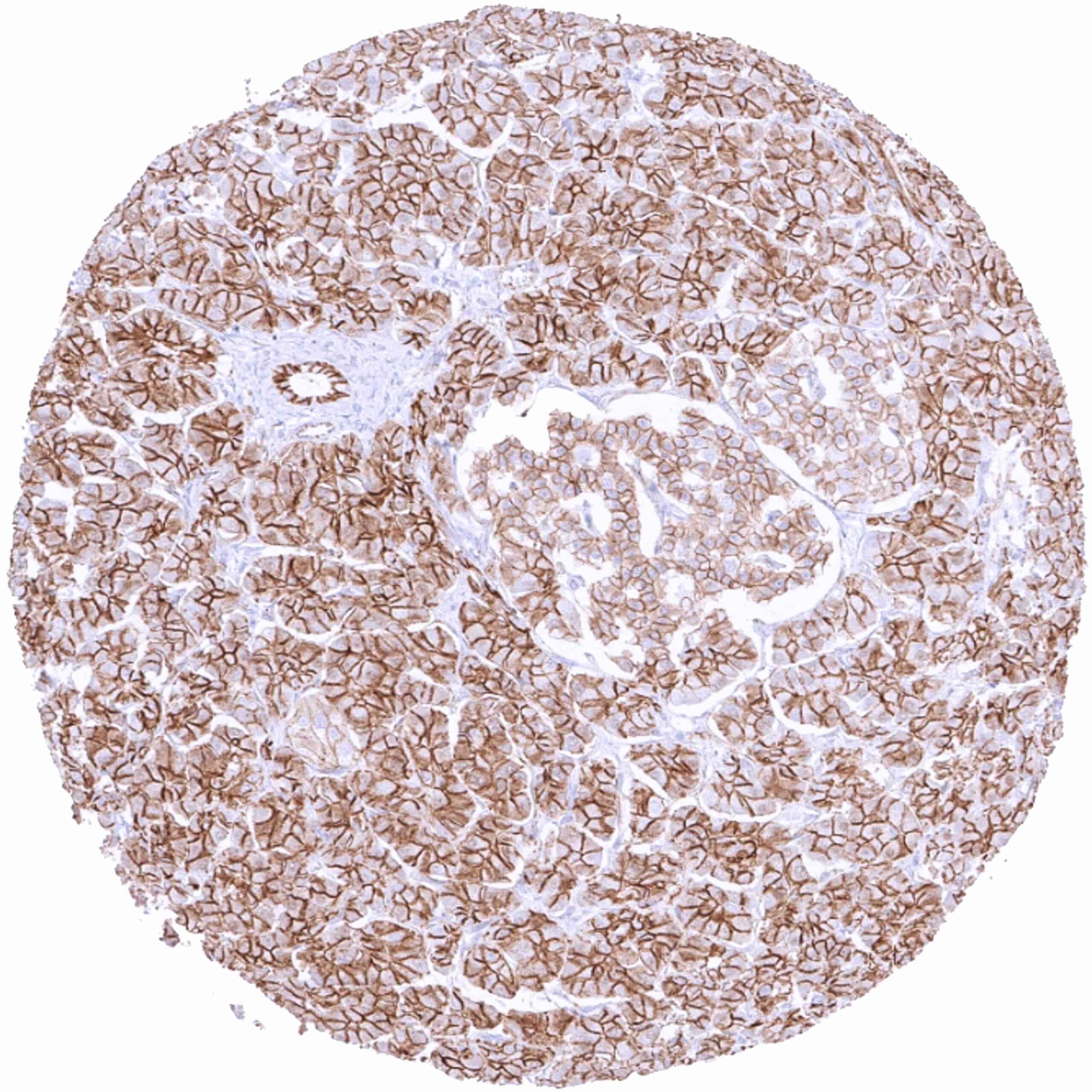
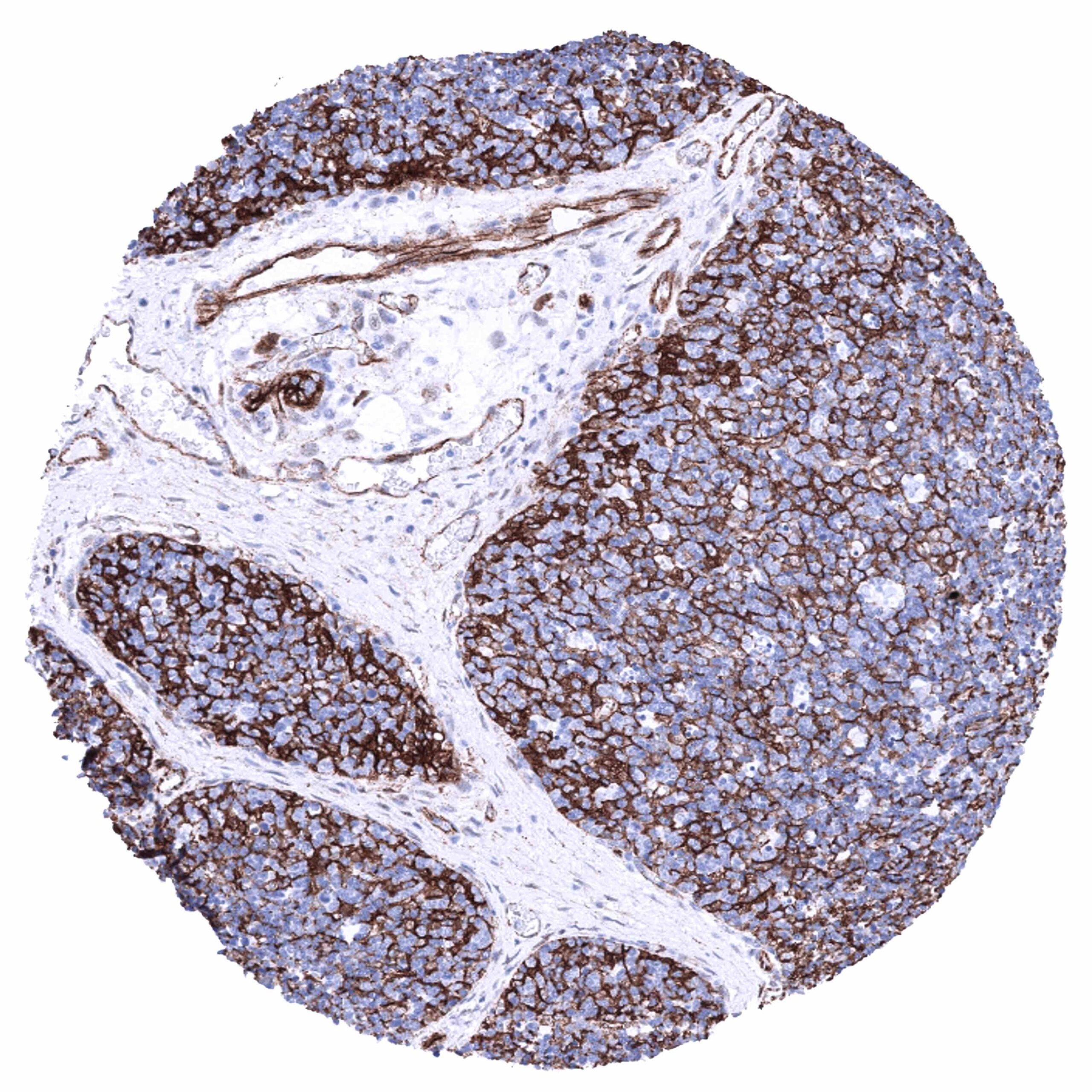
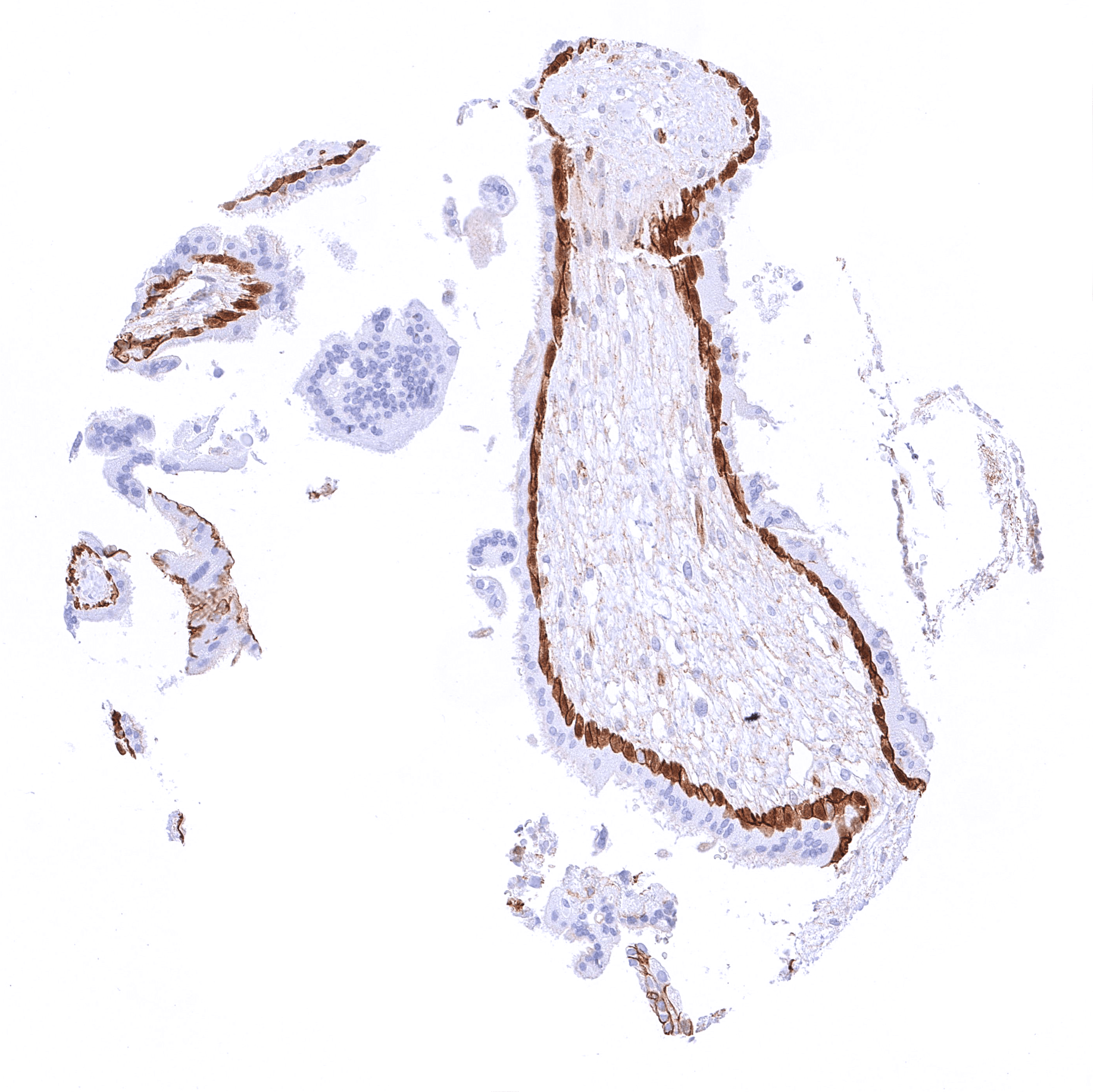
Positive control = Caput epididymis: A strong membranous β-Catenin staining should be seen in all epithelial cell types. In a fraction of the cells, an additional distinct nuclear staining must be seen.
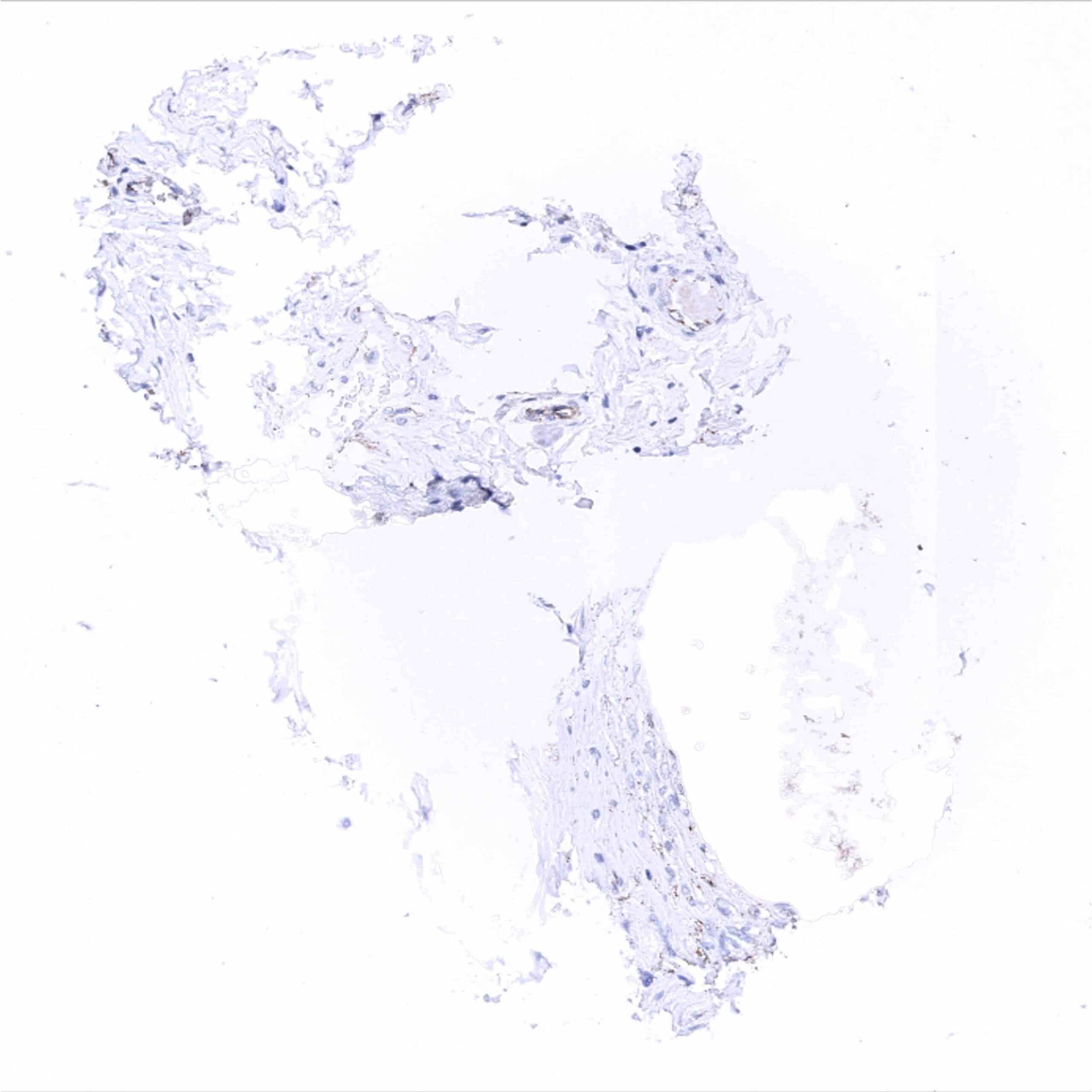
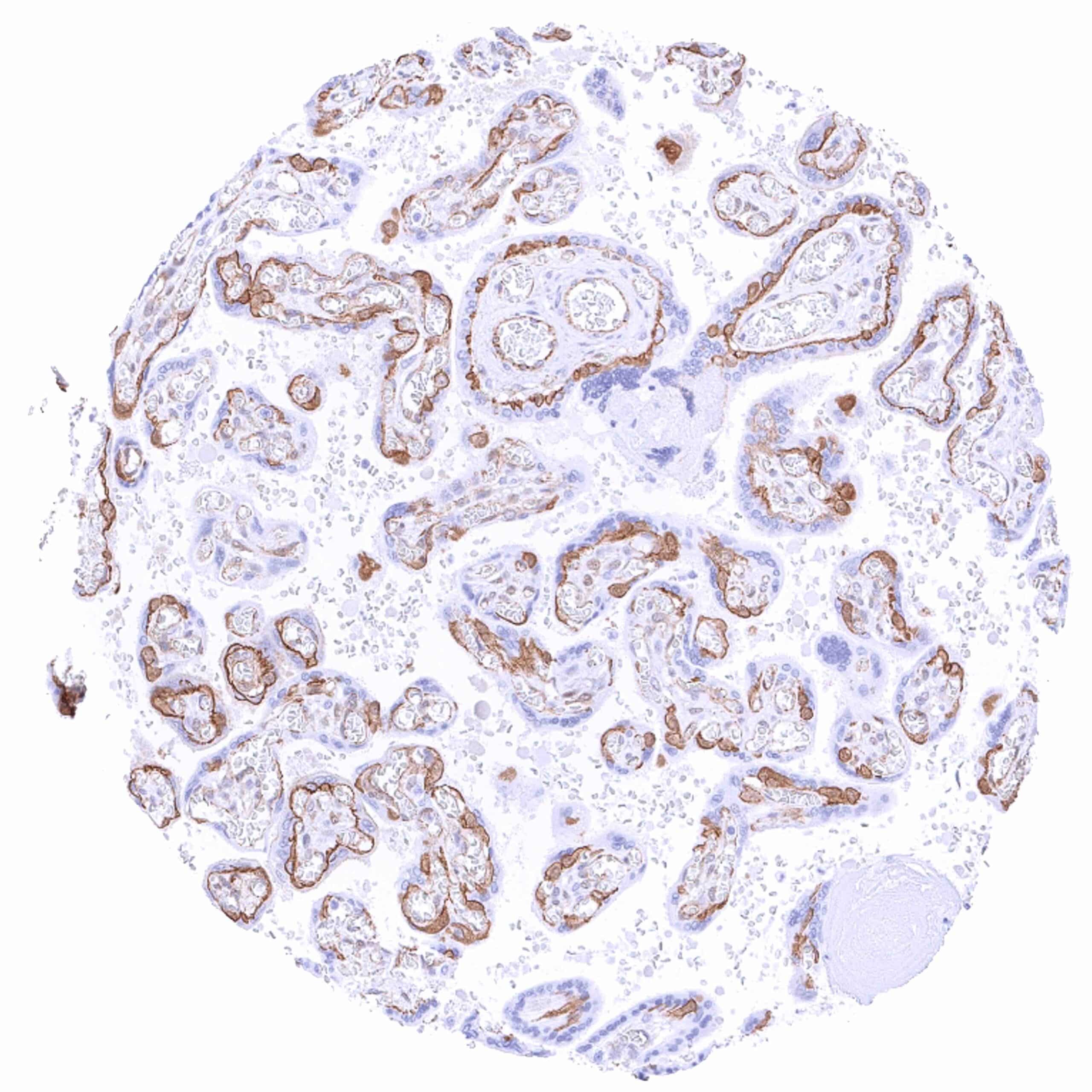
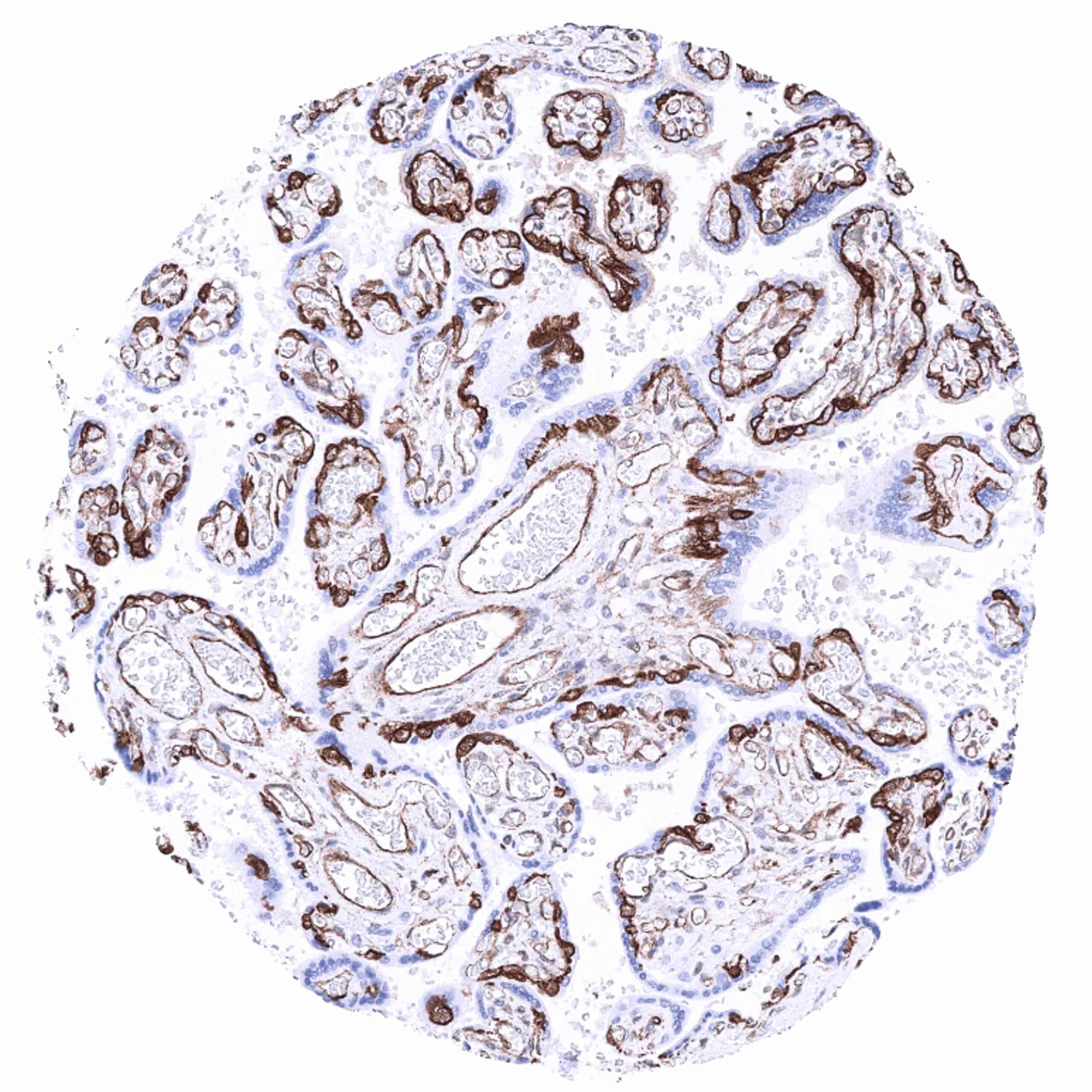
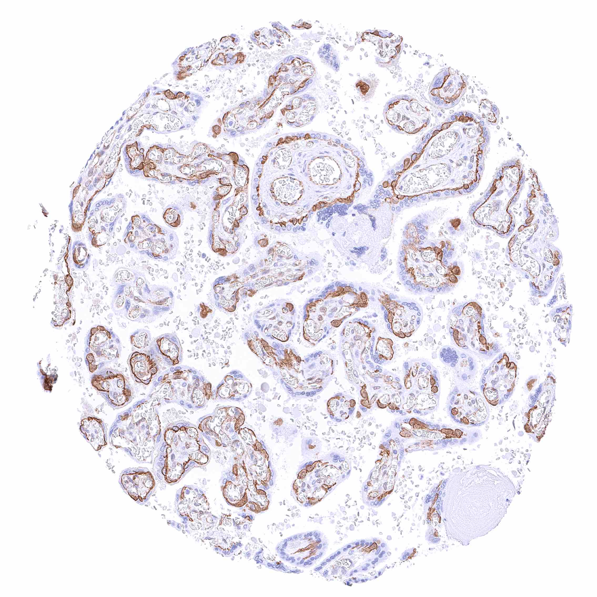
Negative control = Placenta (mature): β-Catenin staining should be absent in the cells of the syncytiotrophoblast while it is strong in cells of the cytotrophoblast.
Cellular localization = Predominantly cell membrane under normal conditions
Reactivity = Human
Application = Immunohistochemistry
Dilution = 1:100 – 1:200
Intended Use = Research Use Only
Relevance of Antibody
Beta-Catenin is a critical regulator of the Wnt pathway.
Biology Behind
β-Catenin is a dual function protein coded by the CTNNB1 gene on chromosome 3p22.1. It has relevant roles in both cell–cell adhesion and gene transcription. In cardiac muscle, it is a main component of the intercalated disc, which is critical for electrical and mechanical coupling between cardiomyocytes. At the cell membrane, β-catenin associates with cadherin class of cell-adhesion proteins to regulate cell adhesion. β-Catenin acts as an intracellular signal transducer in the Wnt signaling pathway. The cellular level of β-catenin is mostly controlled by its ubiquitination and proteosomal degradation. The E3 ubiquitin ligase TrCP1 can recognize β-catenin as a substrate only if a specific motif is phosphorylated by the glycogen synthase kinase 3 alpha and beta (GSK3α and GSK3β). To bring GSK3α and GSK3β together with β-catenin, two other proteins, axin and adenomatous polyposis coli (APC) are needed as a scaffold. Because each APC protein can associate with a multitude of β-catenin’s, this scaffold formation results in a huge, multimeric protein assembly dedicated to β-catenin phosphorylation which is called the “beta-catenin destruction complex”. In case of active Wnt signaling, β-Catenin accumulates in the cytoplasm and subsequently translocates to the nucleus where it acts as a transcriptional co-activator and binds to multiple transcription factors including members of the TCF/LEF family. Nuclear β-Catenin translocation and subsequent activation of β-Catenin regulated genes can also be caused by loss of function mutations of proteins of the beta-catenin destruction complex or by gain of function mutations of CTNNB1. Such mutations commonly occur in several cancer types. Wnt signaling and β-catenin dependent gene expression play a critical role during the formation of different body regions in the early embryo, maintenance of pluripotency, epithelial-to-mesenchymal transition, and cardiac physiology. Alterations of Wnt signaling and beta catenin play a role in various diseases including mental illness (depression), arteriosclerosis, cardiac disease, and cancer.
Staining Pattern in Normal Tissues
Images describing the Beta-Catenin staining pattern in normal tissues obtained by the antibody MSVA-578R are shown in our “Normal Tissue Gallery”.
| Brain | Cerebrum | Weak to moderate beta catenin staining of nerve fibers in the grey matter. |
| Cerebellum | Moderate to strong beta catenin staining of cerebellar glomeruli (post-synaptic granule cell dendrites and pre-synaptic terminals of mossy fibers). Weak to moderate beta catenin staining of nerve fibers in the molecular layer. | |
| Endocrine Tissues | Thyroid | Strong membranous beta catenin staining of epithelial cells. |
| Parathyroid | Strong membranous beta catenin staining of epithelial cells. | |
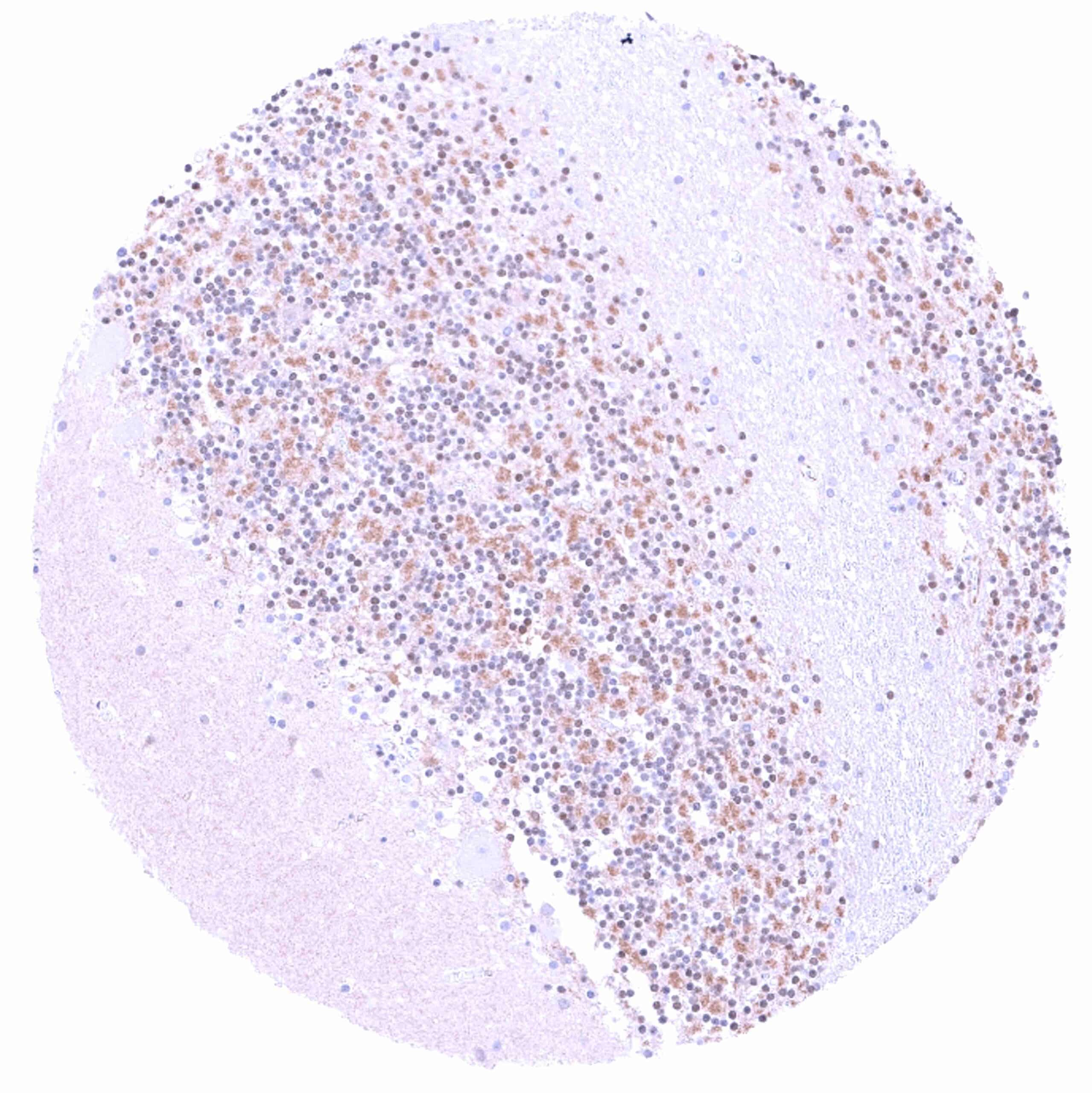
| Adrenal gland | Moderate membranous beta catenin staining of adrenocortical cells, in a fraction of cells accompanied by a nuclear and cytoplasmic positivity. | |
| Pituitary gland | Membranous beta catenin staining of variable intensity in epithelial cells. Weak fiber staining in the neurohypophysis. | |
| Respiratory system | Respiratory epithelium | Strong membranous beta catenin staining of respiratory epithelial cells. Ciliae are beta catenin negative. |
| Lung | Strong membranous beta catenin staining of pneumocytes. | |
| Gastrointestinal Tract | Salivary glands | Distinct membranous beta catenin staining of epithelial cells. |
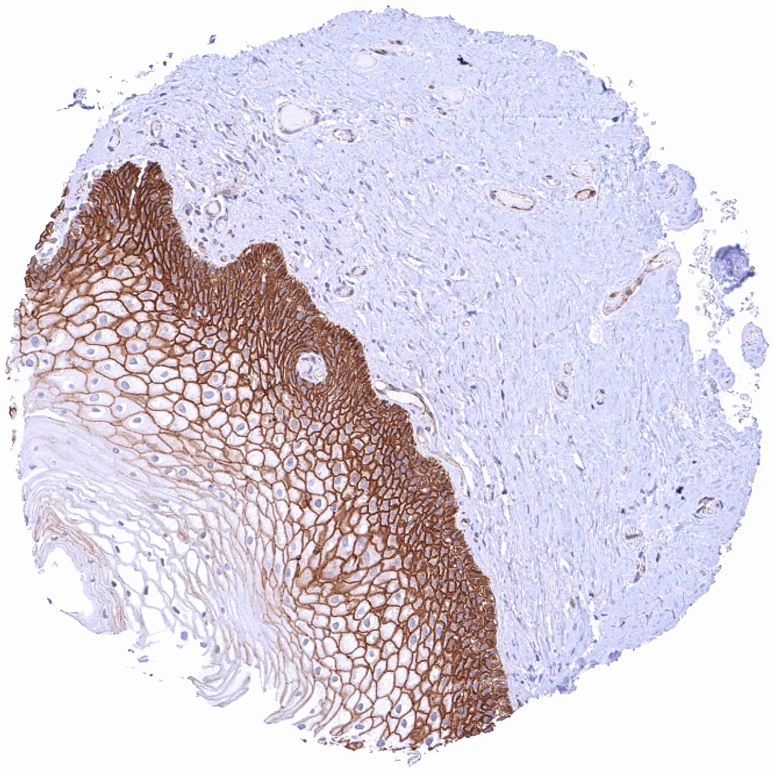
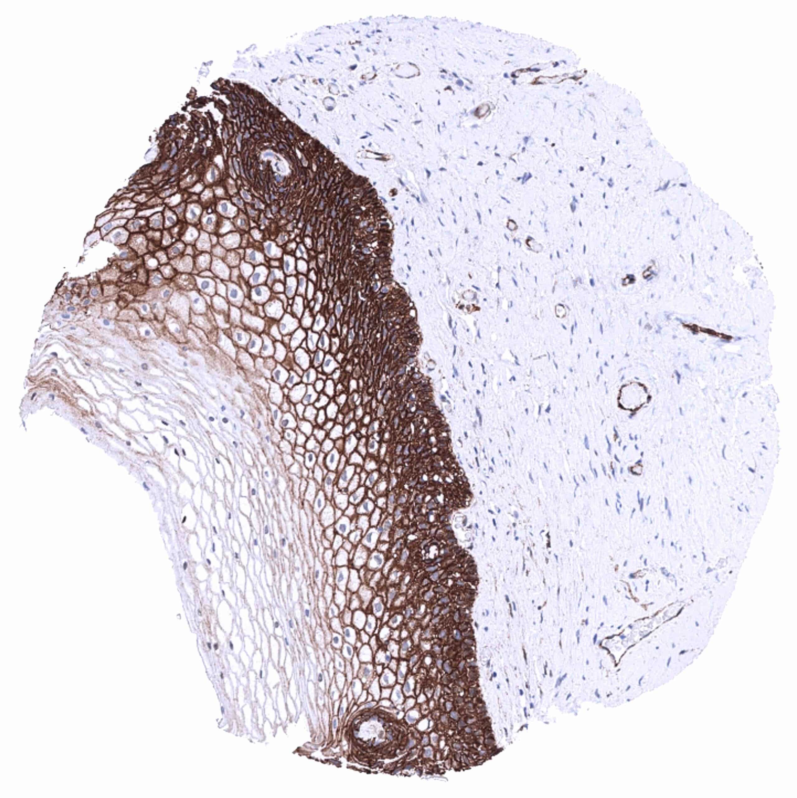
| Esophagus | Strong membranous beta catenin staining of squamous epithelial cells with decreasing staining intensity from the basal towards the superficial cell layers. Top cell layers are beta catenin negative. | |
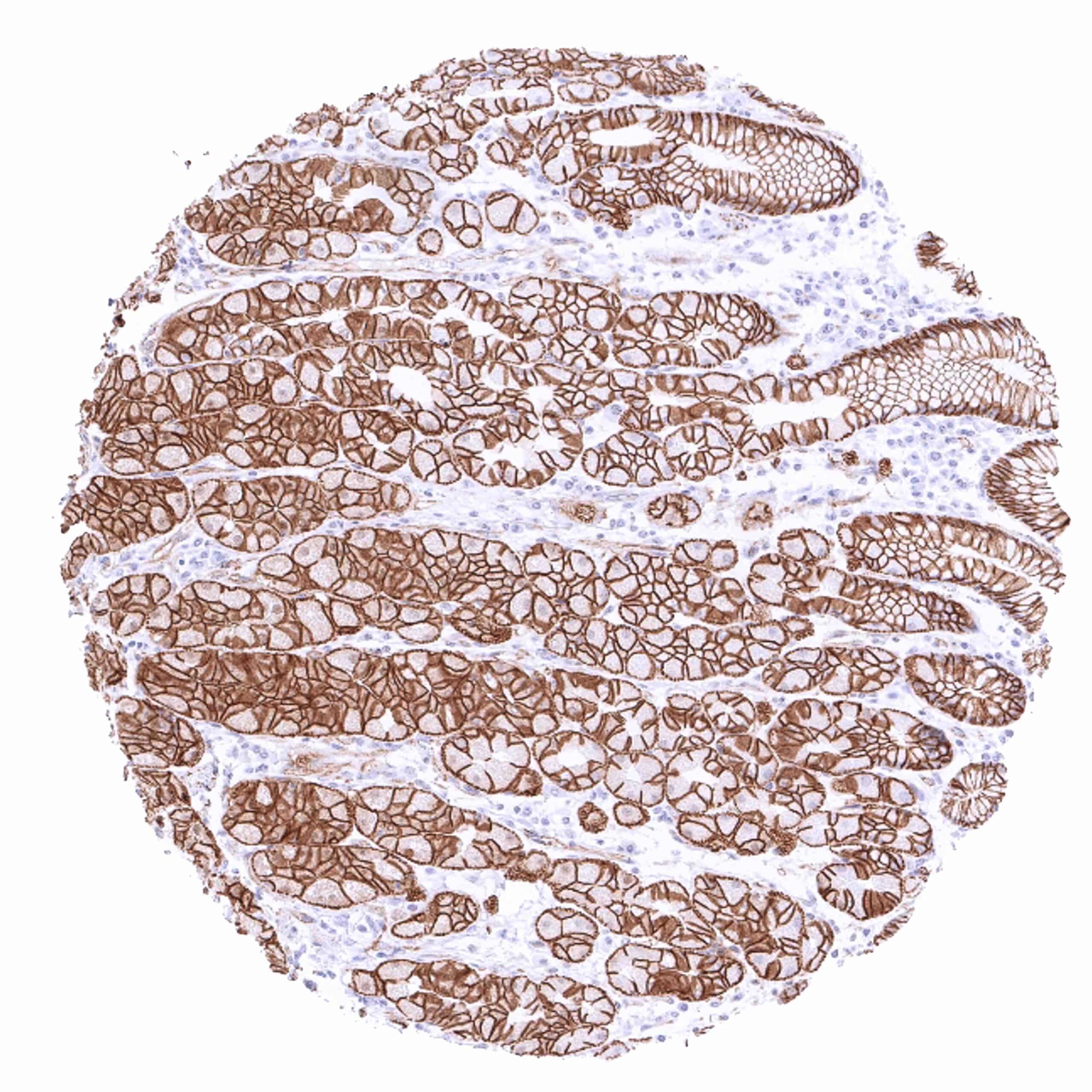
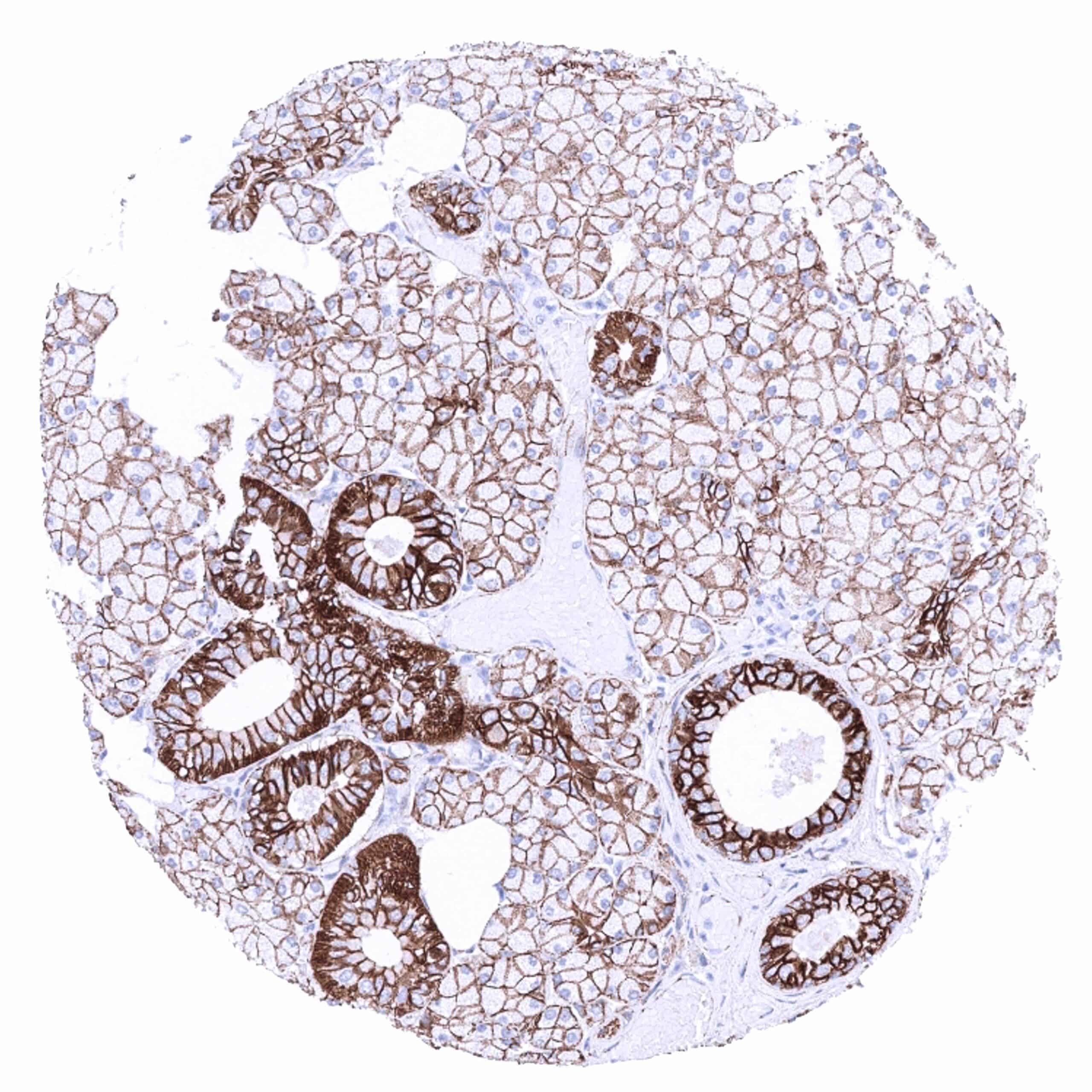
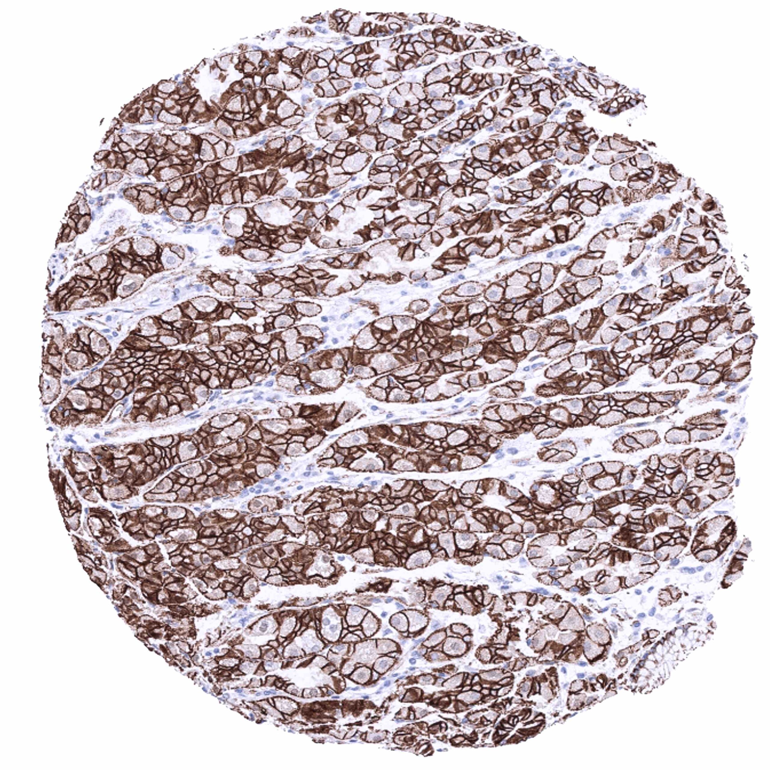
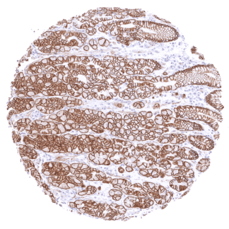
| Stomach | Strong membranous beta catenin staining of all gastric epithelial cells. | |
| Duodenum | Strong membranous beta catenin staining of all epithelial cells. | |
| Small intestine | Strong membranous beta catenin staining of all epithelial cells. | |
| Appendix | Strong membranous beta catenin staining of all epithelial cells. | |
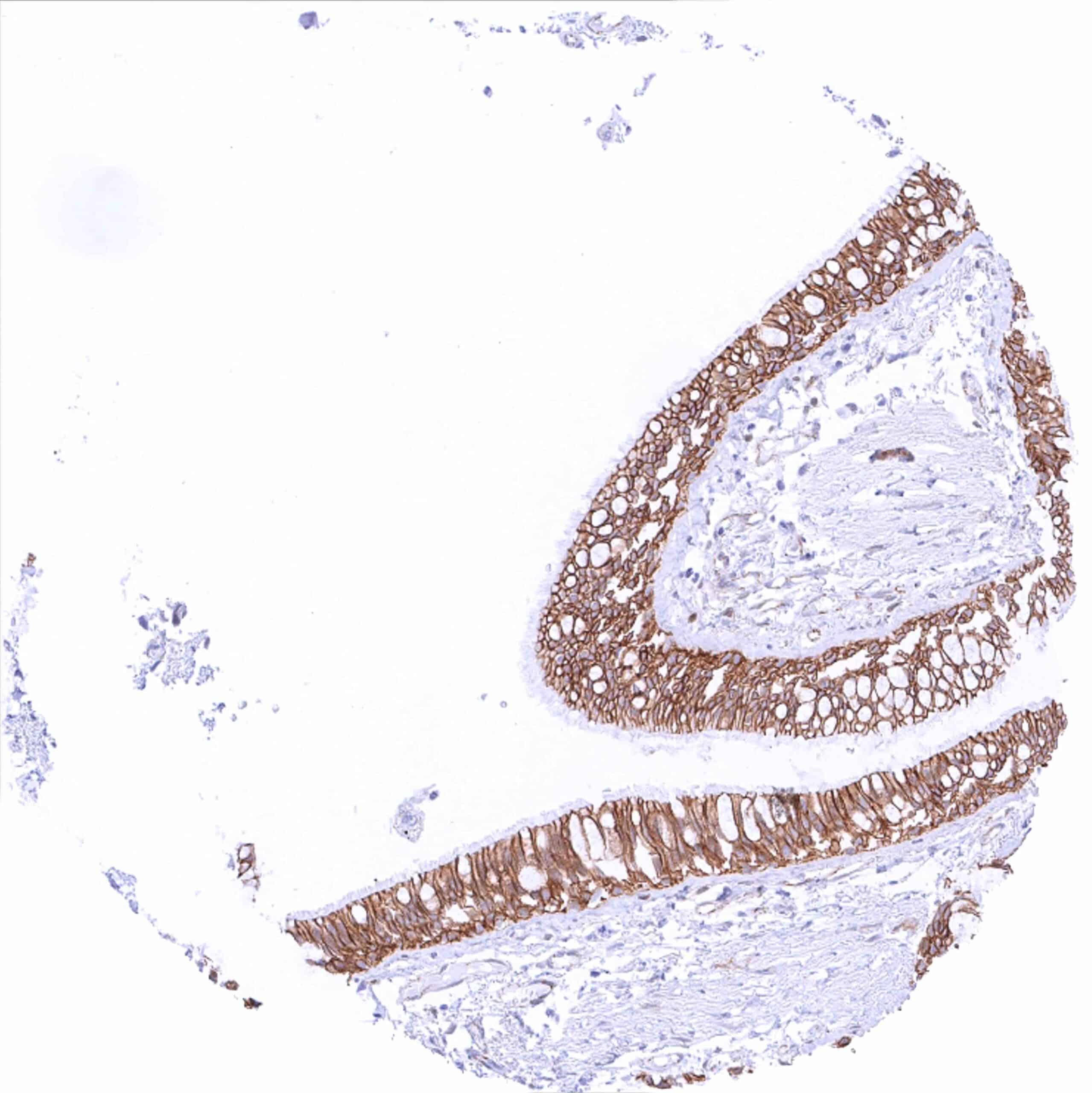
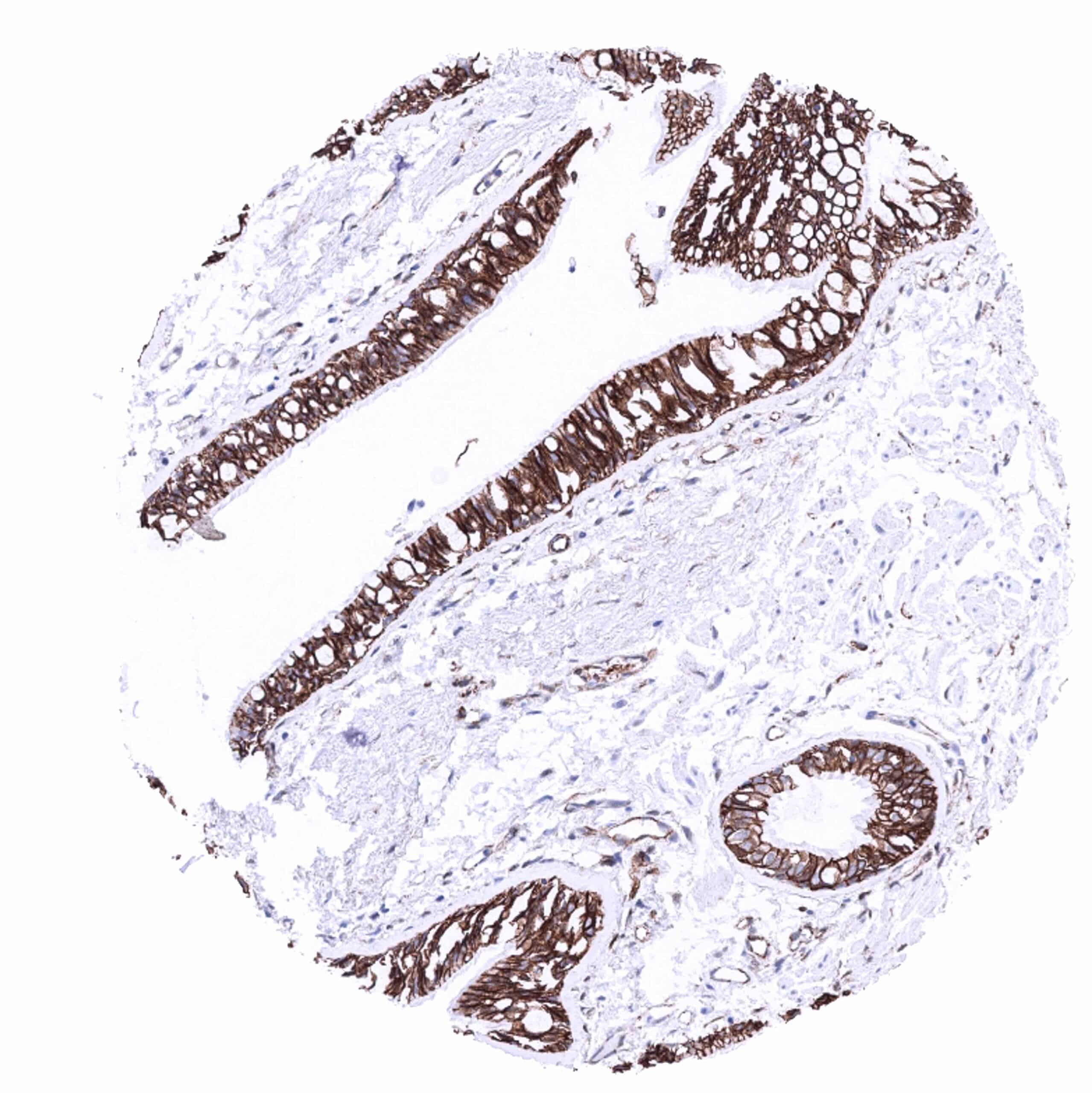
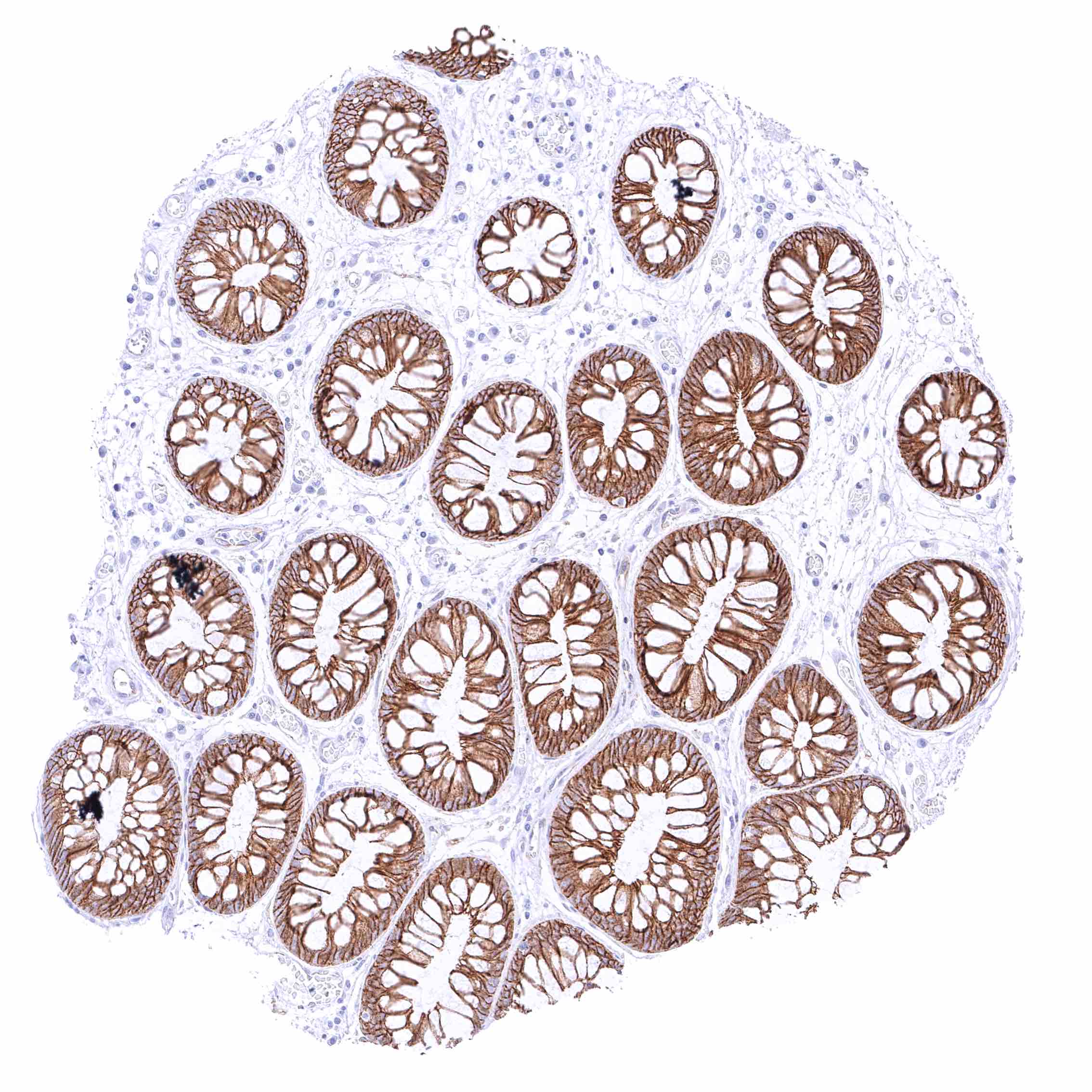
| Colon | Strong membranous beta catenin staining of all epithelial cells. | |
| Rectum | Strong membranous beta catenin staining of all epithelial cells. | |
| Liver | Weak to moderate membranous beta catenin staining of hepatocytes. | |
| Gallbladder | Strong membranous beta catenin staining of epithelial cells. | |
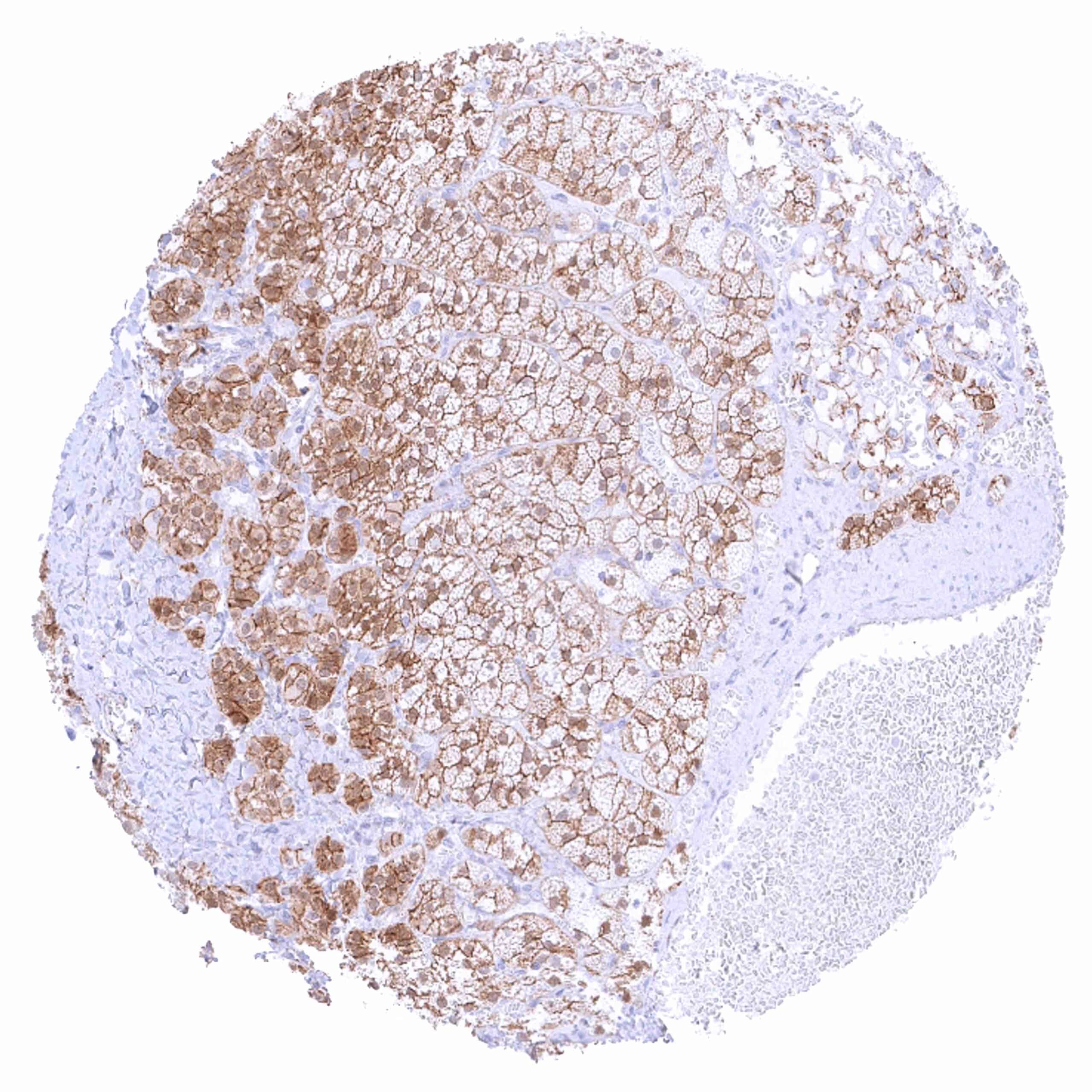
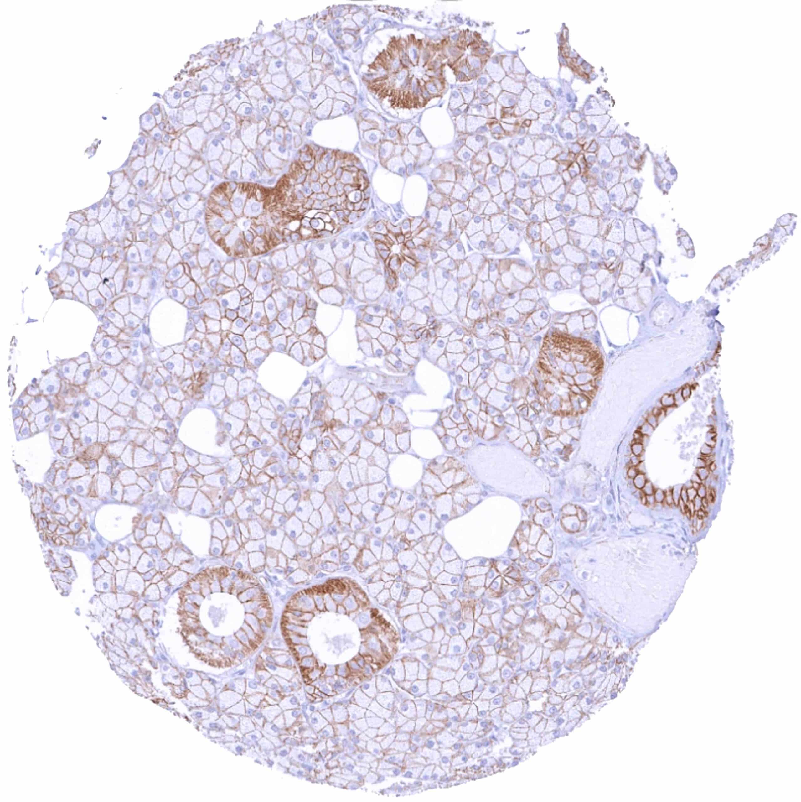
| Pancreas | Strong membranous beta catenin staining of acinar and excretory duct cells while staining is less intense in islets. | |
| Genitourinary | Kidney | Moderate to strong membranous beta catenin staining of tubular and collecting duct cells while staining is only weak at the capsule of Bowman. |
| Urothelium | Strong membranous beta catenin staining of urothelial cells. | |
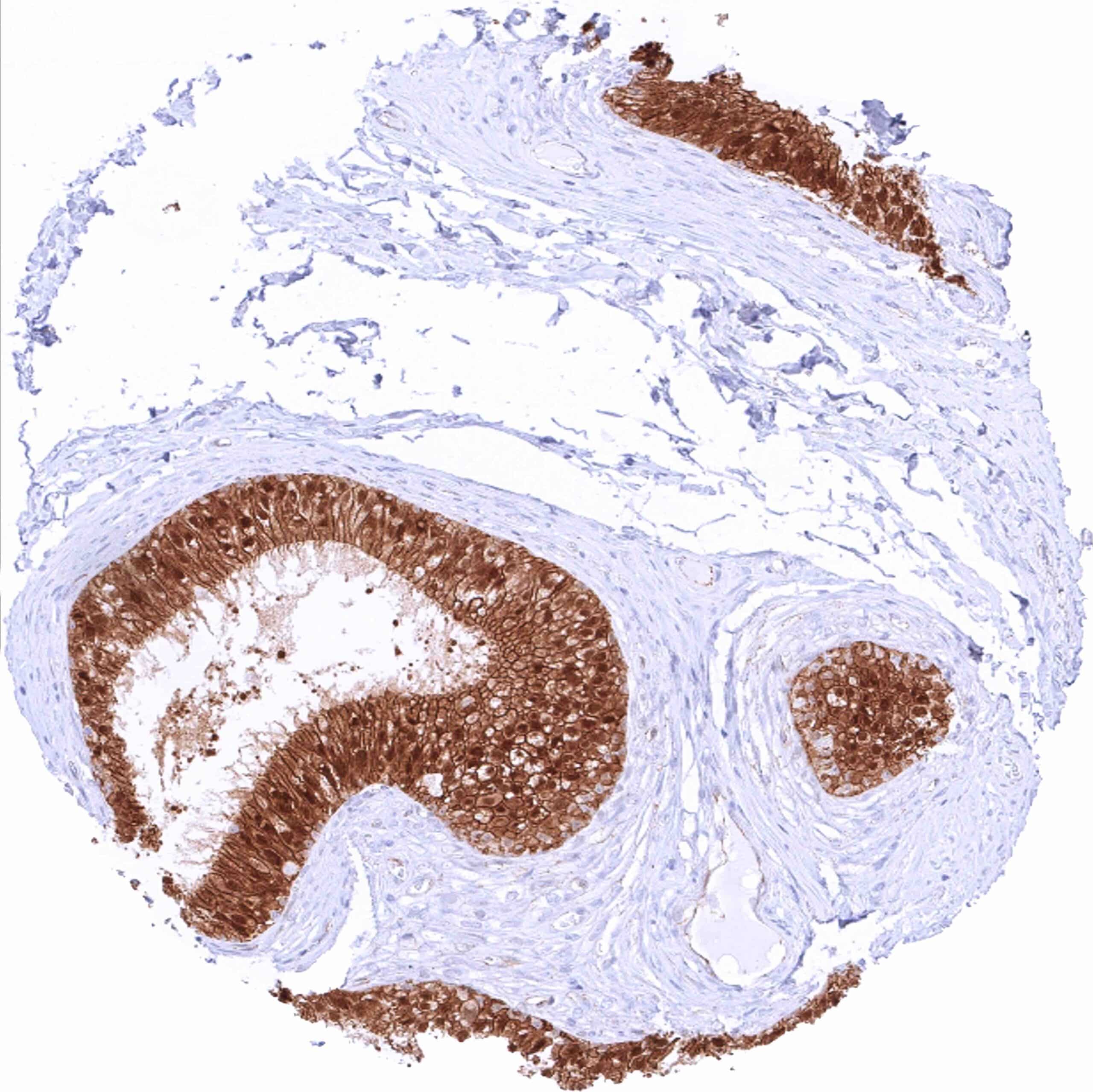
| Male genital | Prostate | Strong membranous beta catenin staining of epithelial cells. |
| Seminal vesicles | Strong membranous beta catenin staining of epithelial cells. | |
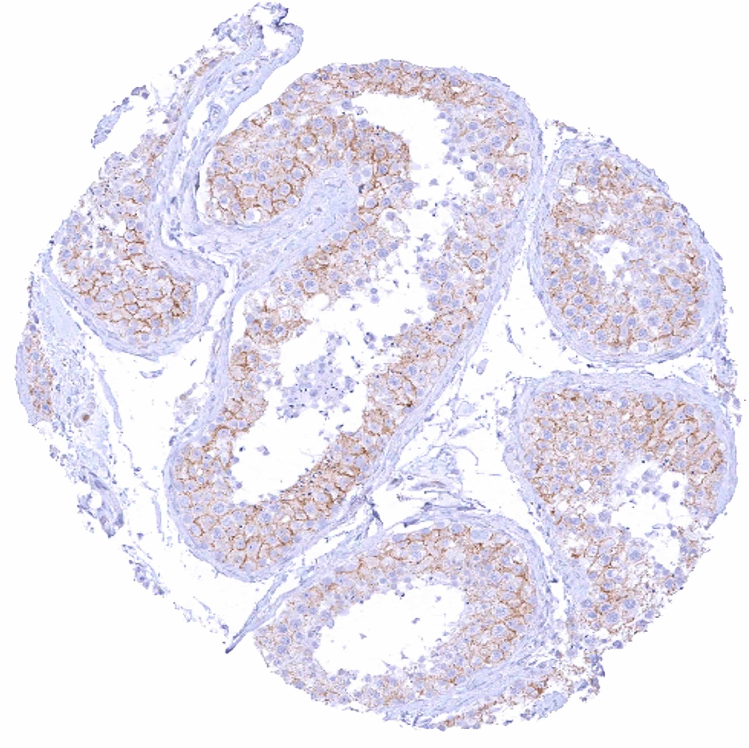
| Testis | Moderate to strong membranous beta catenin staining of Sertoli cells while germ cells are only weakly stained or negative. | |
| Epididymis | Strong membranous beta catenin staining of all epithelial cells with an additional strong nuclear staining in the tall columnar cells of the caput. | |
| Female genital | Breast | Strong membranous beta catenin staining of epithelial cells. |
| Uterus, myometrium | Negative. | |
| Uterus, ectocervix | Strong membranous beta catenin staining of squamous epithelial cells with decreasing staining intensity from the basal towards the superficial cell layers. Top cell layers are beta catenin negative. | |
| Uterus endocervix | Strong membranous beta catenin staining of epithelial cells. | |
| Uterus, endometrium | Strong membranous beta catenin staining of epithelial cells. During pregnancy, membranous beta catenin staining of epithelial and decidua cells is only weak to moderate. | |
| Fallopian Tube | Strong membranous beta catenin staining of epithelial cells. | |
| Ovary | Distinct membranous beta catenin staining of stroma cells. Moderate membranous beta catenin staining of corpus luteum cells. | |
| Placenta early | Strong membranous beta catenin staining of cytotrophoblast cells, accompanied by a nuclear and cytoplasmic positivity in many cells. The syncytiotrophoblast is beta catenin negative. | |
| Placenta mature | Strong membranous beta catenin staining of cytotrophoblast cells. In some of these cells, there is also a nuclear and cytoplasmic positivity. The syncytiotrophoblast is beta catenin negative. | |
| Amnion | Moderate membranous beta catenin staining of amnion cells. | |
| Chorion | Moderate membranous beta catenin staining of chorion cells. | |
| Skin | Epidermis | Strong membranous beta catenin staining of squamous epithelial cells. The staining intensity decreases in the most superficial cell layers. |
| Sebaceous glands | Distinct membranous beta catenin staining of sebaceous gland cells. | |
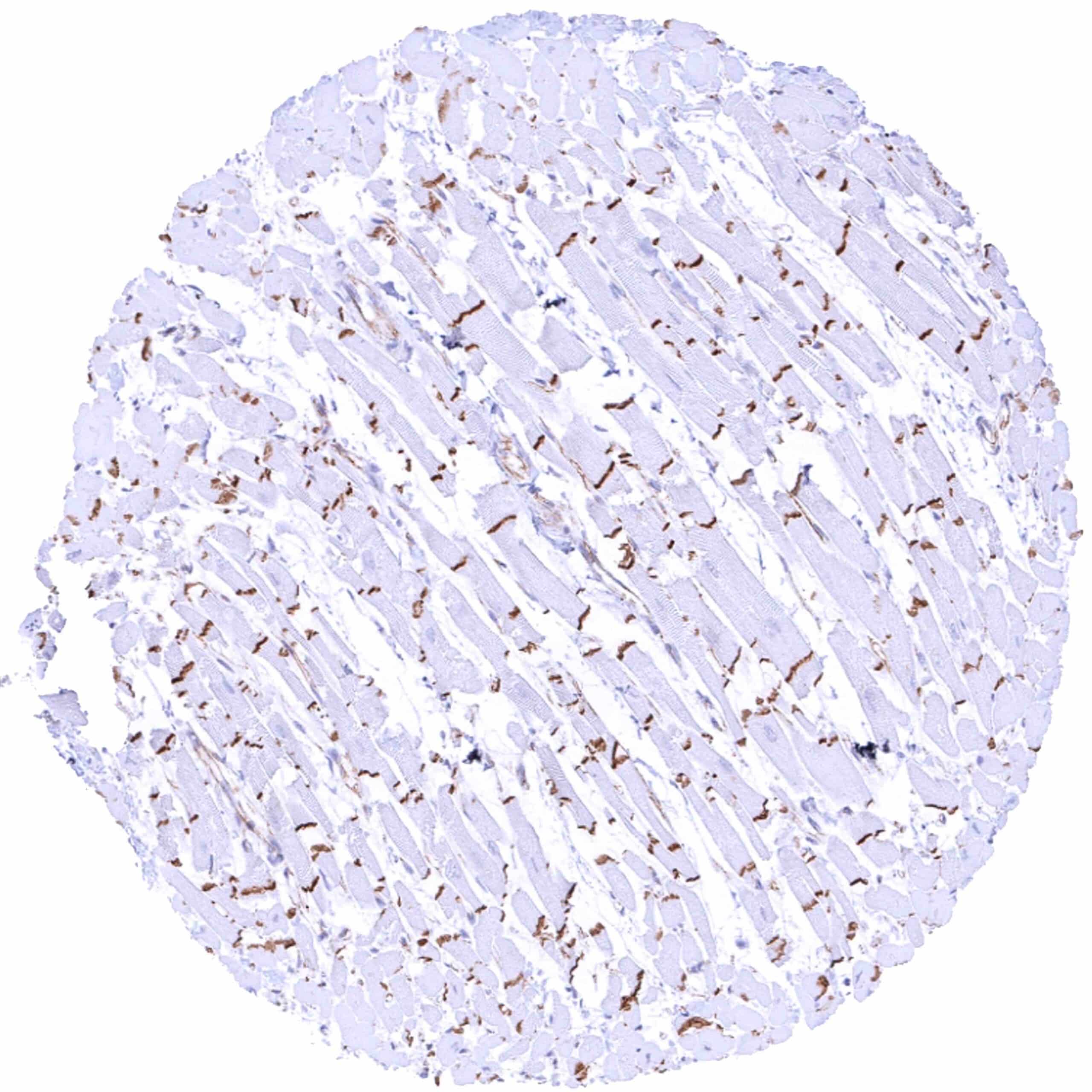
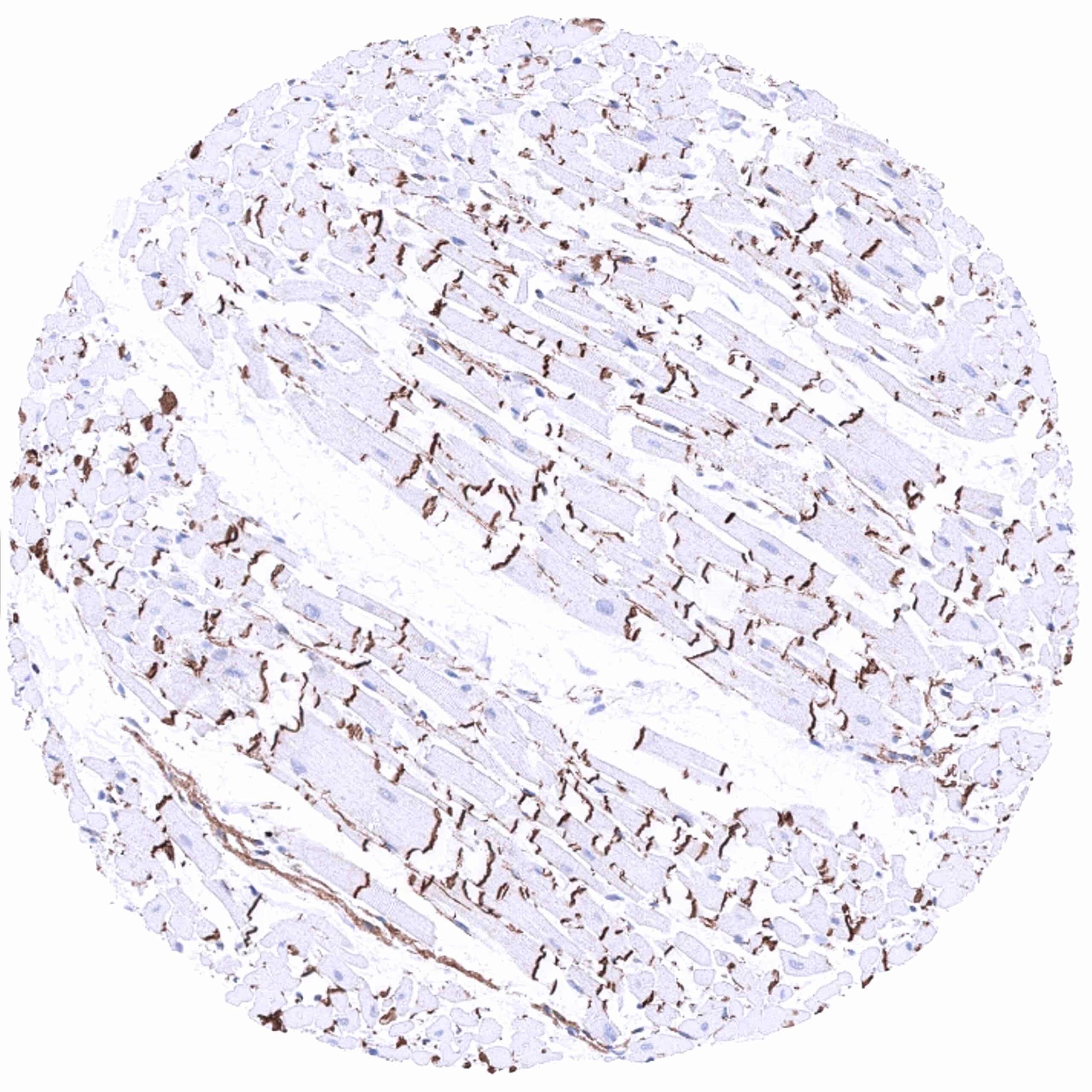
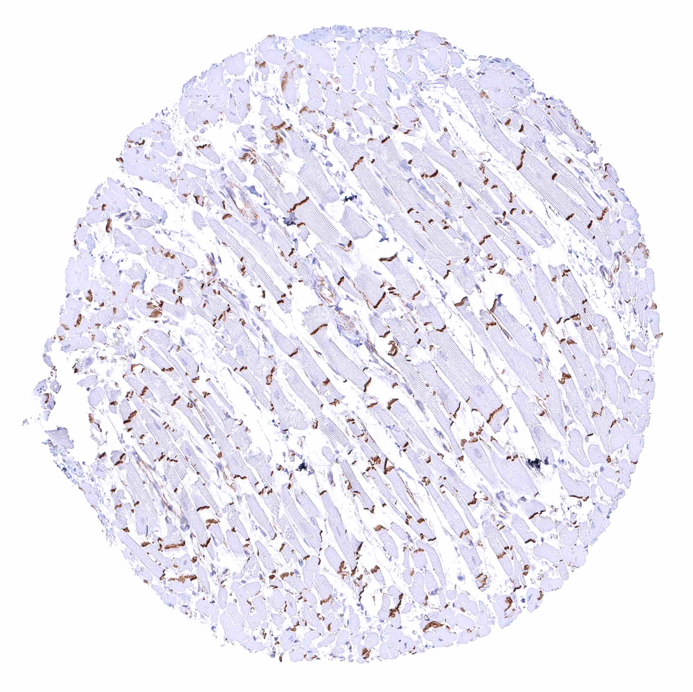
| Muscle/connective tissue | Heart muscle | Strong beta catenin staining intercalated discs. |
| Skeletal muscle | Negative. | |
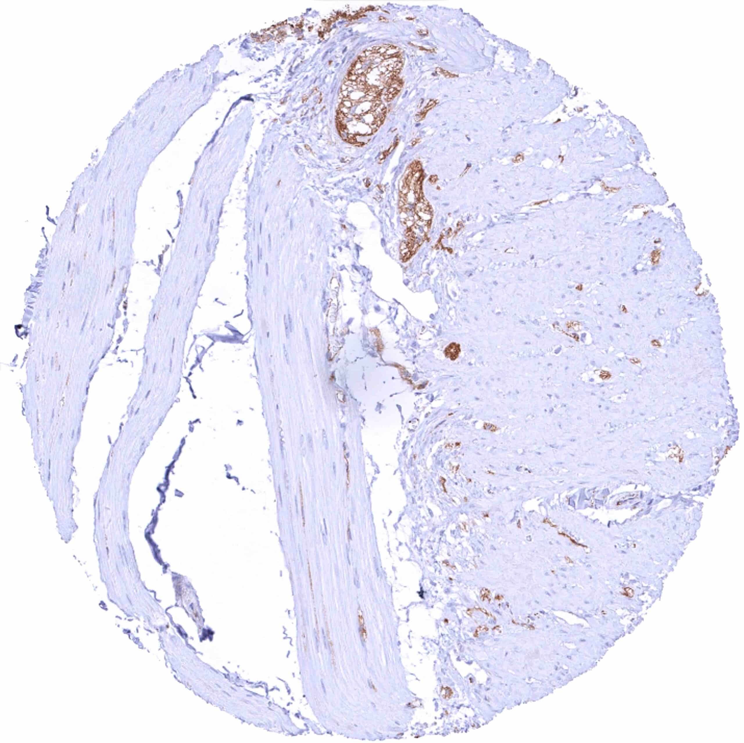
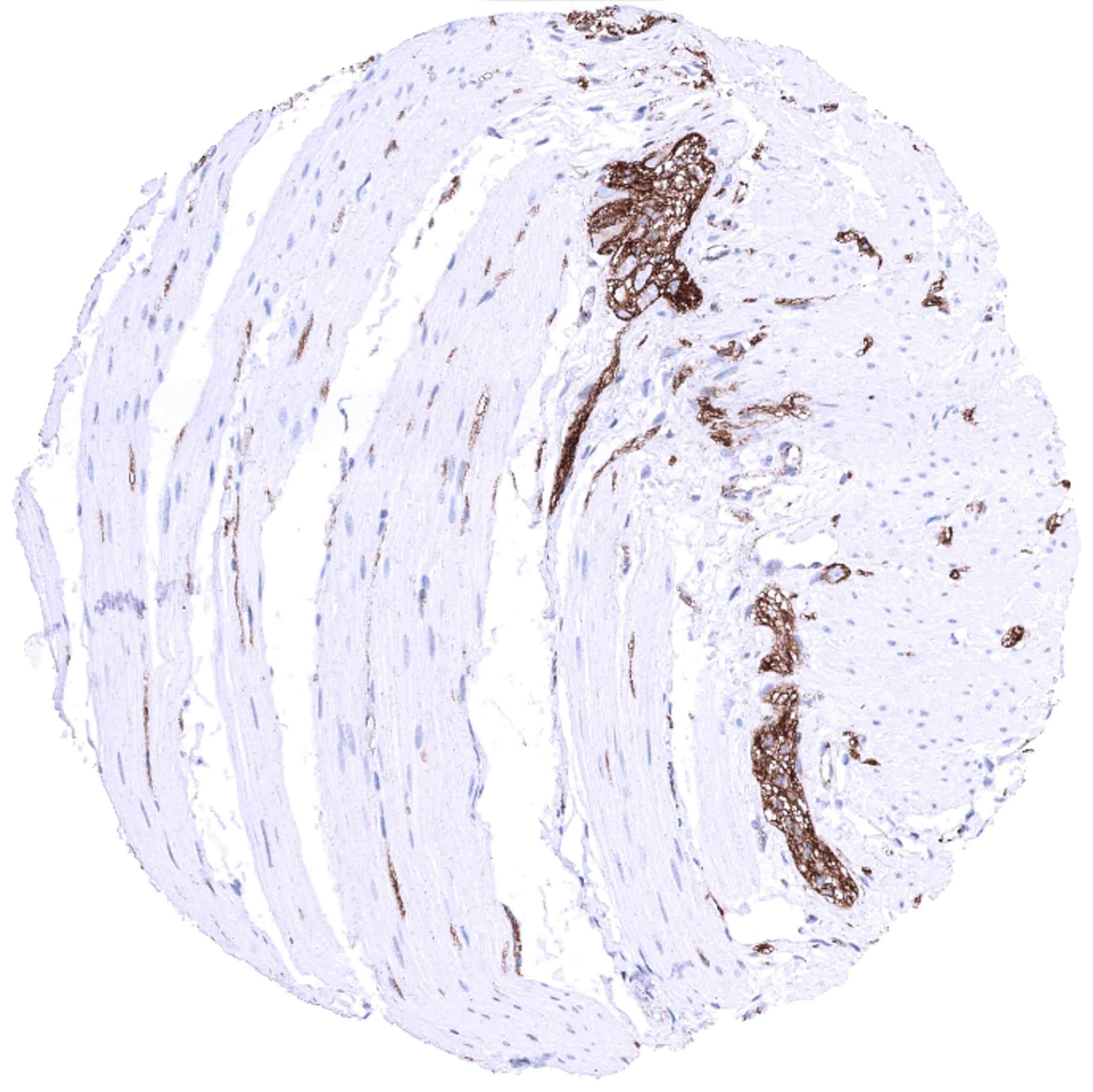
| Smooth muscle | Strong beta catenin staining of nerves included in the muscular wall of organs. | |
| Vessel walls | Negative. | |
| Fat | Negative. | |
| Stroma | Negative. | |
| Endothelium | Weak to moderate positivity of endothelial cells. | |
| Bone marrow/ lymphoid tissue | Bone marrow | Negative. |
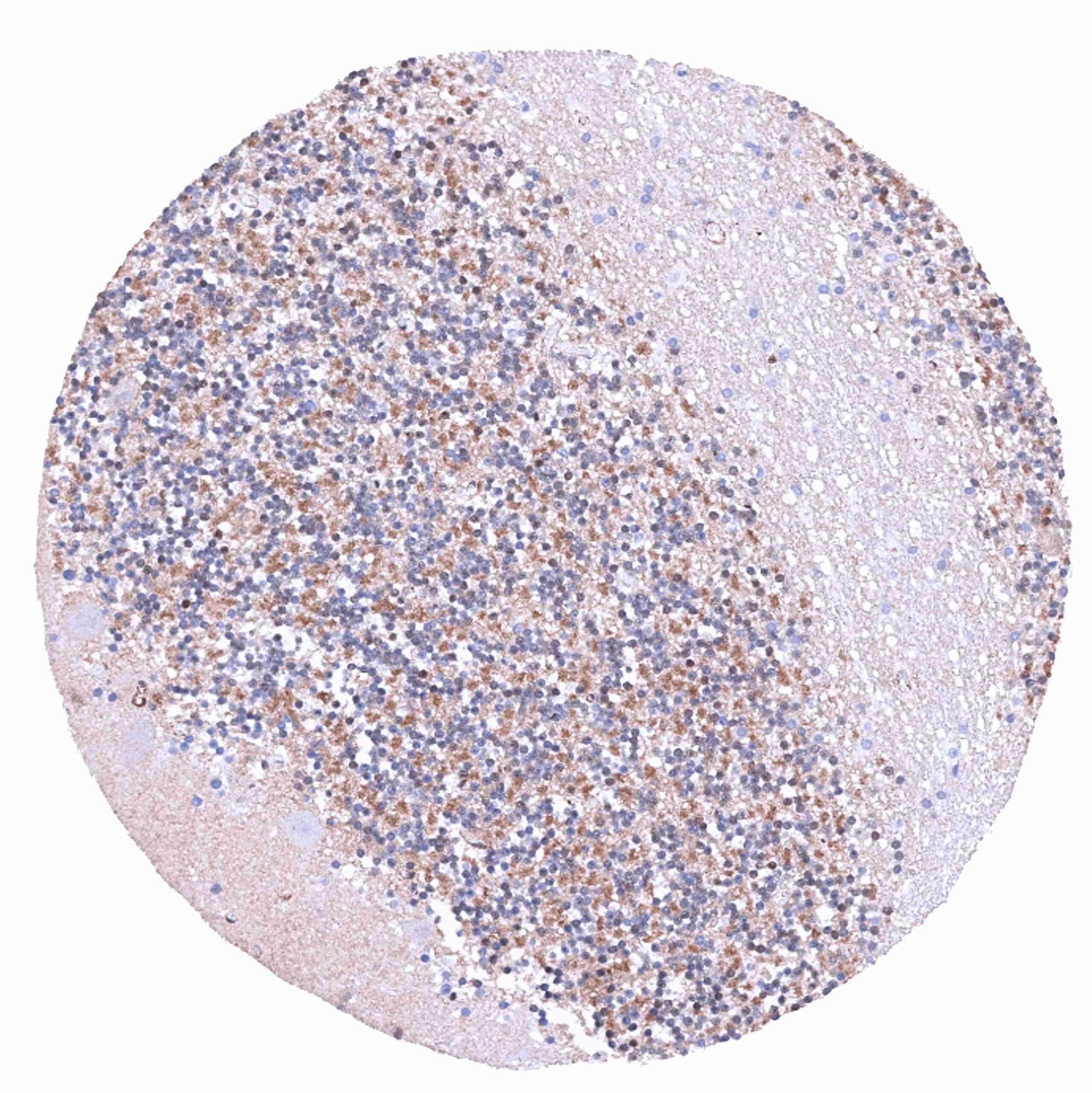
| Lymph node | Weak to moderate membranous beta catenin staining of endothelial cells. Lymphocytes are beta catenin negative. | |
| Spleen | Weak to moderate membranous beta catenin staining of endothelial cells. | |
| Thymus | Strong membranous beta catenin staining of epithelial cells including corpuscles of Hassall’s. Weak to moderate positivity of endothelial cells. Lymphocytes are beta catenin negative. | |
| Tonsil | Strong membranous beta catenin staining of squamous epithelial cells with decreasing staining intensity from the basal towards the superficial cell layers. Weak to moderate positivity of endothelial cells. Lymphocytes are beta catenin negative. | |
| Remarks |
β-Catenin is expressed in most epithelial cells, endothelial cells, intercalated discs of heart muscle cells, along nerve fibers as well as few other non-epithelial cell types. The findings described above are thus consistent with the the Human Protein Atlas (Tissue expression Beta-catenin).
Positive control = Caput epididymis: A strong membranous β-Catenin staining should be seen in all epithelial cell types. In a fraction of the cells, an additional distinct nuclear staining must be seen.
Negative control = Placenta (mature): β-Catenin staining should be absent in the cells of the syncytiotrophoblast while it is strong in cells of the cytotrophoblast.
Staining Pattern in Relevant Tumor Types
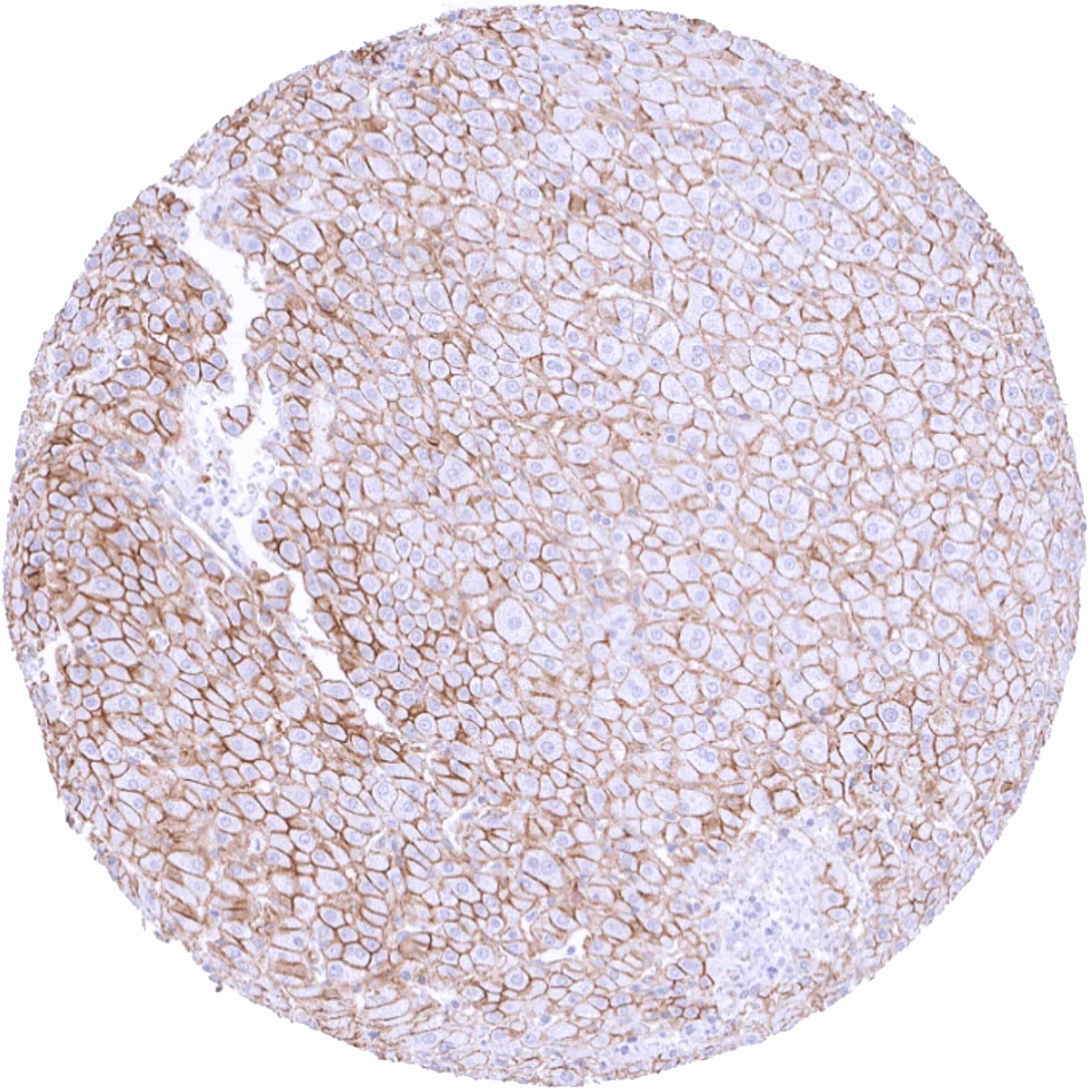
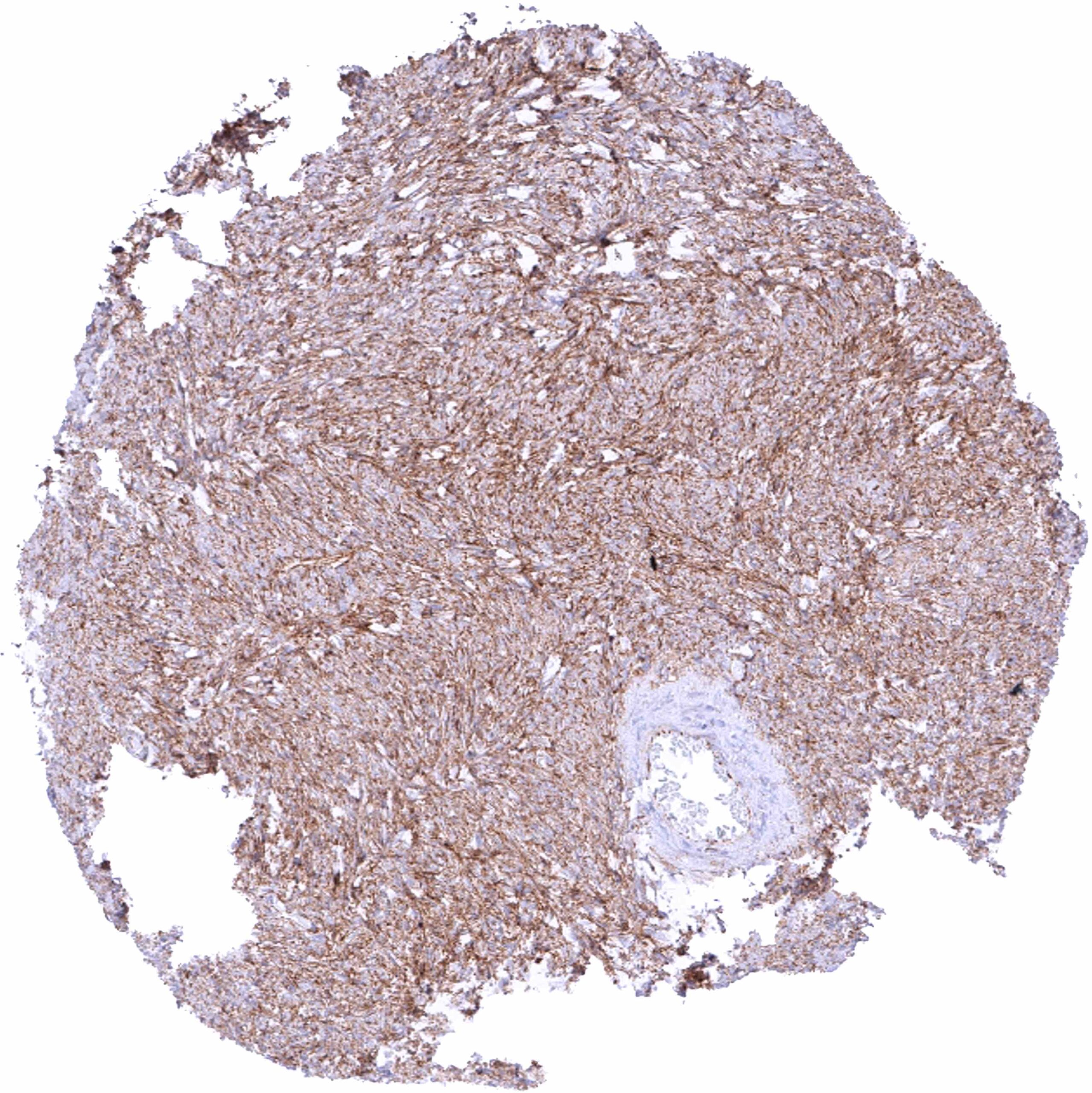
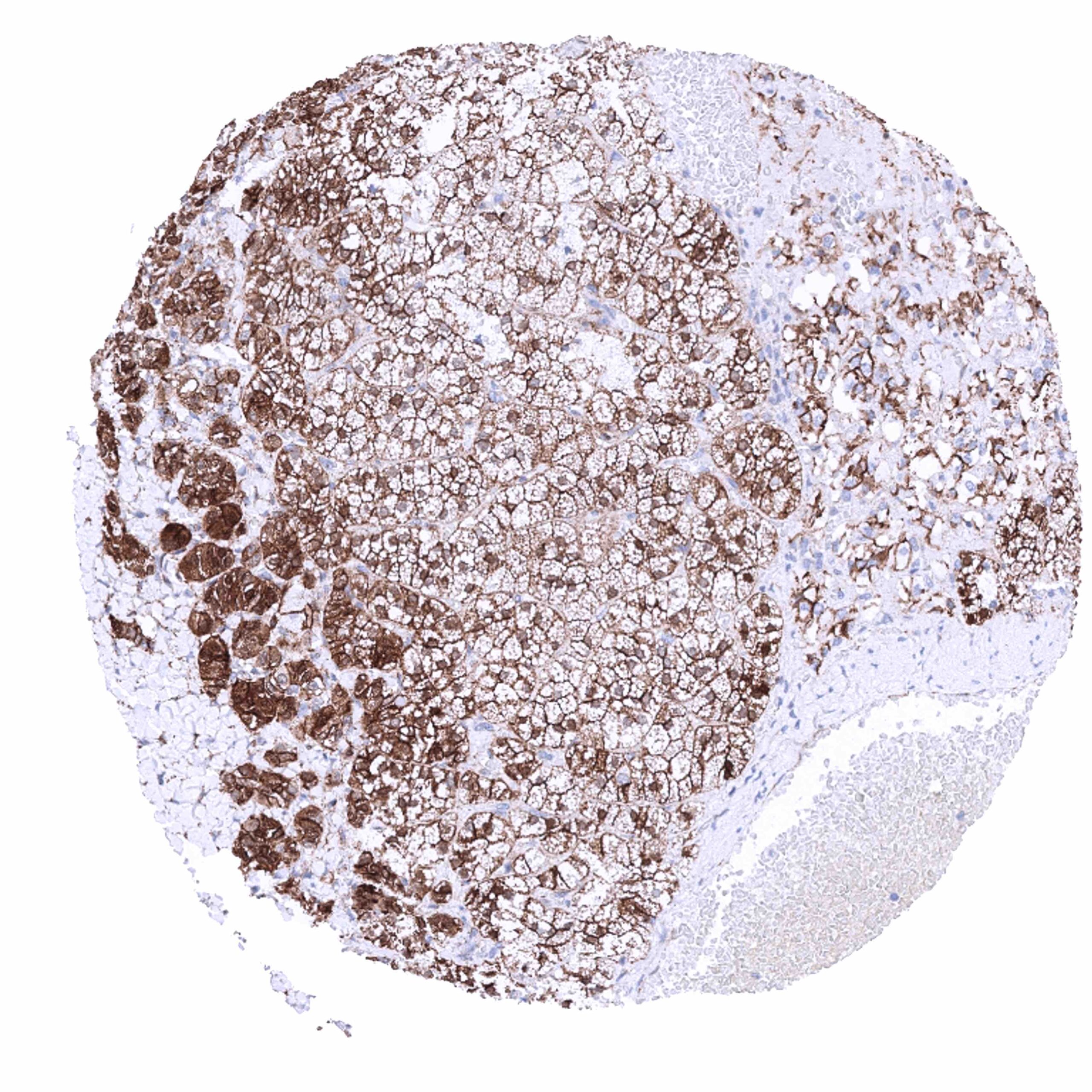
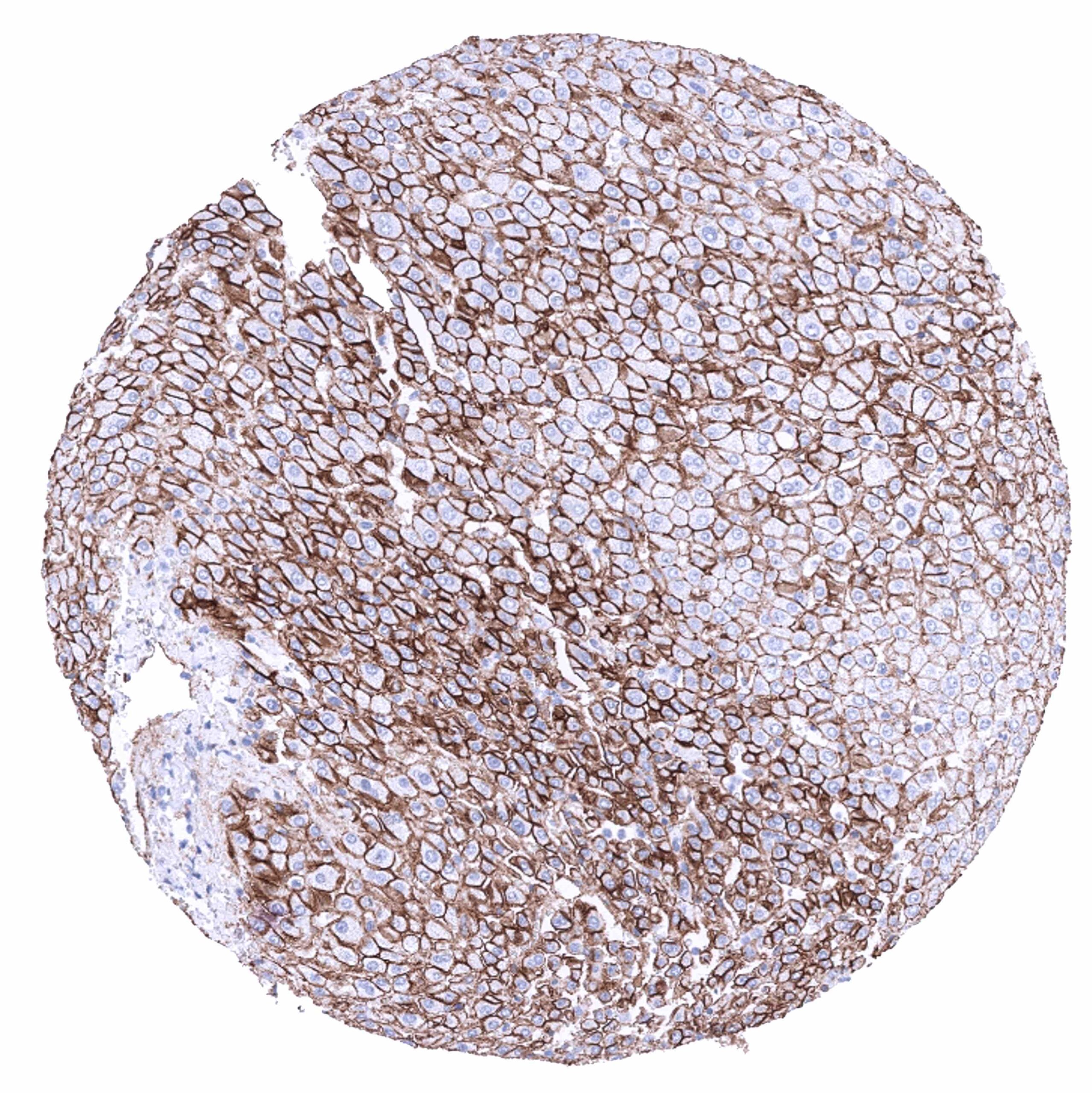
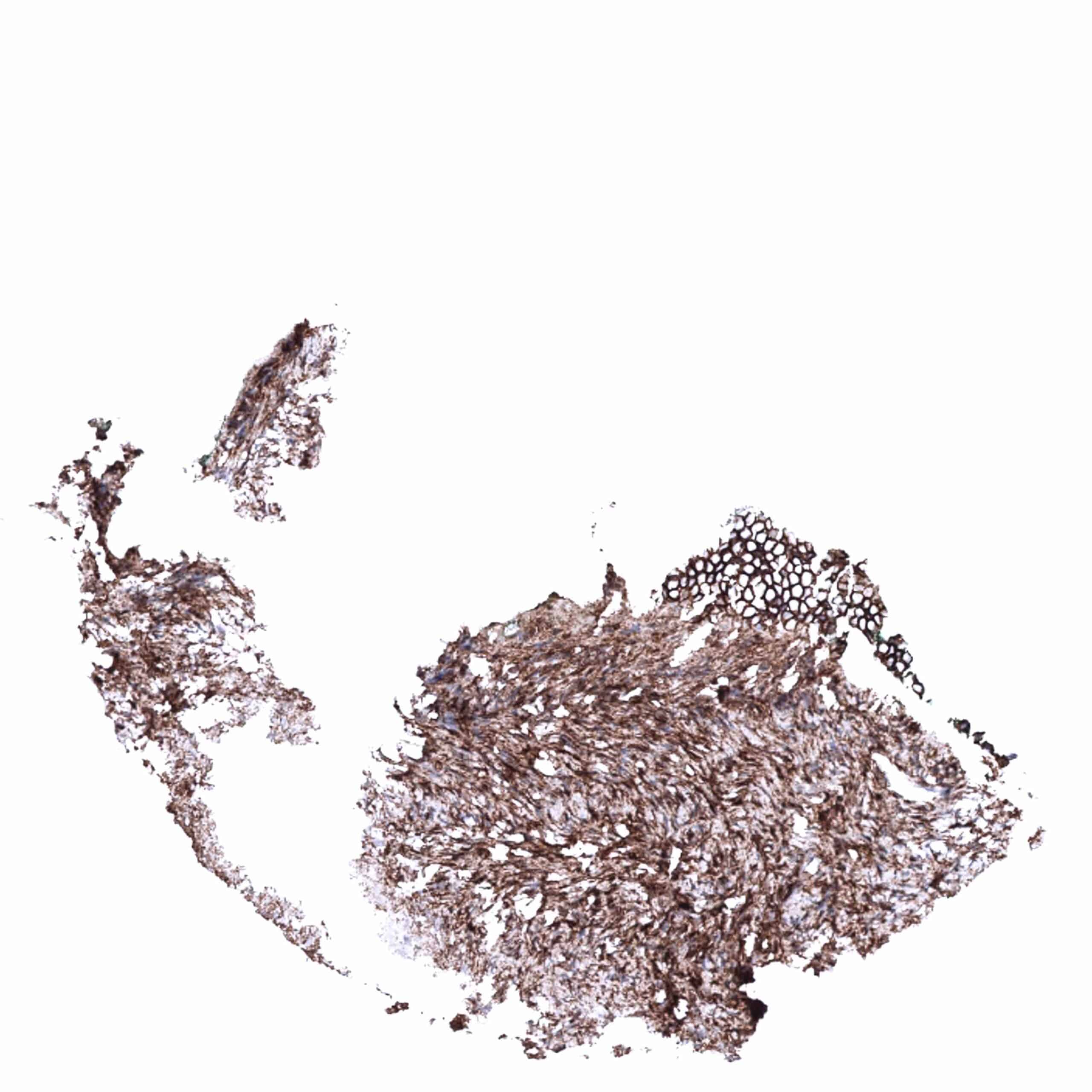
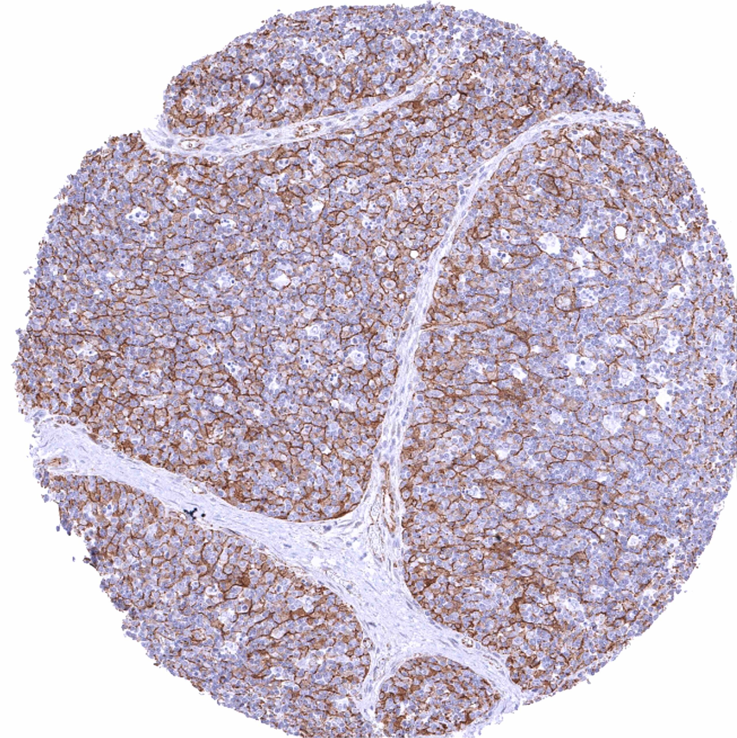
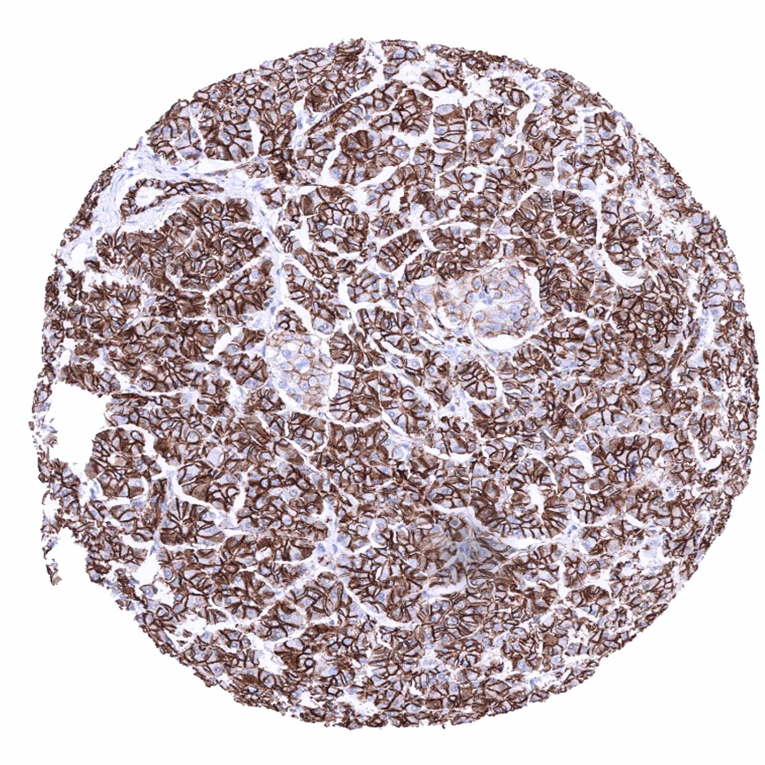
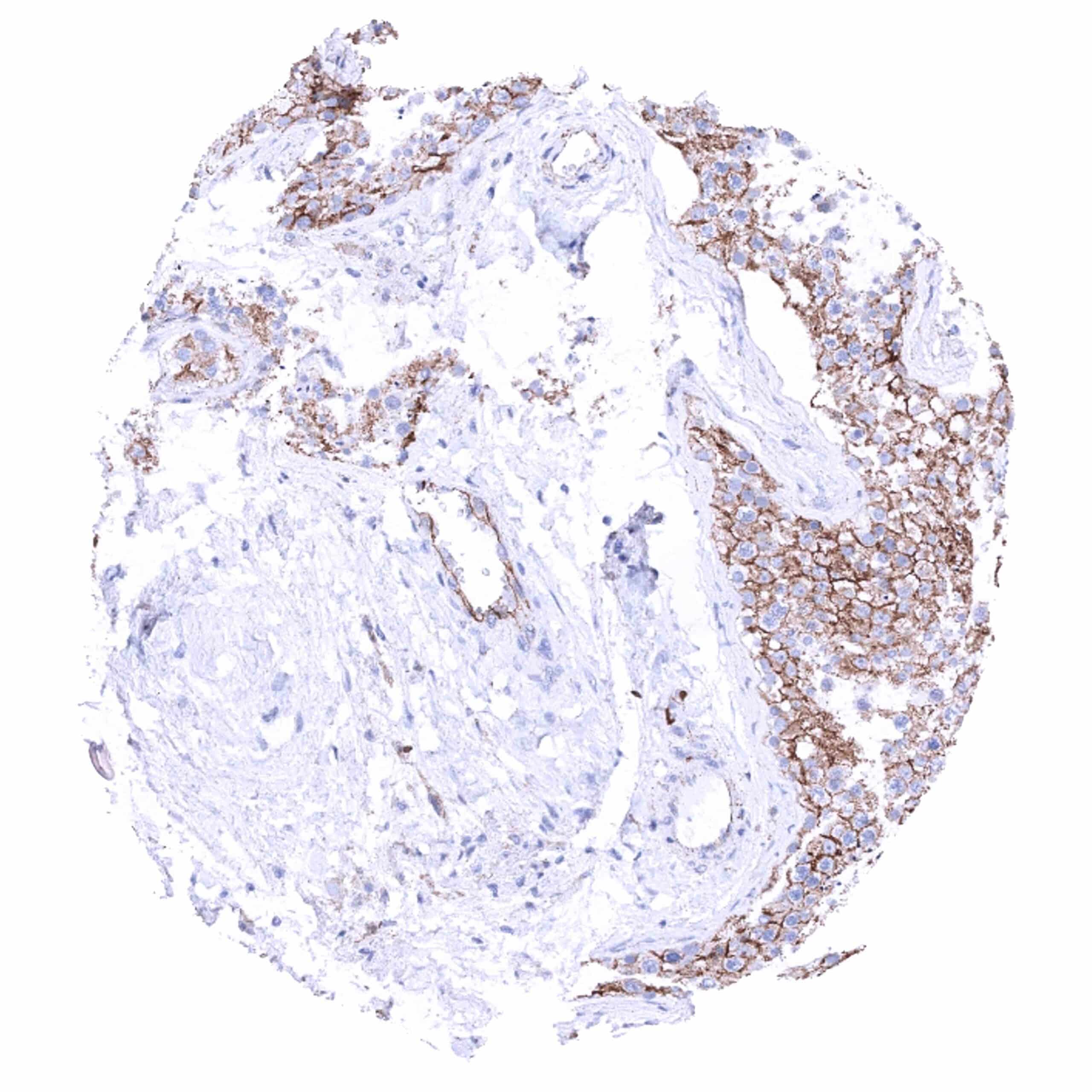
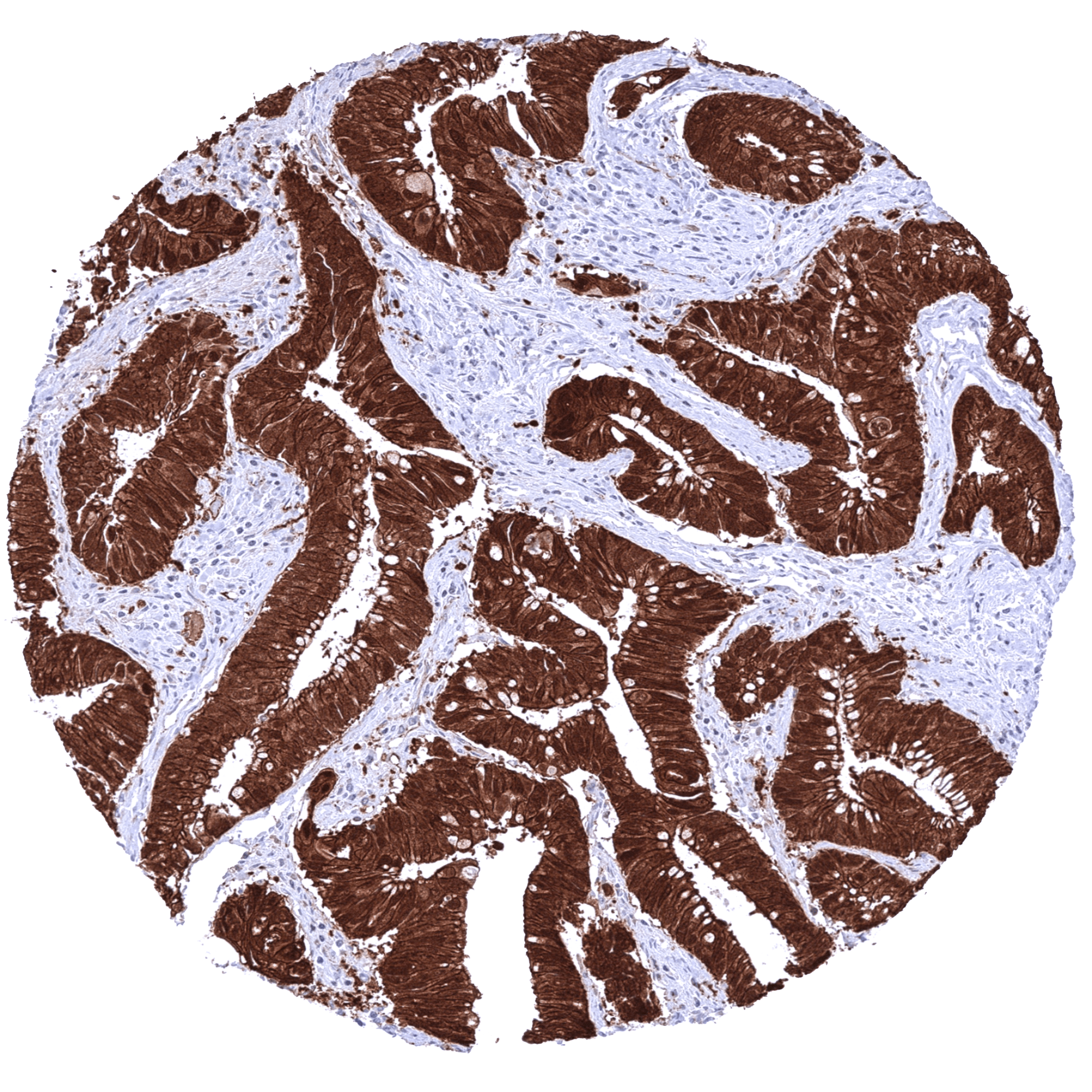
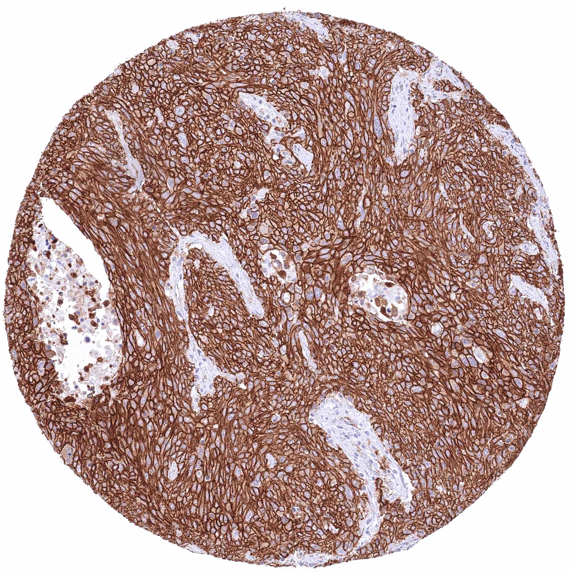
Most epithelial tumors express β-Catenin on their cell membranes. In a fraction of cancers, there is an additional cytoplasmic and nuclear β-Catenin staining. Membranous β-Catenin staining can be reduced or absent in another fraction of cancers.
The TCGA findings on Beta-Catenin RNA expression in different tumor categories have been summarized in the Human Protein Atlas.
Compatibility of Antibodies
No data available at the moment
Protocol Recommendations
IHC users have different preferences on how the stains should look like. Some prefer high staining intensity of the target stain and even accept some background. Others favor absolute specificity and lighter target stains. Factors that invariably lead to more intense staining include higher concentration of the antibody and visualization tools, longer incubation time, higher temperature during incubation, higher temperature and longer duration of the heat induced epitope retrieval (slide pretreatment). The impact of the pH during slide pretreatment has variable effects and depends on the antibody and the target protein.
All images and data shown here and in our image galleries are obtained by the manual protocol described below. Other protocols resulting in equivalent staining are described as well.
Manual protocol
Freshly cut sections should be used (less than 10 days between cutting and staining). Heat-induced antigen retrieval for 5 minutes in an autoclave at 121°C in pH 7,8 Target Retrieval Solution buffer. Apply MSVA-578R at a dilution of 1:150 at 37°C for 60 minutes. Visualization of bound antibody by the EnVision Kit (Dako, Agilent) according to the manufacturer’s directions.
Potential Research Applications
- The clinical significance of different Wnt/β-Catenin pathway alterations are not fully understood.
- The prevalence of β-Catenin alterations in different cancers is not fully clarified.
- Drugs targeting the Wnt/β-Catenin pathway are under development.
- The exact functions of β-Catenin and its interactions in the nucleus are unknown.
Evidence for Antibody Specificity in IHC
There are two ways how the specificity of antibodies can be documented for immunohistochemistry on formalin fixed tissues. These are: 1. Comparison with a second independent method for target expression measurement across a large number of different tissue types (orthogonal strategy), and 2. Comparison with one or several independent antibodies for the same target and showing that all positive staining results are also seen with other antibodies for the same target (independent antibody strategy).
Orthogonal validation: For the antibody MSVA-578R specificity is in line with data from three independent RNA screening studies, including the Human Protein Atlas (HPA) RNA-seq tissue dataset, the FANTOM5 project, and the Genotype-Tissue Expression (GTEx) project, which are all compiled in the Human Protein Atlas (Tissue expression Beta-catenin). In agreement with MSVA-578R immunostaining data, high RNA expression occurs in all normal tissues. However, it must be understood that orthogonal validation is not optimal for assessing ubiquitously expressed proteins.
Comparison of antibodies: True expression of beta catenin in cell types with documented β-Catenin immunostaining by MSVA-578R is validated by largely identical staining patterns obtained for virtually all normal tissues by a second, independent commercially available β-Catenin antibody, termed “validation antibody”. In particular, the use of the validation antibody confirmed characteristic variabilities and patterns of beta catenin staining in several tissue types. These included the decrease of β-Catenin staining from basal to superficial cell layers in squamous epithelium, the lower level of expression in islet cells as compared to acinus and ductal cells in the pancreas, and the lower staining intensity in the capsule of Bowman than in tubuli and collecting ducts of the kidney, as well as the additional nuclear staining in the cytotrophoblast of the placenta, a subset of adrenocortical cells, and in tall columnar cells of the caput epididymis.