Adrenal gland
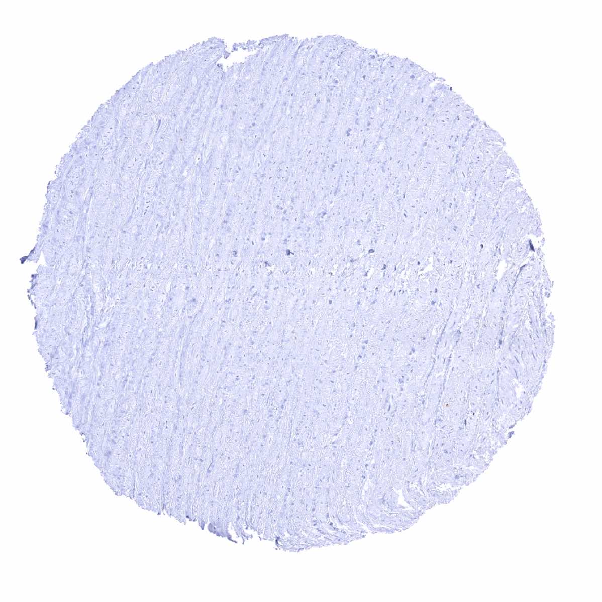
Aorta, media
Appendix, mucosa
Appendix, muscular wall

Bone marrow
Breast
Bronchus, mucosa

Cerebellum, cortex (Stratum moleculare)
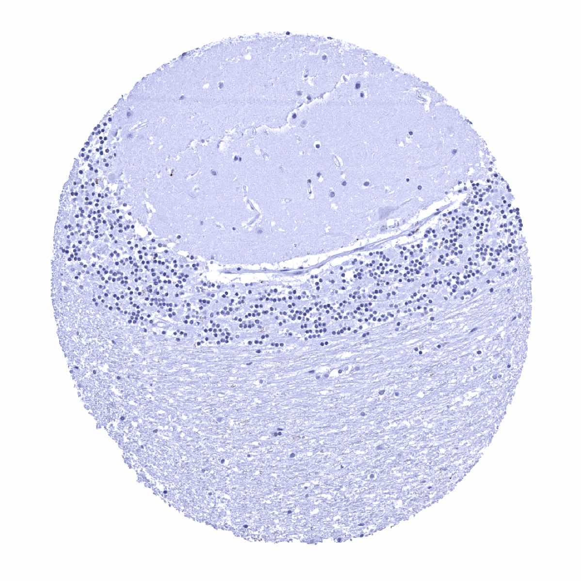
Cerebellum, grey (Stratum neuronorum)
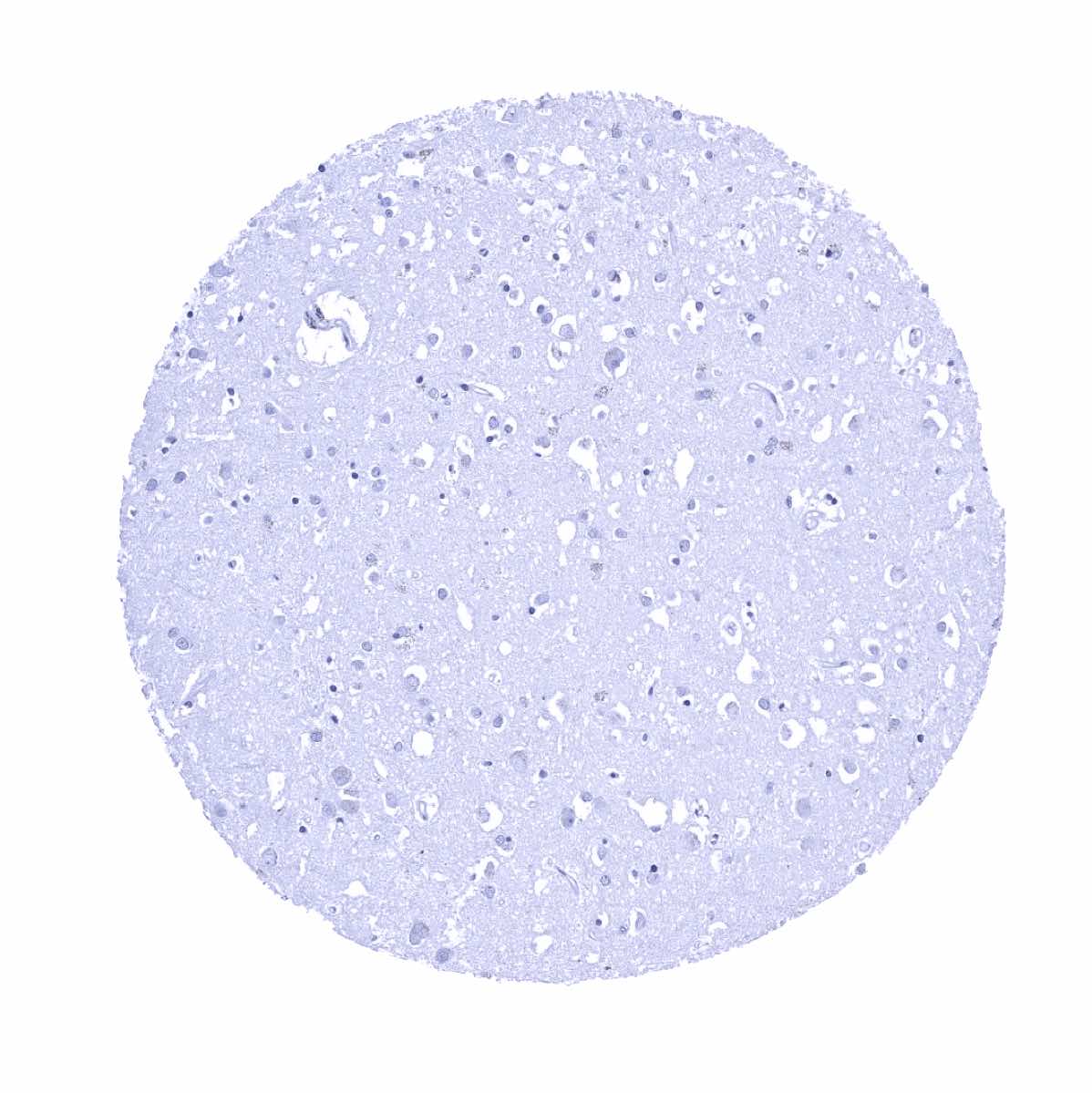
Cerebrum, grey
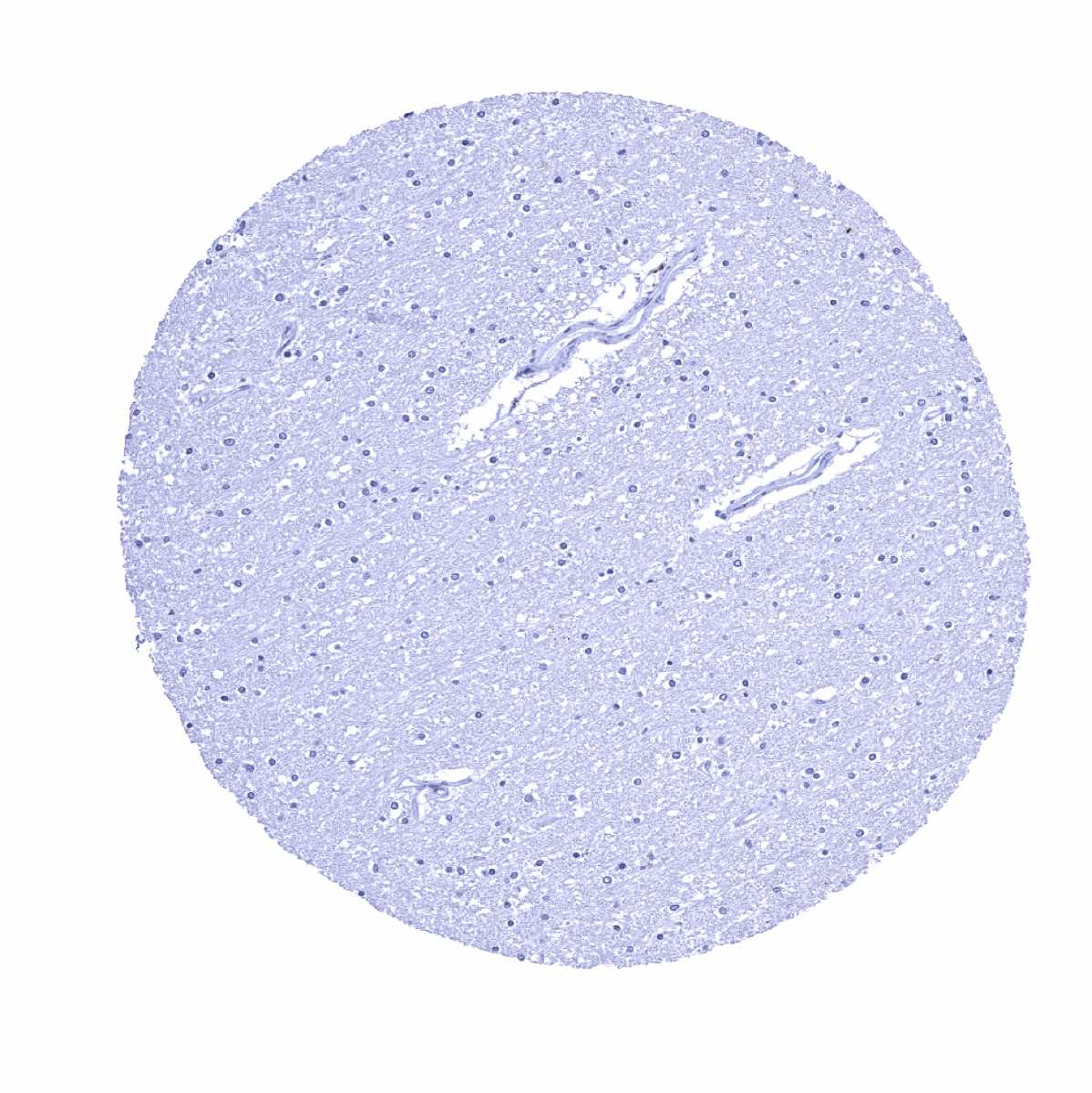
Cerebrum, white
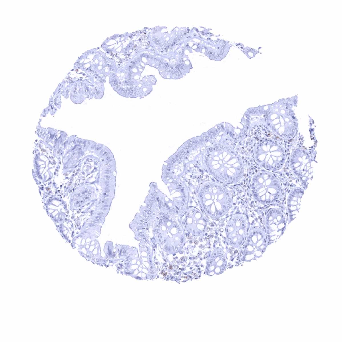
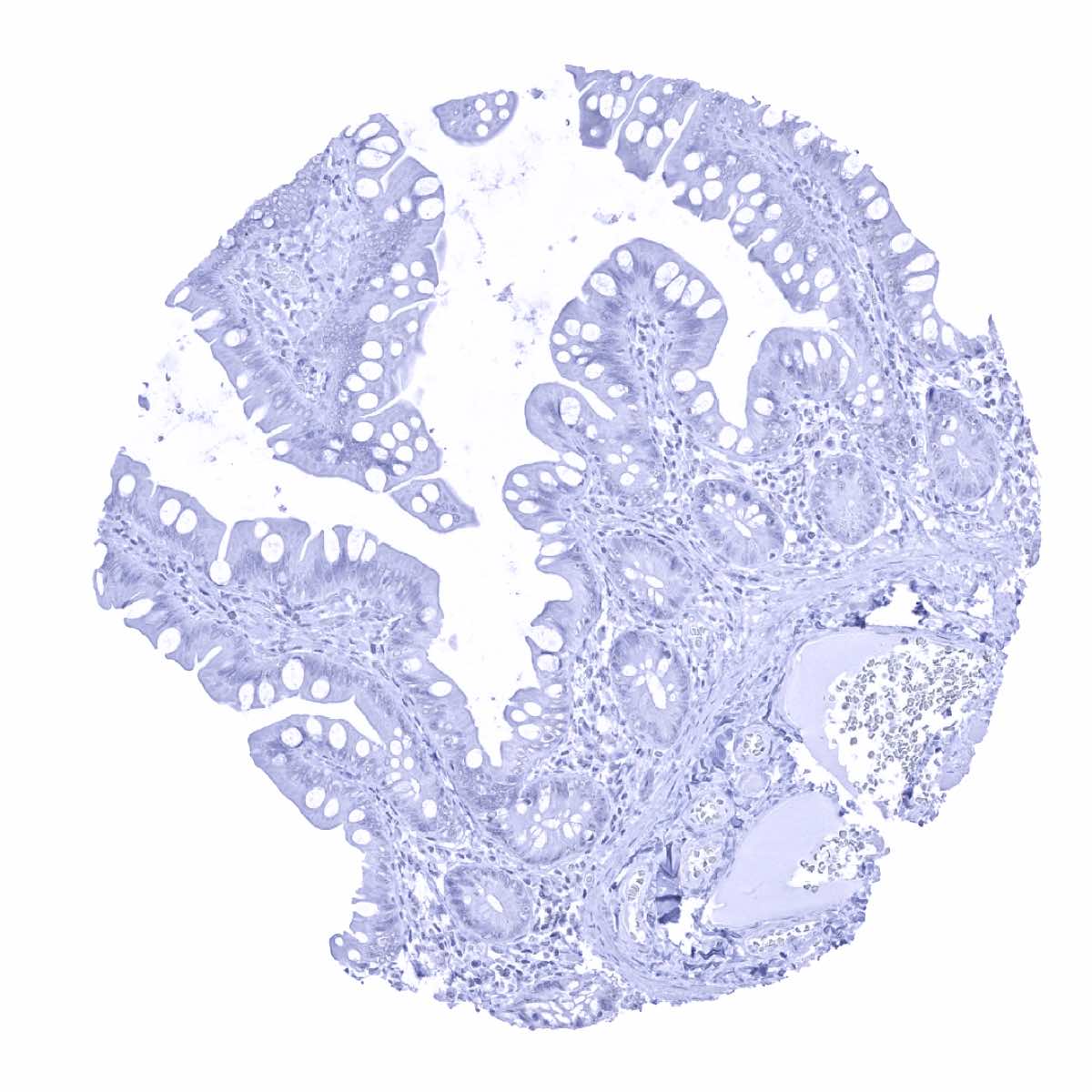
Colon descendens, mucosa
Colon descendens, muscular wall
Duodenum, Brunner gland
Duodenum, mucosa
Endocervix
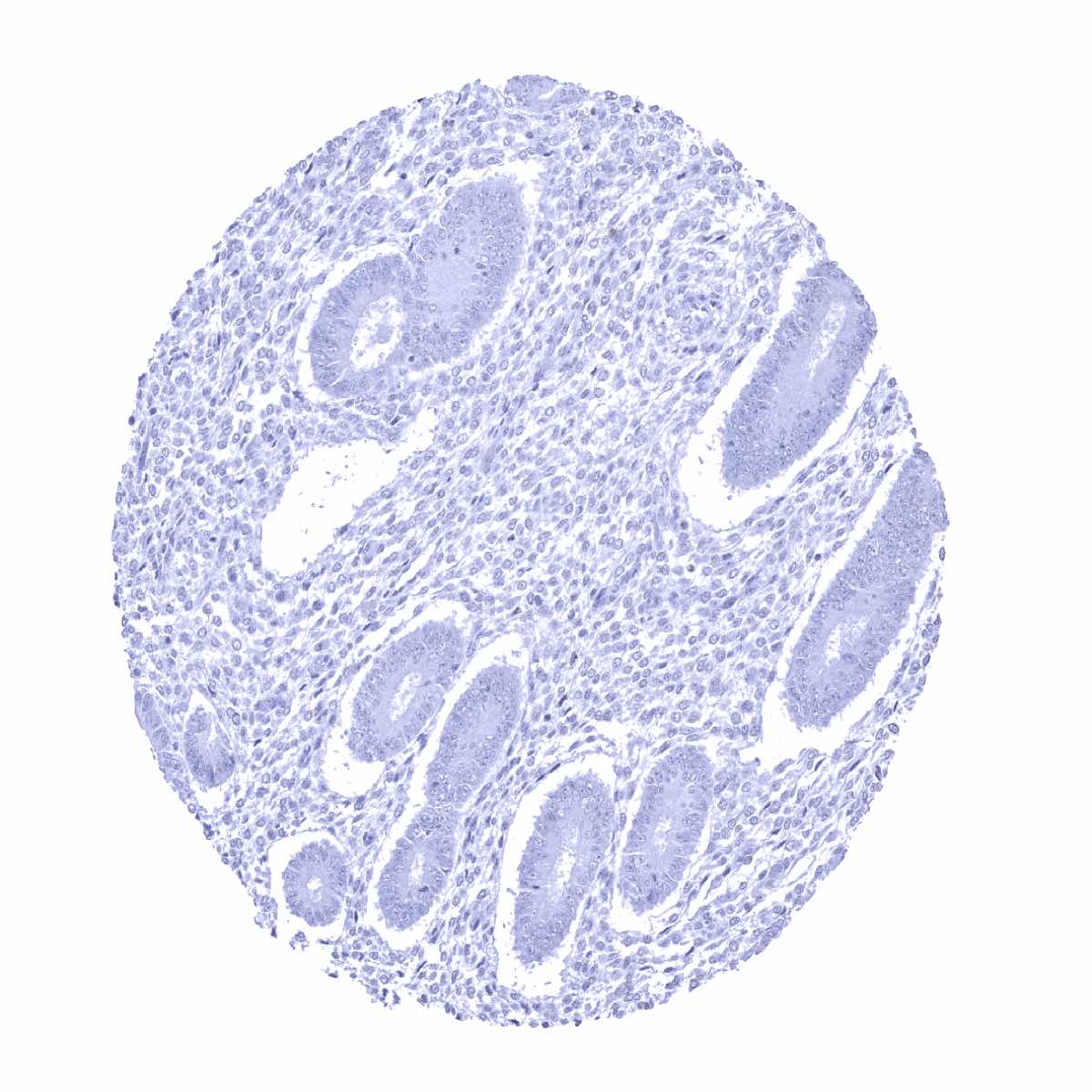
Endometrium, proliferation

Endometrium, secretion
Epididymis
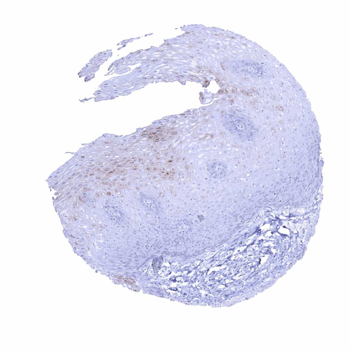
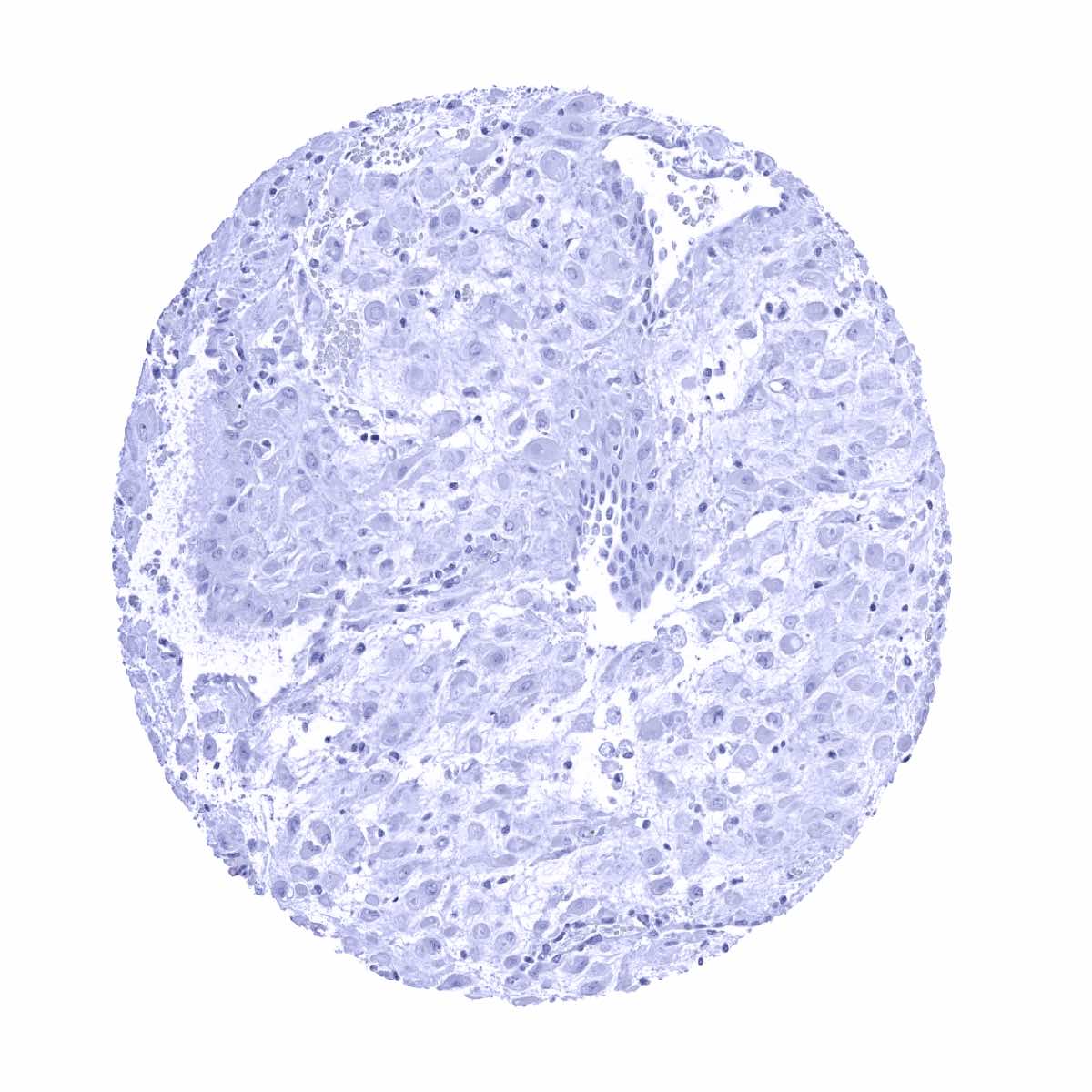
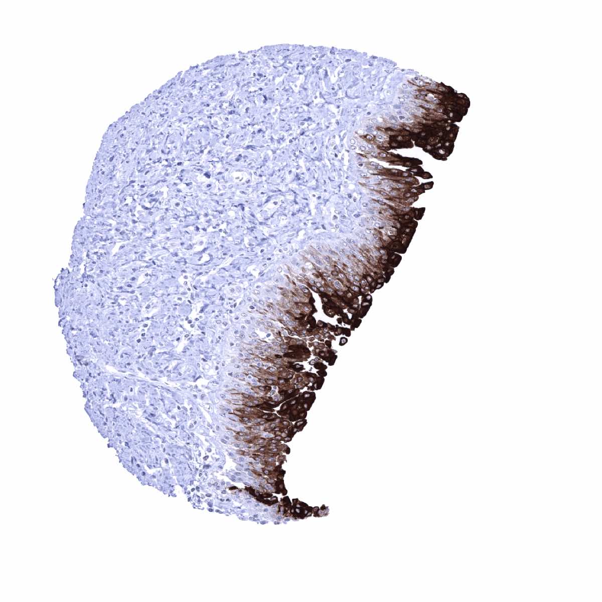
Esophagus, squamous epithelium - Weak Upk1A staining in distinct cell layers (middle third) of the squamous epithelium
Fallopian tube, mucosa
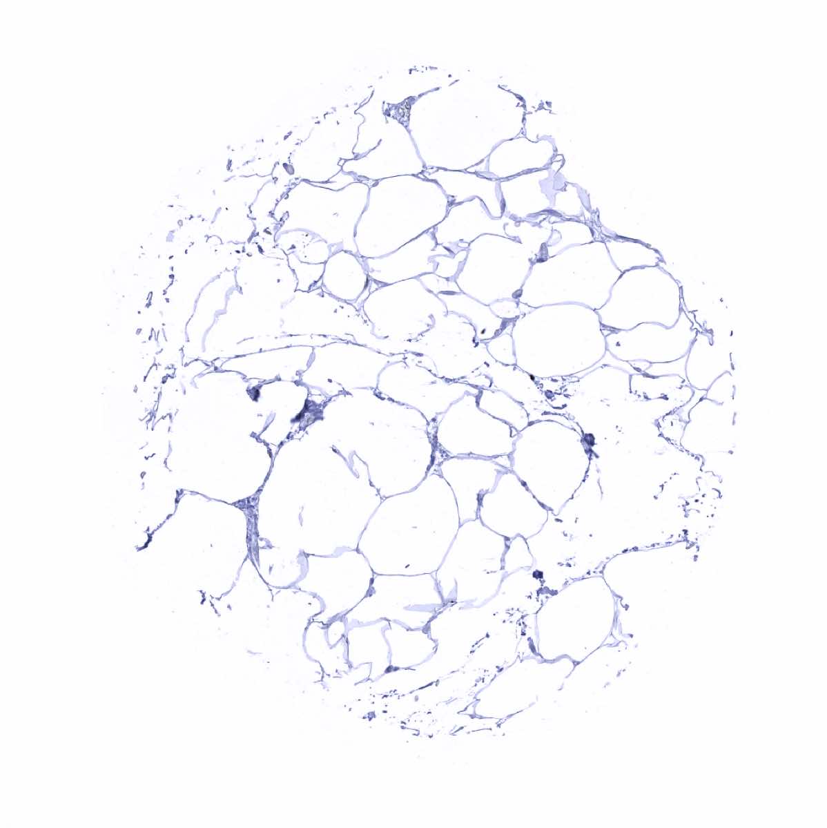
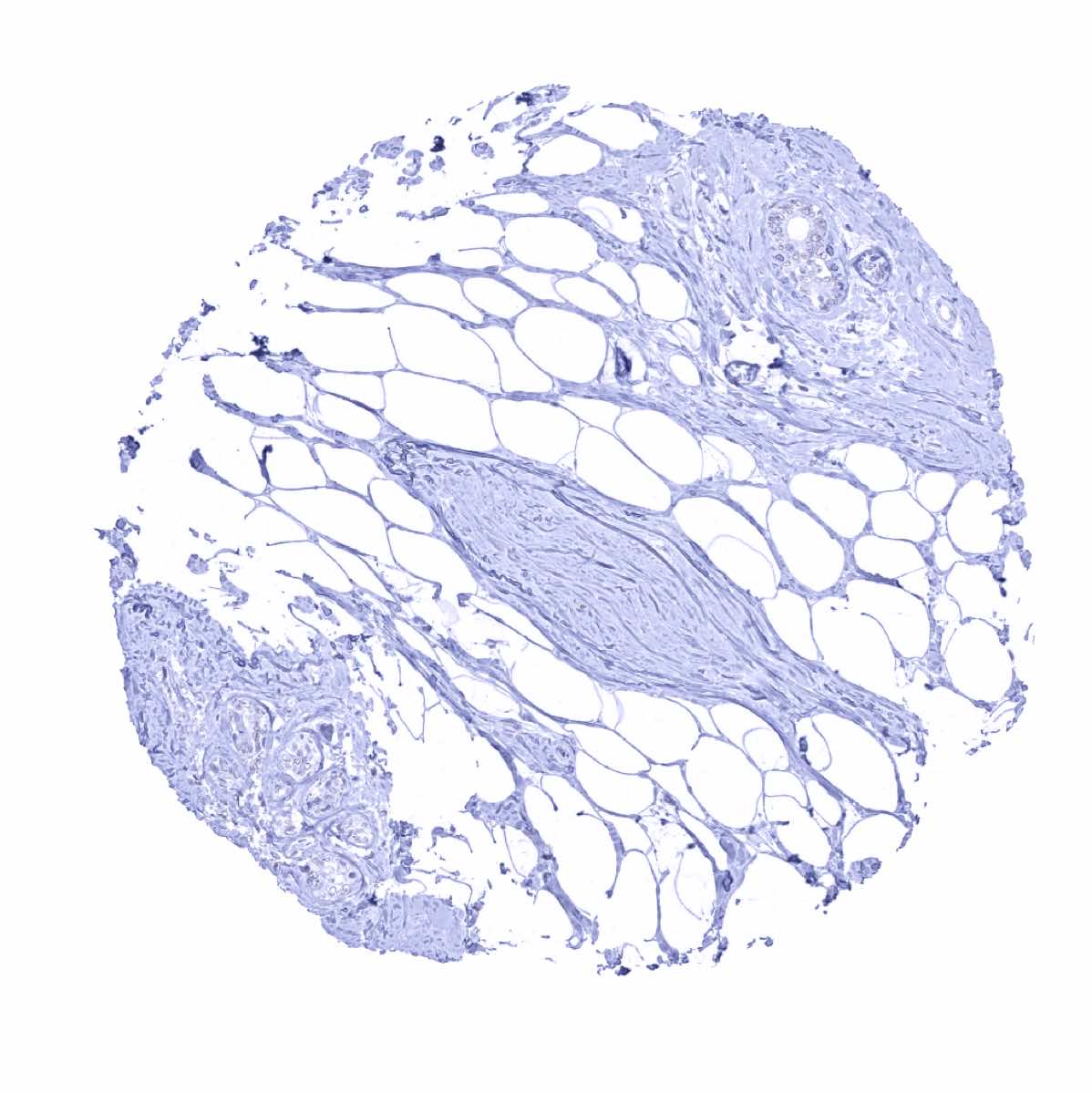
Fat
Gallbladder, epithelium
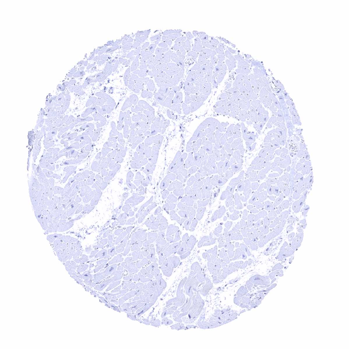
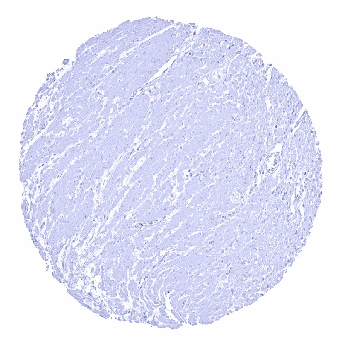
Heart
Ileum, mucosa
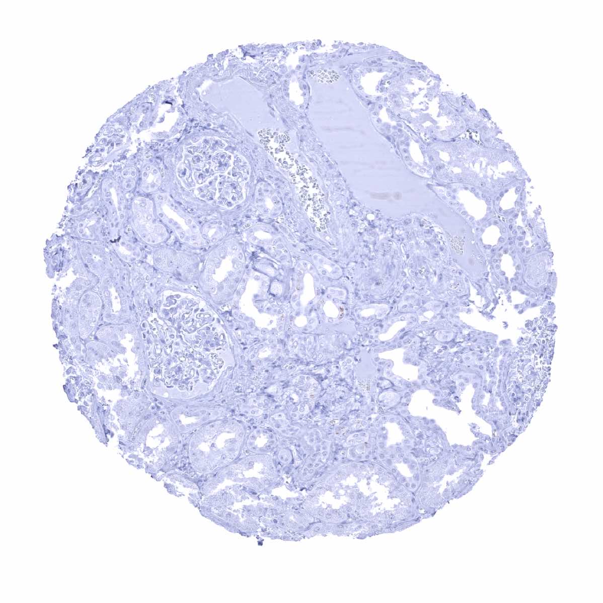
Kidney, cortex

Kidney, medulla
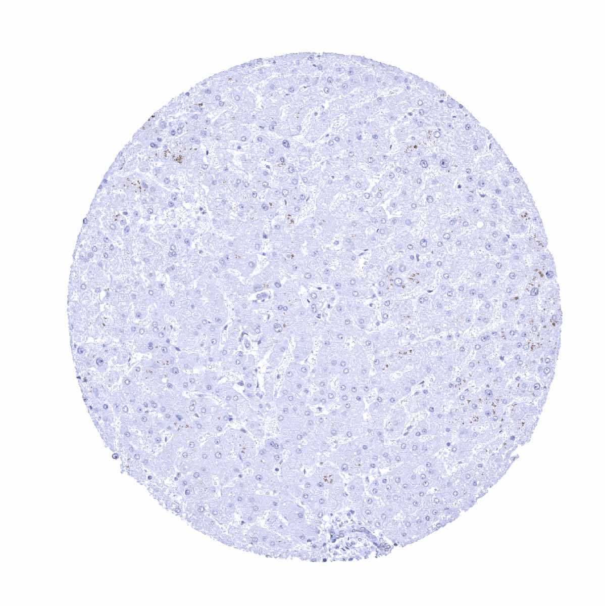
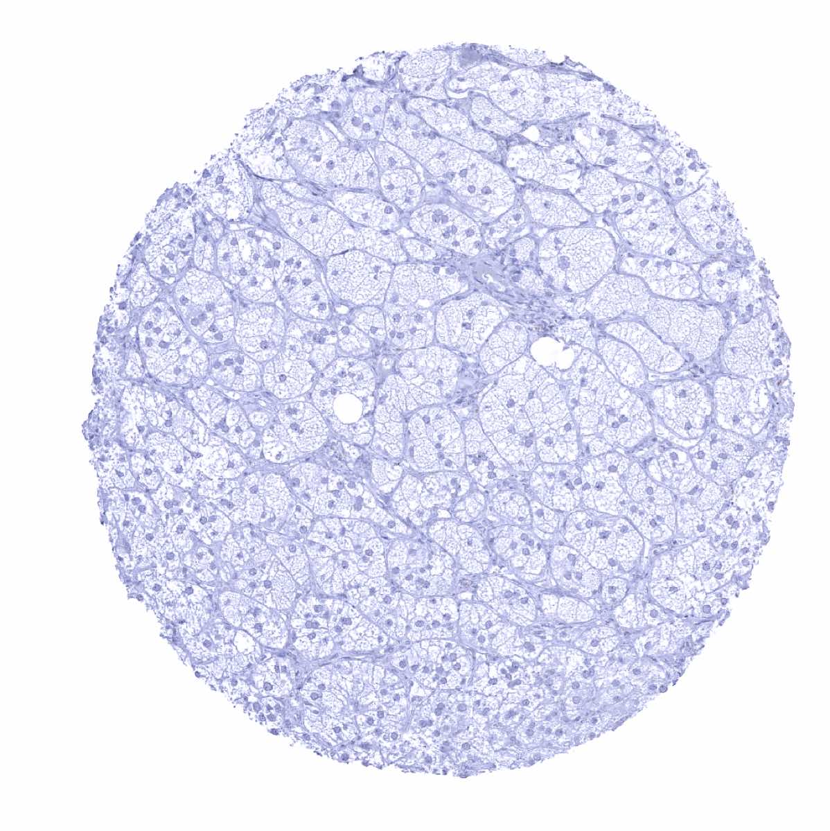
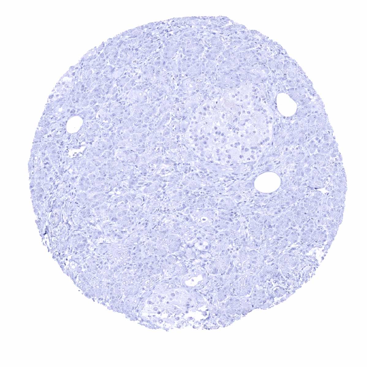
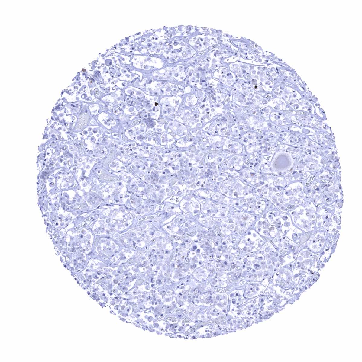
Liver
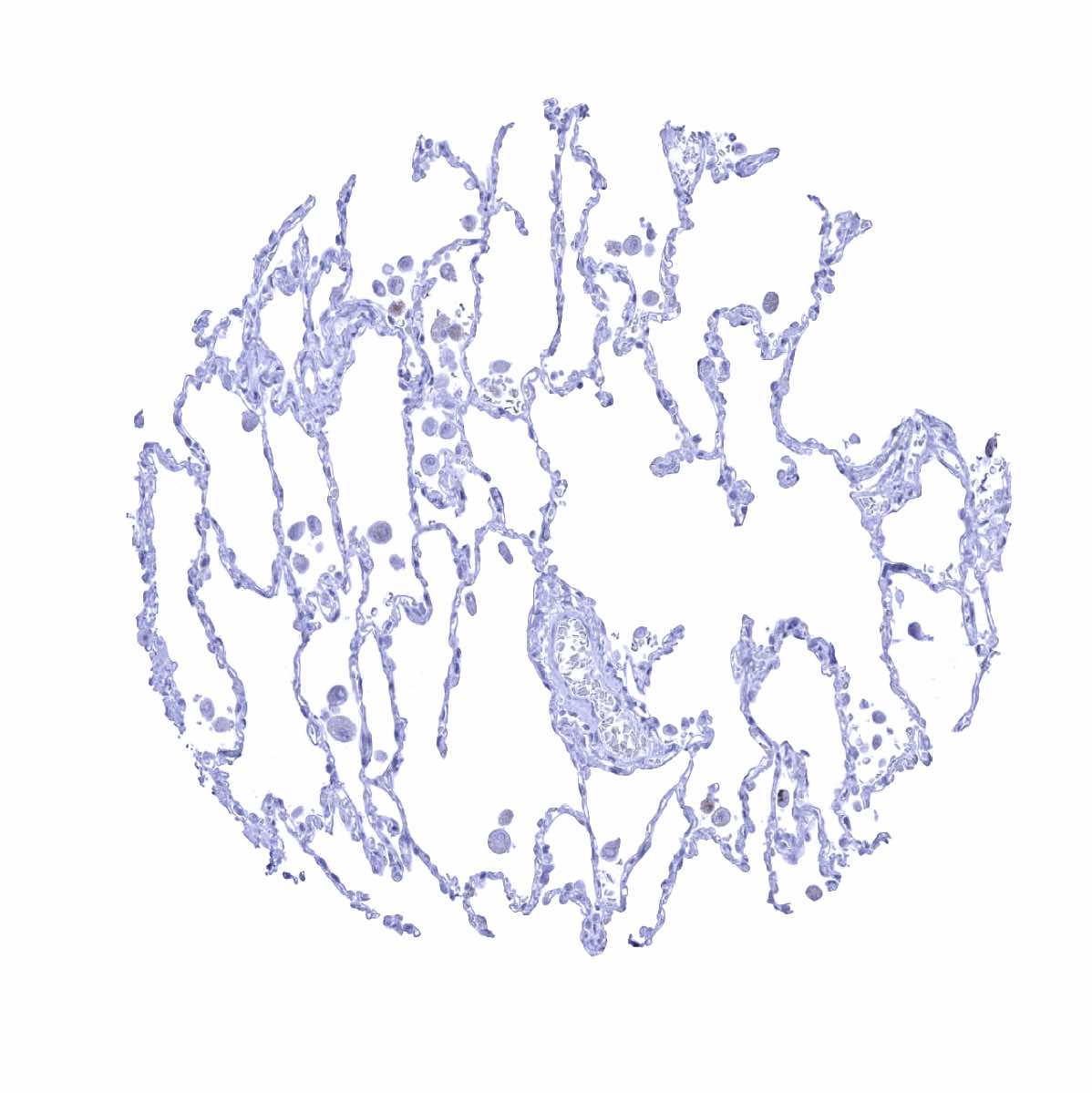
Lung
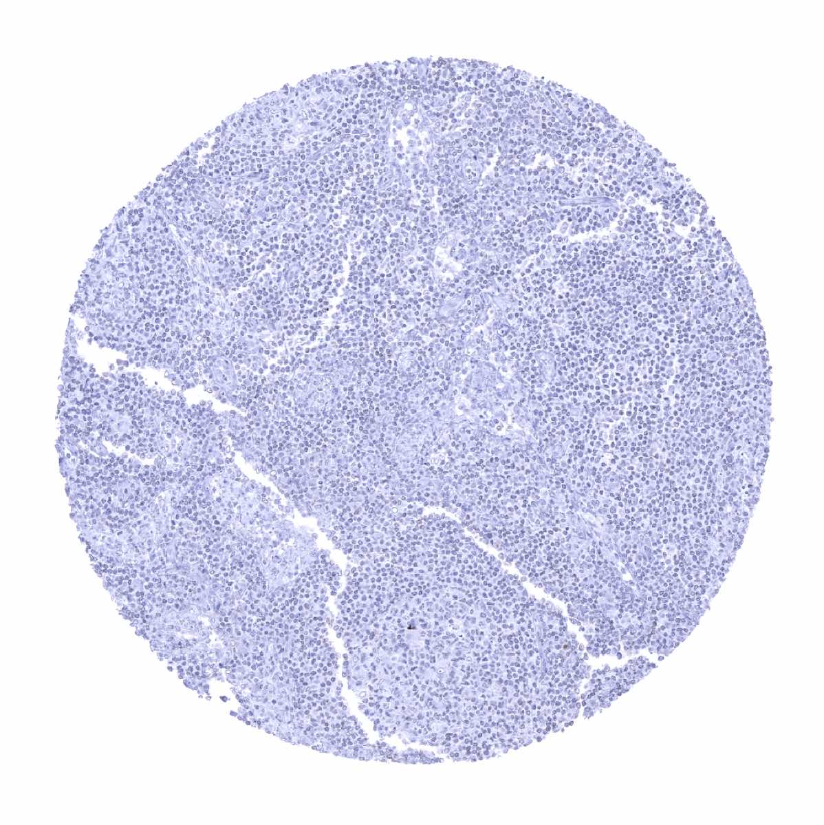
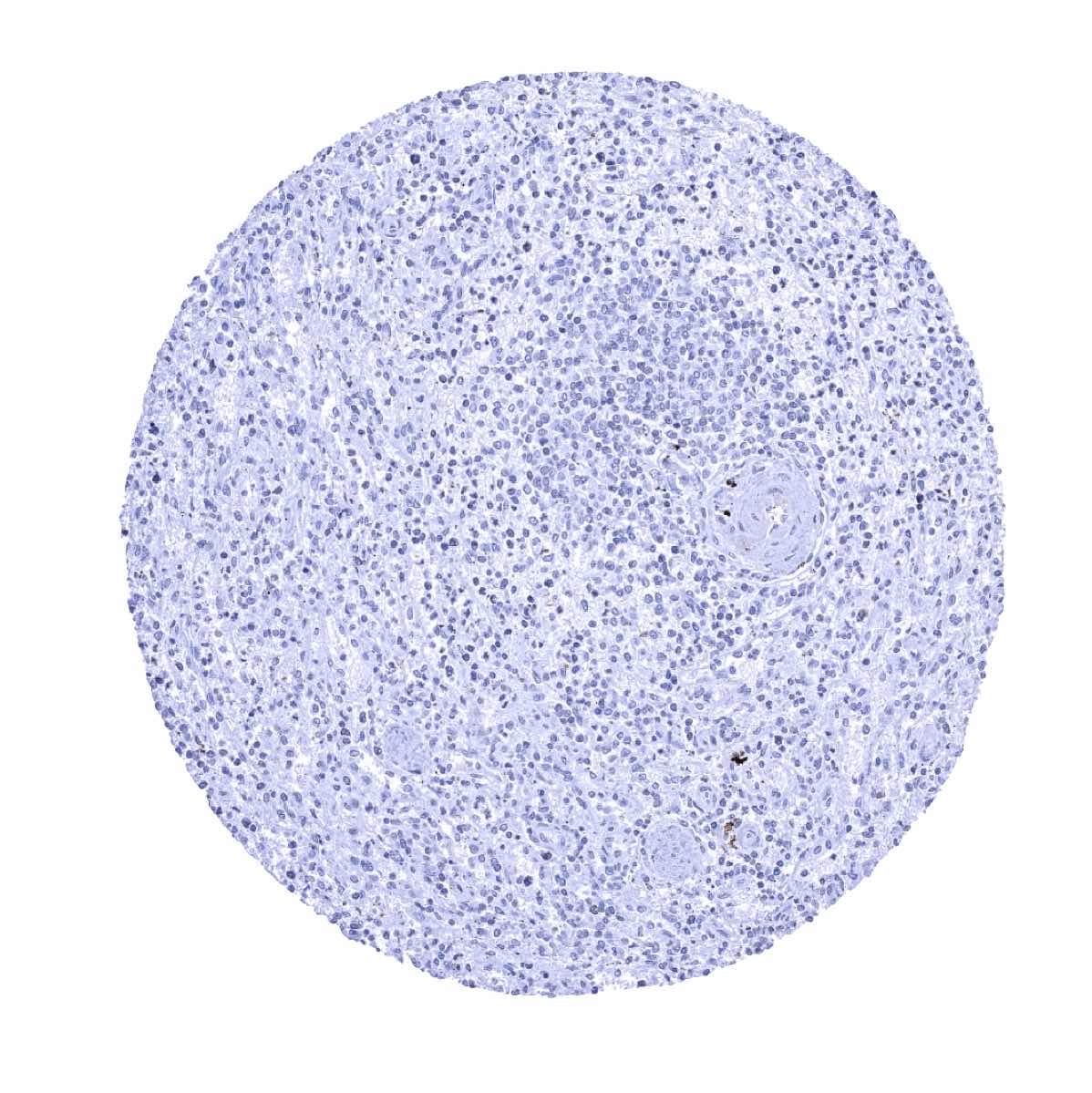
Lymph node
Ovary, stroma
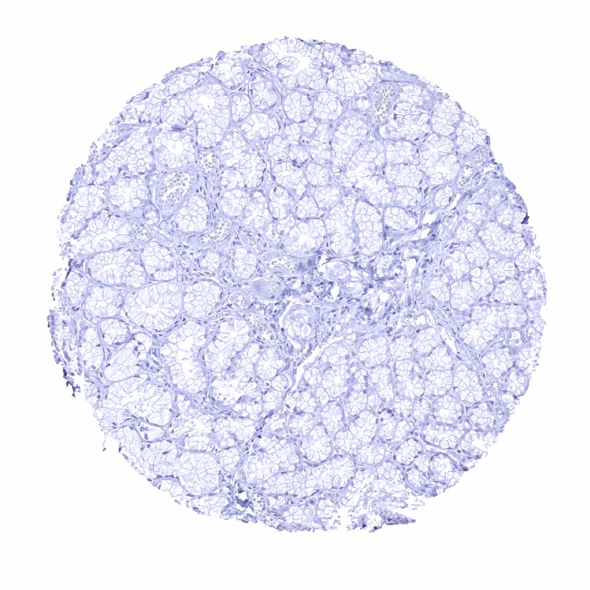
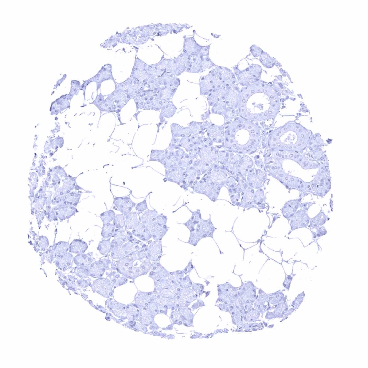
Pancreas
Parathyroid
Parotid gland
Pituitary, anterior lobe
Pituitary gland, posterior lobe
Placenta early, decidua
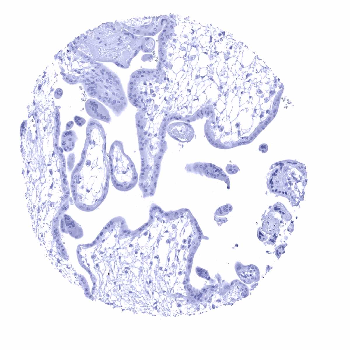
Placenta, early
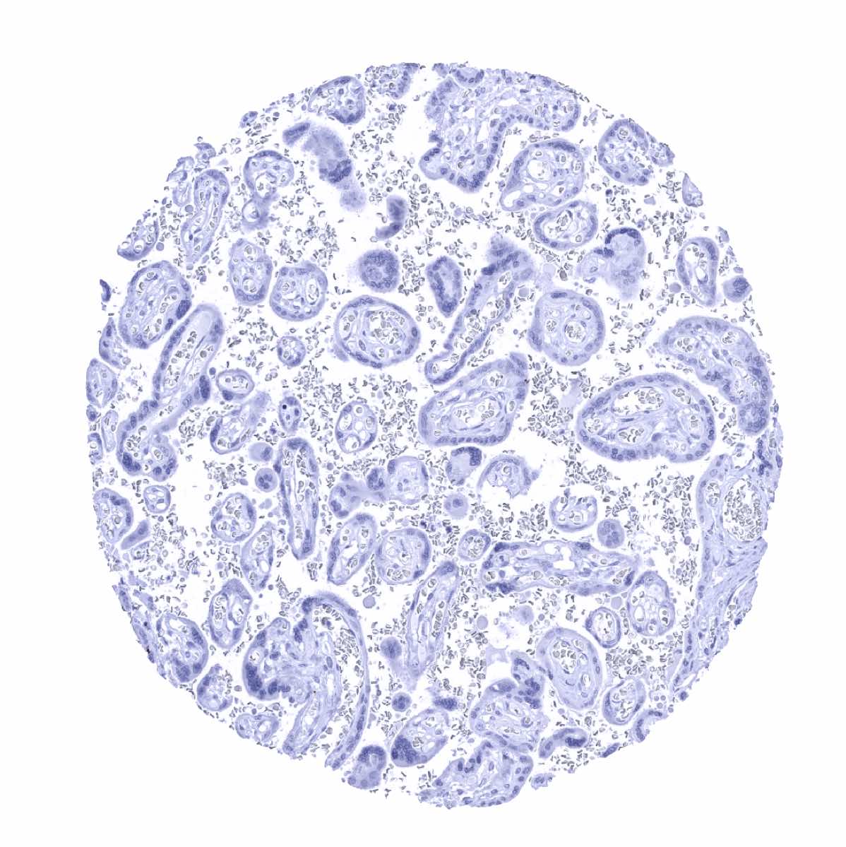
Placenta, mature
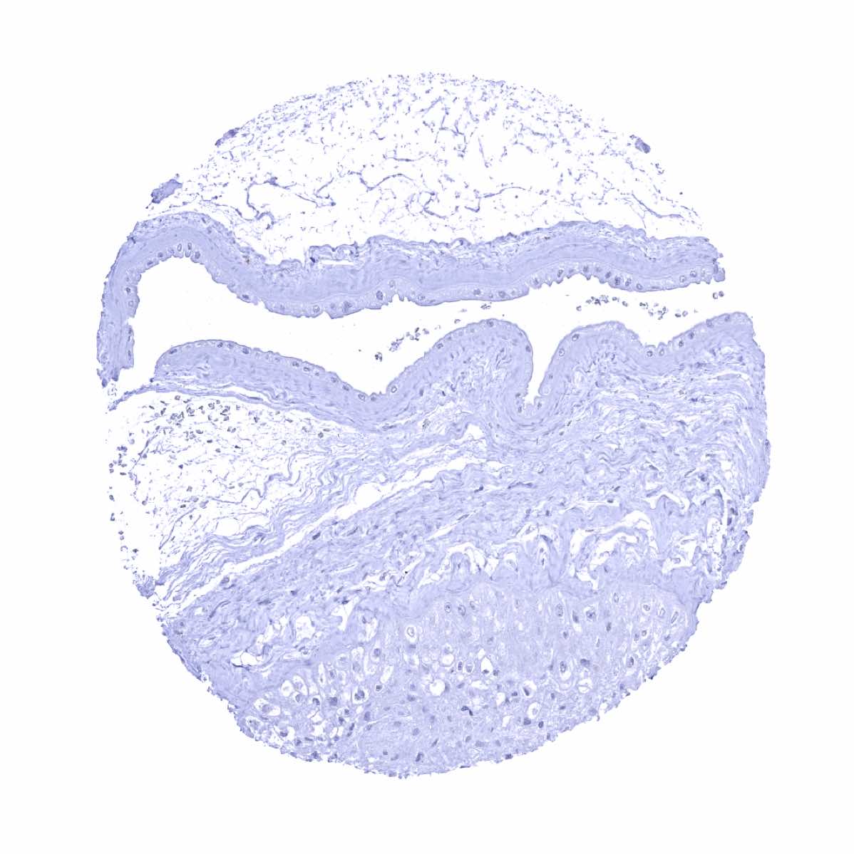
Placenta, mature, amnion and chorion

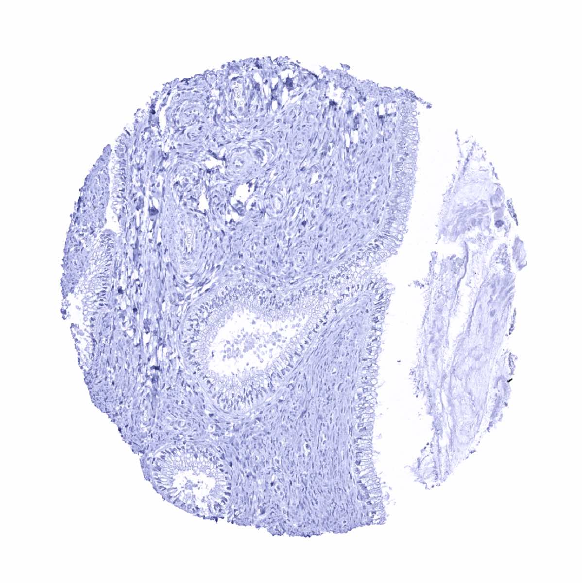
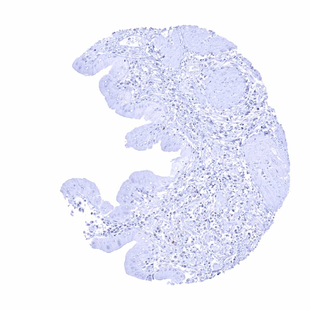
Prostate - Weak to moderate Upk1A staining in a squamous metaplasia of prostatic epithelial cells
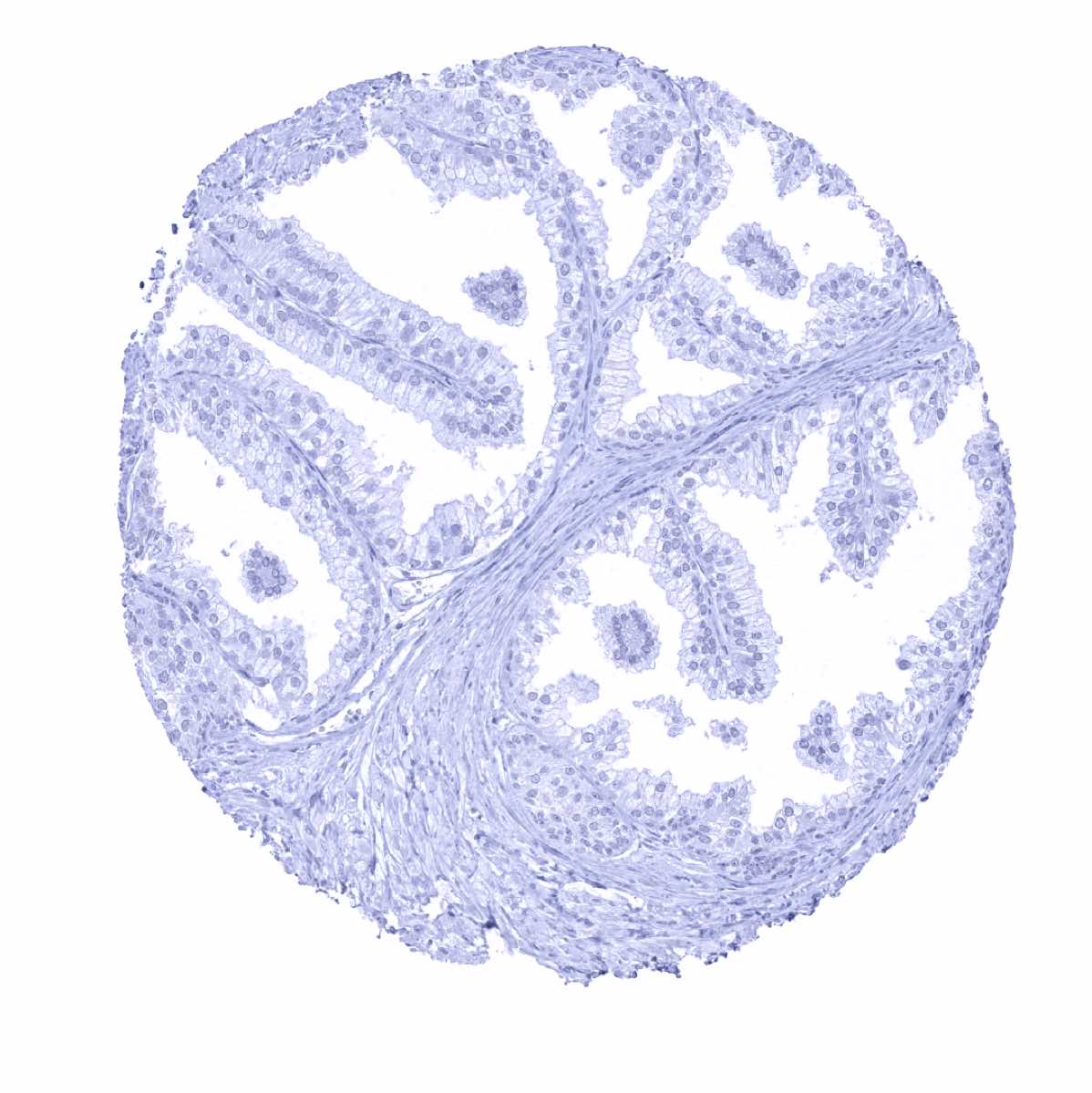
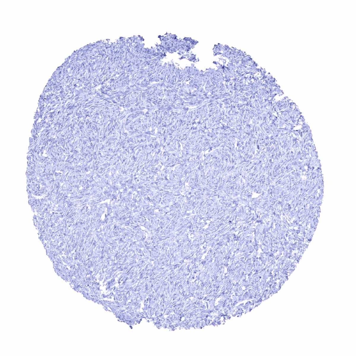
Prostate
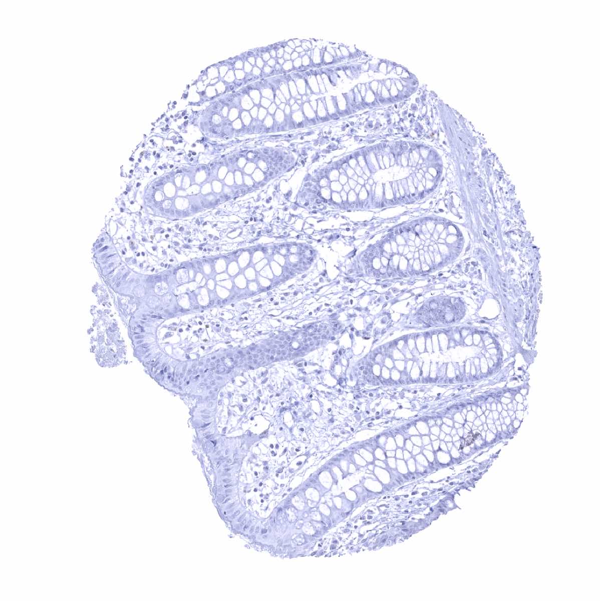
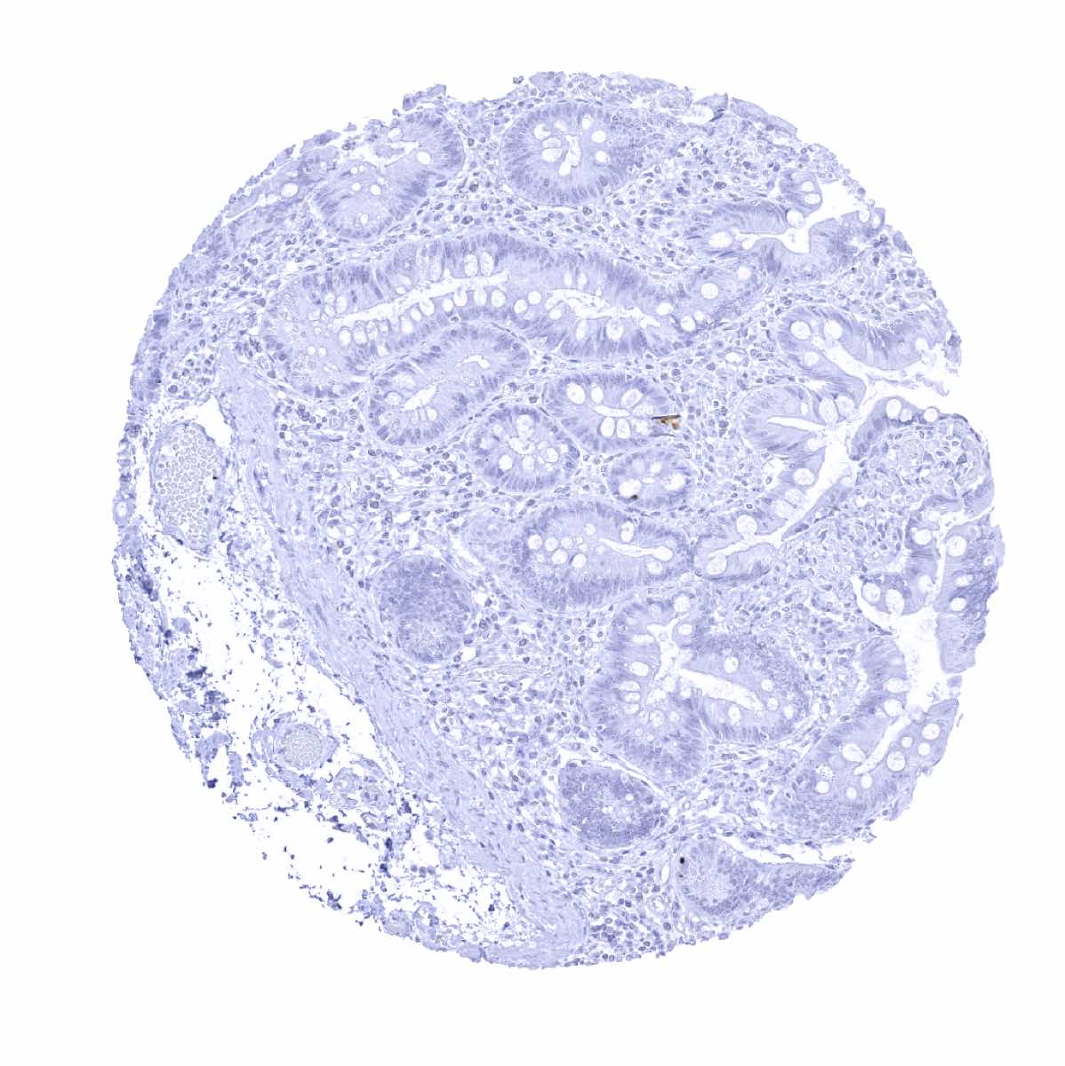
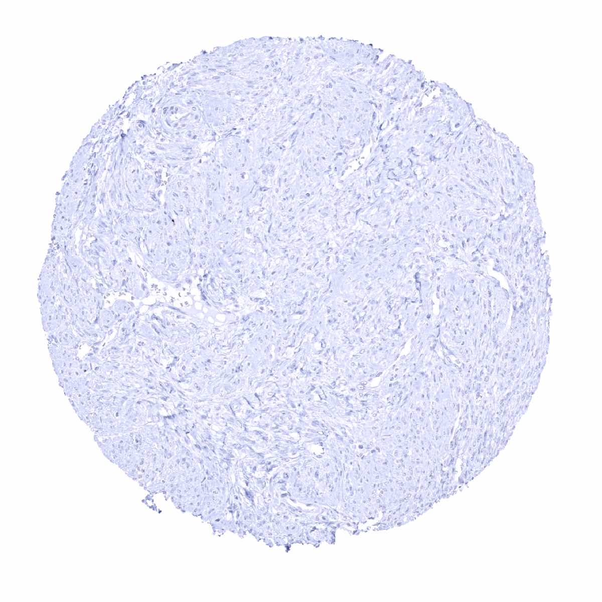
Rectum, mucosa - Complete absence of Upk1A immunostaining
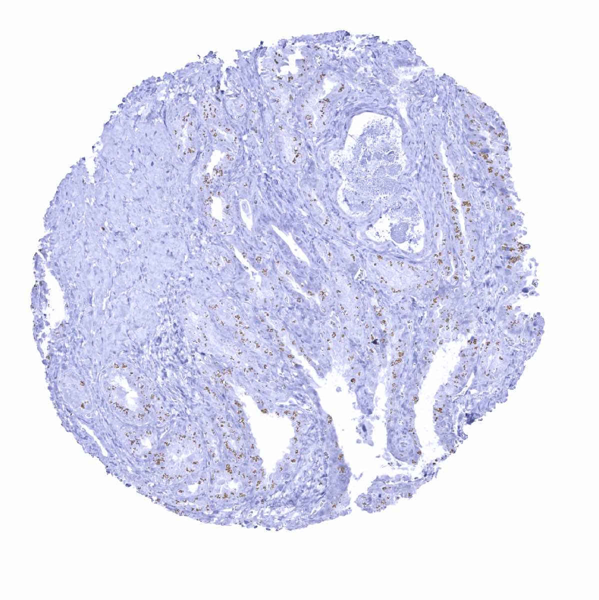
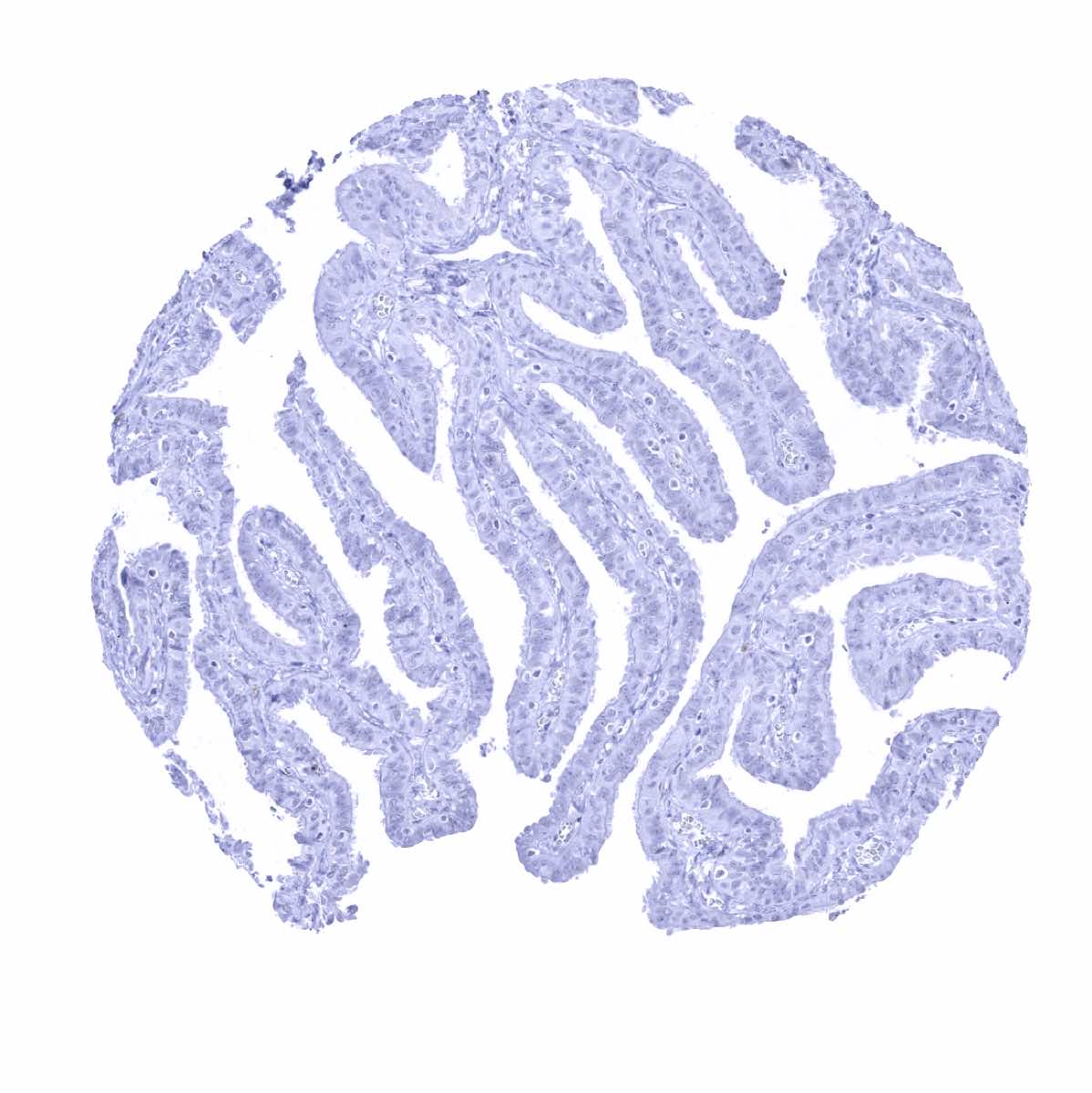
Seminal vesicle - Strong Upk1A immunostaining of few epithelial cells in the seminal vesicle (perhaps representing urothelium)
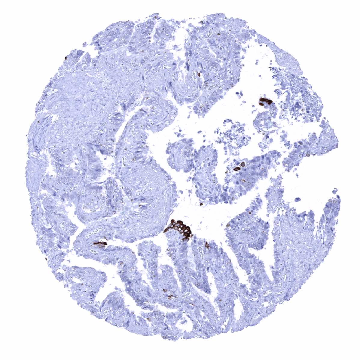
Seminal vesicle

Sinus paranasales

Skin
Spleen

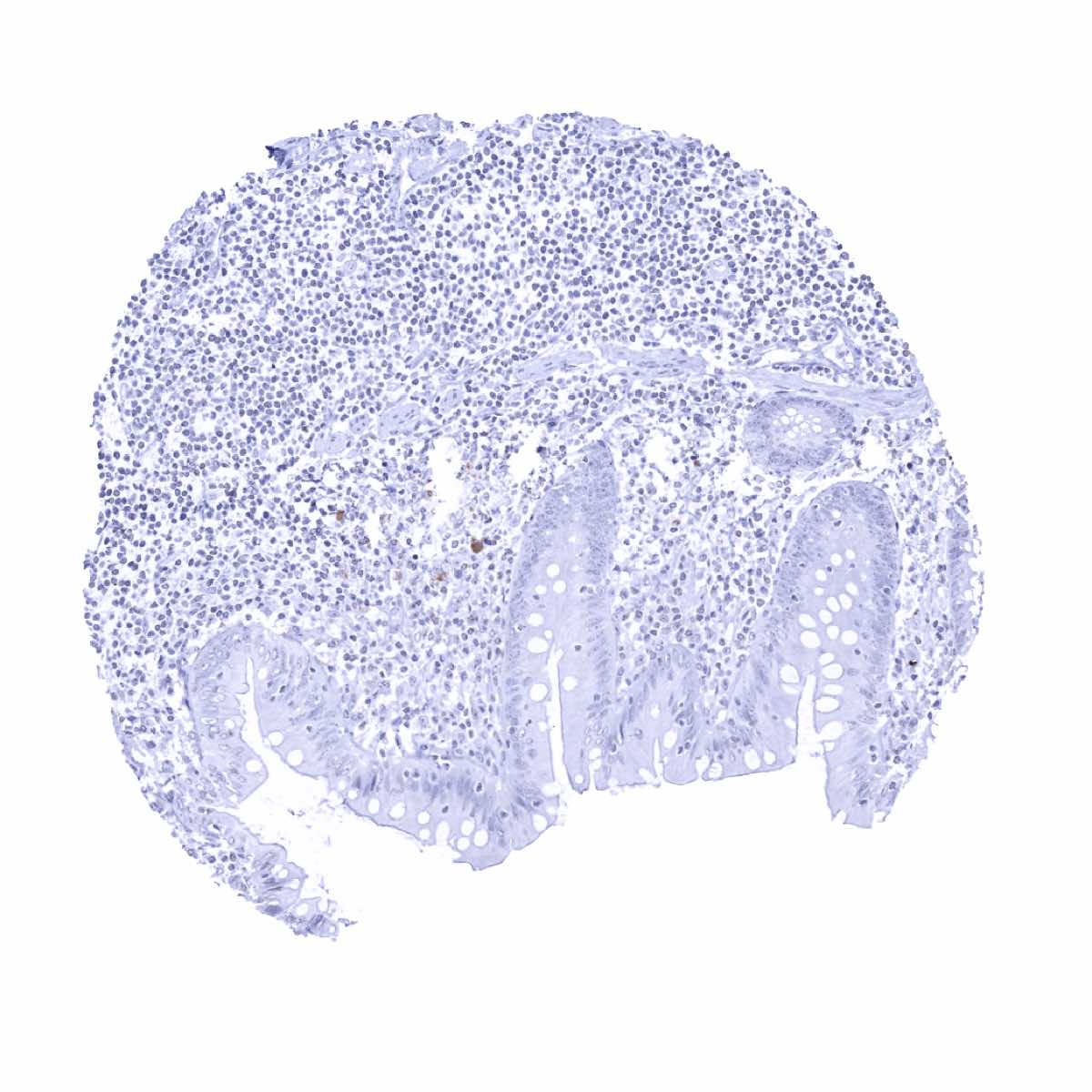
Stomach, antrum

Stomach, corpus

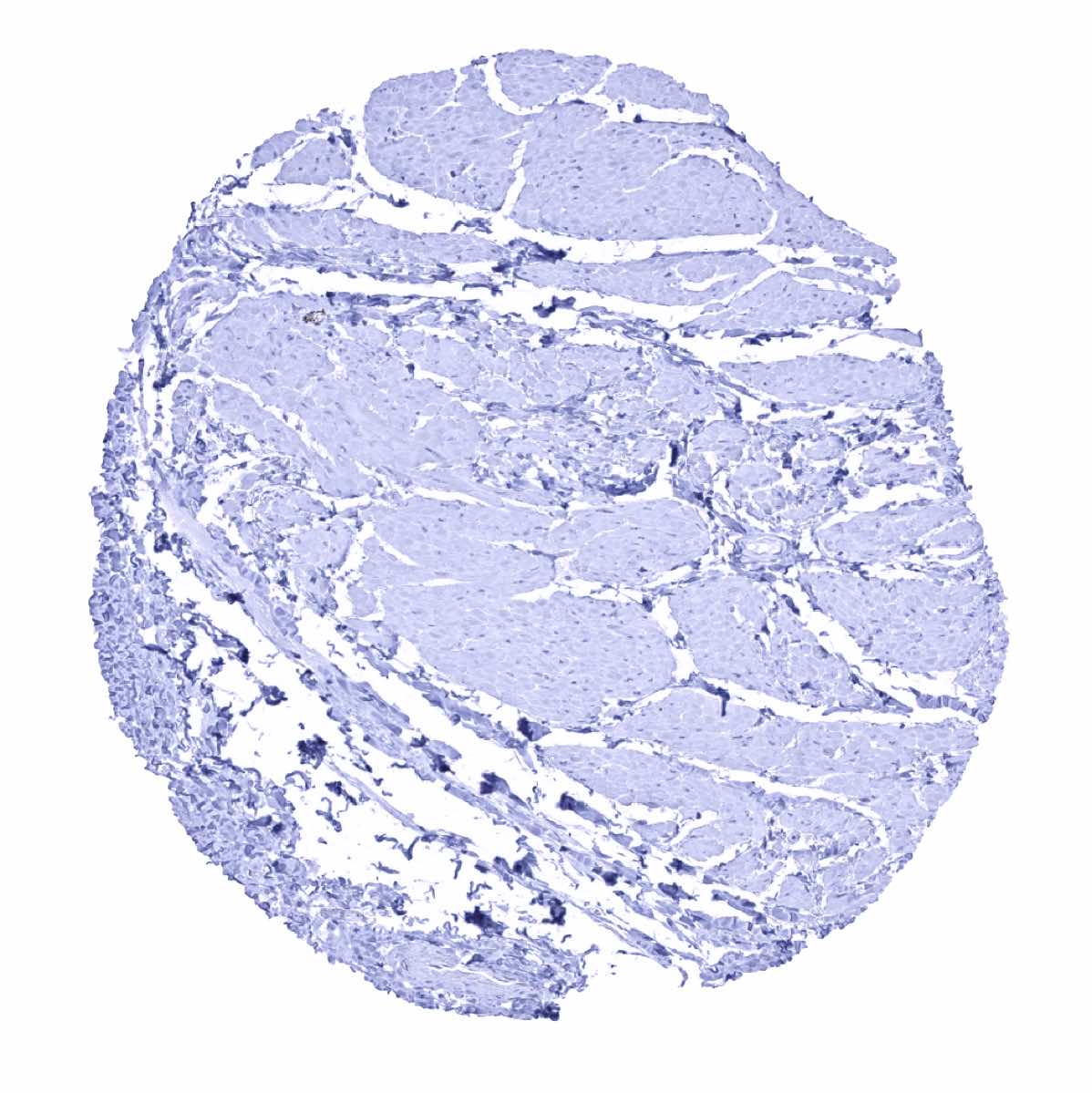
Striated muscle
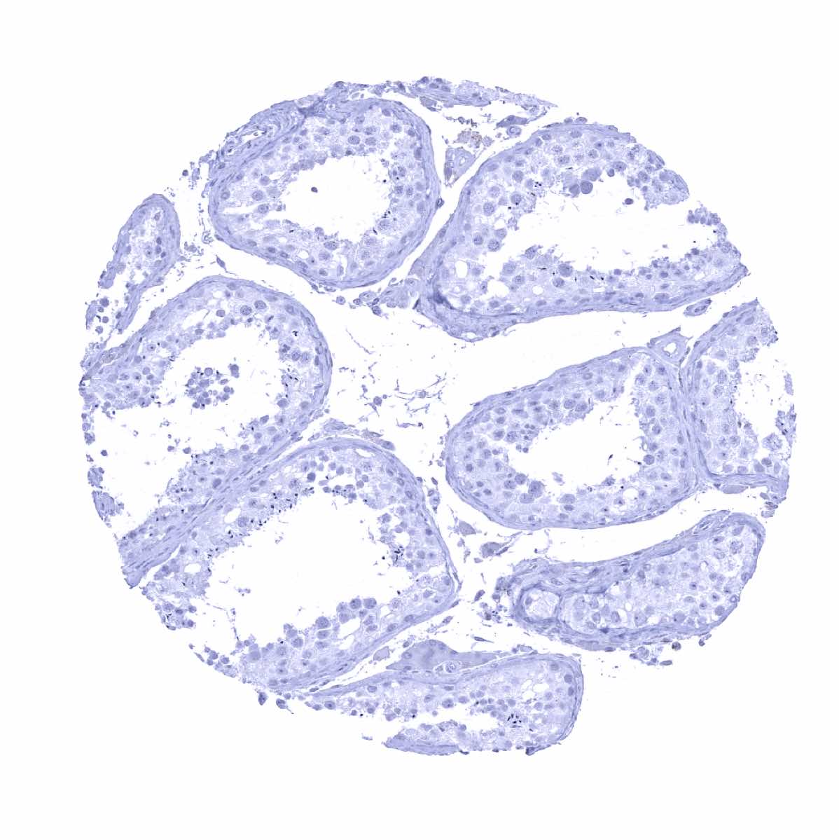
Testis
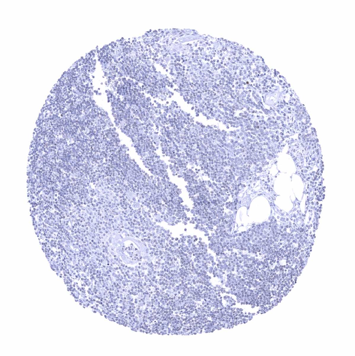
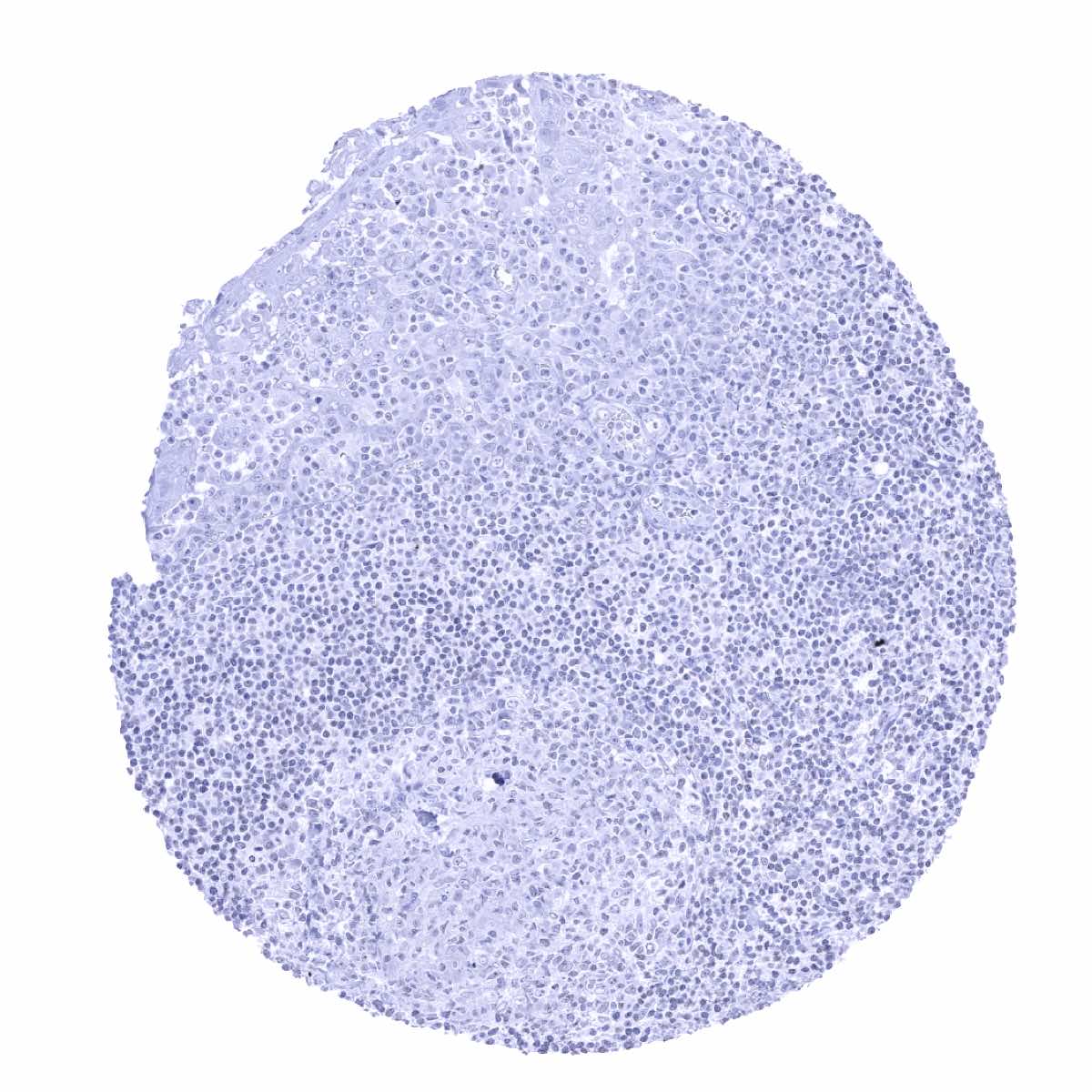
Thymus

Thyroid gland
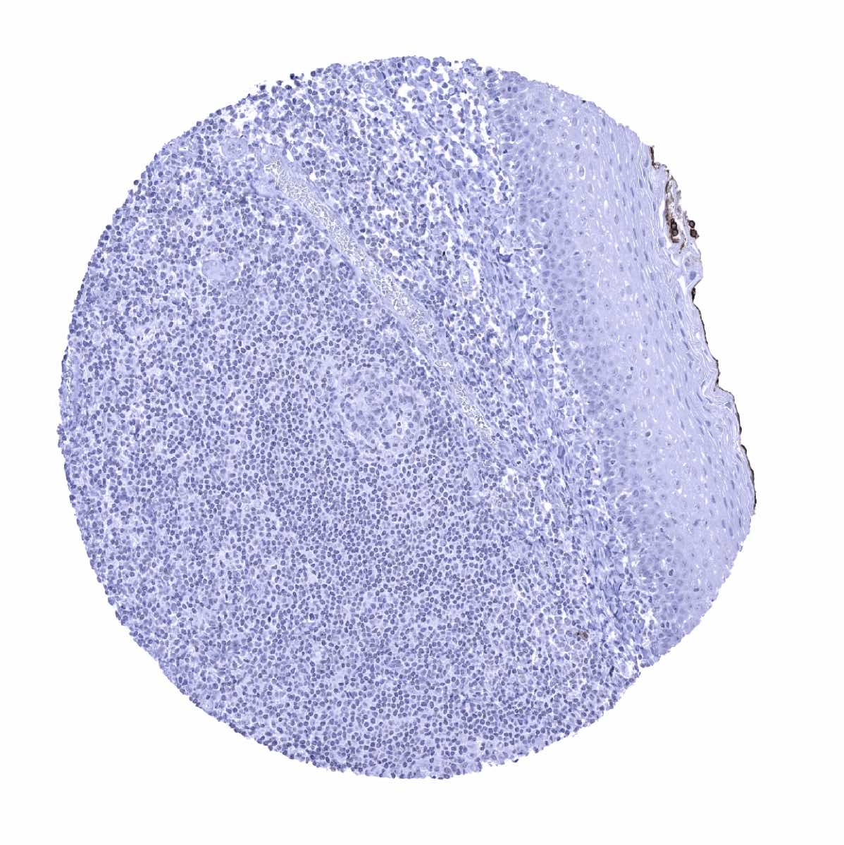
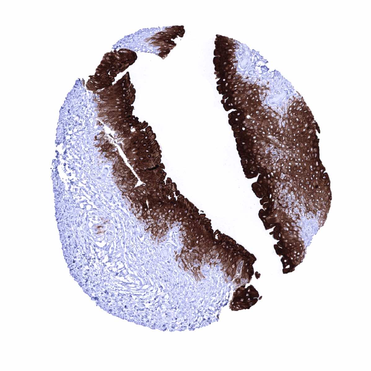
Tonsil, surface epithelium
Tonsil
Urinary bladder, muscular wall
Urinary bladder, urothelium - Strong Upk1A immunostaining of the top 2/3 of the urothelium. The basal cell layers do not show staining
Urinary bladder, urothelium - Strong Upk1A immunostaining of the urothelium. The staining intensity decreases to some extent toward the basal cell layers

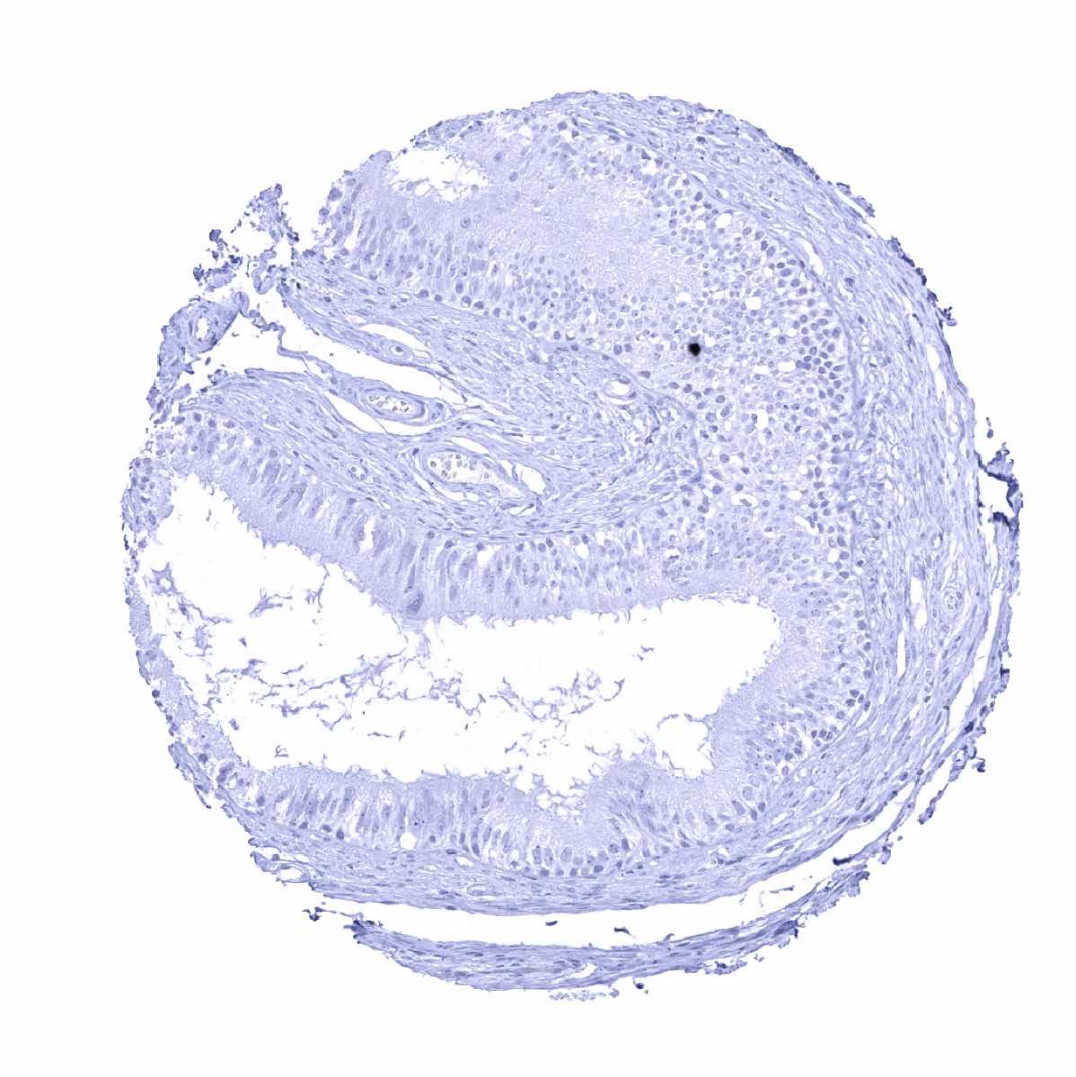
Uterus, ectocervix - Weak to moderate Upk1A staining in distinct cell layers (middle third) of the squamous epithelium
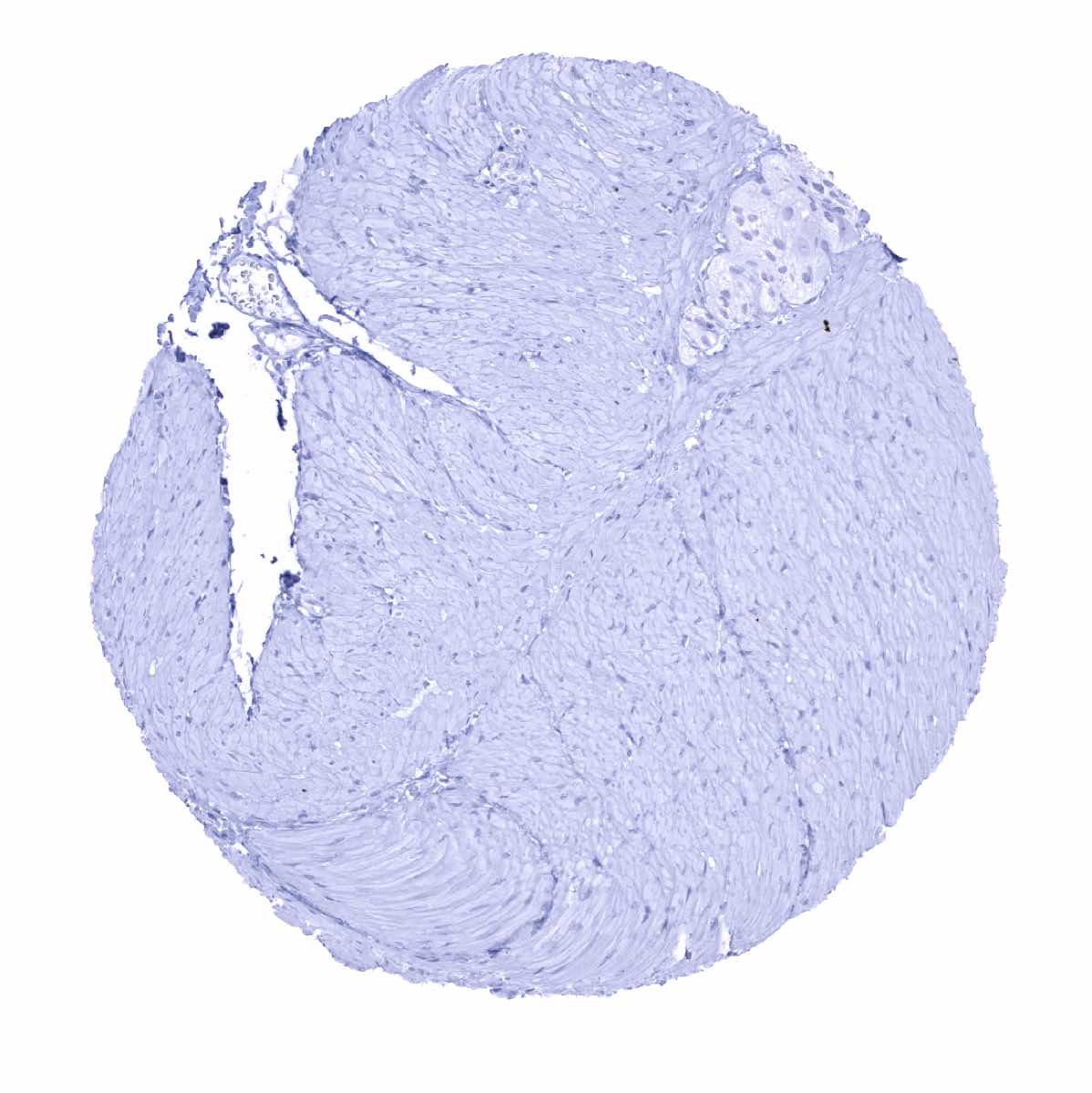
Uterus, myometrium