Adrenal gland
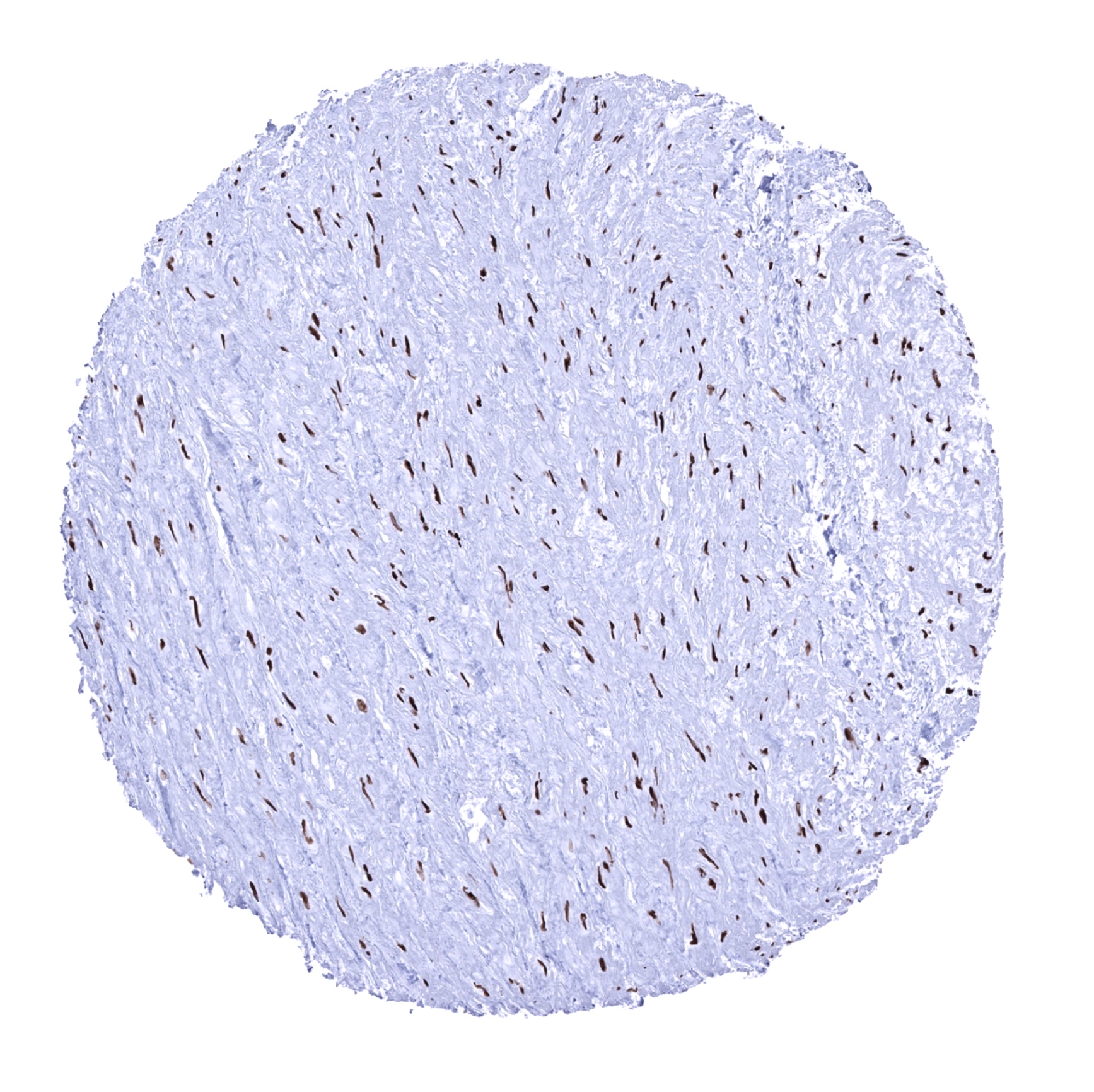
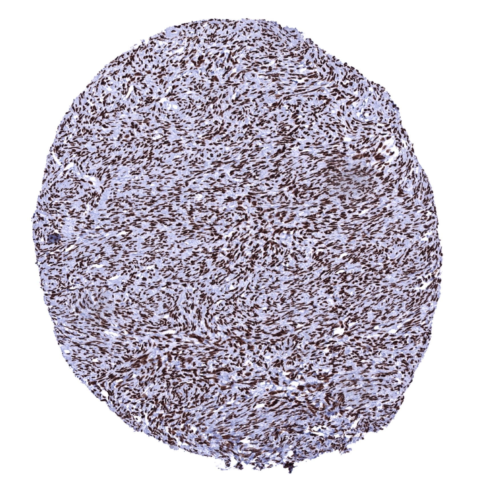
Aorta, media - A marked Ku80 staining is seen in all nuclei of all cells.
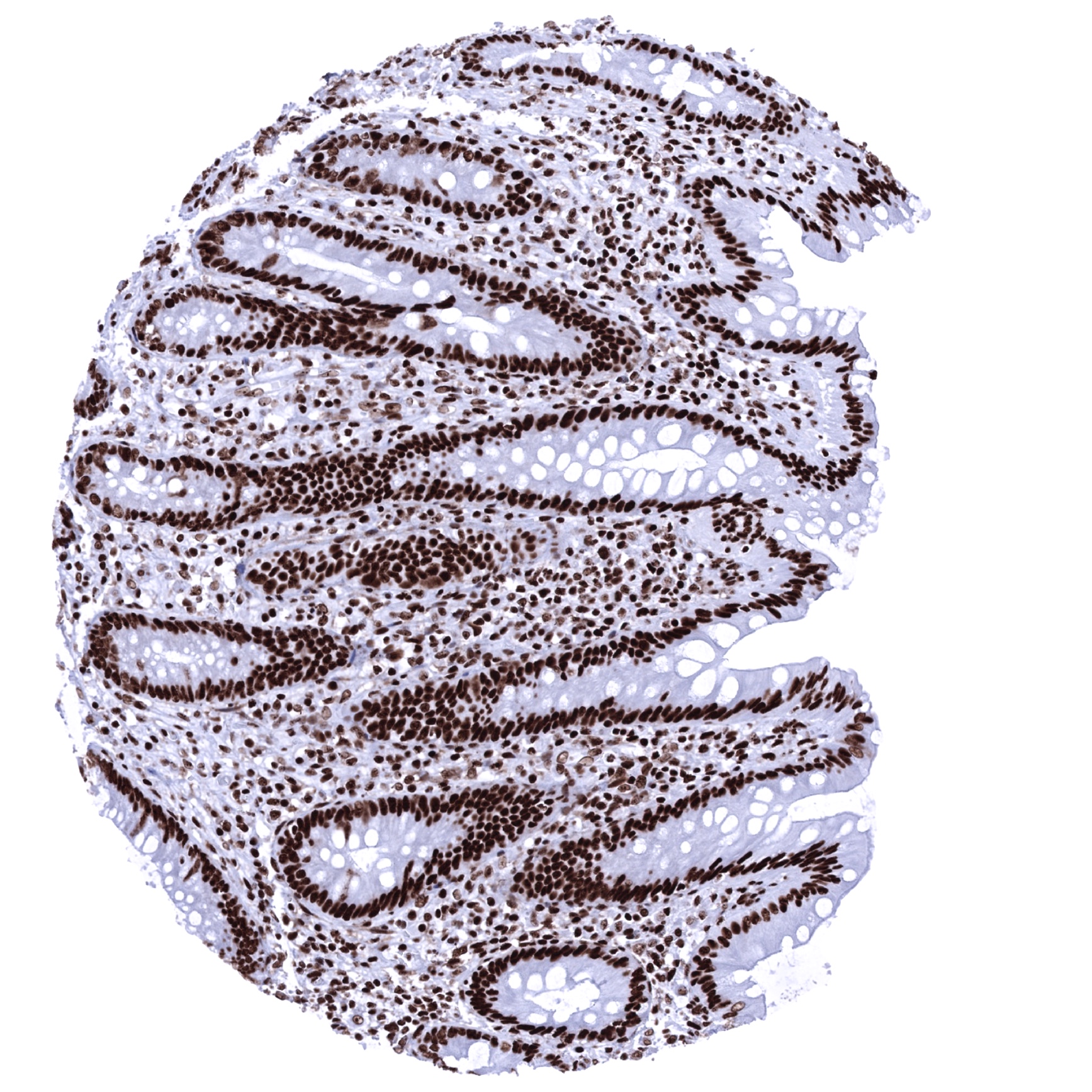
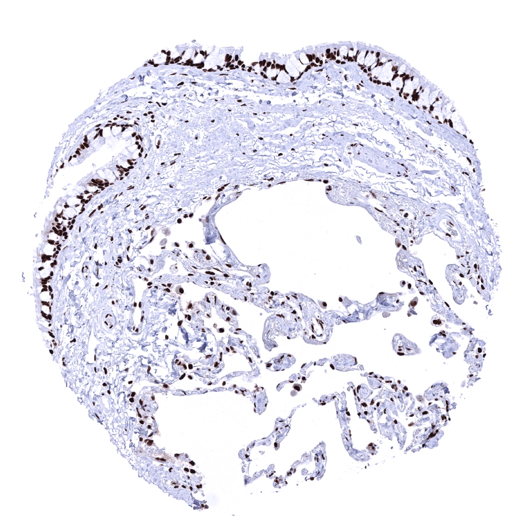
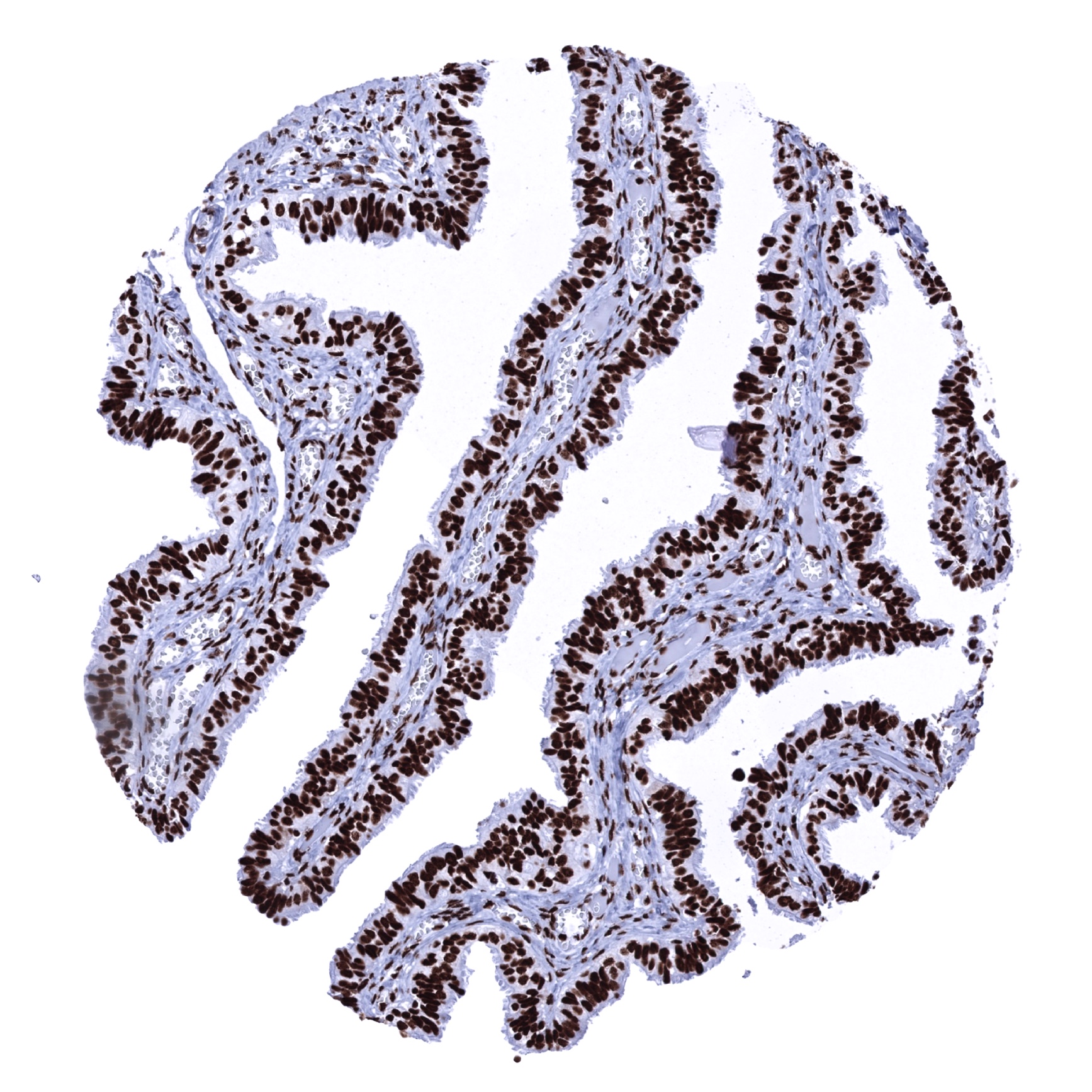
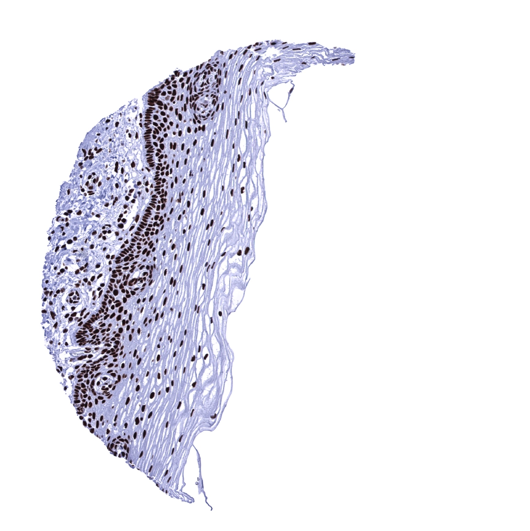
Appendix, mucosa
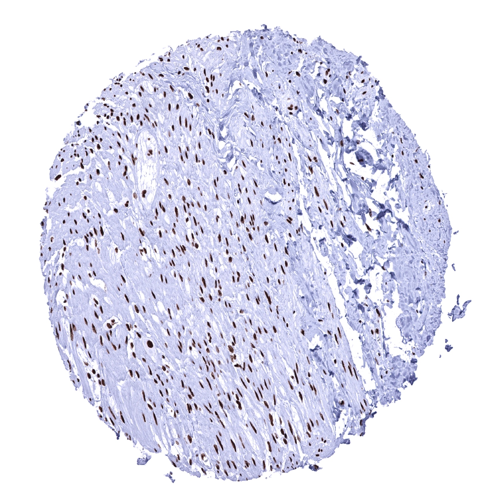
Appendix, muscular wall

Bone marrow

Breast
Bronchus, glands
Bronchus, mucosa
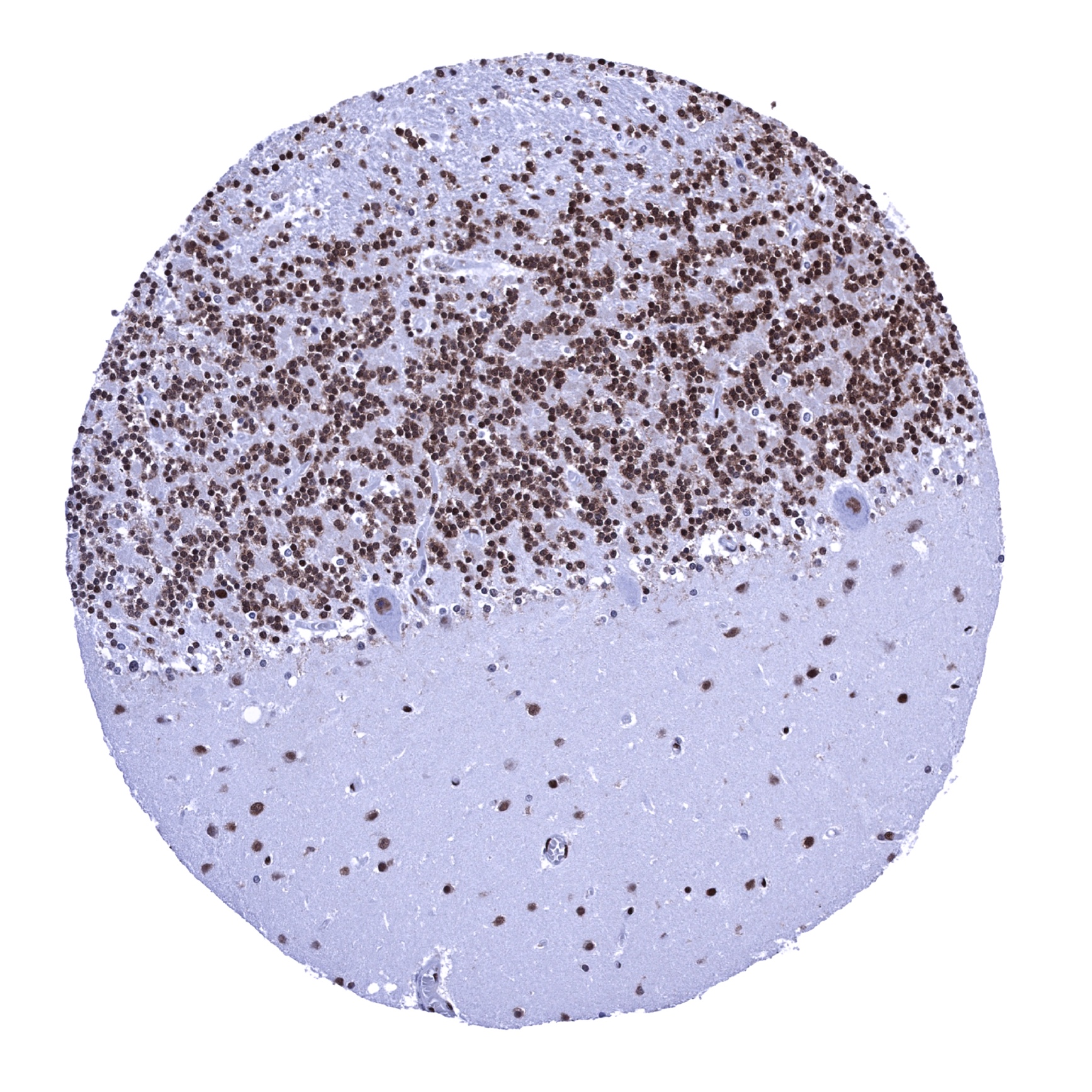
Cerebellum (molecular layer, Purkinje cell layer, granule cell layer)
Cerebellum (white matter)
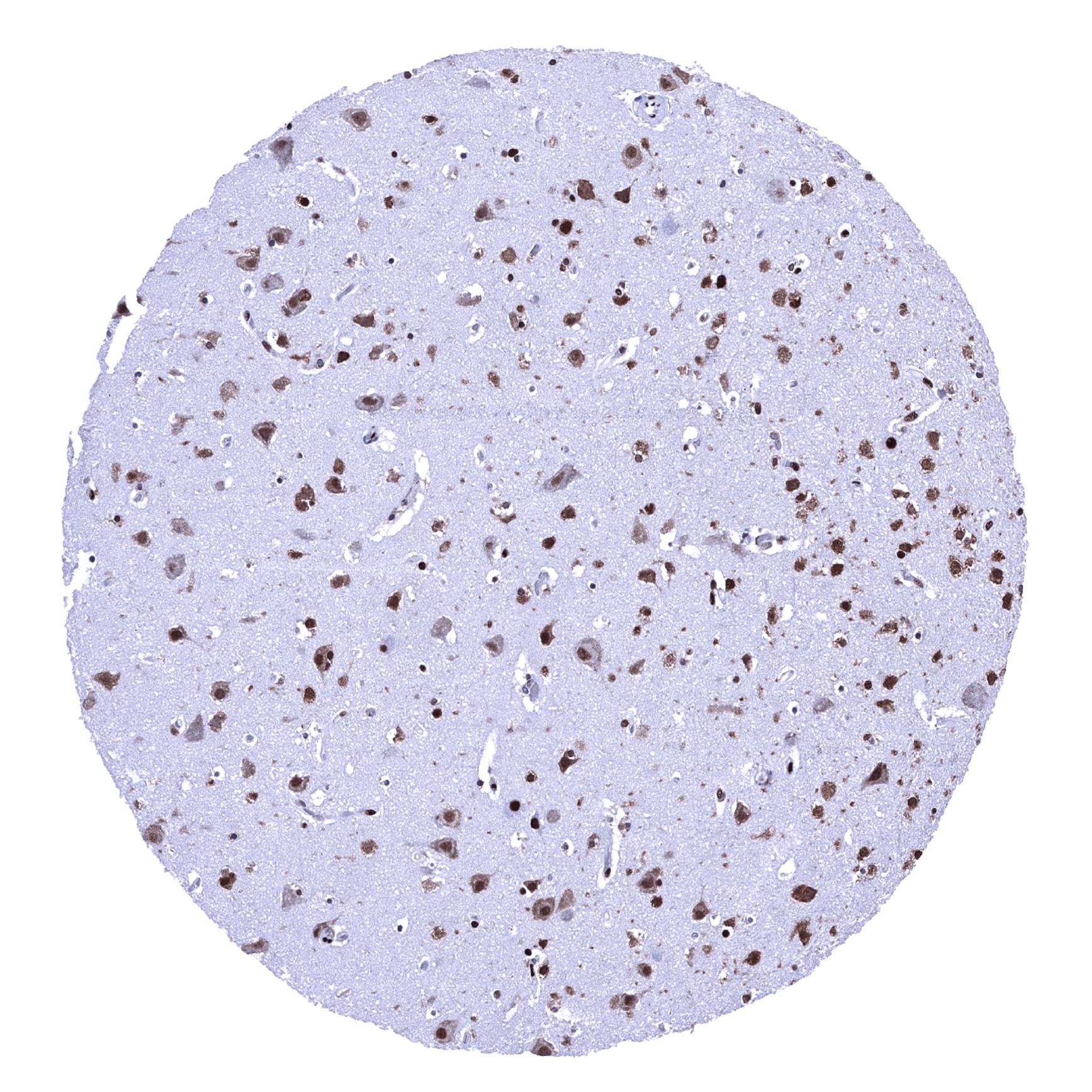
Cerebrum, grey matter
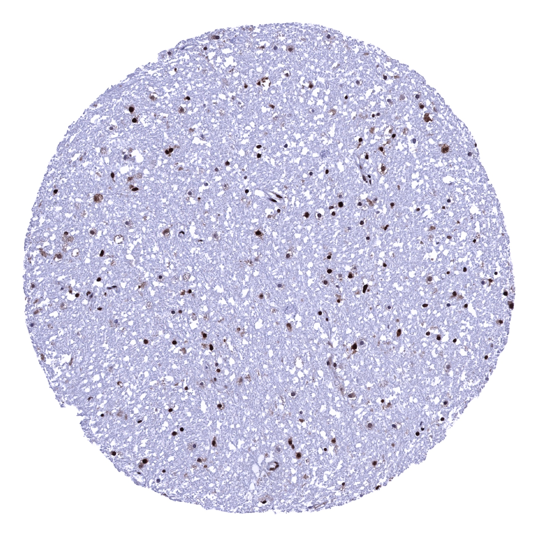
Cerebrum, white matter

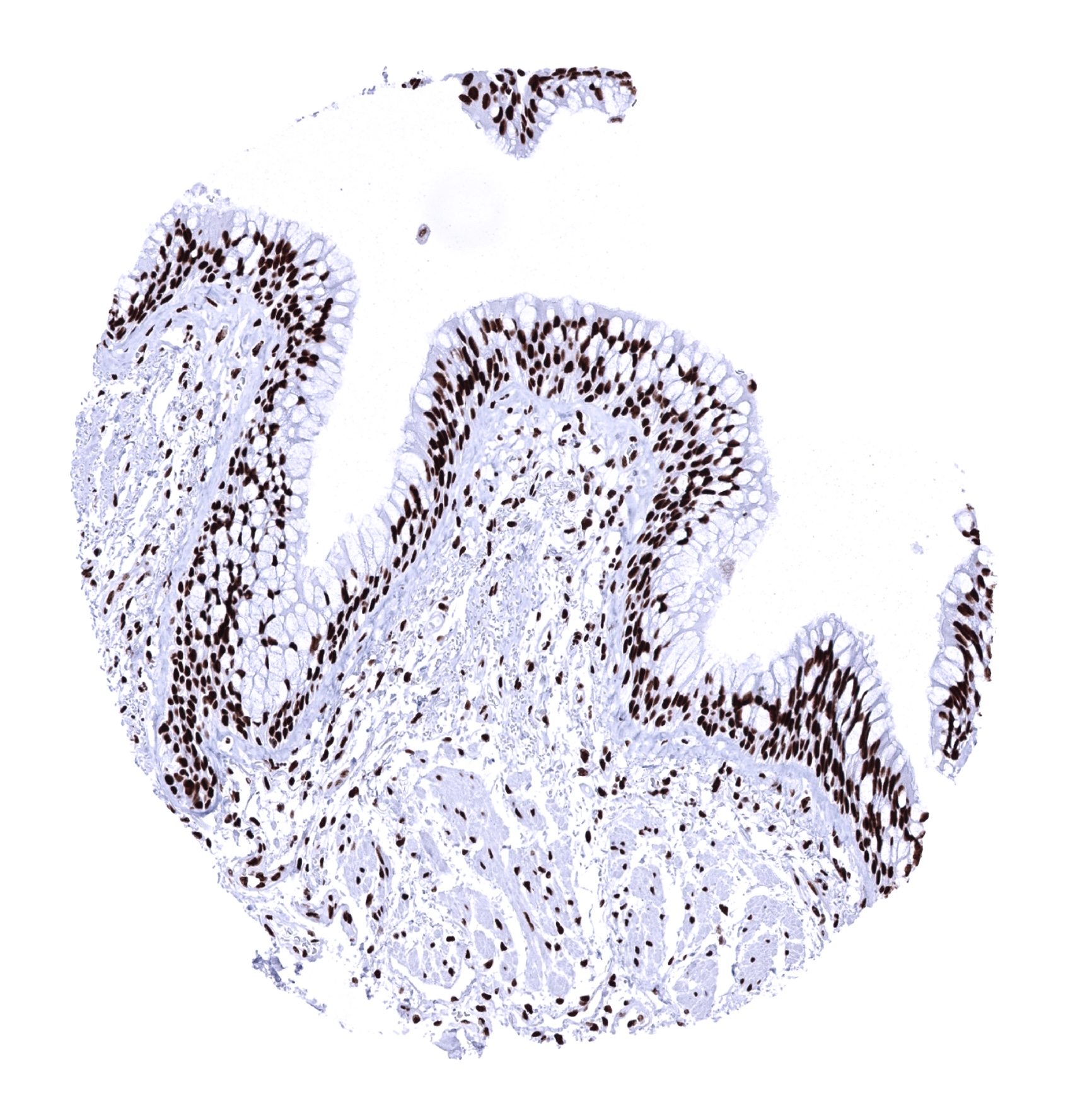
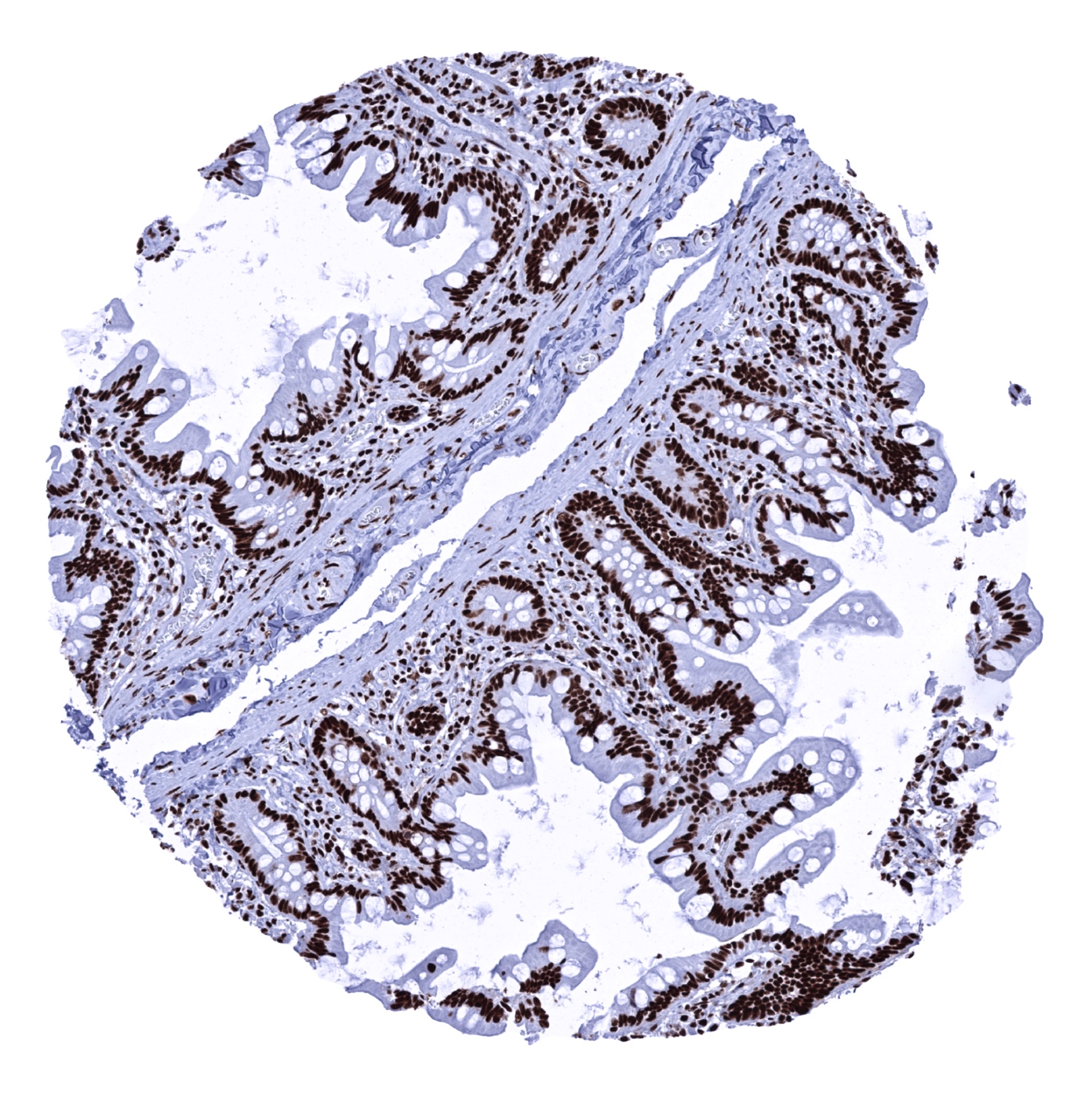
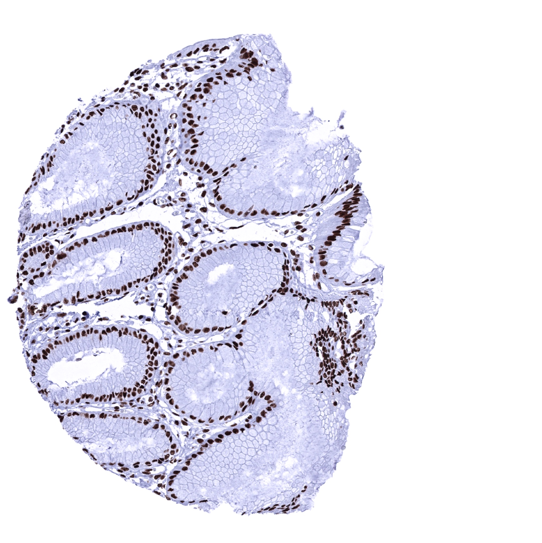
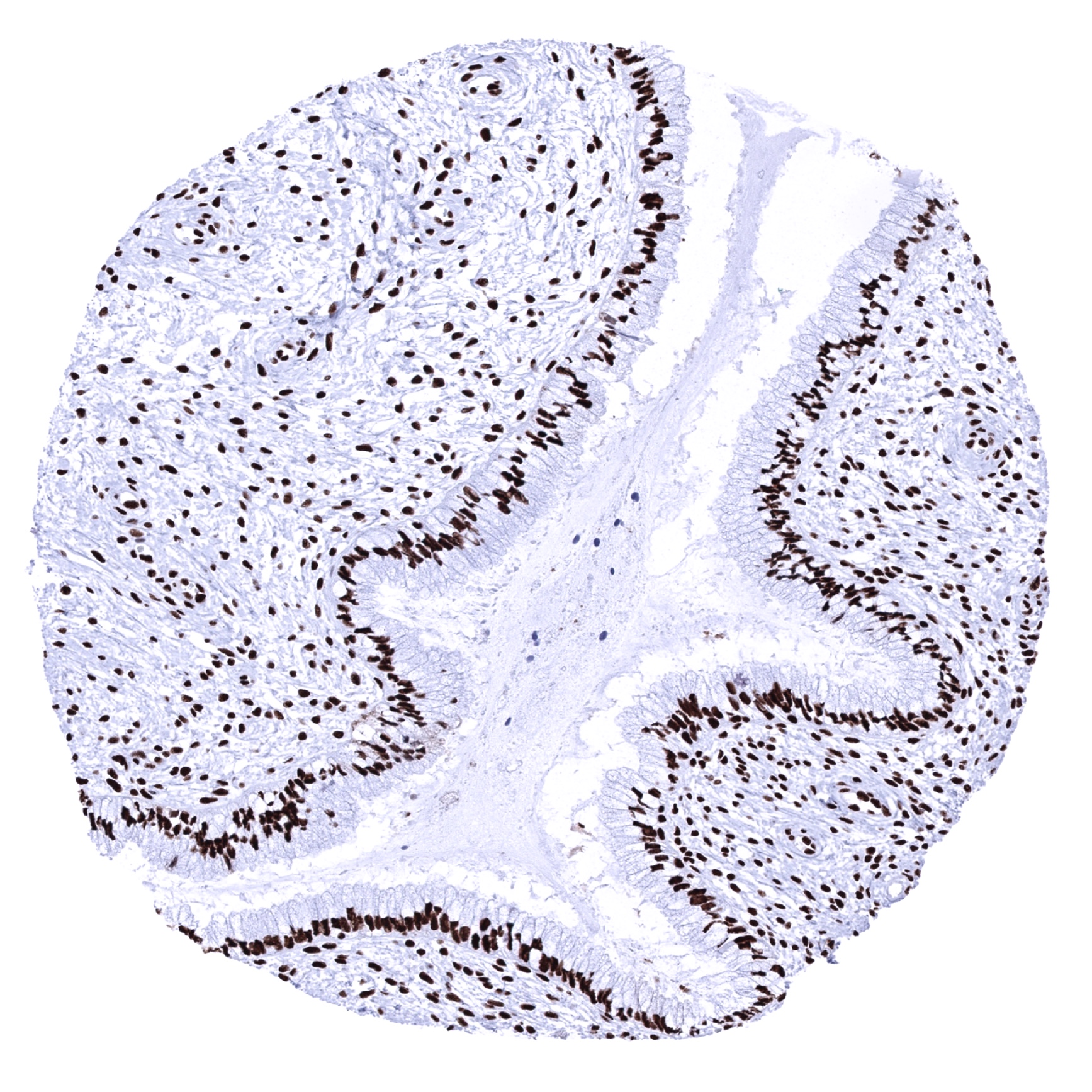
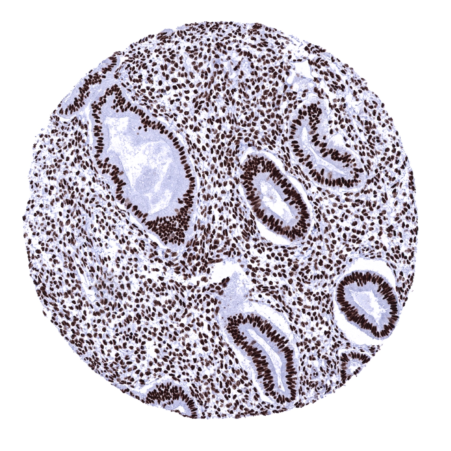
Colon descendens, mucosa
Colon descendens, muscular wall

Duodenum, Brunner gland

Duodenum, mucosa
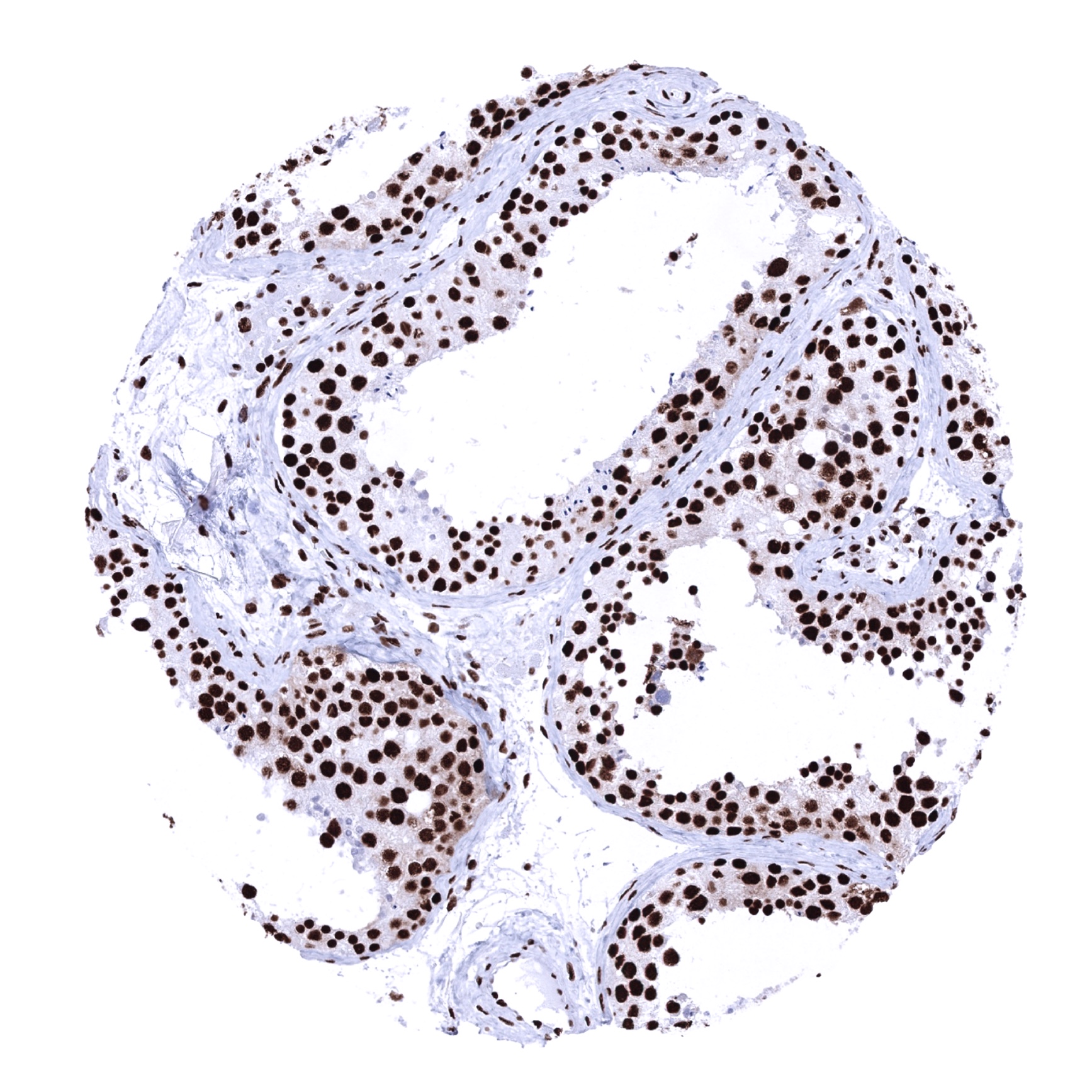
Epididymis

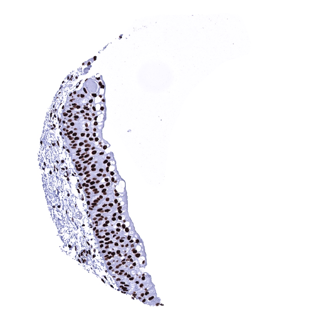
Esophagus, squamous epithelium
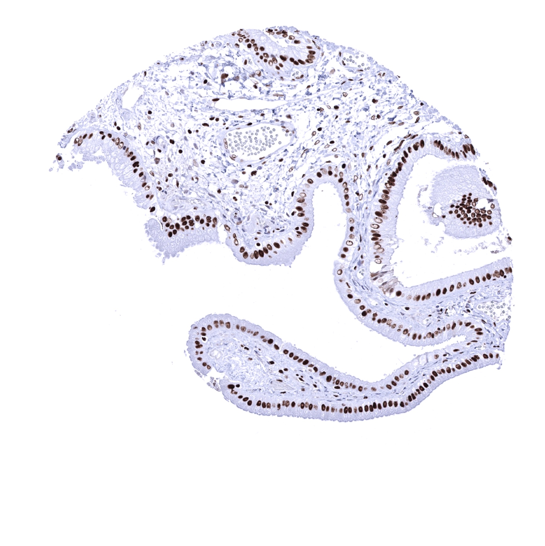
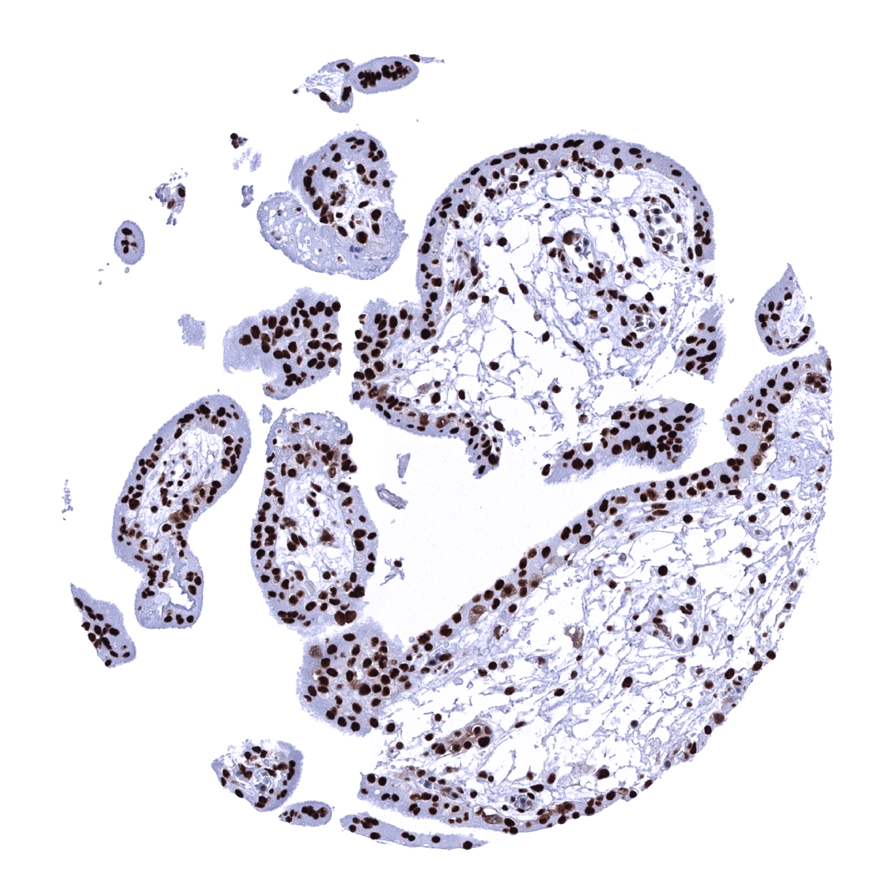
Fallopian tube, mucosa
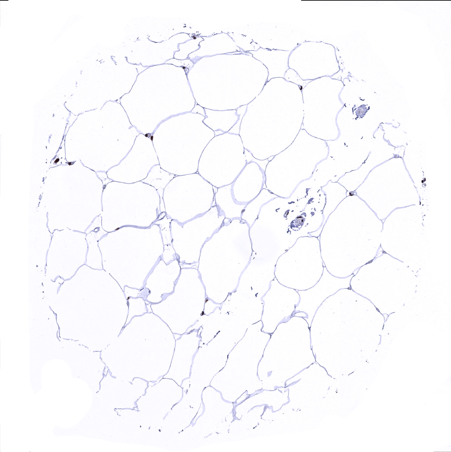
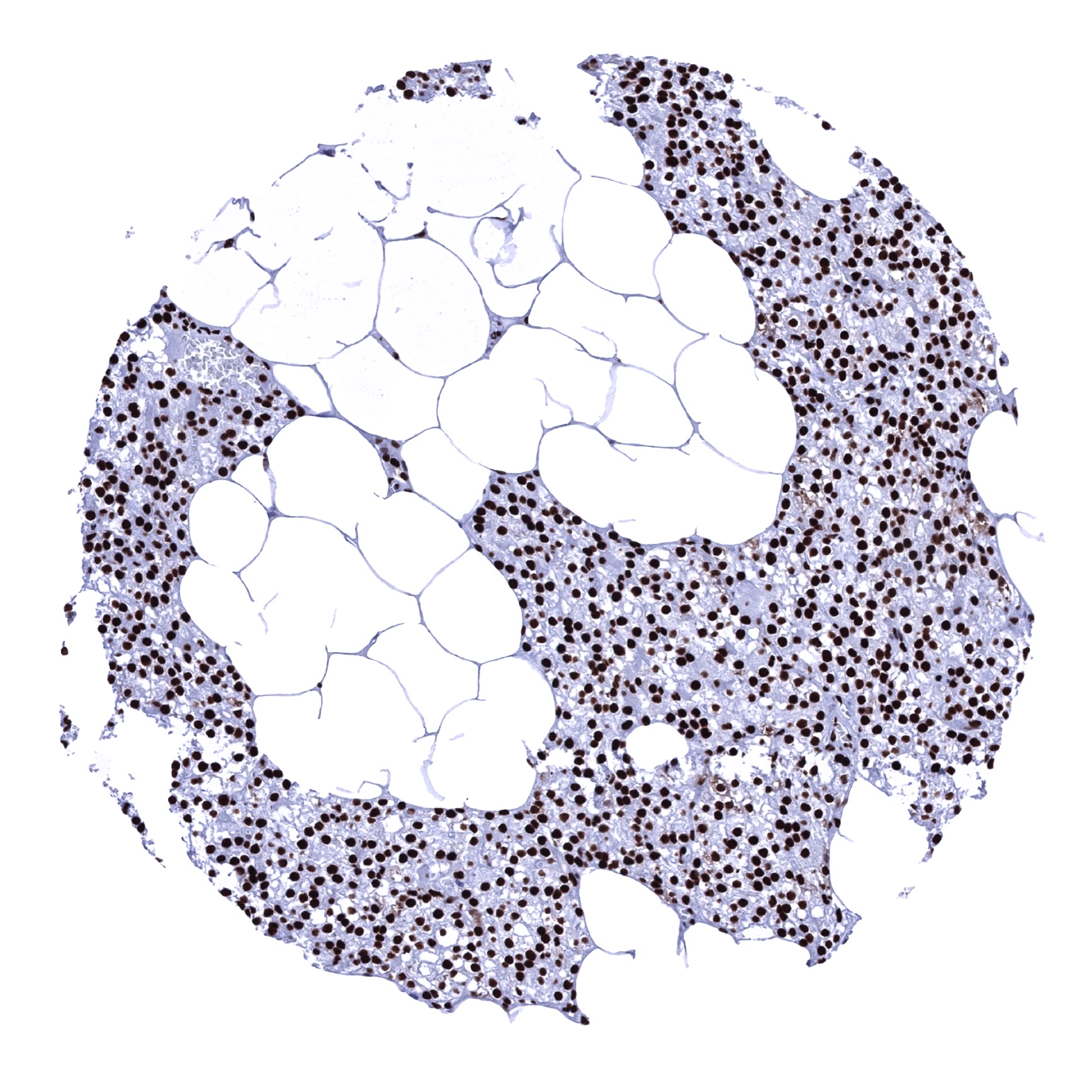
Fat
Gallbladder, epithelium
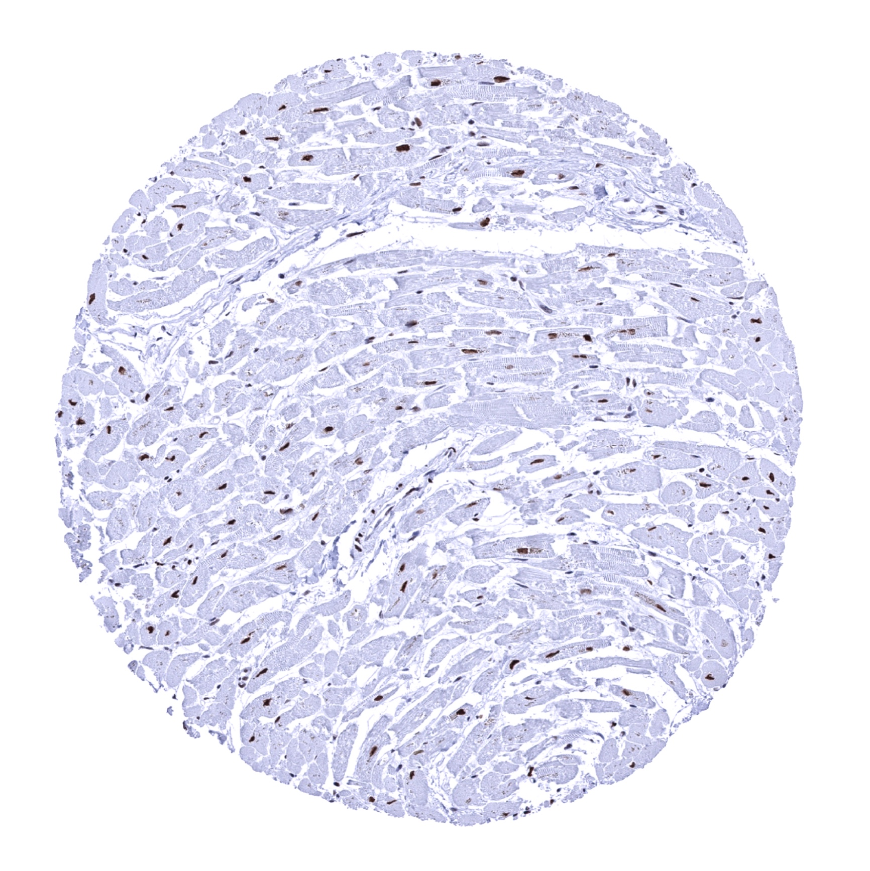
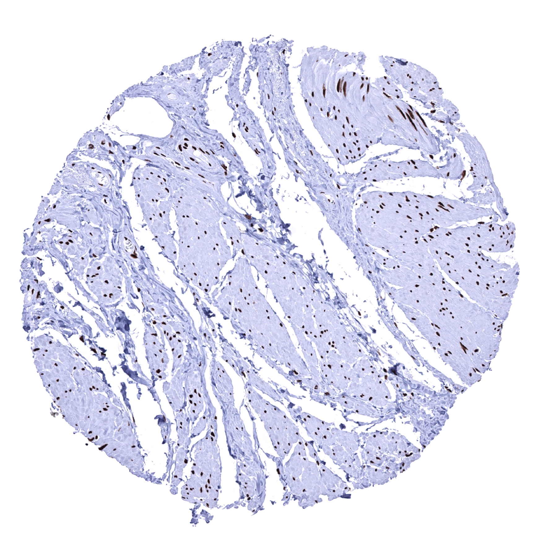
Heart muscle
Ileum, mucosa
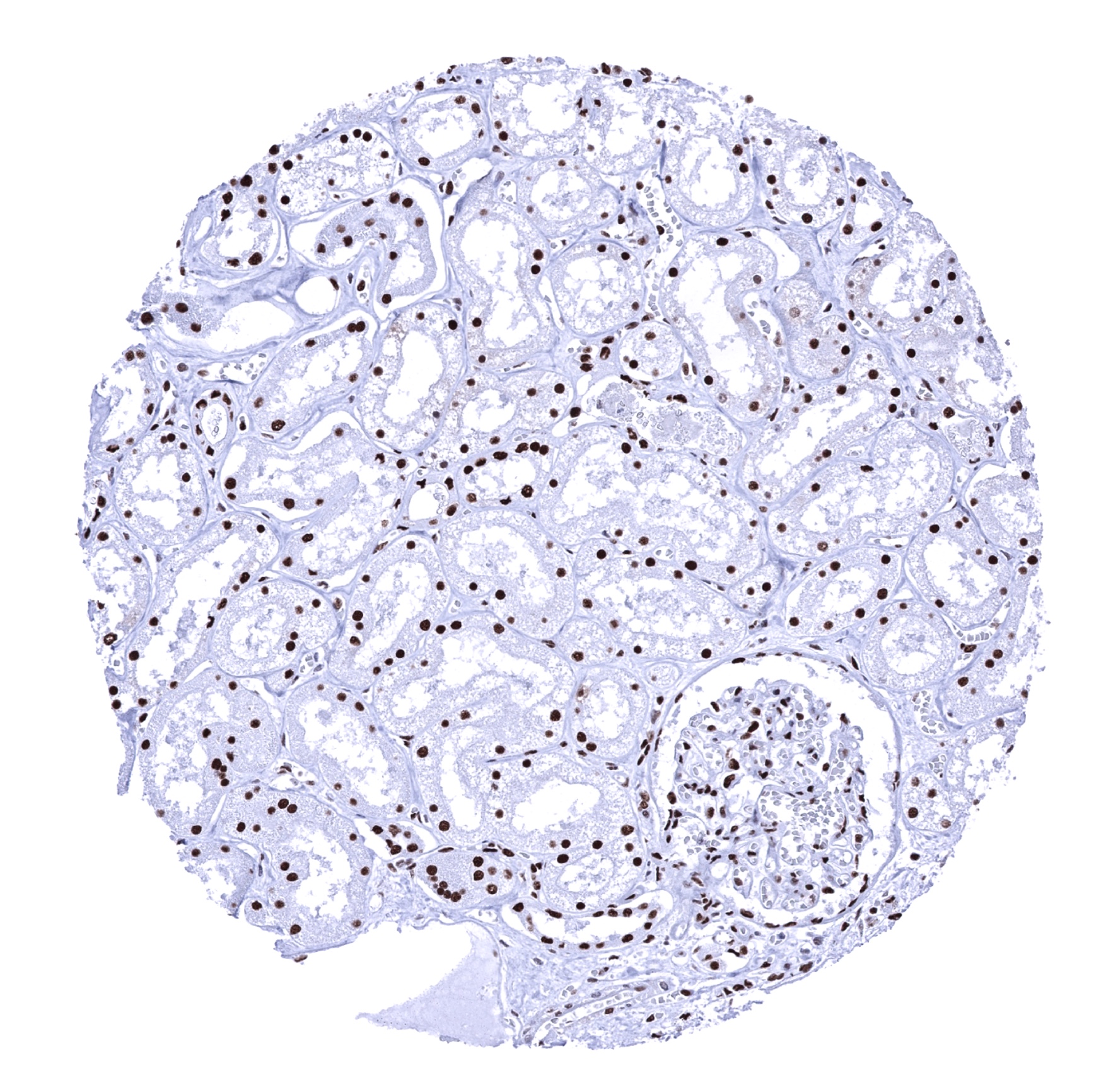
Kidney, cortex
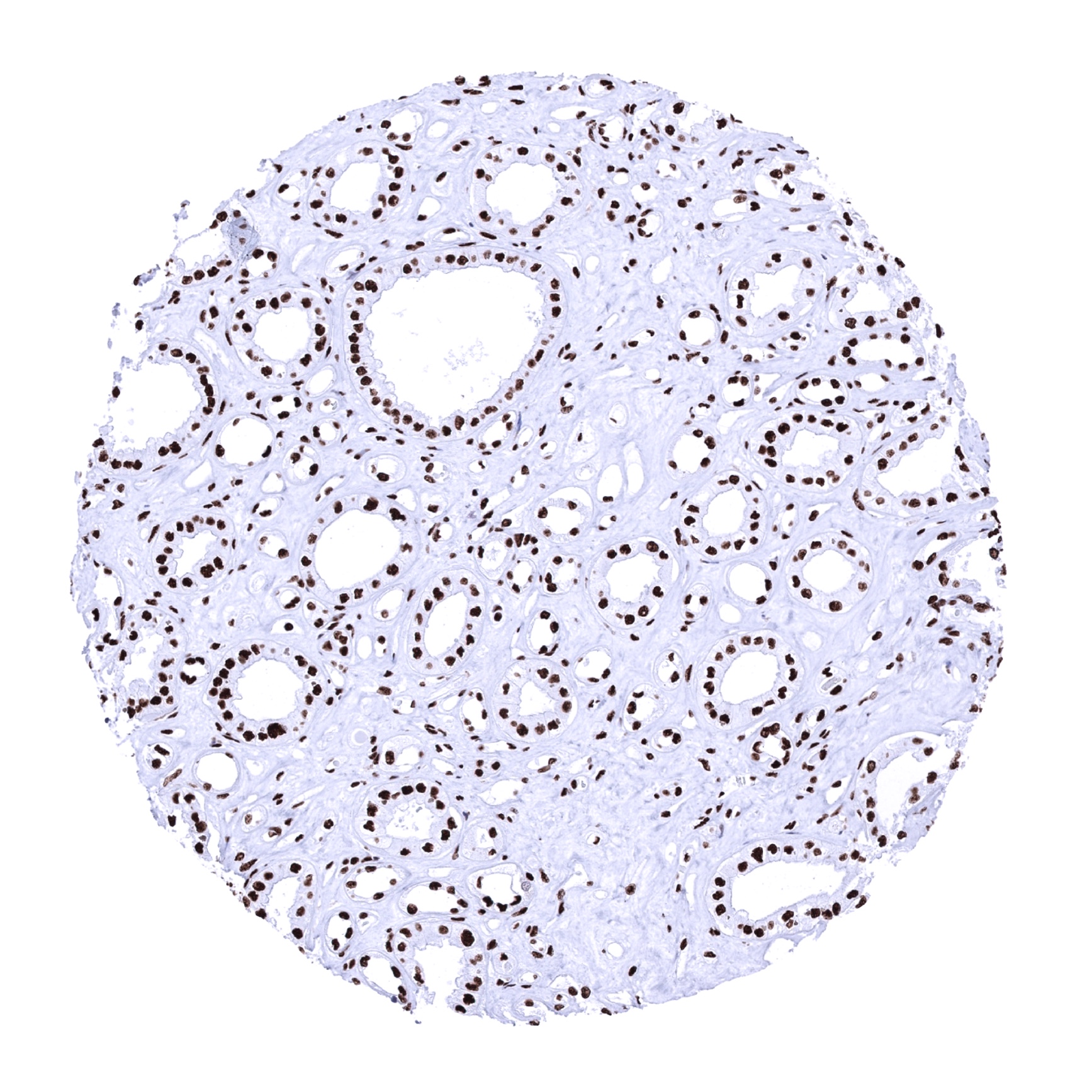
Kidney, medulla
Kidney, pelvis, urothelium

Kidney, pelvis, urothelium
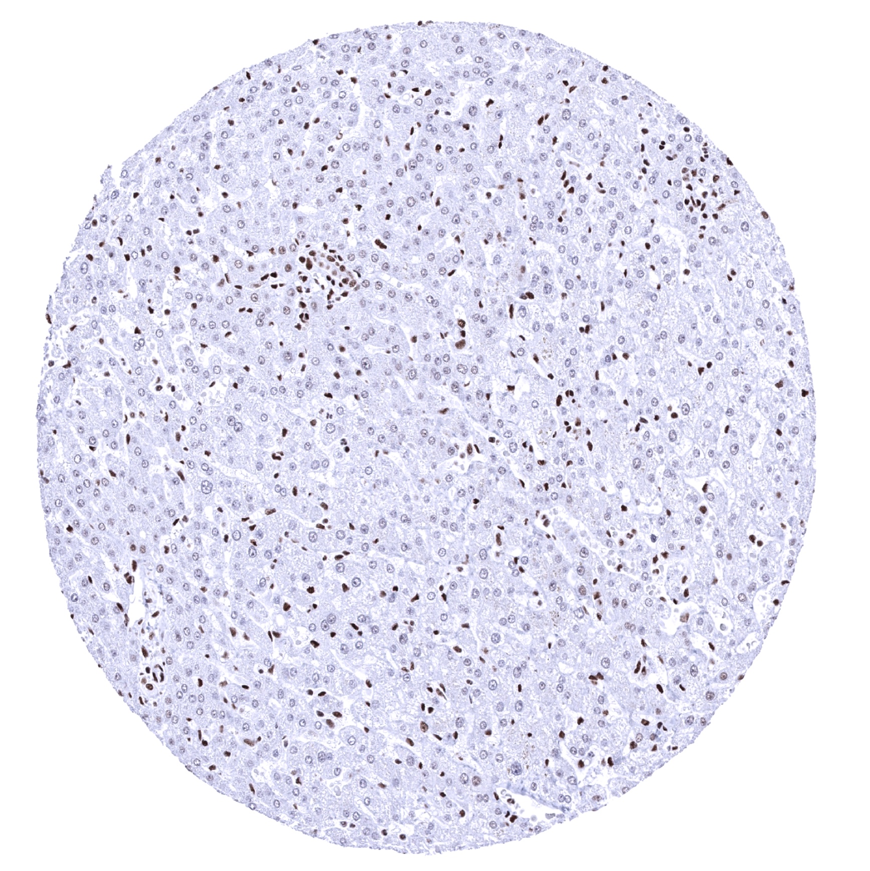
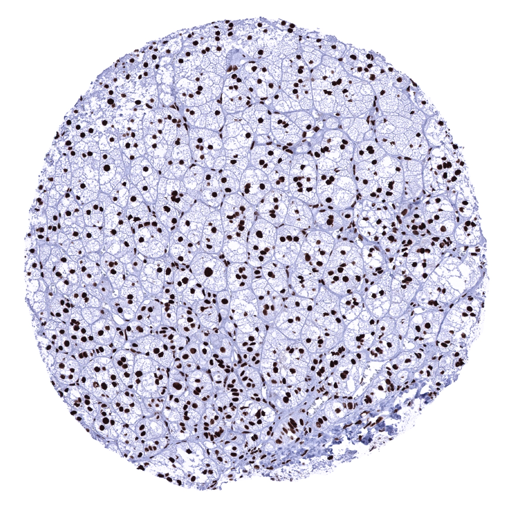
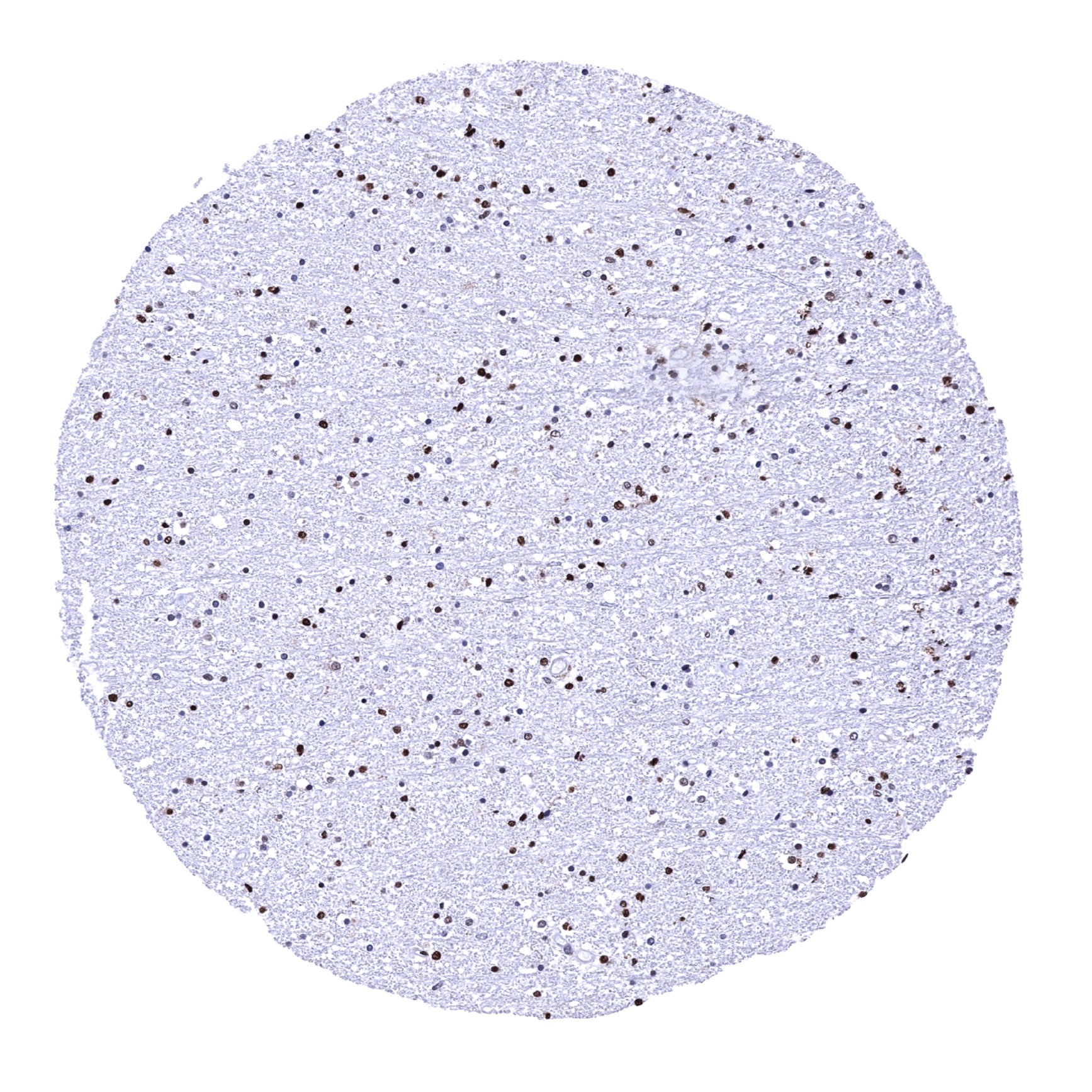
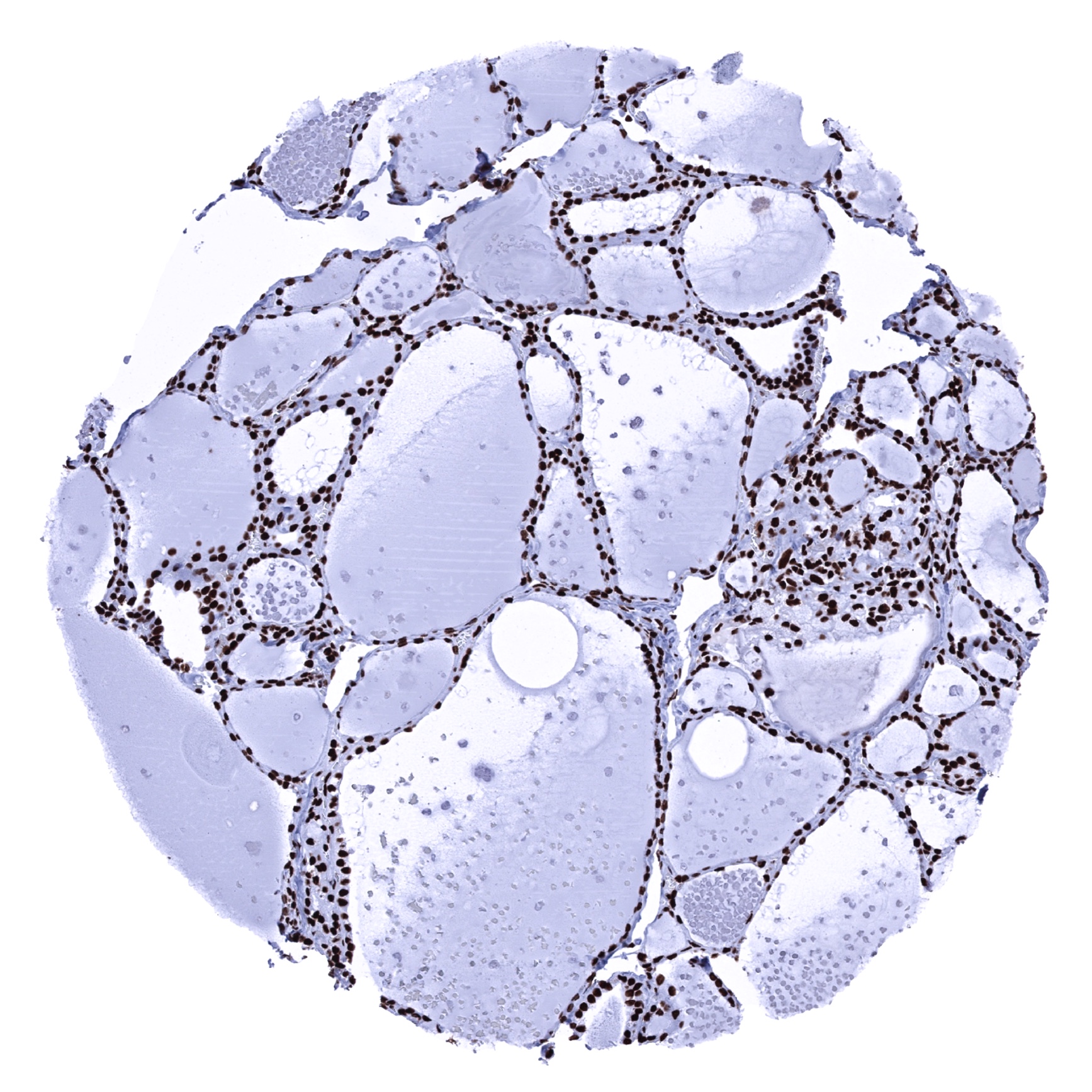
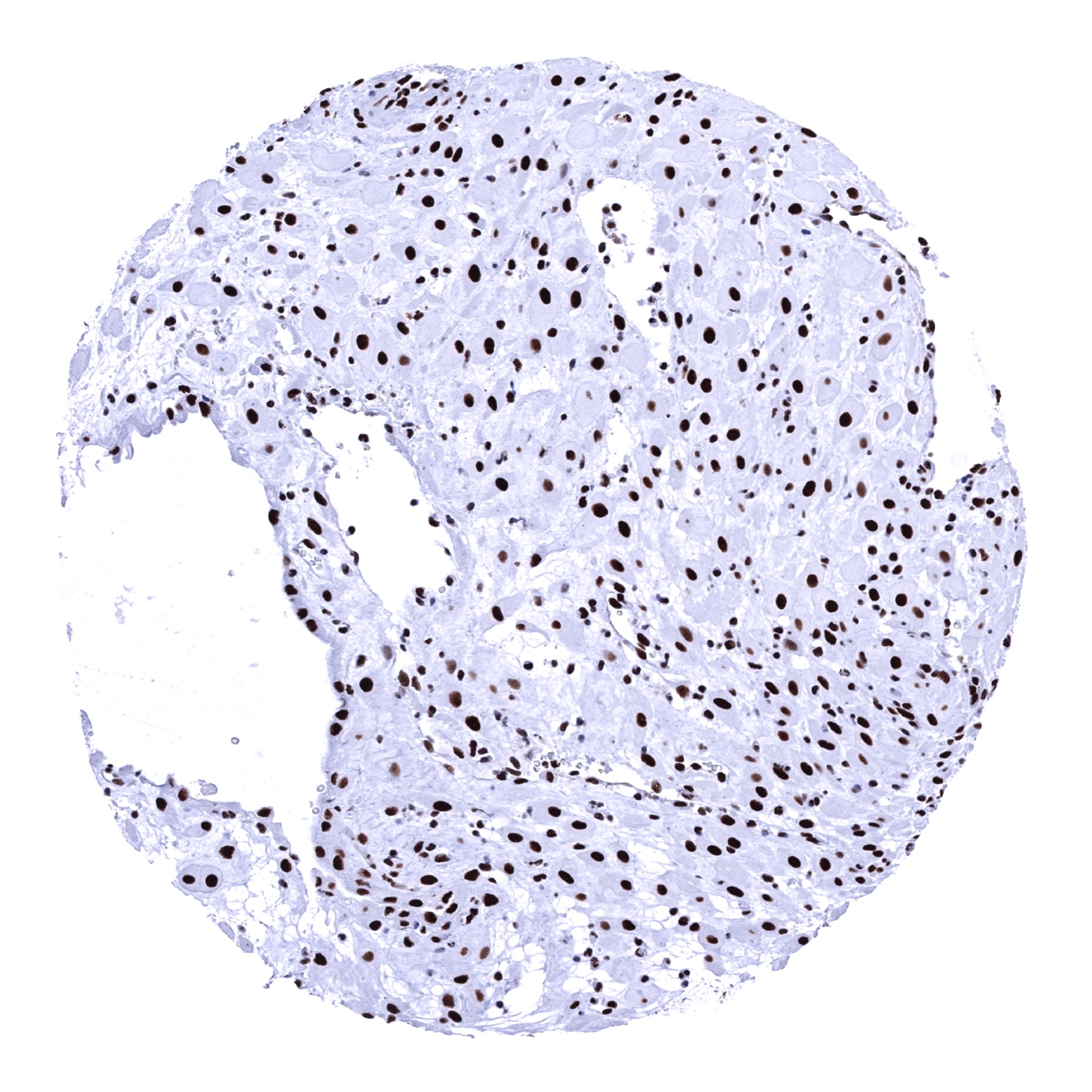
Liver - Ku80 staining is markedly lower in hepatocytes than in other cell types in the liver.
Lung

Lymph node
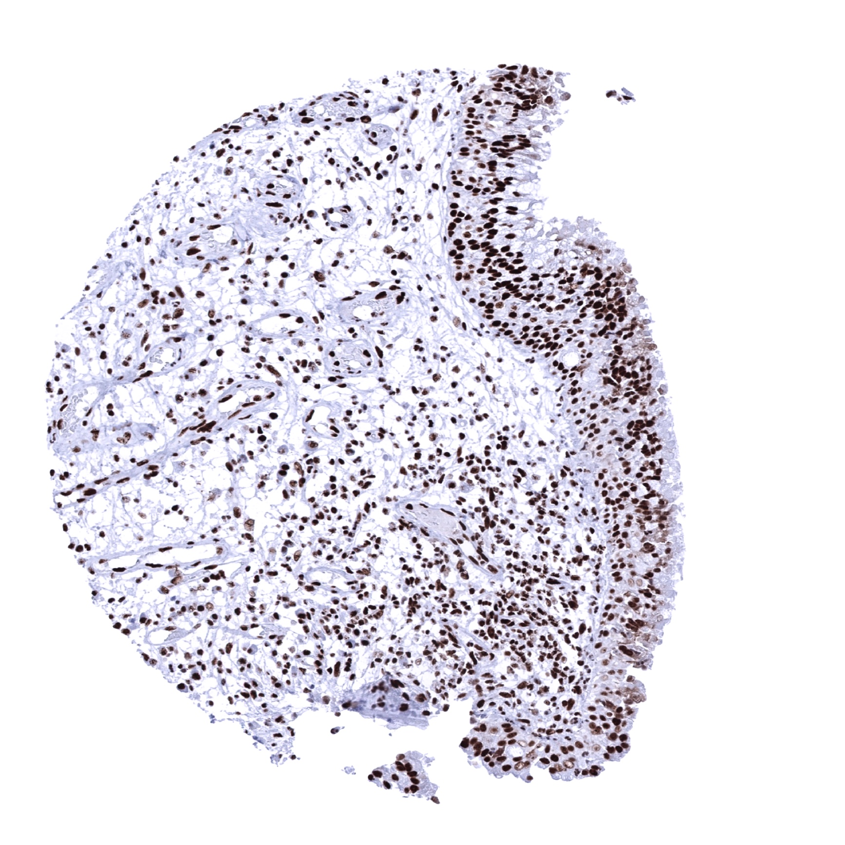
Ovary, stroma
Pancreas

Pancreas
Parathyroid gland
Parotid gland

Pituitary gland, anterior lobe

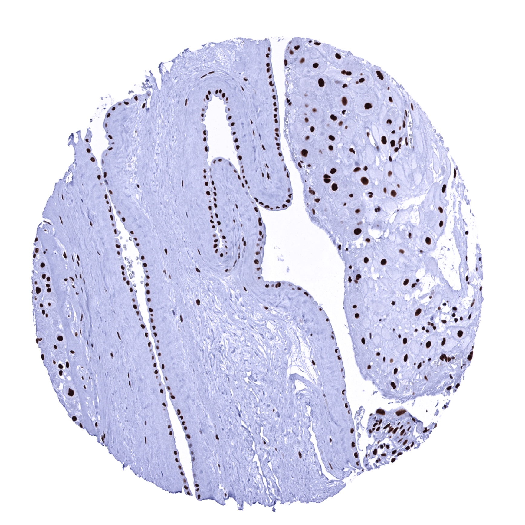
Pituitary gland, posterior lobe
Placenta (amnion and chorion)
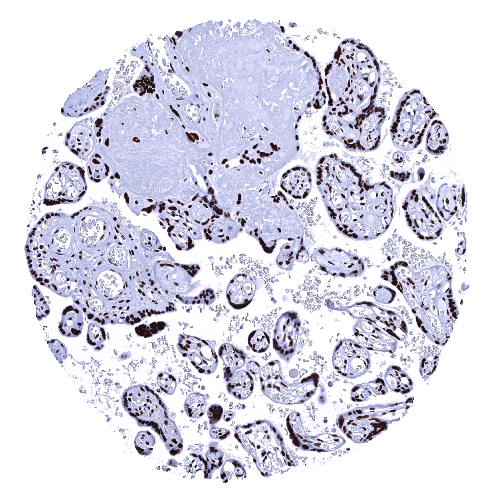
Placenta, early
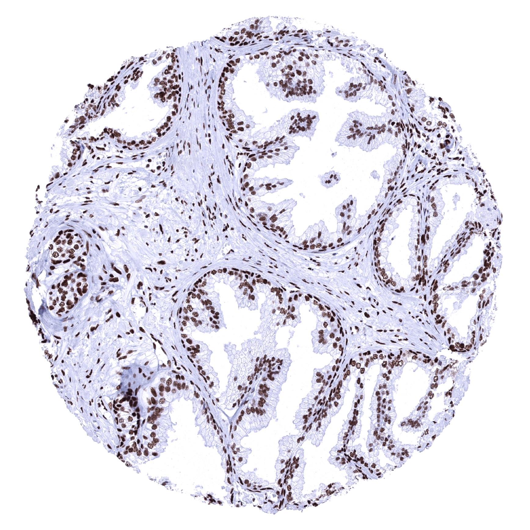
Placenta, mature
Prostate

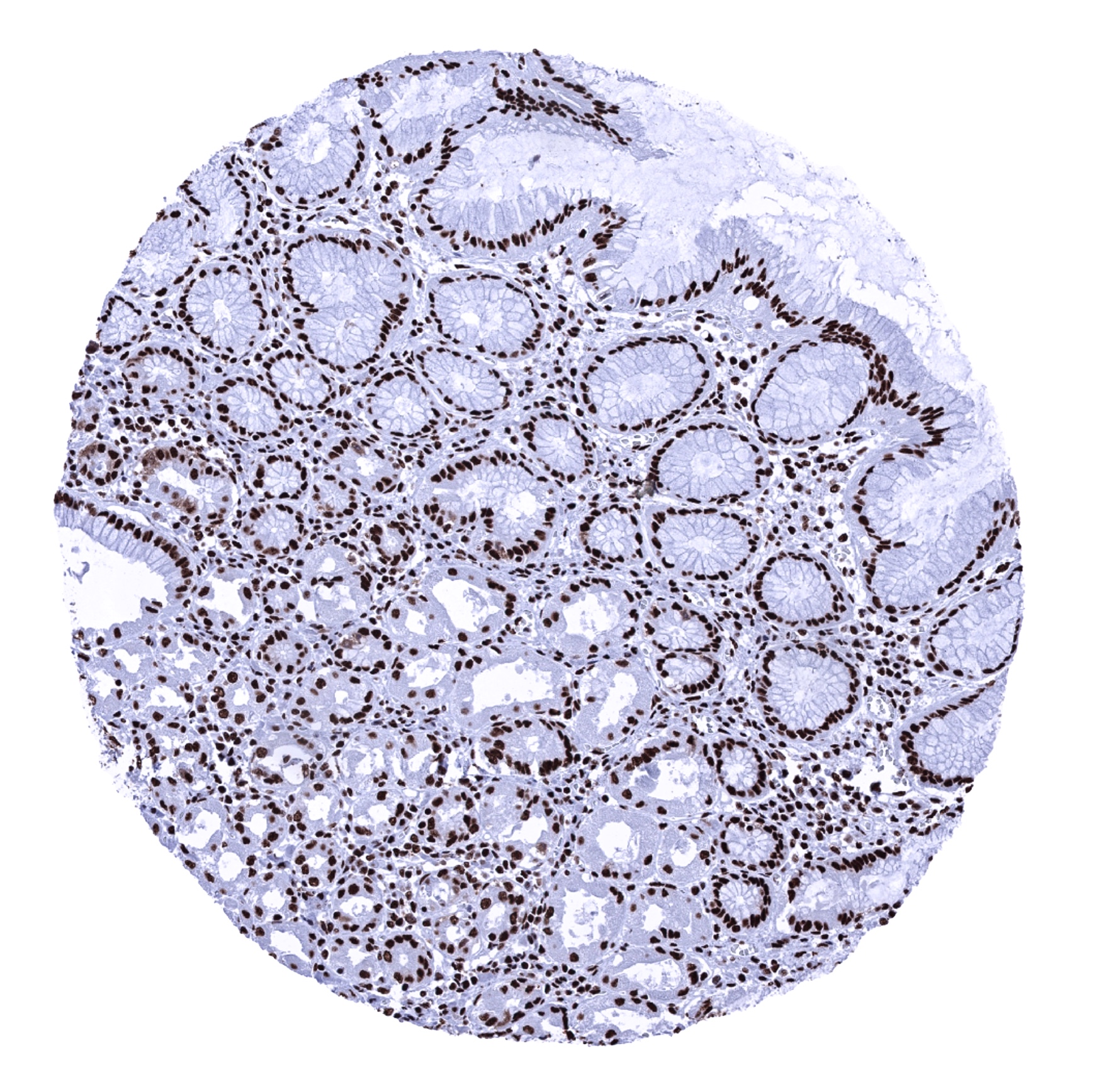
Rectum, mucosa

Seminal vesicle
Sinus paranasales
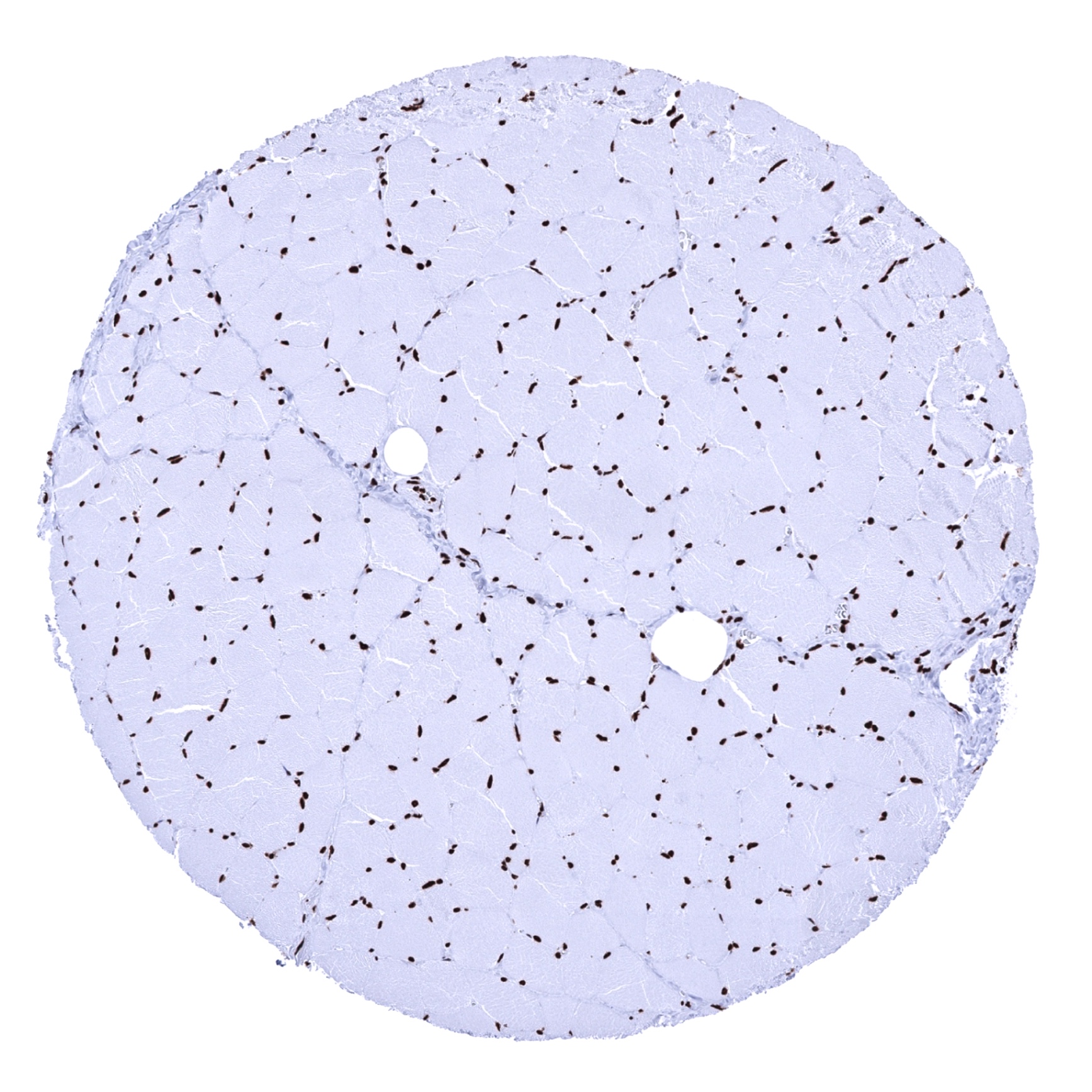
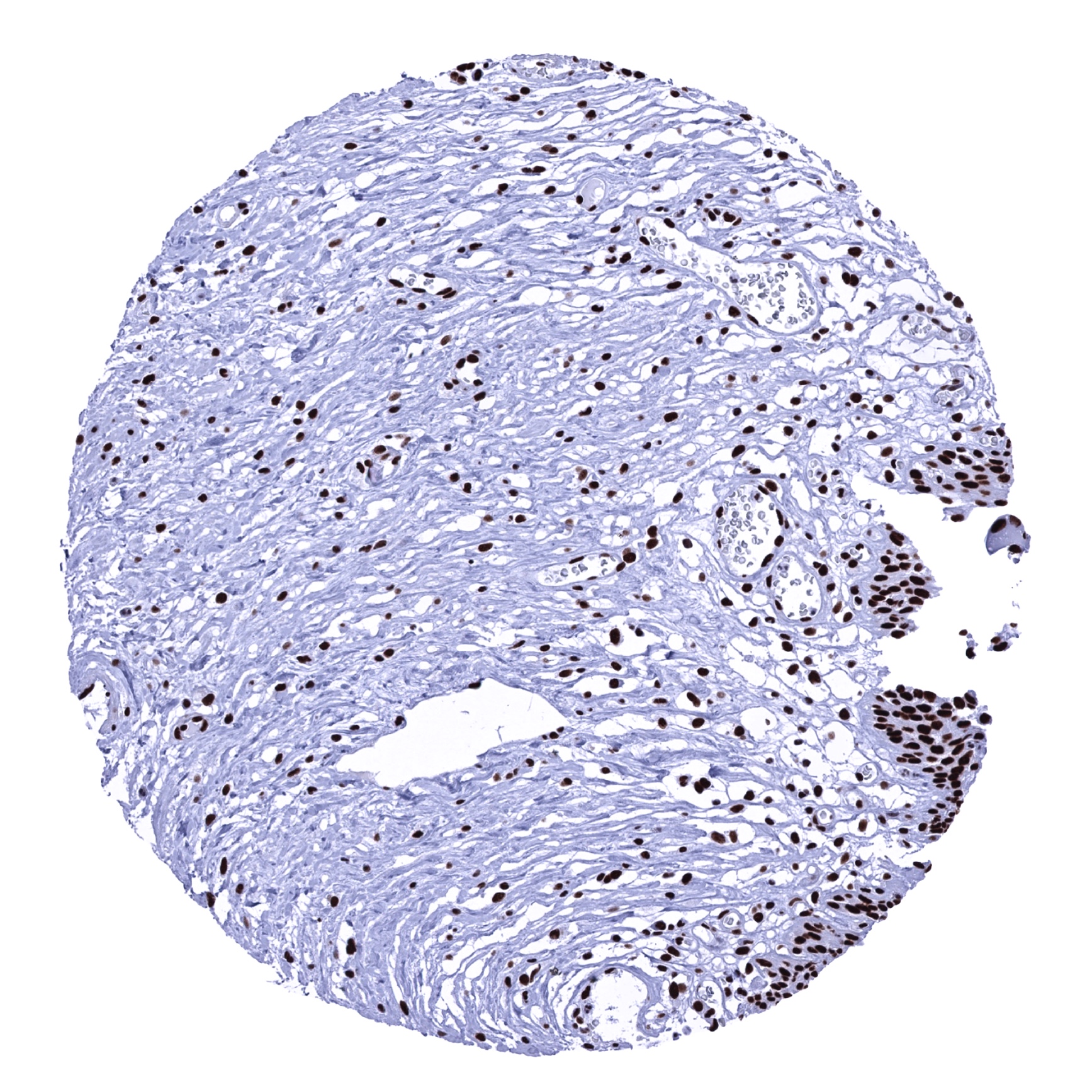
Skeletal muscle
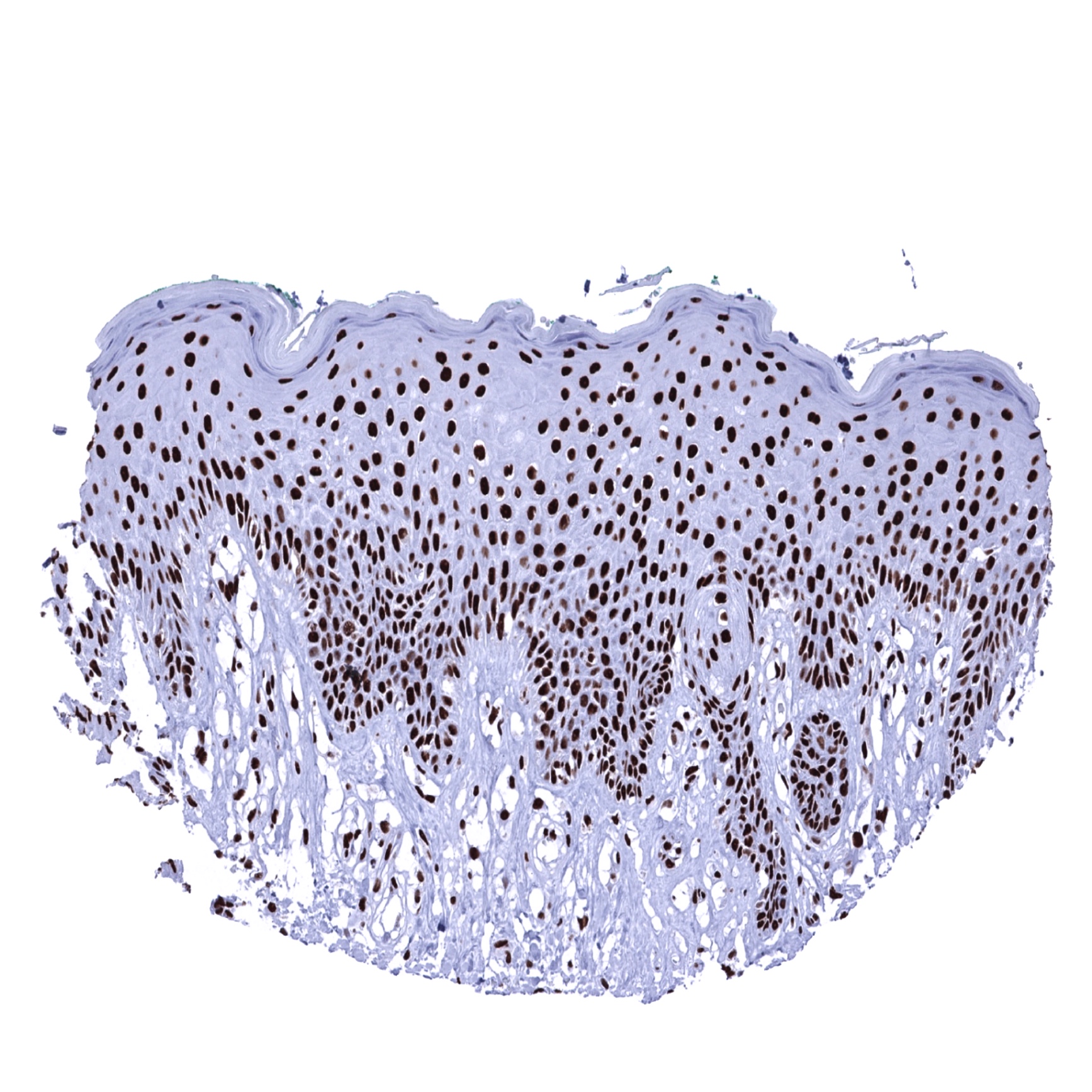
Skin

Spleen
Stomach, antrum
Stomach, corpus
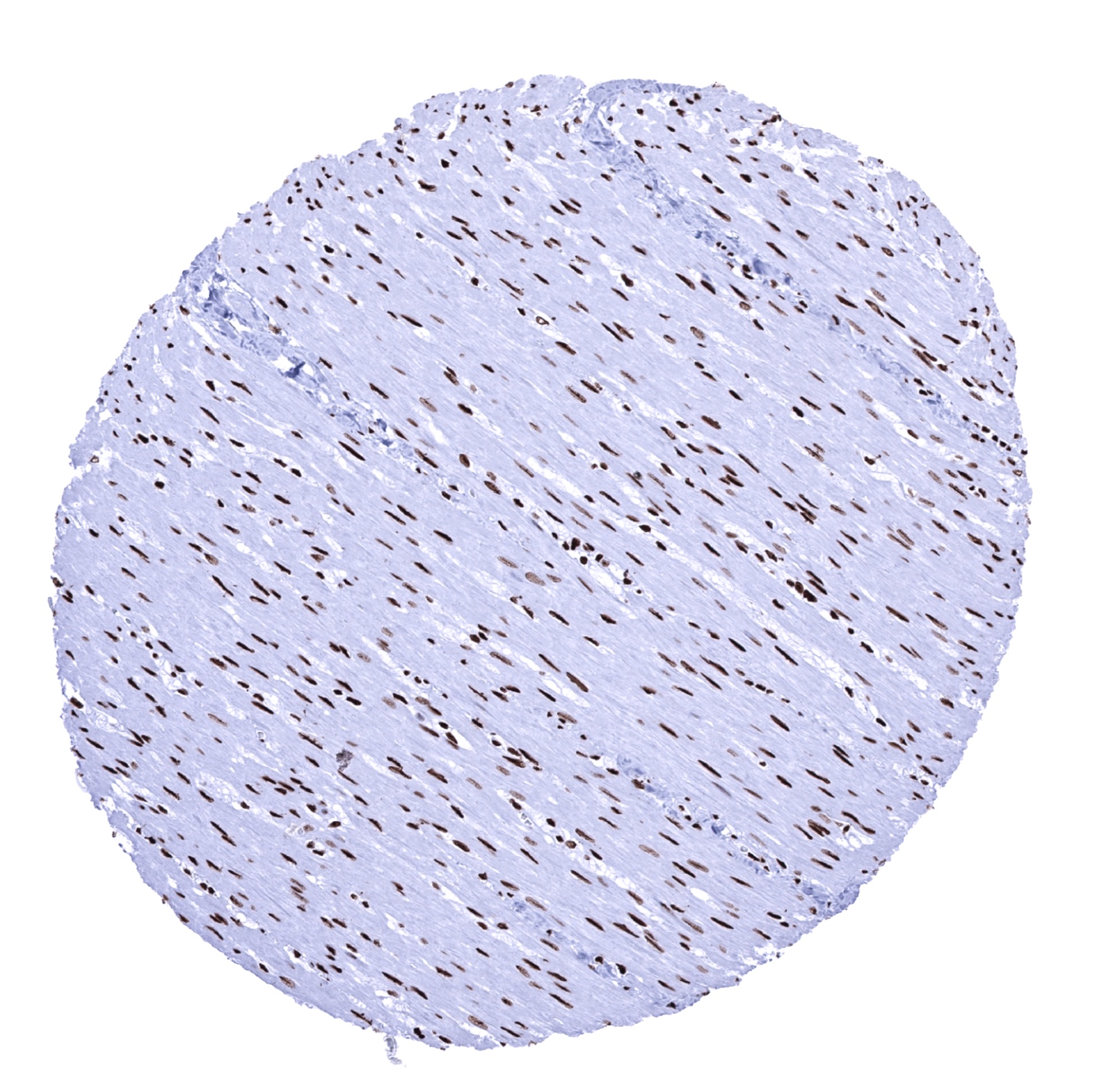
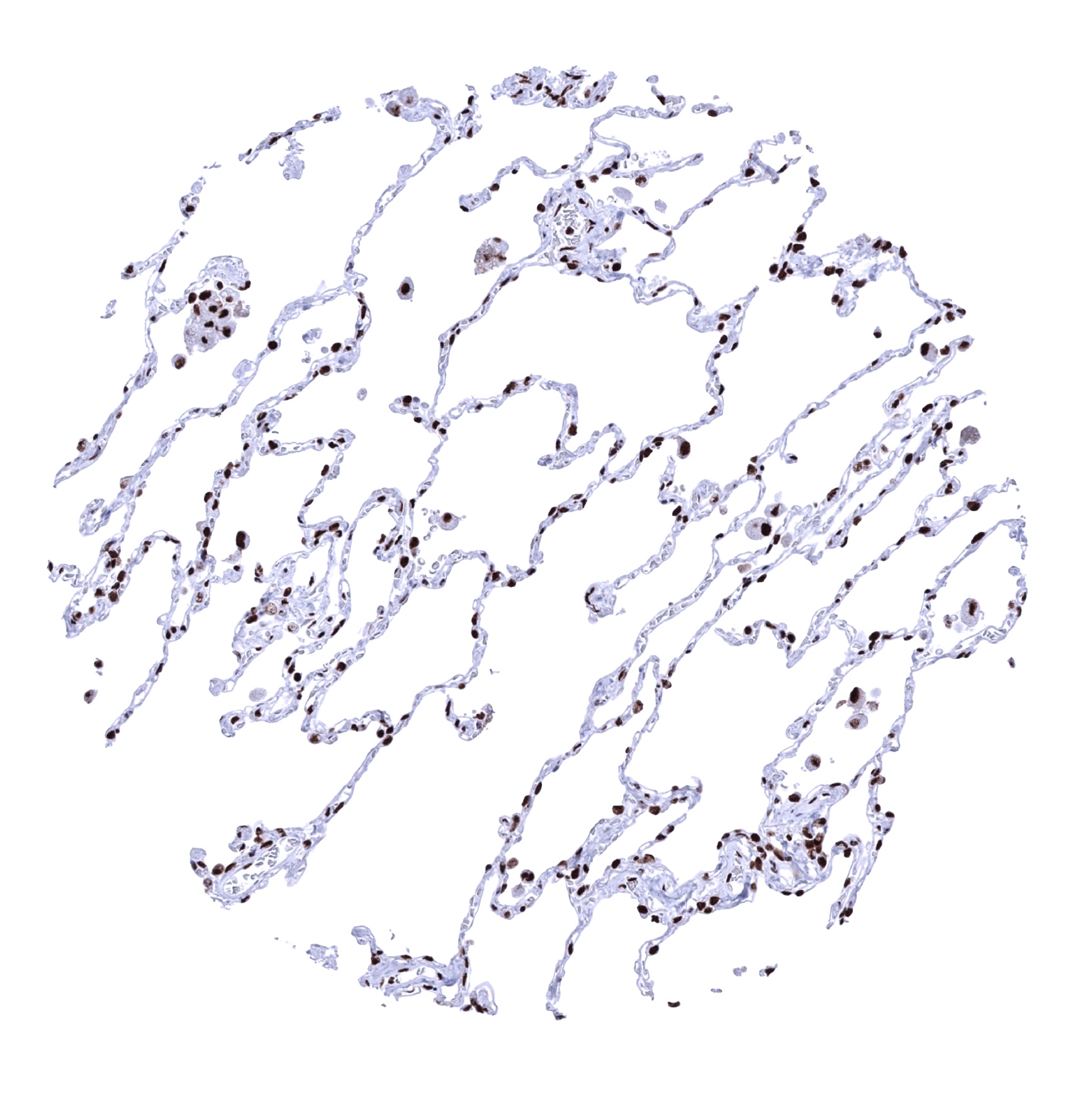
Testis - As compared to other cell types, Ku80 staining is lower in spermatids.
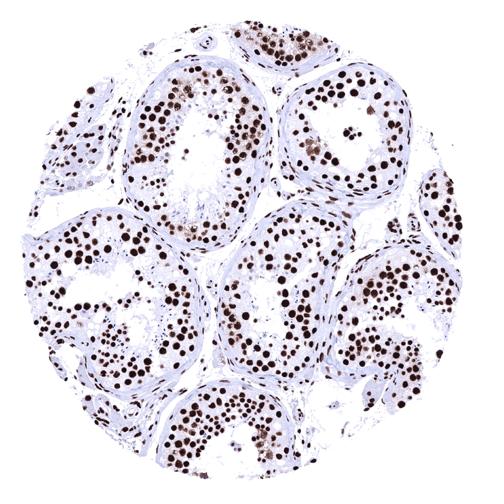
Testis - Ku80 staining is markedly lower in spermatids than in other cell types in the testis.

Thymus
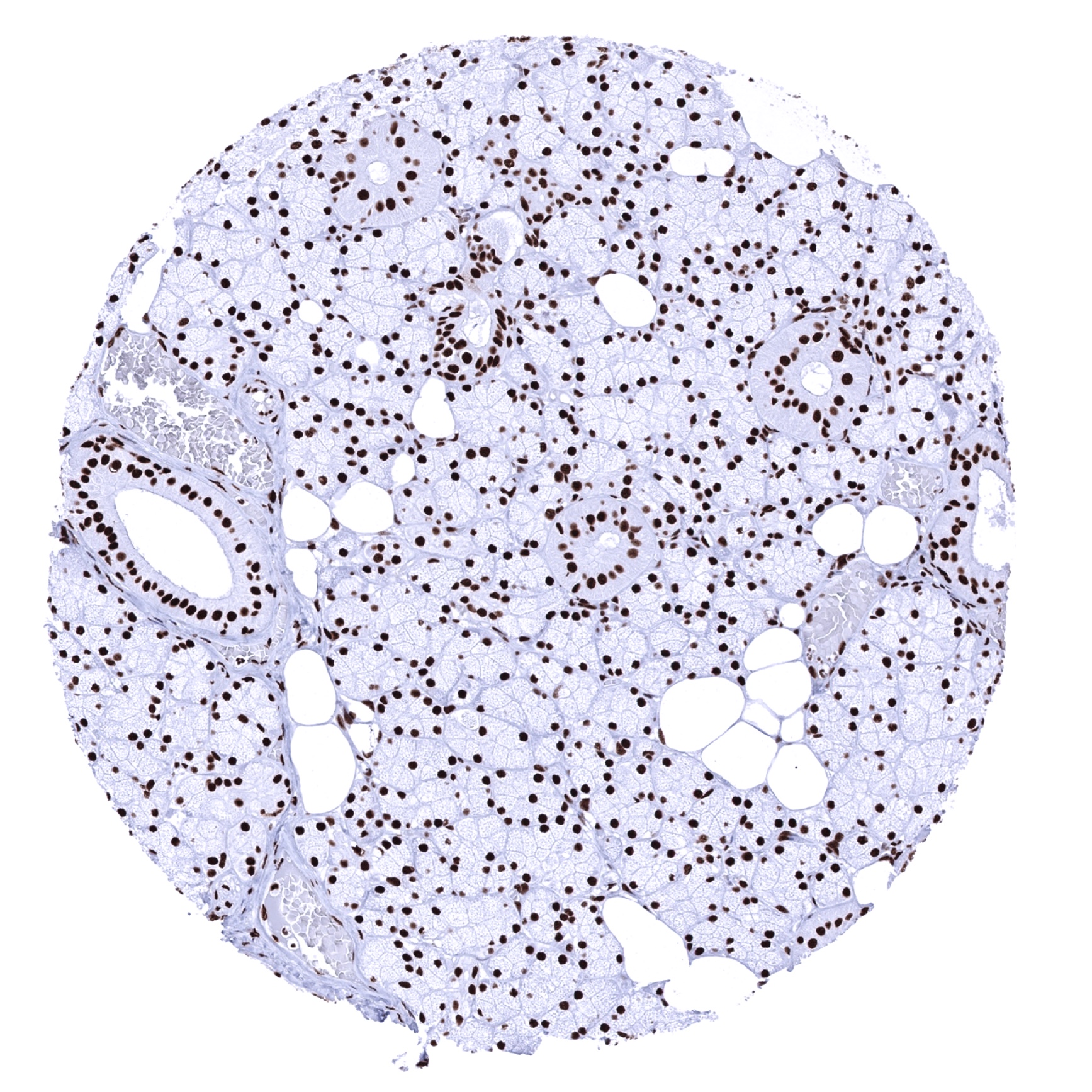
Thyroid gland
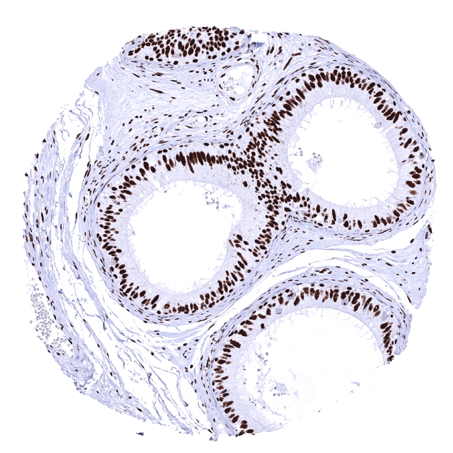
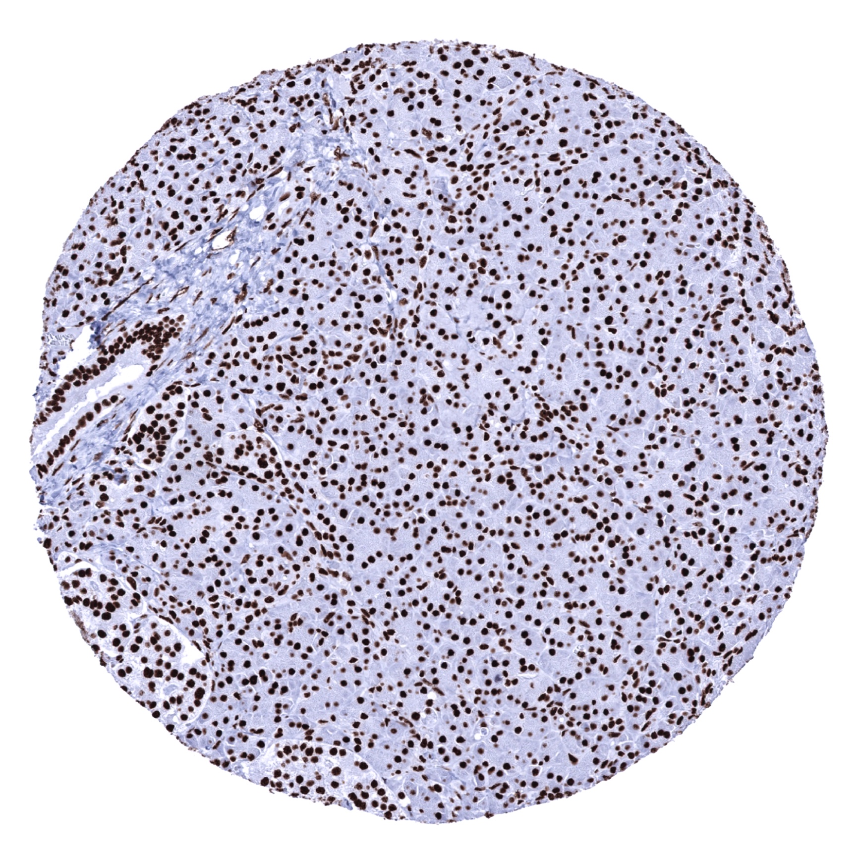
Tonsil, surface epithelium - A marked Ku80 staining is seen in all nuclei of all epithelial and lymphatic cells.
Tonsil
Urinary bladder, muscular wall
Urinary bladder, urothelium
Uterus, ectocervix
Uterus, endocervix
Uterus, endometrium (pregnancy)
Uterus, endometrium (proliferation)
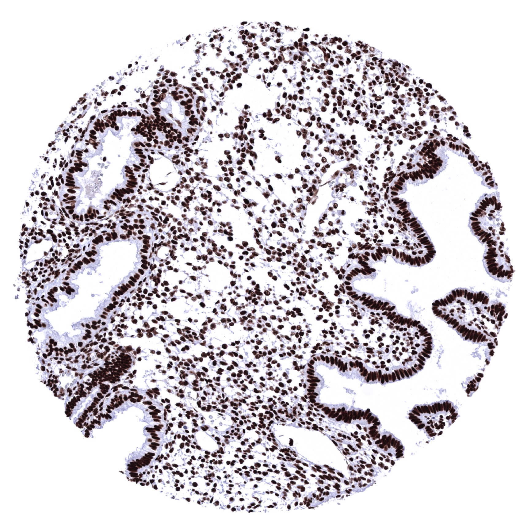
Uterus, endometrium (secretion)
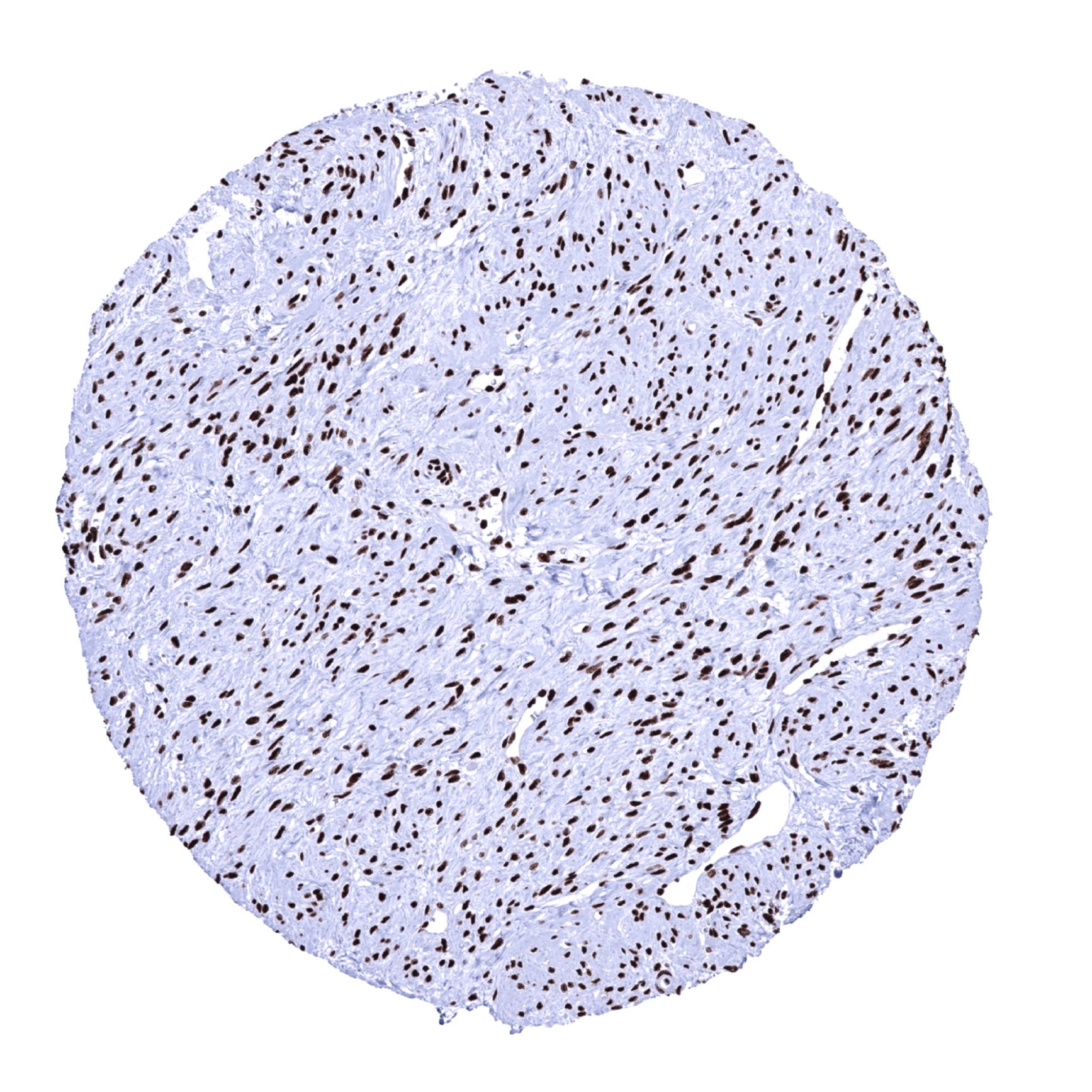
Uterus, myometrium