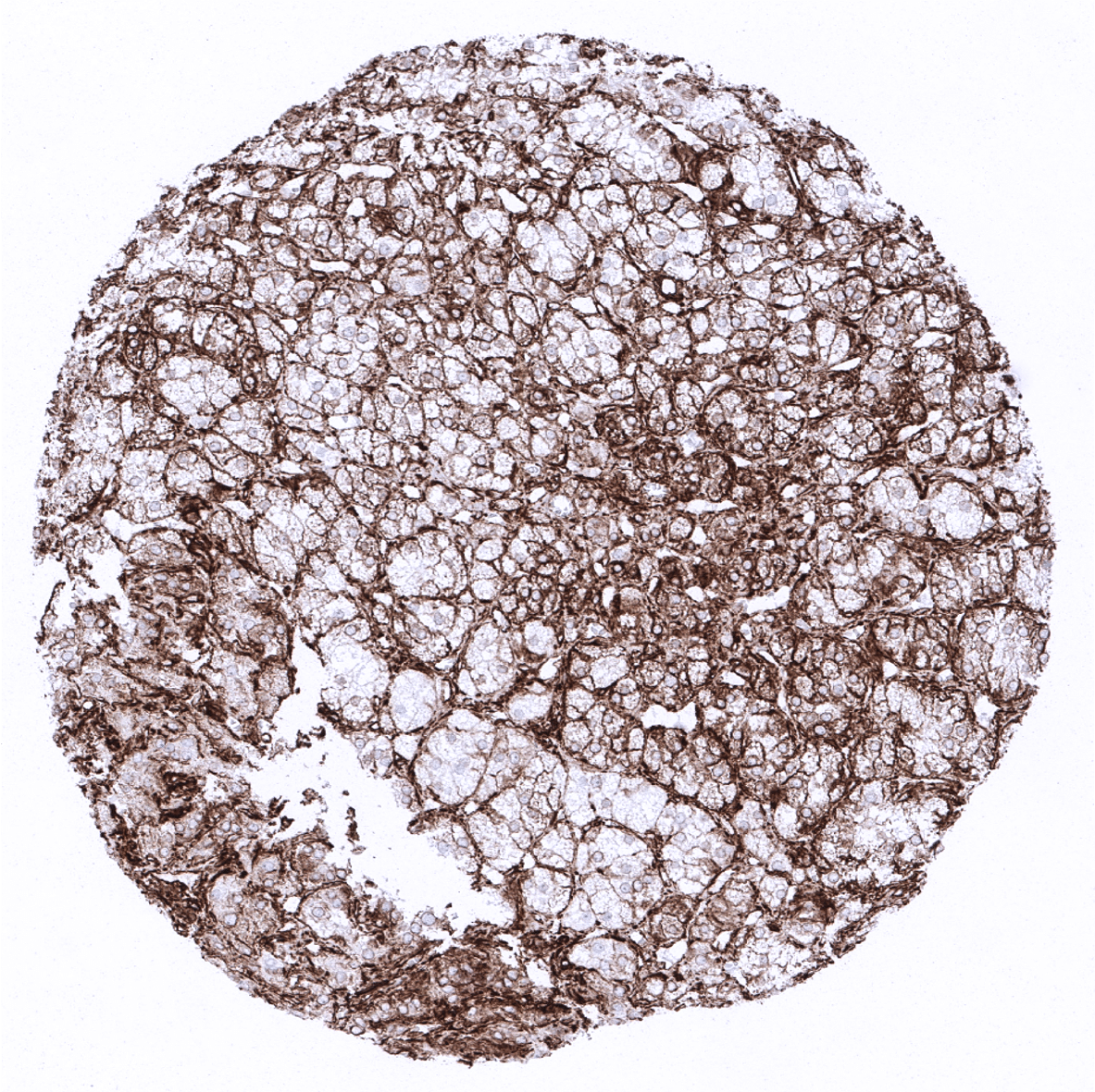
Adrenal gland - In the adrenal gland a variable vimentin immunostaining can be seen in cortical and medullary cells.
Adrenal gland: In the adrenal gland a variable vimentin immunostaining can be seen in cortical and medullary cells.
Adrenal gland - In the adrenal gland a variable vimentin immunostaining can be seen in cortical and medullary cells.
Adrenal gland - In the adrenal gland a variable vimentin immunostaining can be seen in cortical and medullary cells.
Aorta, media

Appendix, mucosa

Appendix, muscular wall
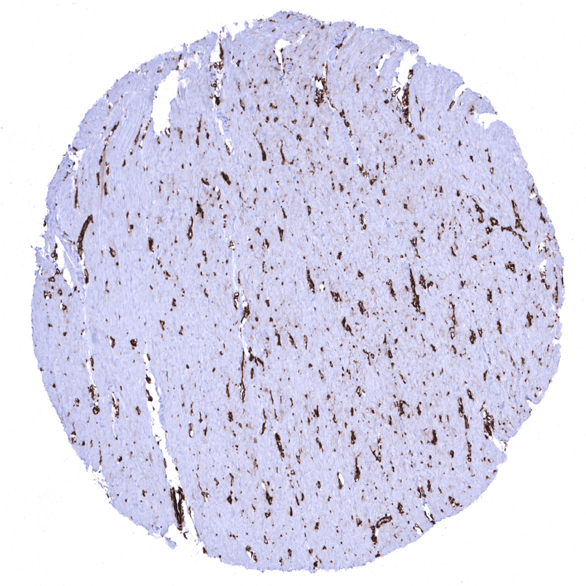
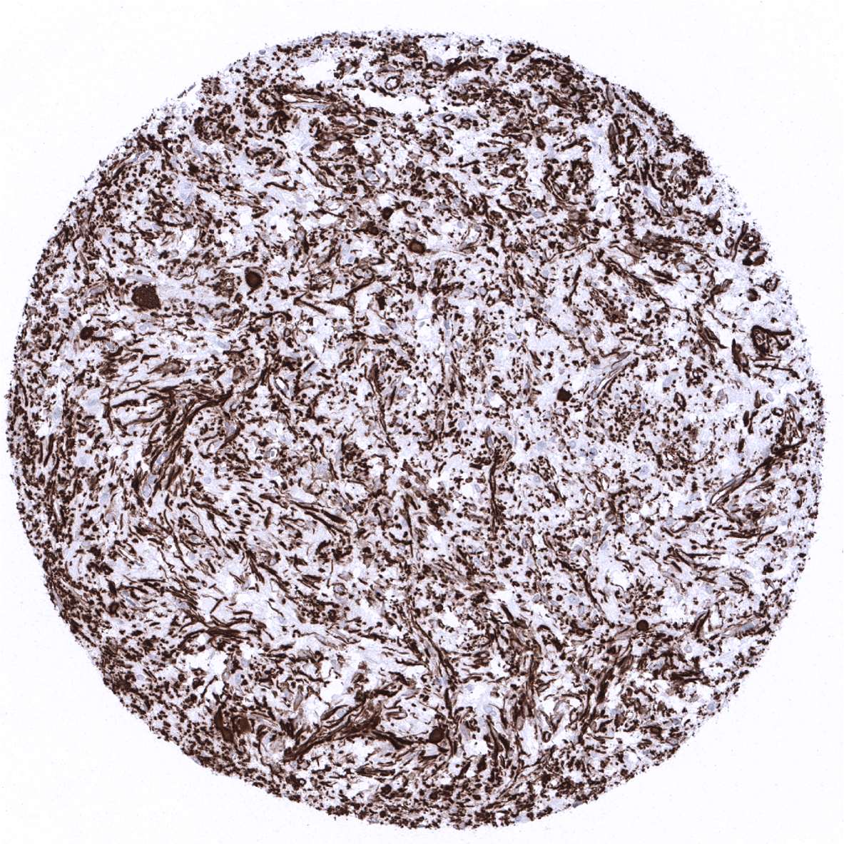
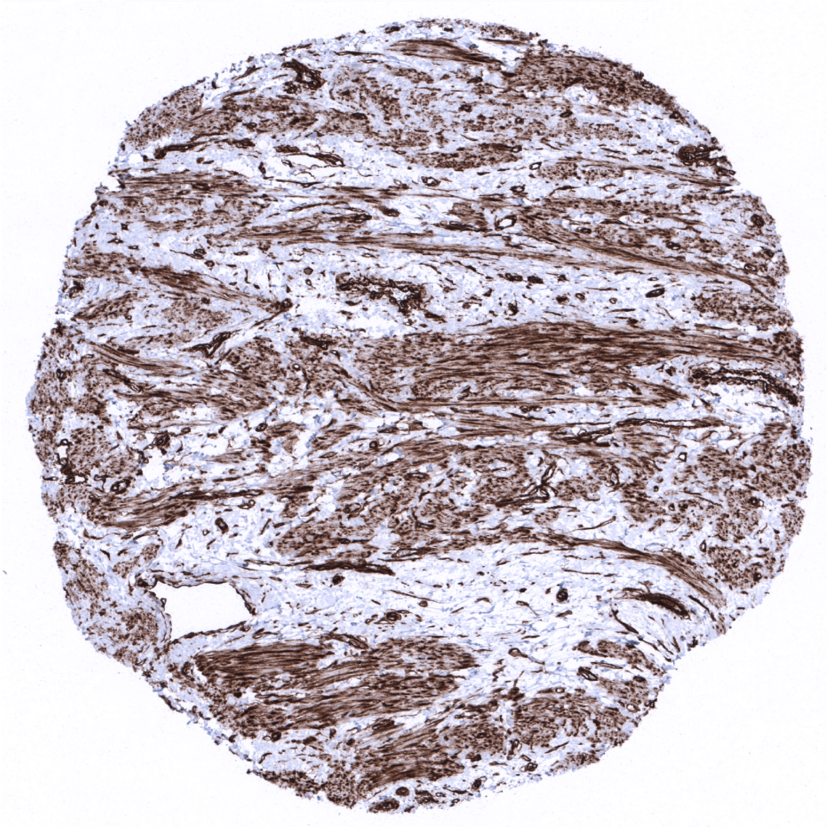
Bone marrow
Breast
Breast
Bronchus, mucosa - Some vimentin immunostaining may be found in epithelial cells of the respiratory mucosa.
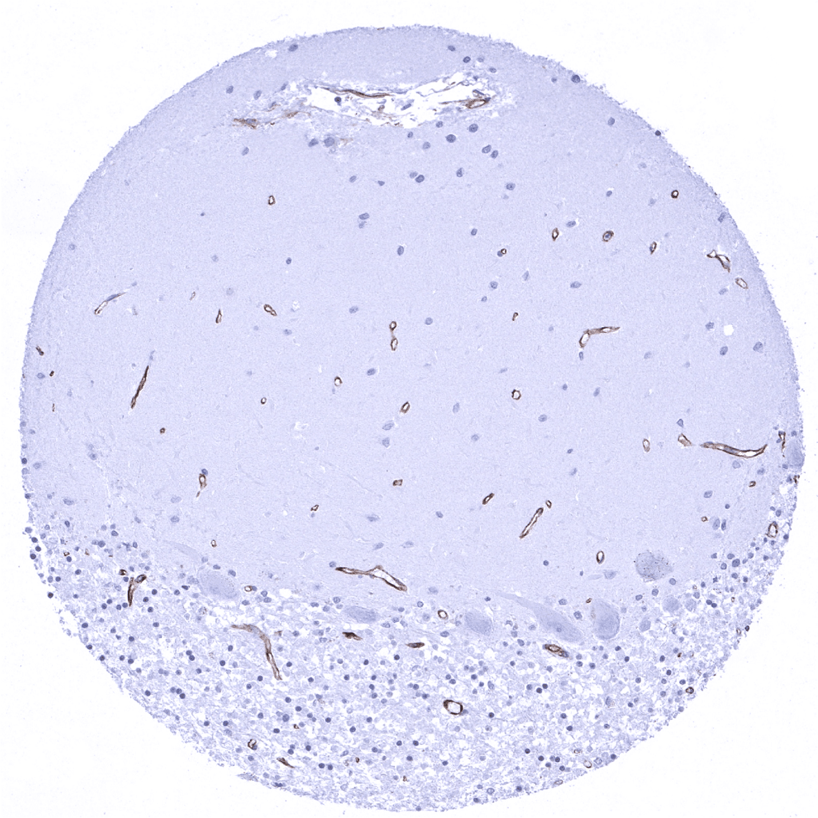
Cerebellum (molecular layer, Purkinje cell layer, granule cell layer)
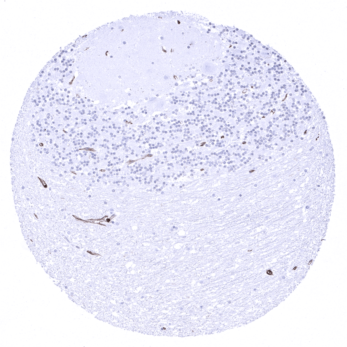
Cerebellum (molecular layer, Purkinje cell layer, granule cell layer, white matter)
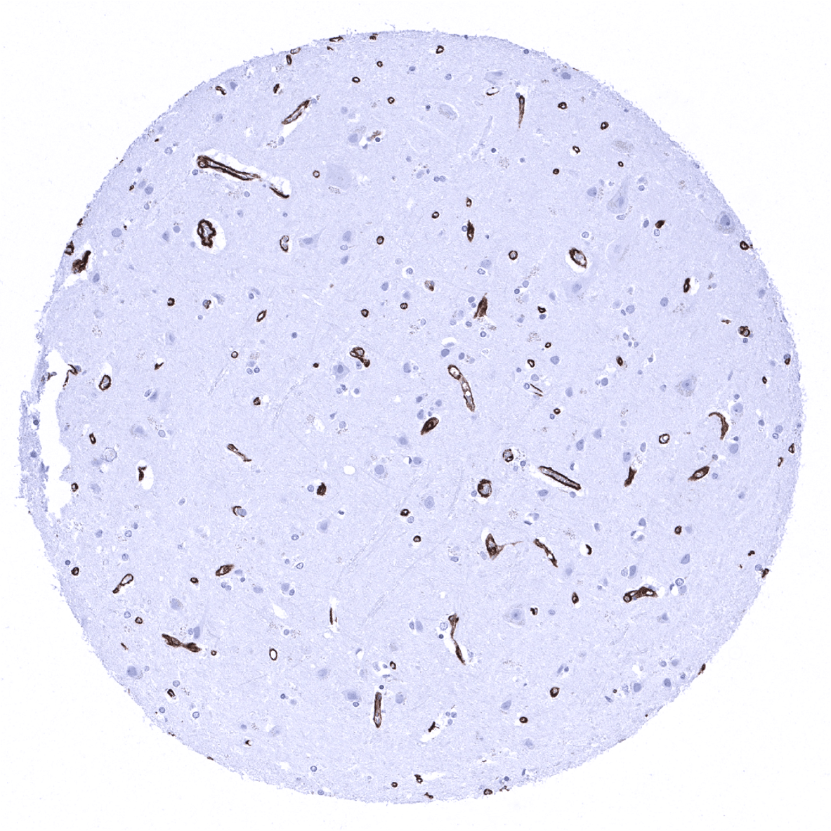
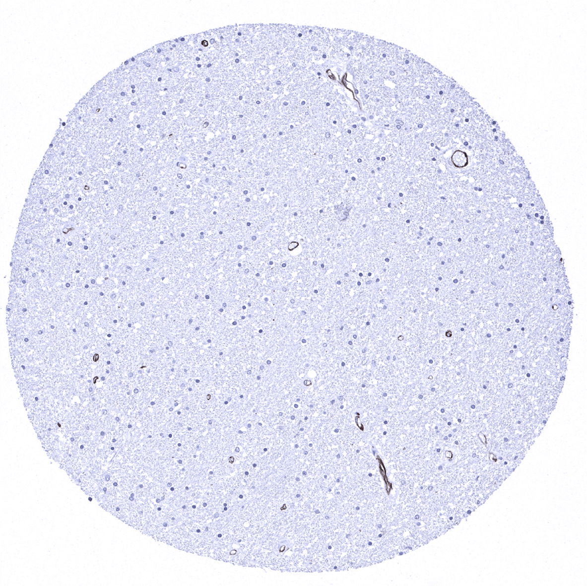
Cerebrum, grey matter
Cerebrum, white matter

Colon descendens, mucosa
Colon descendens, muscular wall
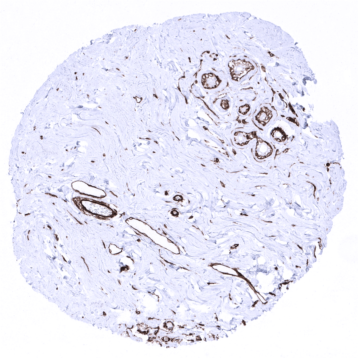
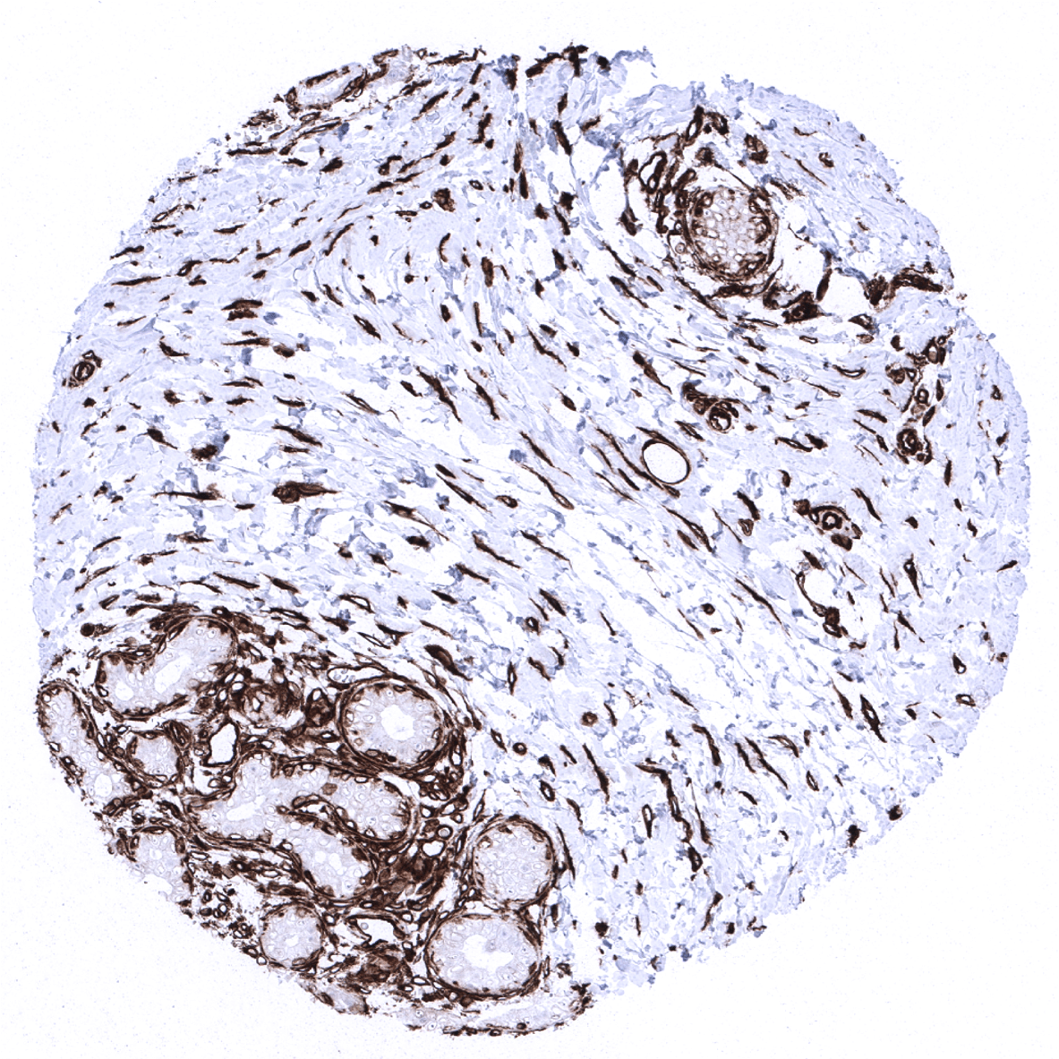
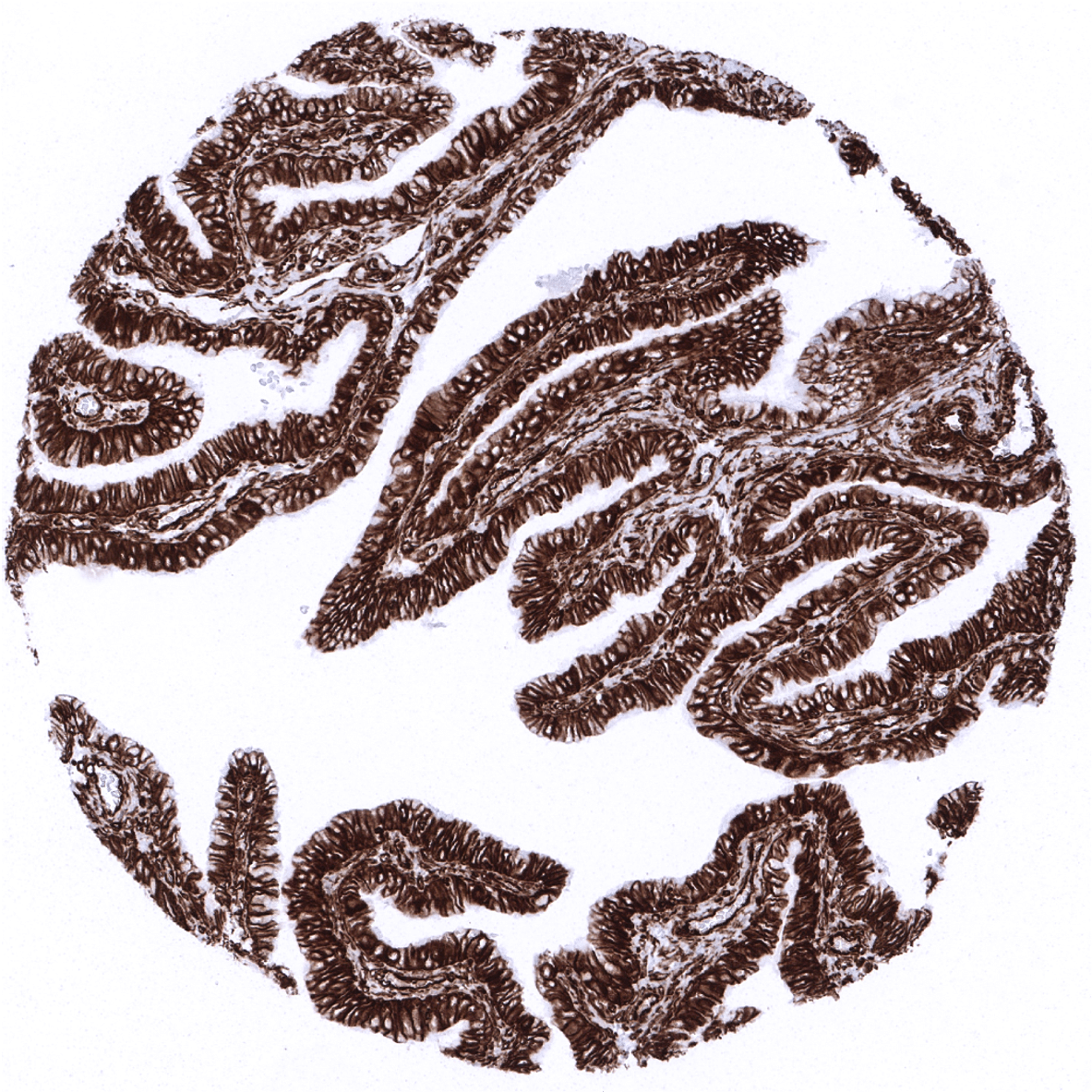
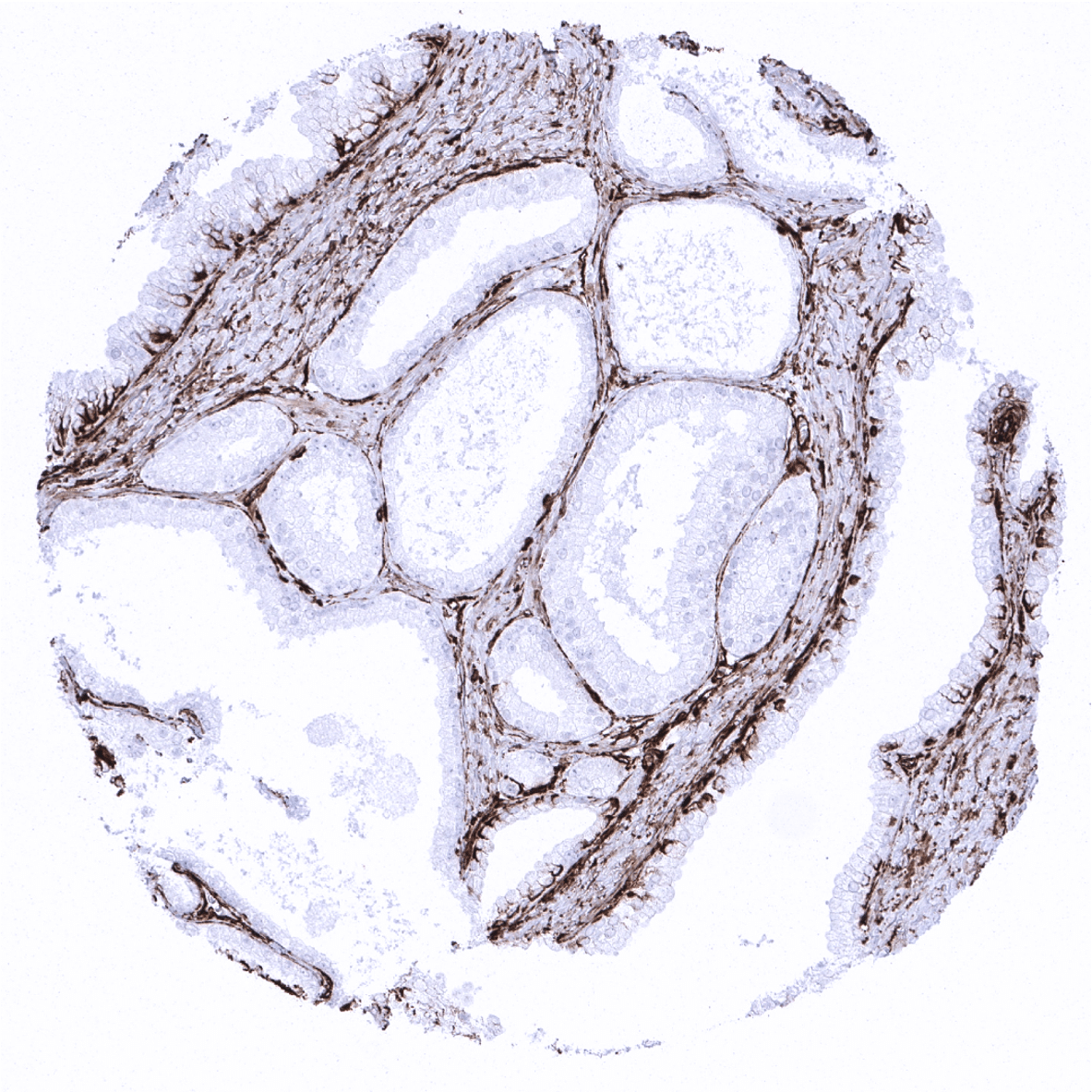
Duodenum, Brunner gland

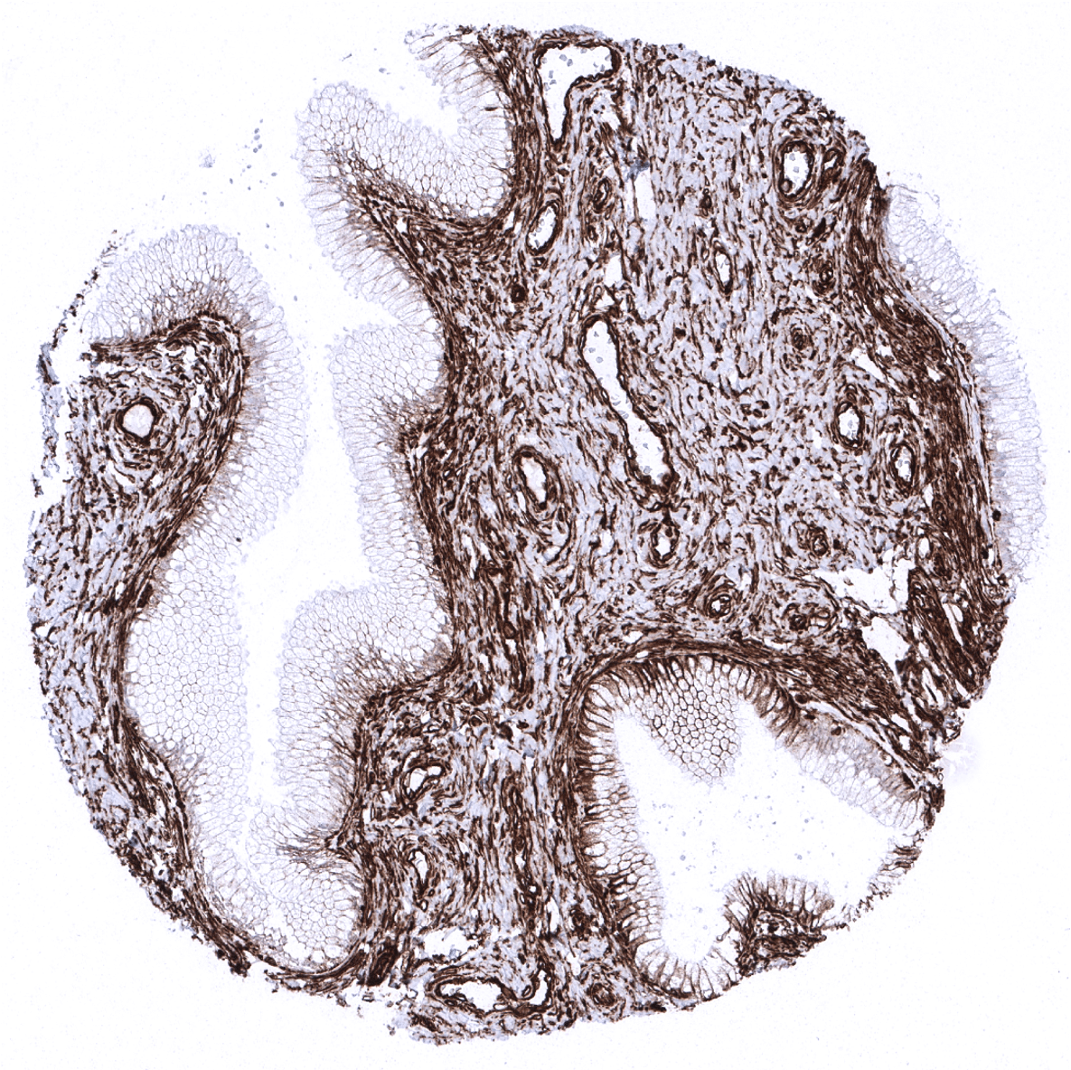
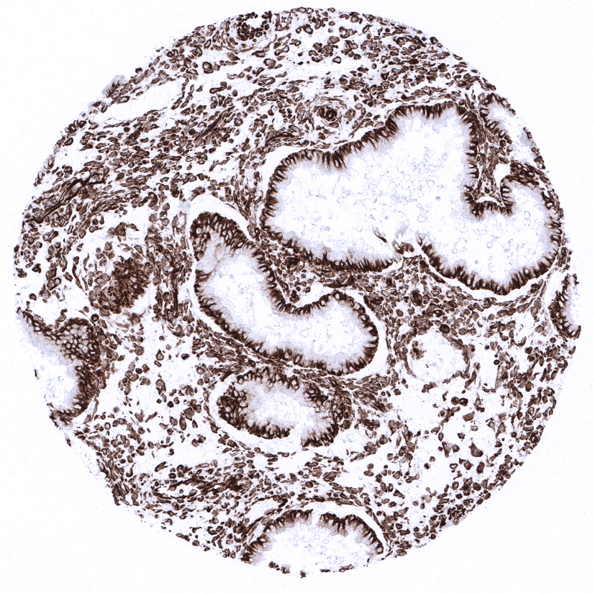
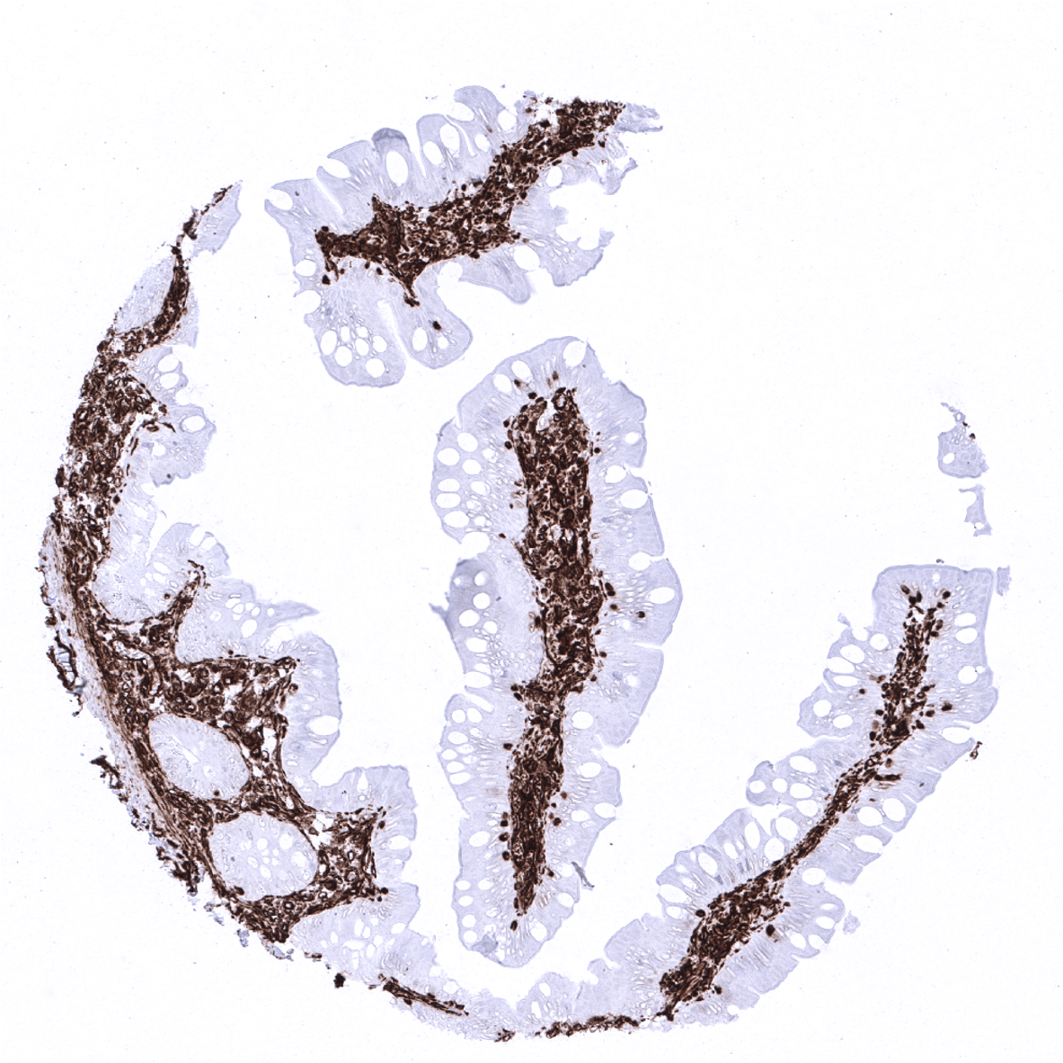
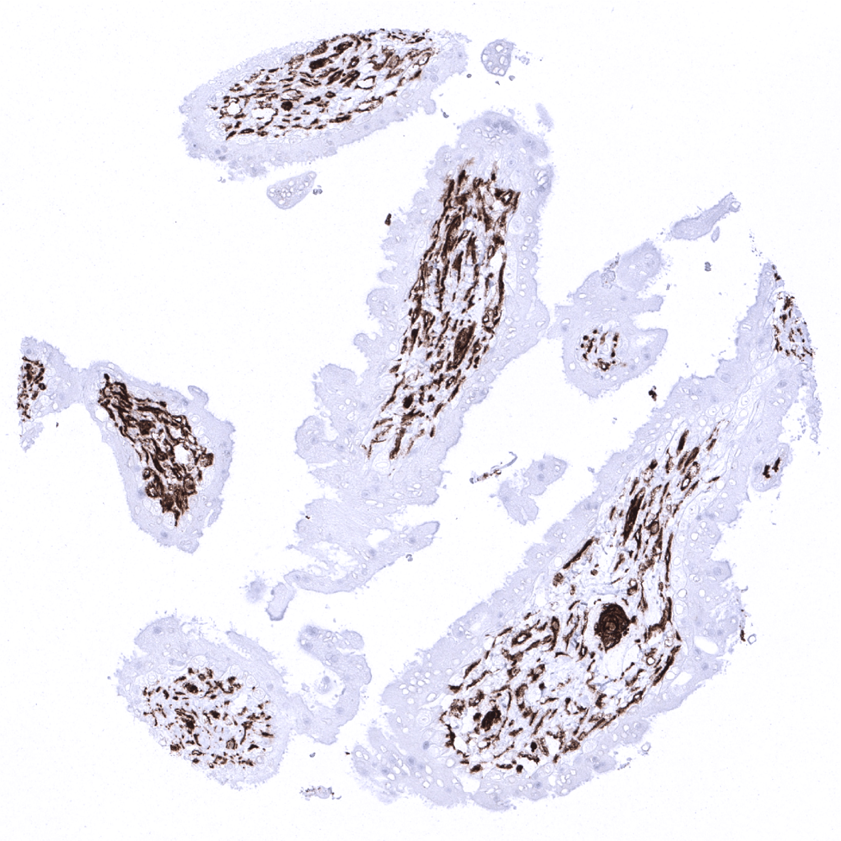
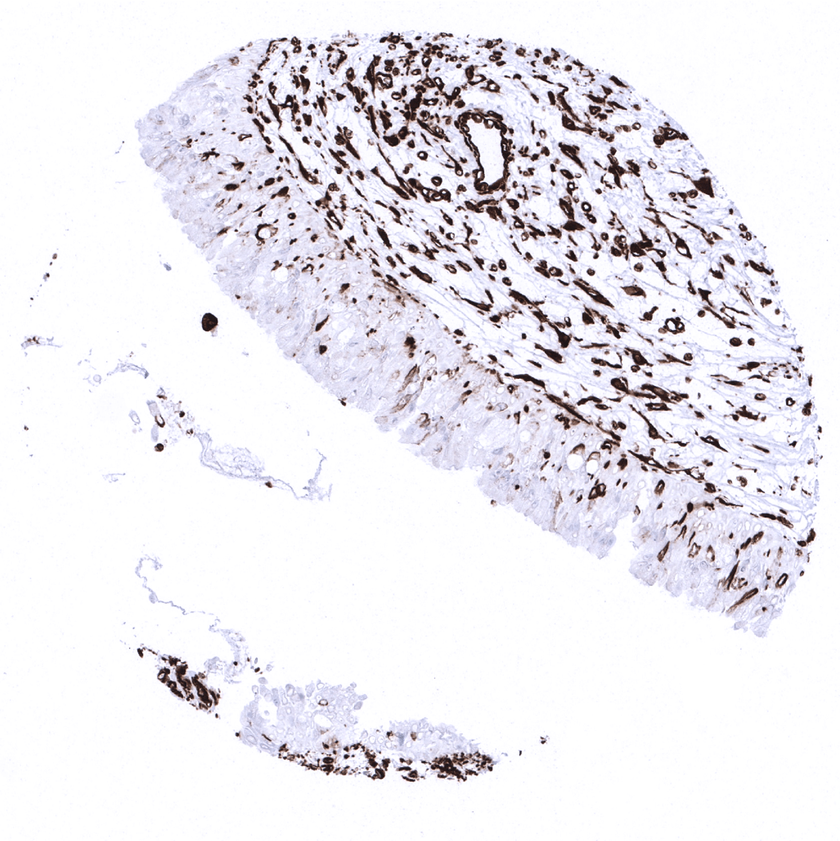
Duodenum, mucosa - In the duodenum, endothelial cells of large vessels and stromal cells must show strong staining while dispersed intraepithelial T-cells must show an at least moderate staining.

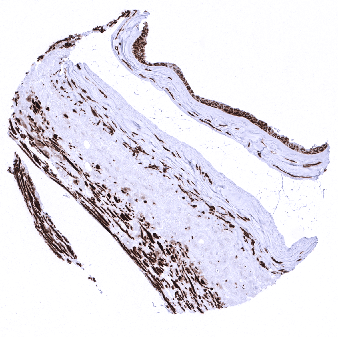
Ectocervix - Strong vimentin staining is also seen in intra-epithelial Langerhans cells.
Endocervix

Endometrium, proliferation - Glandular cells and stromal cells of the endometrium show strong vimentin immunostaining.
Endometrium, secretion

Epididymis - Some principal cells in the epididymis can show moderate to strong vimentin immunostaining.

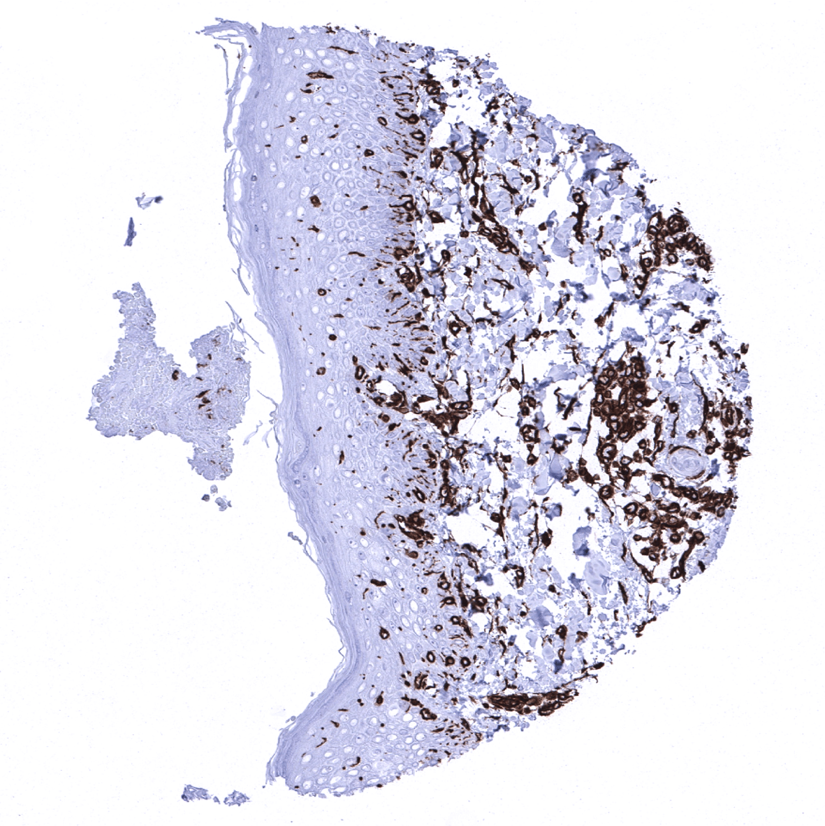
Esophagus, squamous epithelium - Strong vimentin staining is also seen in intra-epithelial Langerhans cells.
Fallopian tube, mucosa - Strong vimentin immunostaining of glandular cells and stromal cells of the fallopian tube.
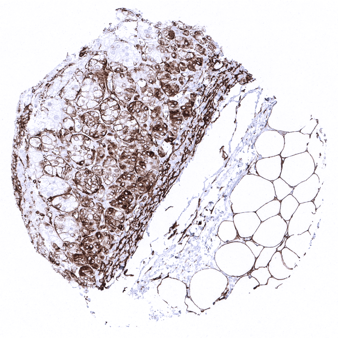
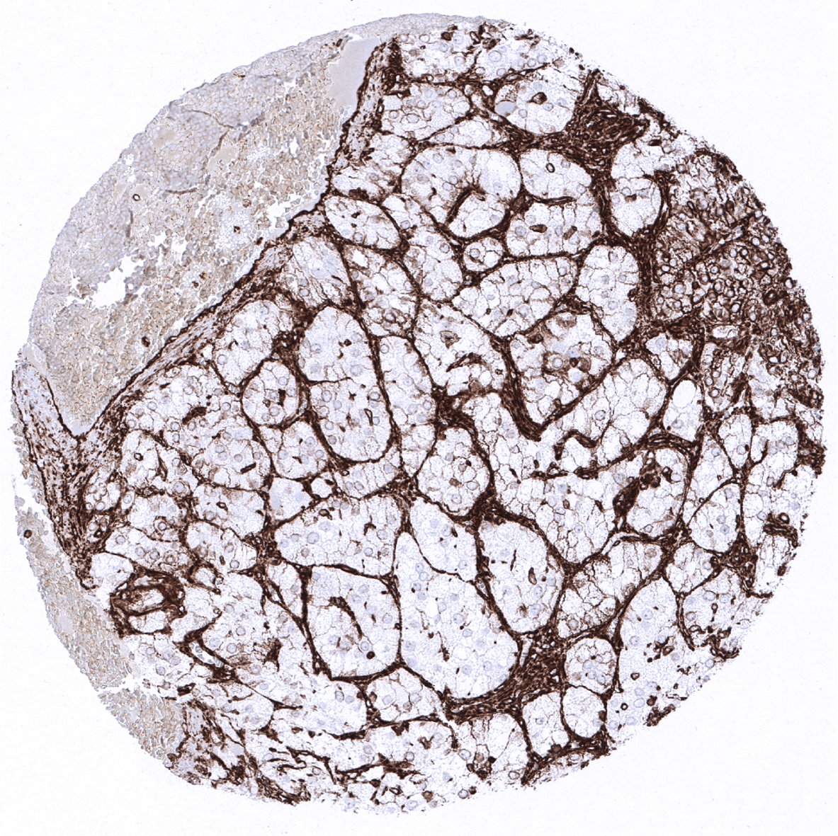
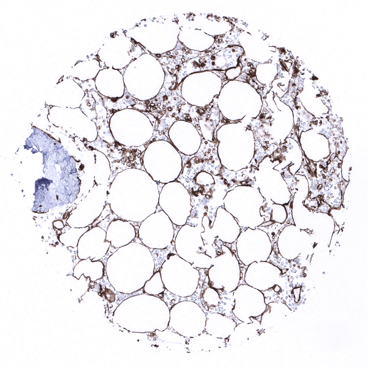
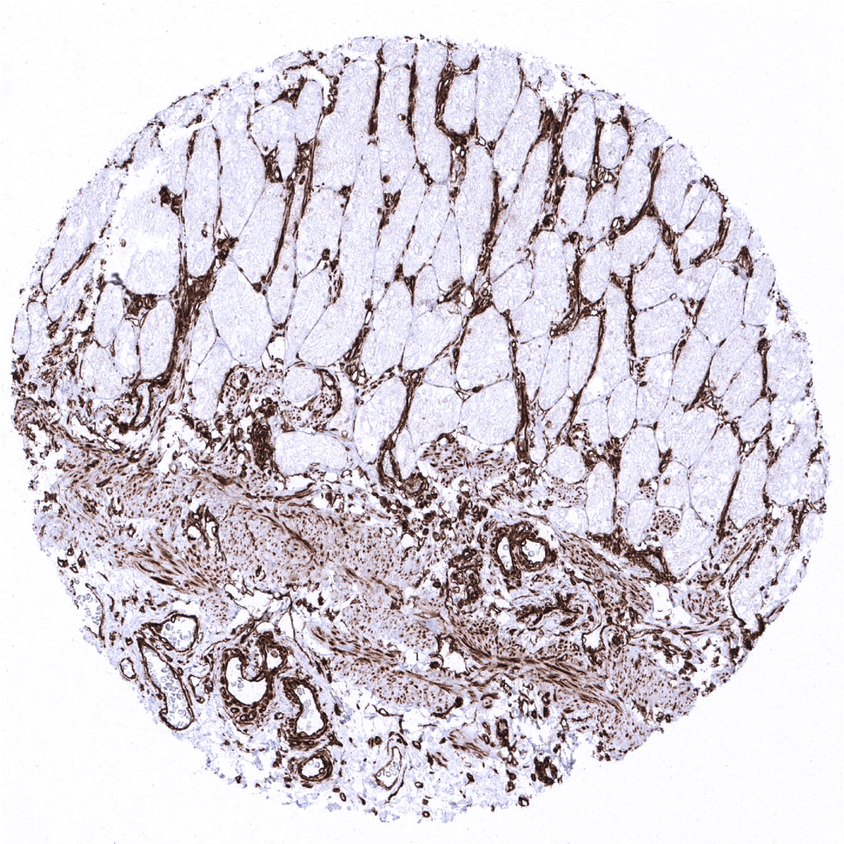
Fat
Gallbladder, epithelium
Heart
Ileum, mucosa
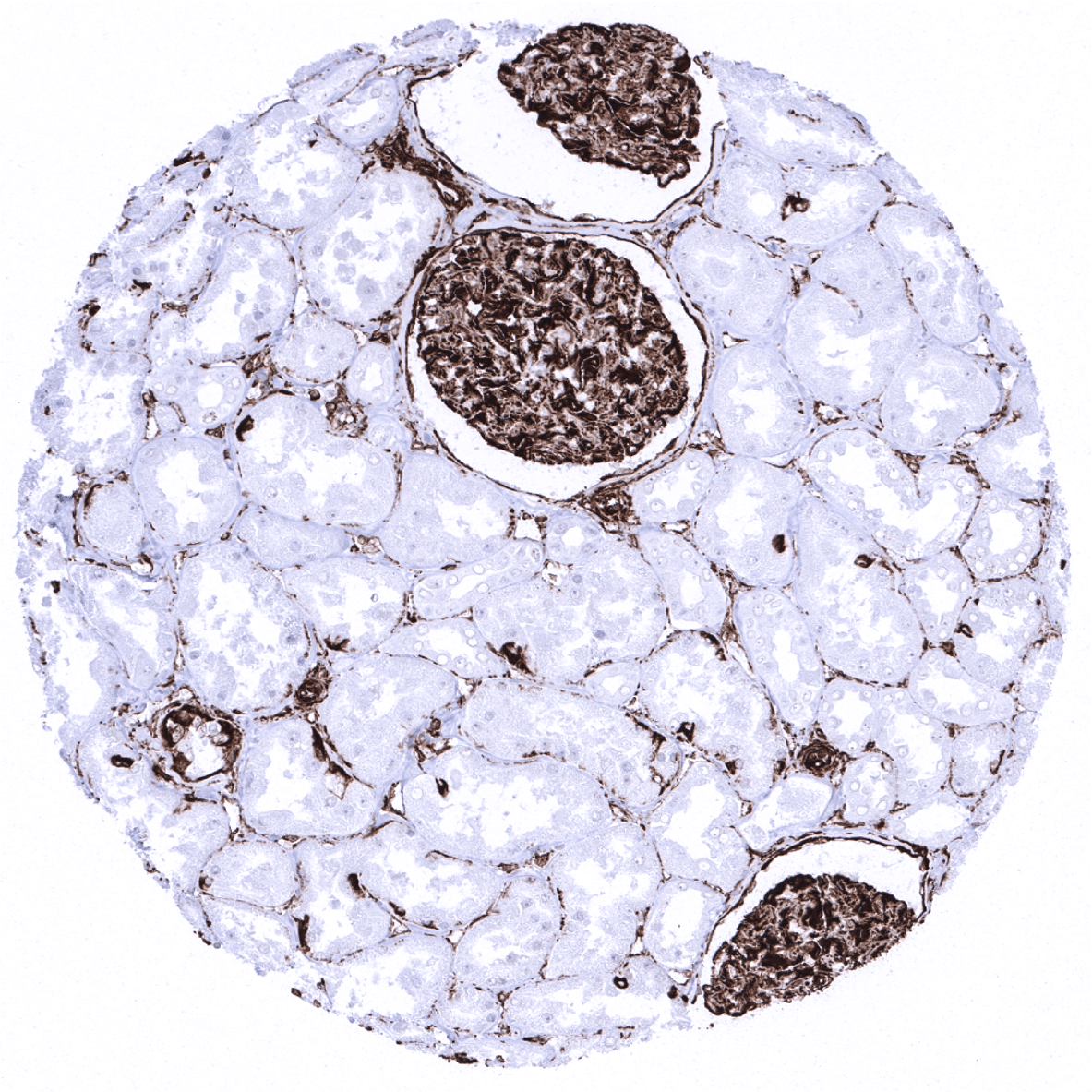
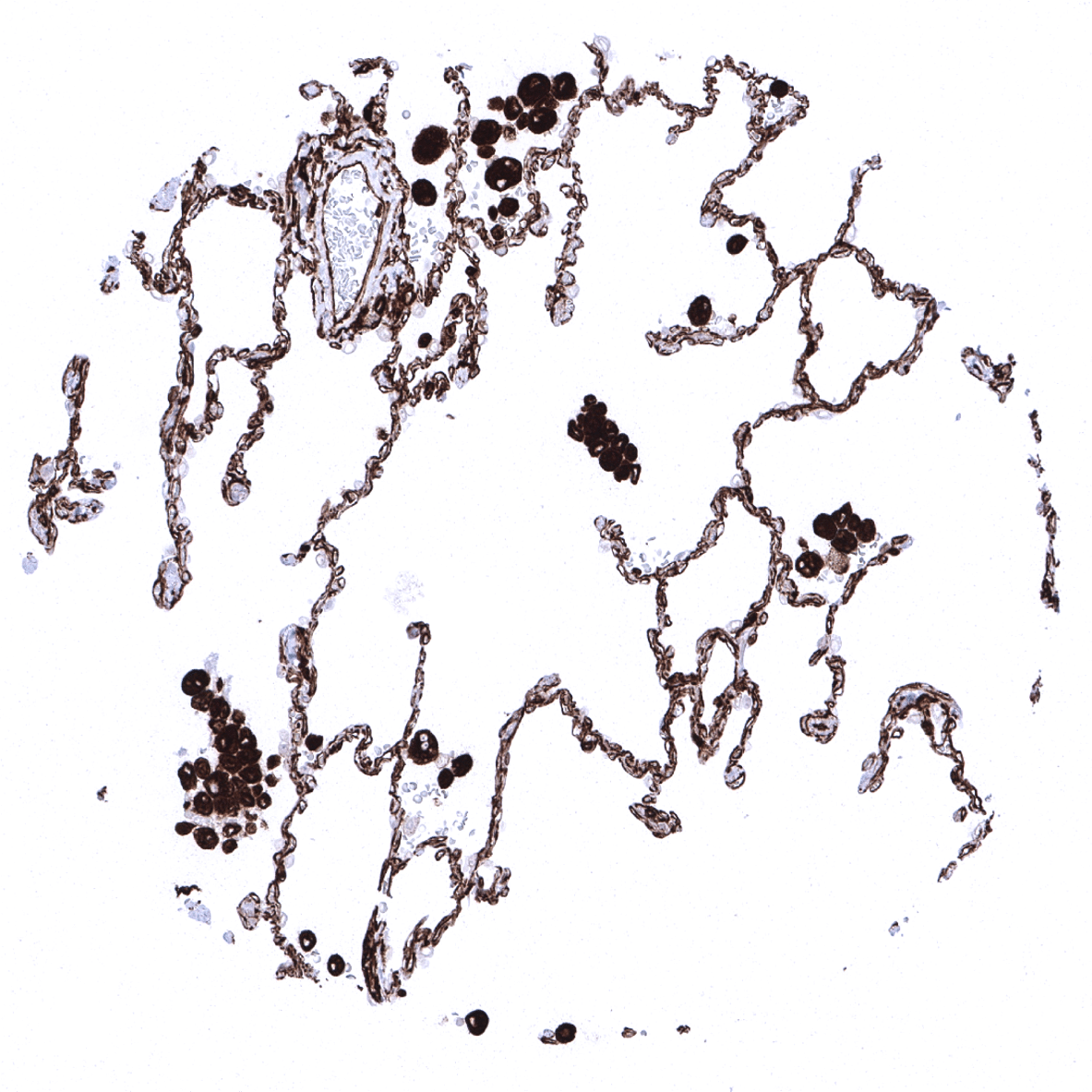
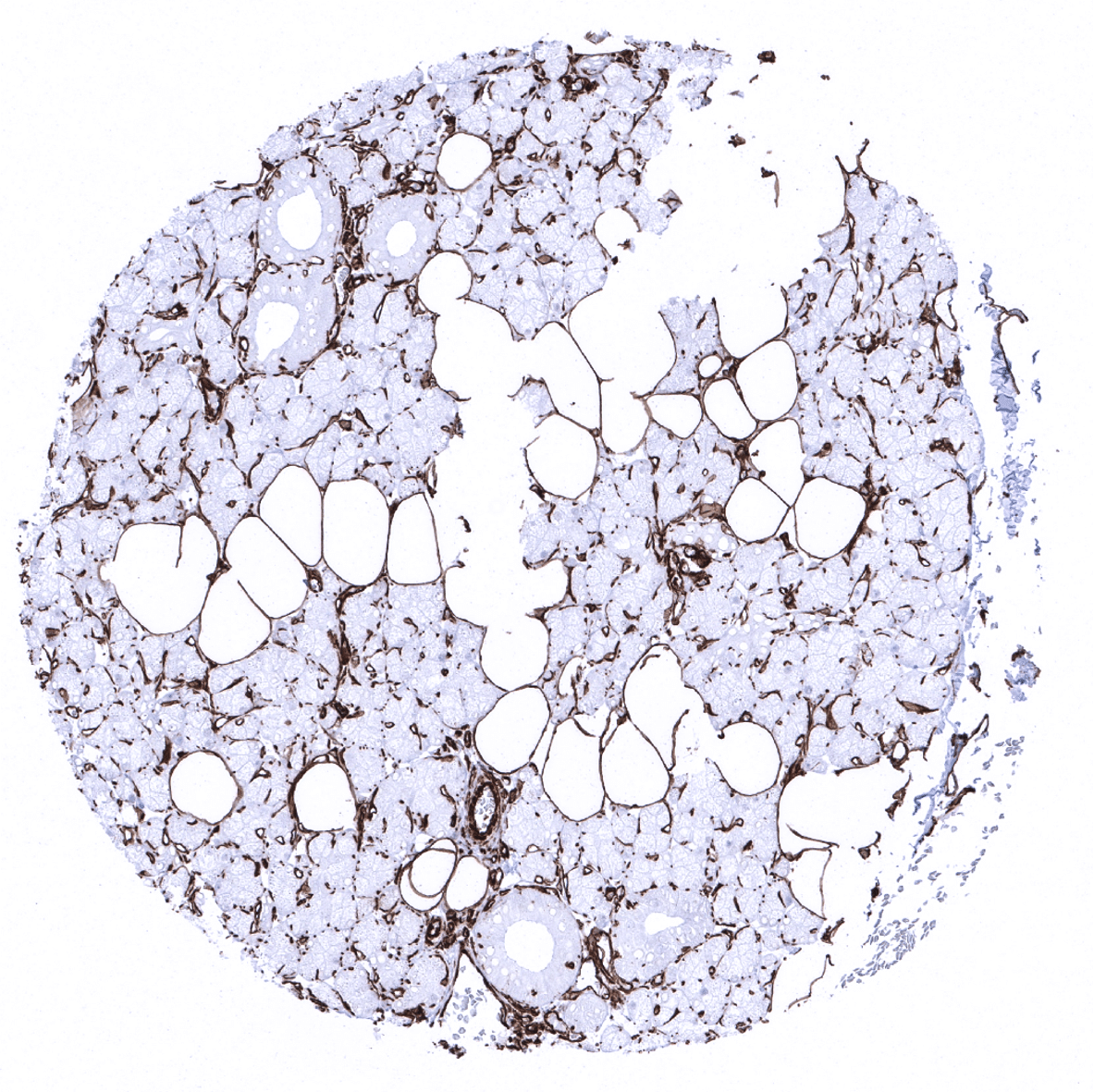
Kidney, cortex - In the normal kidney, a strong vimentin staining is seen in all cell types of glomeruli and Bowman capsule and in vessels. Epithelial cells are vimentin negative.

Kidney, medulla
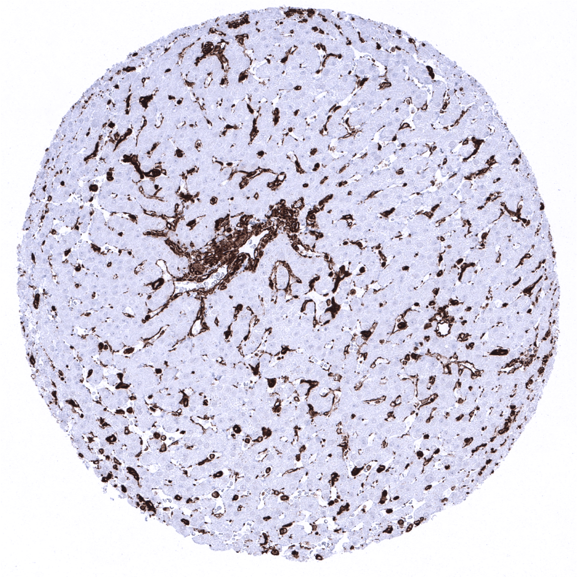
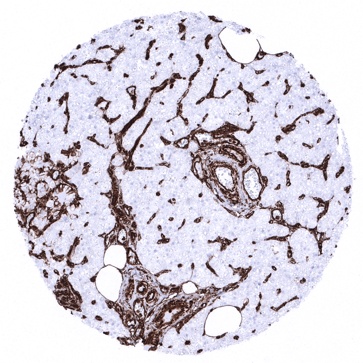
Liver - In the liver, Kupffer cells must show a strong staining and sinusoidal endothelial cells should show an at least a weak staining.
Lung
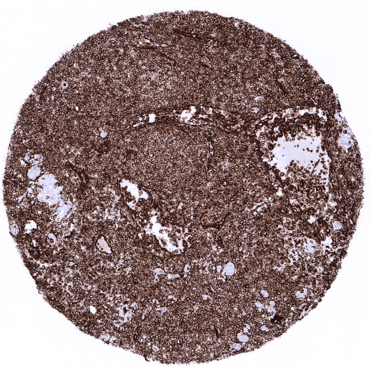
Lymph node - Strong vimentin expression in all lymphocytic cell types.
Ovary, stroma

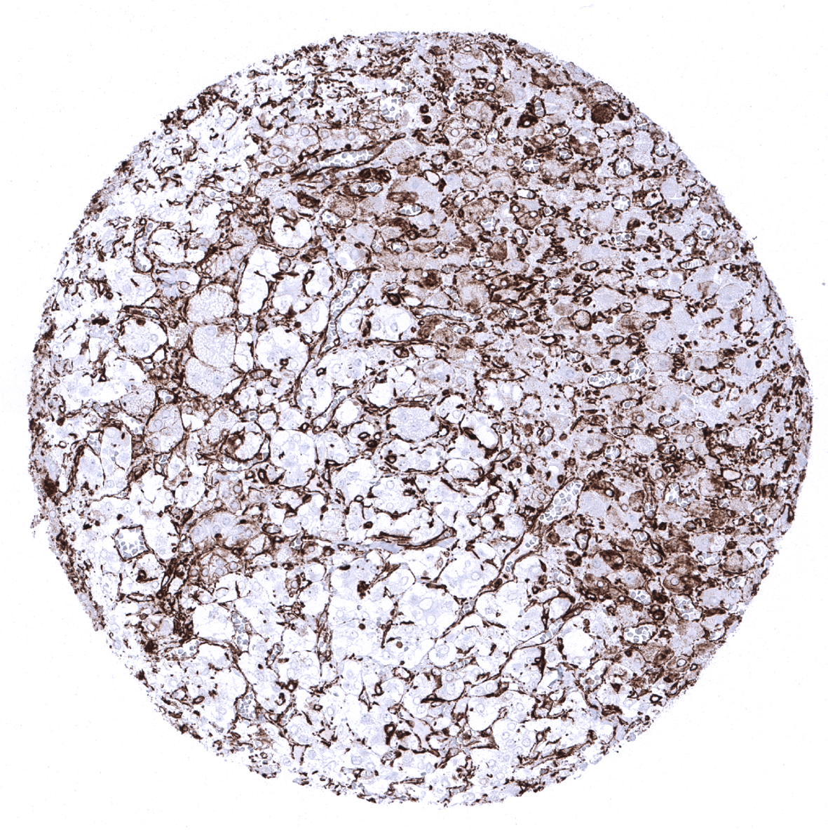
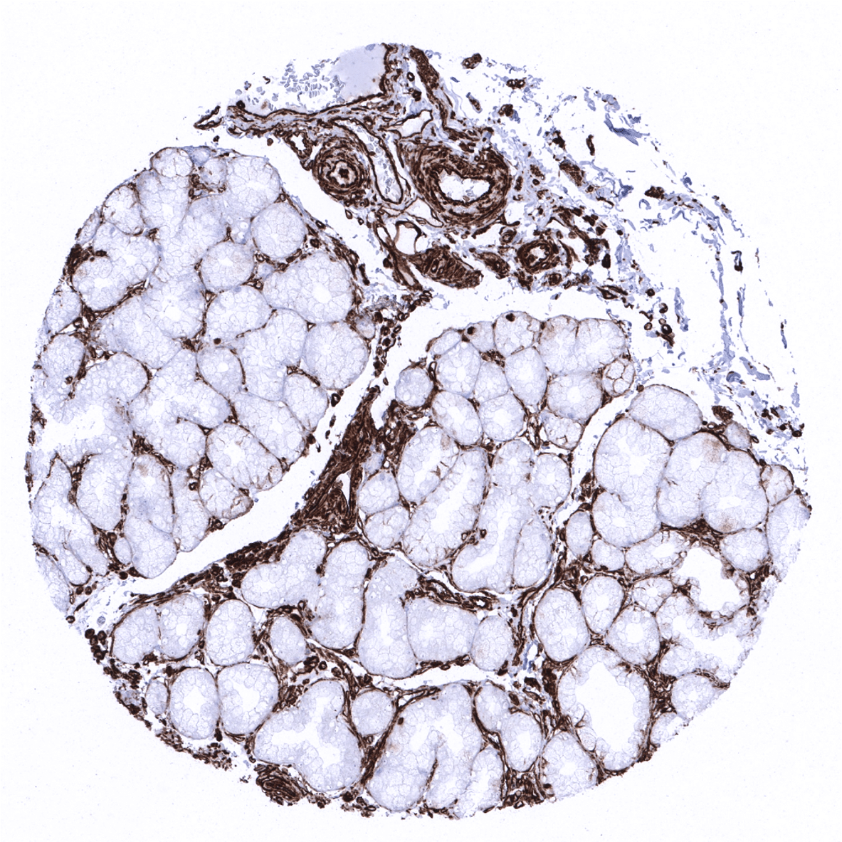
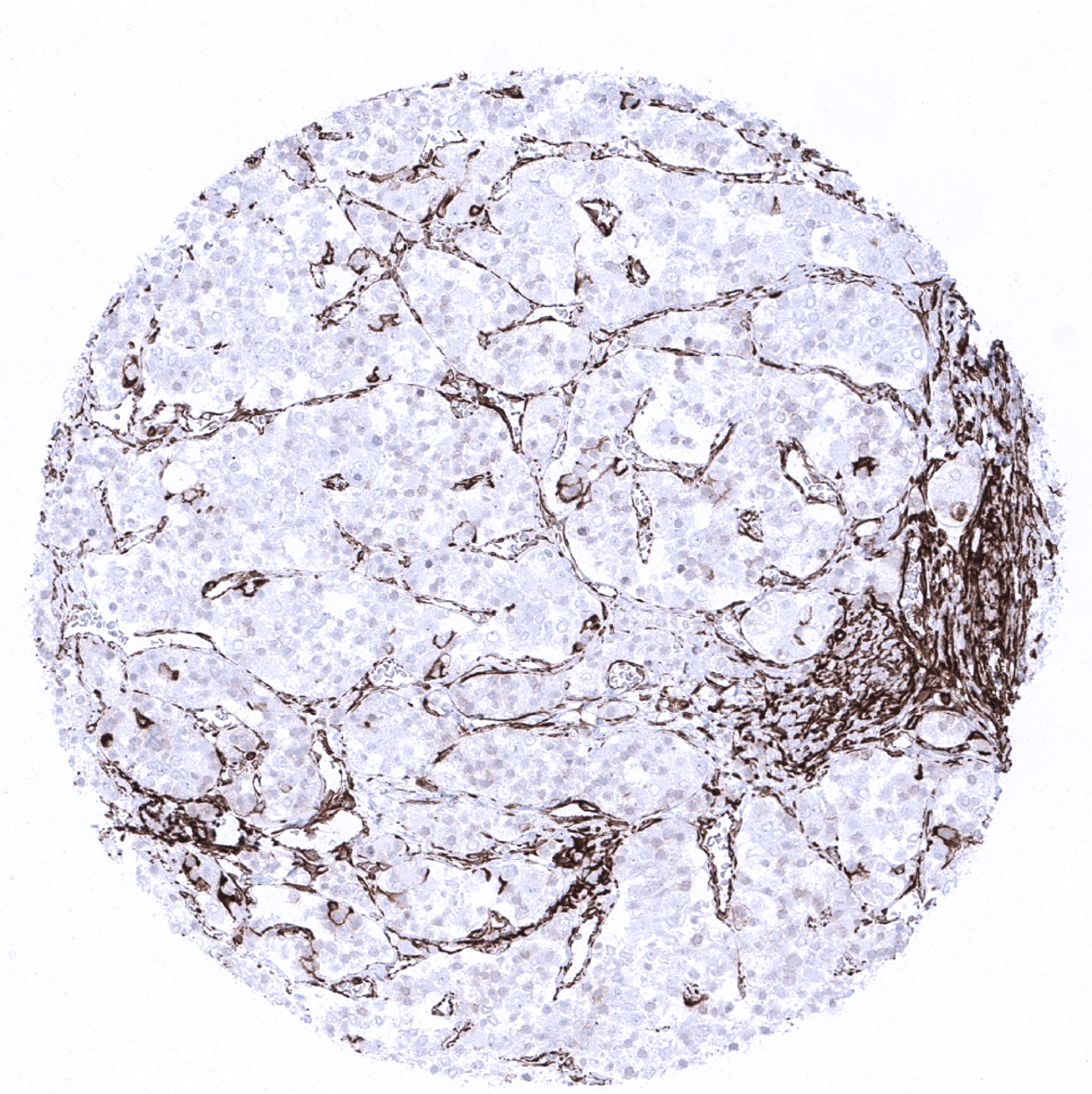
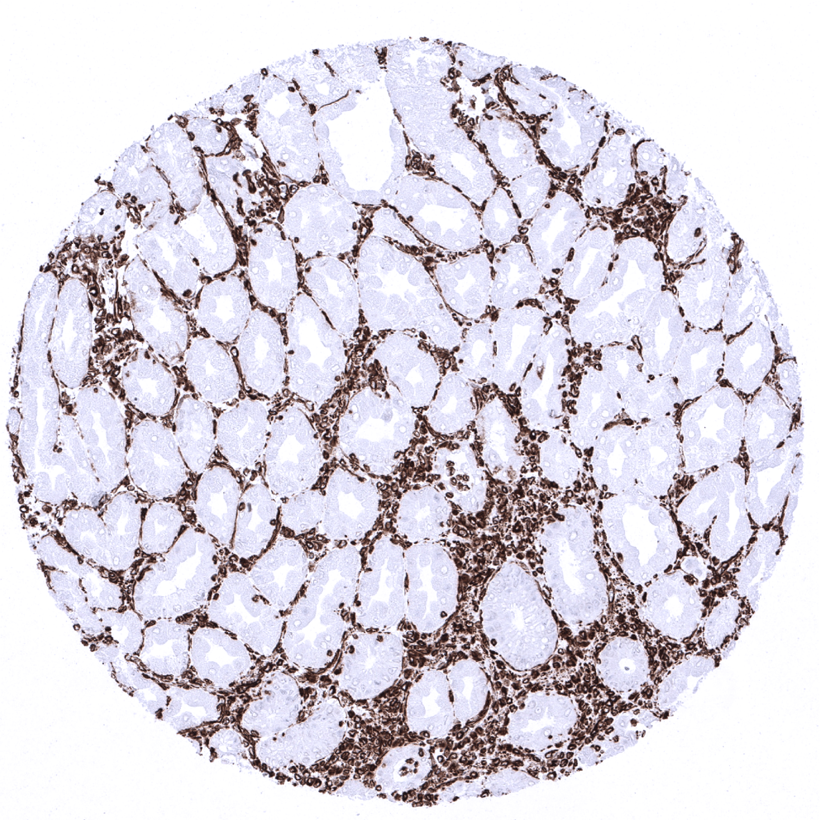
Pancreas - In the pancreas, the vast majority of epithelial cells of exocrine acini should display a weak to strong basolateral membranous and cytoplasmic staining.
Parathyroid
Parotid gland - Strong vimentin immunostaining in fat cells, myoepithelial cells, stroma and blood vessels of the parotid gland.
Pituitary gland, anterior lobe
Pituitary gland, posterior lobe

Pregnant uterus (decidua)
Placenta, early

Placenta, mature - In the placenta, a strong vimentin staining is seen in vessels while trophoblastic cells are strictly negative.
Placenta (amnion and chorion)
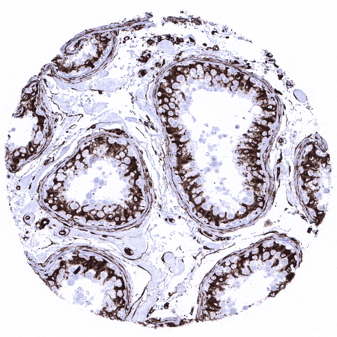
Prostate - Some vimentin immunostaining may be found in prostate acinar cells.
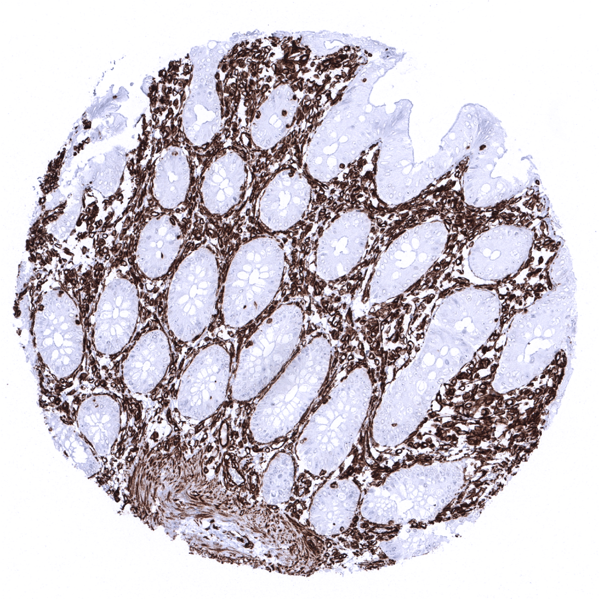
Rectum, mucosa
Seminal vesicle
Sinus paranasales
Skin

Spleen
Stomach, antrum
Stomach, corpus

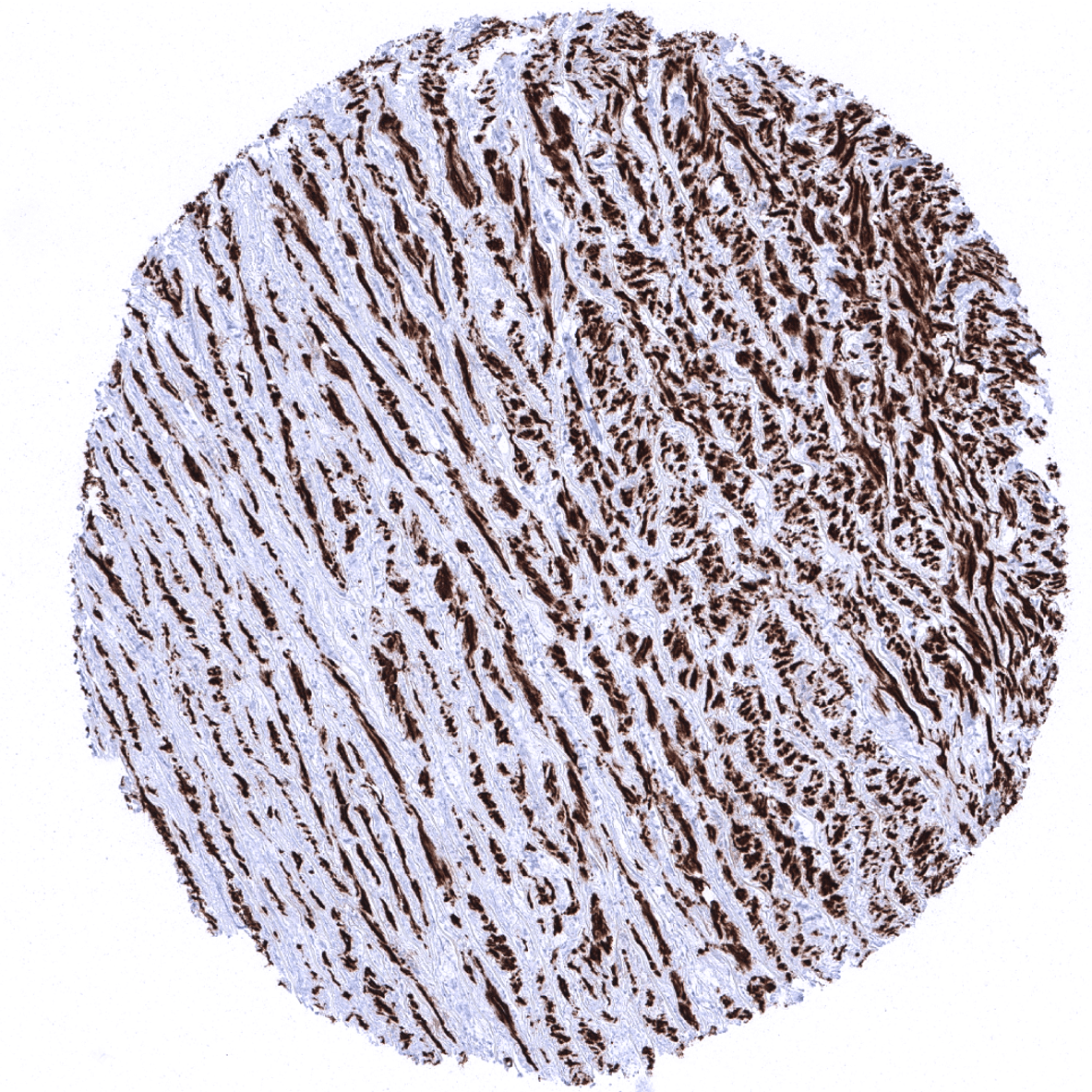
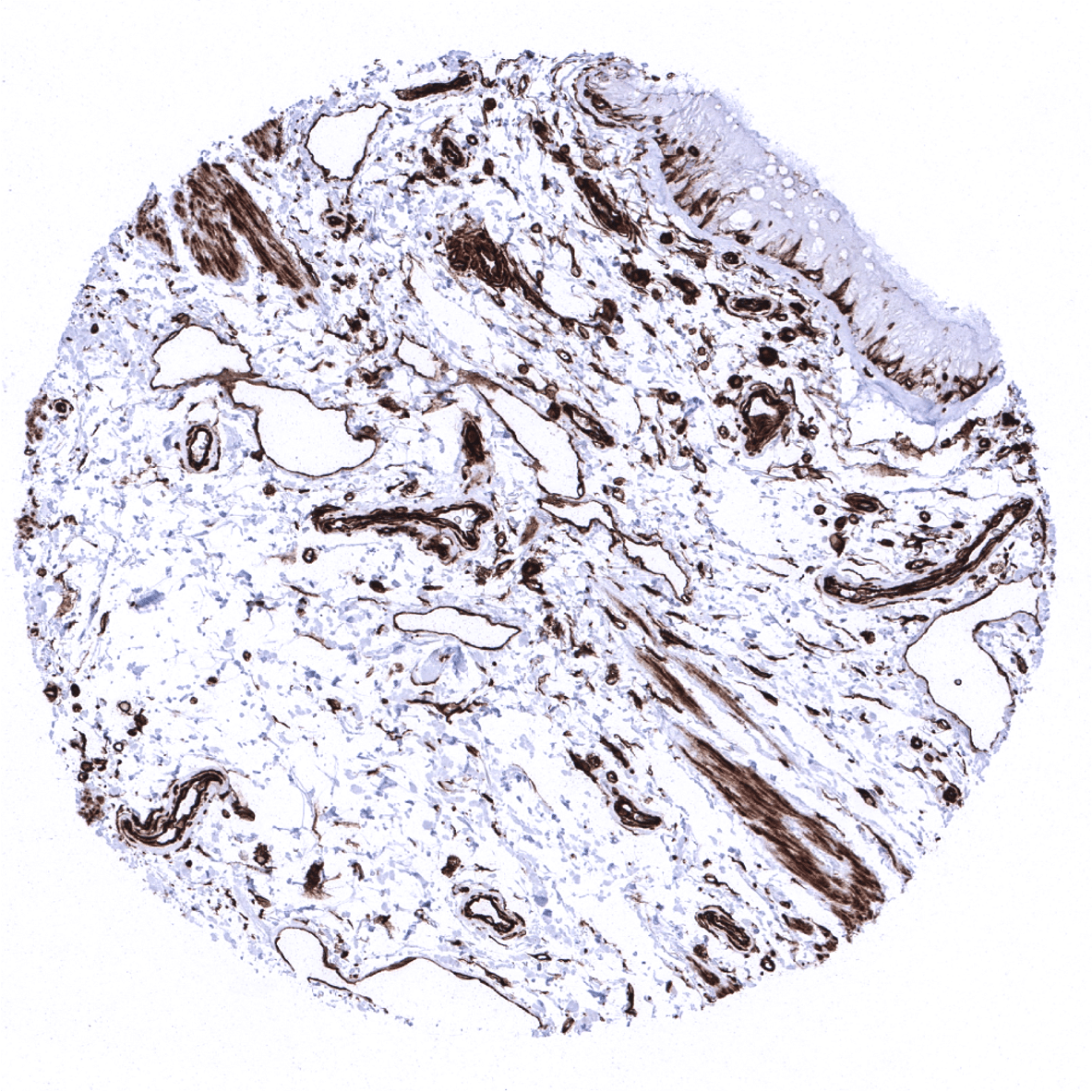
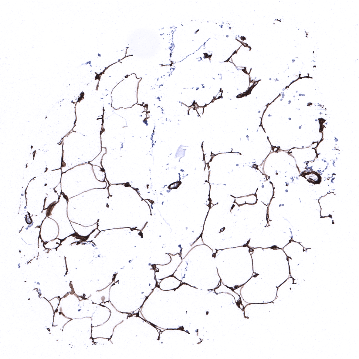
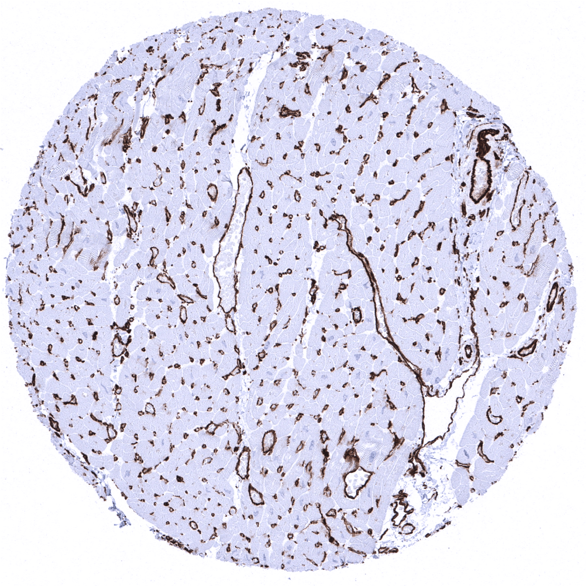
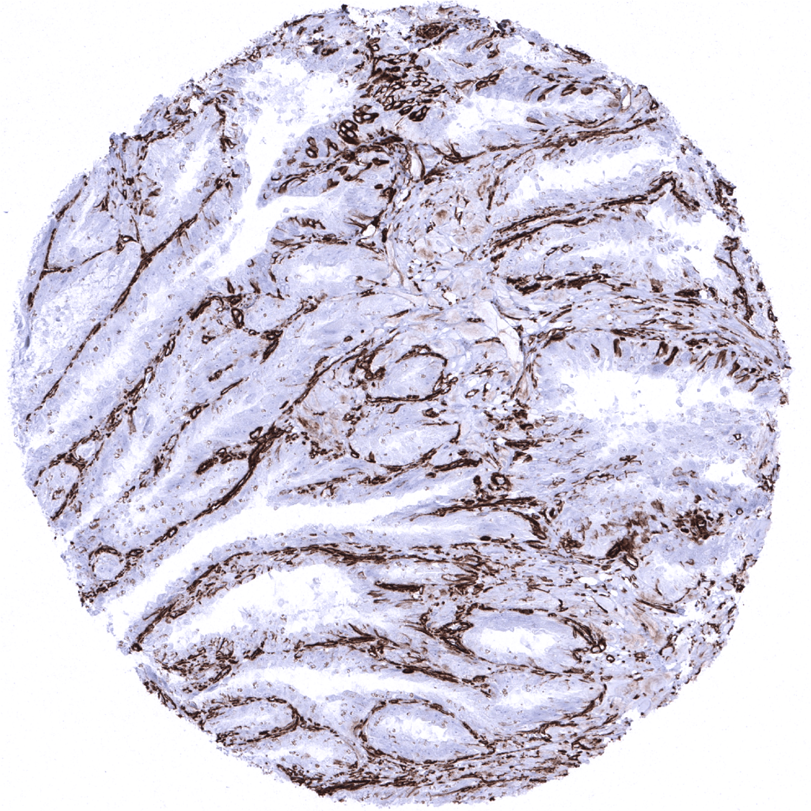
Skeletal muscle - In skeletal muscle, vimentin immunostaining is largely limited to blood vessels.
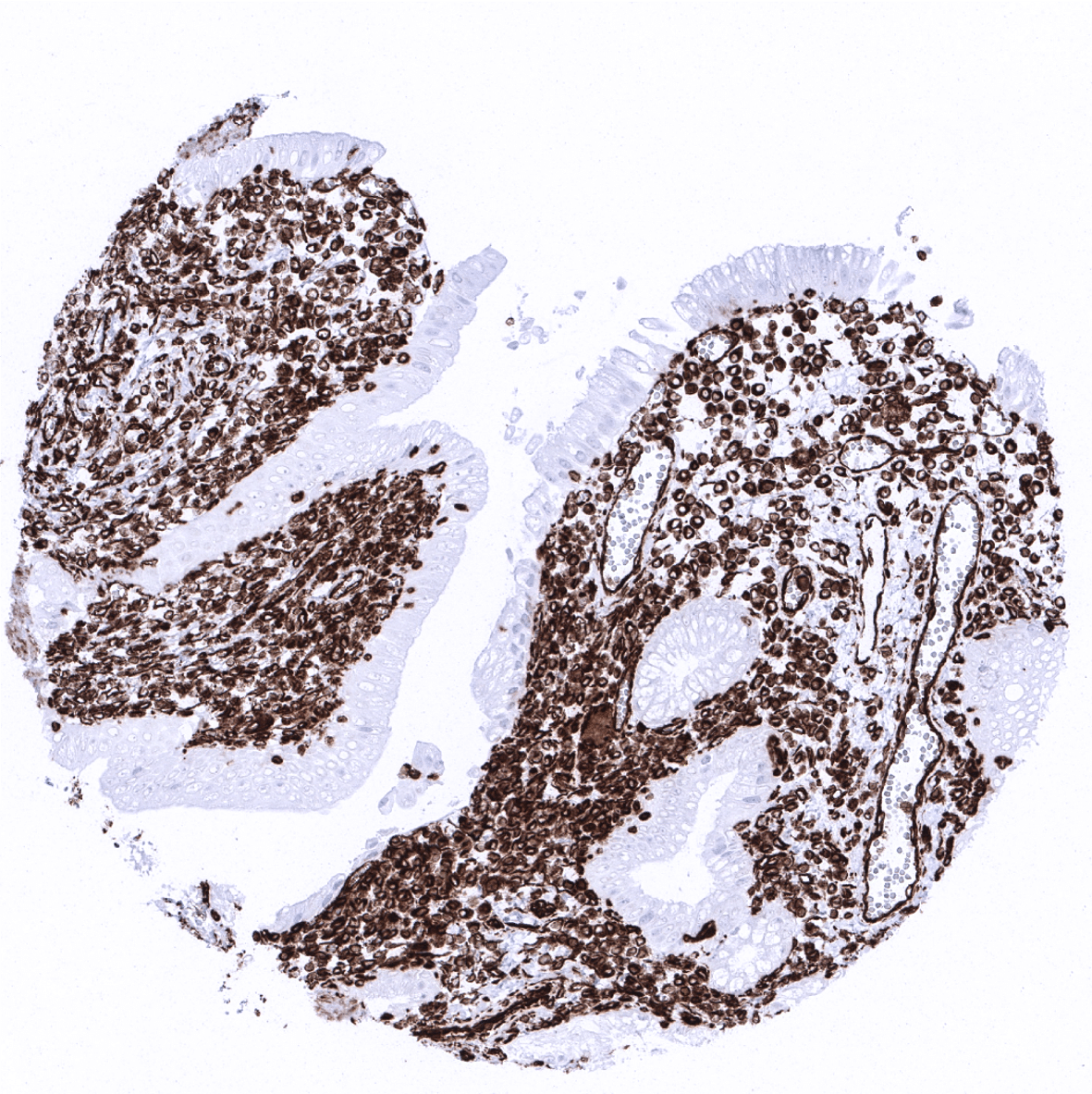
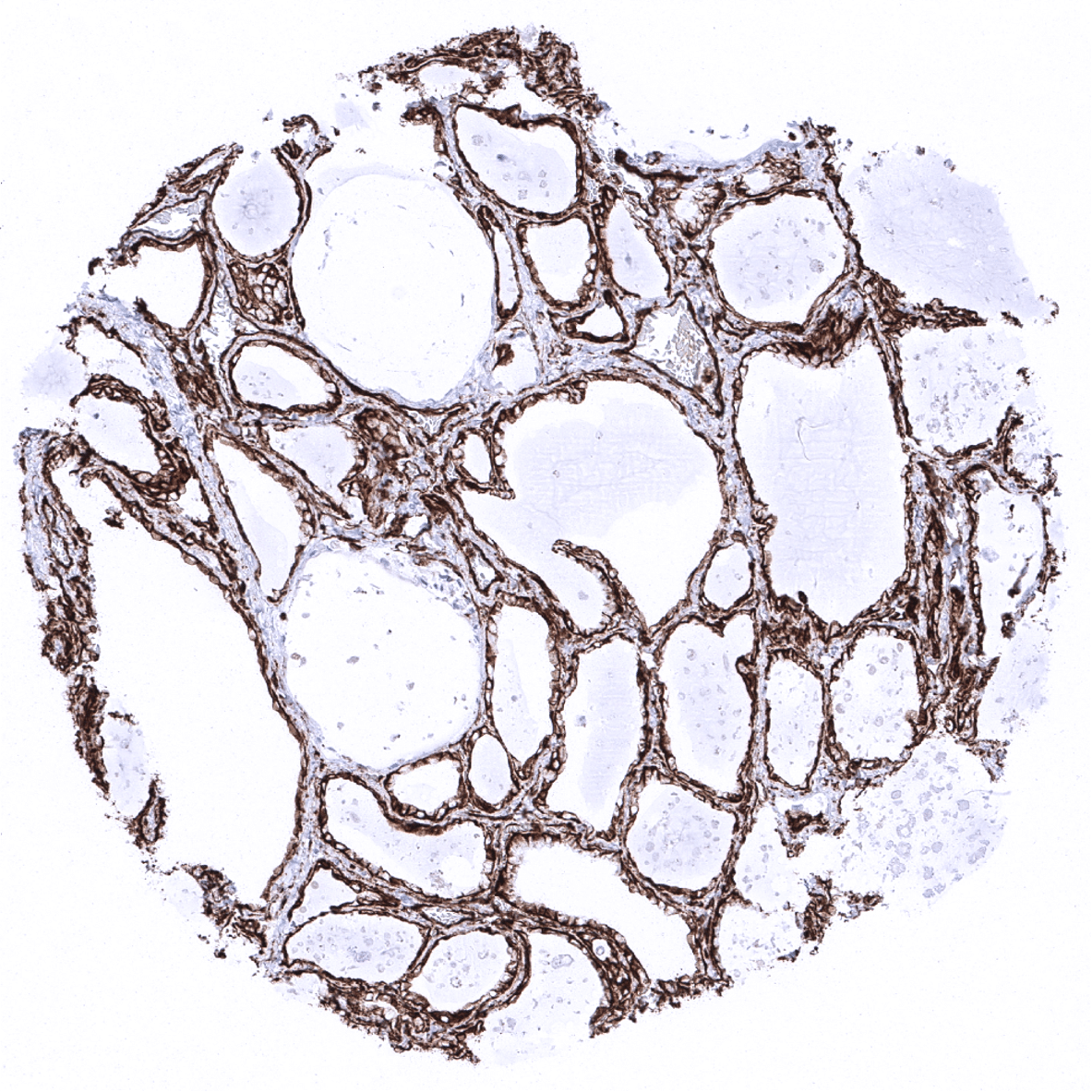
Testis - In the testis, Sertoli and Leydig cells are strongly positive for vimentin (Leydig cells are not seen on this image).

Thymus - Vimentin expression occurs in all lymphocytic cell types.
Thyroid gland - Follicular epithelial cells and stromal cells show strong vimentin immunostaining.

Tonsil, surface epithelium

Tonsil
Urinary bladder, muscular wall
Urinary bladder, urothelium
Uterus, myometrium