Adrenal gland

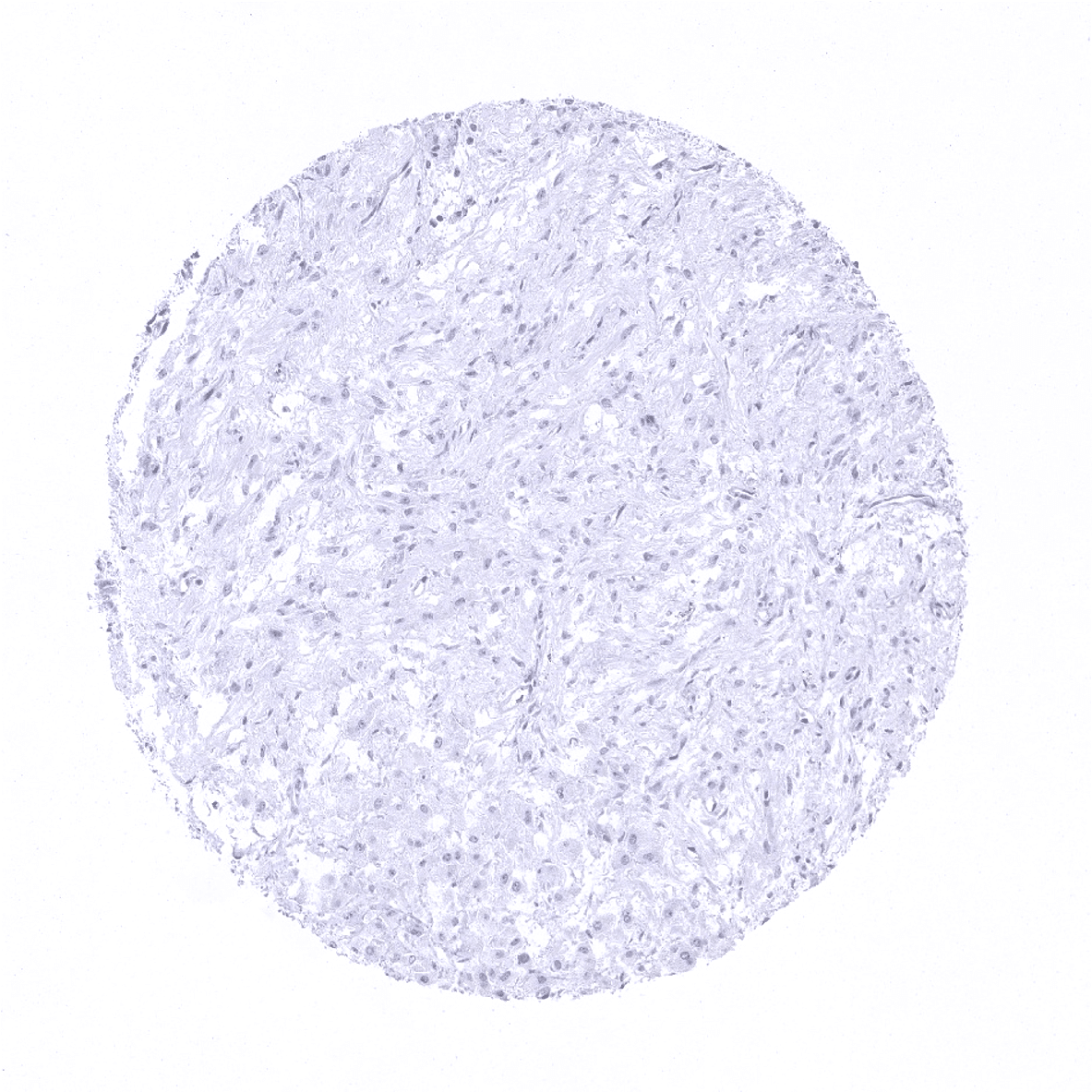
Aorta, media
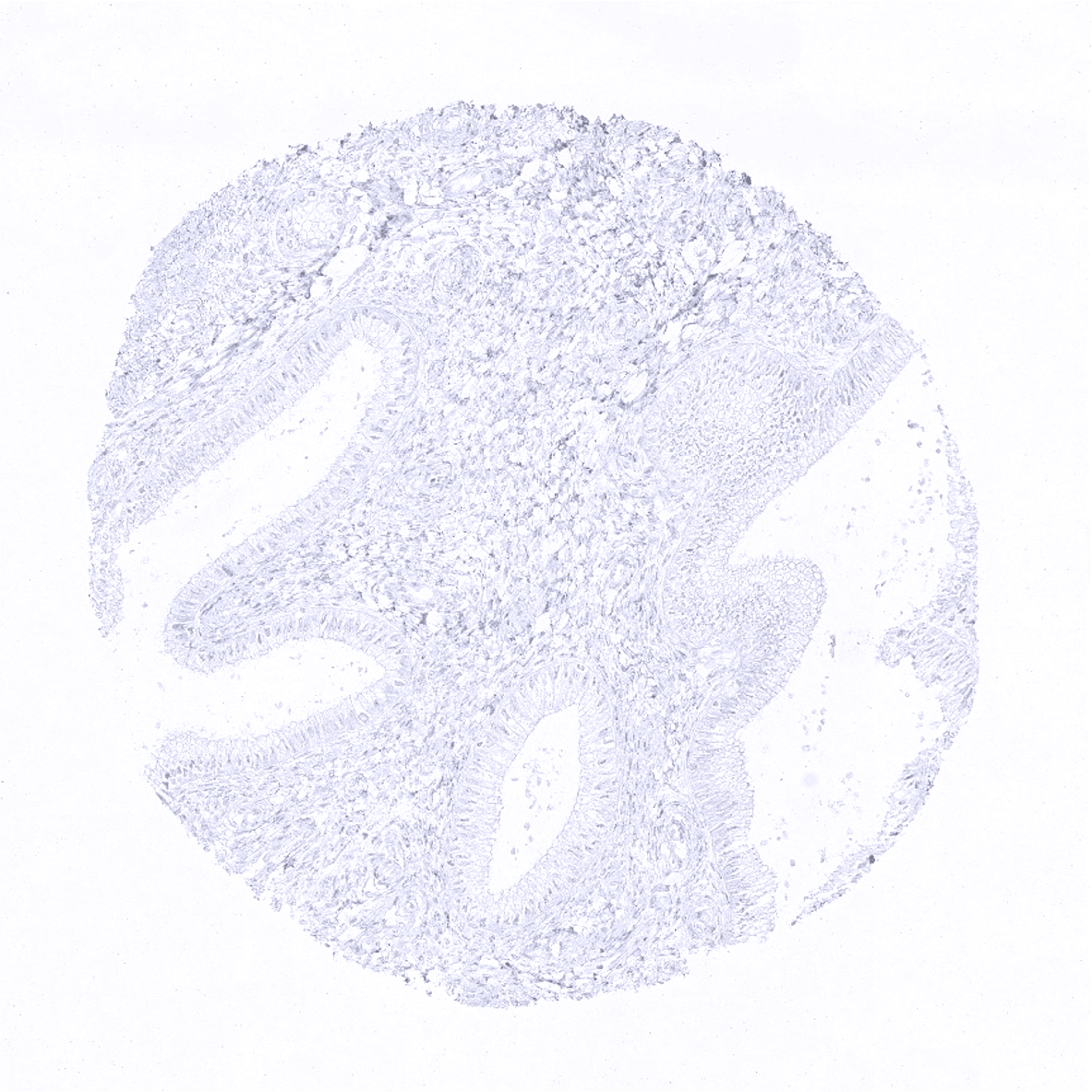
Appendix, mucosa - Villin immunostaining is predominantly membranous but also cytoplasmic and often shows a strong focus on the apical/luminal membranes.
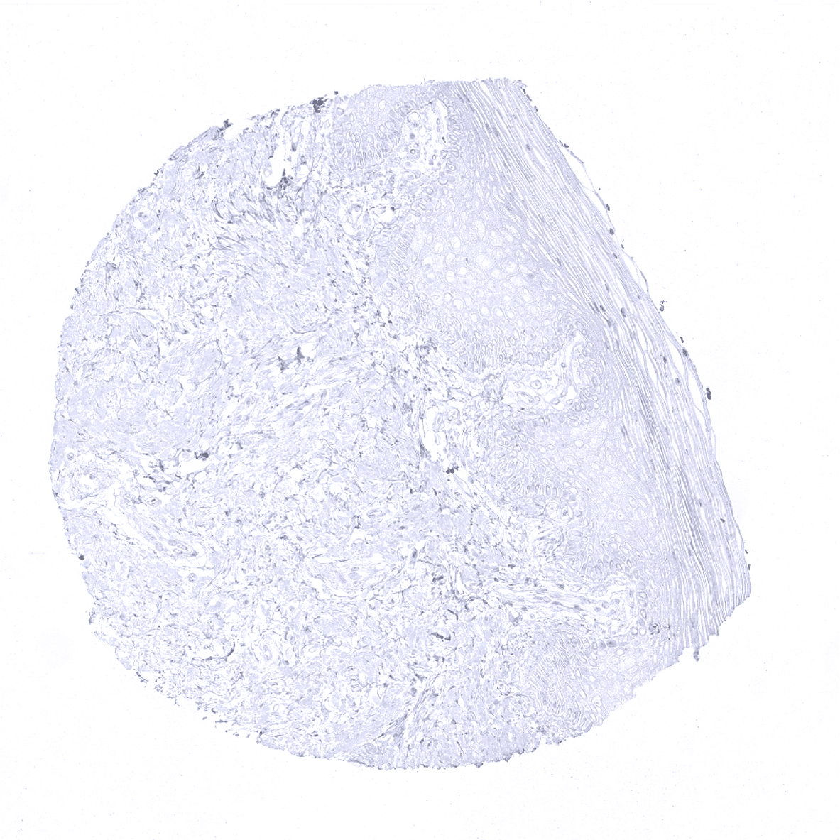
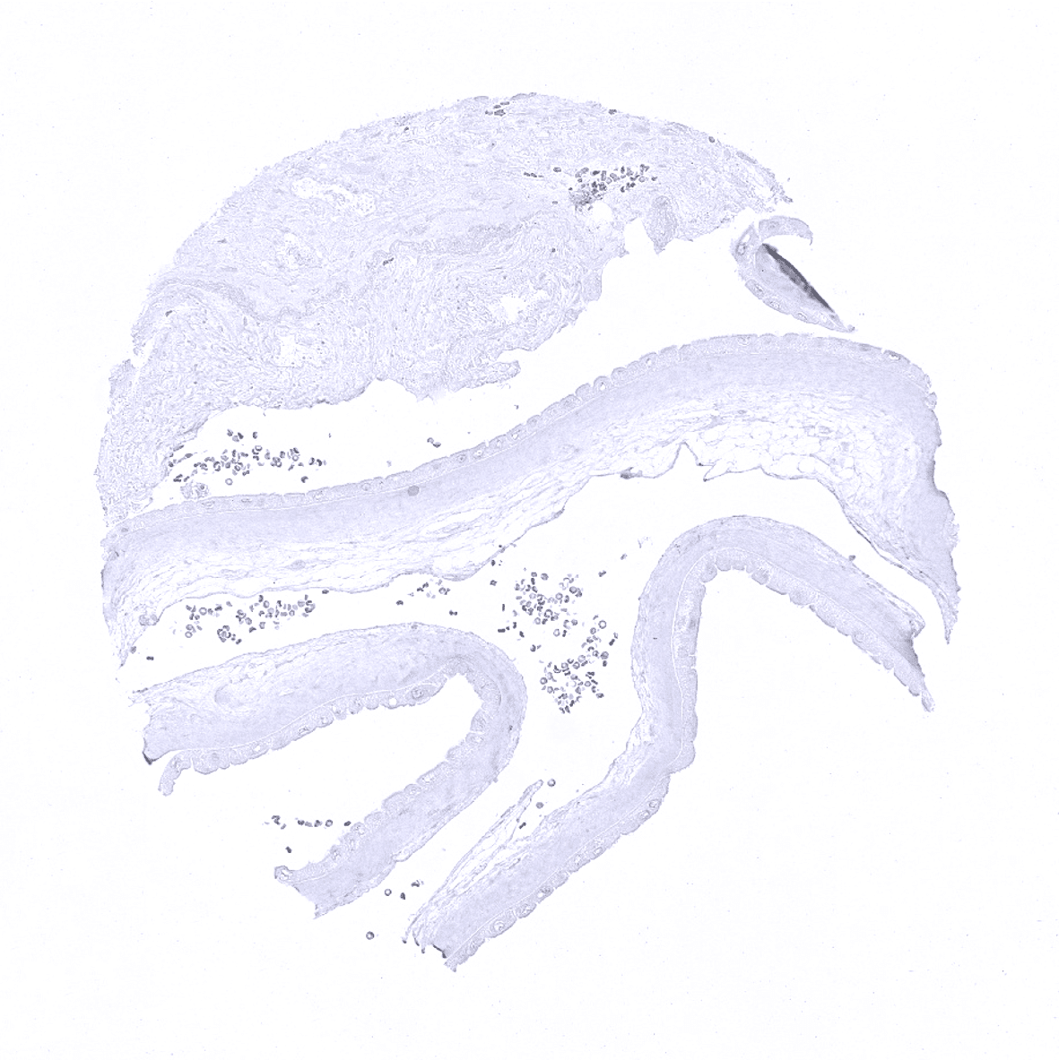
Appendix, muscular wall
Bone marrow
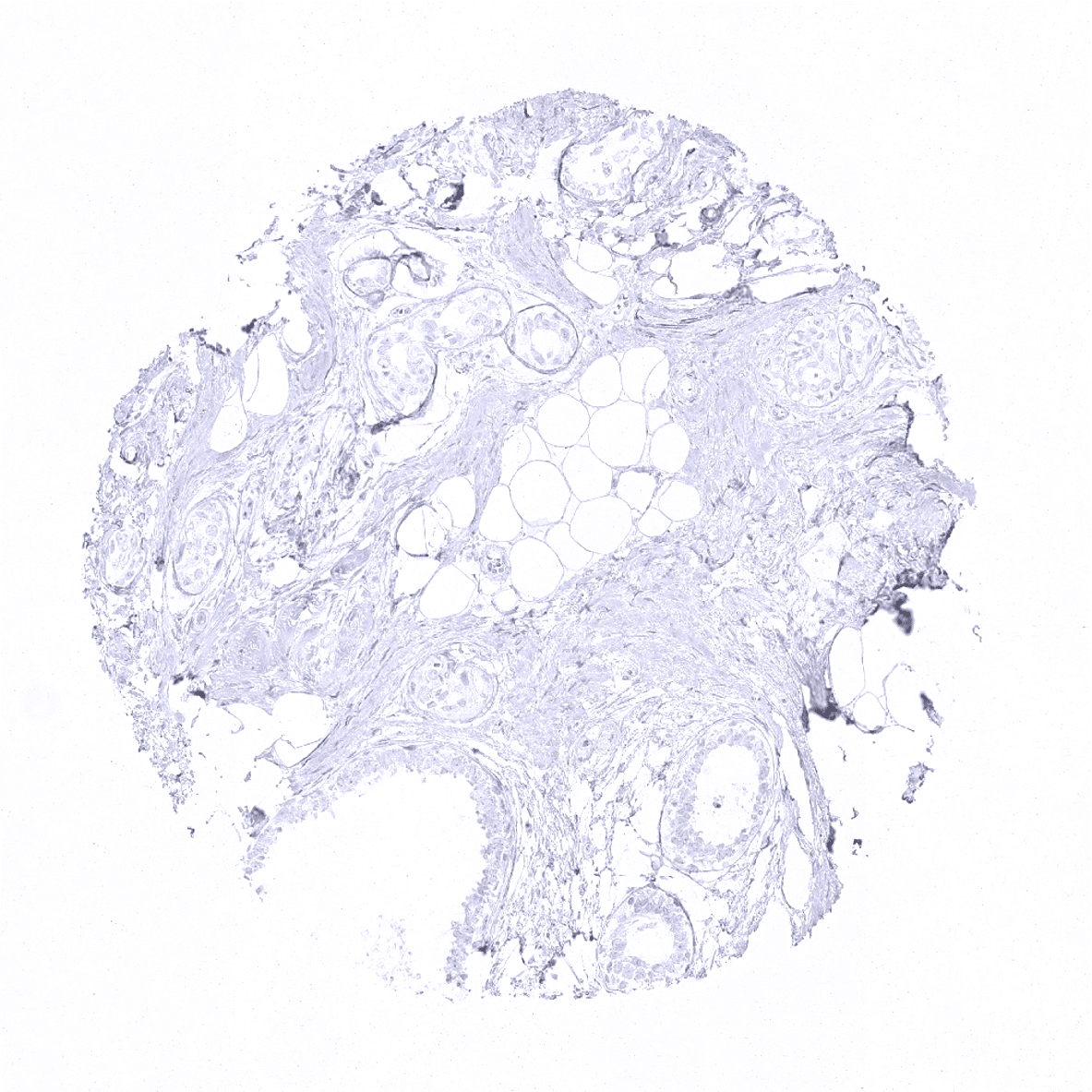
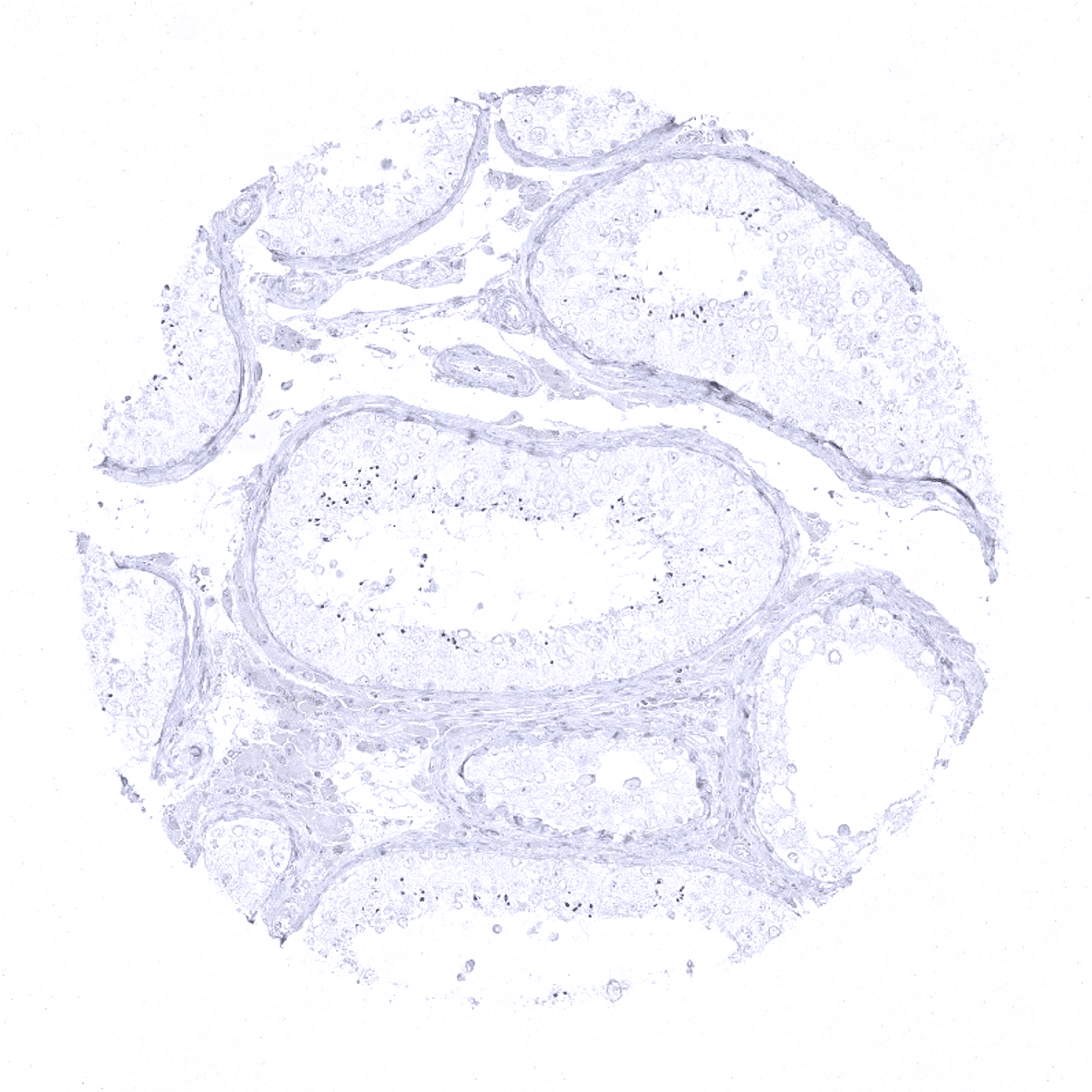
Breast
Bronchus, mucosa
Cerebellum (molecular layer, Purkinje cell layer, granule cell layer, white matter)
Cerebellum (white matter)
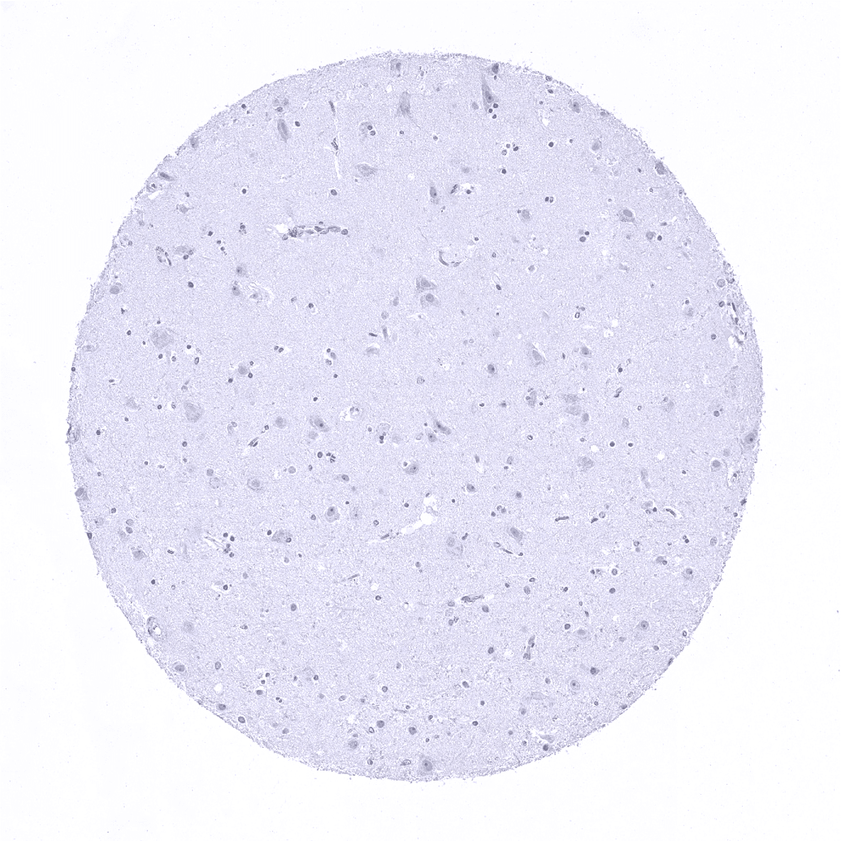
Cerebrum, grey matter
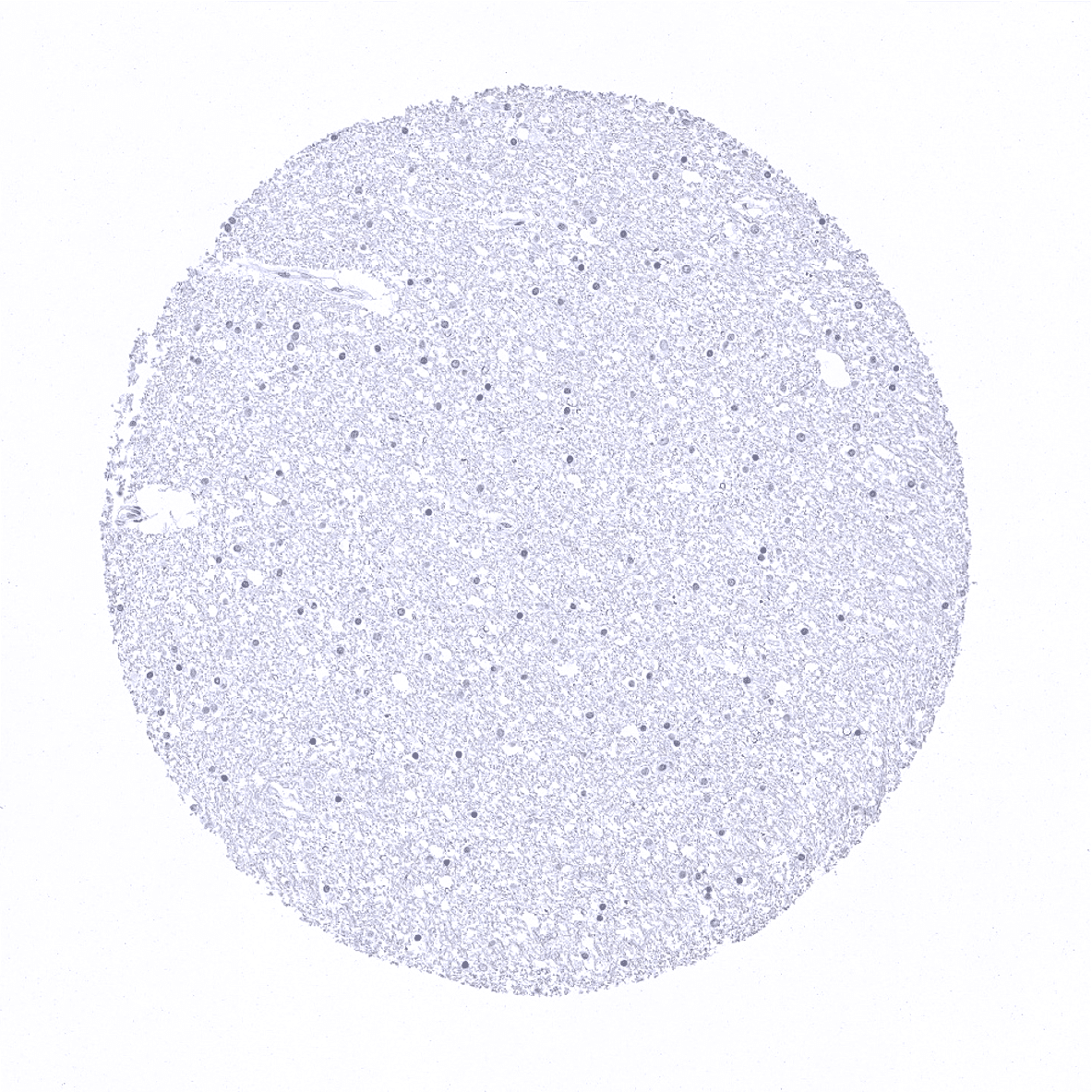
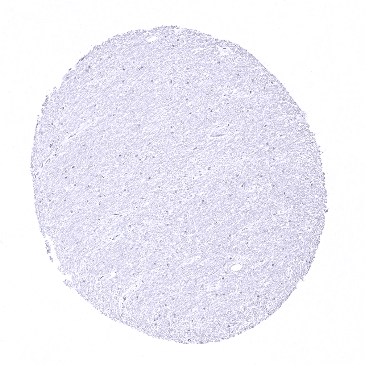
Cerebrum, white matter
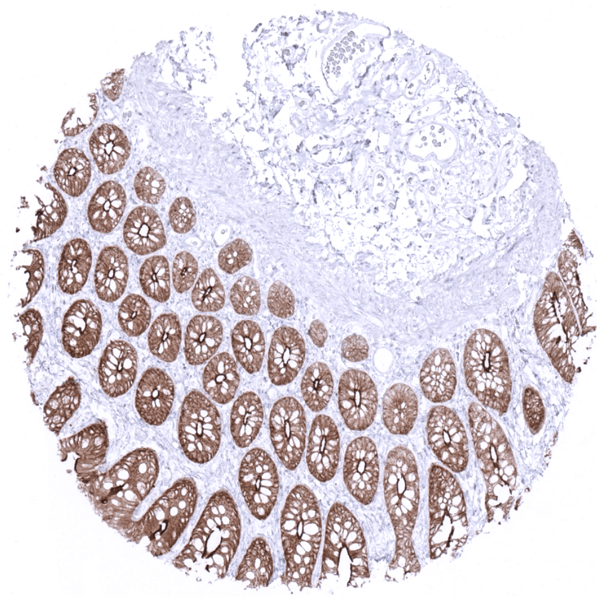
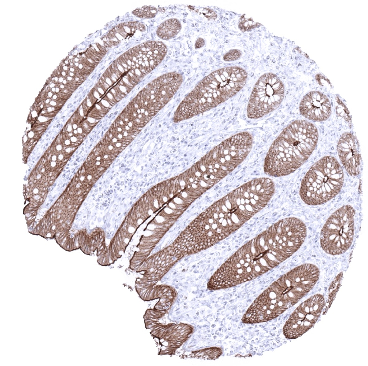
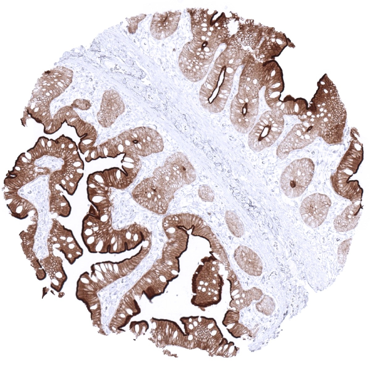
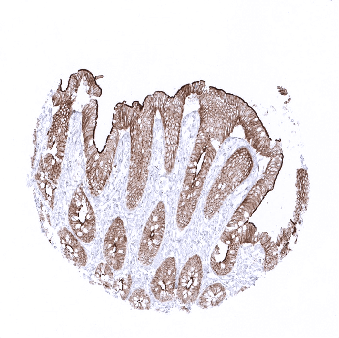
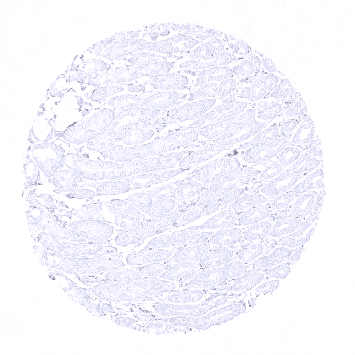
Colon descendens, mucosa - Colon epithelial cells show a strong, predominantly membranous but also cytoplasmic villin immunostaining.
Colon descendens, muscular wall

Duodenum, Brunner gland

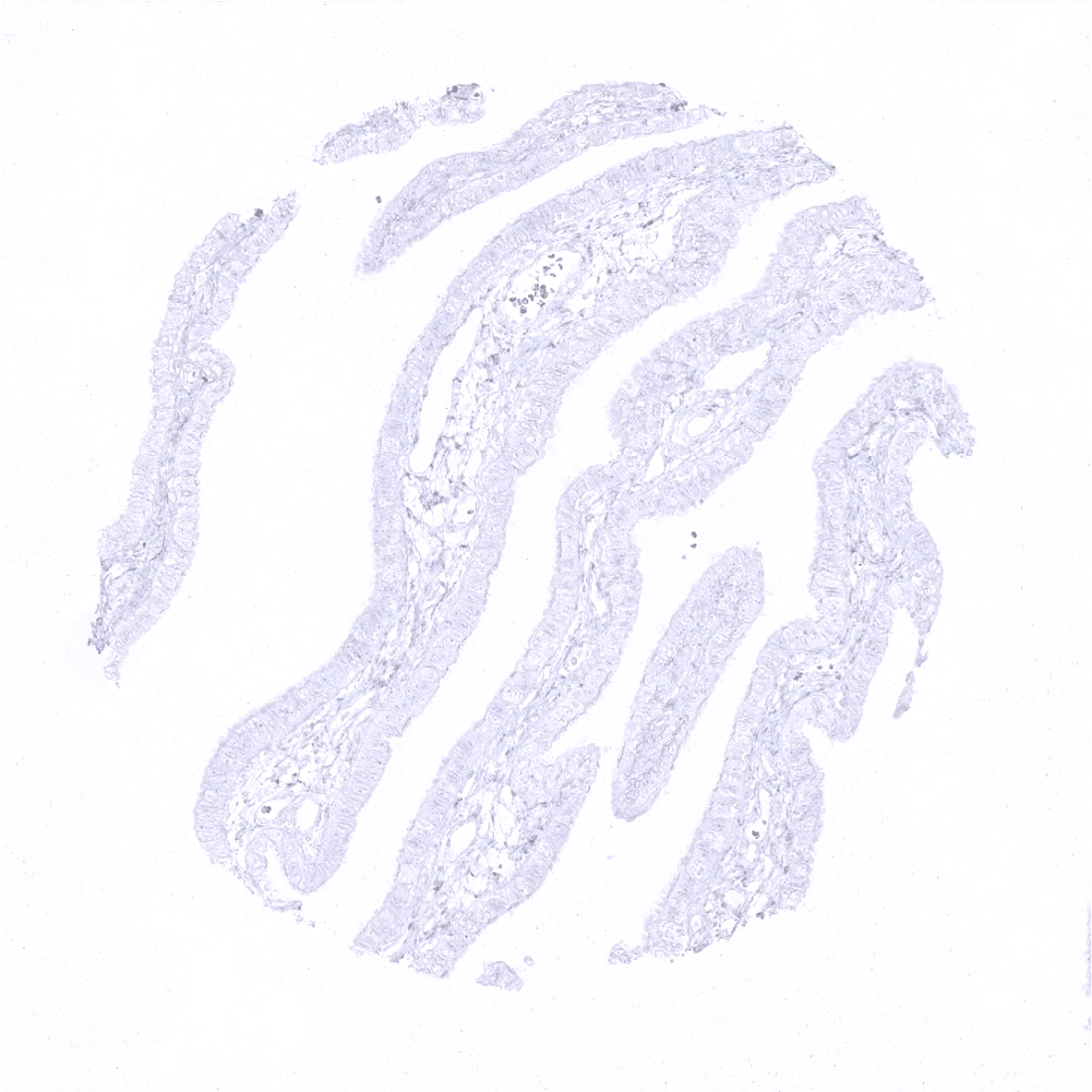
Duodenum, mucosa - Villin immunostaining is predominantly membranous but also cytoplasmic and often shows a strong focus on the apical/luminal membranes in the duodenum mucosa.
Ectocervix
Endocervix

Endometrium, proliferation
Endometrium, secretion
Endometrium, secretion
Epididymis - A moderate villin staining is seen in ciliated columnar cells in the epididymis.

Epididymis

Esophagus, squamous epithelium
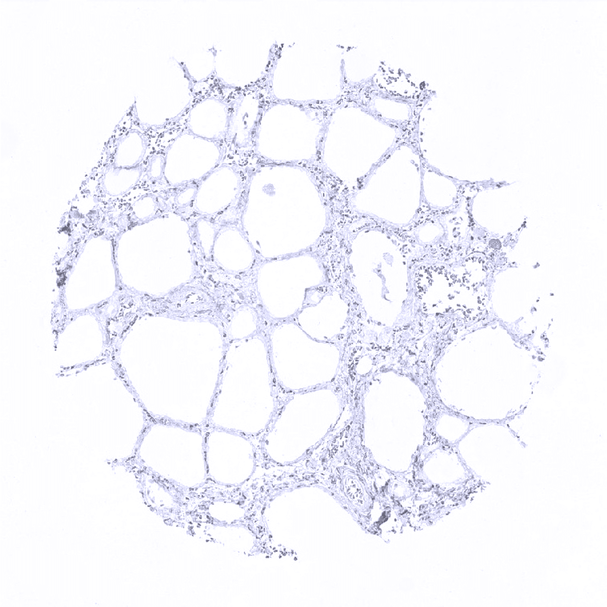
Fat
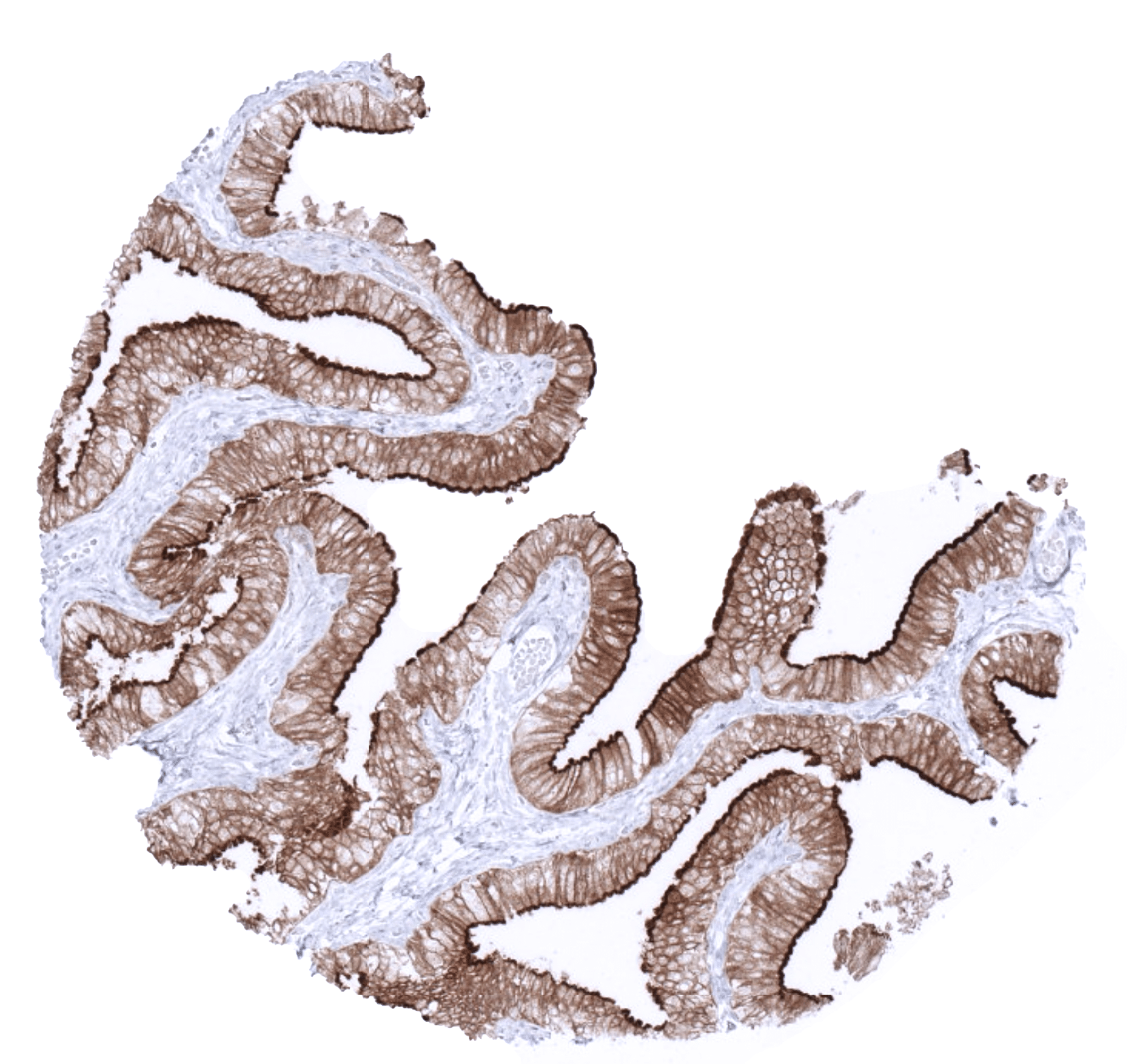
Gallbladder, epithelium - Villin immunostaining is particularly strong in the gallbladder epithelium.
Heart
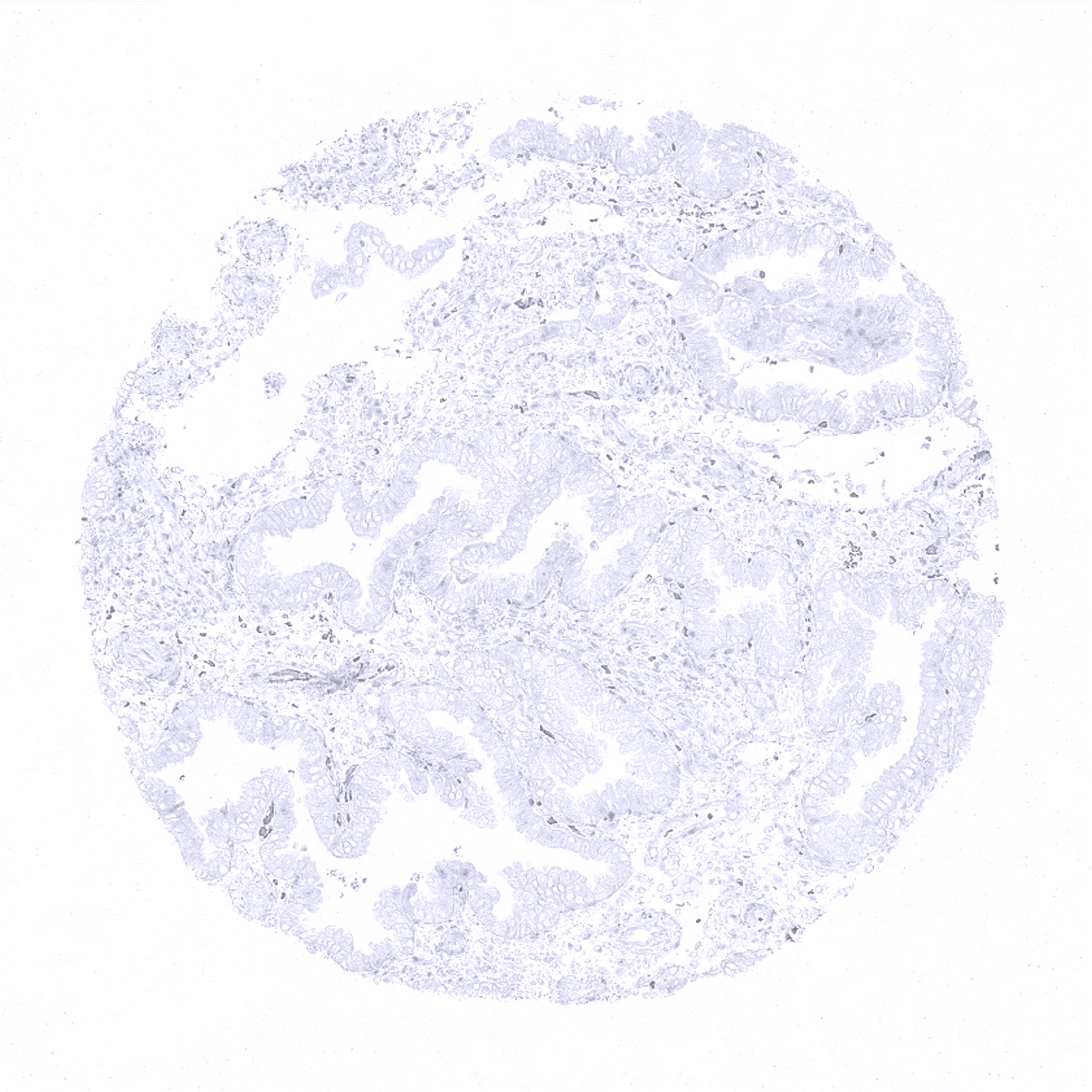
Ileum, mucosa - Villin immunostaining is predominantly membranous but also cytoplasmic and often shows a strong focus on the apical/luminal membranes in epithelial cells of the small intestine.

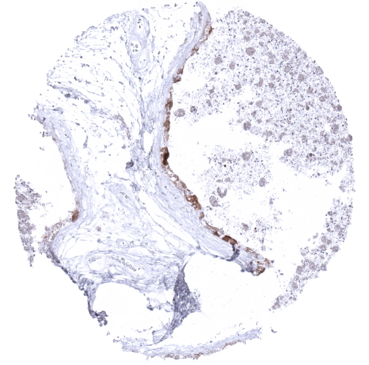
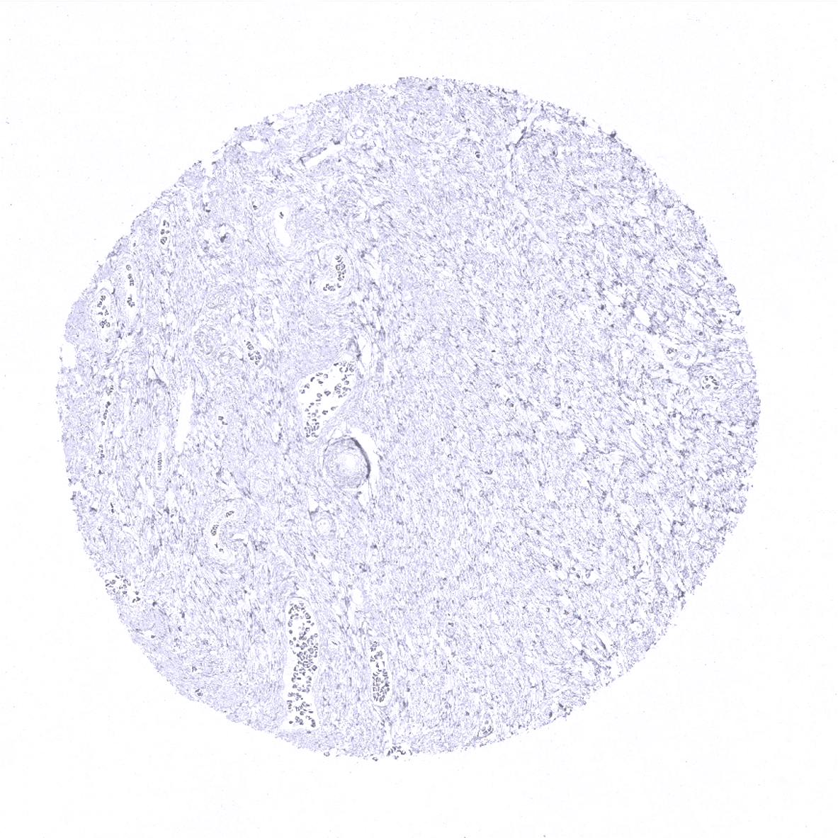
Kidney cortex - A strong villin positivity is seen in proximal tubuli of the kidney but not in distal tubuli or glomeruli.
Kidney, medulla - Absence of villin immunostaining in collecting ducts of the kidney.

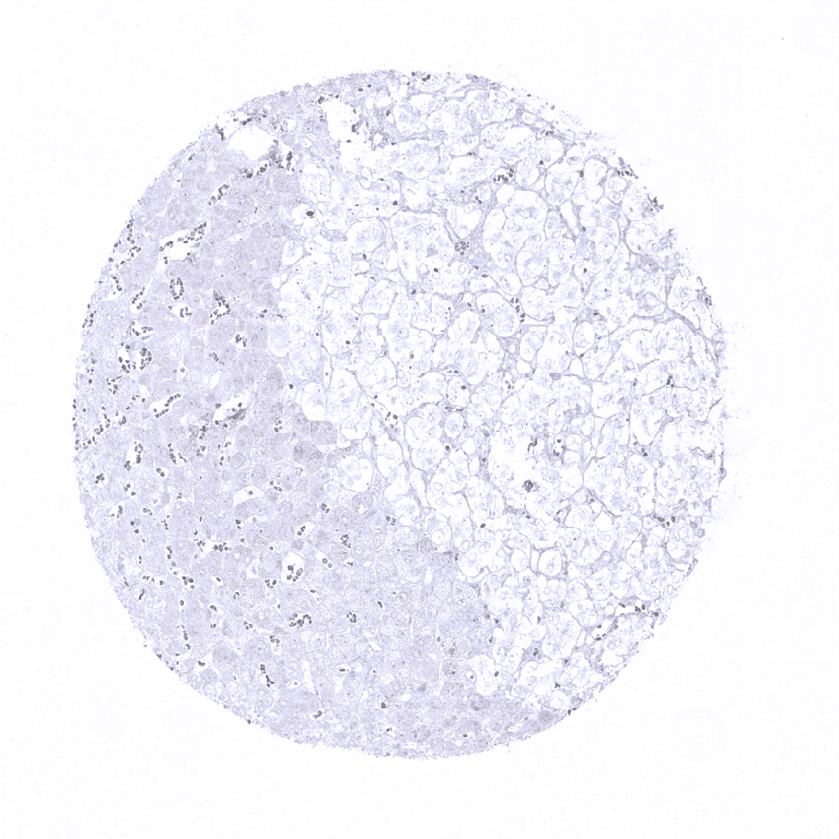
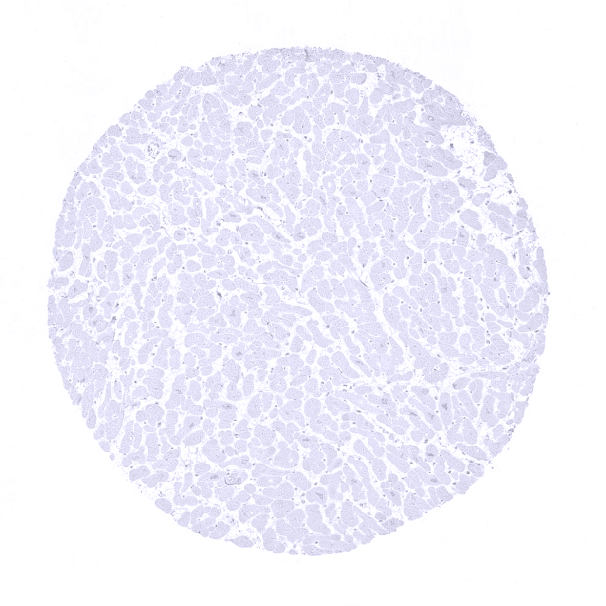
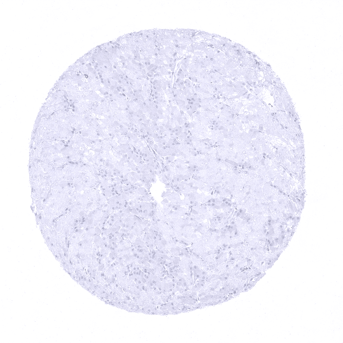
Liver - In the liver a weak to moderate villin staining is seen at the apical pole of hepatocytes. The expression level exhibits a zonal variability.
Lung
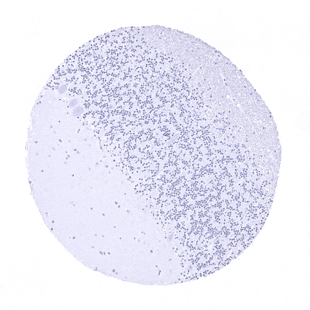
Lymph node
Ovary, stroma
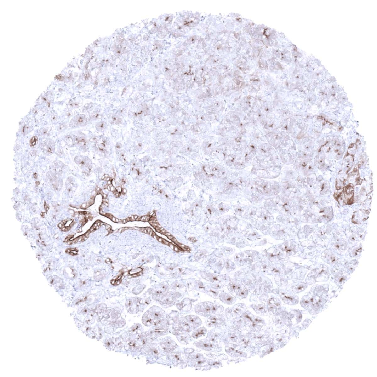
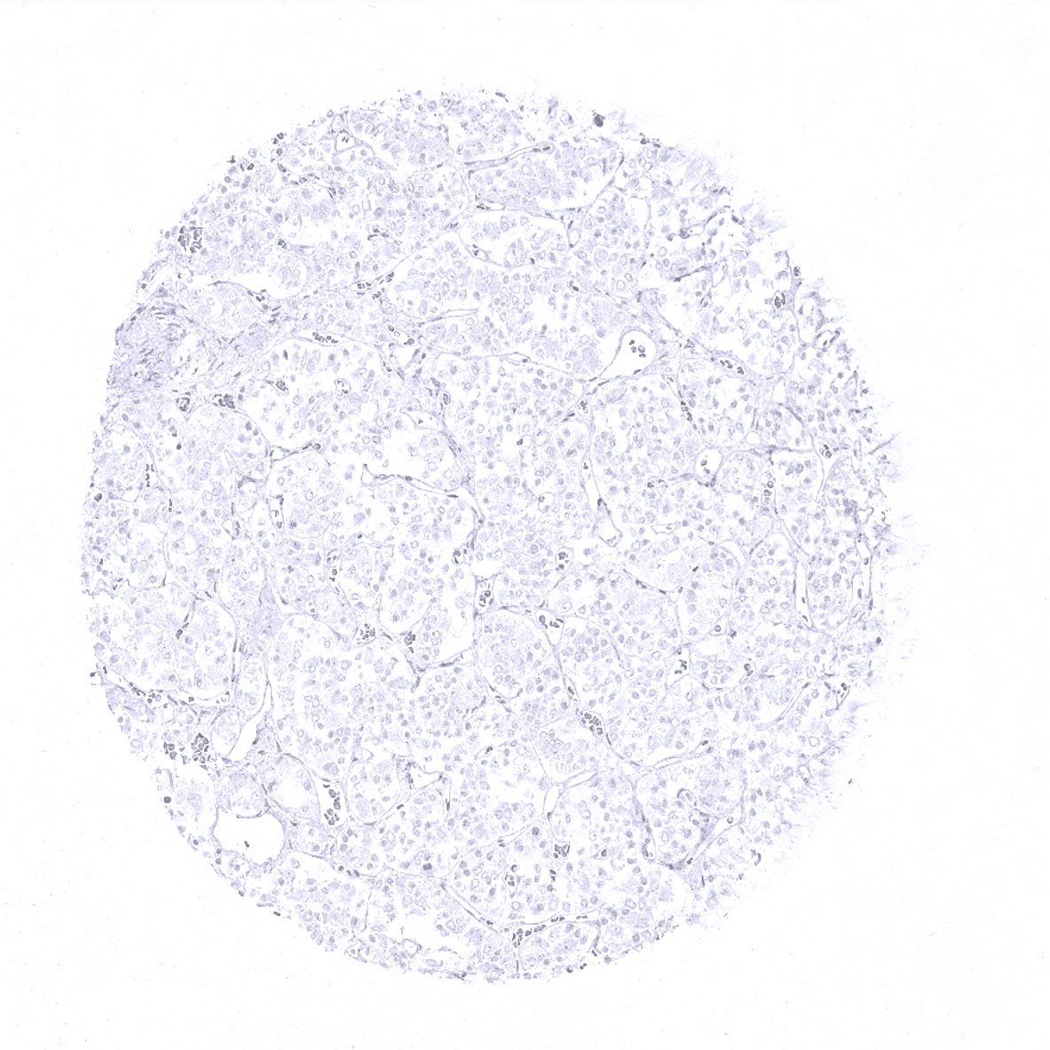
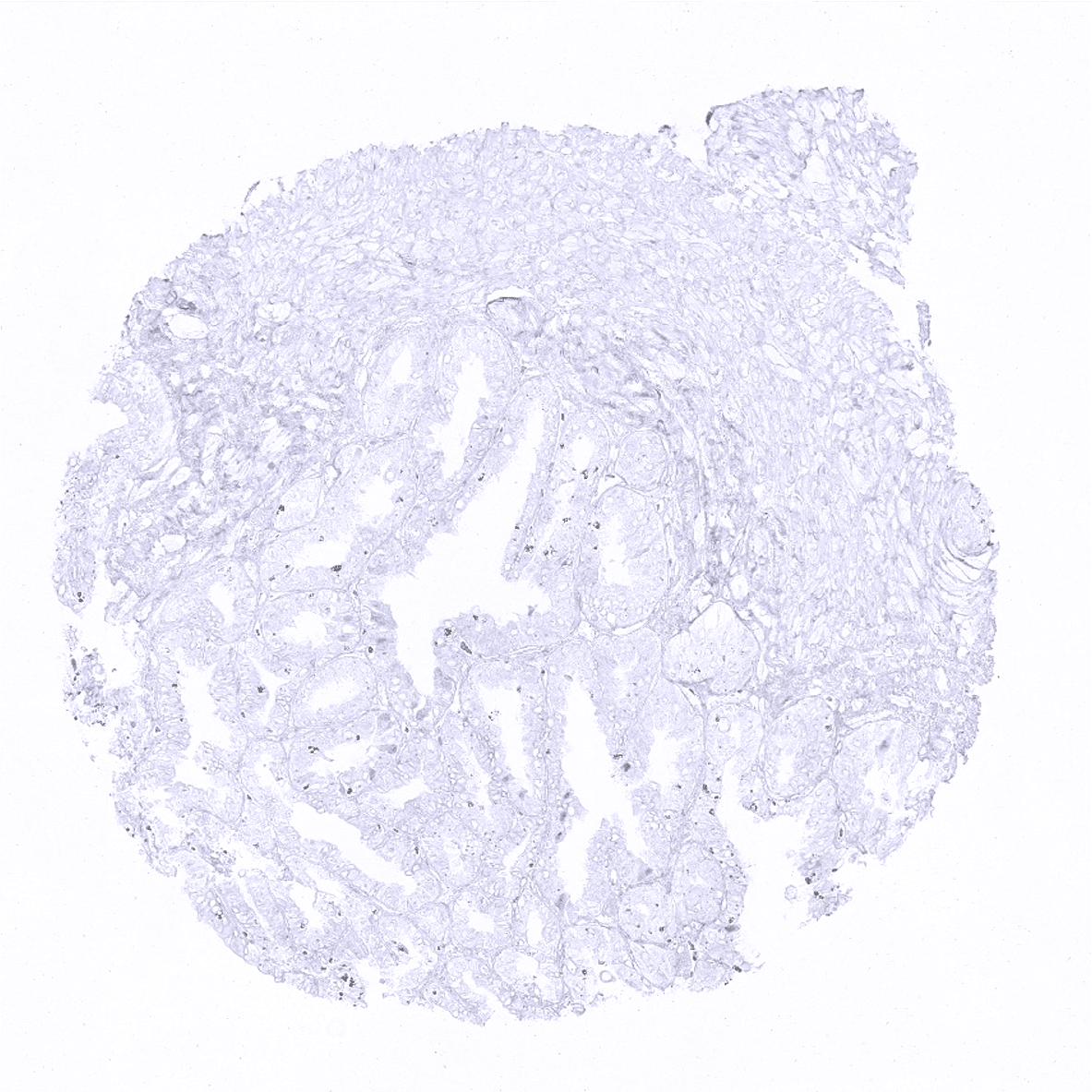
Pancreas - In the pancreas, a strong villin positivity is seen in interlobular ducts. In addition, a weak to moderate villin staining is seen at the apical pole of acinar cells.
Parathyroid
Parotid gland
Pituitary gland, anterior lobe
Pituitary gland, posterior lobe

Pregnant uterus (decidua)
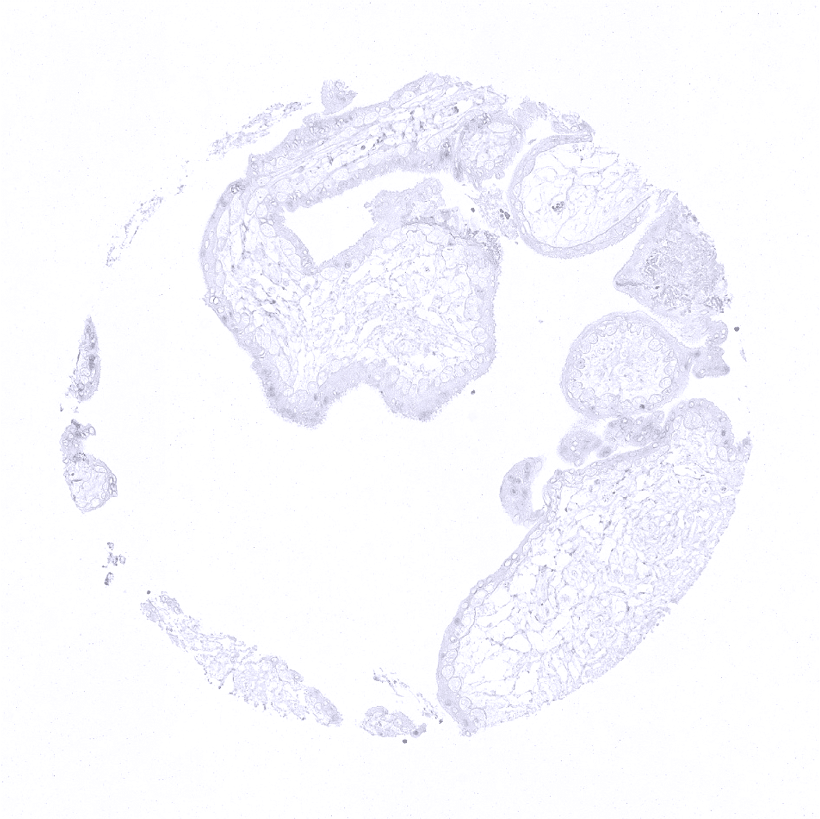
Placenta, early
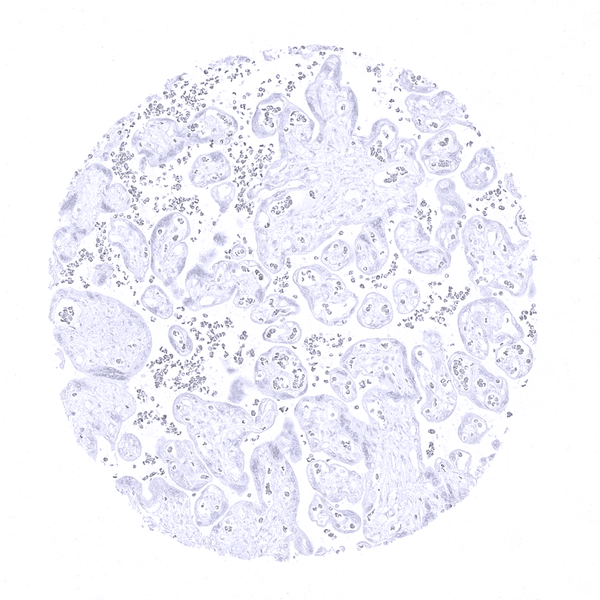
Placenta, mature
Placenta (amnion and chorion)
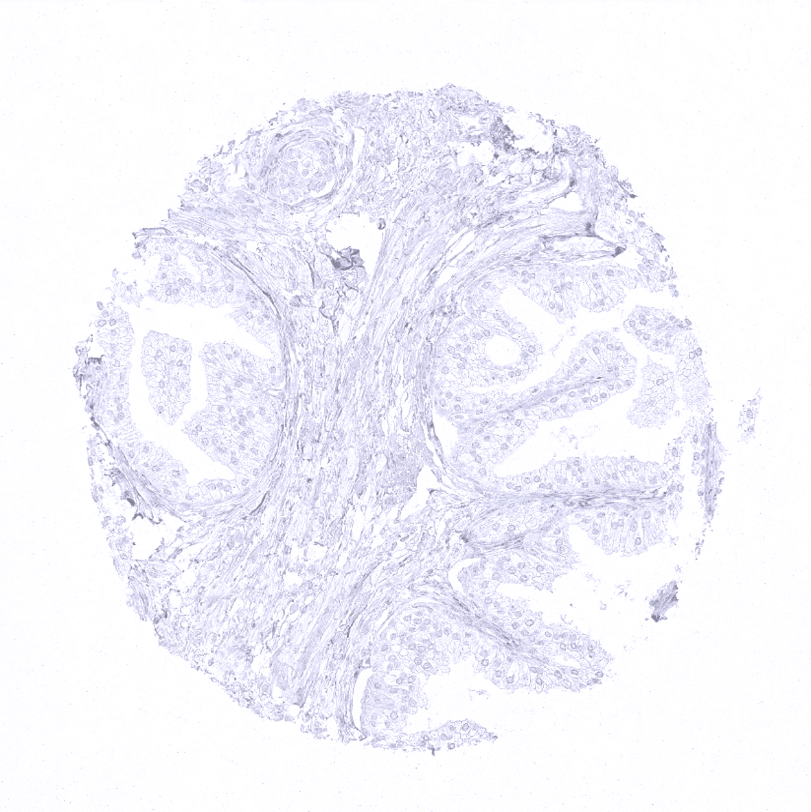
Prostate
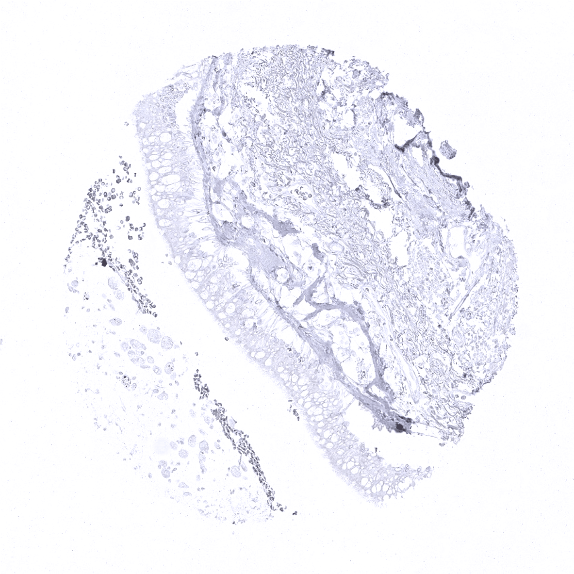
Rectum, mucosa
Seminal vesicle
Sinus paranasales
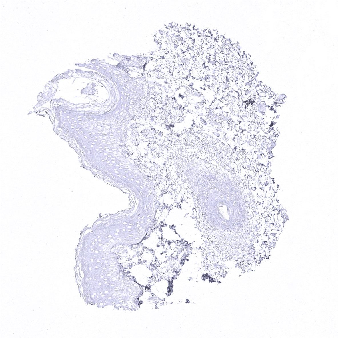
Skin
Spleen

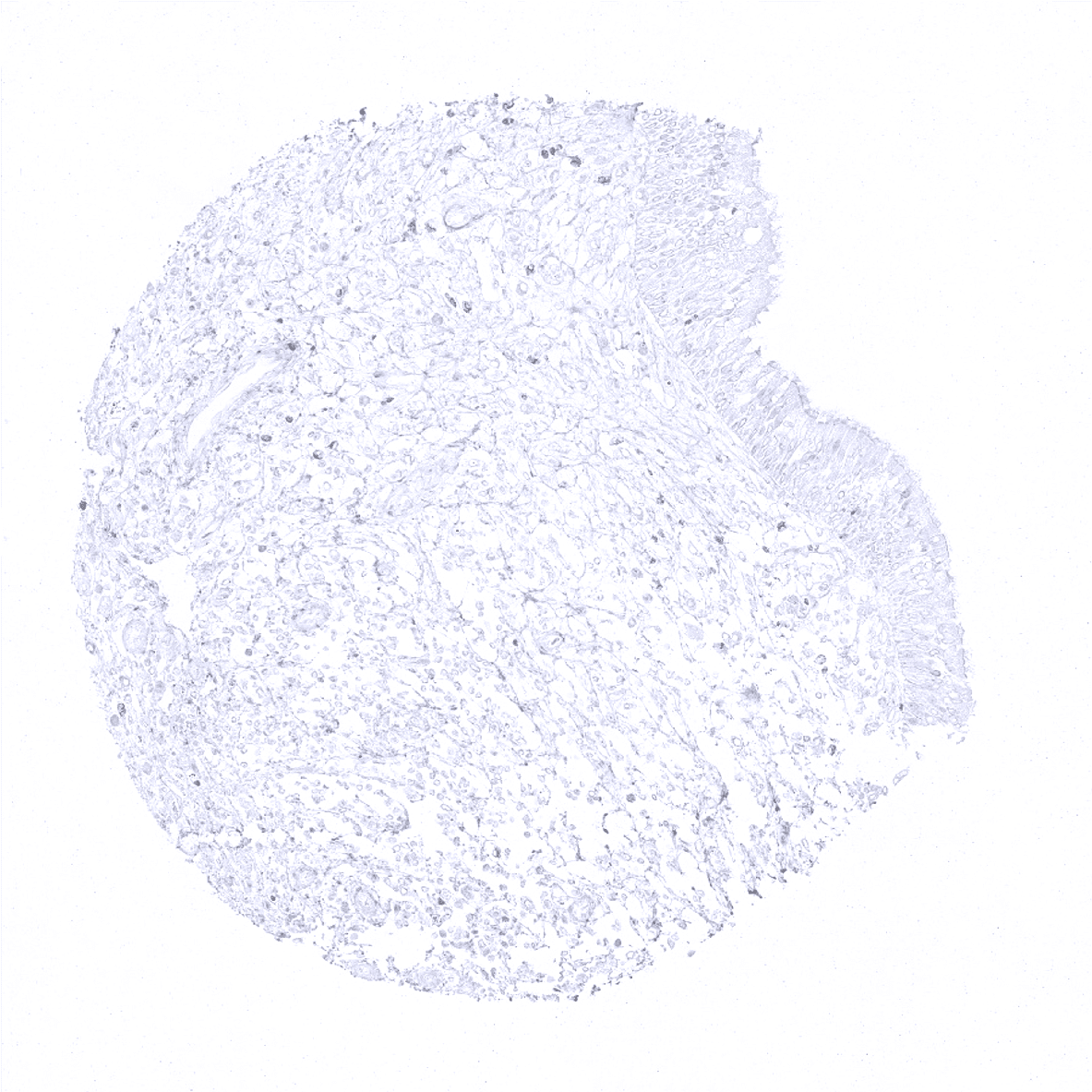
Stomach, antrum - The surface epithelium of the stomach shows a weak, sometimes moderate villin immunostaining with decreasing intensity towards the base of the glands (which are villin negative).
Stomach, corpus - Villin negative gastric corpus glands.
Testis
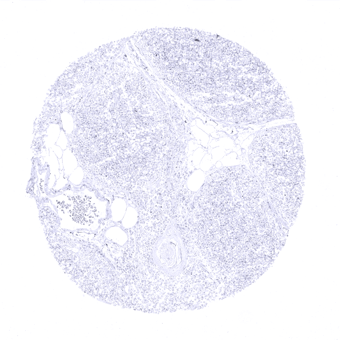
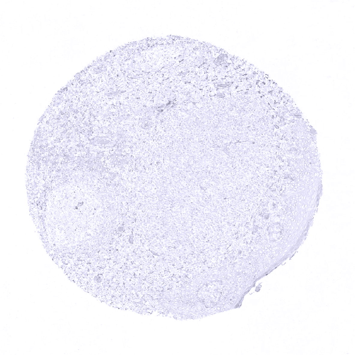
Thymus
Thyroid gland
Tonsil, surface epithelium
Tonsil

Urinary bladder, muscular wall
Urinary bladder, urothelium
Uterus, myometrium