
Adrenal gland
Adrenal gland
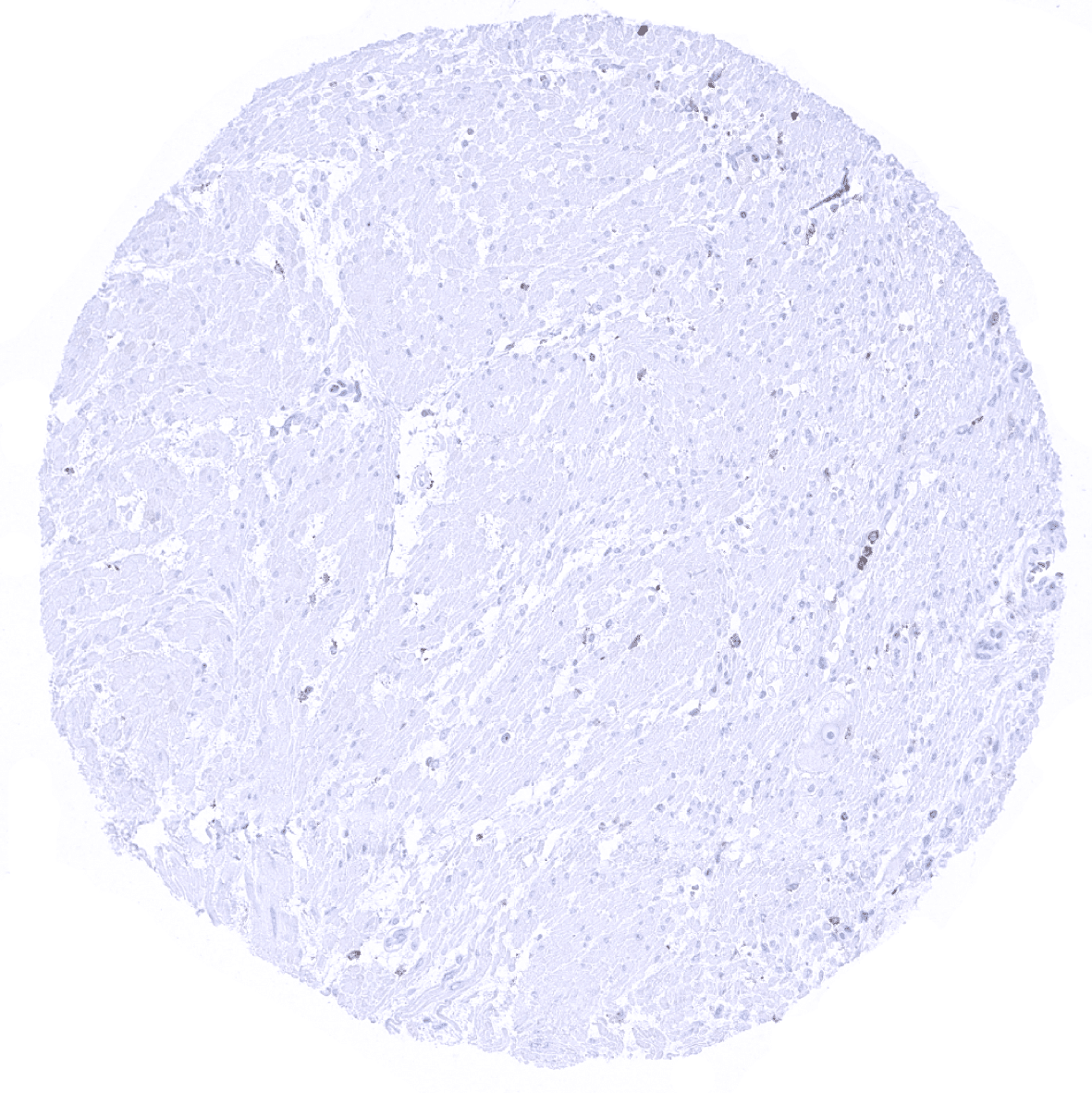
Aorta, media

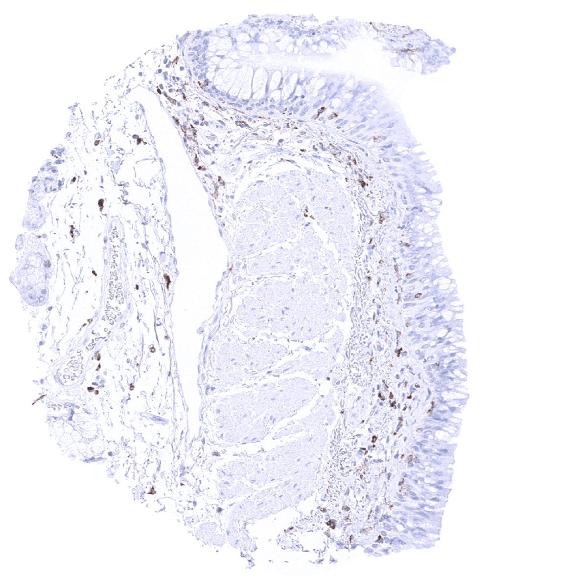
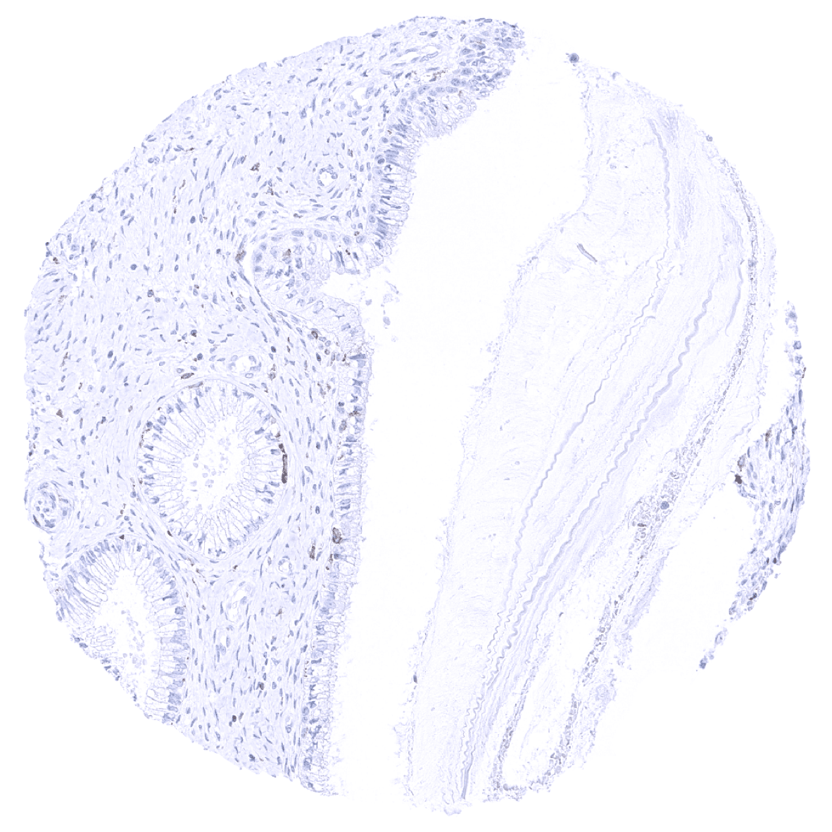
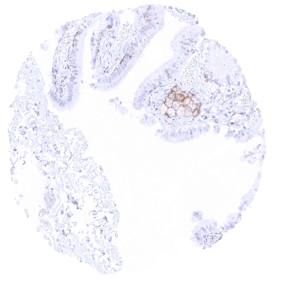
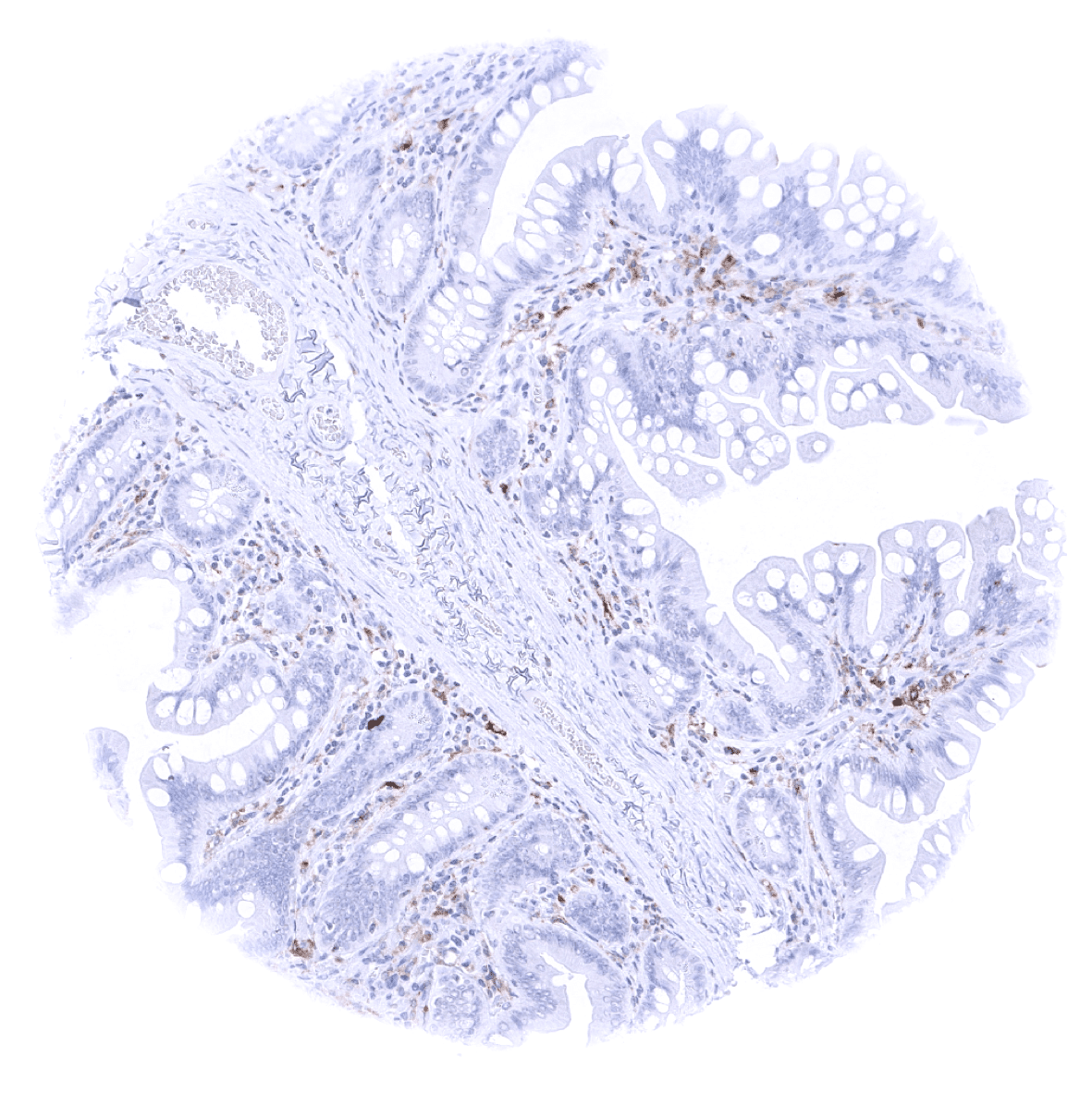
Appendix, mucosa
Appendix, muscular wall
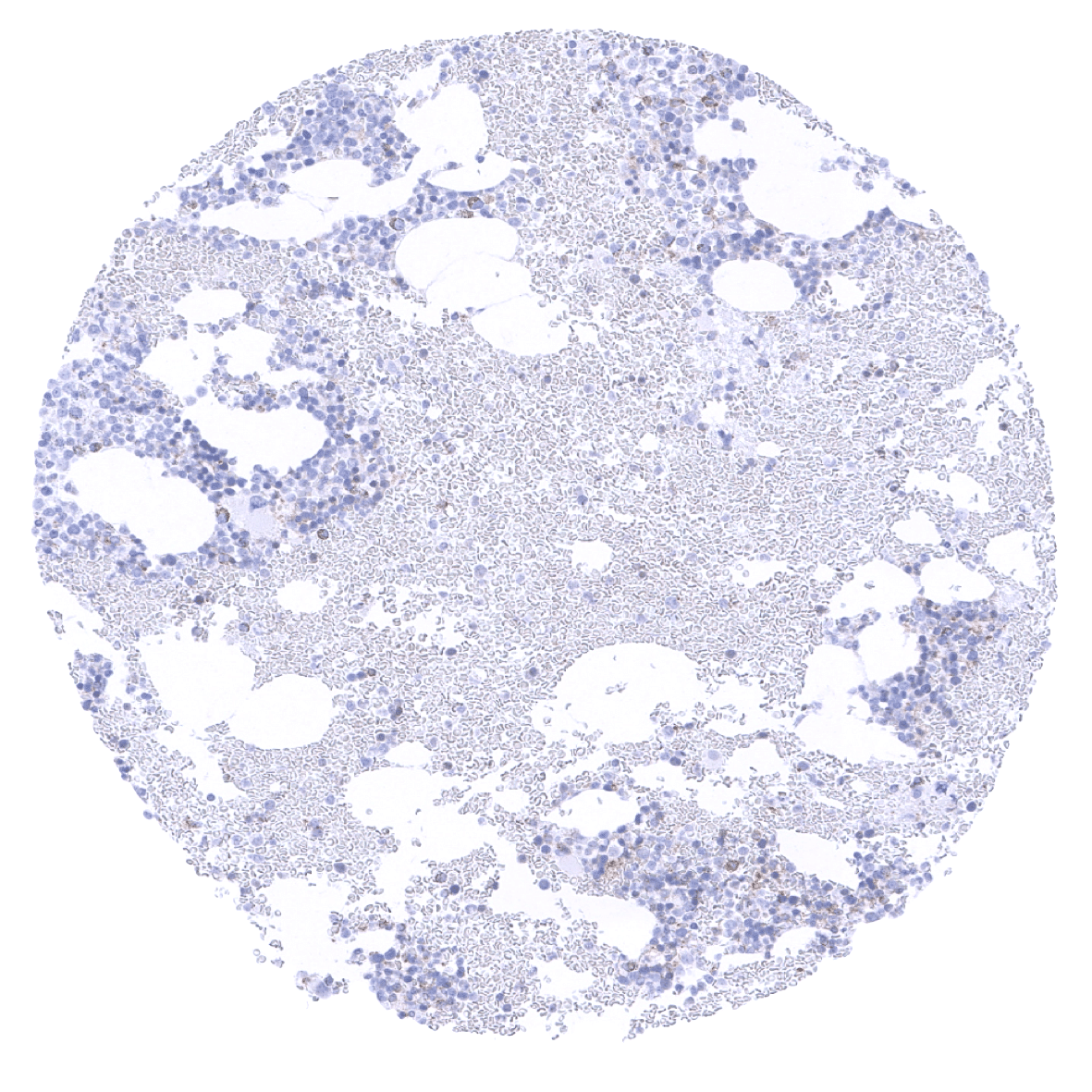
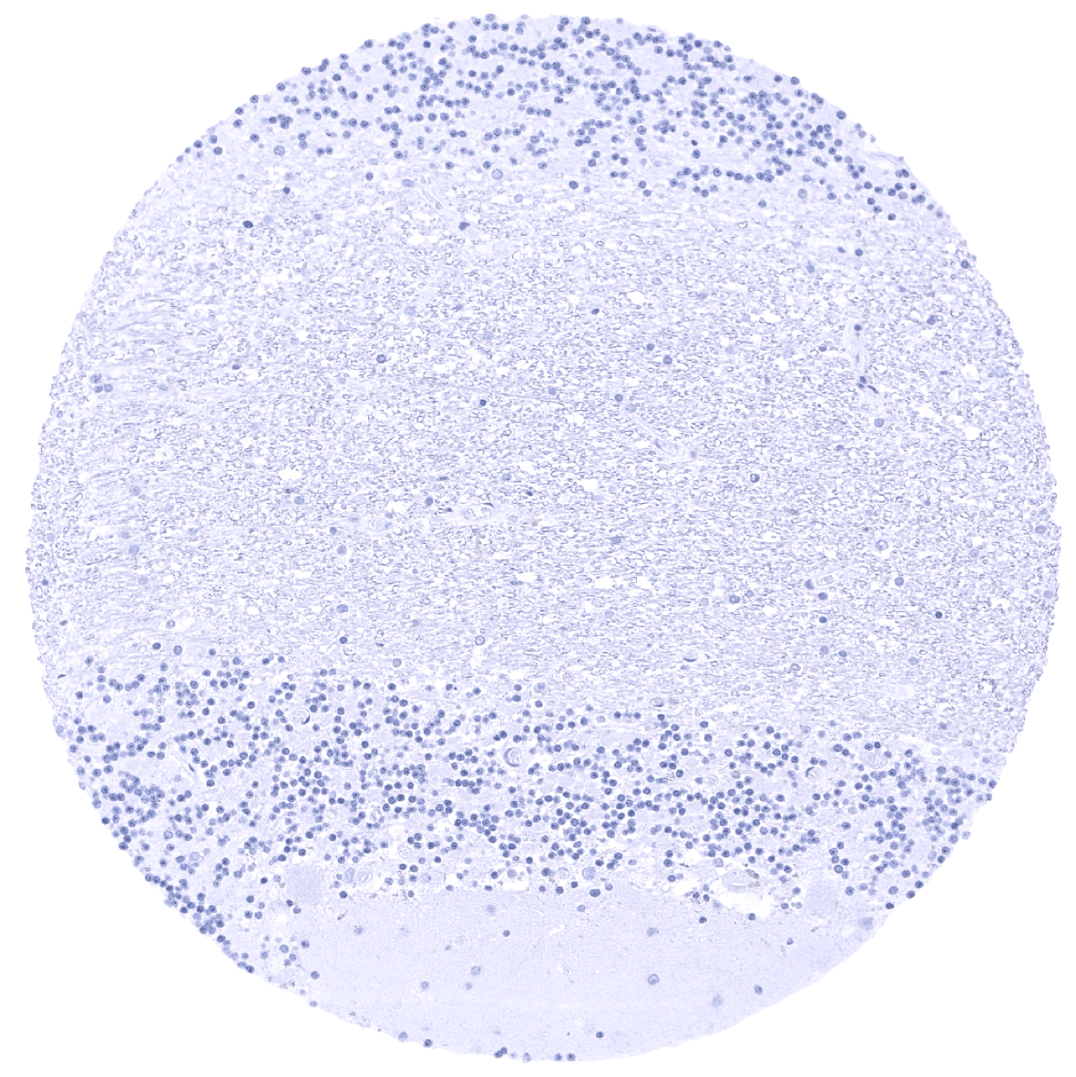
Bone marrow

Breast
Bronchus, mucosa

cerebellum (molecular layer, Purkinje cell layer, granule cell layer)
cerebellum (molecular layer, Purkinje cell layer, granule cell layer, white matter)
Cerebrum, grey matter
Cerebrum, white matter

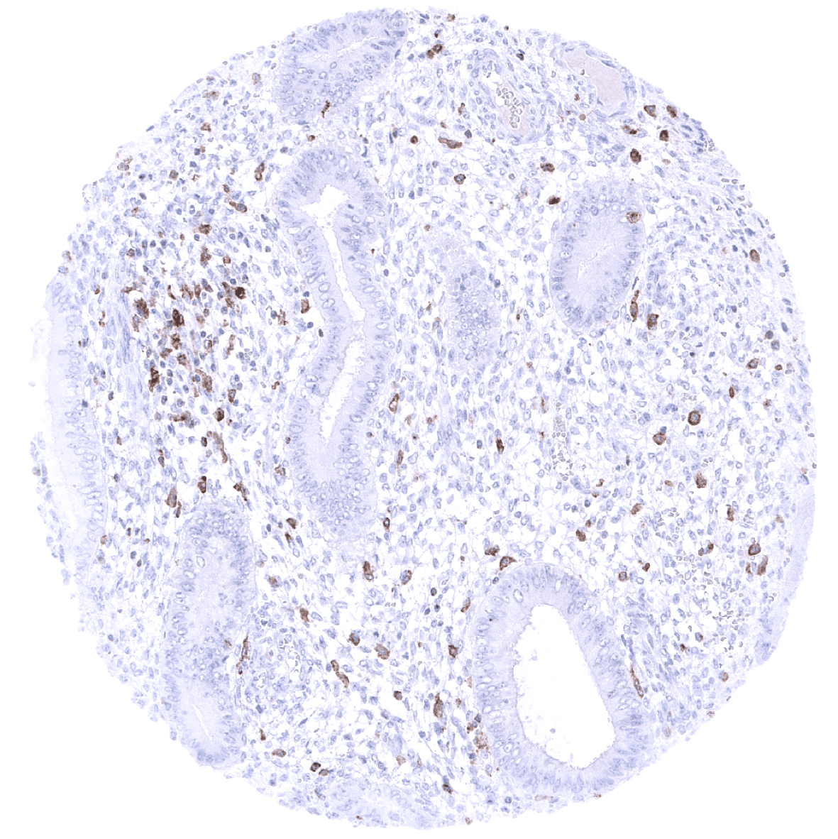
Colon descendens, mucosa: In the colon mucosa, variable levels of TIM-3 immunostaining are seen in T-cells and in macrophages. Epithelial staining does not occur.
Colon descendens, muscular wall
Duodenum, Brunner gland
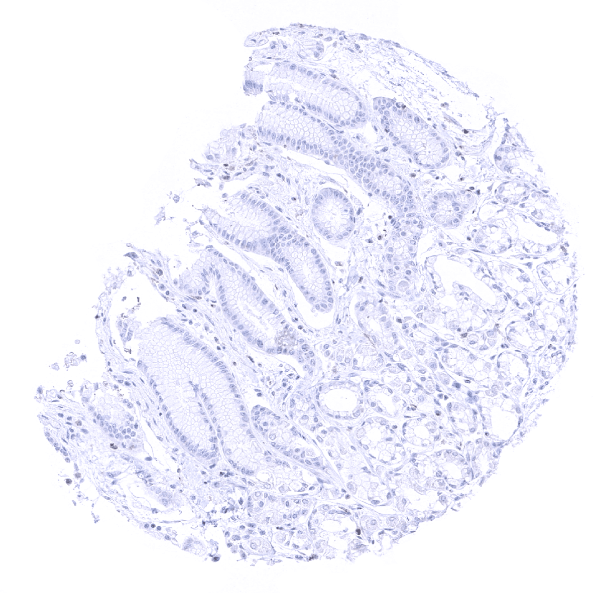
Duodenum, mucosa
Ectocervix
Endocervix
Endometrium, proliferation

Endometrium, secretion
Epididymis: A strong TIM-3 staining occurs at the apical membranes an ciliae of tall columnar cells of the epididymis.

Esophagus, squamous epithelium
Fallopian tube, mucosa
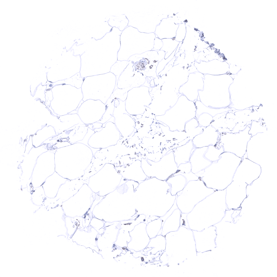
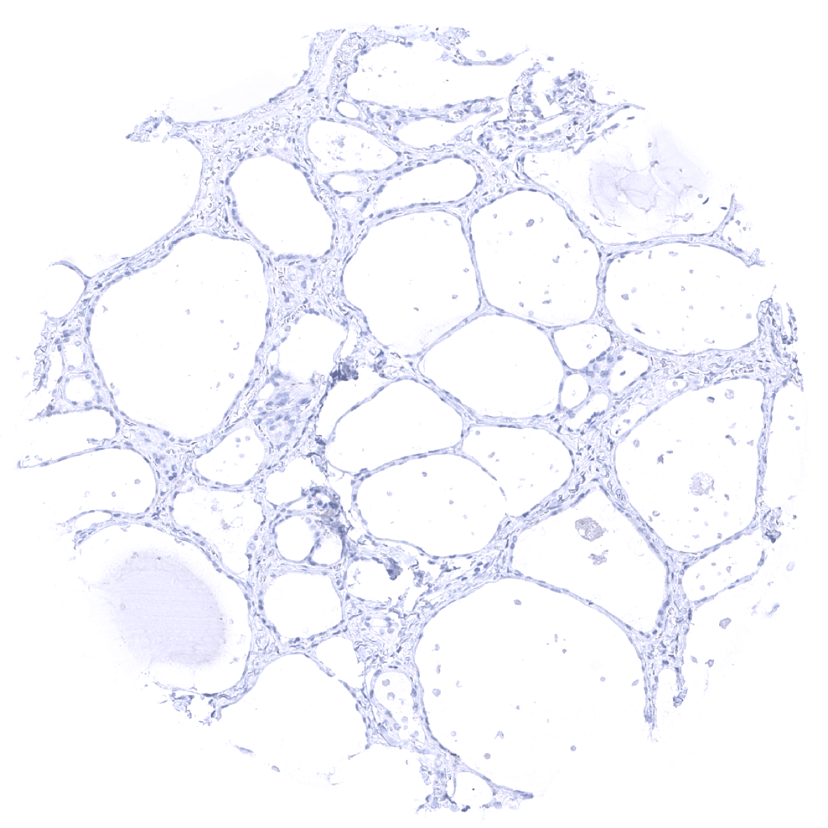
Fat
Gallbladder, epithelium
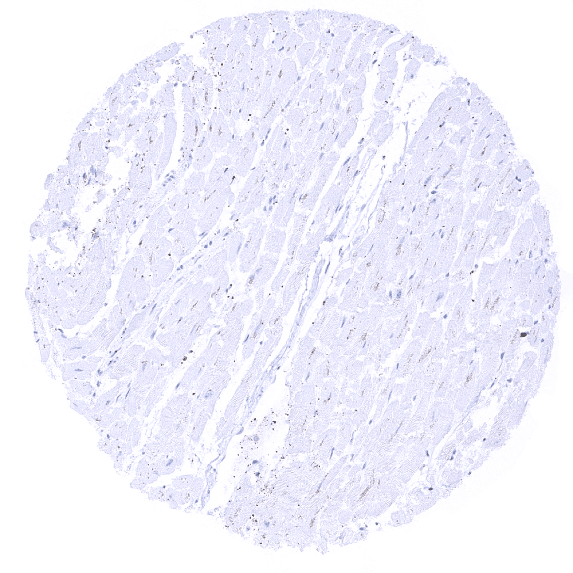
Heart
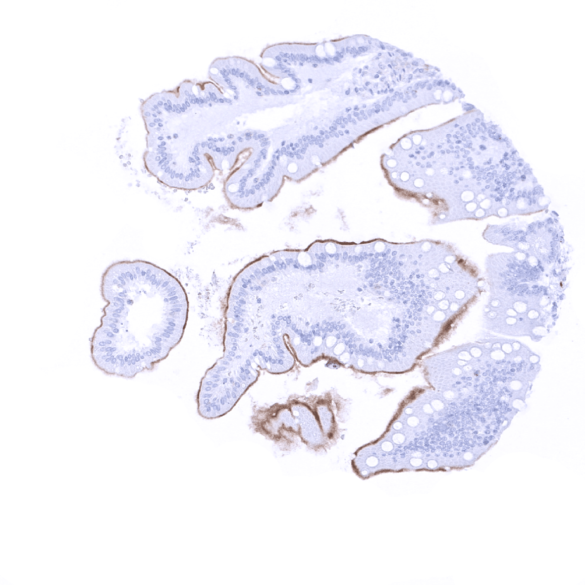
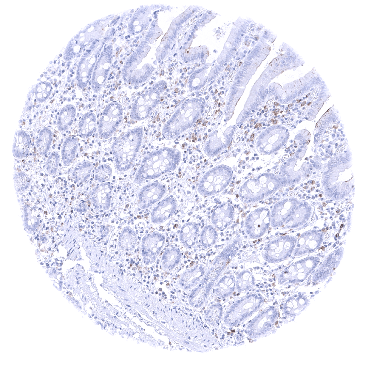
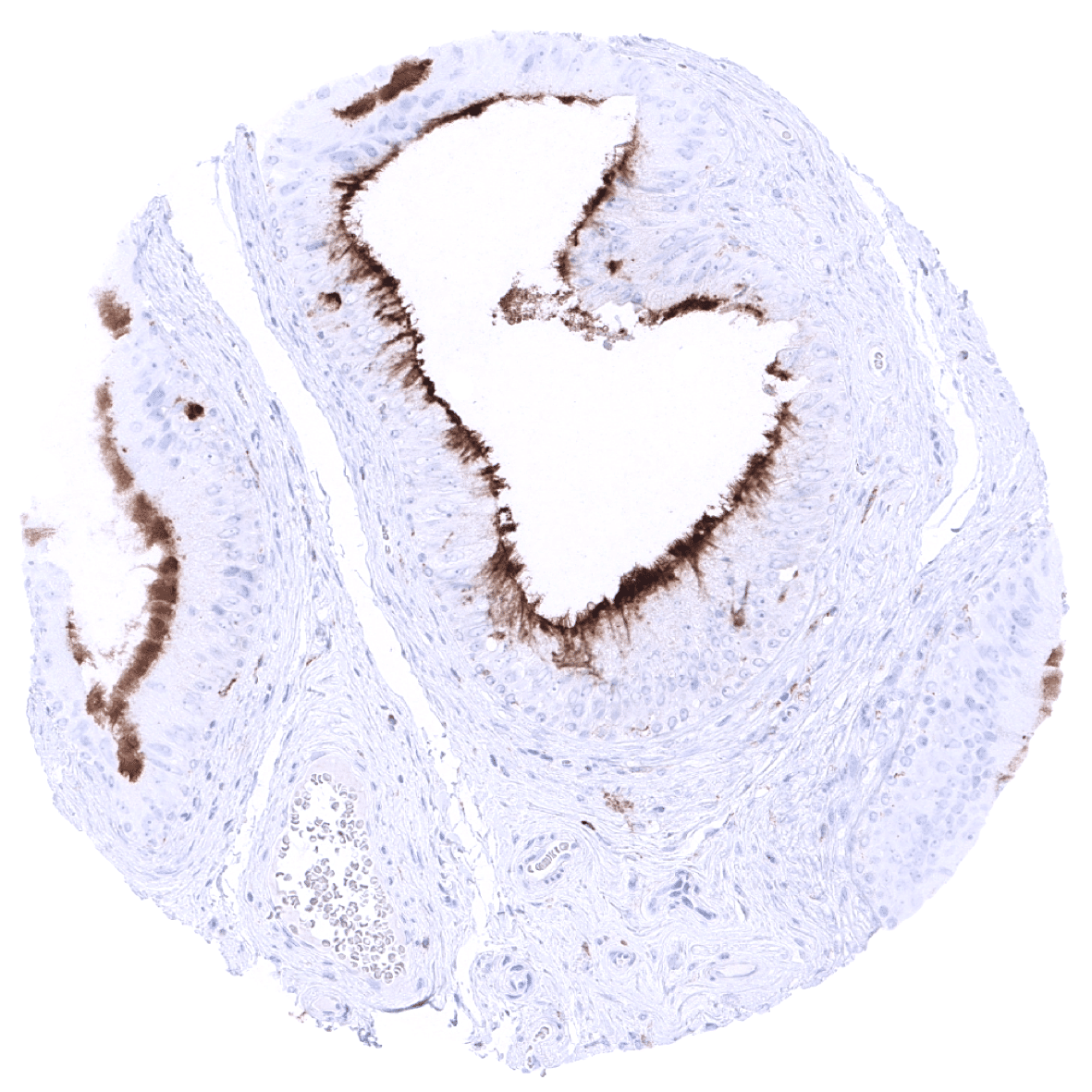
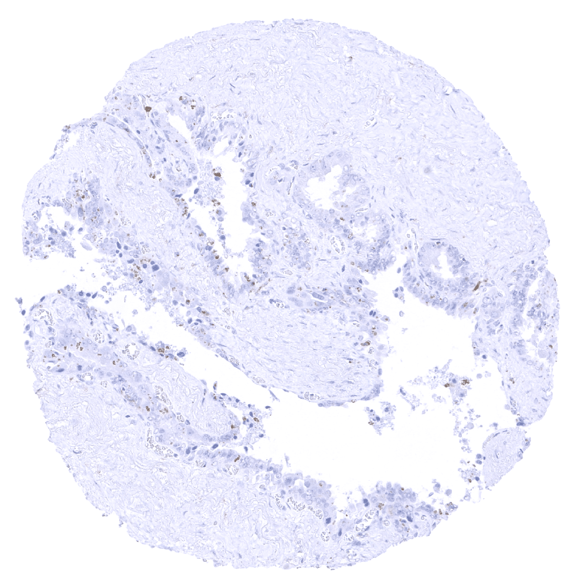
Ileum, mucosa: A membranous TIM-3 immunostaining of the surface epithelium of the small intestine may represent a (tolerable) cross-reactivity.
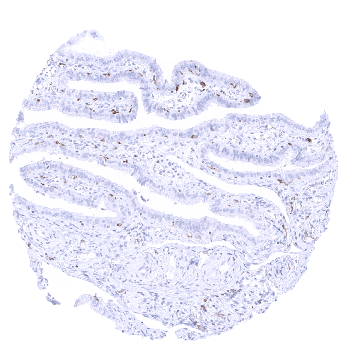
Ileum, mucosa
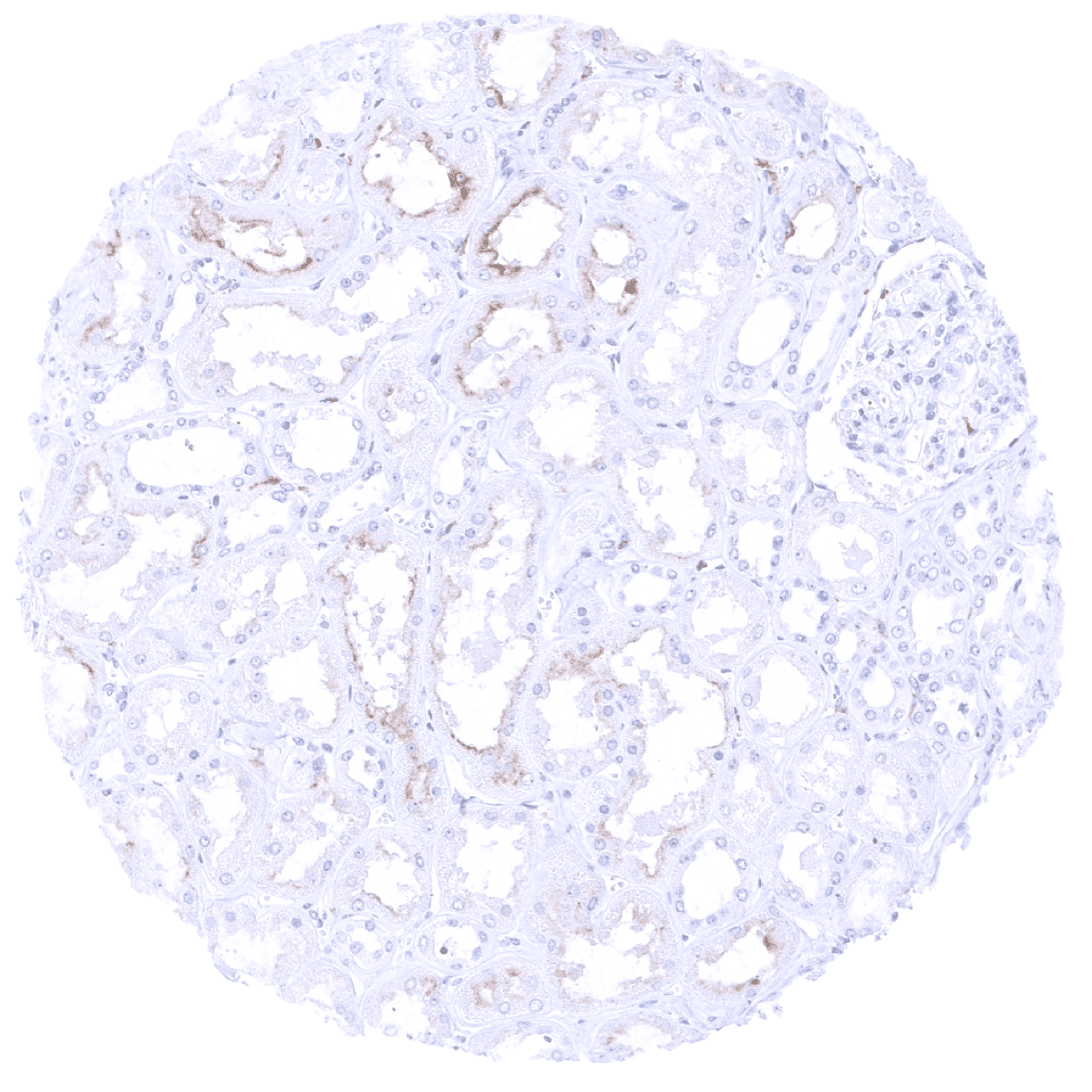
Kidney, cortex: In the kidney, apical membranes of a fraction of proximal tubuli show a moderate TIM-3 immunostaining.
Kidney, medulla
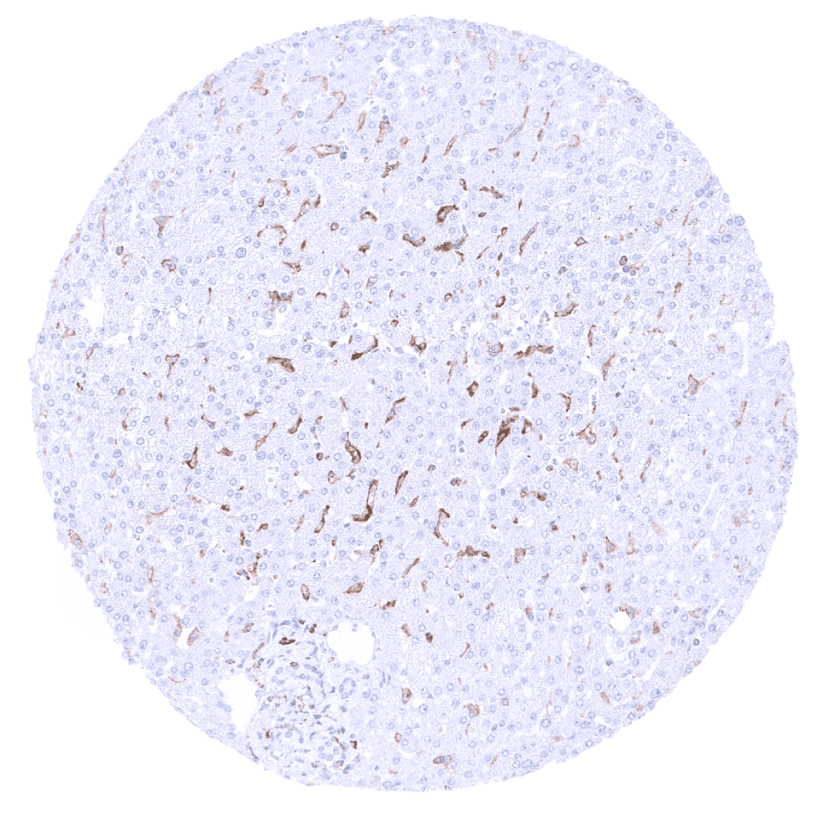
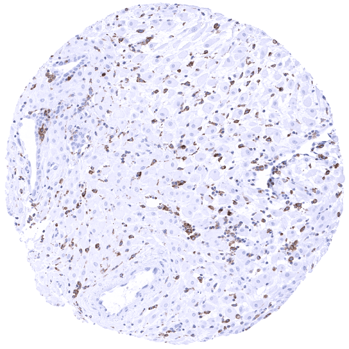
Liver: TIM-3 immunostaining occurs in venous sinusoids of the spleen and in Kupffer cells of the liver.

Lung: In the lung, a particularly strong TIM-3 immunostaining occurs in alveolar macrophages.
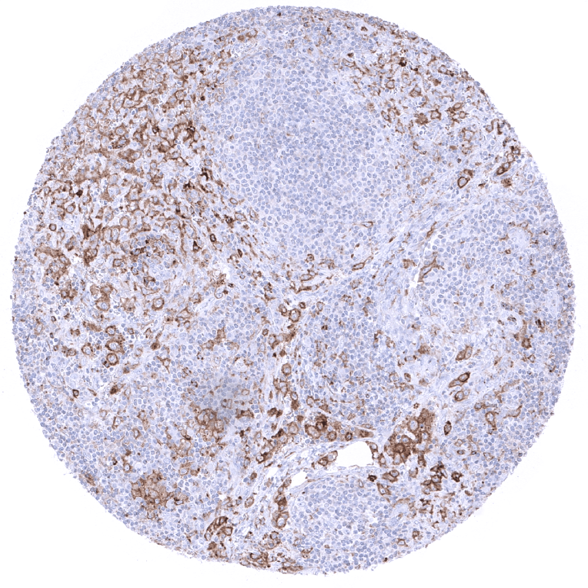
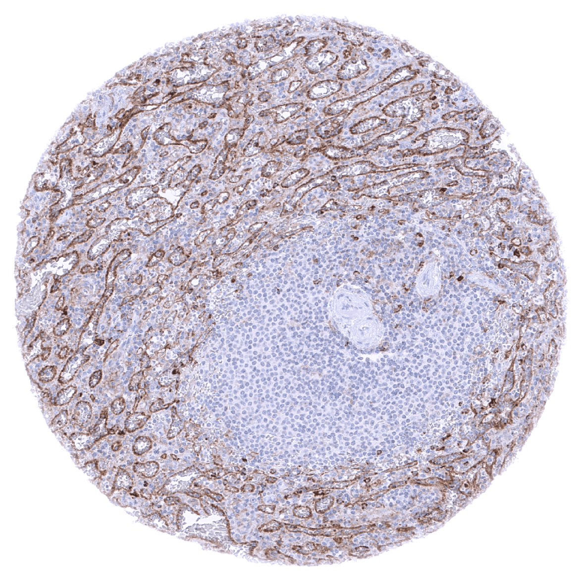
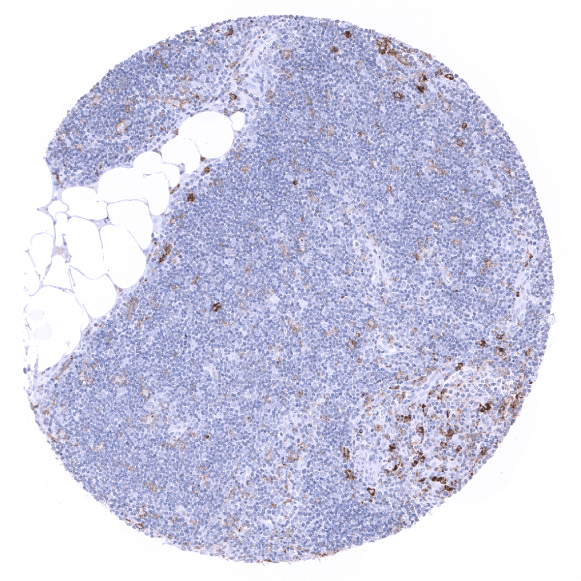
lymph node: In the lymph node, TIM-3 expression is seen in subsets of T-lymphocytes and macrophages. In this sample, macrophages show a particularly strong TIM-3 positivity.
Ovary, stroma
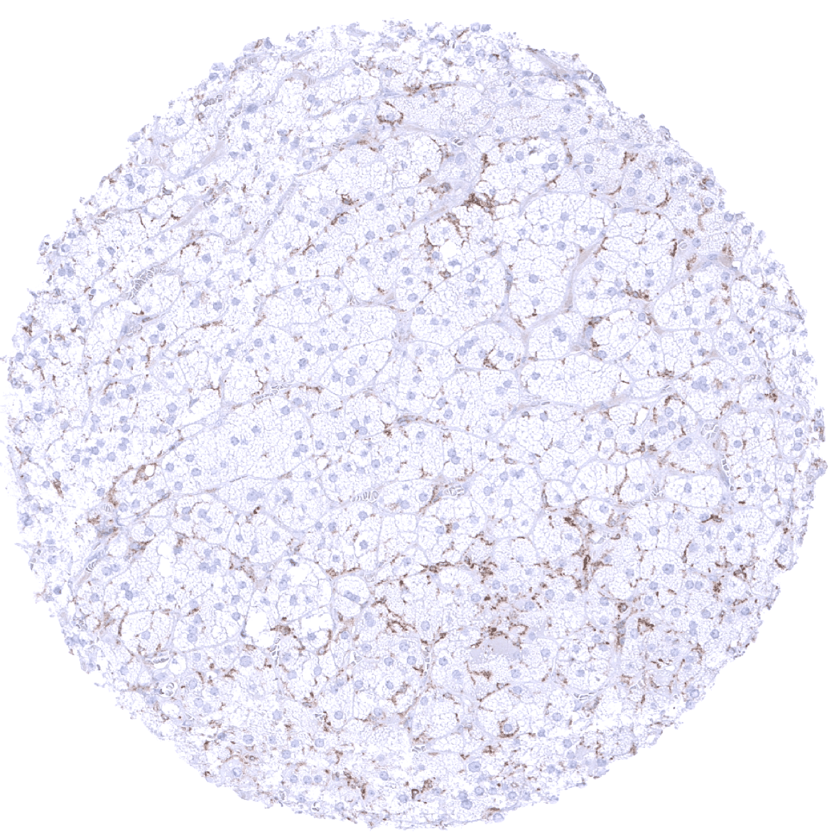
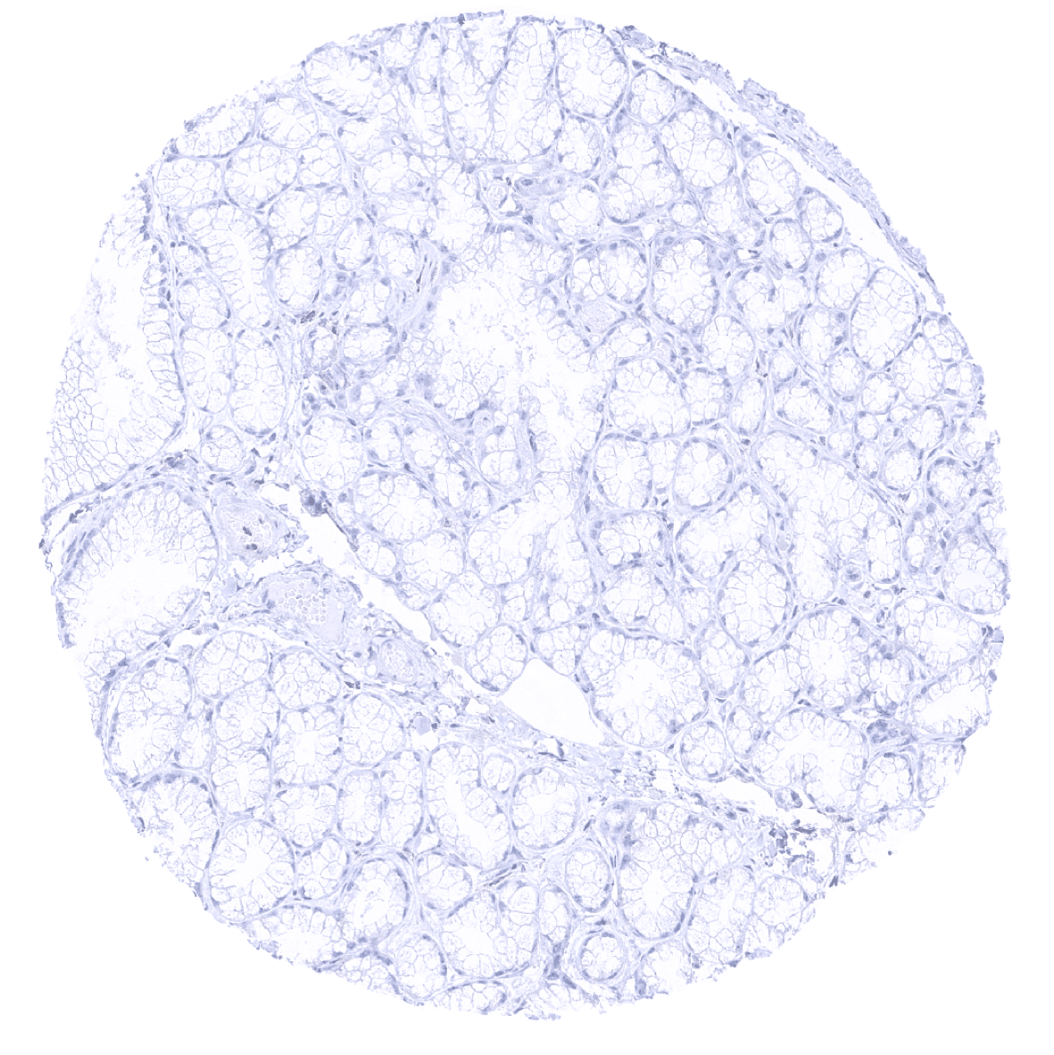
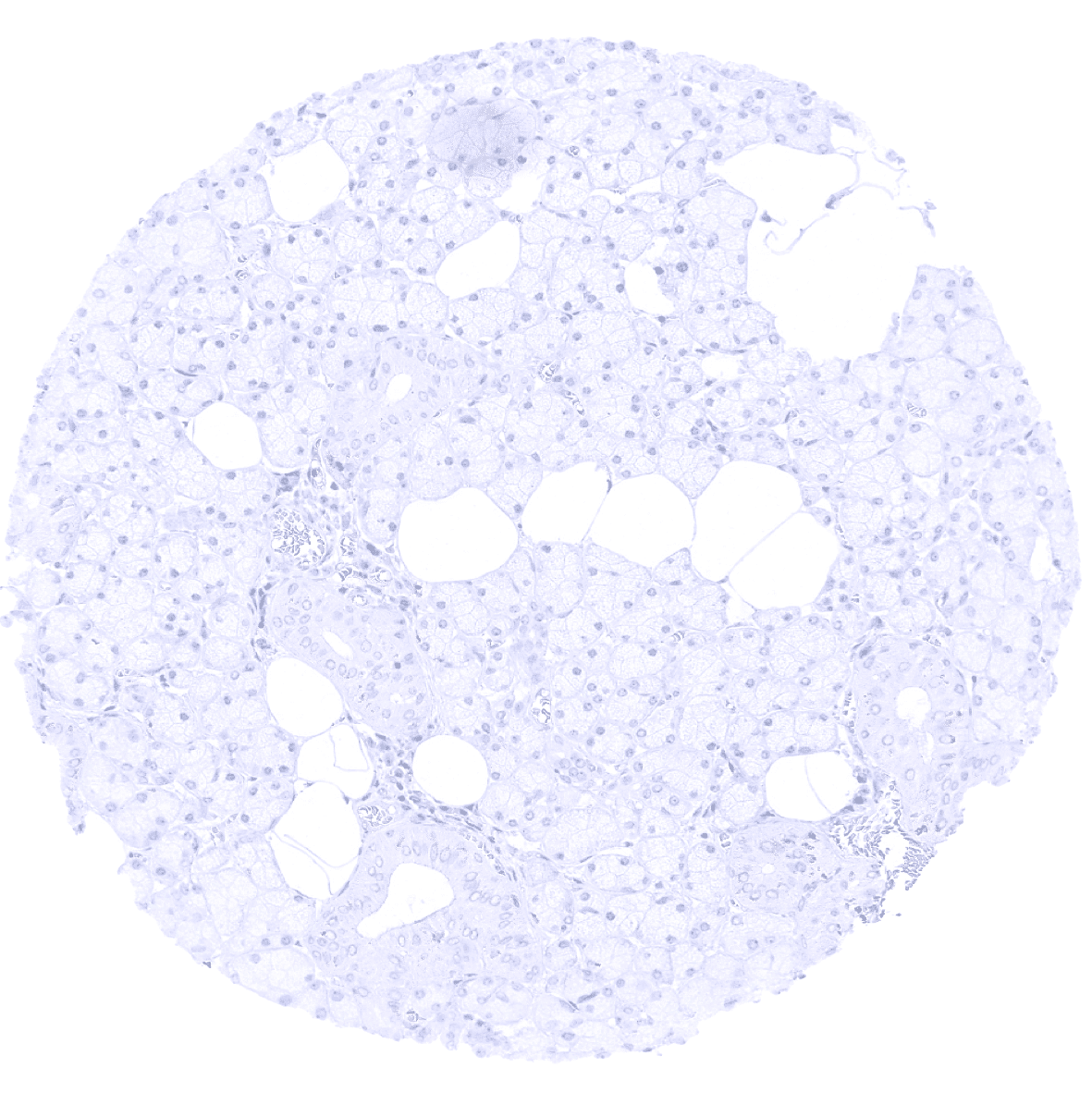
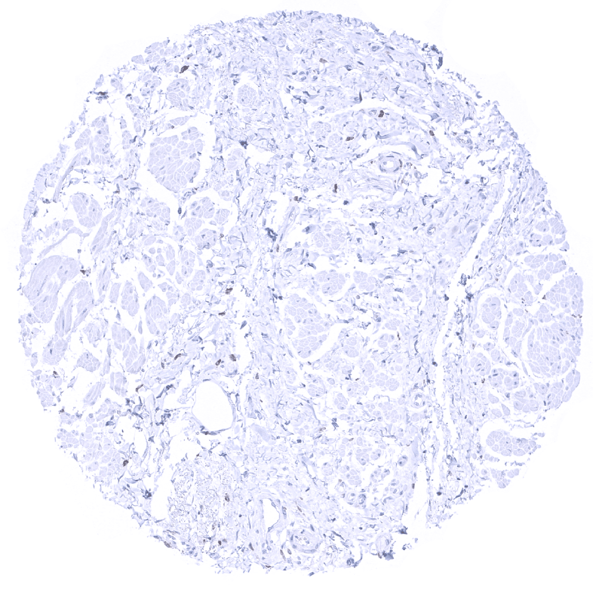
Pancreas
Parathyroid
Parotid gland

Pituitary gland, anterior lobe
Pituitary gland, anterior lobe
Pituitary gland, posterior lobe
Pregnant uterus (decidua): Numerous interspersed macrophages show a strong TIM-3 immunostaining.
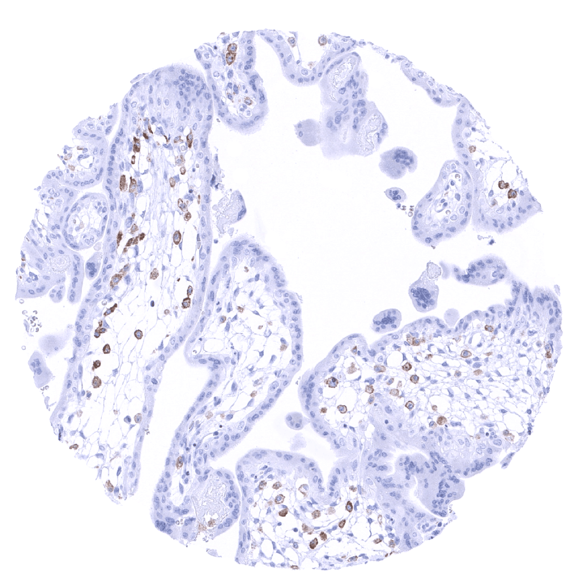
Placenta, early: In placenta macrophages, a strong TIM-3 immunostaining is seen.
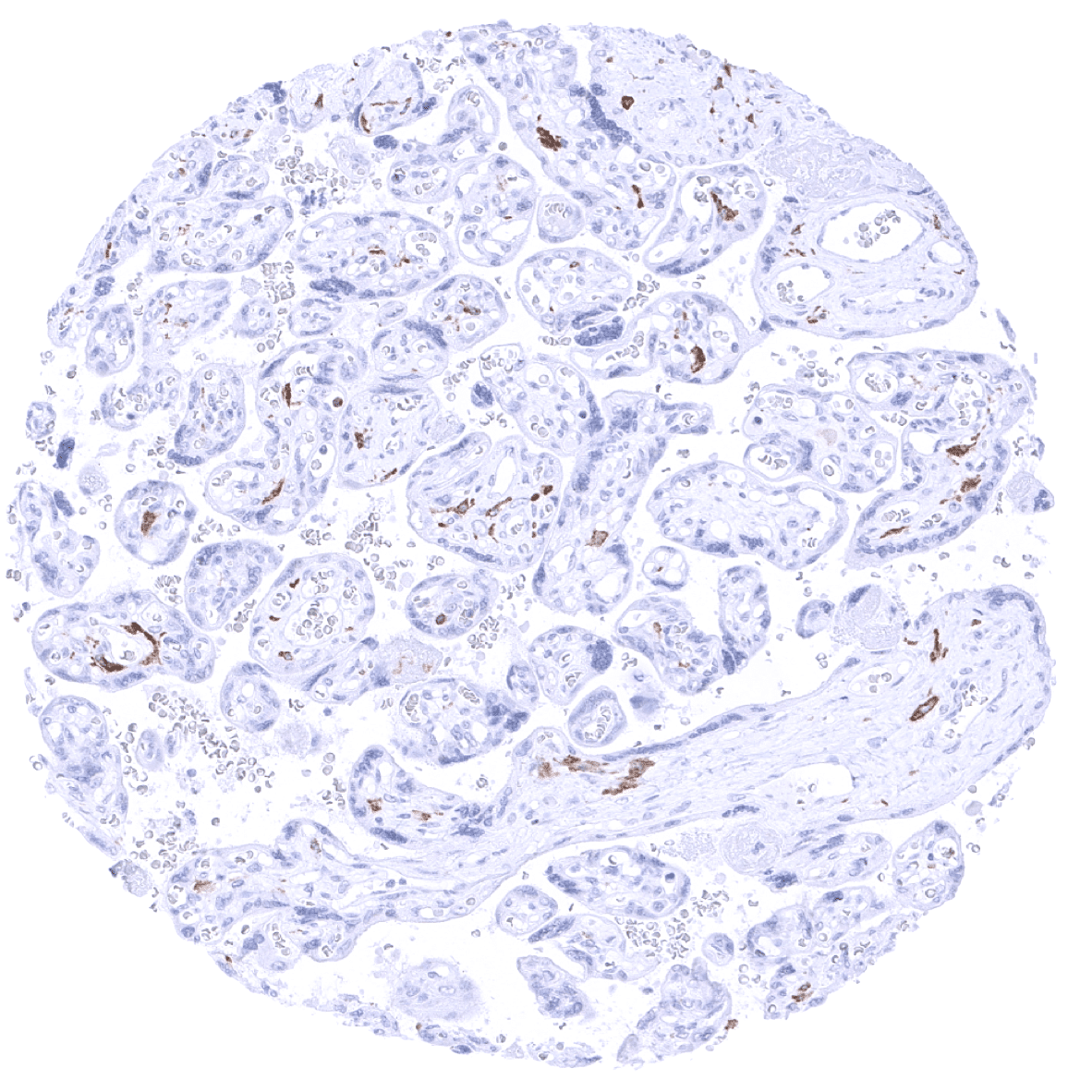
Placenta, mature

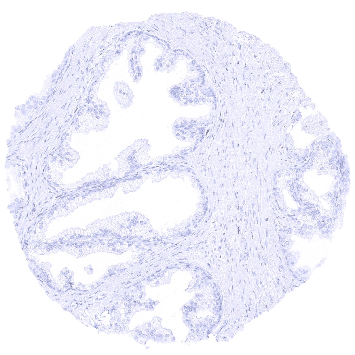
Placenta (amnion and chorion)
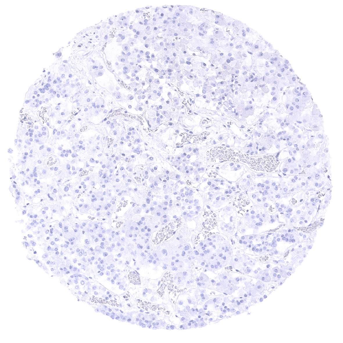
Prostate
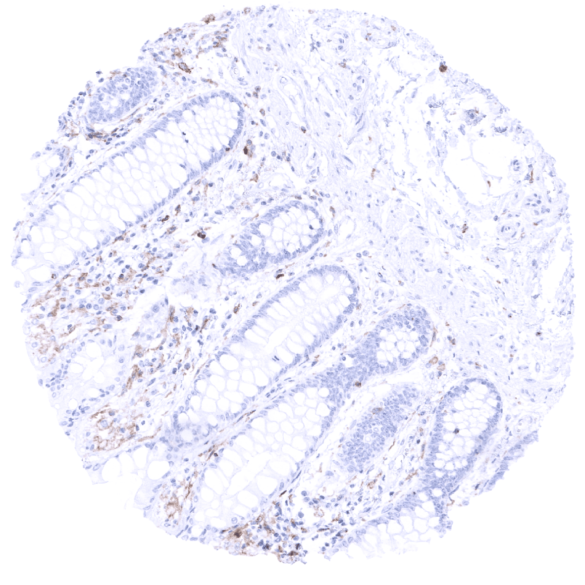
Rectum, mucosa
Seminal vesicle
Sinus paranasales
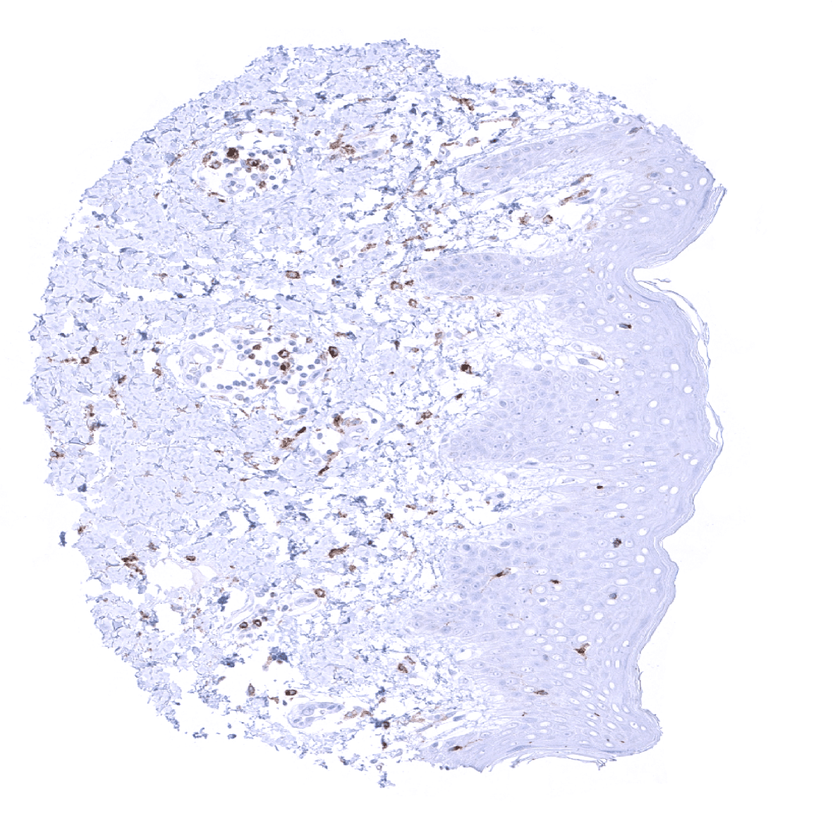
Skin: In the skin, TIM-3 immunostaining occurs in a fraction of T-lymphocytes and of macrophages.
Spleen: In the spleen, TIM-3 immunostaining occurs in venous sinusoids as well as in T-lymphocytes.
Stomach, antrum

Stomach, corpus
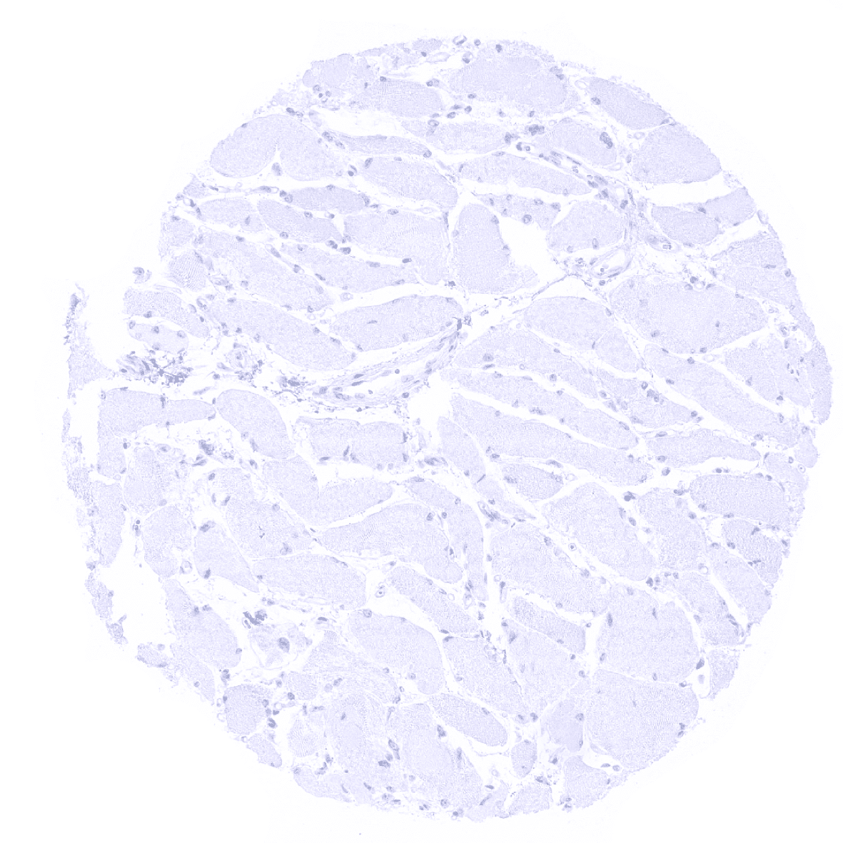
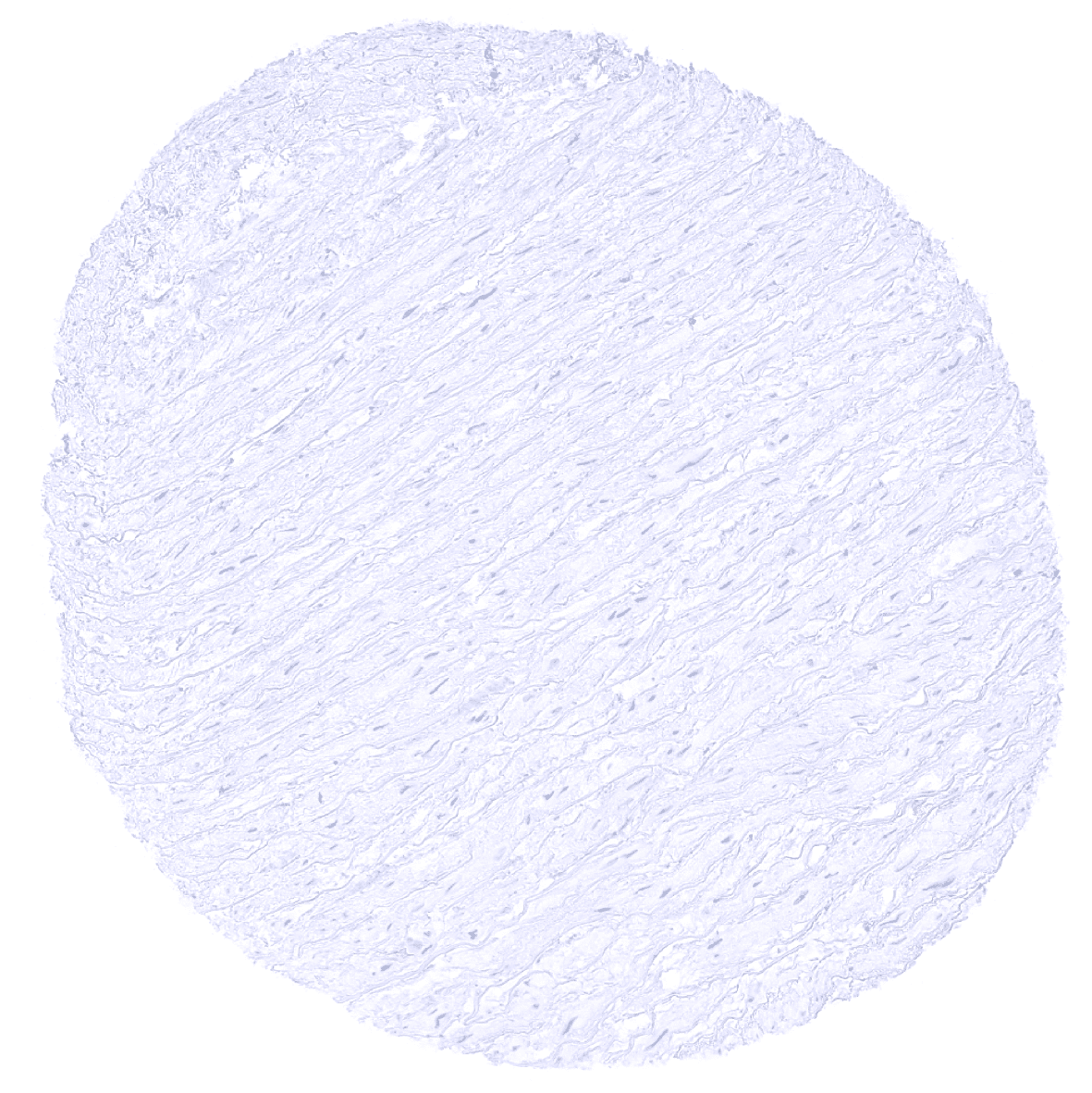
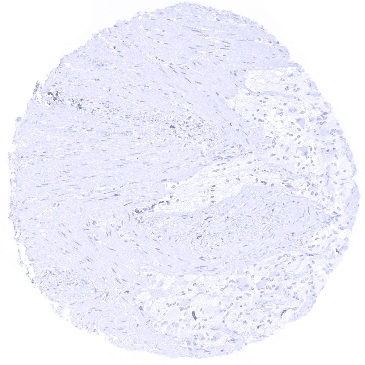
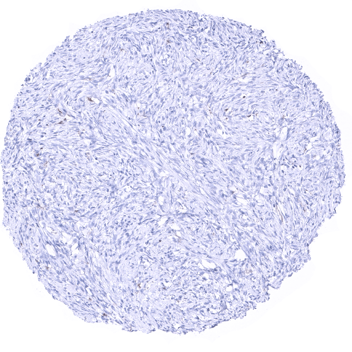
Striated muscle
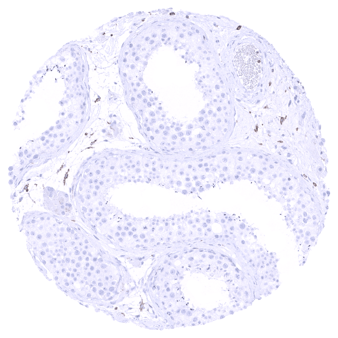
Testis
Thymus: In the thymus, TIM-3 immunostaining is rarely seen in cortical T-cells.
Thyroid gland
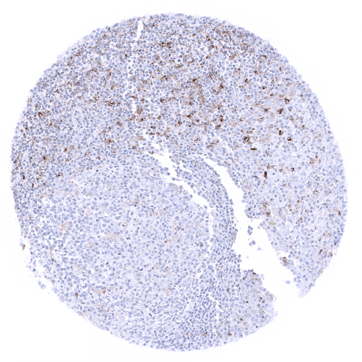
Tonsil: In the tonsil, variable levels of TIM-3 immunostaining are seen in T-cells and in macrophages.
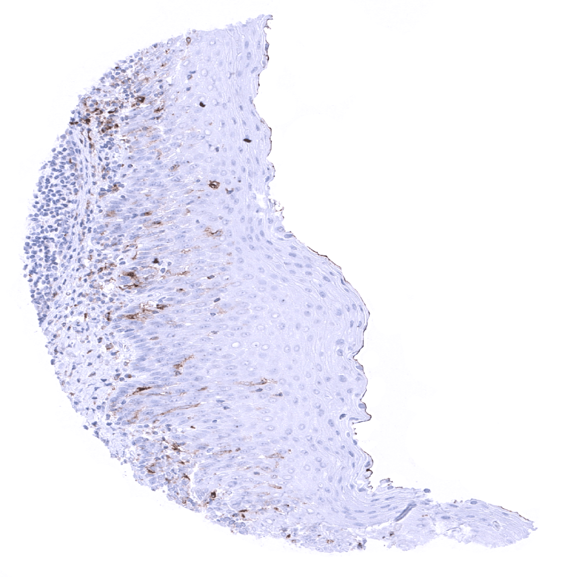
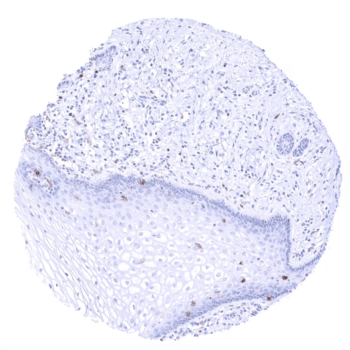
Tonsil, surface epithelium: In the tonsil surface epithelium, TIM-3 immunostaining occurs in a fraction of T-lymphocytes and of macrophages. The black staining of the surface mebrane represents an inking effect.
Urinary bladder, muscular wall

Urinary bladder, urothelium
Uterus, myometrium