Adrenal gland
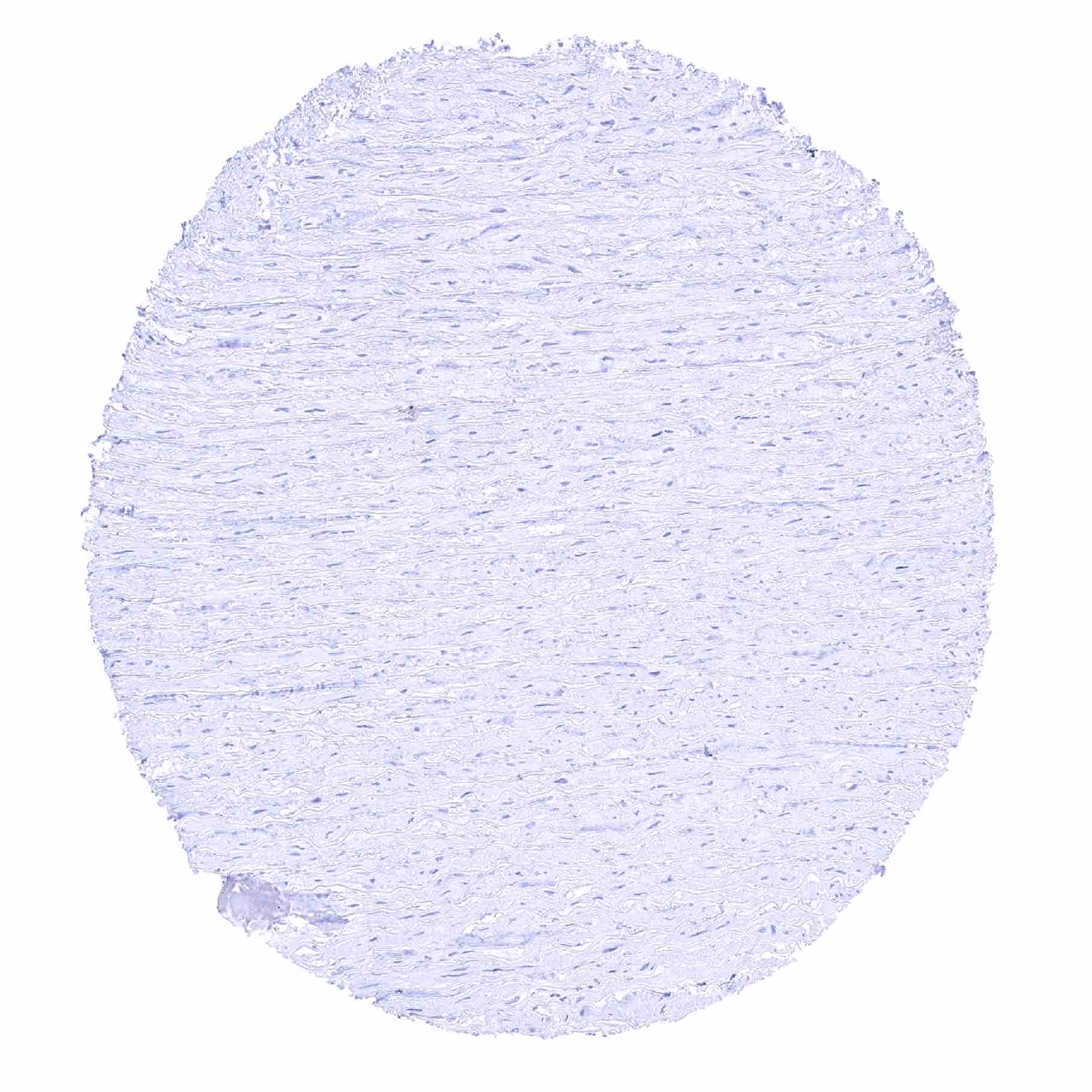
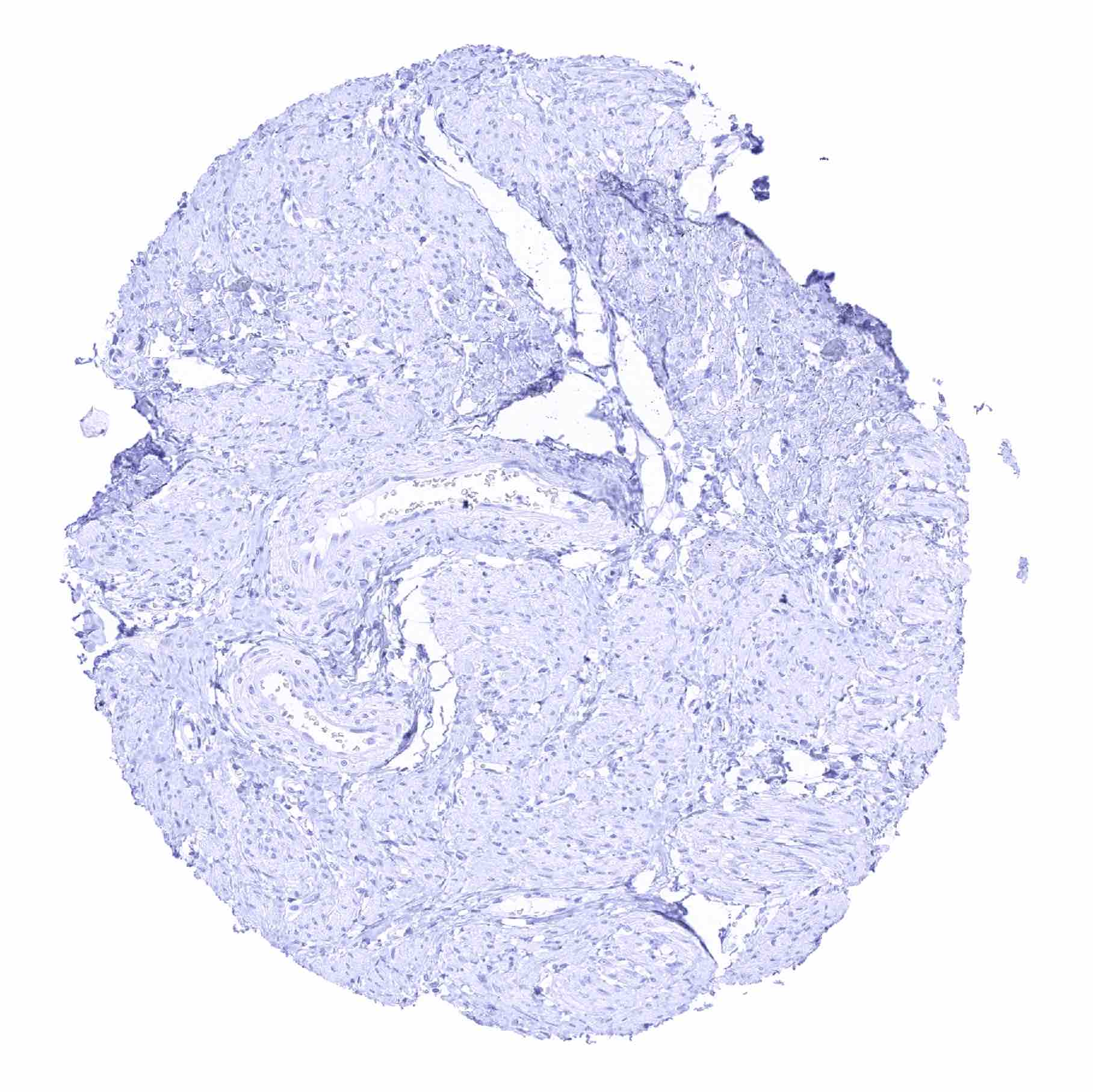
Aorta, media
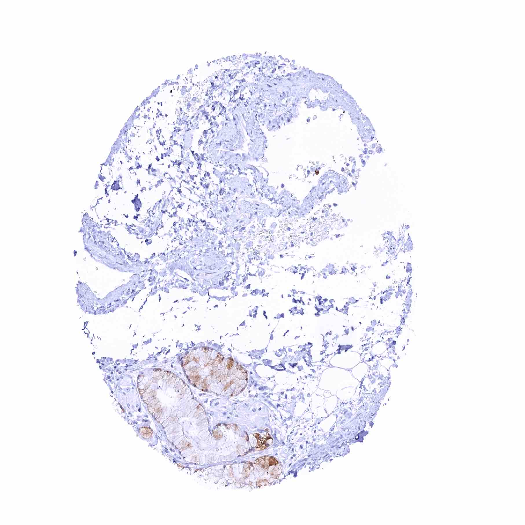
Appendix, mucosa – Moderate cytoplasmic TFF1 staining of a subset of goblet cells, mainly located at the surface epithelium

Appendix, muscular wall

Bone marrow
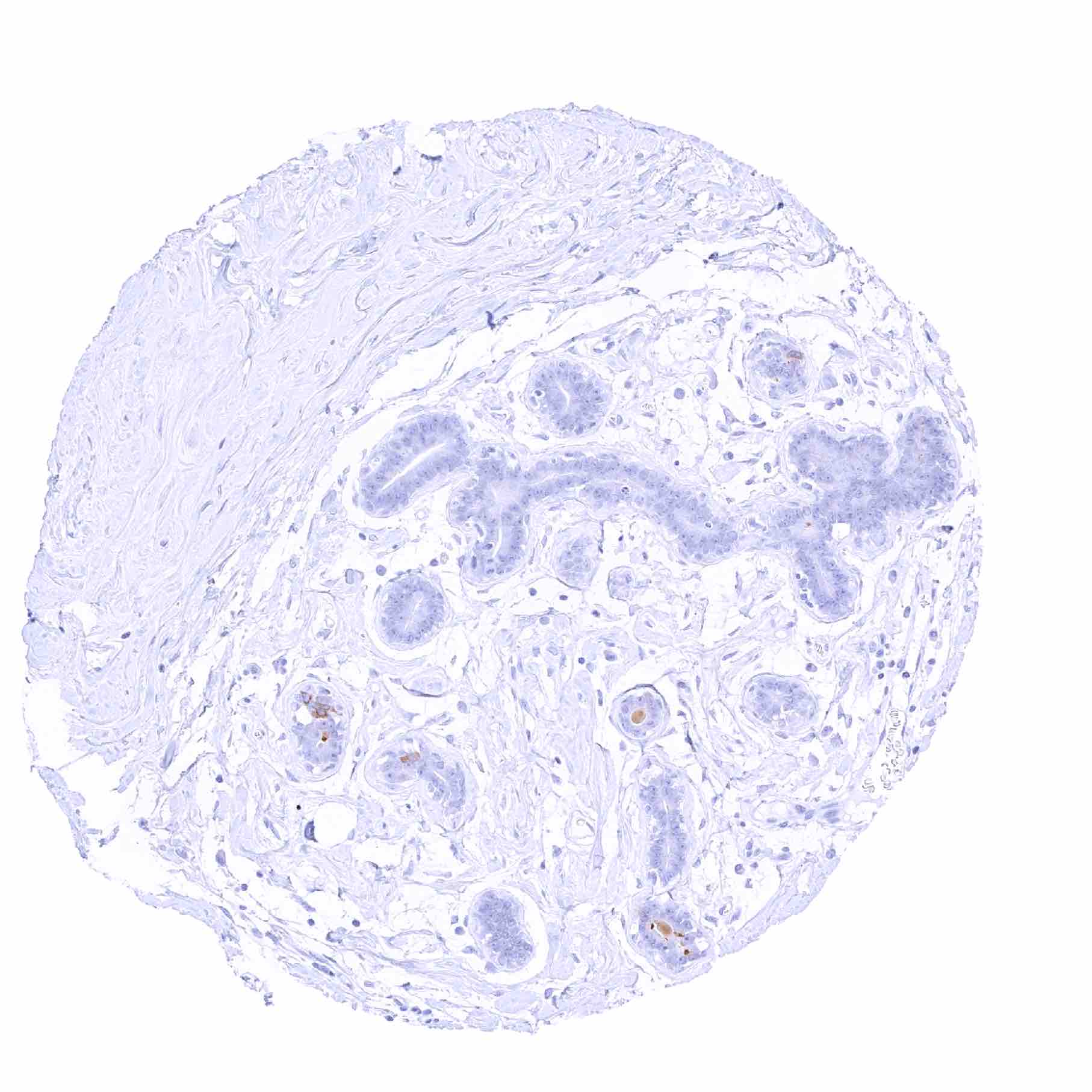
Breast – Weak to moderate TFF1 staining of very few luminal breast epithelial cells
Bronchus, glands glands – Goblet cells in bronchial glands exhibit a weak to moderate TFF1 positivity
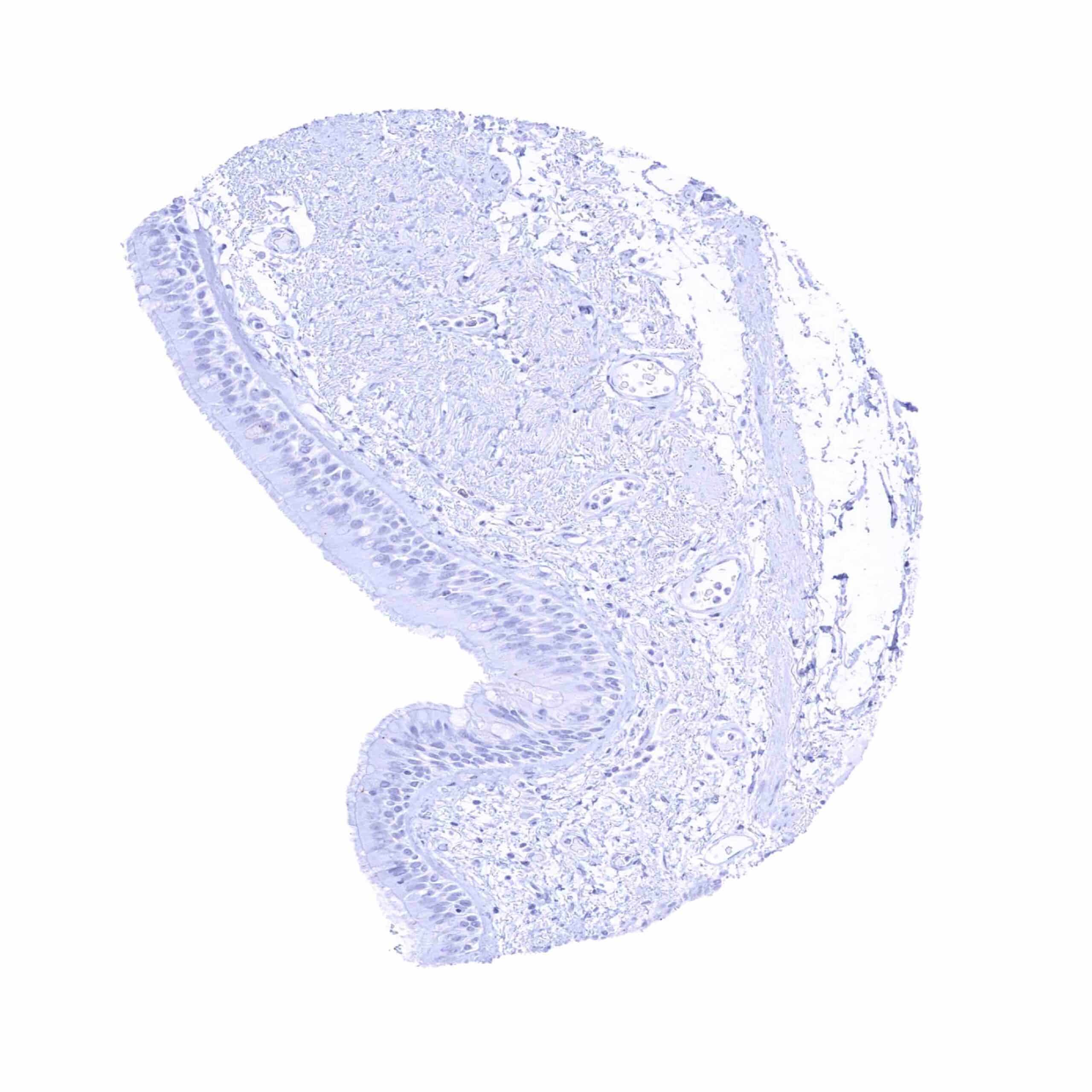
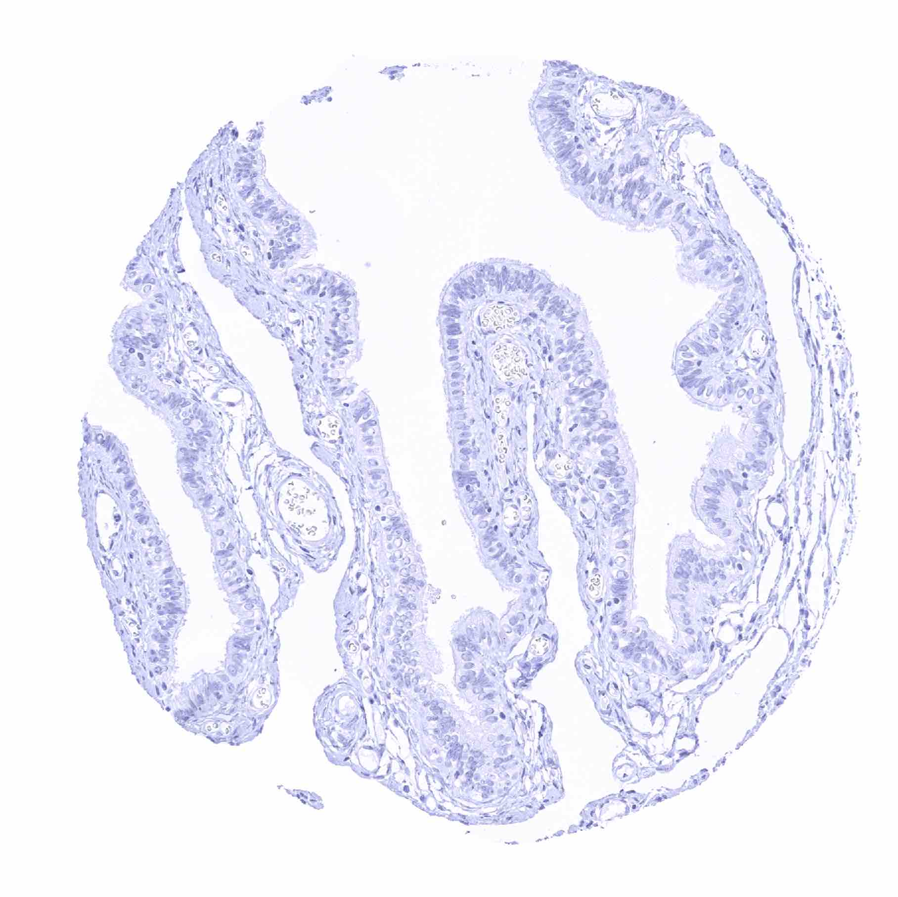
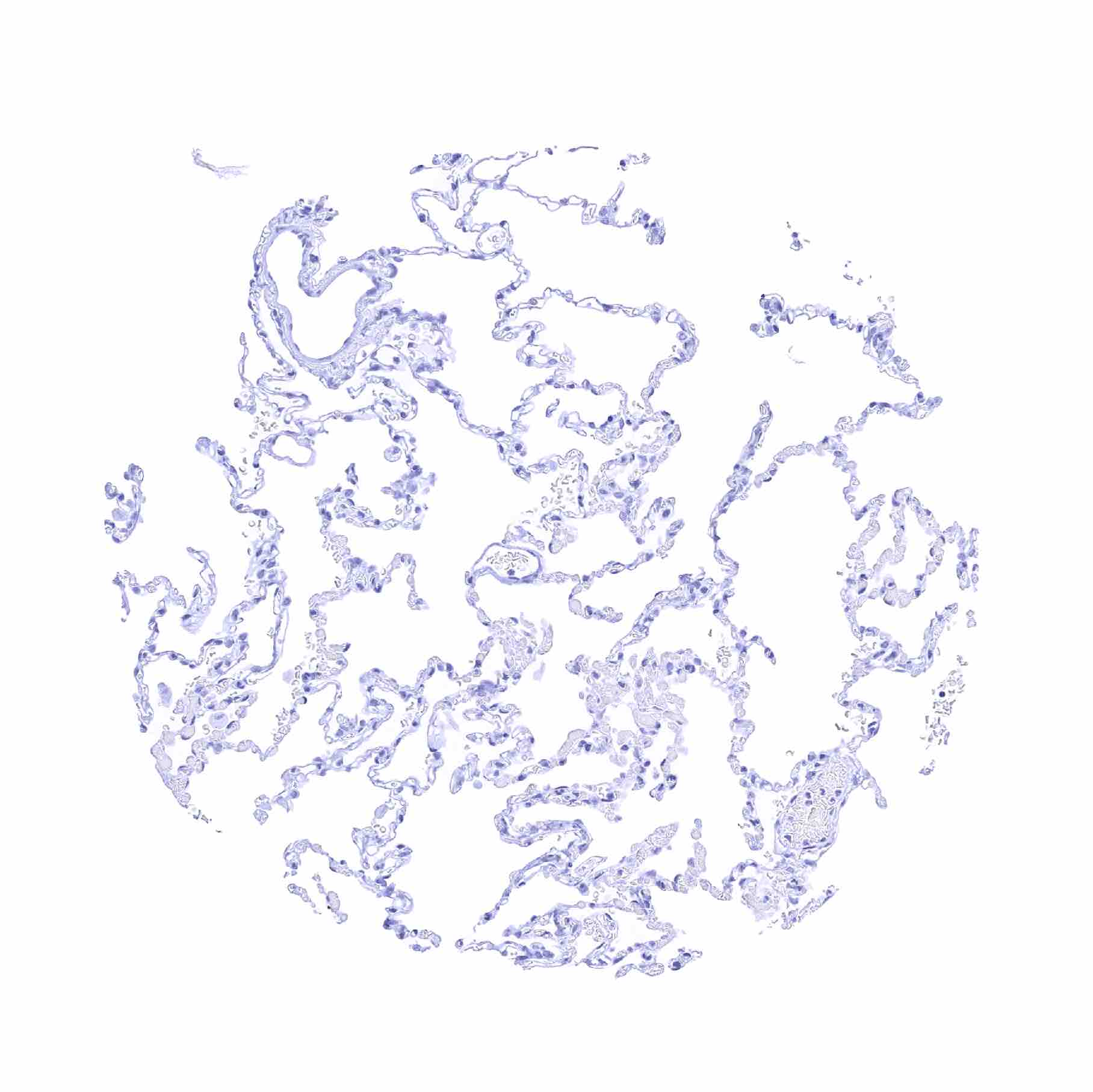
Bronchus, mucosa – Absence of TFF1 staining in this sample of respiratory epithelium
Bronchus, mucosa – Some goblet cells of the respiratory epithelium show a weak TFF1 staining
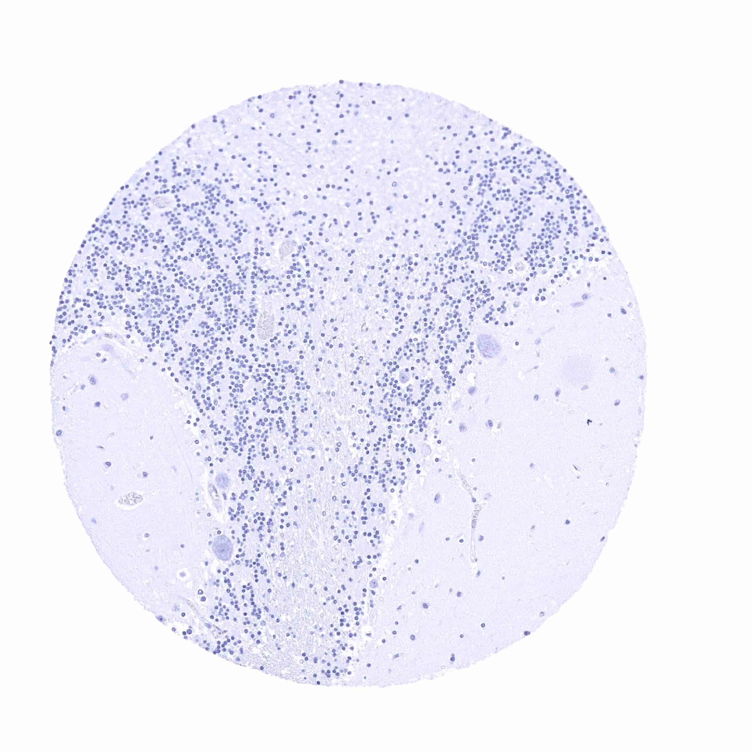
Cerebellum (molecular layer, Purkinje cell layer, granule cell layer, white matter)
Cerebellum (white matter)
Cerebrum, grey matter

Cerebrum, white matter
Colon descendens, muscular wall

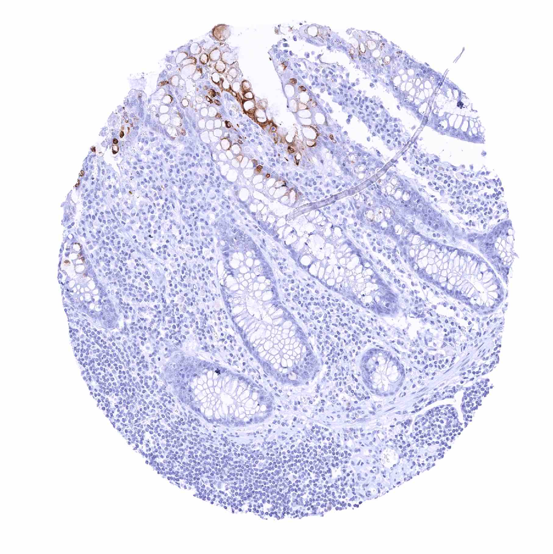
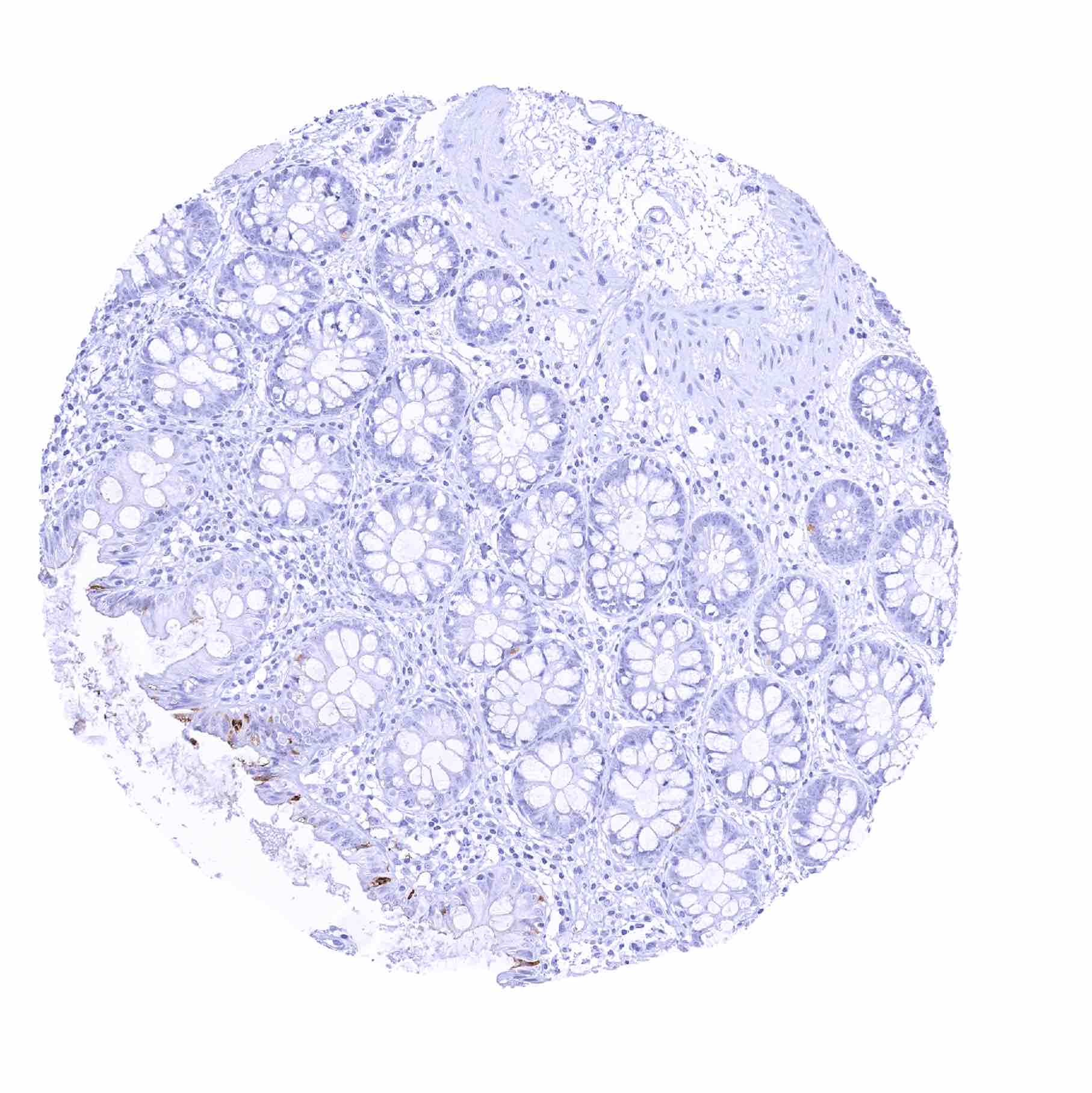
Colon, mucosa – Moderate cytoplasmic TFF1 staining of a small subset of goblet cells
Duodenum, Brunner gland
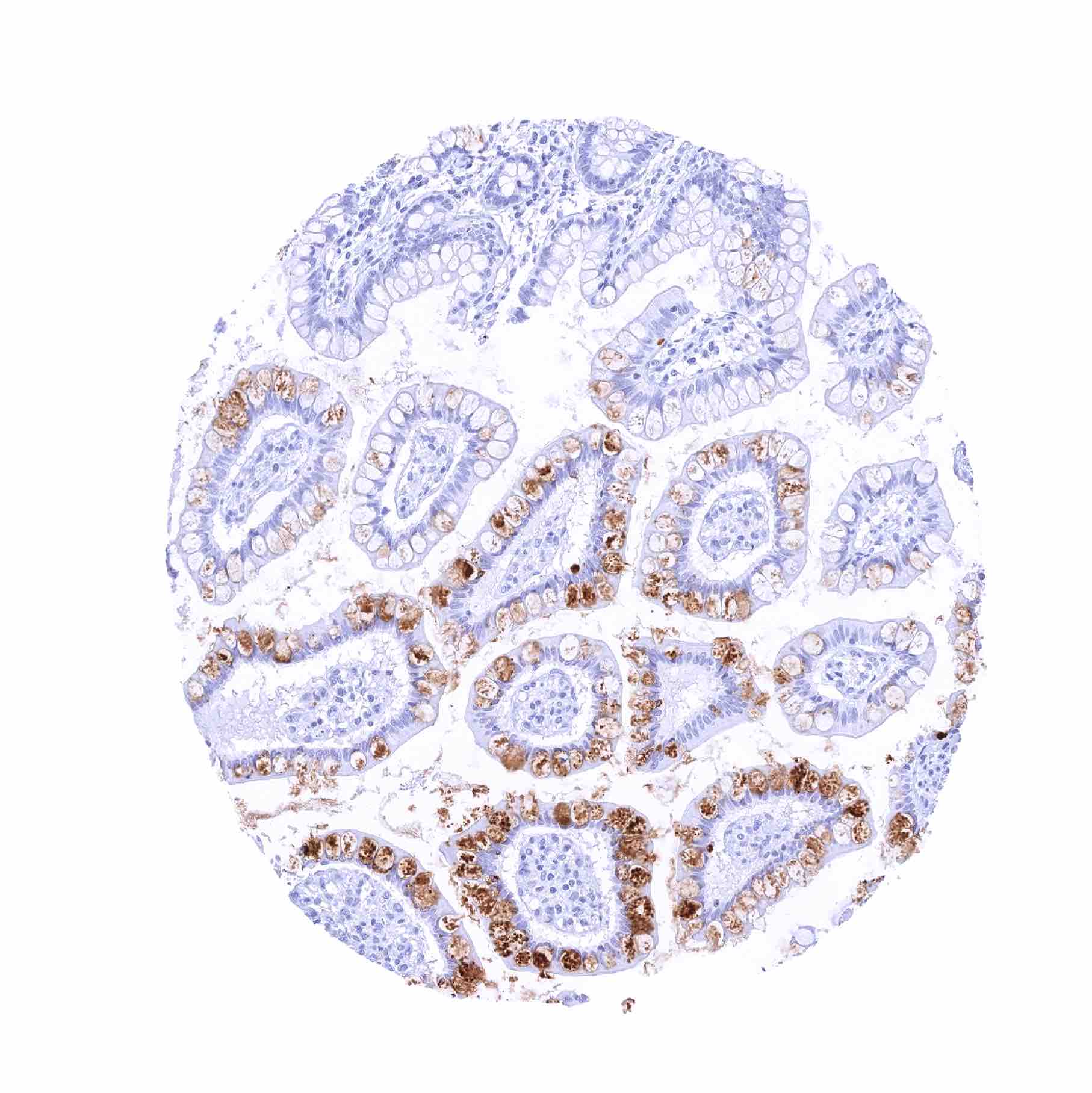
Duodenum, mucosa – Moderate to strong TFF1 staining of selected goblet cells
Epididymis

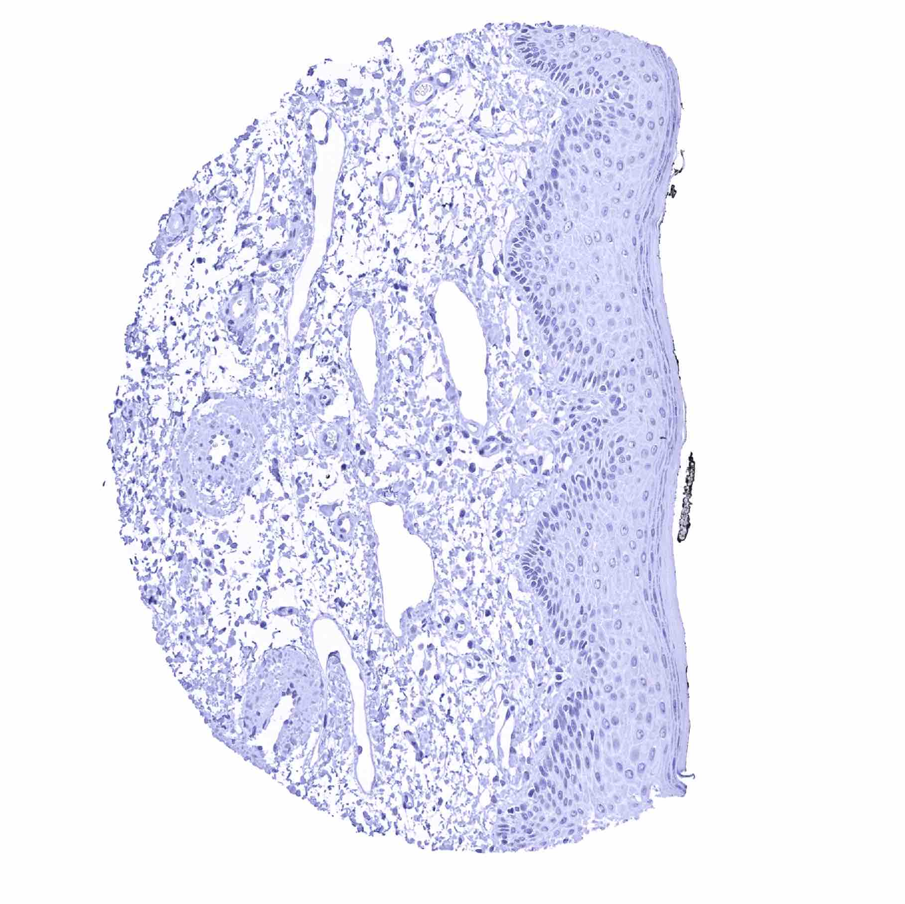
Esophagus, squamous epithelium
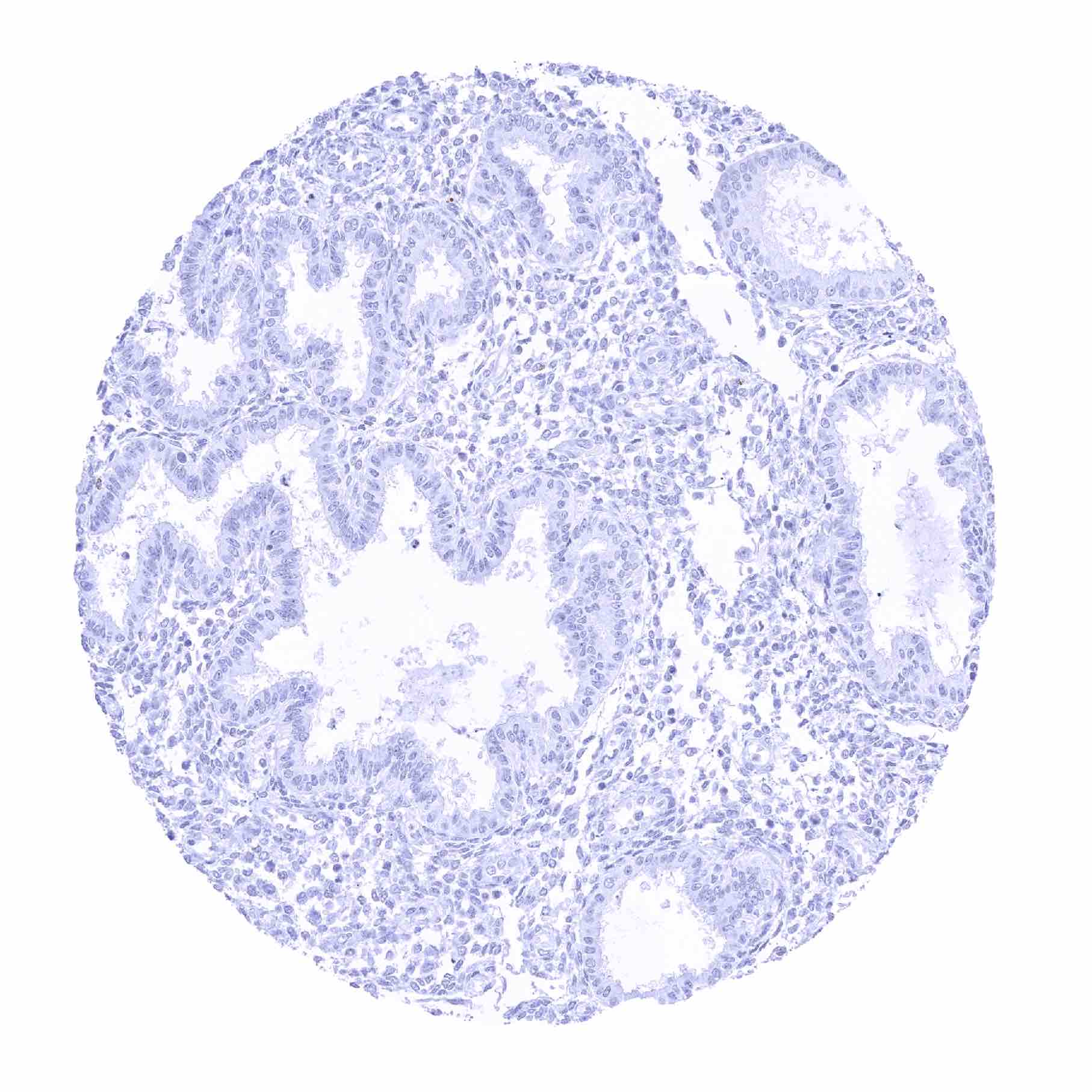
Fallopian tube, mucosa
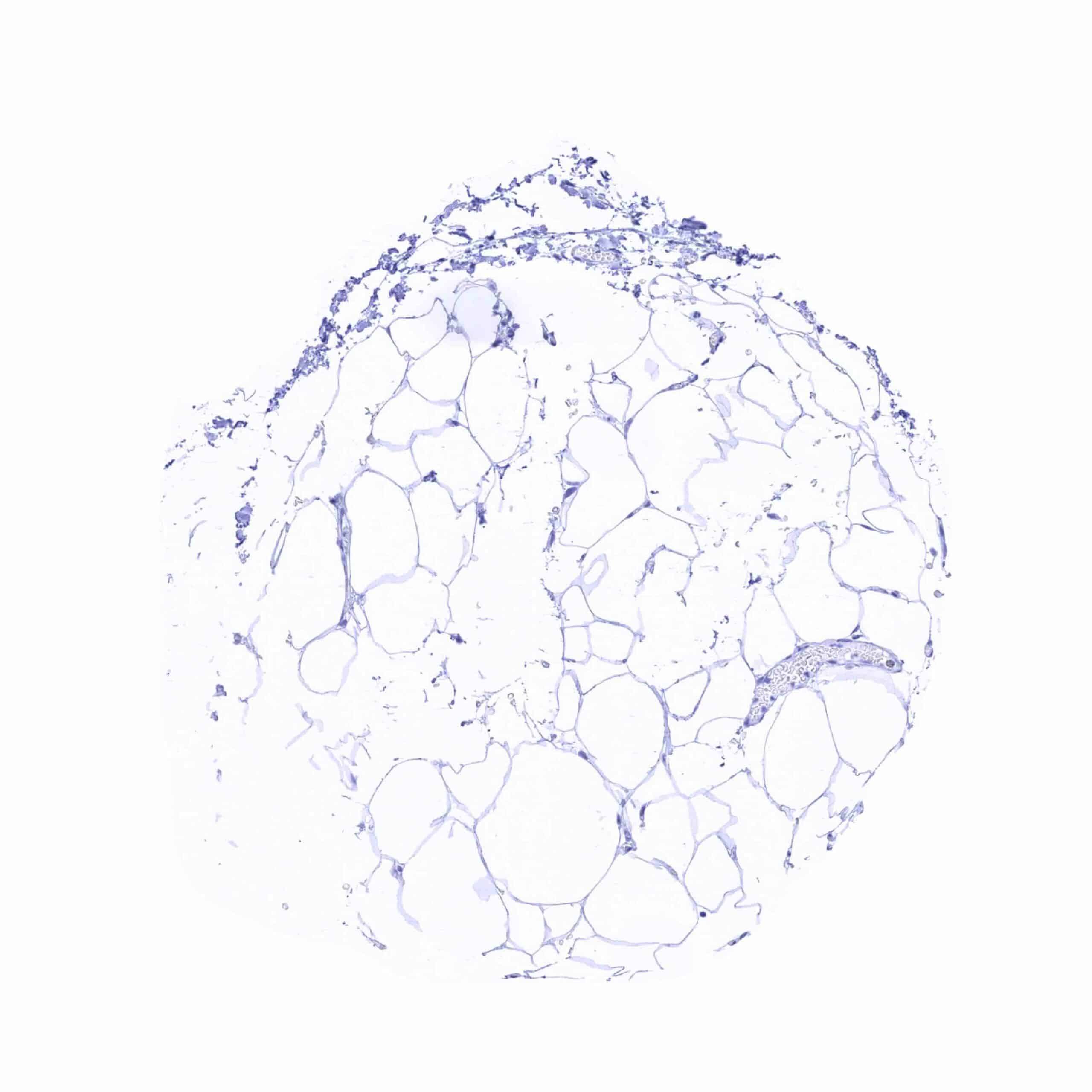
Fat
Gallbladder, epithelium – Weak focal cytoplasmic TFF1 staining of some surface epithelial cells

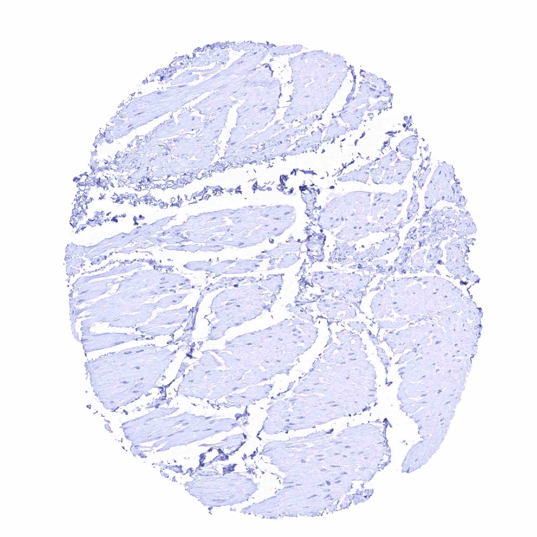
Heart muscle
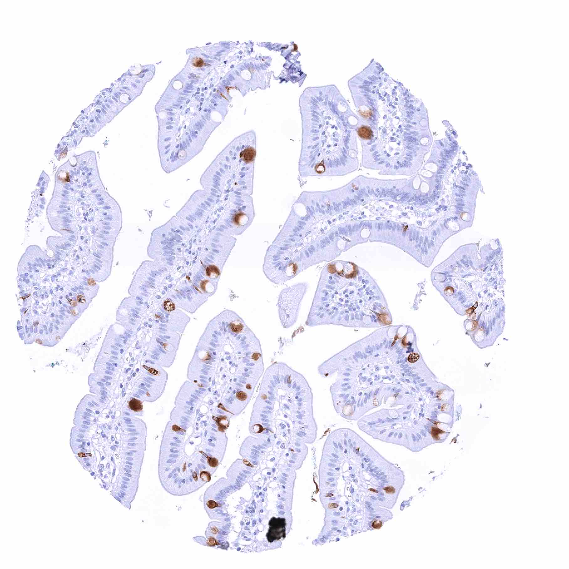
Ileum, mucosa – Moderate to strong TFF1 staining of a subset of goblet cells, predominantly at the surface or the top third of the crypts

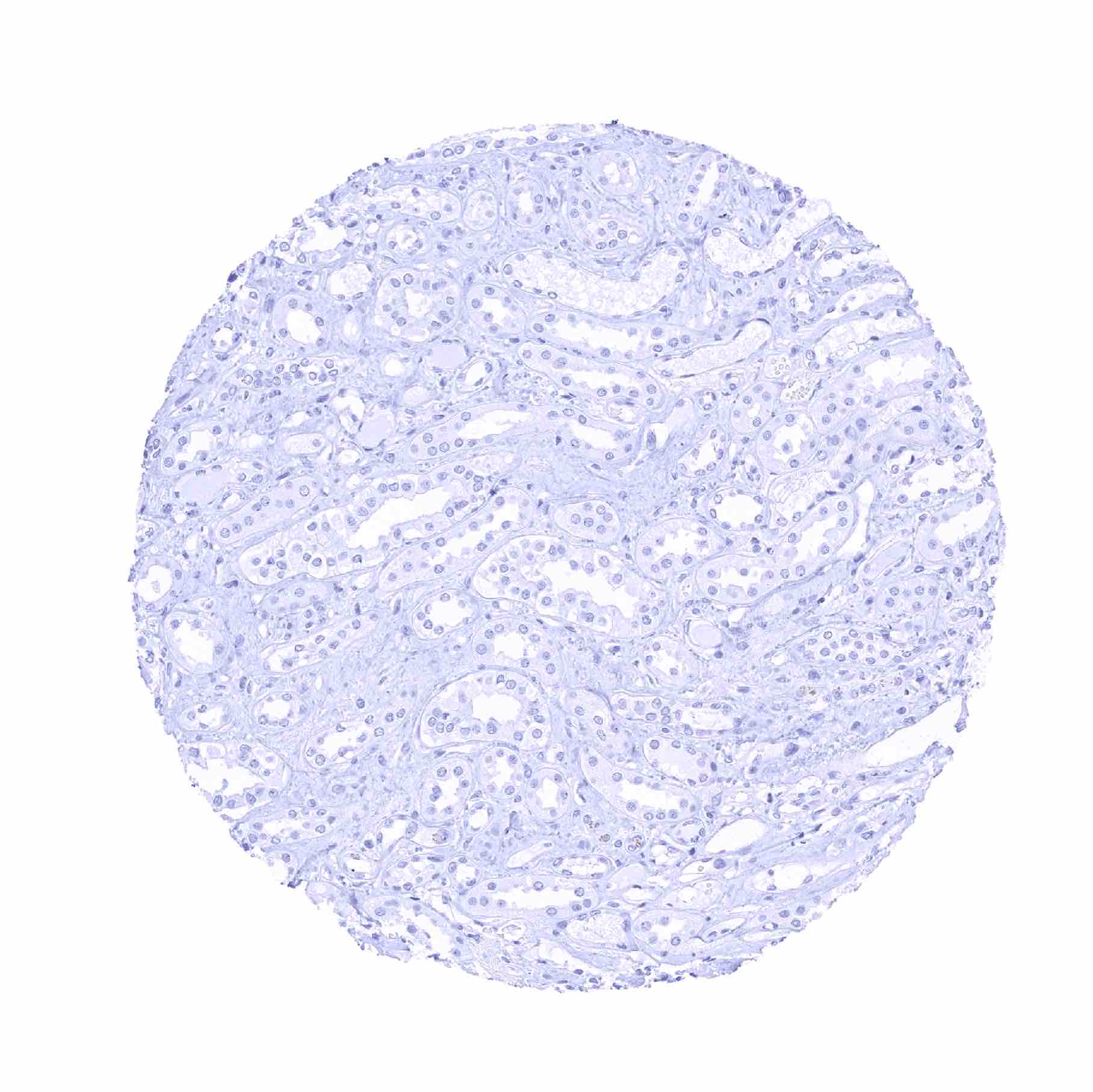
Kidney, cortex
Kidney, medulla
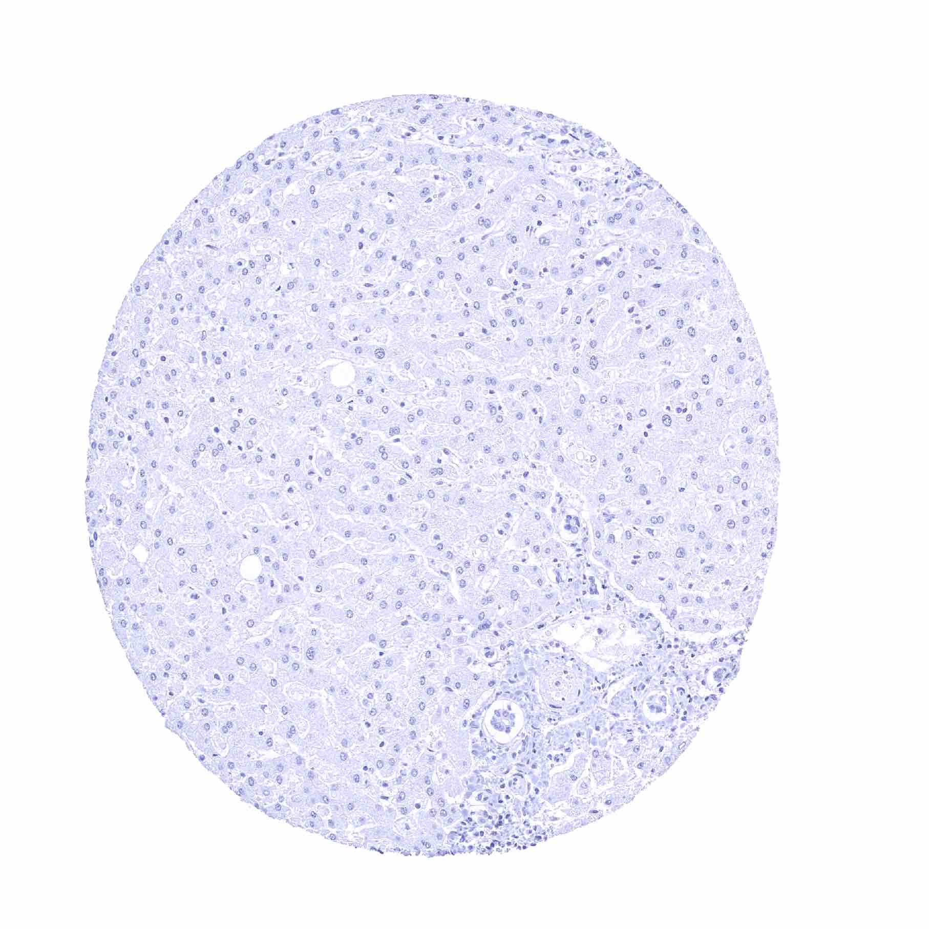
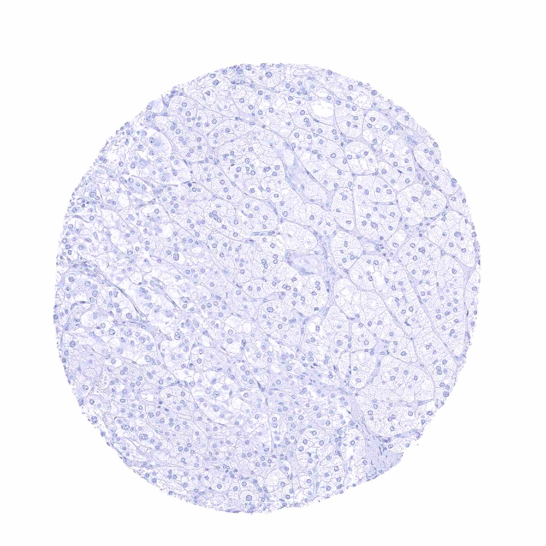
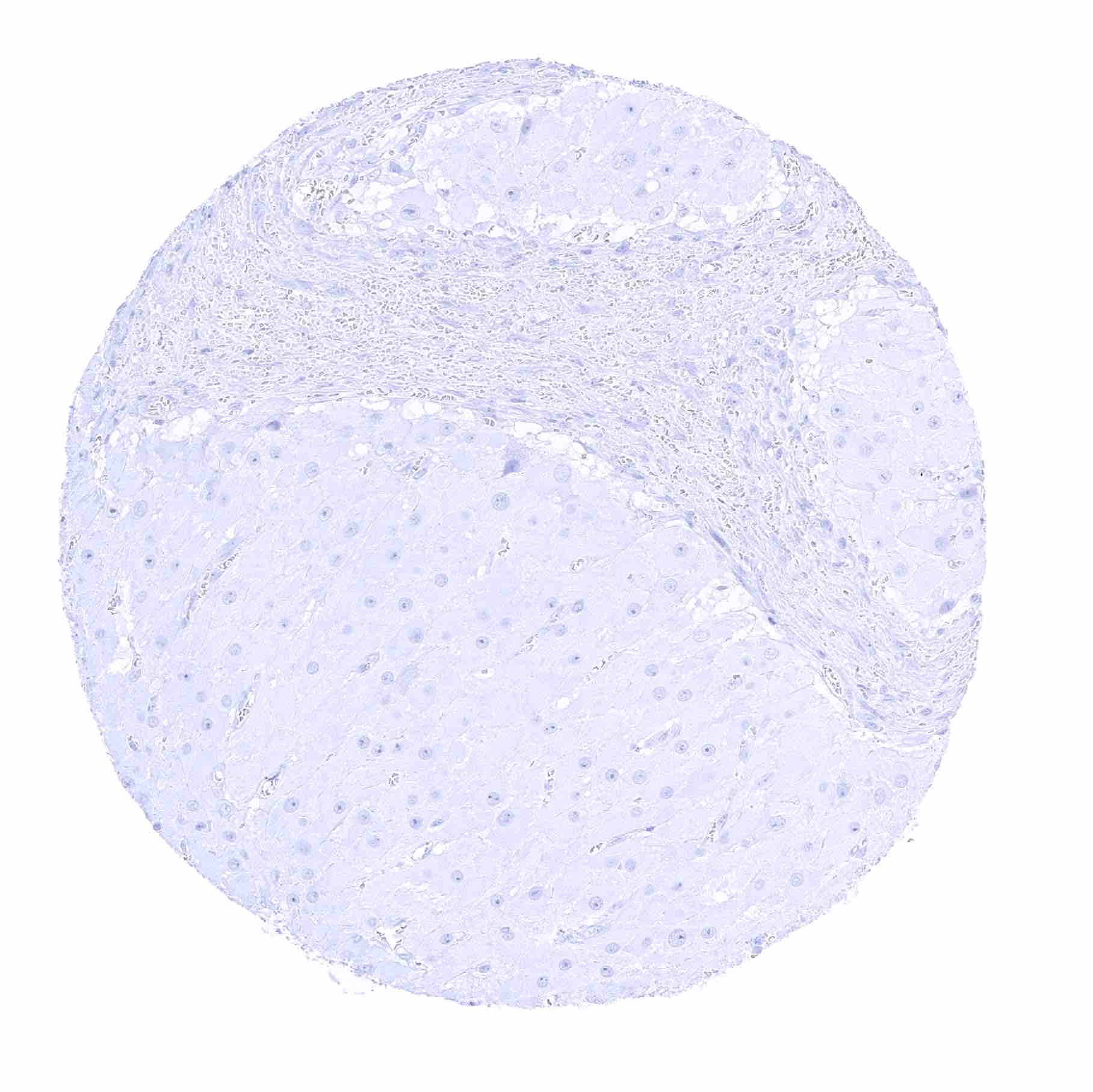
Liver
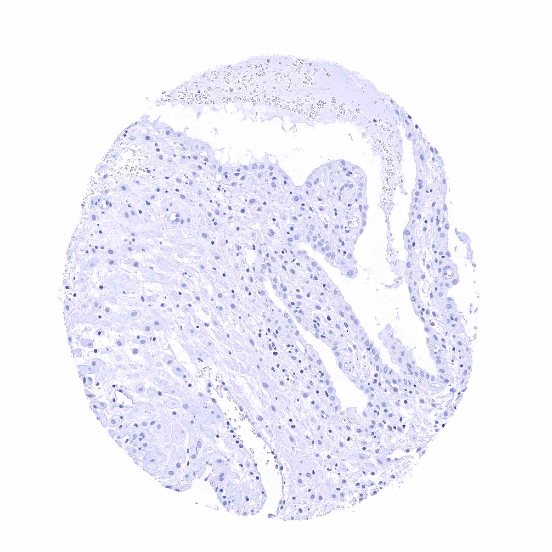
Lung
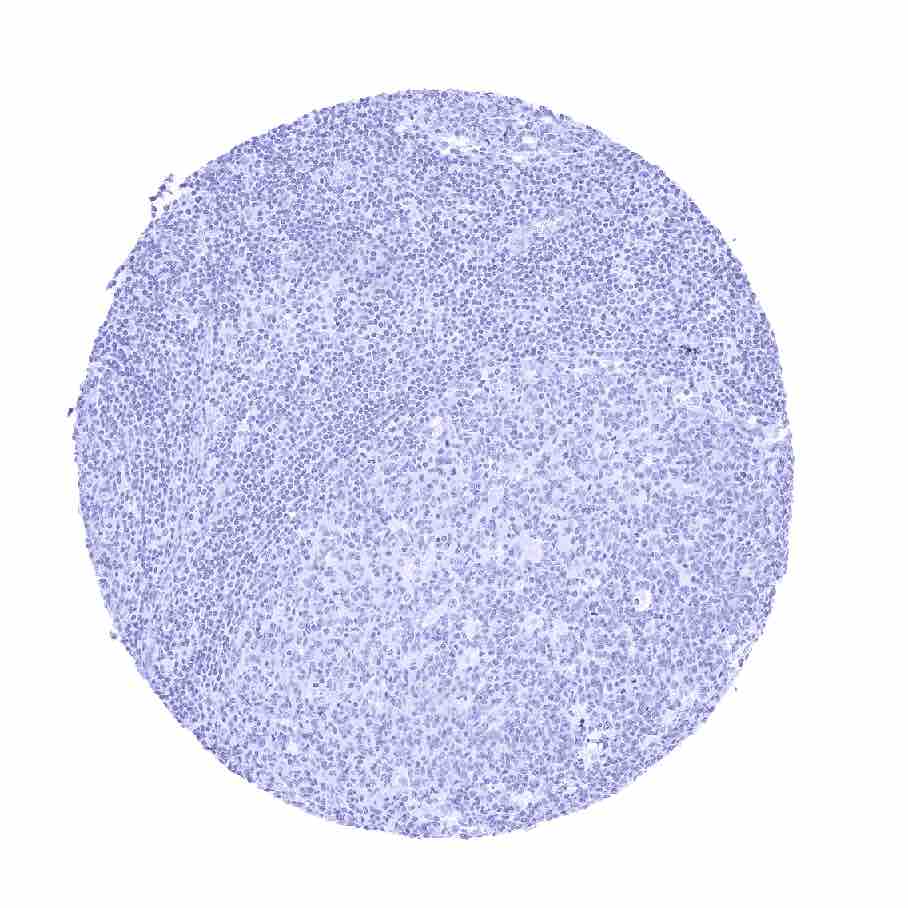
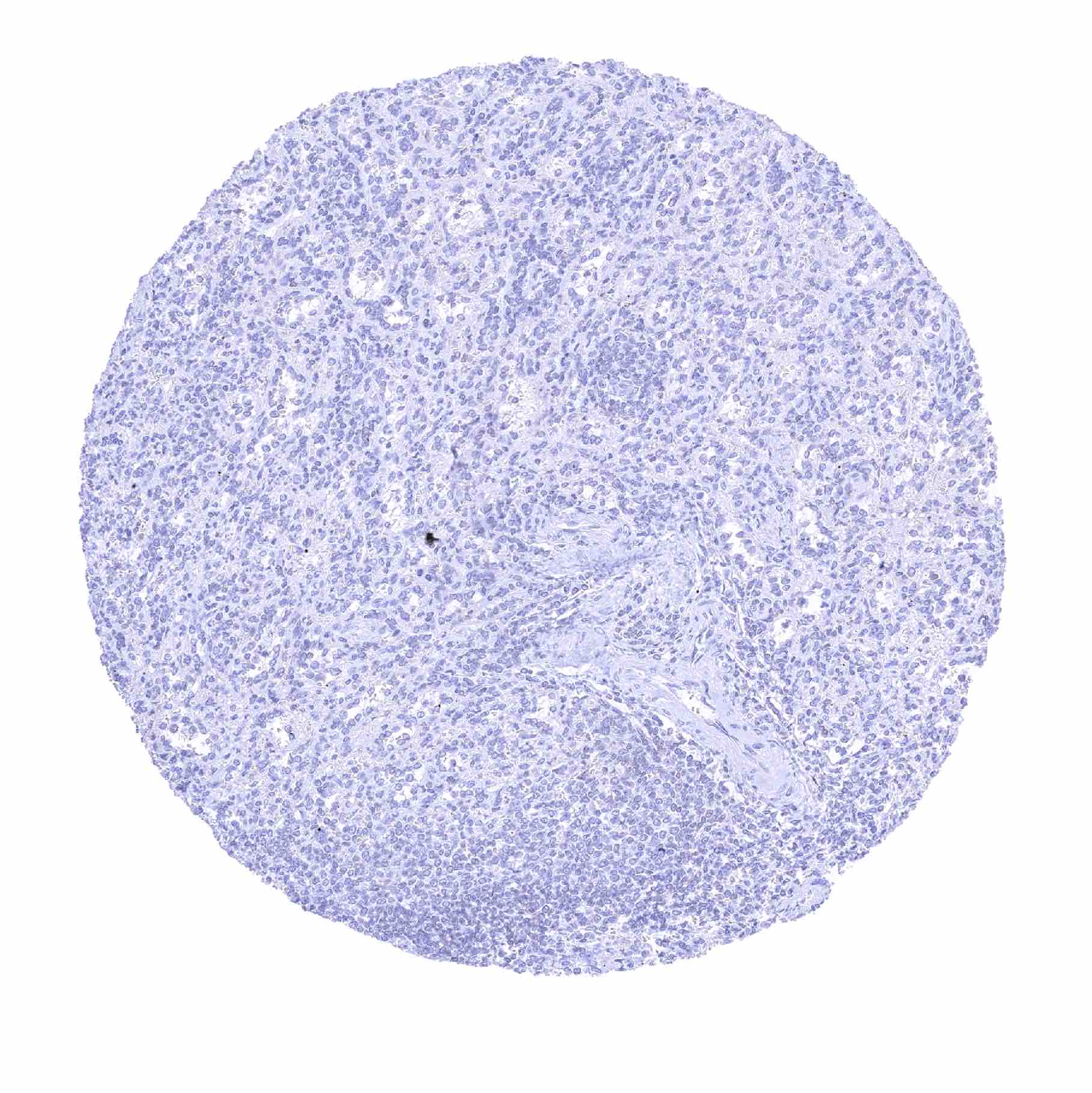
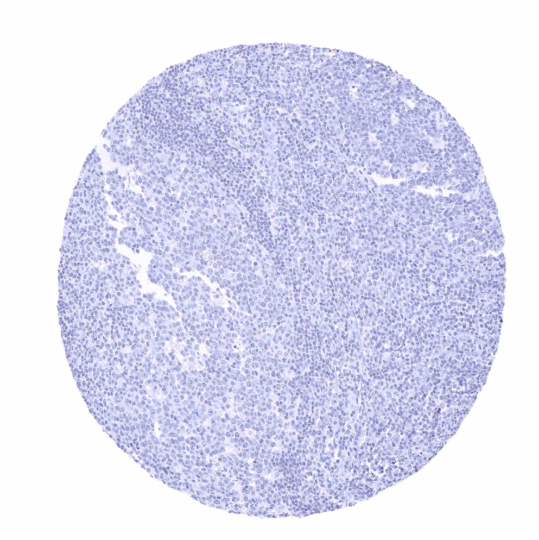
Lymph node
Ovary, corpus luteum
Ovary, follicular cyst

Ovary, stroma (2)

Ovary, stroma
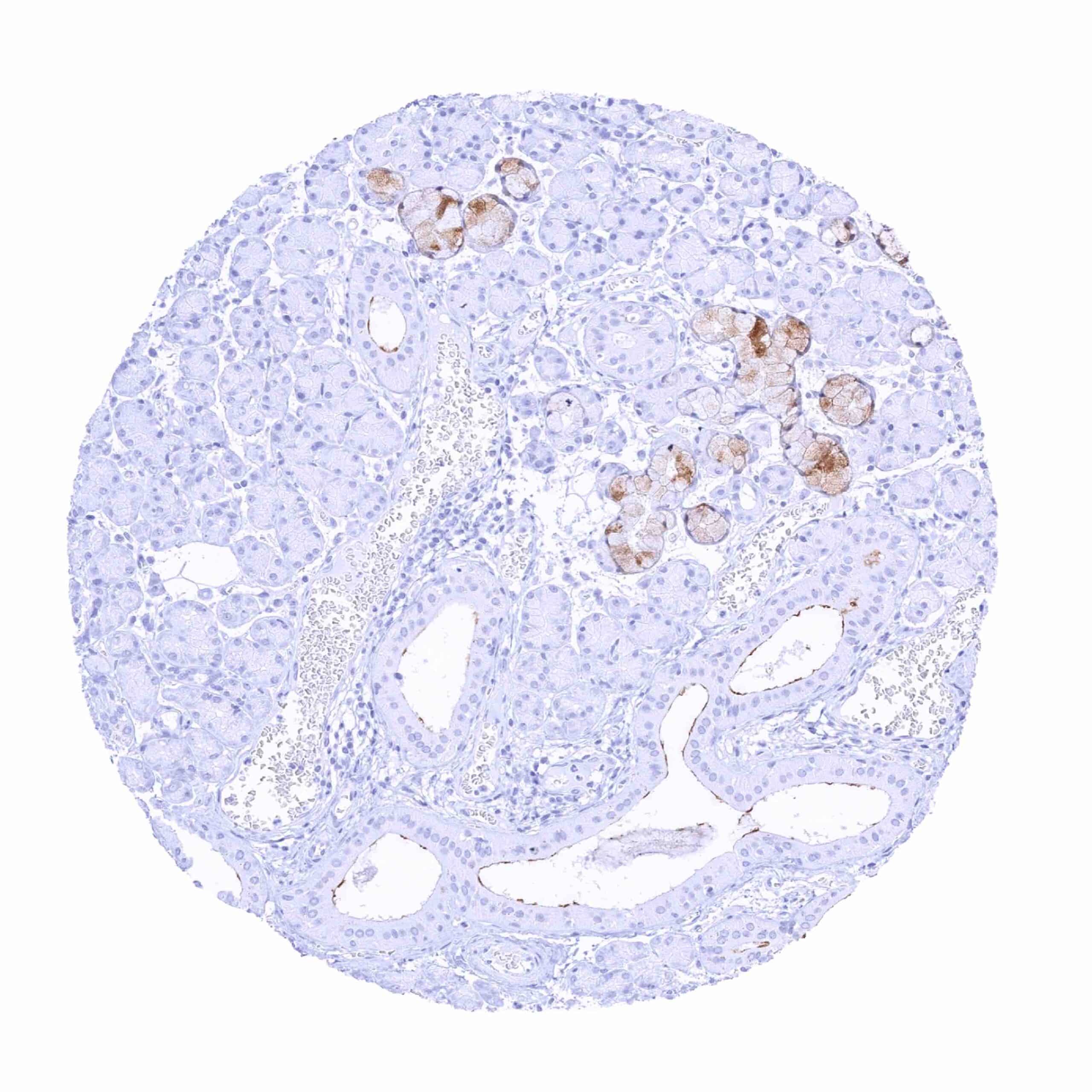
Pancreas
Parathyroid gland
Parotid gland
Pituitary gland, anterior lobe
Pituitary gland, posterior lobe
Placenta (amnion and chorion)
Placenta, early
Placenta, mature
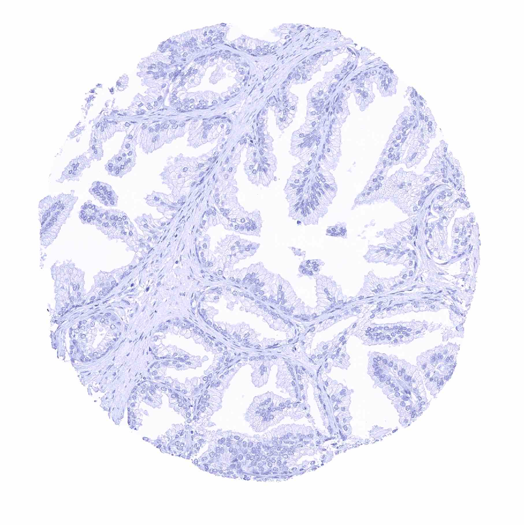
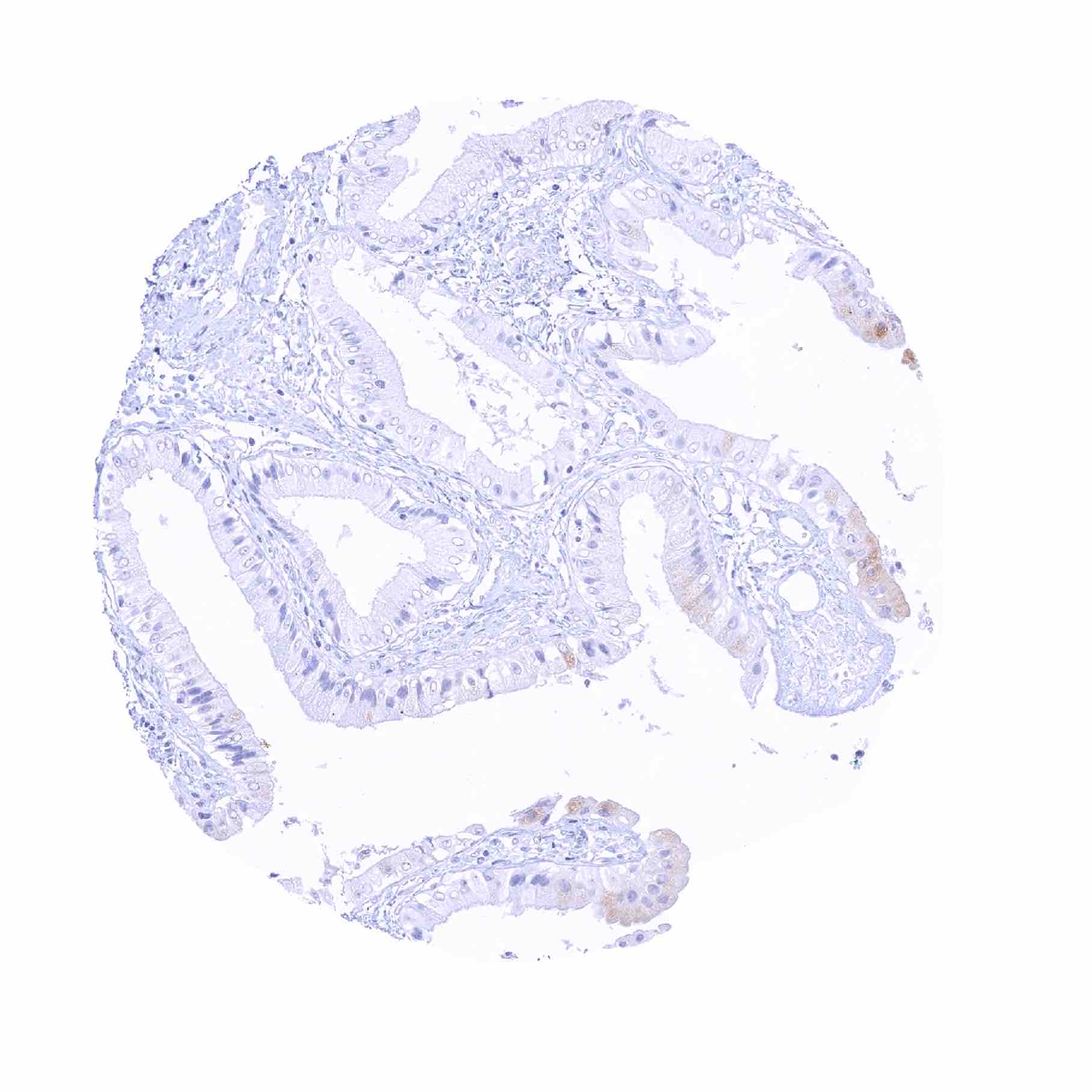
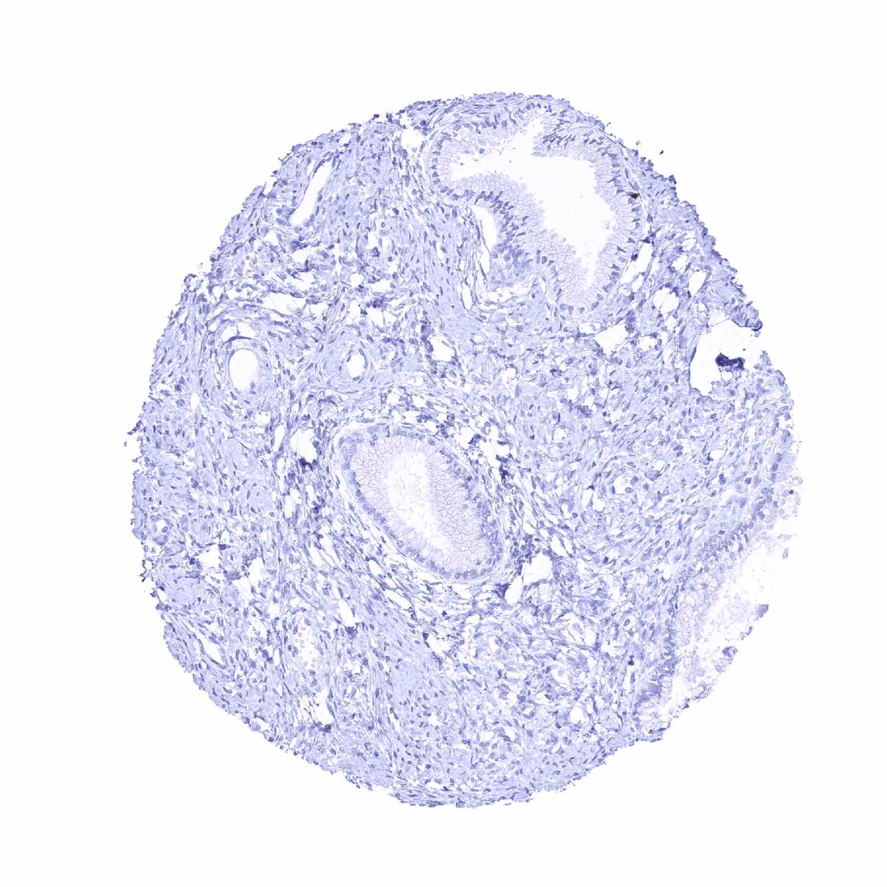
Prostate
Rectum, mucosa – Moderate cytoplasmic TFF1 staining of a small subset of goblet cells
Seminal vesicle

Sinus paranasales – A subset of the goblet cells of the respiratory epithelium show a weak TFF1 positivity
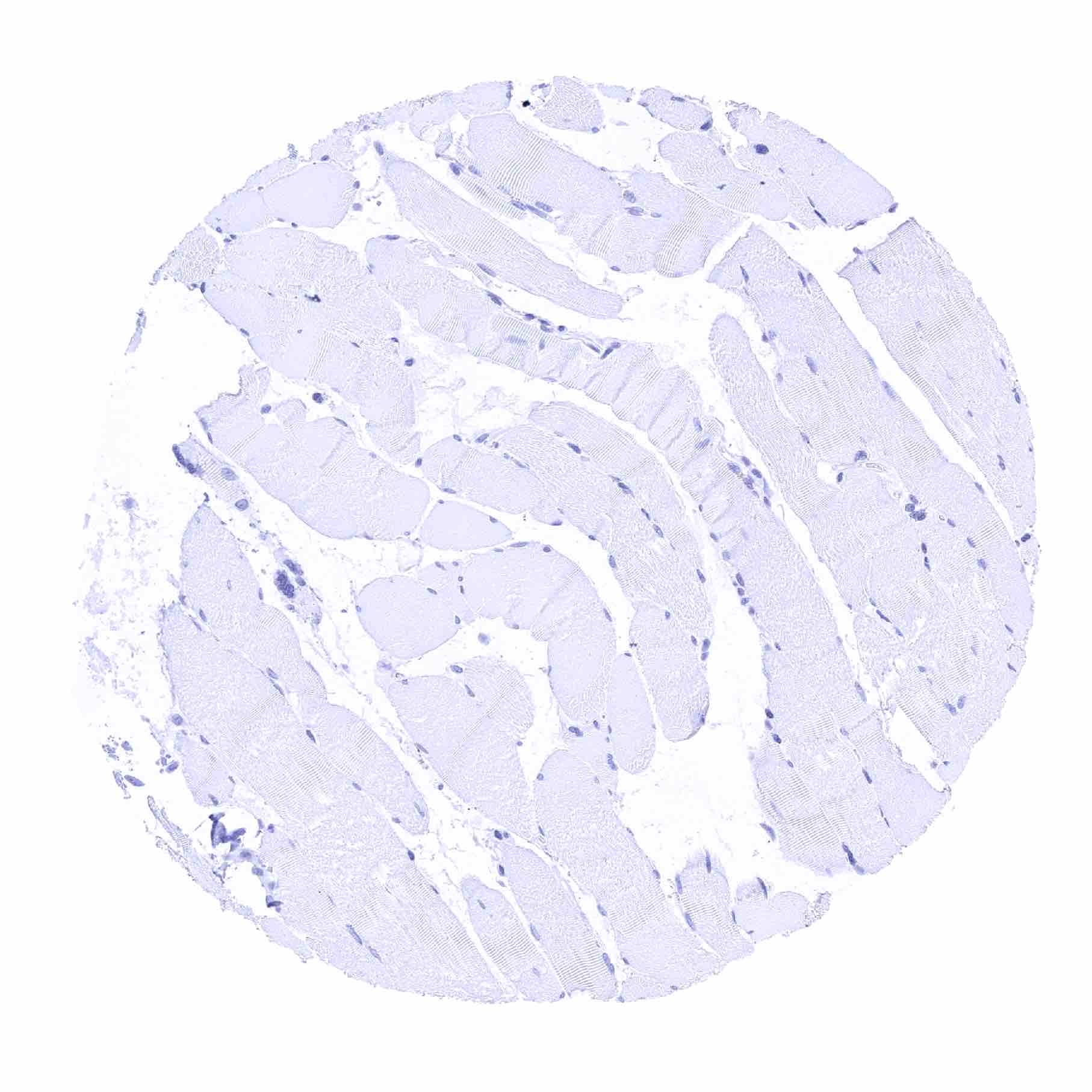
Skeletal muscle
Skin, hairfollicel and sebaceous glands
Skin
Spleen
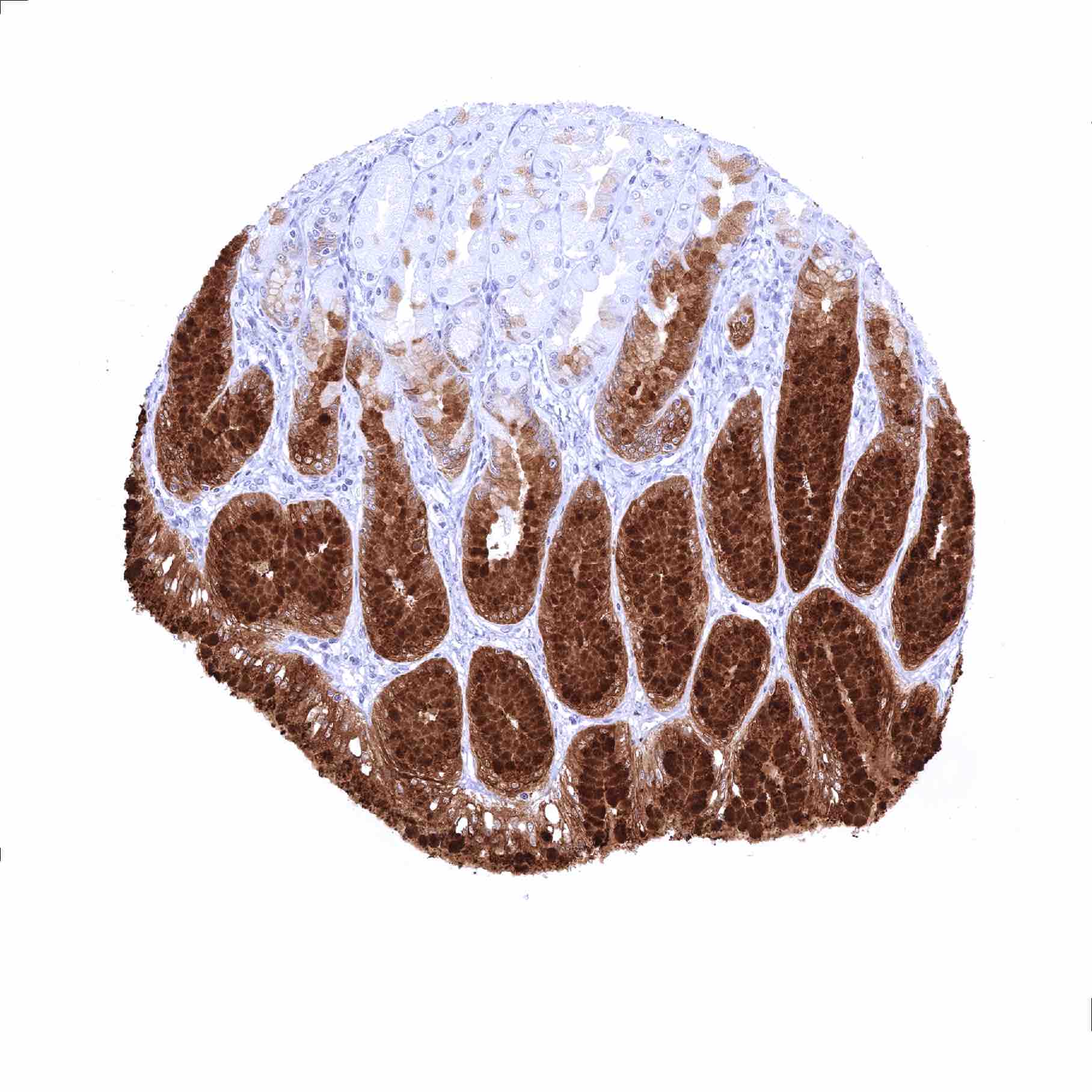
Stomach, antrum – Very intense TFF1 staining of surface epithelial cells while the stomach glands mostly remain negative_
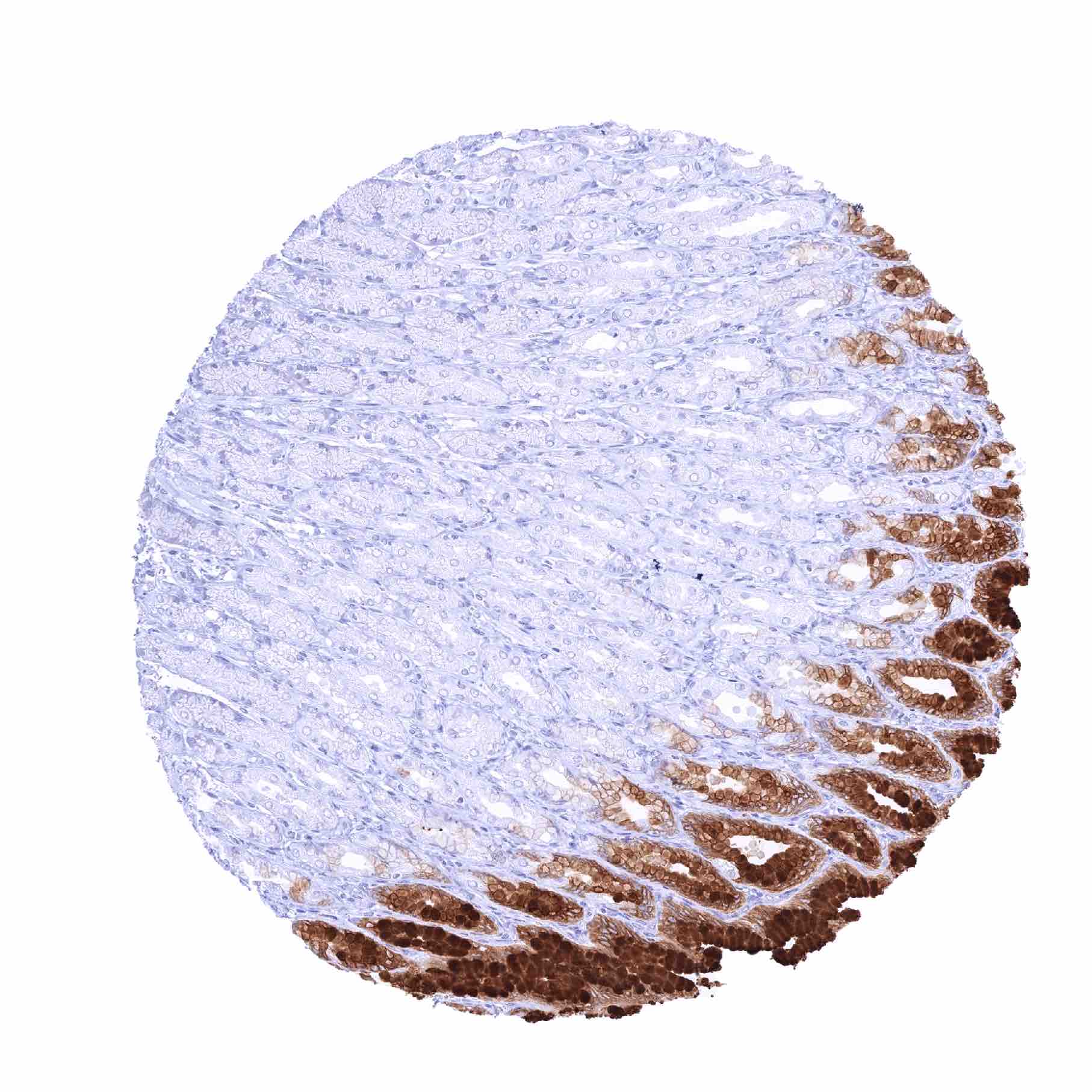
Stomach, corpus – Very intense TFF1 staining of surface epithelial cells. The stomach glands remain TFF1 negative

Sublingual gland – TFF1 immunostaining of variable intensity in mucinous glands
Submandibular gland – TFF1 positivity of varying intensity in mucinous glands
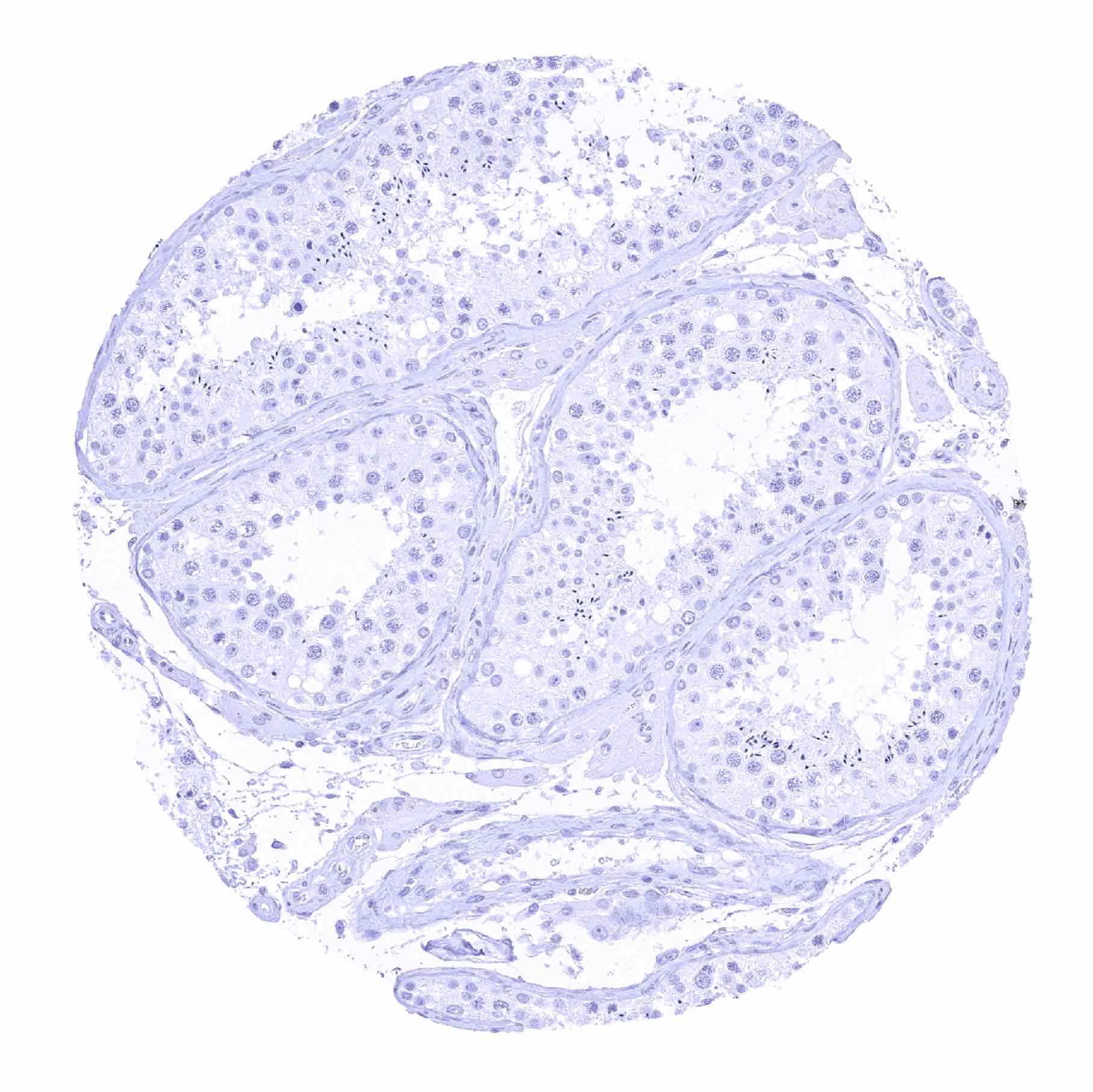
Testis

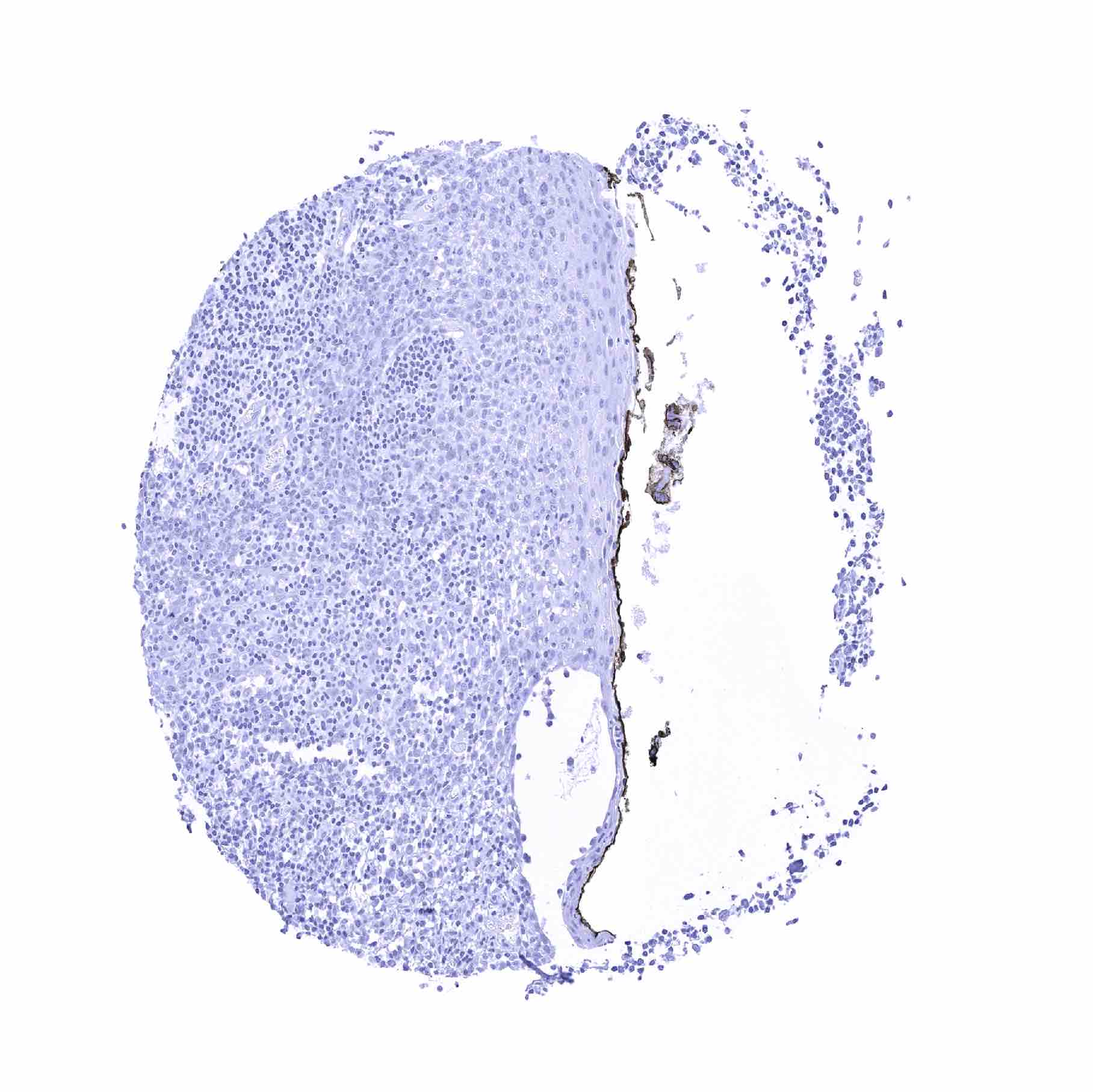
Thymus
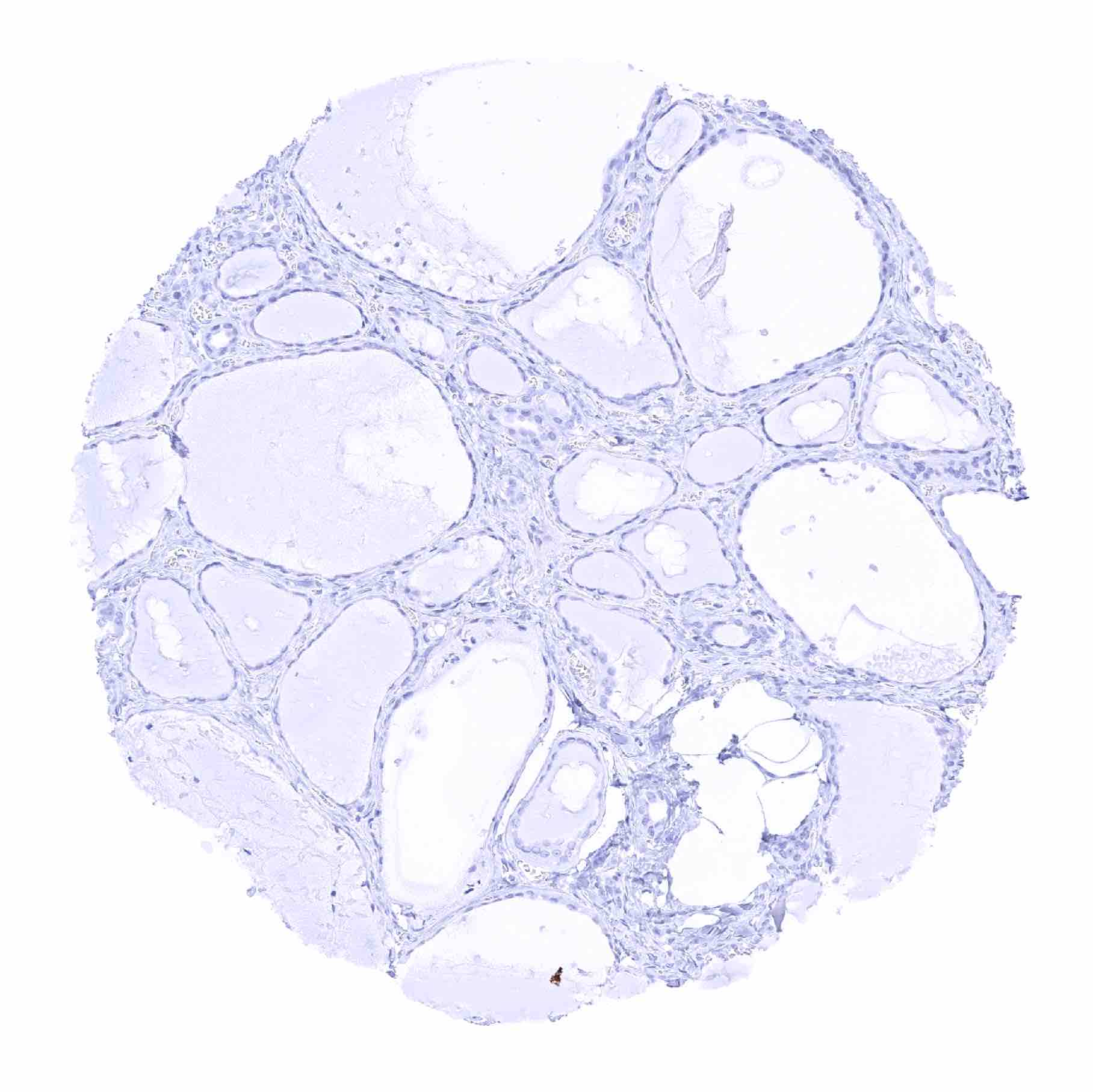
Thyroid gland
Tonsil

Tonsil
Tonsil, surface epithelium
Urinary bladder, muscular wall
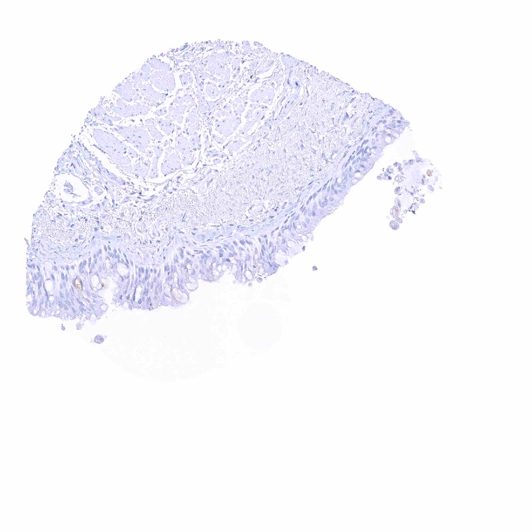
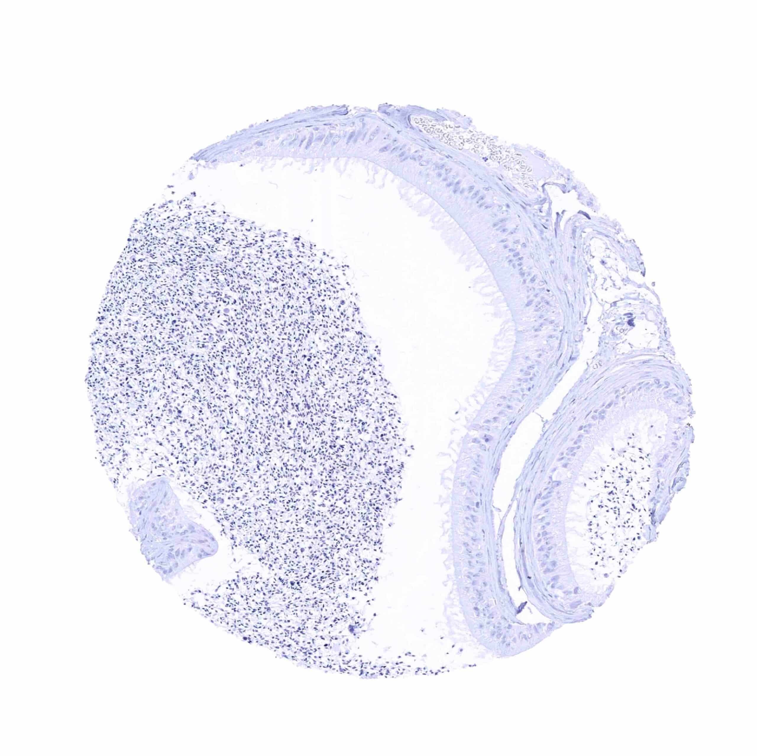
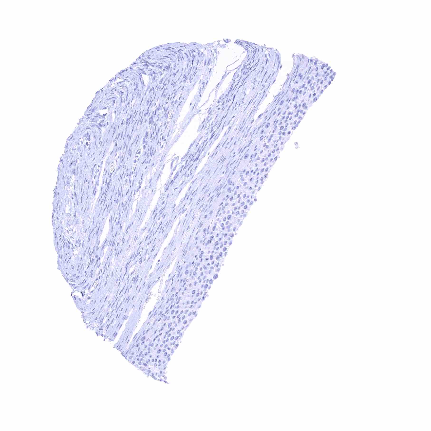
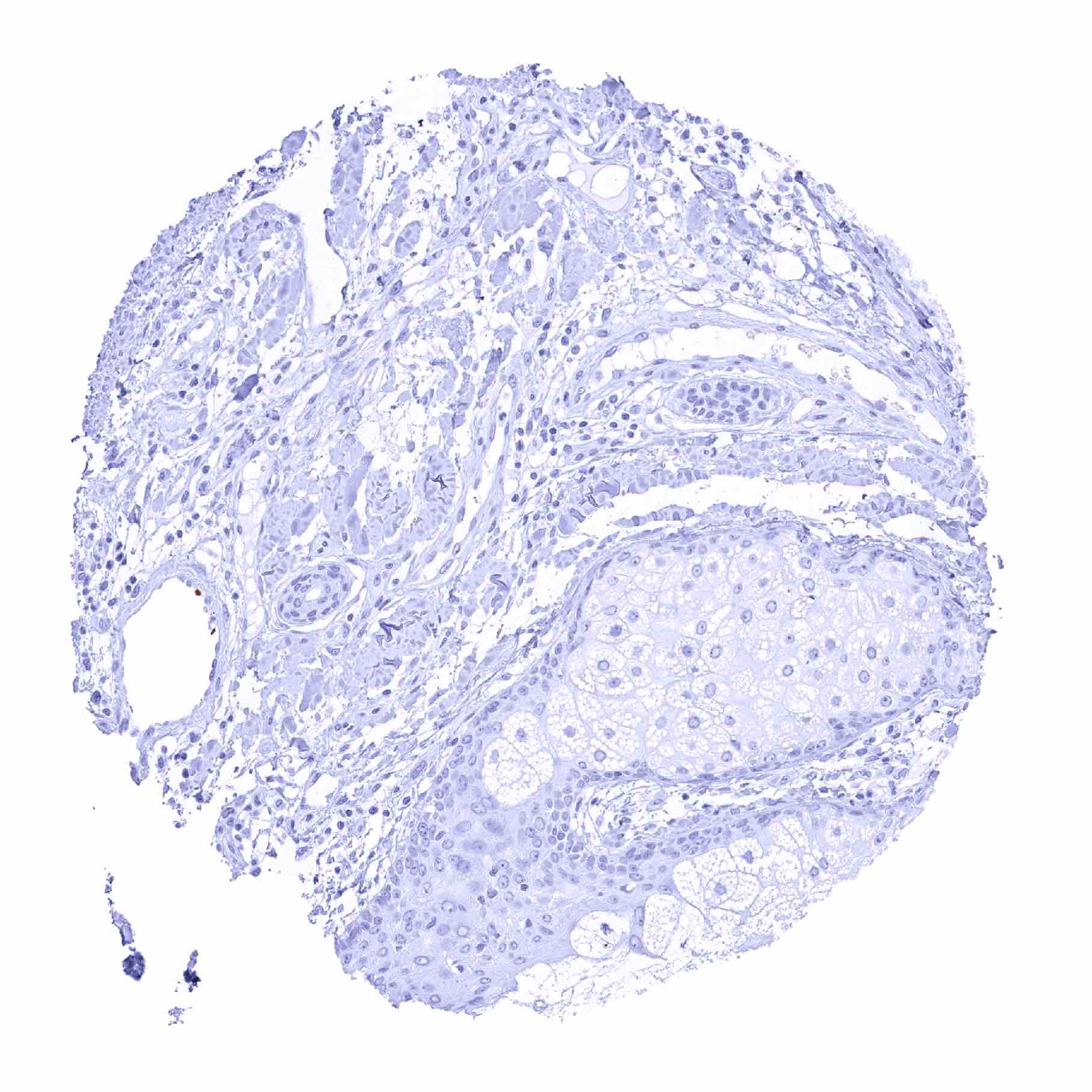
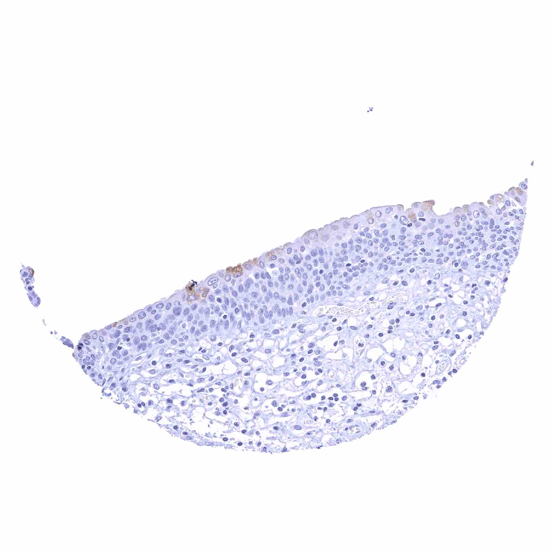
Urinary bladder, urothelium – Absence of urothelial TFF1 staining in this sample
Urinary bladder, urothelium – Weak cytoplasmic TFF1 staining of some umbrella cells. Urinary bladder, urothelium – Weak cytoplasmic TFF1 staining of some umbrella cells

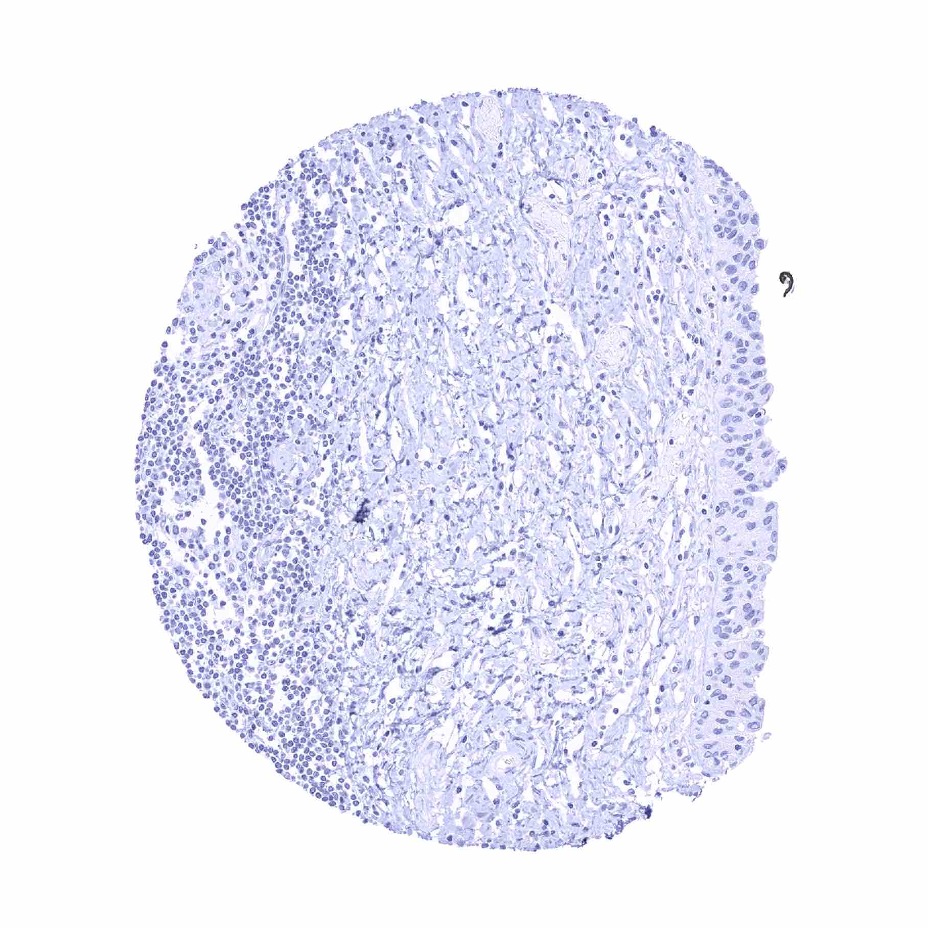
Uterus, ectocervix
Uterus, endocervix
Uterus, endometrium (pregnancy)

Uterus, endometrium (proliferation)
Uterus, endometrium (secretion)
Uterus, myometrium