Adrenal gland
Anal canal, transitional mucosa - A weak to moderate S100P immunostaining can be seen in the surface cell layer of transitional epithelium of the anal canal.

Aorta, media
Appendix, mucosa - A weak to moderate S100P staining can be found in a variable fraction of epithelial cells of the appendix mucosa.
Appendix, mucosa

Appendix, muscular wall
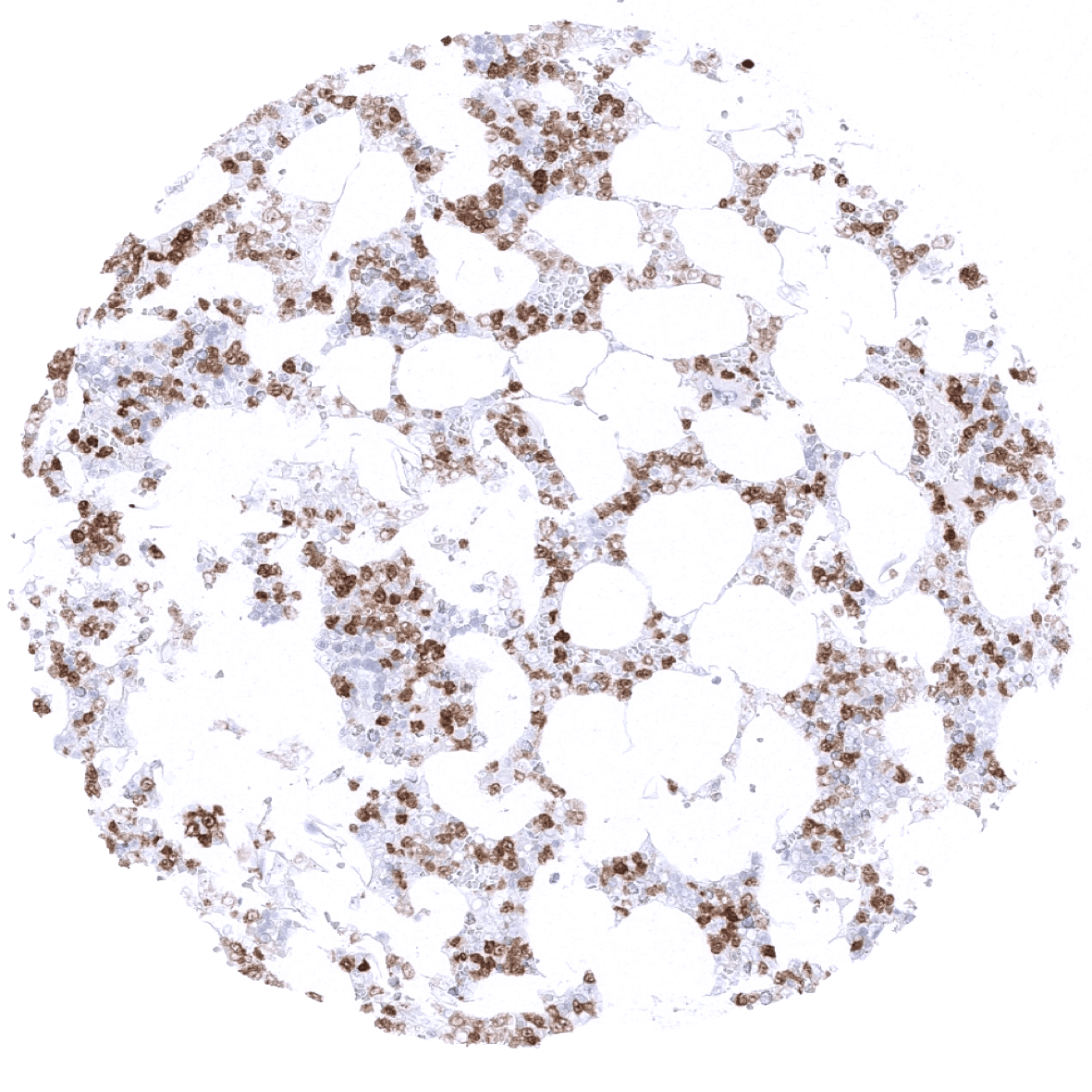
Bone marrow - A moderate to strong S100P immunostaining is seen in granulocytes and their precursor cells.
Breast
Bronchus, mucosa - S100P immunostaining is seen in a large fraction of non-basal cells of respiratory epithelium.
Bronchus, mucosa - S100P immunostaining is seen in a fraction of non-basal cells of respiratory epithelium (including many goblet cells).
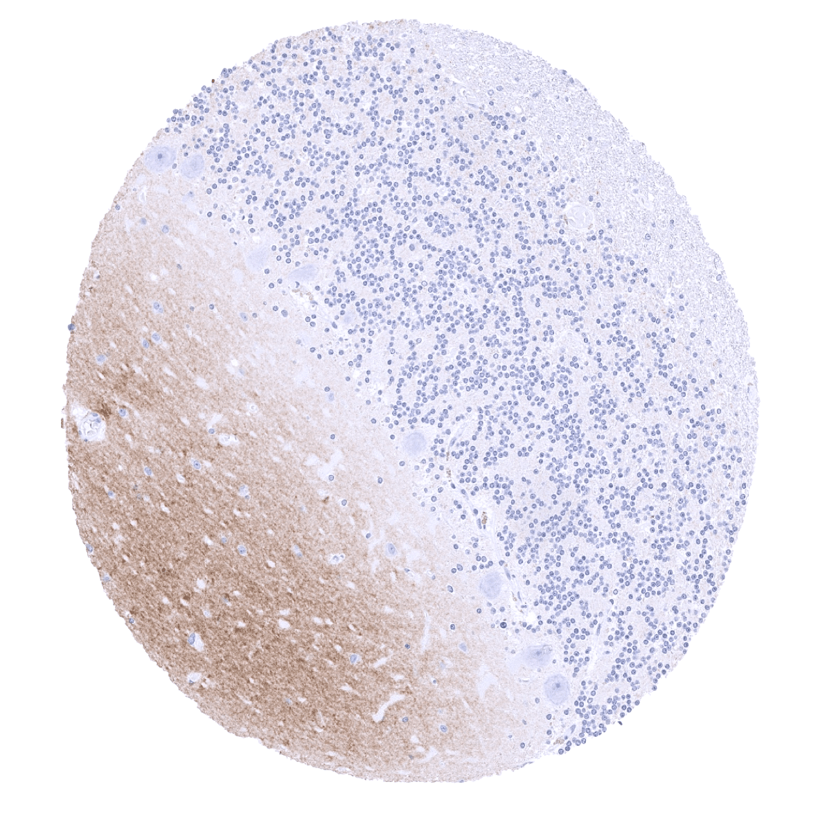
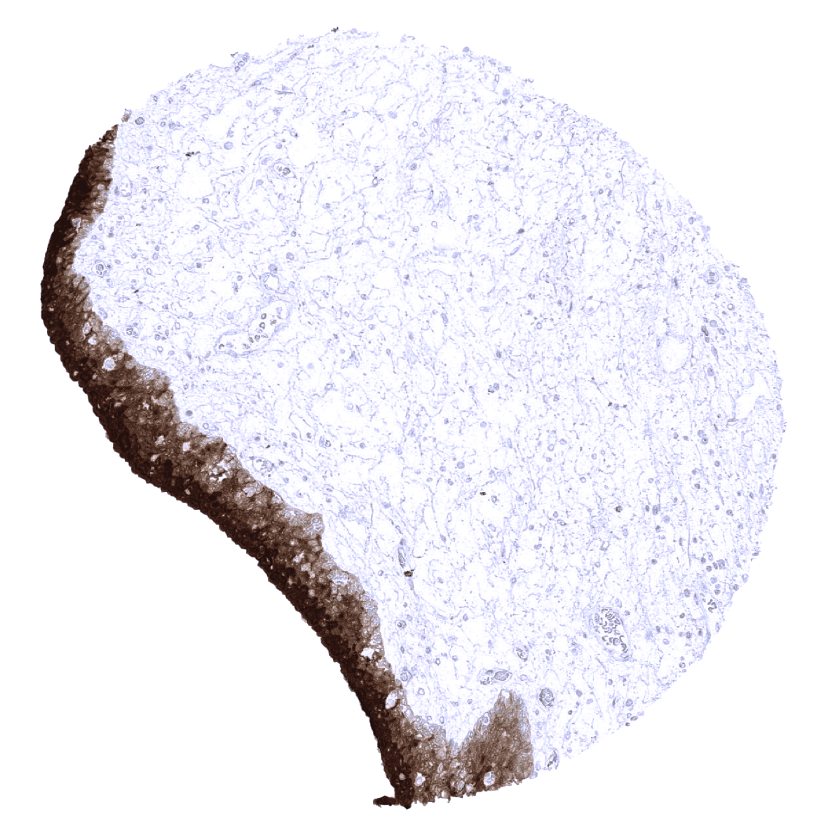
Cerebellum (molecular layer, Purkinje cell layer, granule cell layer, white matter) - A fibrillar staining observed in the white matter of the cerebellum may represent a (tolerable) cross-reactivity

Cerebellum (white matter)
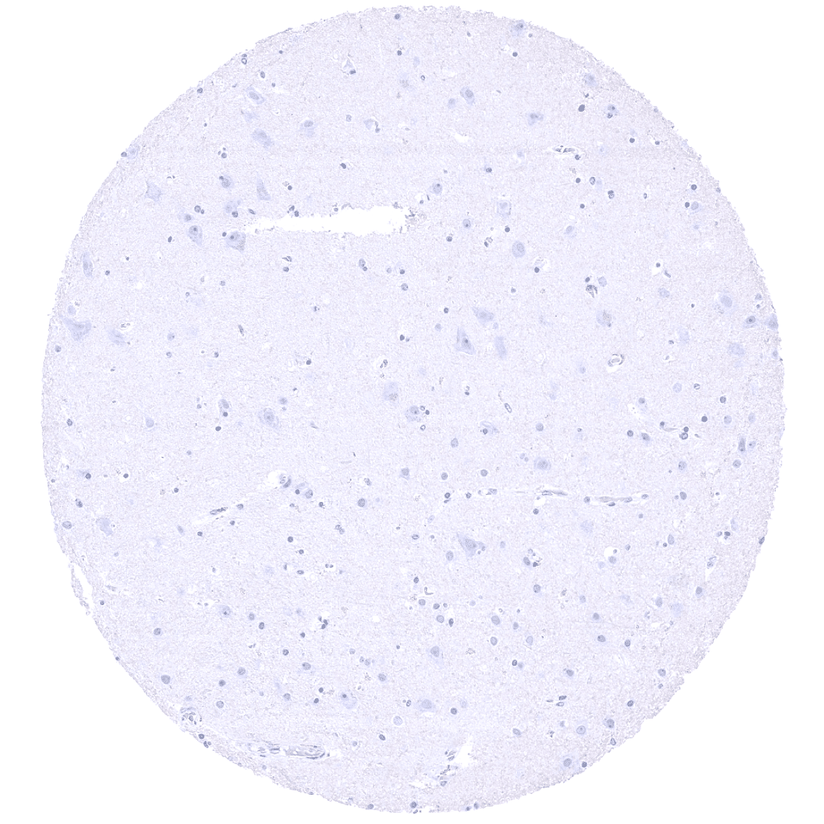
Cerebrum, grey matter
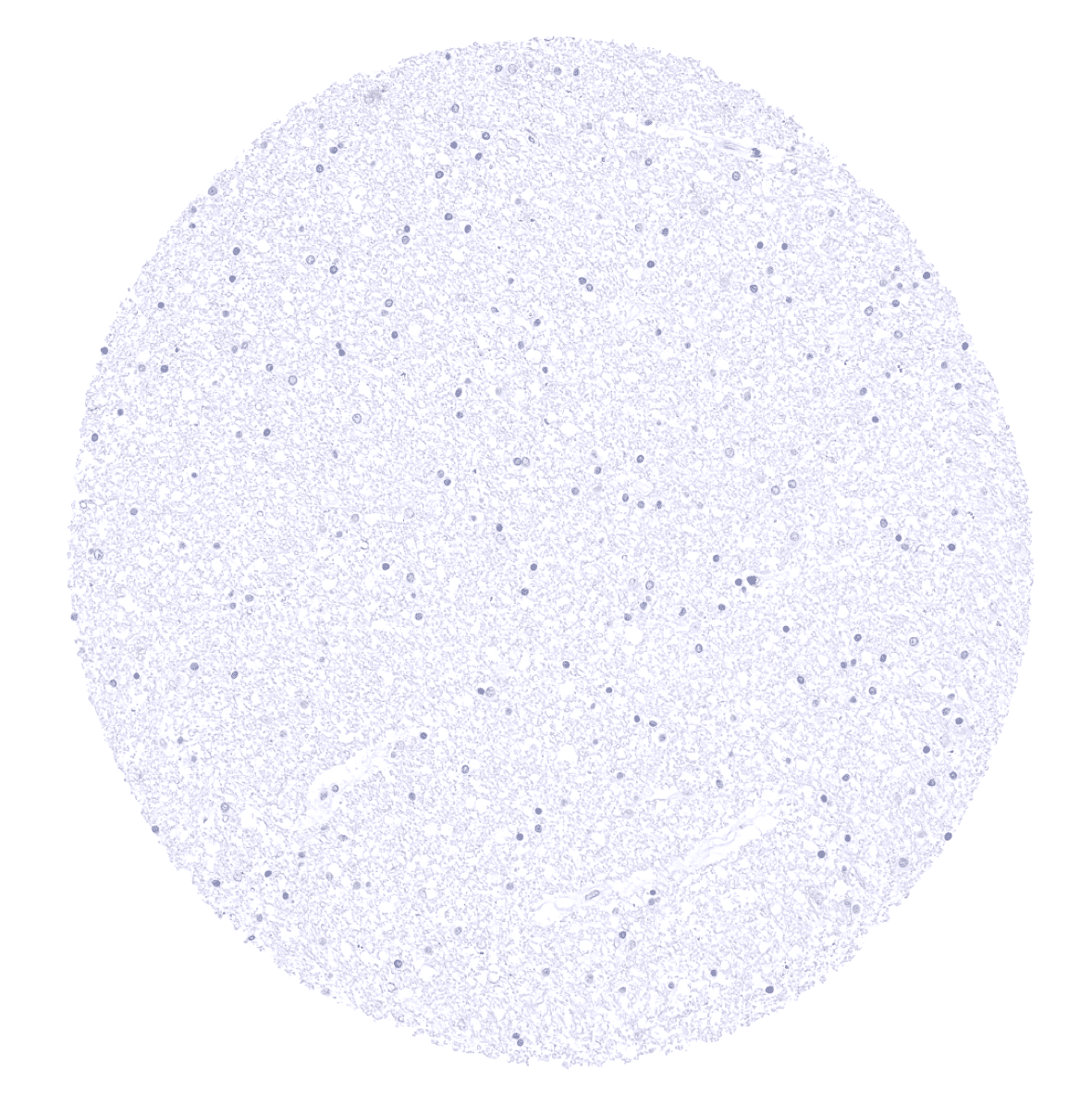
Cerebrum, white matter

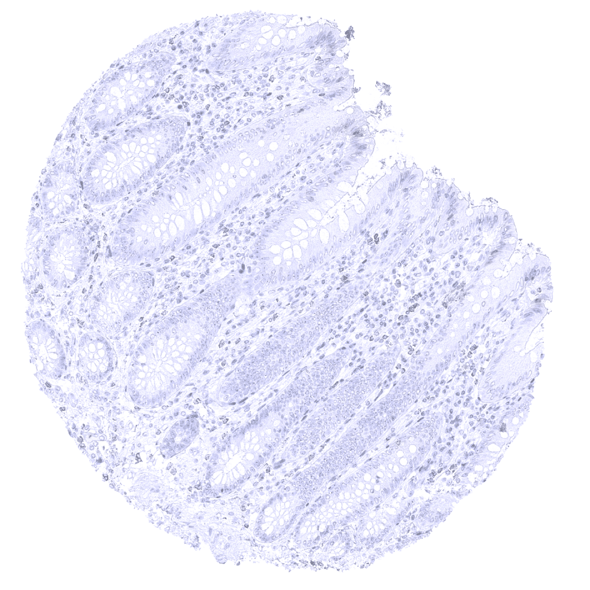
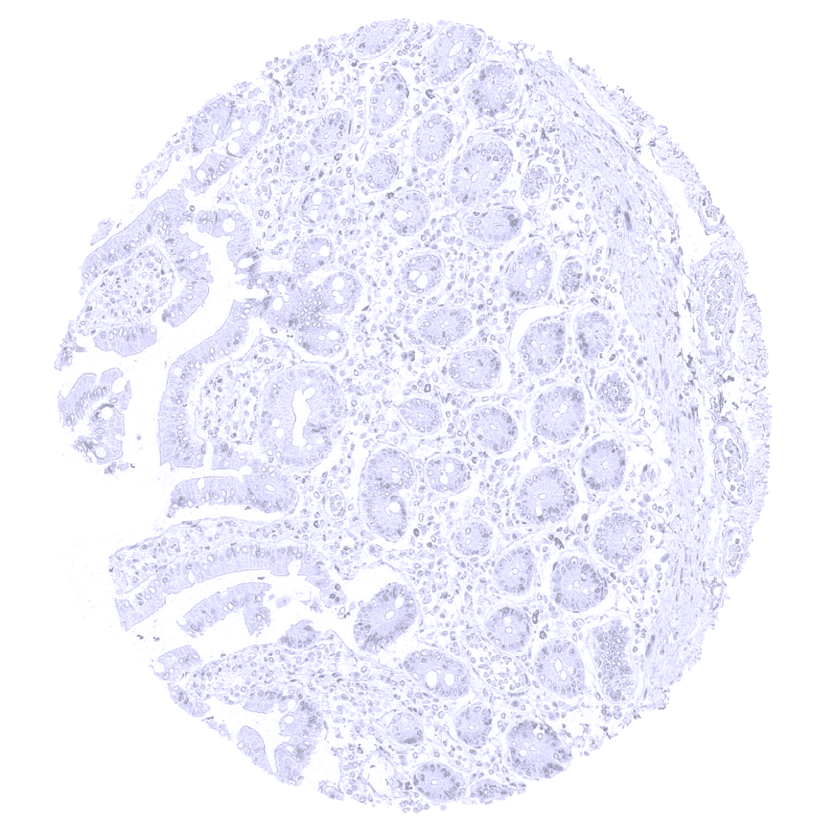
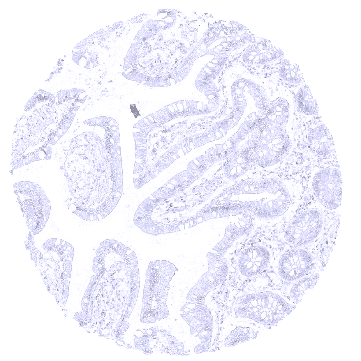
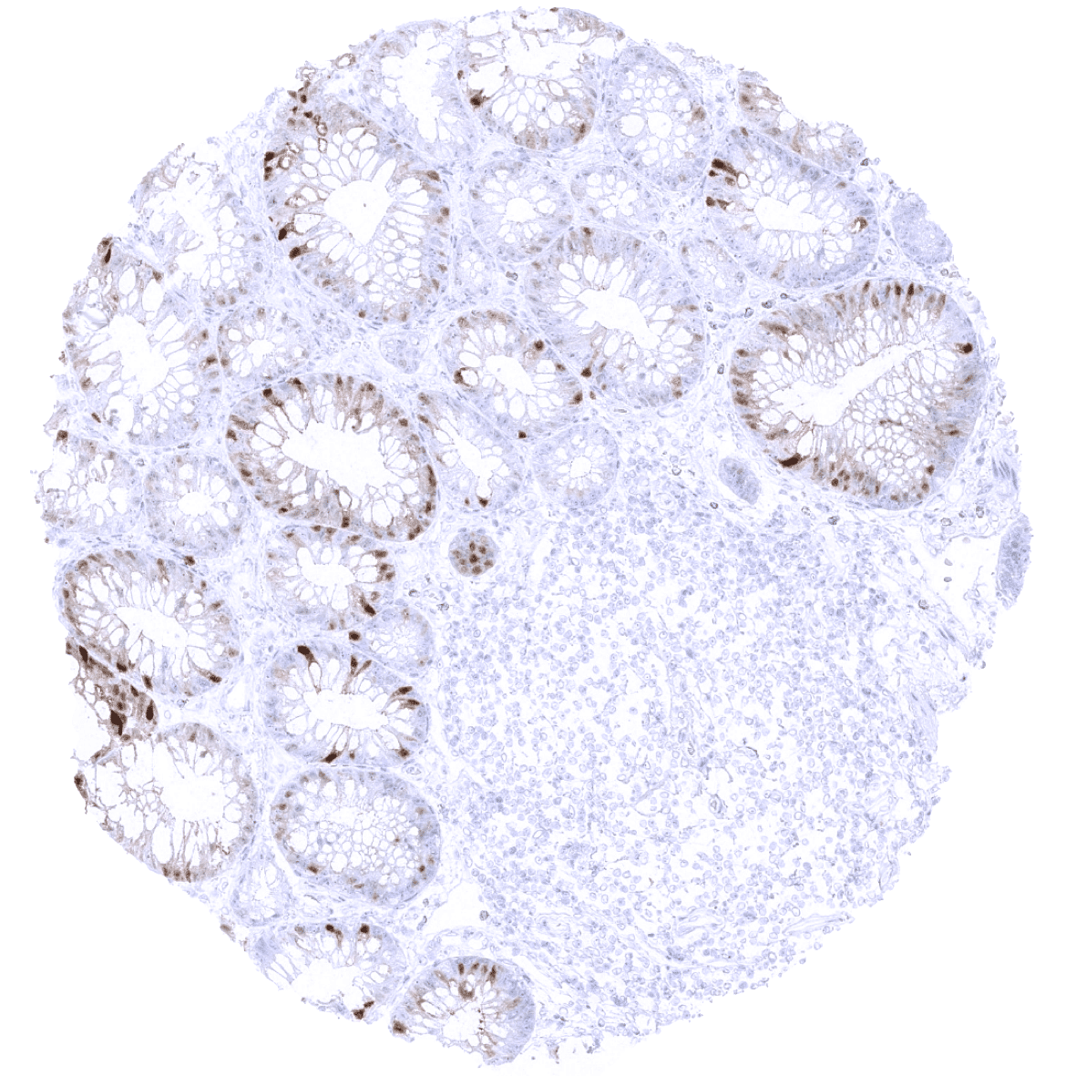
Colon descendens, mucosa - A weak to moderate S100P staining can be found in a variable fraction of epithelial cells of the colon mucosa (stronger in superficial than in crypt cells).
Colon descendens, muscular wall
Duodenum, Brunner gland
Duodenum, mucosa

Epididymis
Esophagus, squamous epithelium
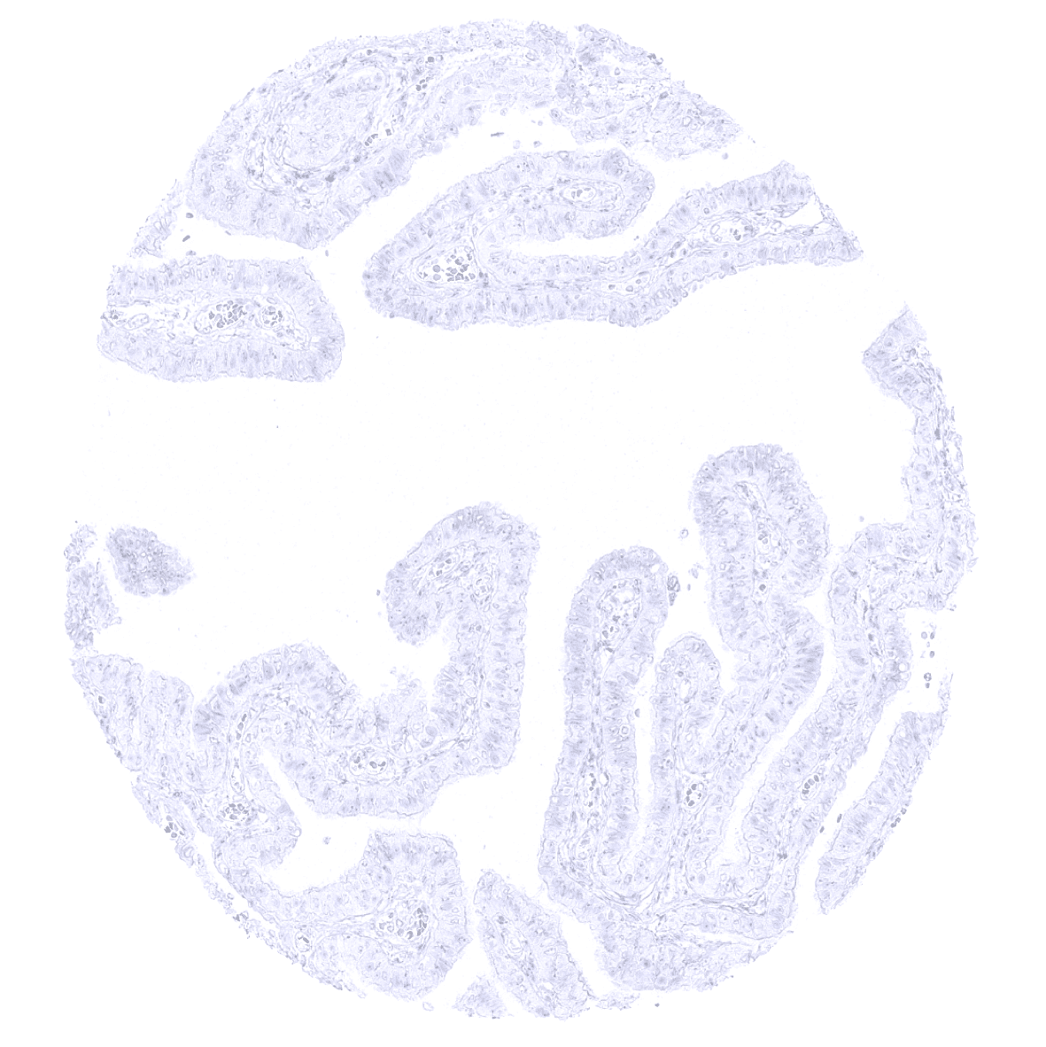
Fallopian tube, mucosa
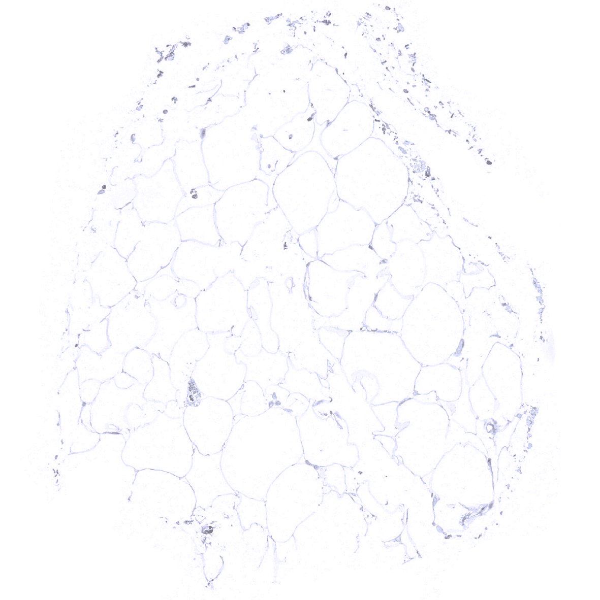
Fat
Gallbladder, epithelium - A moderate to strong S100P immunostaining can occur in gallbladder epithelium (not in all samples). In this sample, extensive S100P positivity may be related to heavy inflammation.
Gallbladder, epithelium - The normal gallbladder epithelium is mostly S100P negative.

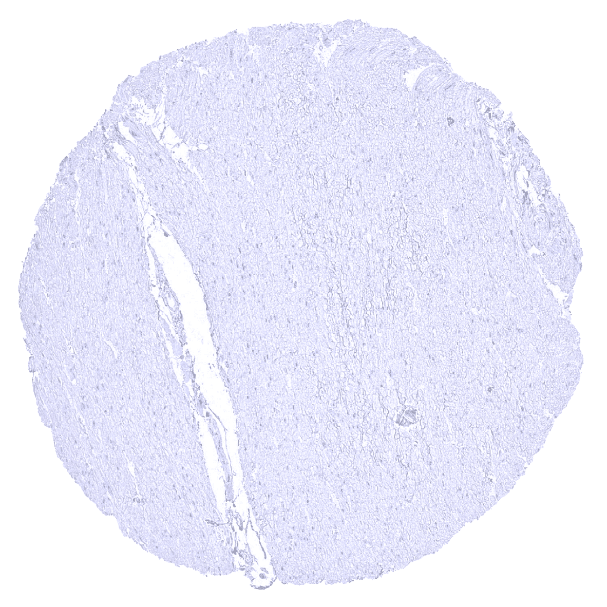
Heart muscle

Ileum, mucosa - A weak to moderate S100P can be found in a variable fraction of epithelial cells of the ileum.
Ileum, mucosa

Kidney, cortex
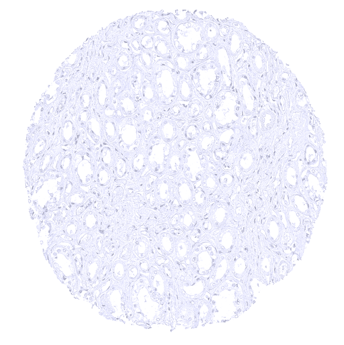
Kidney, medulla
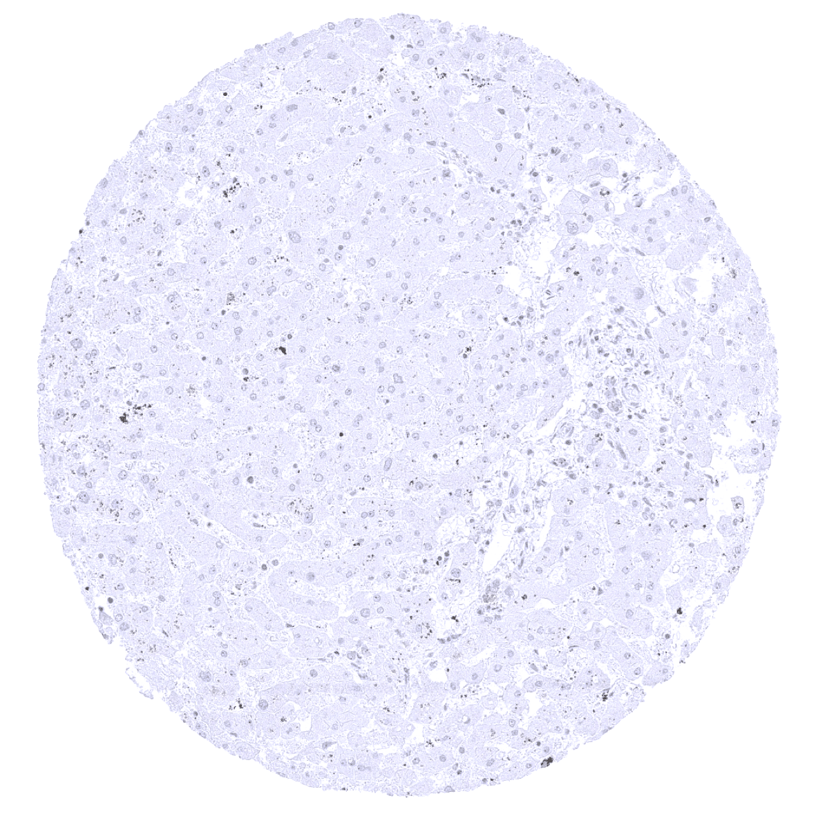
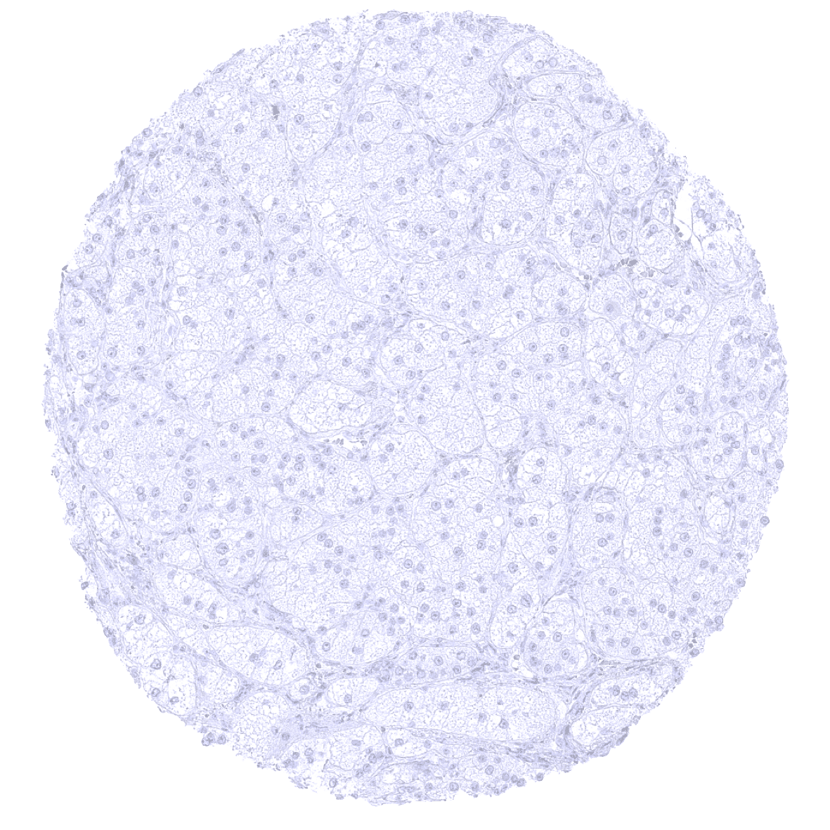
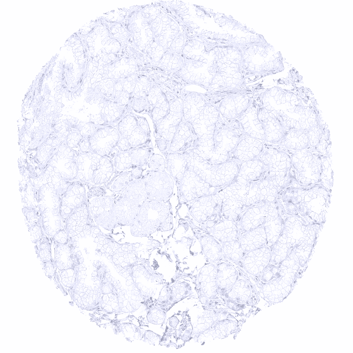
Liver
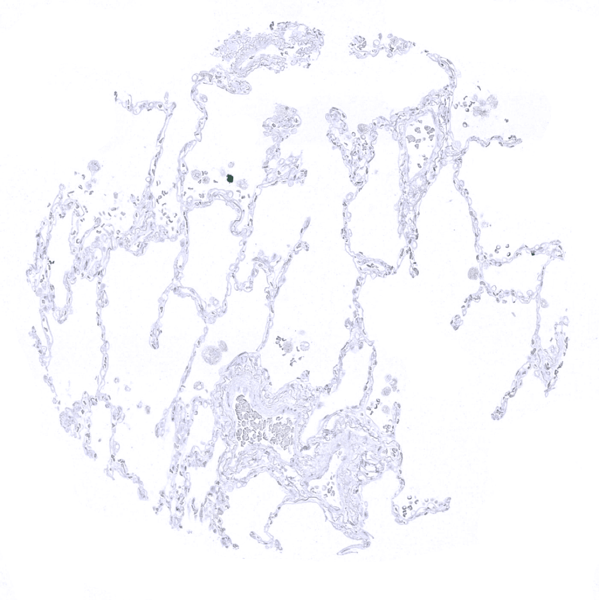
Lung

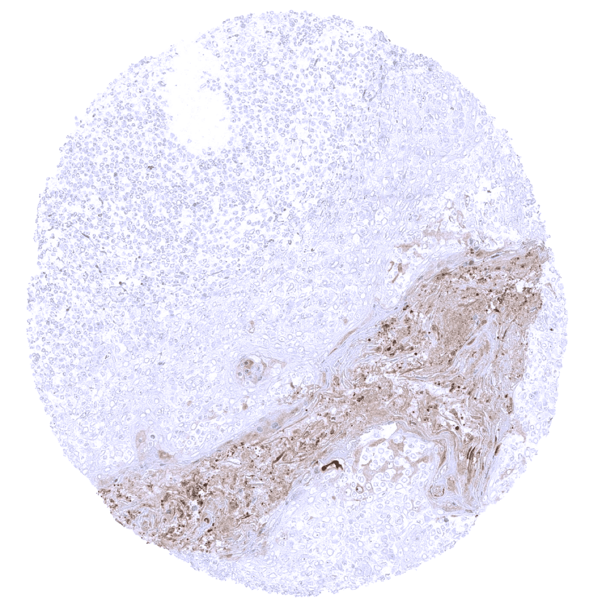
Lymph node
Ovary, stroma
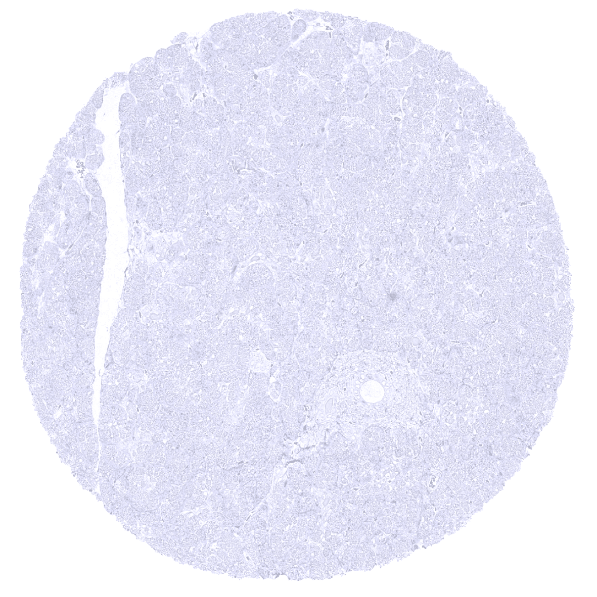
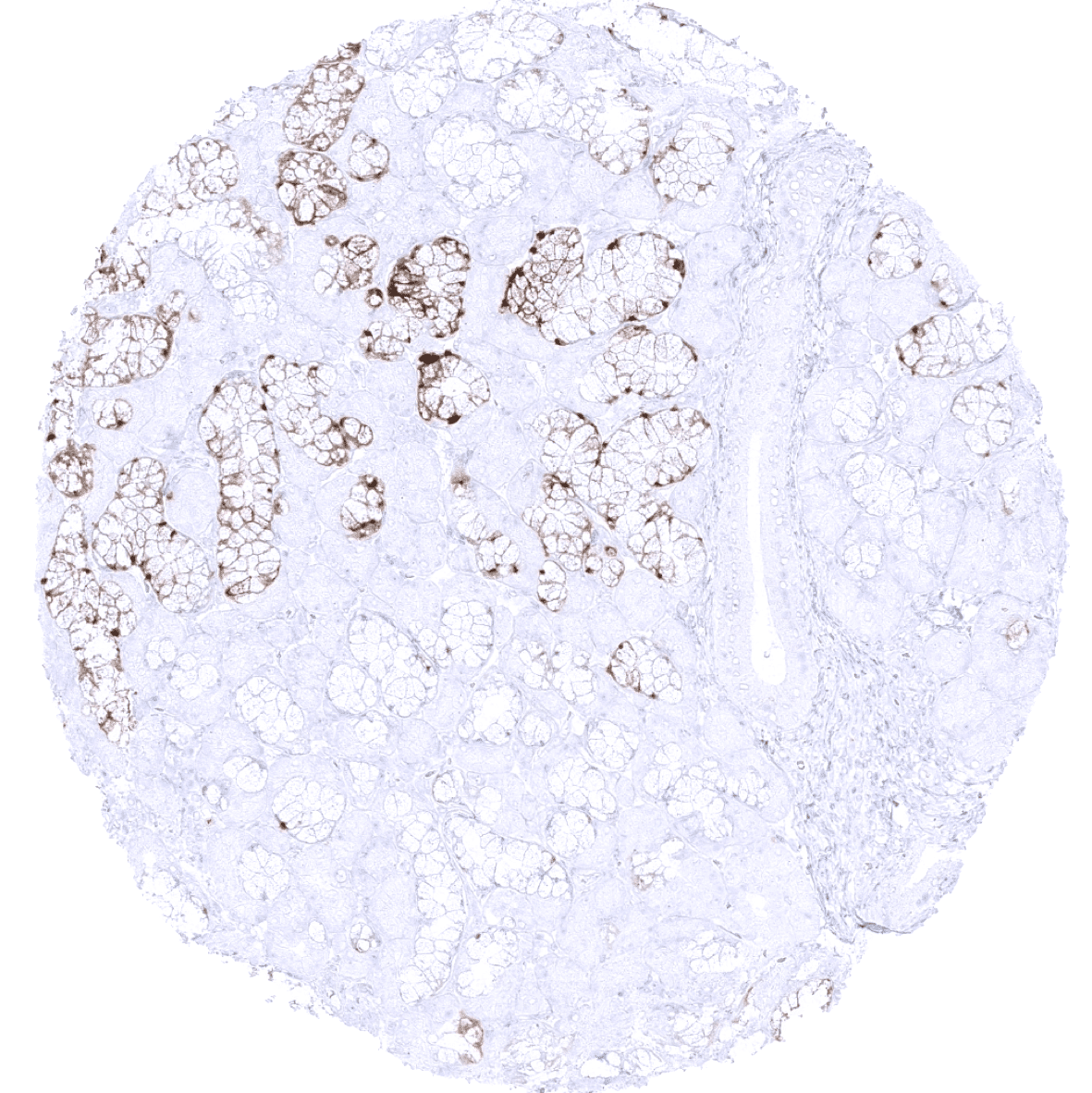
Pancreas

Parathyroid gland

Parotid gland
Pituitary gland, anterior lobe
Pituitary gland, posterior lobe

Placenta (amnion and chorion) - A moderate to strong S100P immunostaining occurs in chorion cells of the placenta.

Placenta, early - A strong S100P immunostaining is seen in trophoblastic cells of the placenta (syncytiotrophoblast more than cytotrophoblast).

Placenta, mature - A strong S100P immunostaining is seen in trophoblastic cells of the placenta (syncytiotrophoblast more than cytotrophoblast).

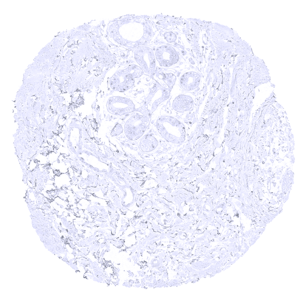
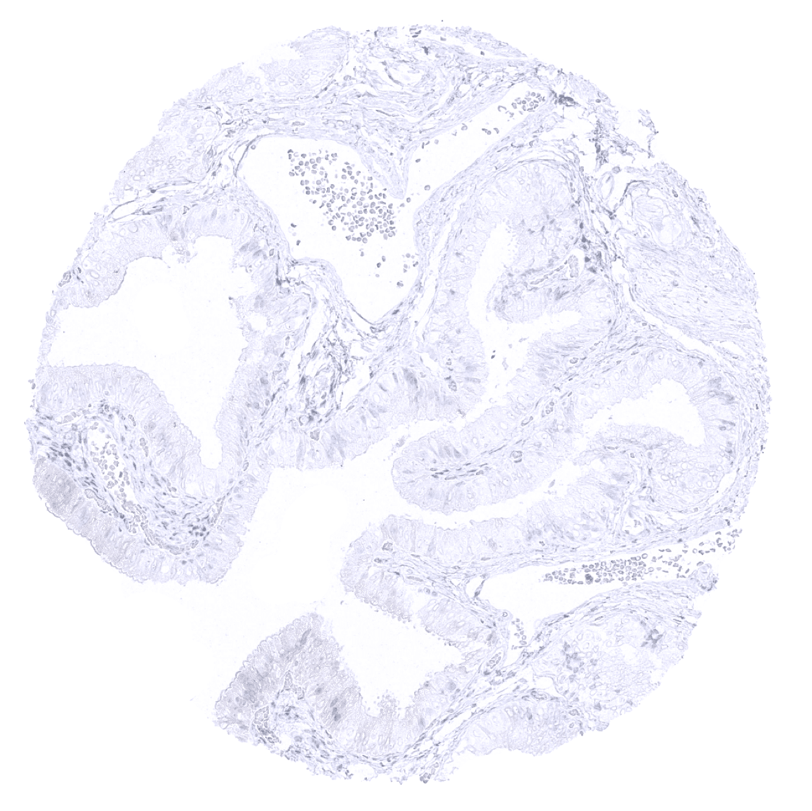
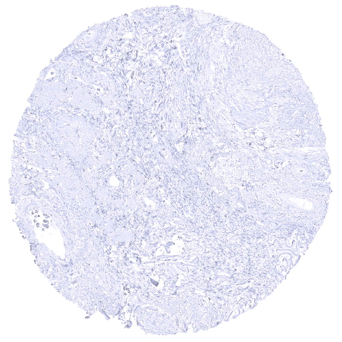
Prostate
Rectum, mucosa - A weak to moderate S100P can be found in a variable fraction of epithelial cells of the colon mucosa.
Seminal vesicle
Sinus paranasales - S100P immunostaining is often seen in a fraction of non-basal cells of respiratory epithelium.

Sinus paranasales - S100P immunostaining is seen in a small fraction of non-basal cells of respiratory epithelium in this sample.
Skeletal muscle

Skin
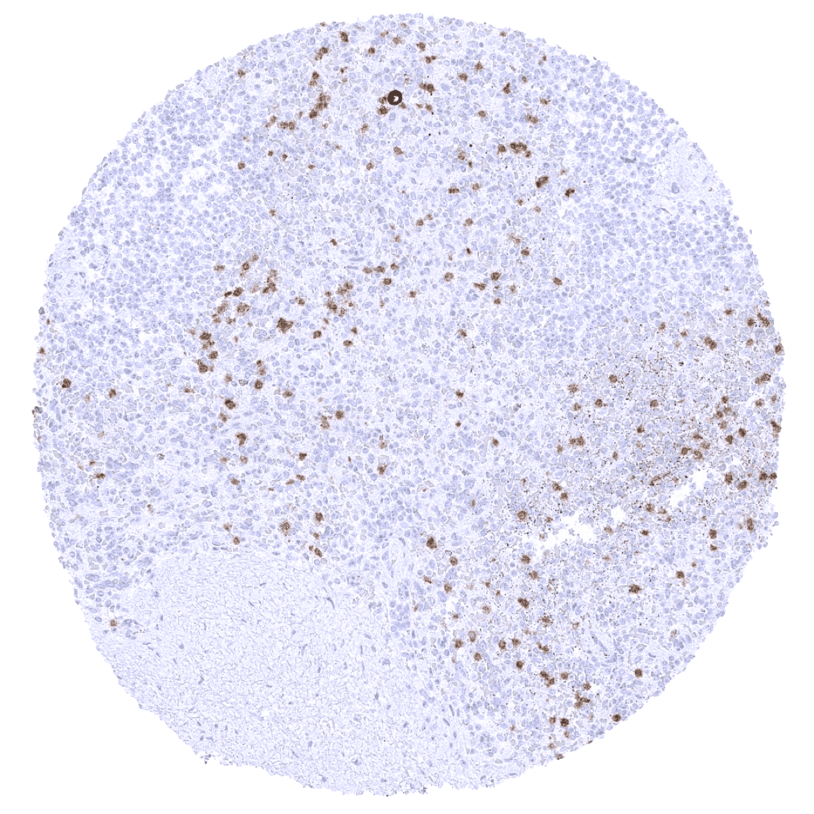
Spleen – S100P immunostaining is seen in granulocytes.
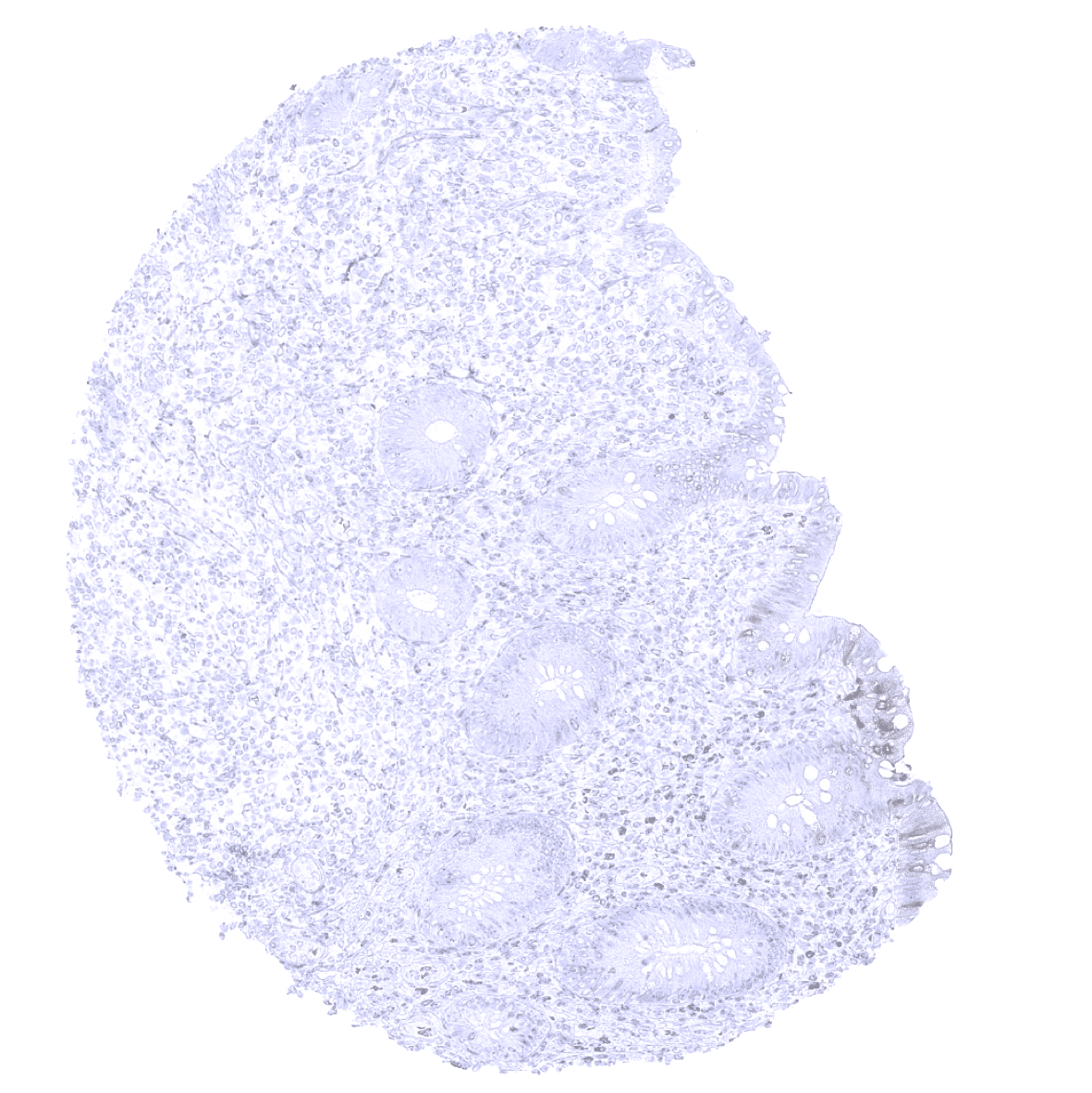
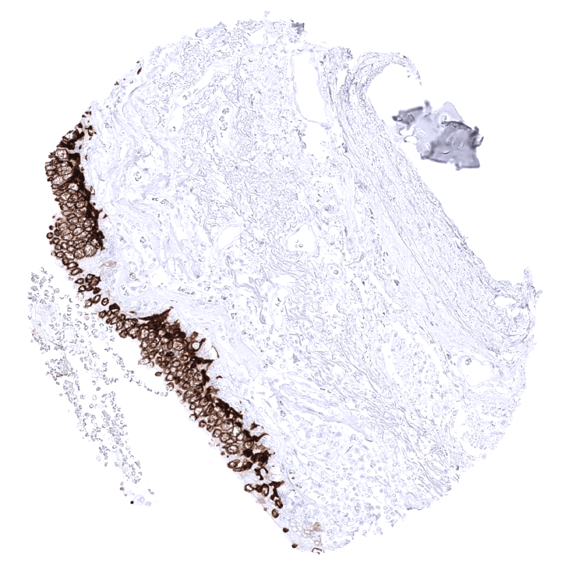
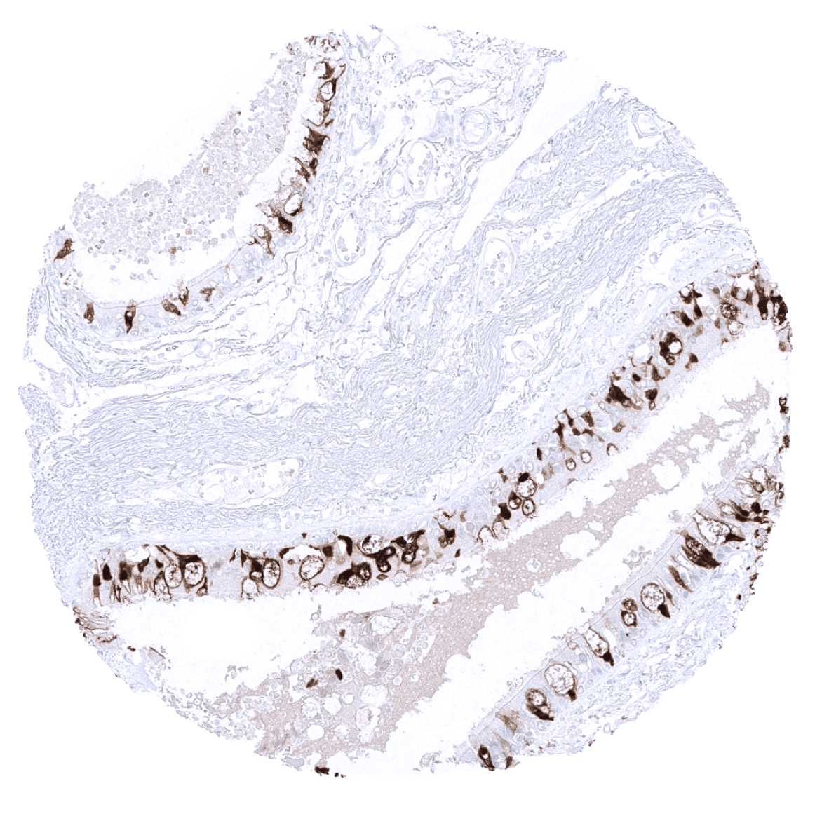
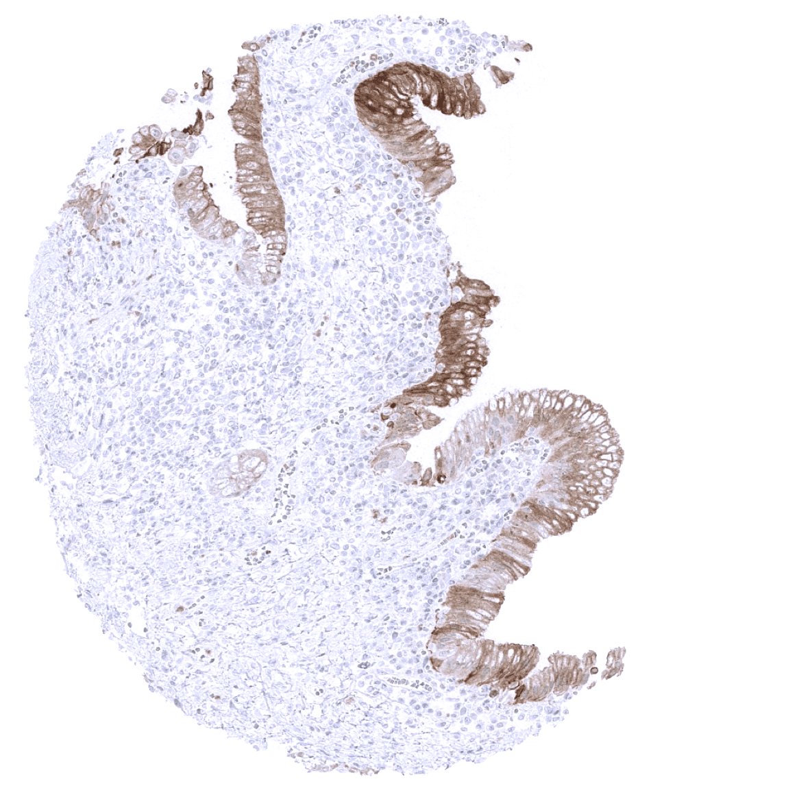
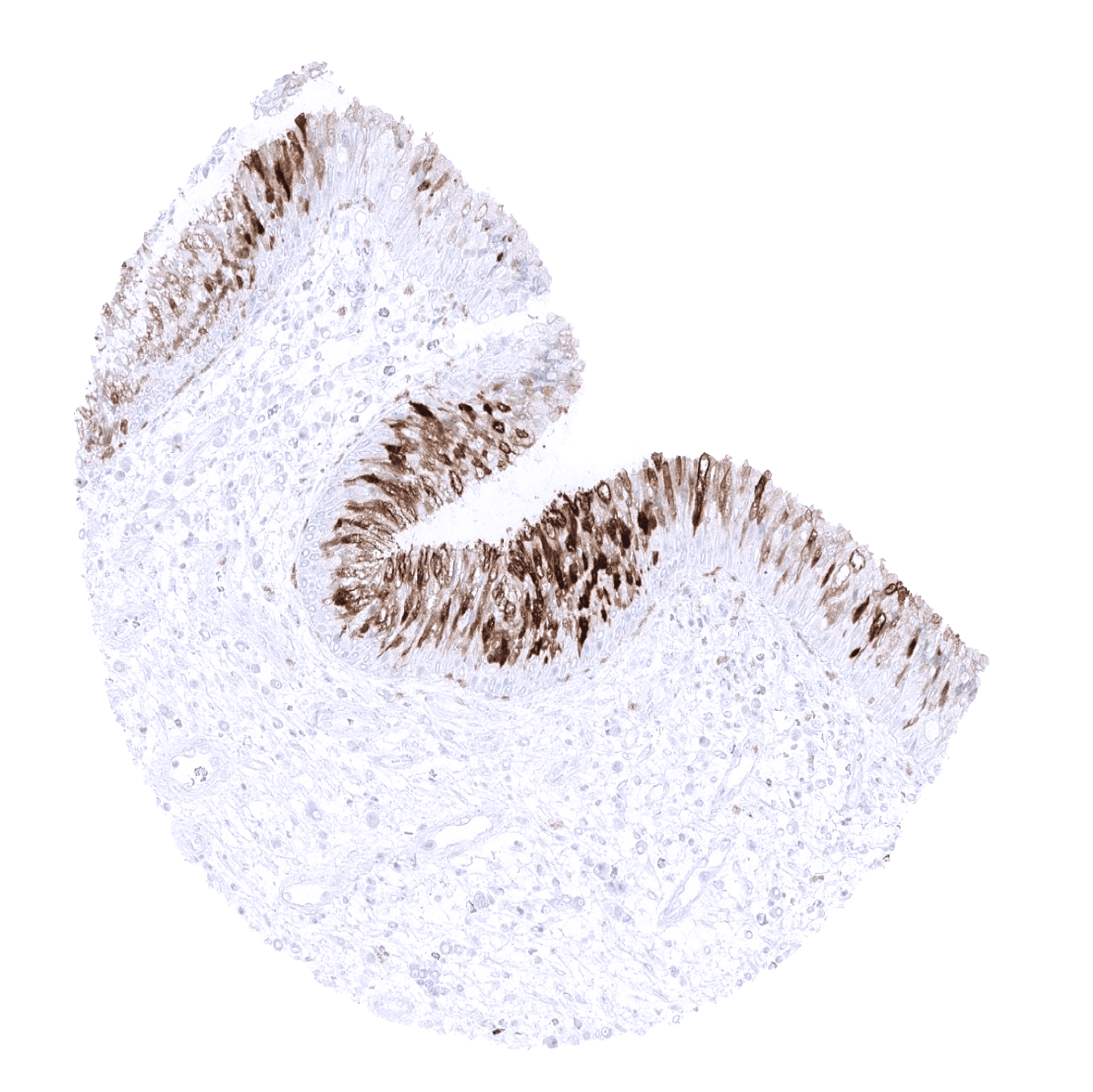
Stomach, antrum - A strong S100P immunostaining is seen in the surface epithelial cells and neck cells of the stomach while staining is markedly less intense or absent in gastric glands.
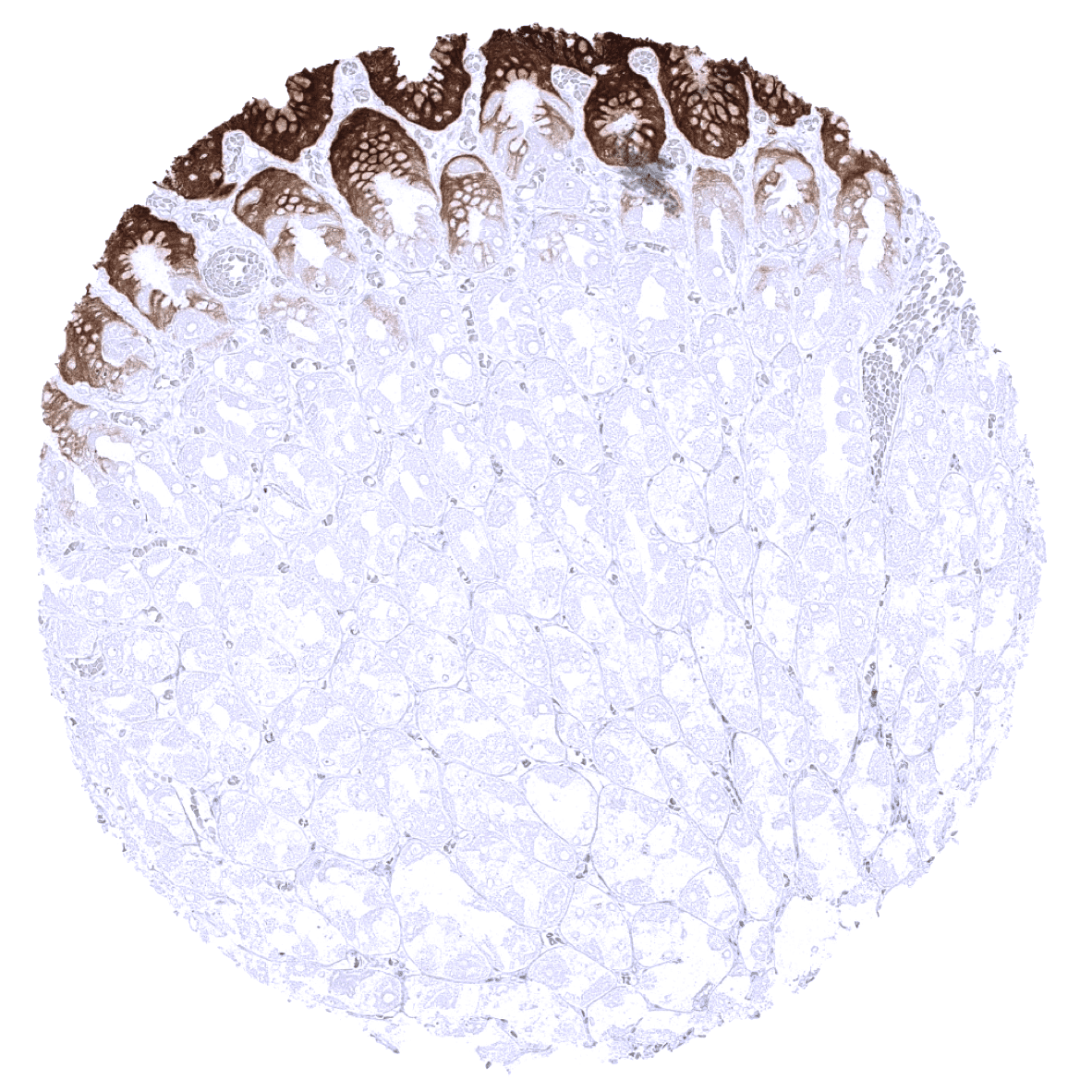
Stomach, corpus - A strong S100P immunostaining is seen in the surface epithelial cells and neck cells of the stomach while staining is markedly less intense or absent in gastric glands.
Sublingual gland - A moderate S100P immunostaining can occur in salivary glands (not in all samples).

Thymus - A moderate S100P staining of corpuscles of Hassall’s but not of other epithelial cells is seen in the thymus.
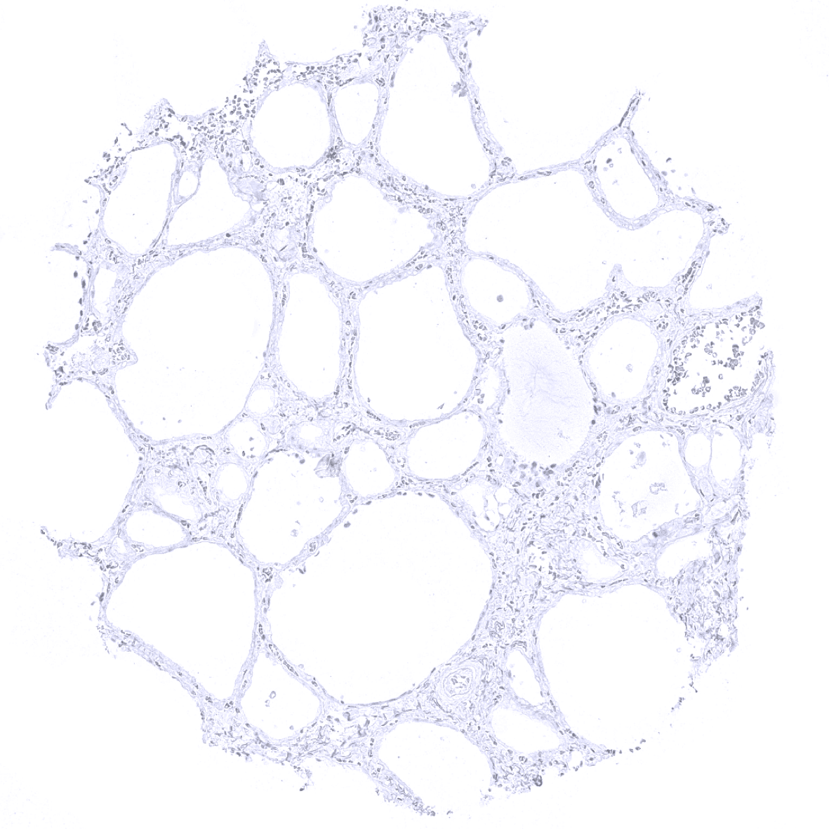
Thyroid gland
Tonsil - A weak to moderate S100P staining occurs in the superficial layers of squamous epithelium of tonsil crypts.
Tonsil, surface epithelium
Urinary bladder, muscular wall
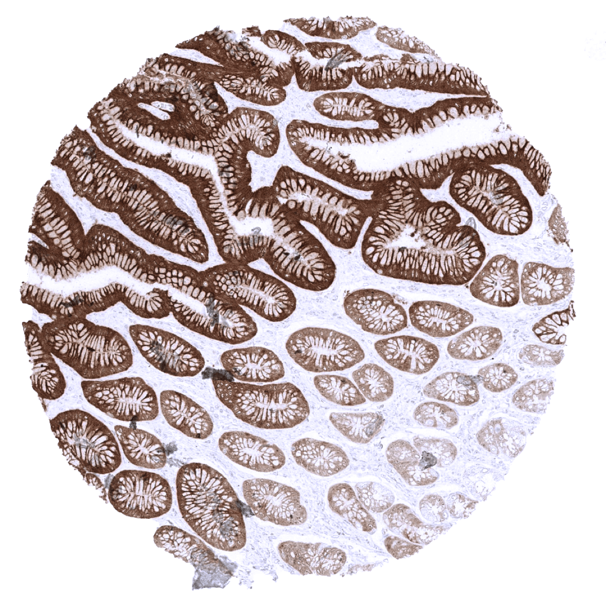
Urinary bladder, urothelium - A strong S100P immunostaining is seen in the urothelium. The staining intensity may be somewhat weaker in the basal cell layer.
Uterus, ectocervix
Uterus, endocervix
Uterus, endometrium (pregnancy)
Uterus, endometrium (proliferation)

Uterus, endometrium (secretion) - S100P immunostaining can occasionally occur in endometrium.
Uterus, myometrium