Adrenal gland
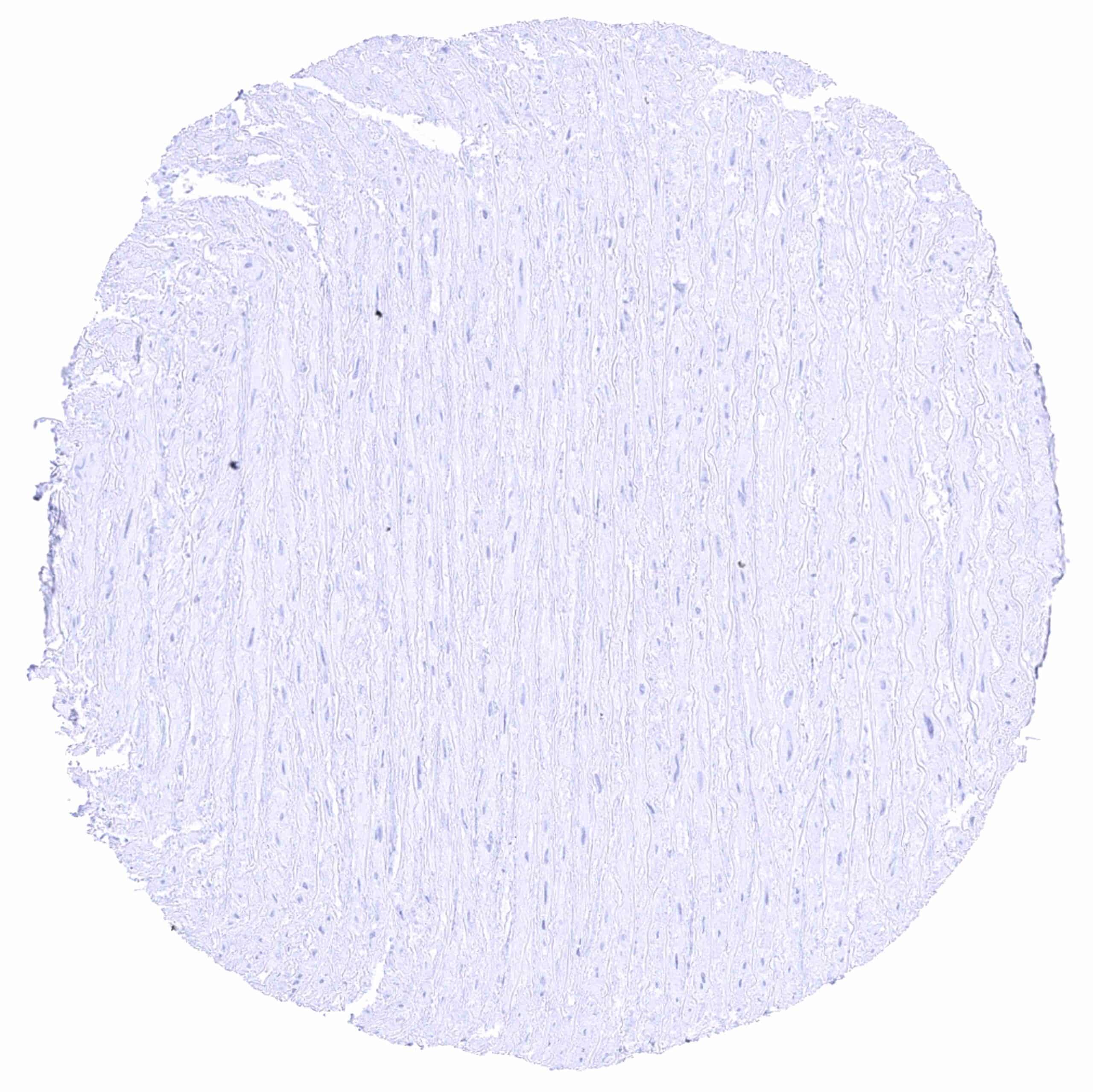
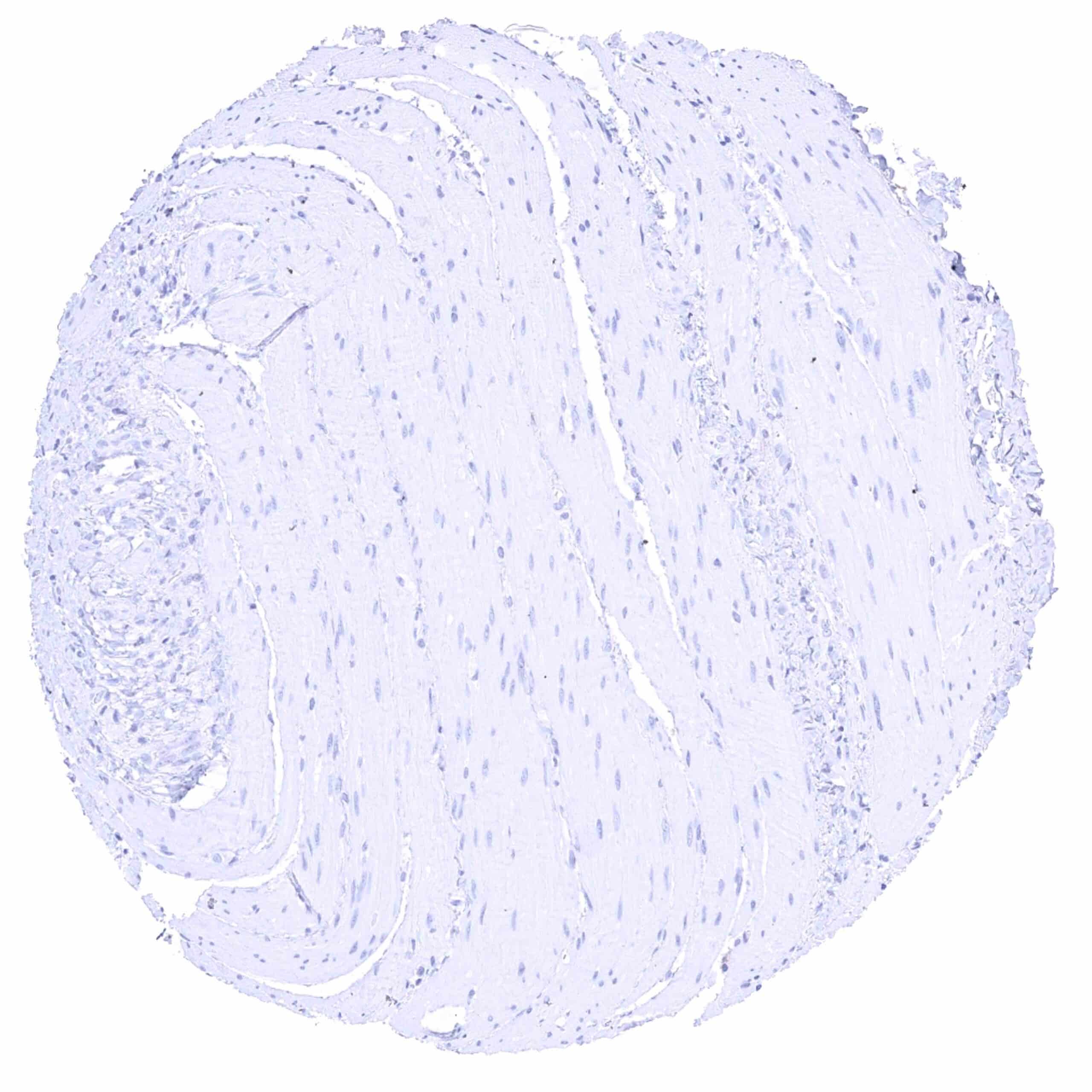
Aorta, media
Appendix, mucosa

Appendix, muscular wall
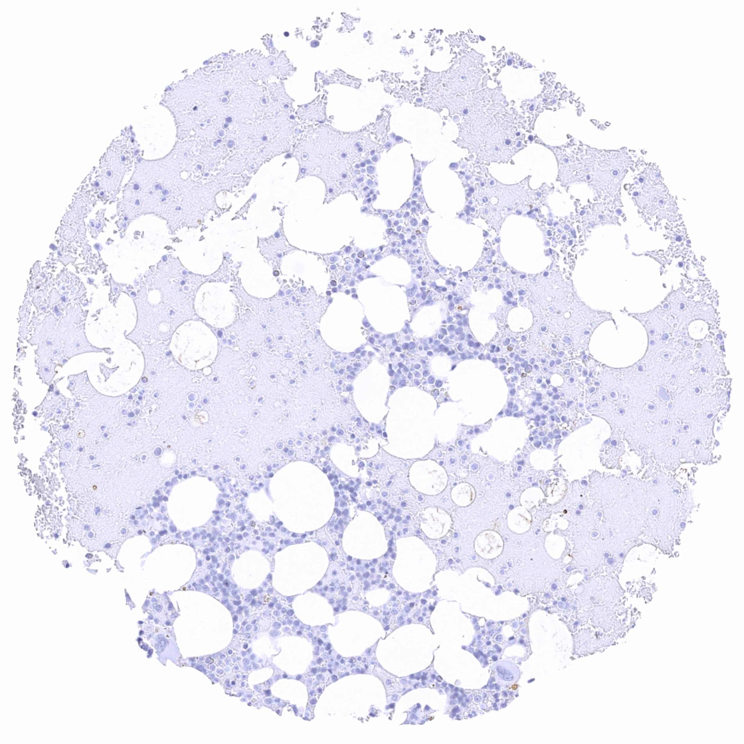
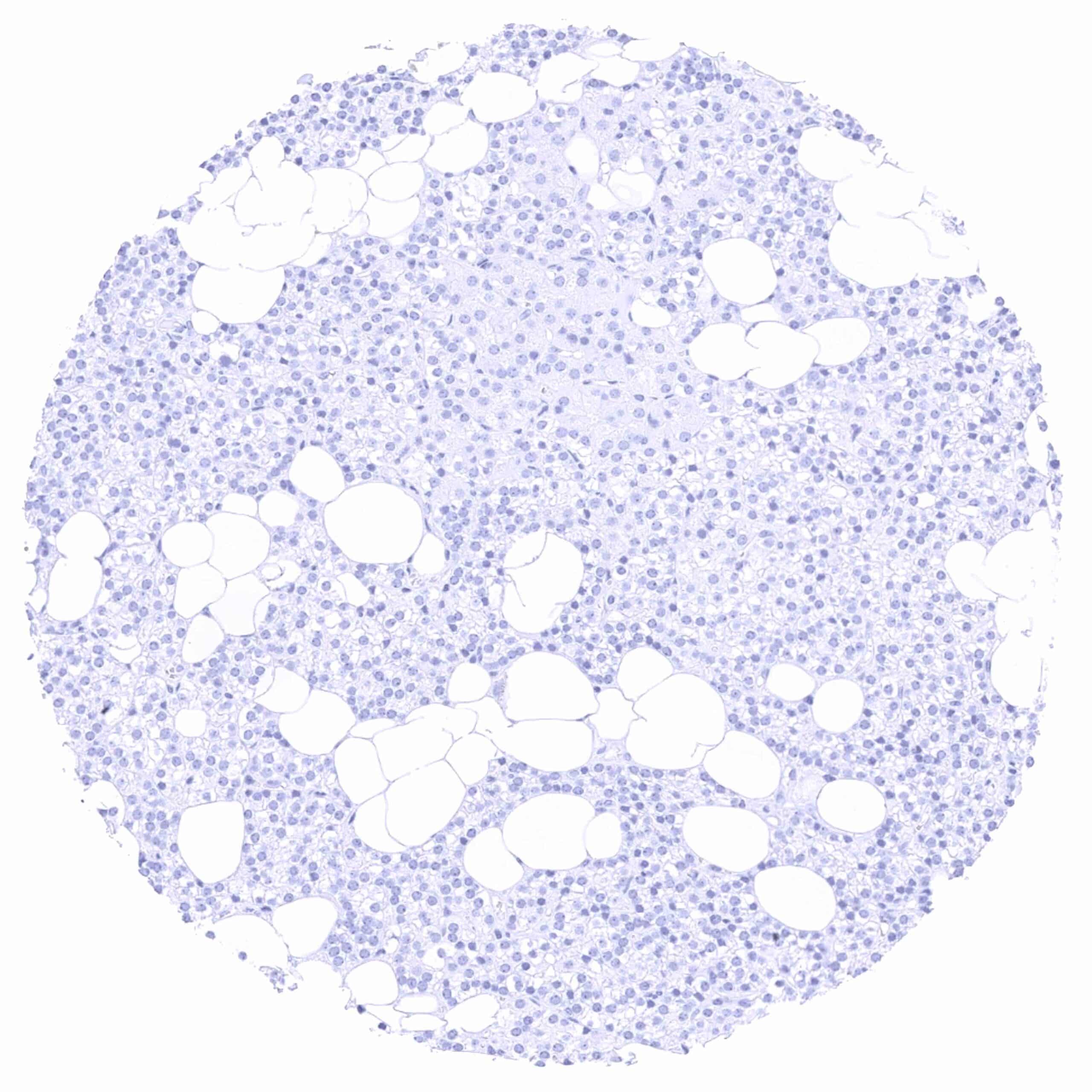
Bone marrow
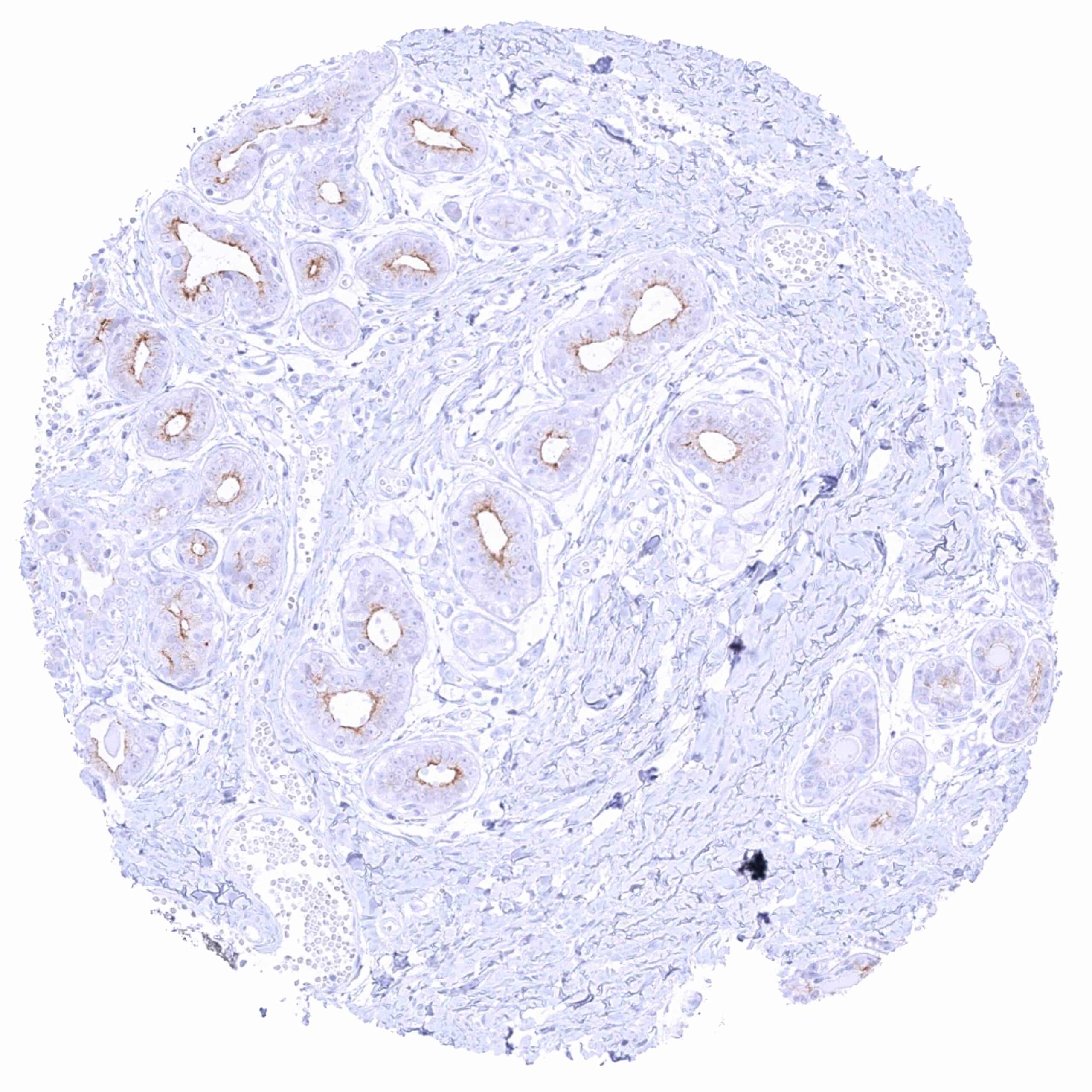
Breast – Weak to moderate apical membranous and faint cytoplasmic staining of luminal glandular cells.
Bronchus, glands – Weak to moderate PSMA staining (membranous apical and cytoplasmic) of bronchial glands.
Bronchus, mucosa
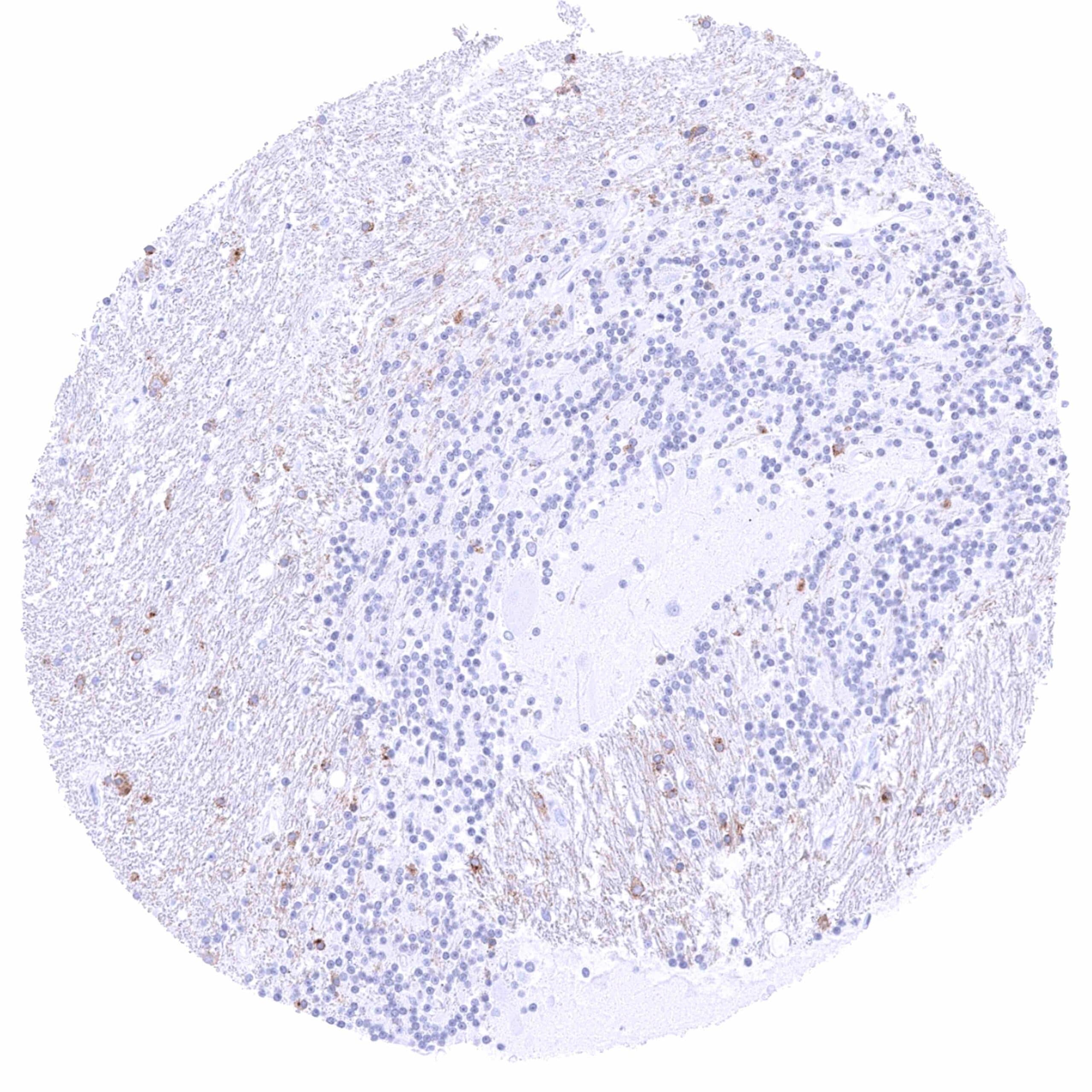
Cerebellum, cortex (molecular layer, Purkinje cell layer, granule cell layer, white matter) – Distinct membranous staining of at least a fraction of glia cells in the white layer. Weak fibrillar PSMA staining in the white matter
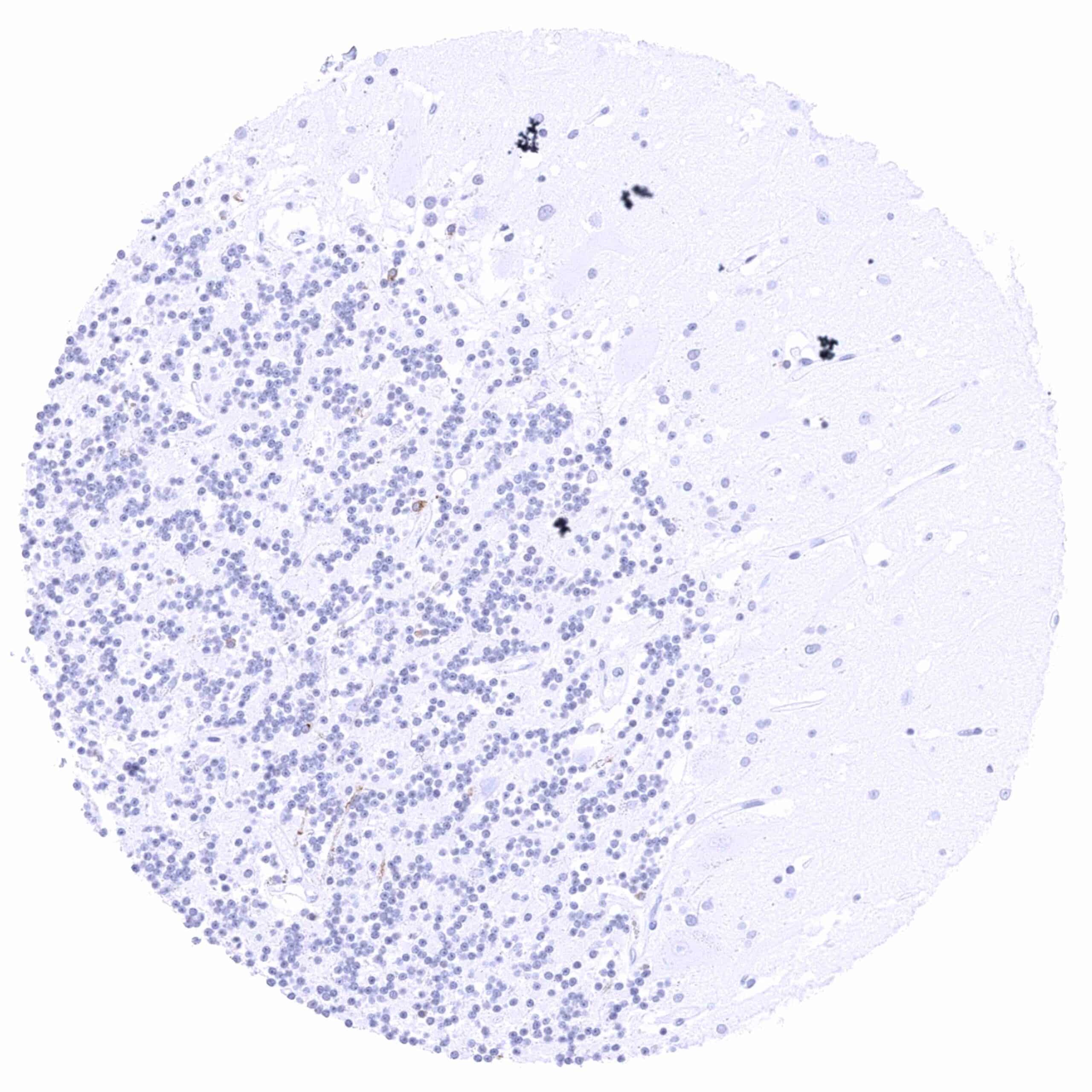
Cerebellum, cortex (molecular layer, Purkinje cell layer, granule cell layer)
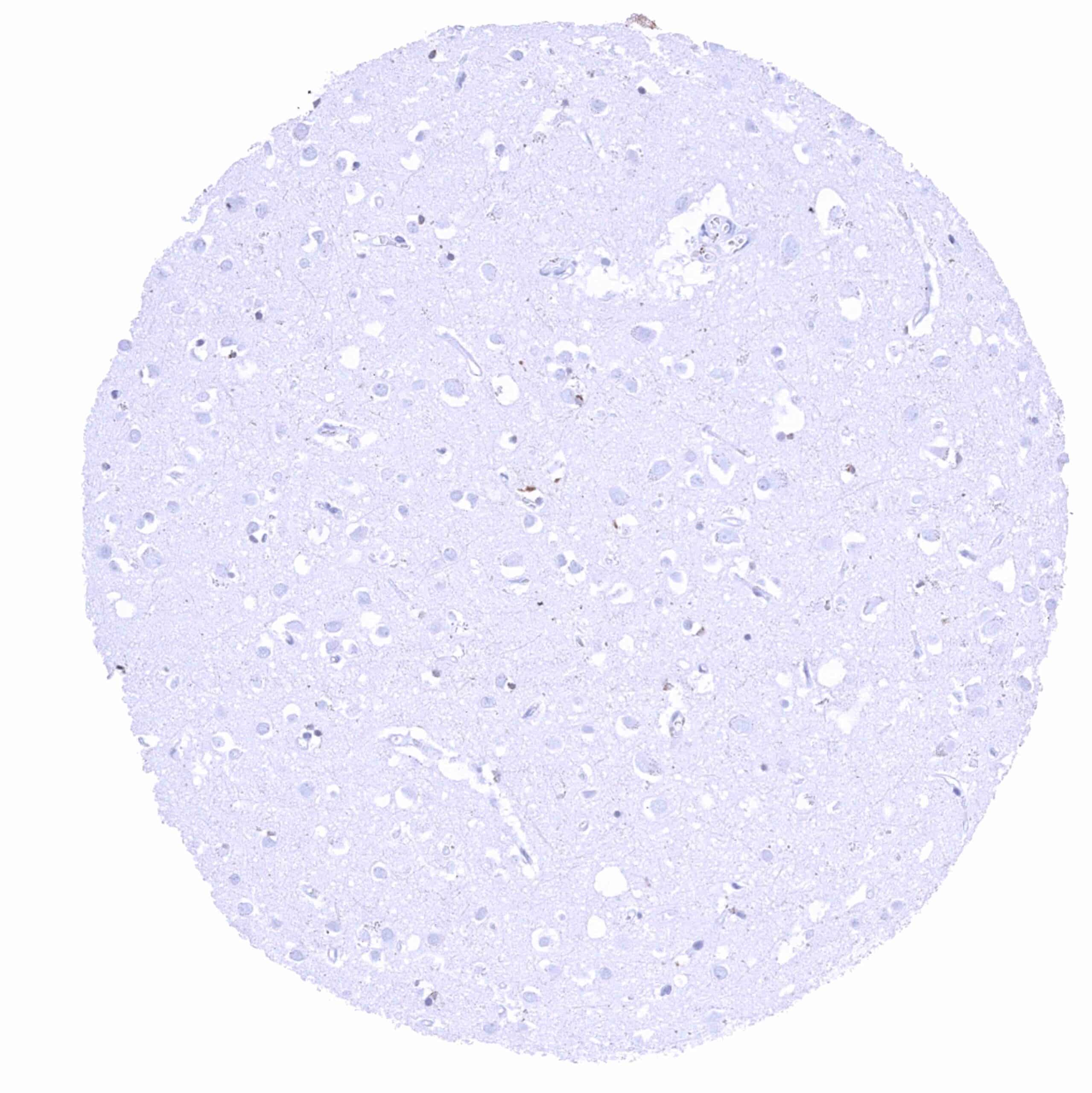
Cerebrum, grey matter
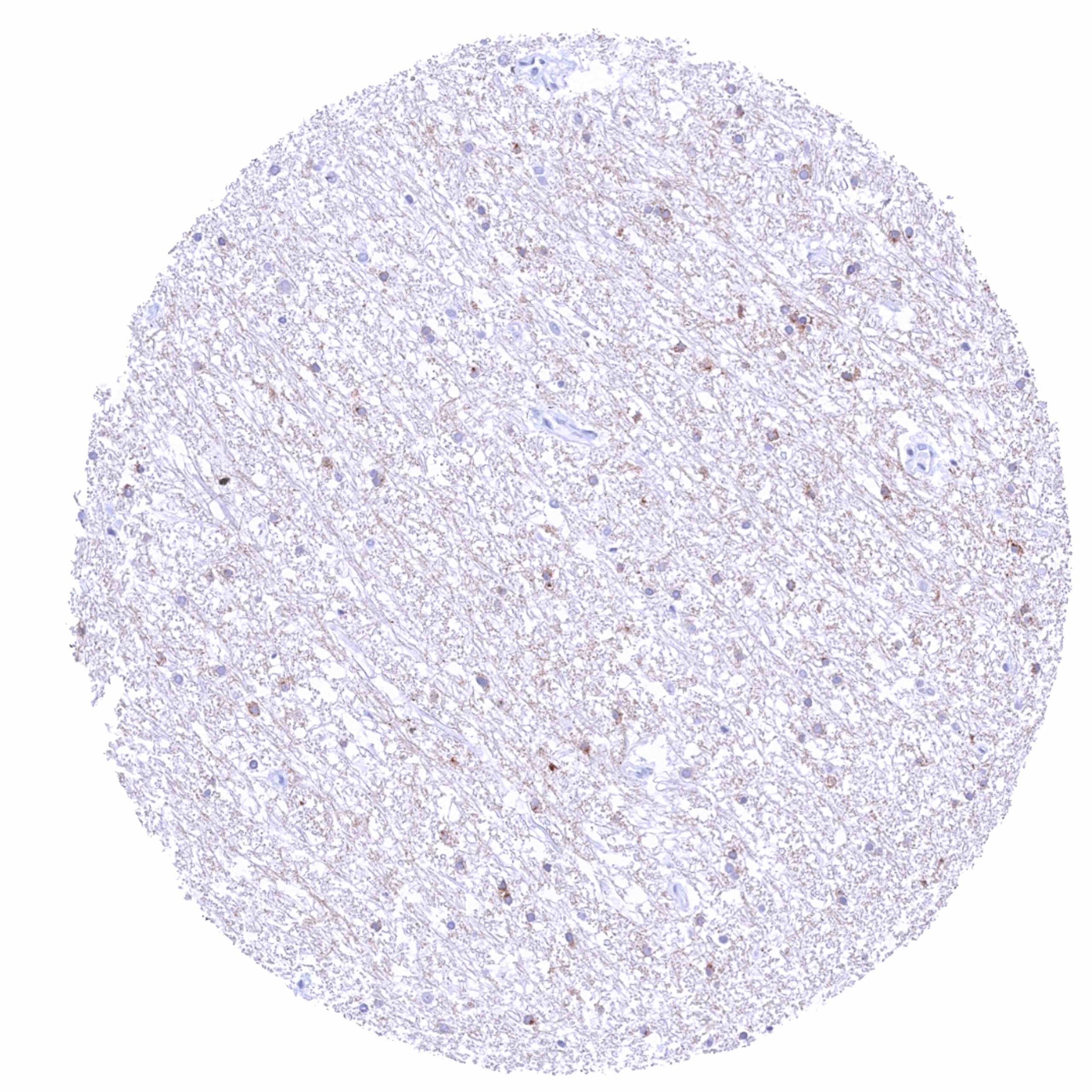
Cerebrum, white matter – Distinct membranous PSMA staining of at least a fraction of glia cells. Weak to moderate fibrillar PSMA staining.

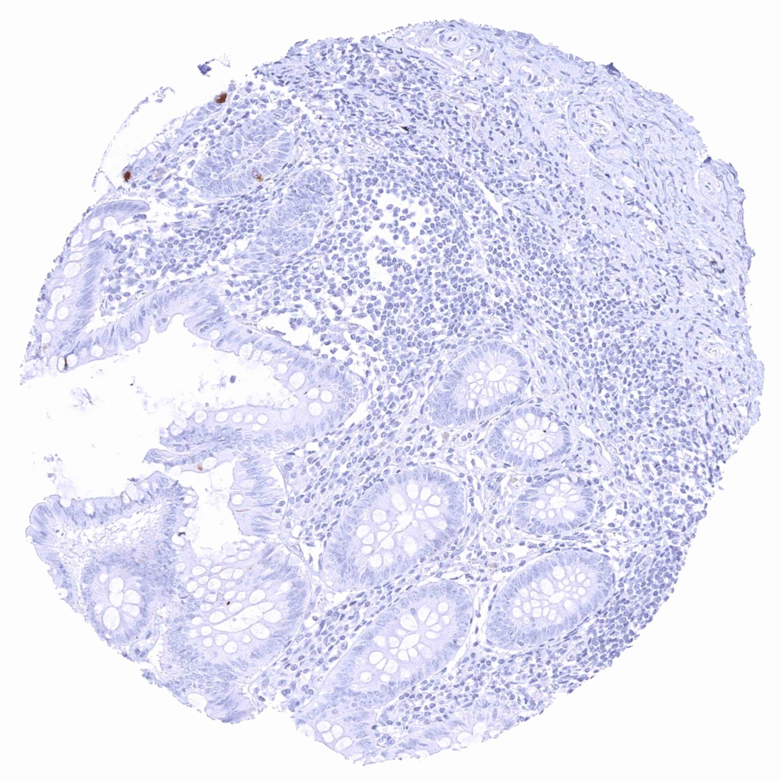
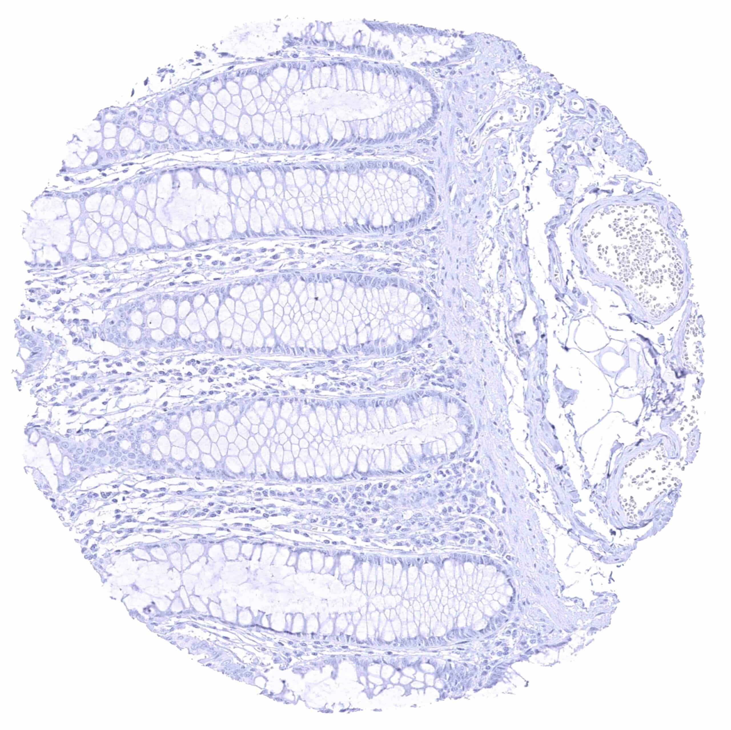
Colon descendens, mucosa.jpeg
Colon descendens, muscular wall
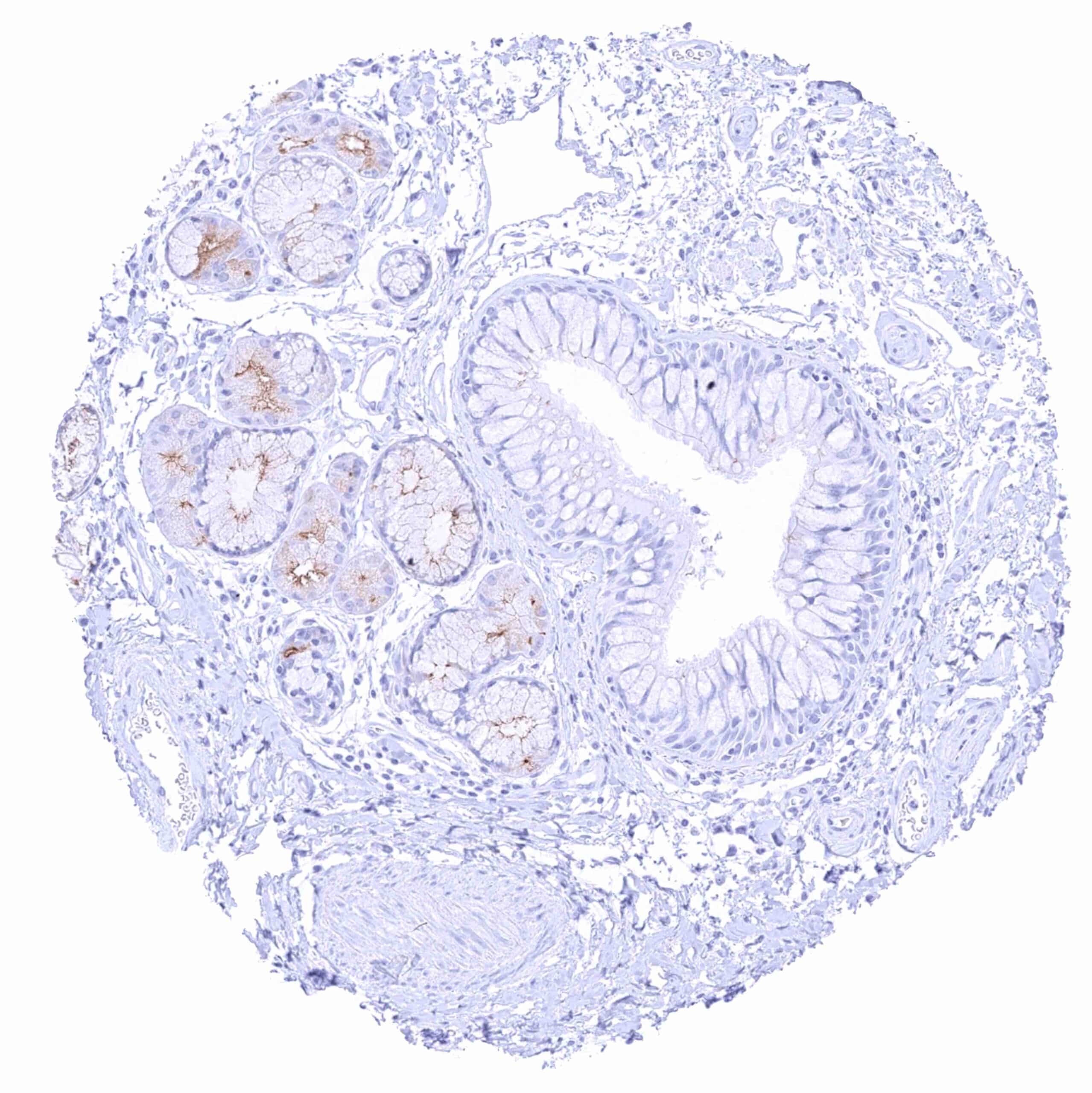
Duodenum, Brunner gland – Weak PSMA staining of a fraction of glandular cells, predominantly at the apical membrane.
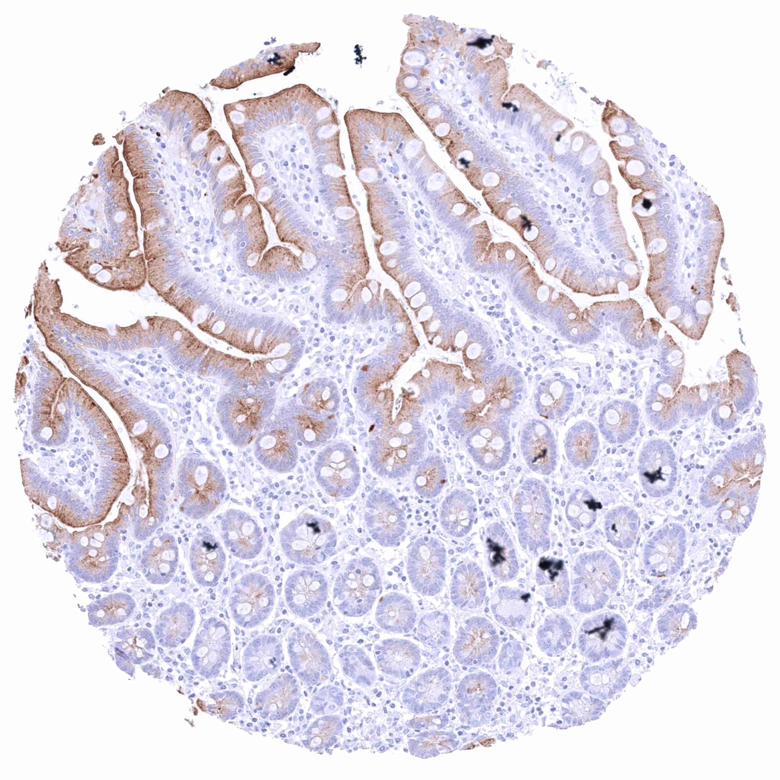
Duodenum, mucosa – Intense PSMA staining of the luminal membranes – stronger on the surface of villi than in the crypt base. The cytoplasm is also PSMA positive especially on the luminal half.
Epididymis (Caput)
Epididymis (Cauda) – Focal weak to moderate apical membranous PSMA staining.
Esophagus, muscular wall
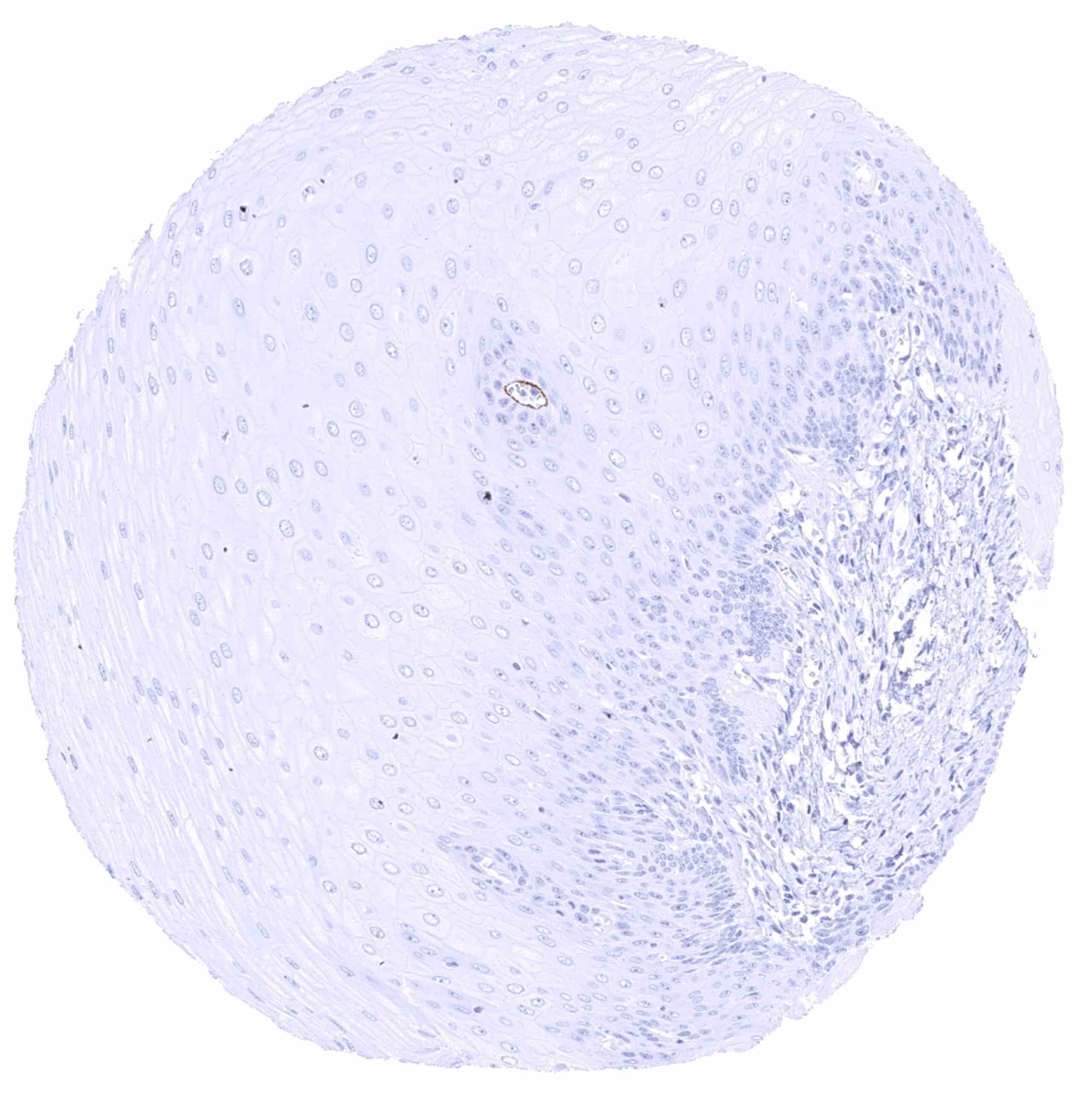
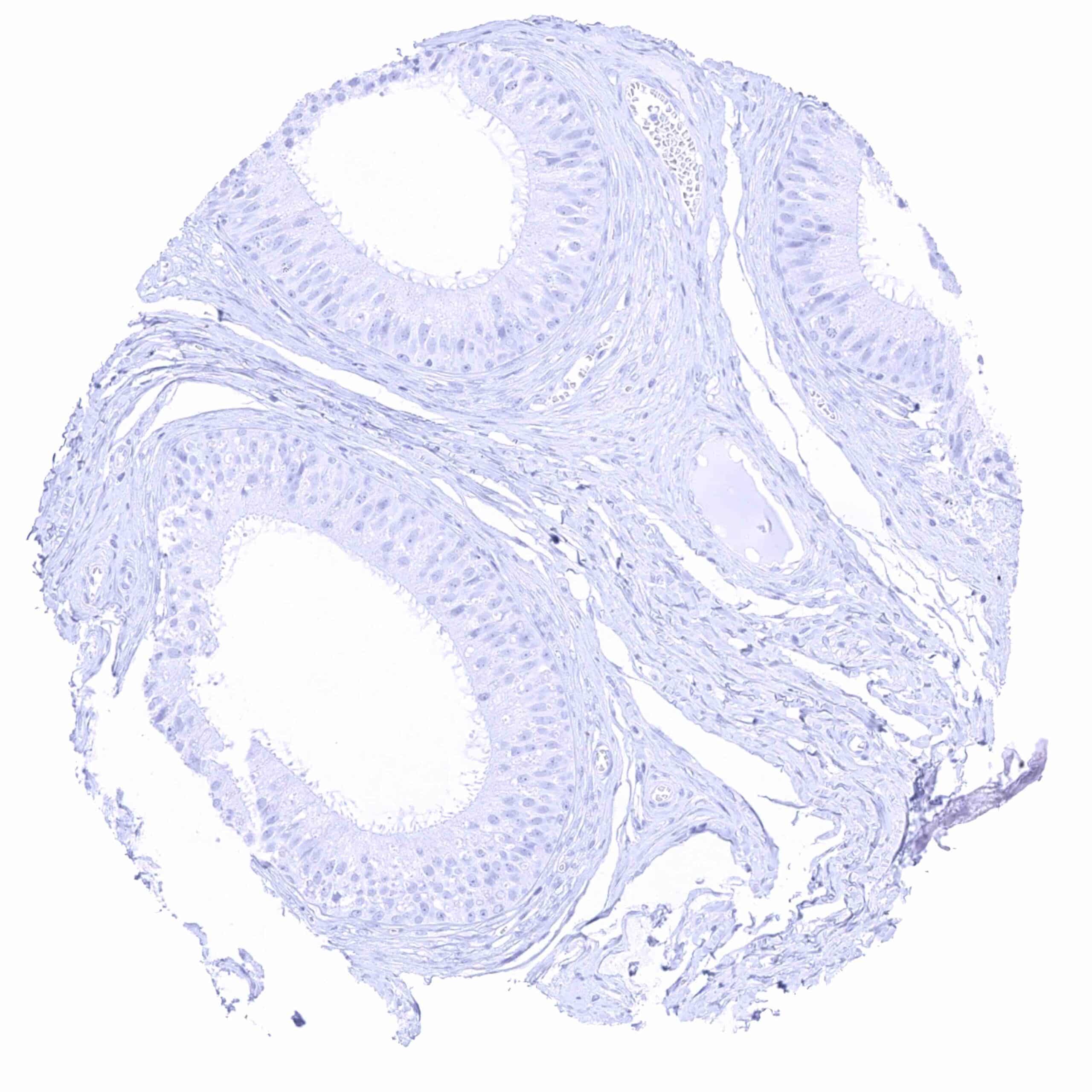
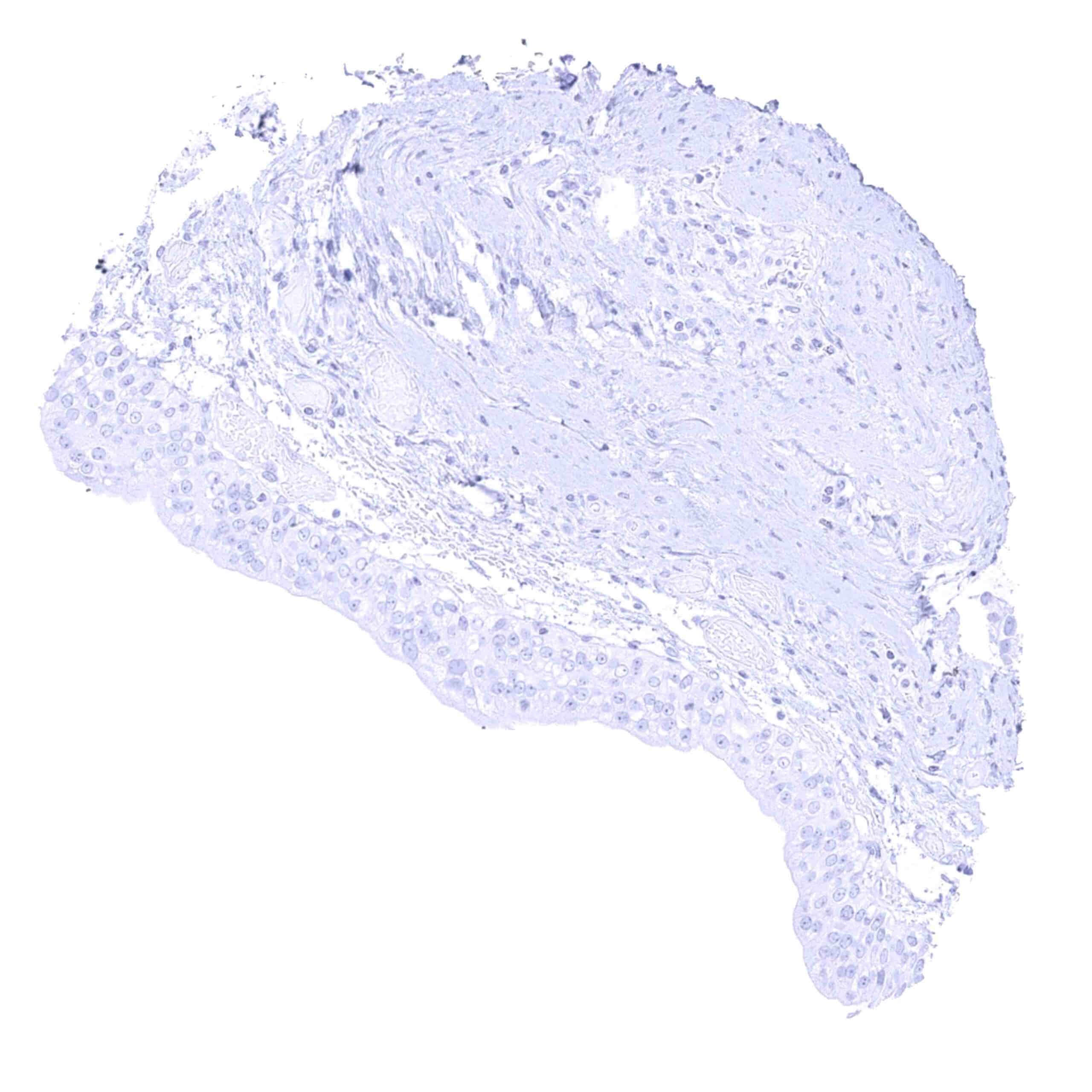
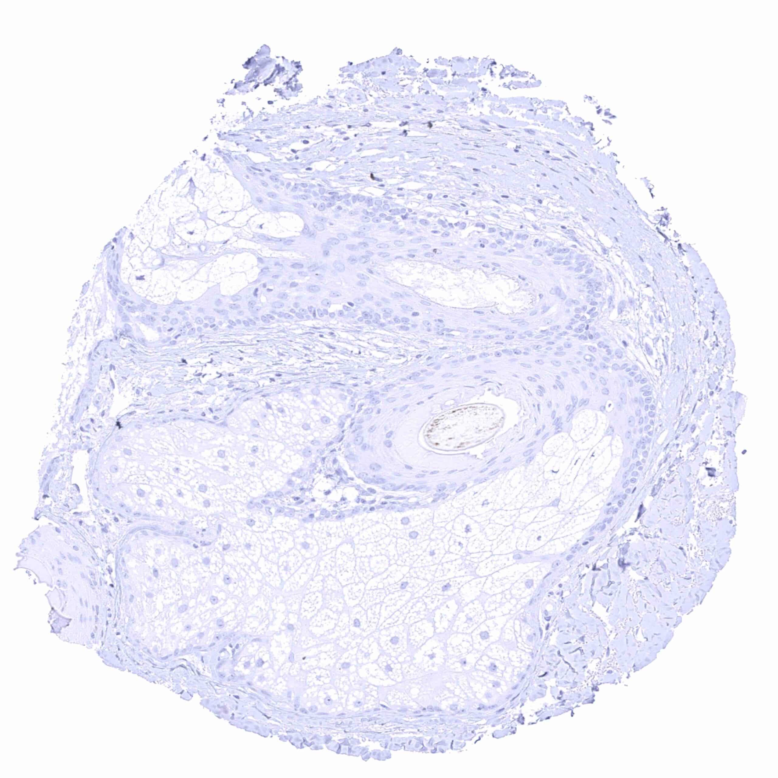
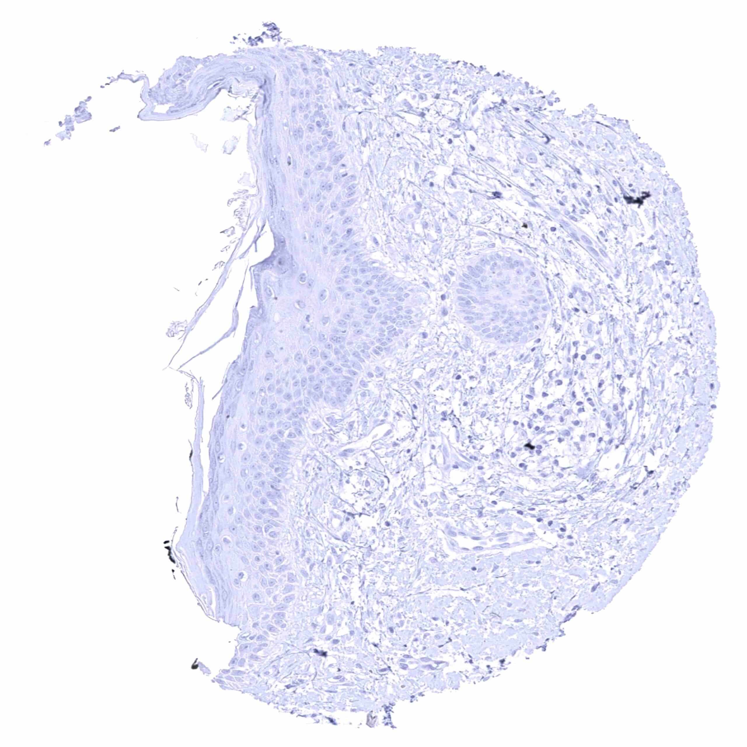
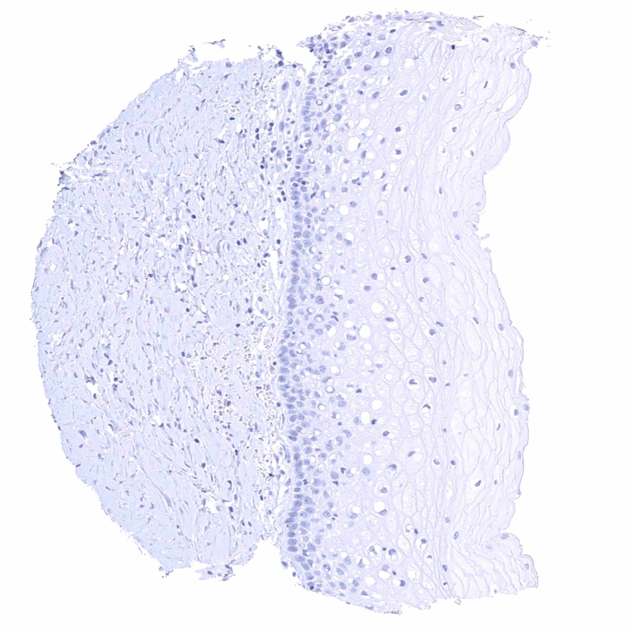
Esophagus, squamous epithelium
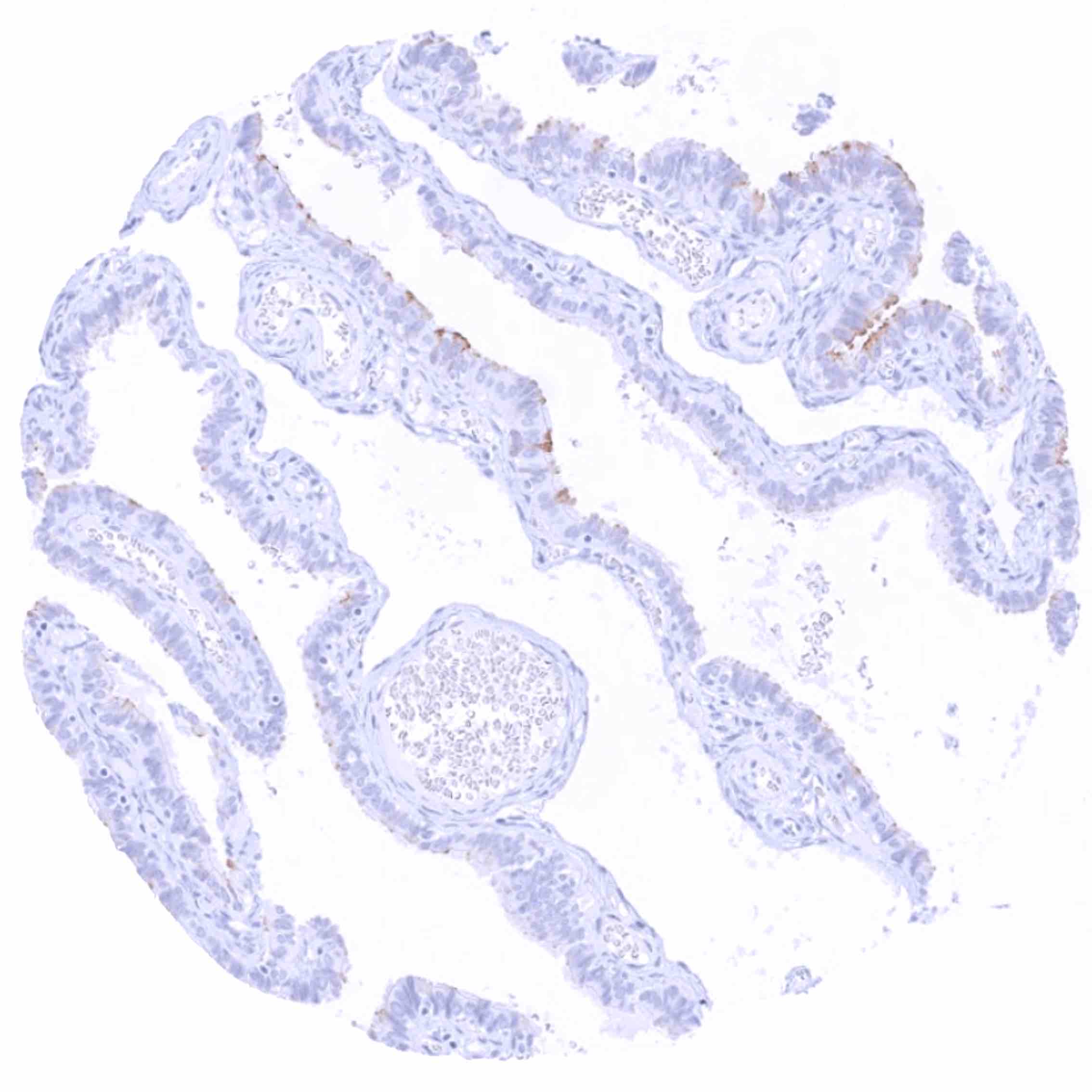
Fallopian tube, mucosa – Weak to moderate apical membranous and weak cytoplasmic PSMA staining of a subset of epithelial cells.
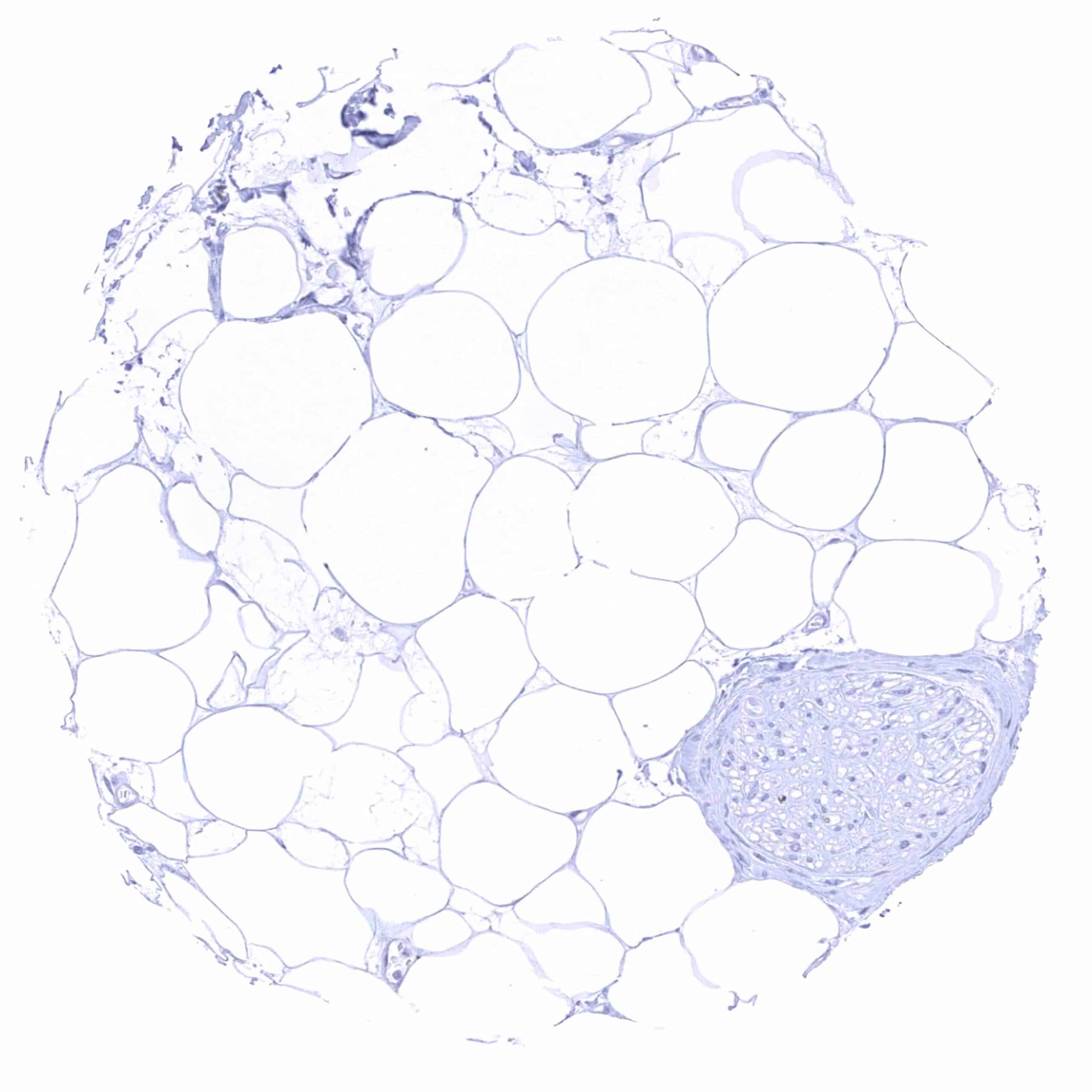
Fat
Gallbladder, epithelium – Weak to moderate PSMA staining of the apical membranes of epithelial cells. .jpeg
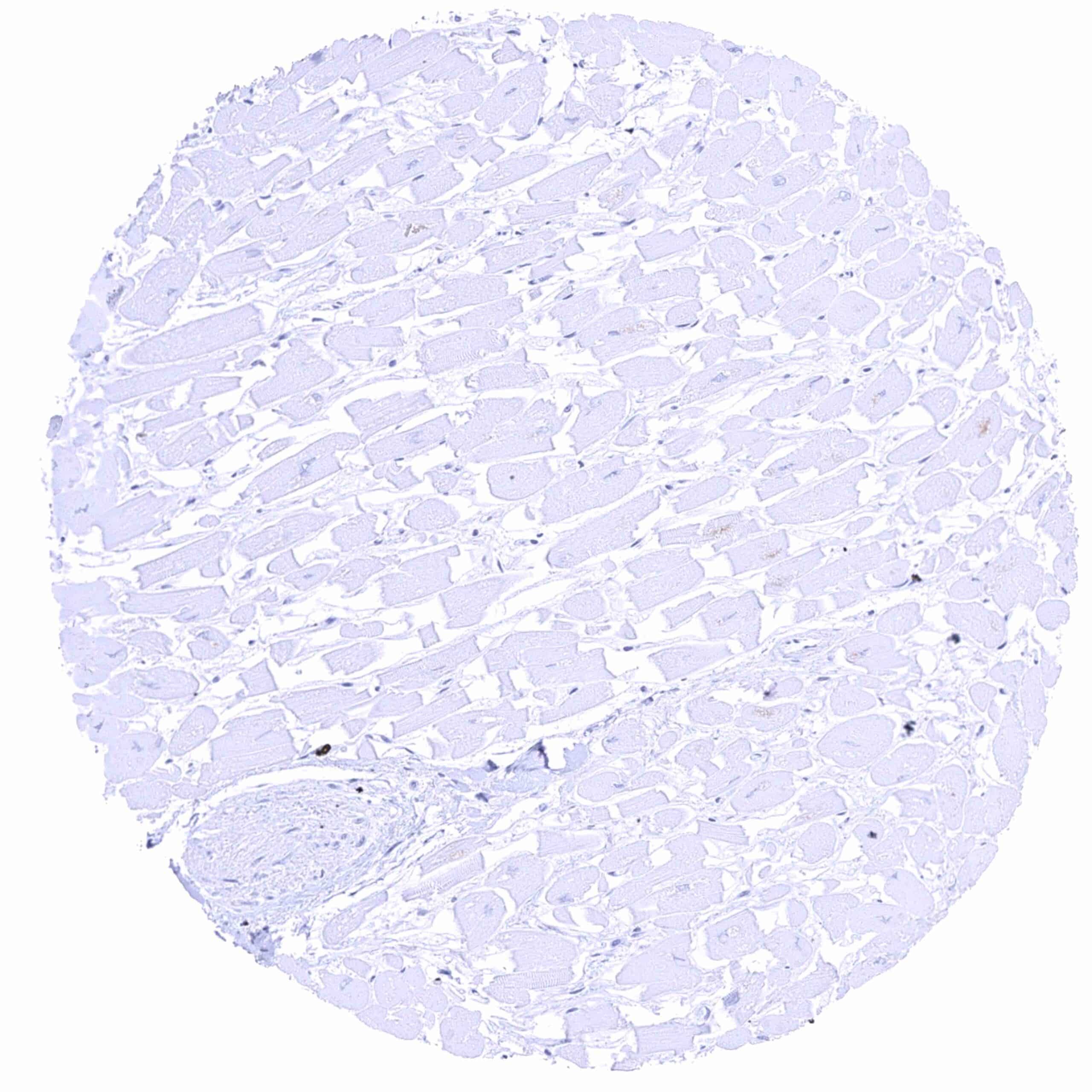
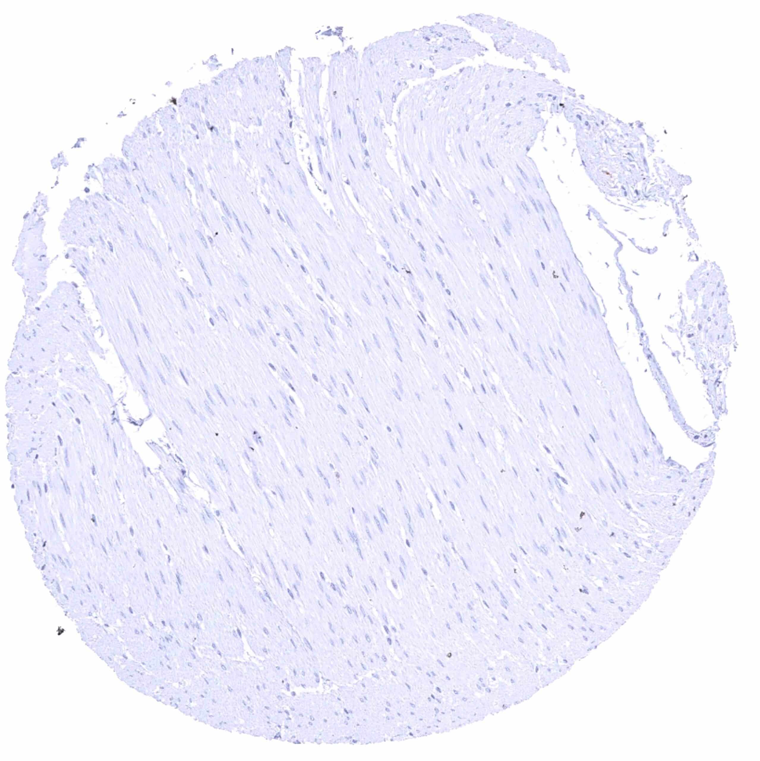
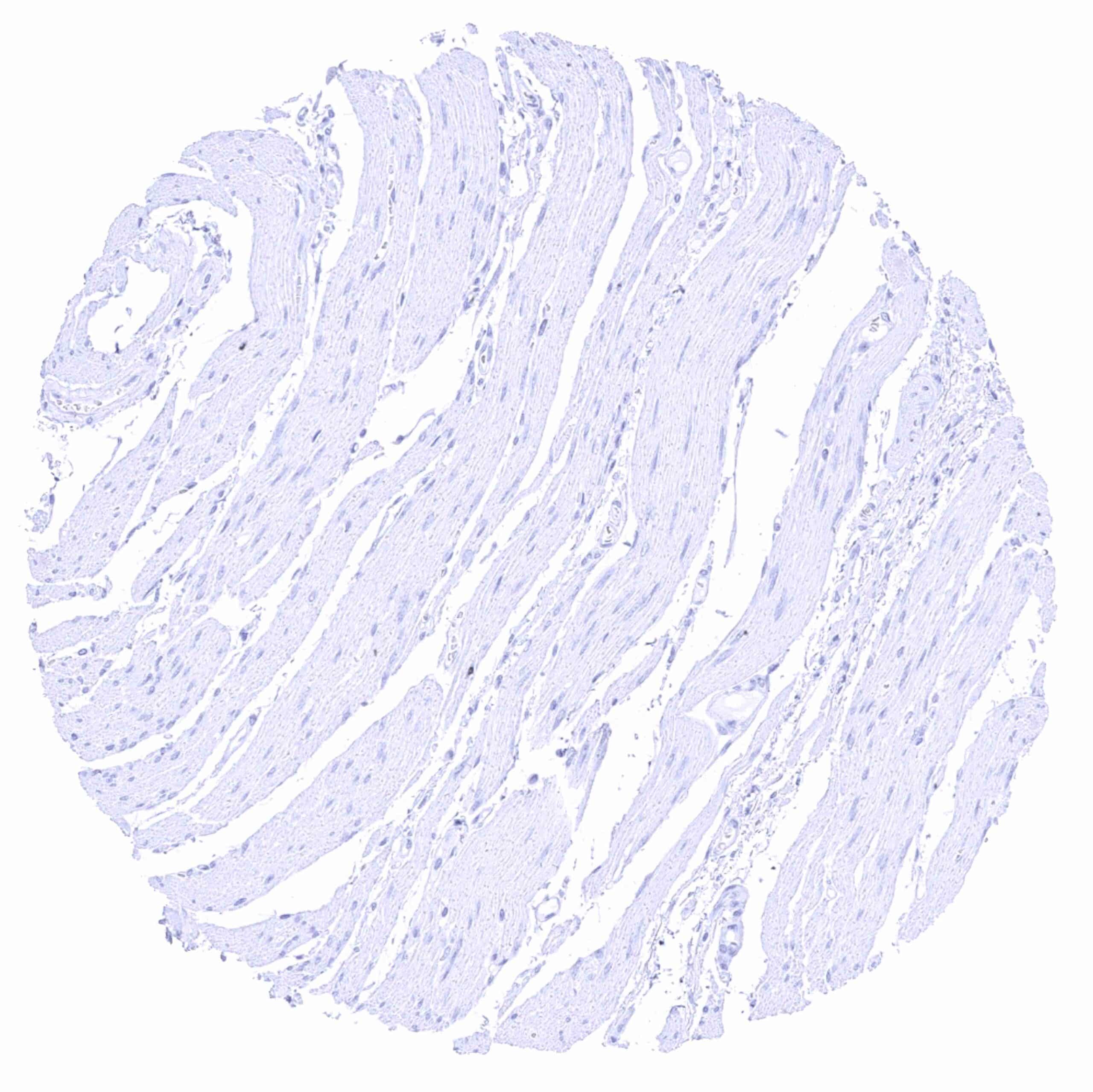
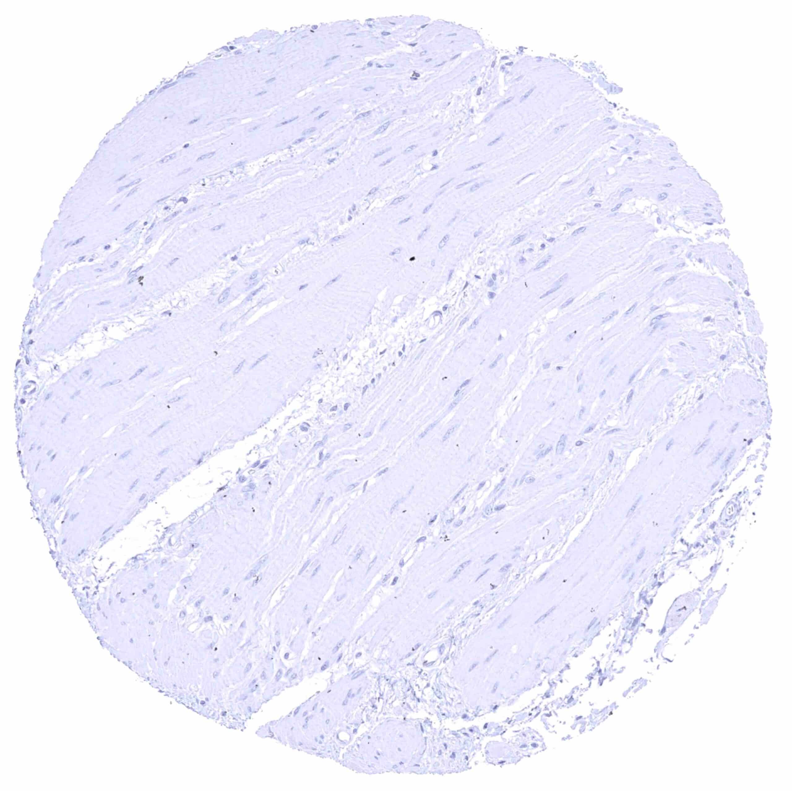
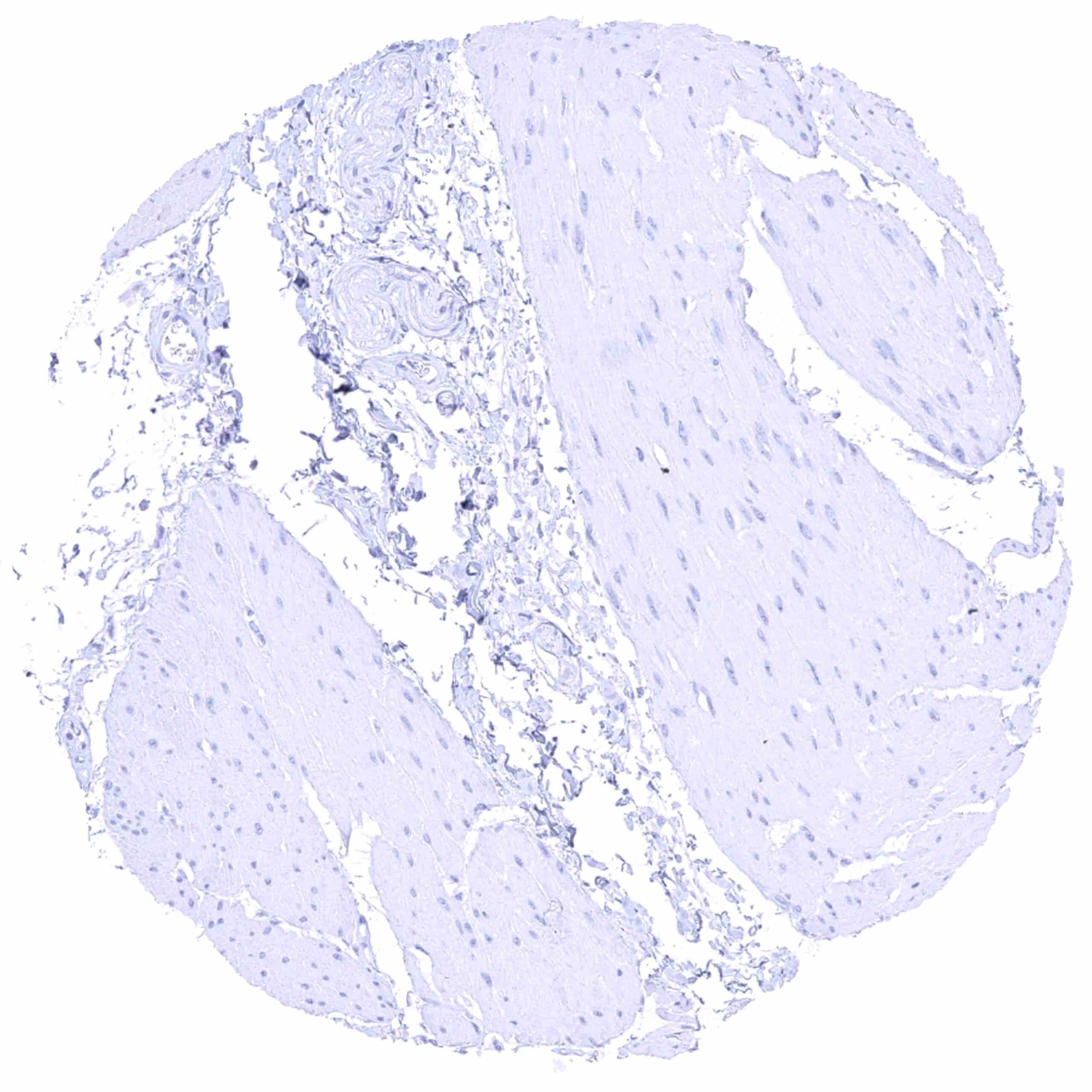
Heart muscle
Ileum, mucosa – Weak PSMA staining of the luminal membranes on the surface of villi while it is absent in crypts.
Ileum, muscular wall
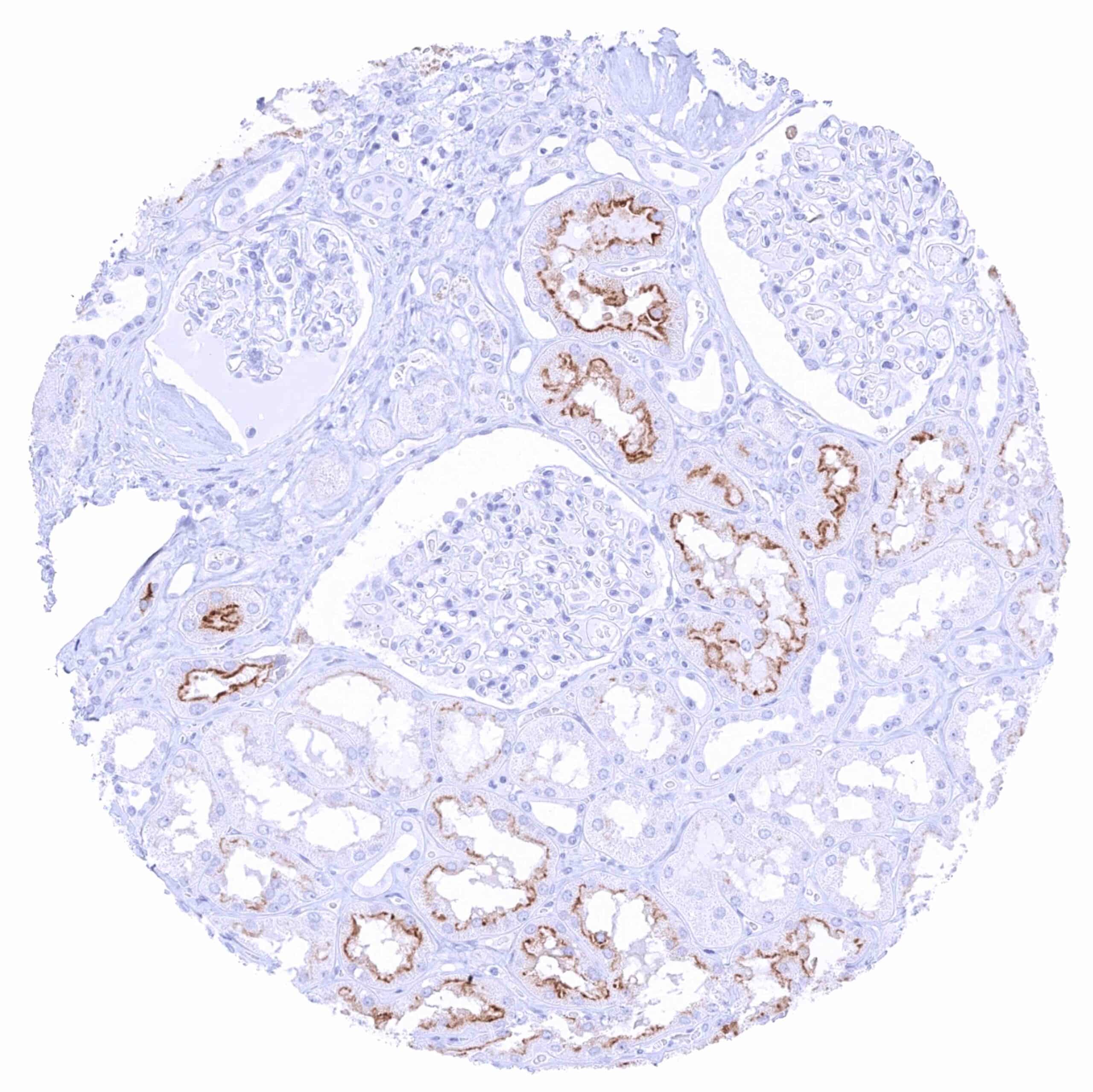
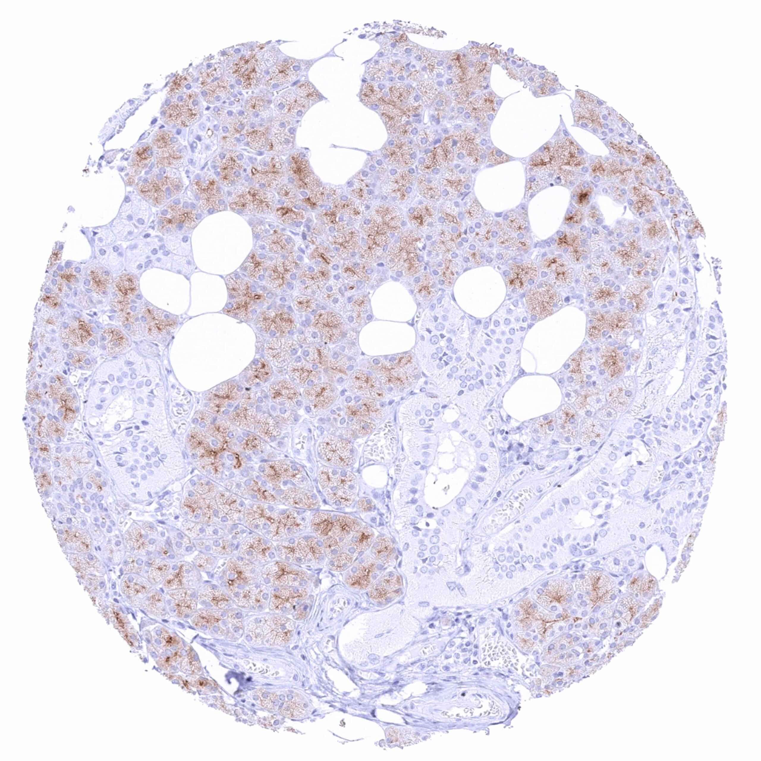
Kidney, cortex – Intense PSMA staining of luminal membranes of proximal tubules while staining is markedly less intense in distal tubuli and absent in collecting ducts.
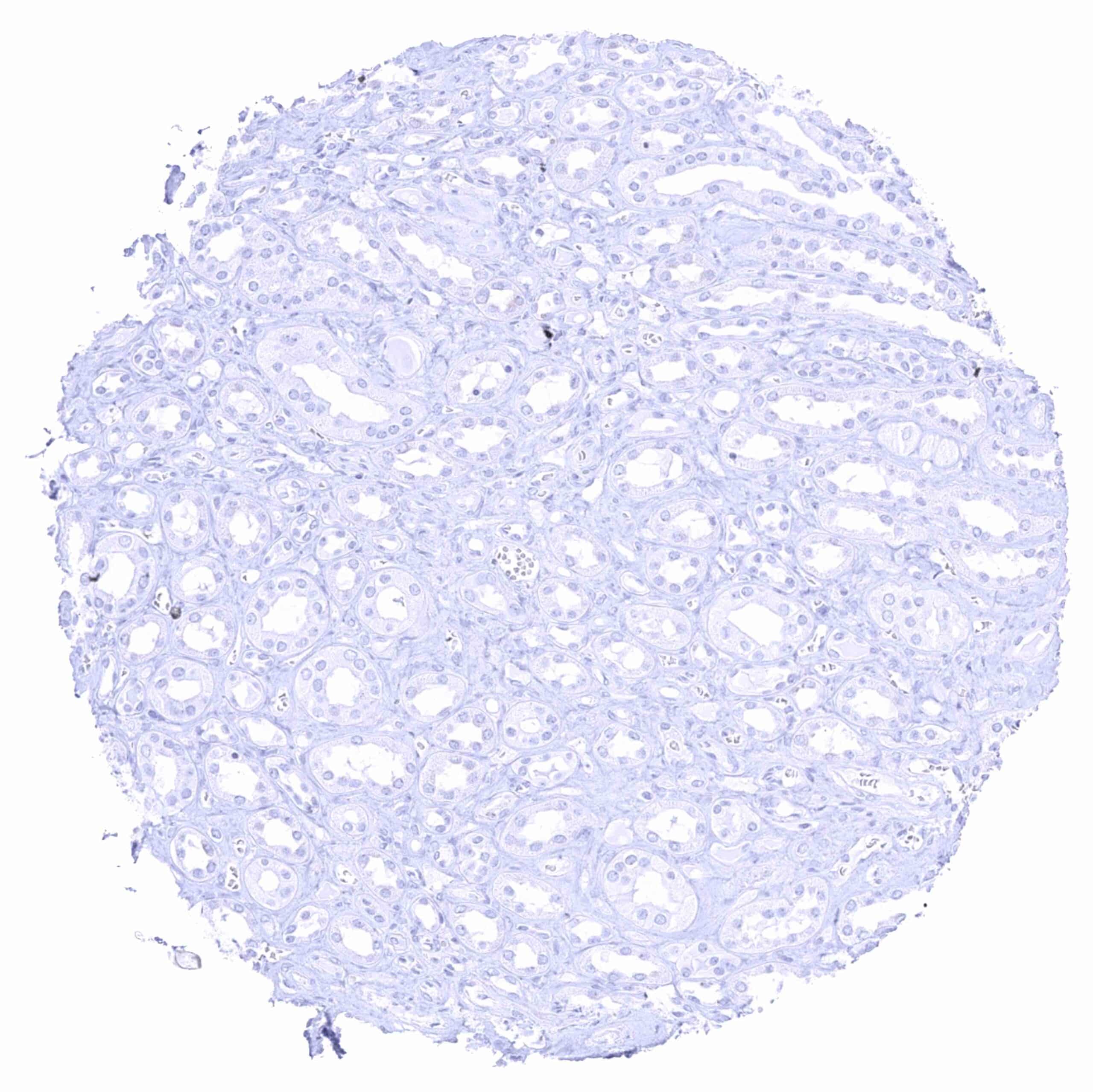
Kidney, medulla – PSMA staining is absent in collecting ducts.
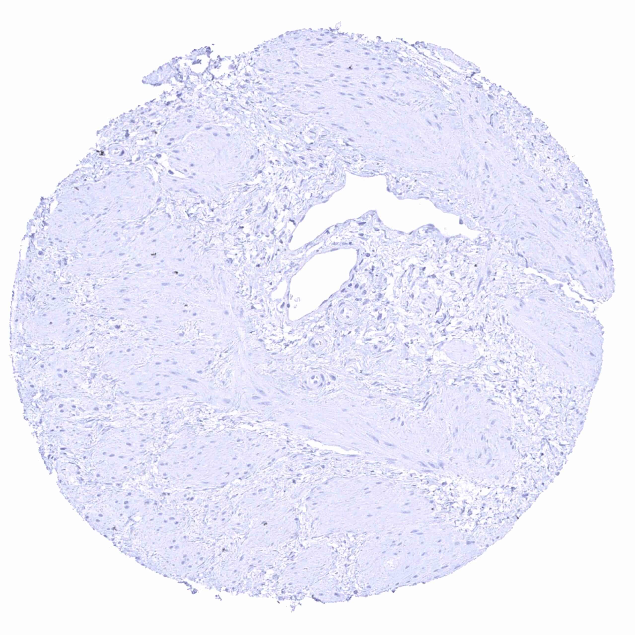
Kidney, pelvis, muscular wall
Kidney, pelvis, urothelium
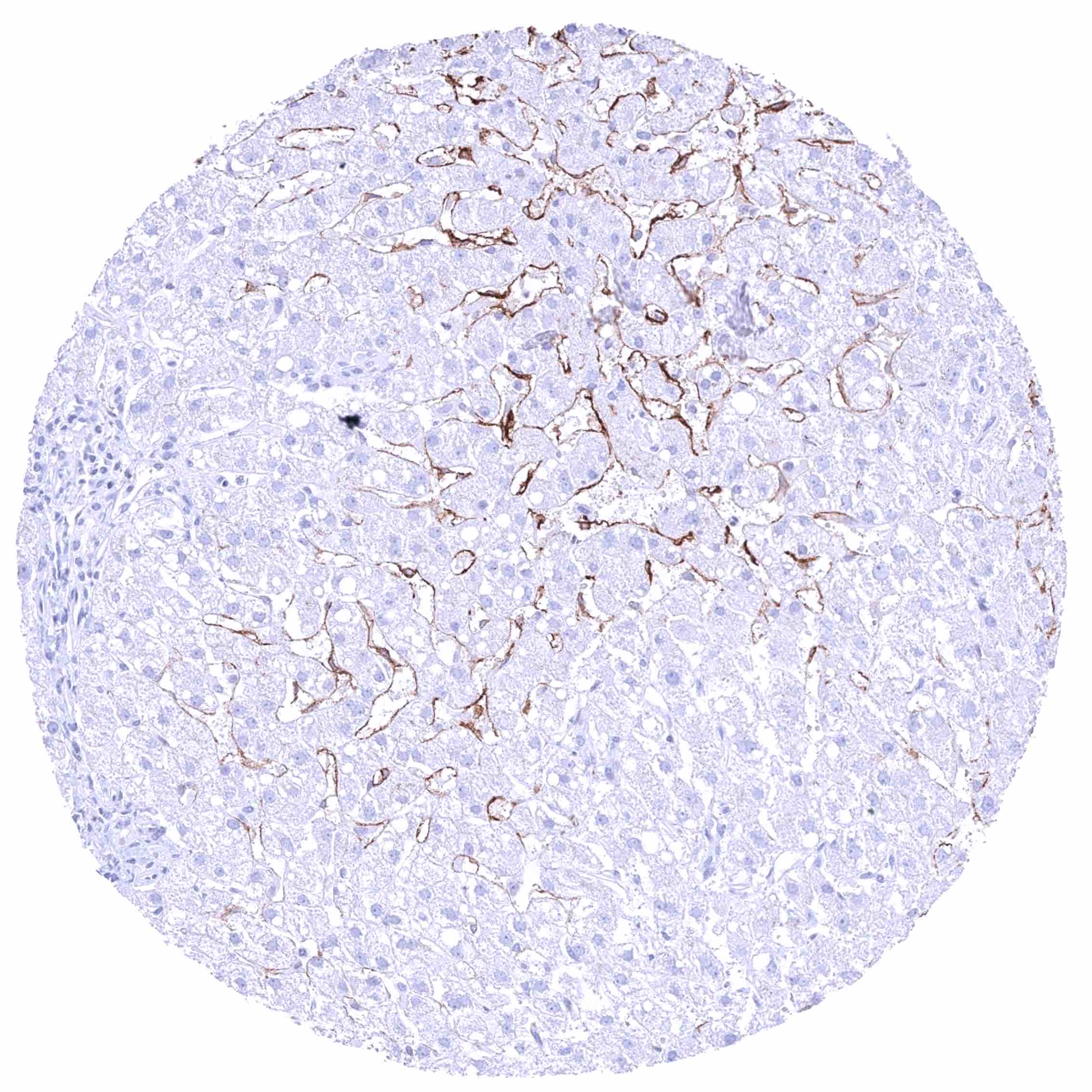
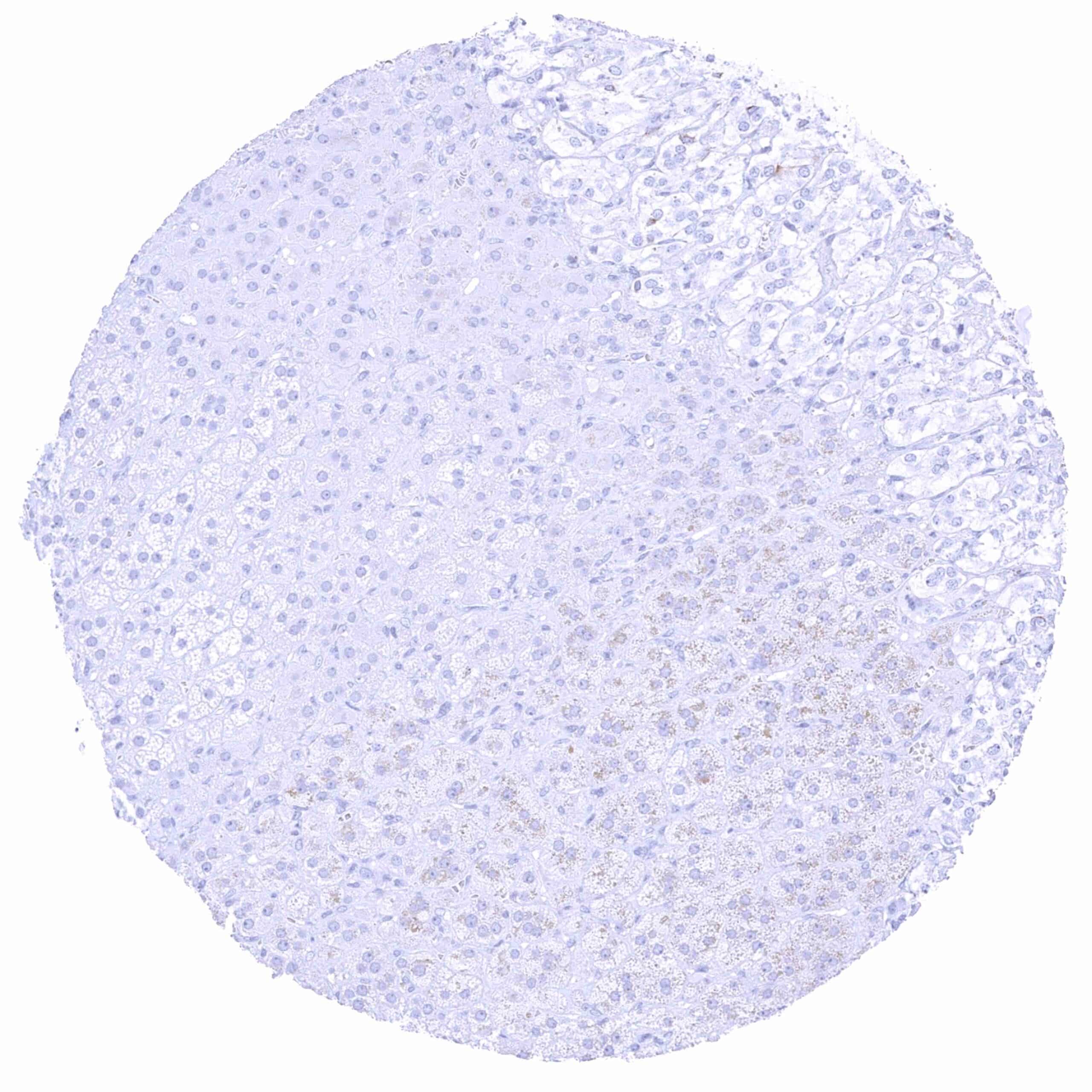
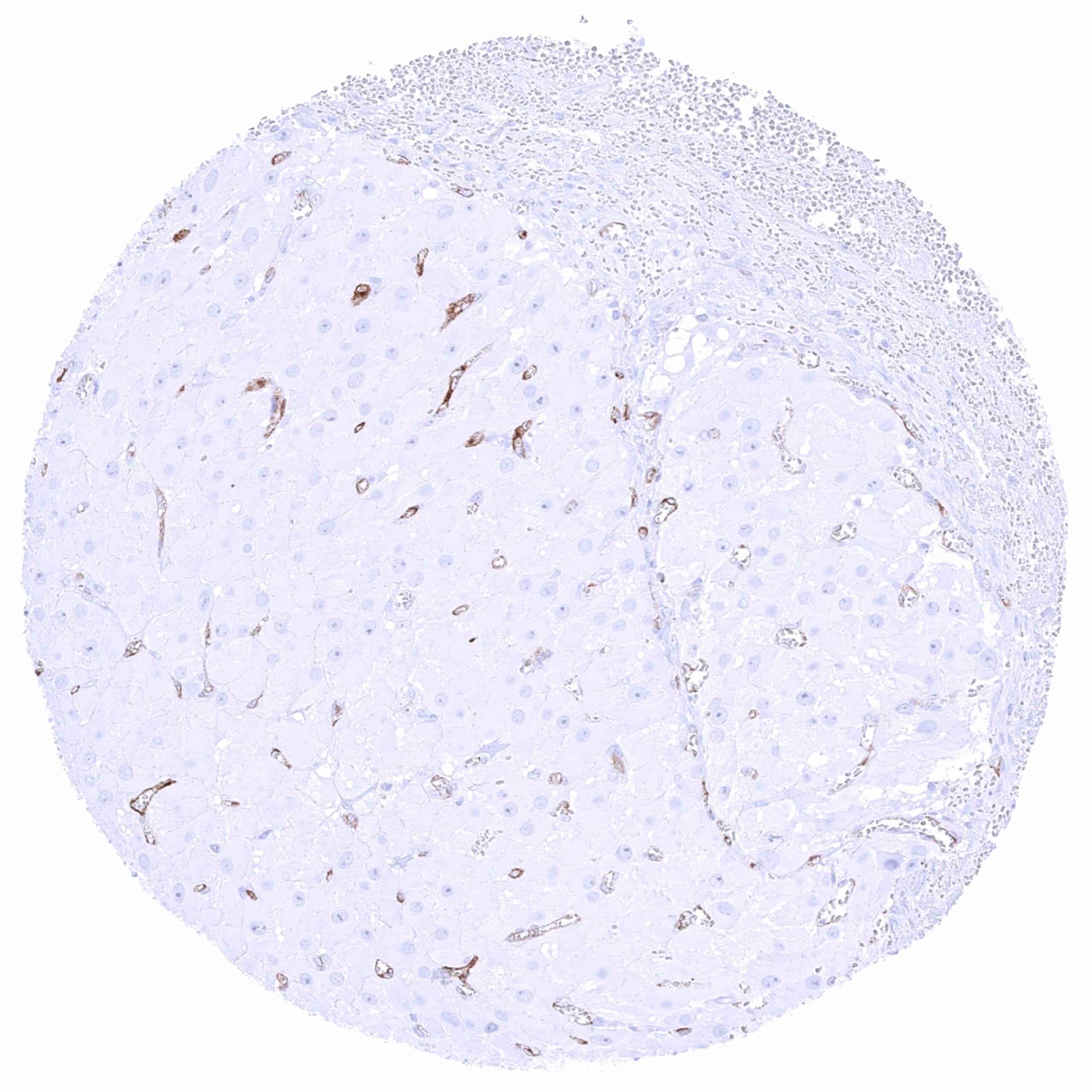
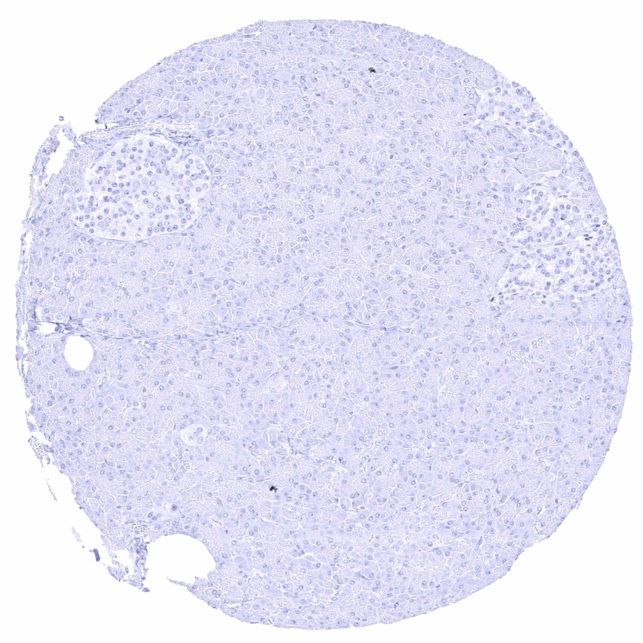
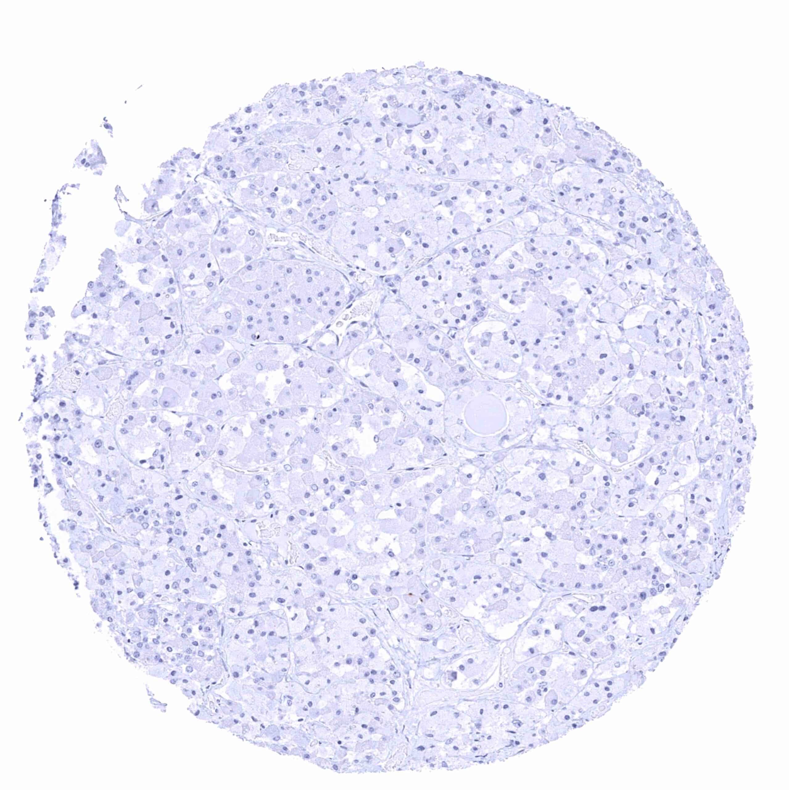
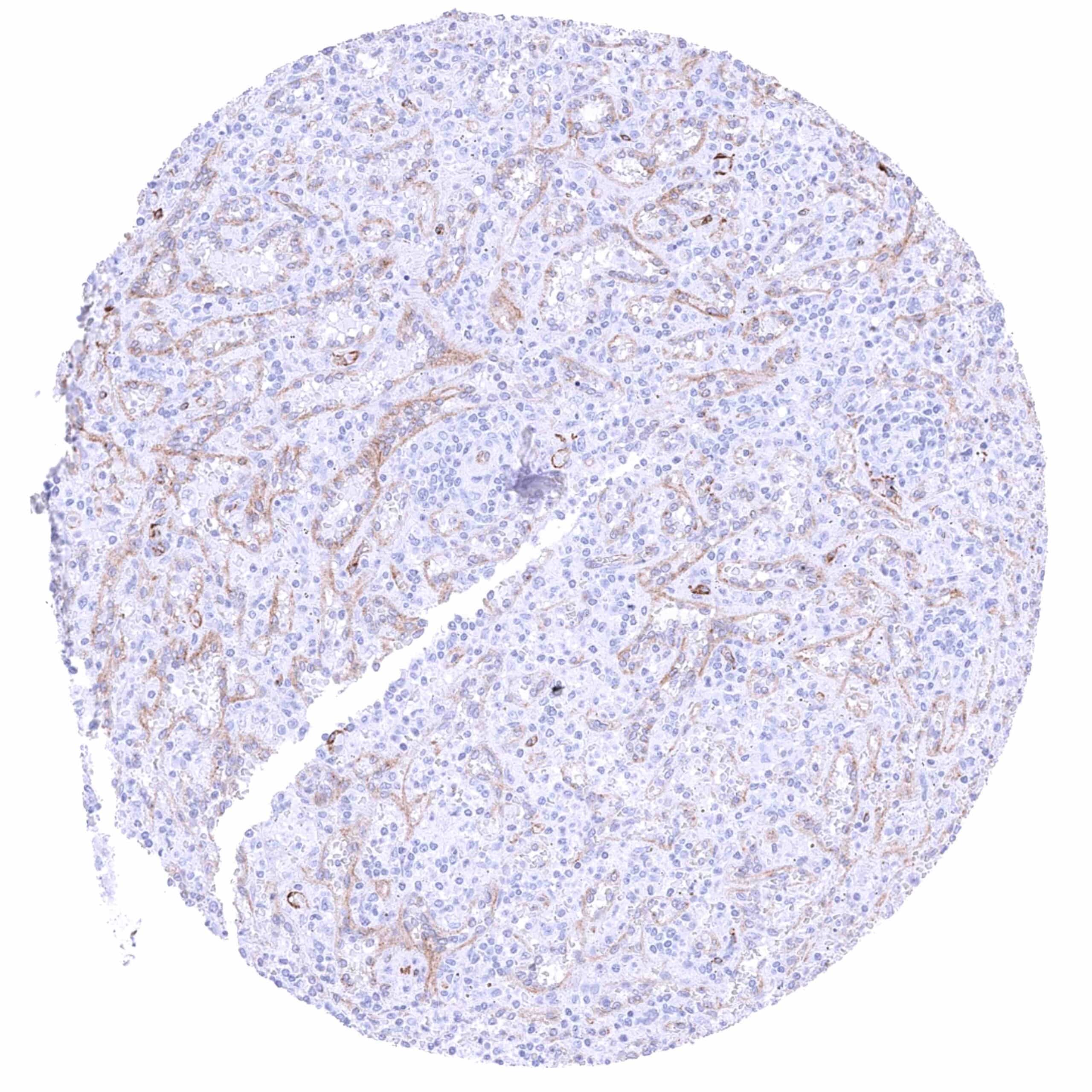
Liver – Moderate to strong PSMA staining of sinusoid cells (regional differences in the staining intensity). Hepatocyte staining is not detectable in this sample.
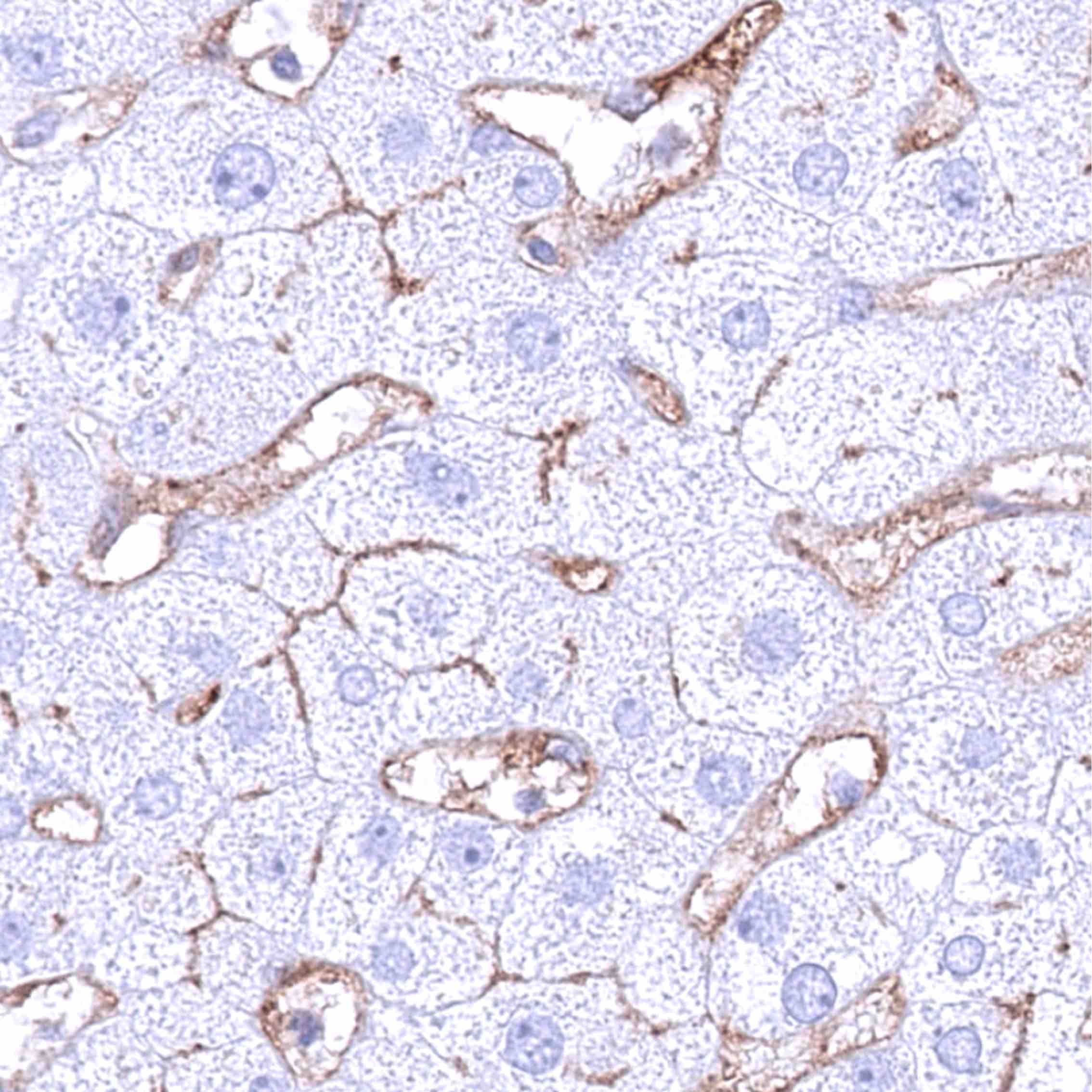
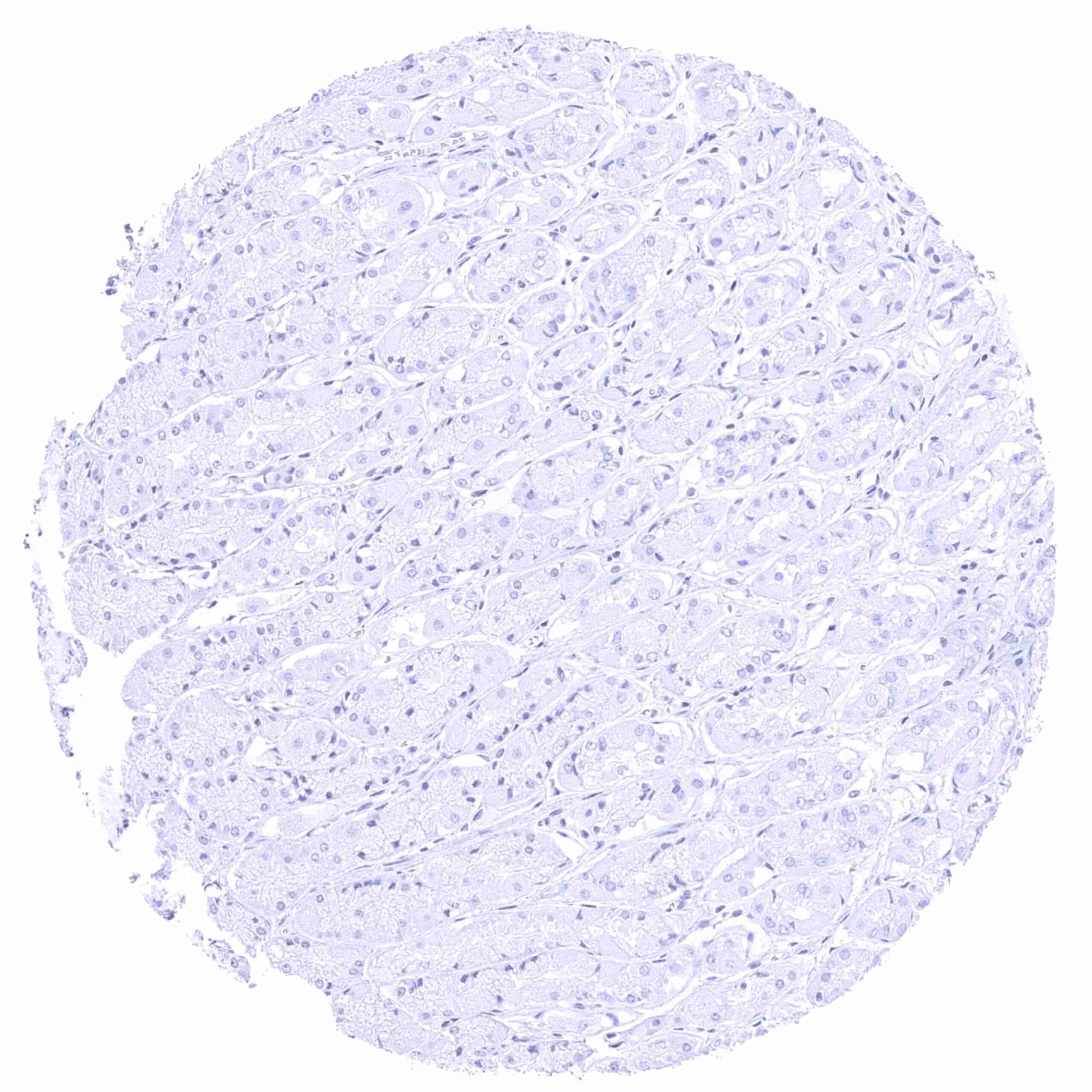
Liver – Moderate to strong PSMA staining of sinusoid cells. Weak to moderate membranous PSMA staining of hepatocytes (especially at the bile secreting pole)
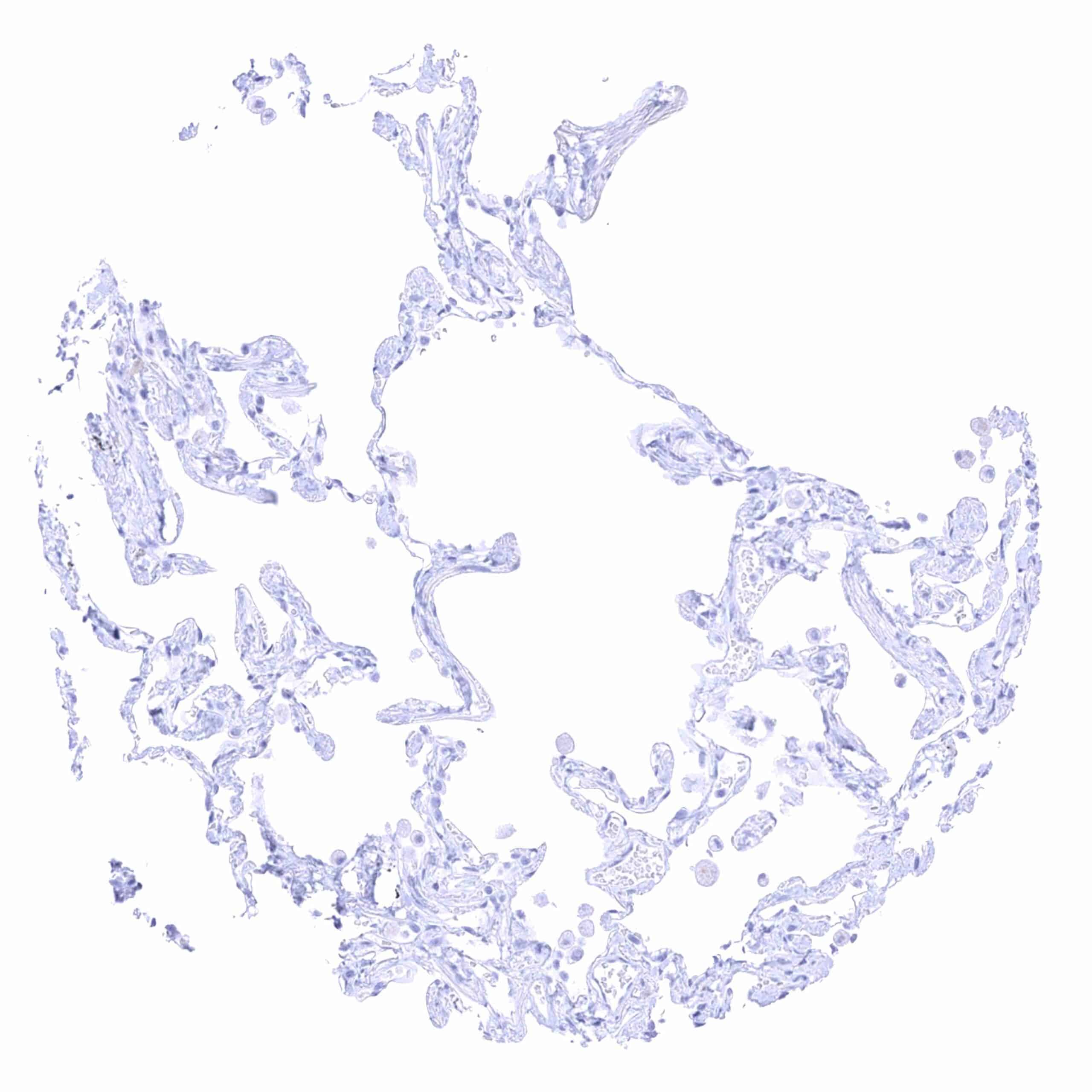
Lung
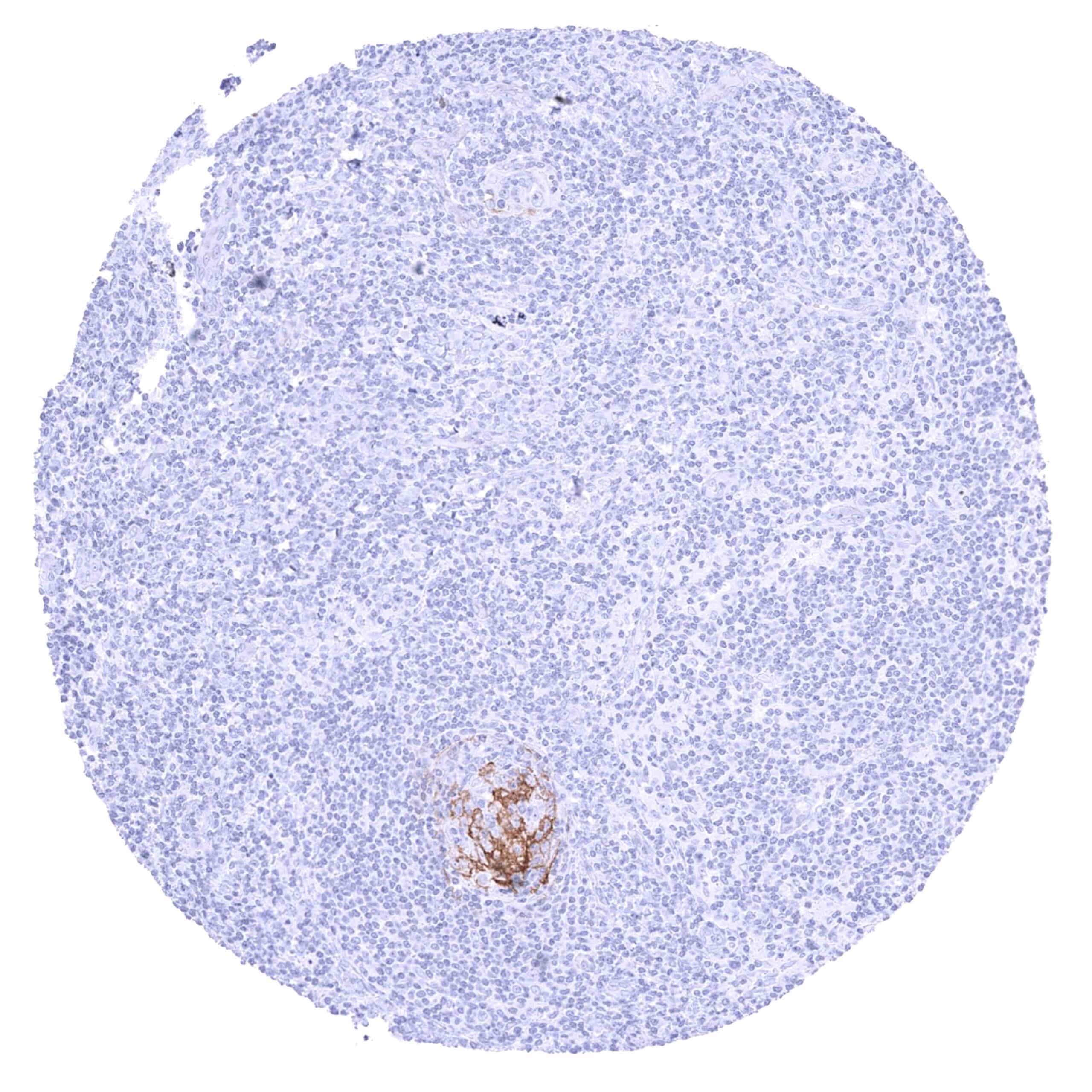
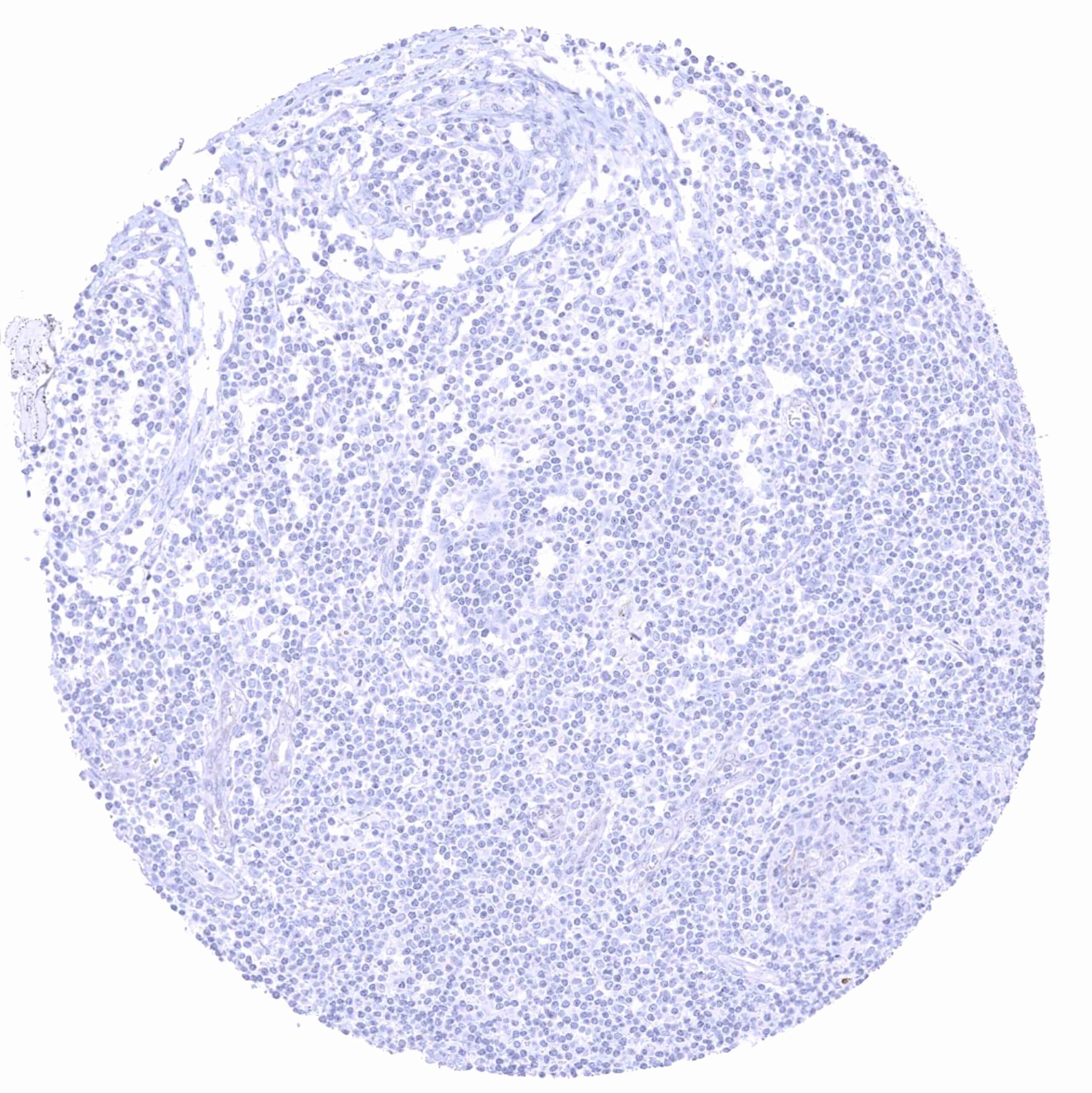
Lymph node – Distinct membranous PSMA staining of grouped germinal centre cells.
Lymph node
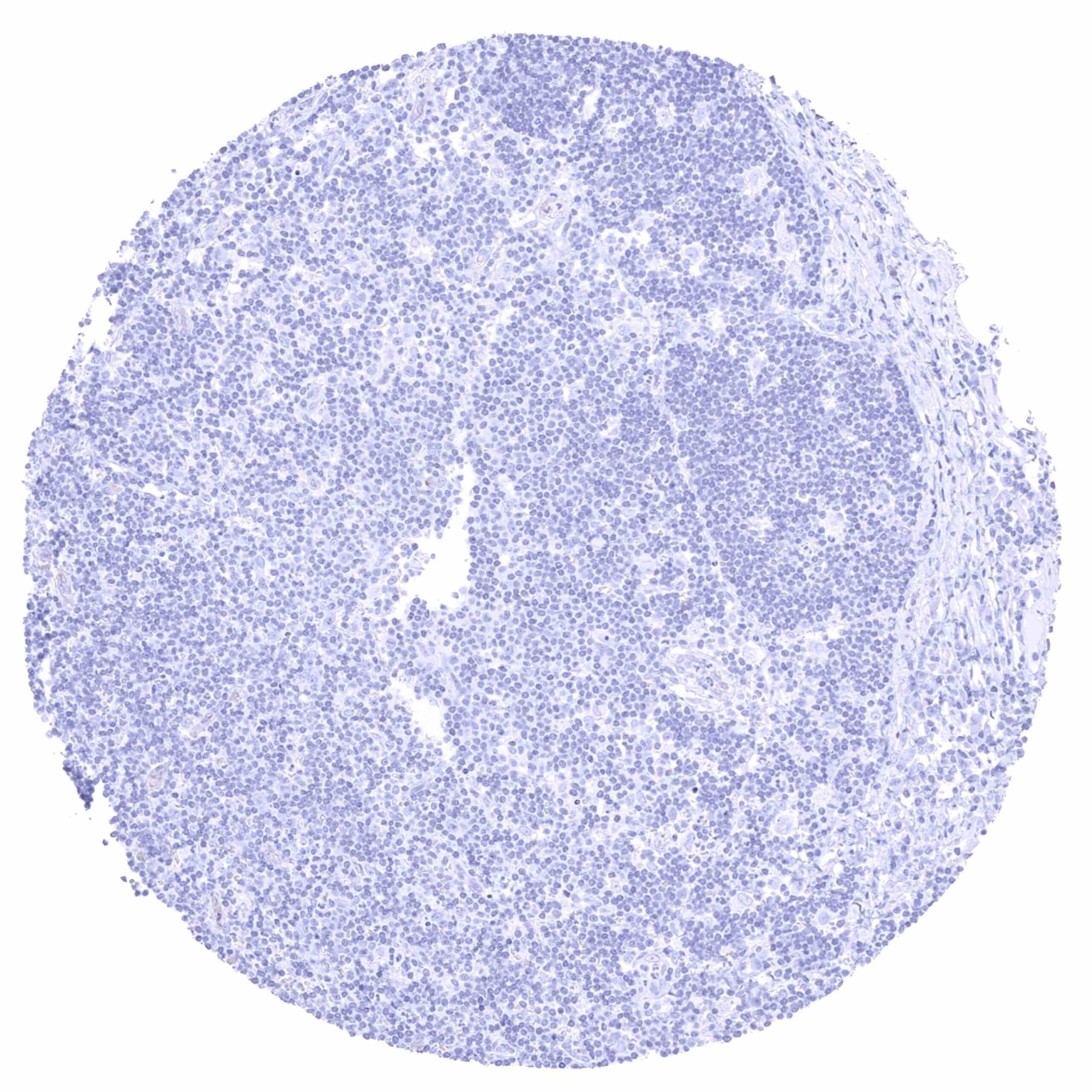
Lymph node (2)
Ovary, corpus luteum
Ovary, stroma
Pancreas (excretory duct)
Pancreas
Parathyroid gland
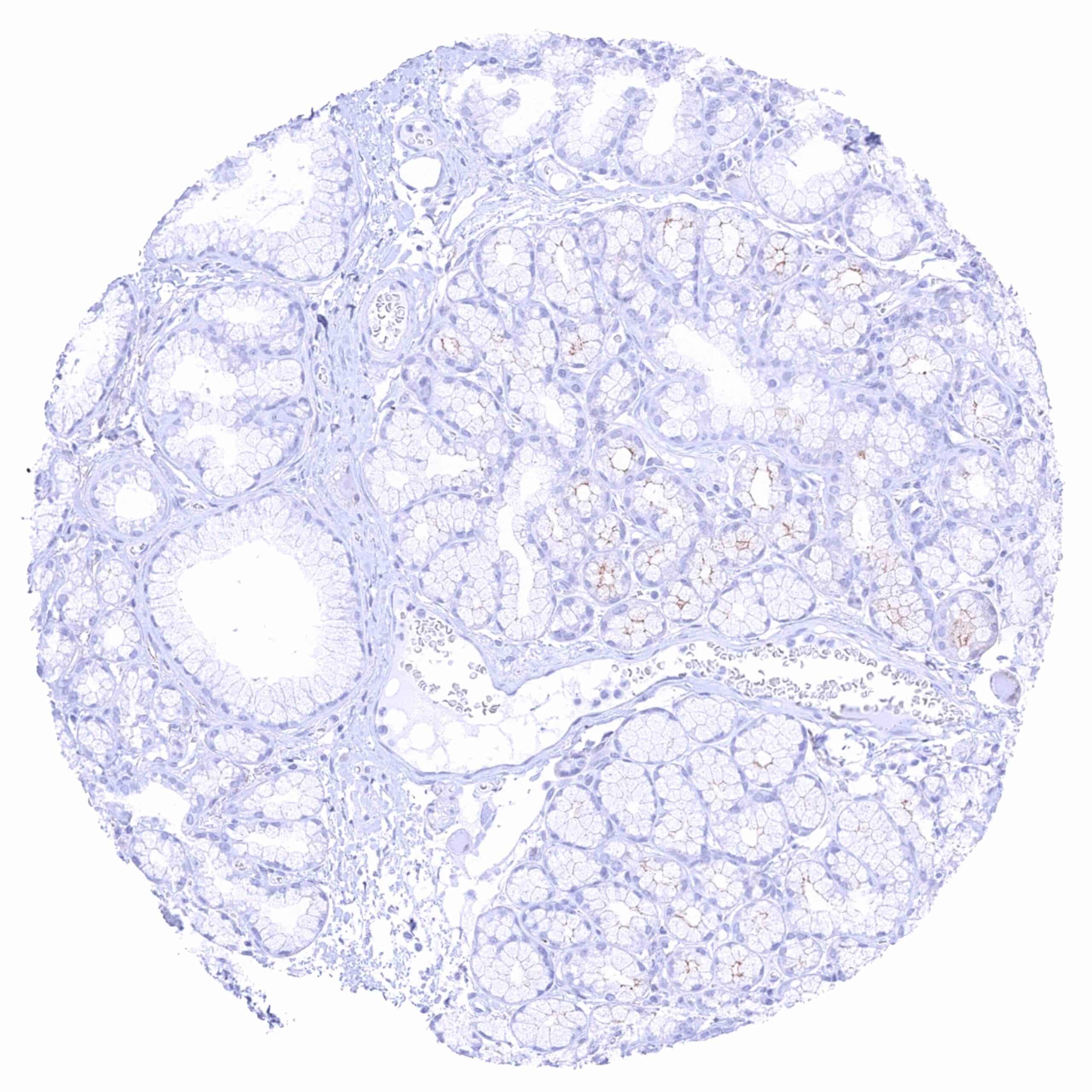
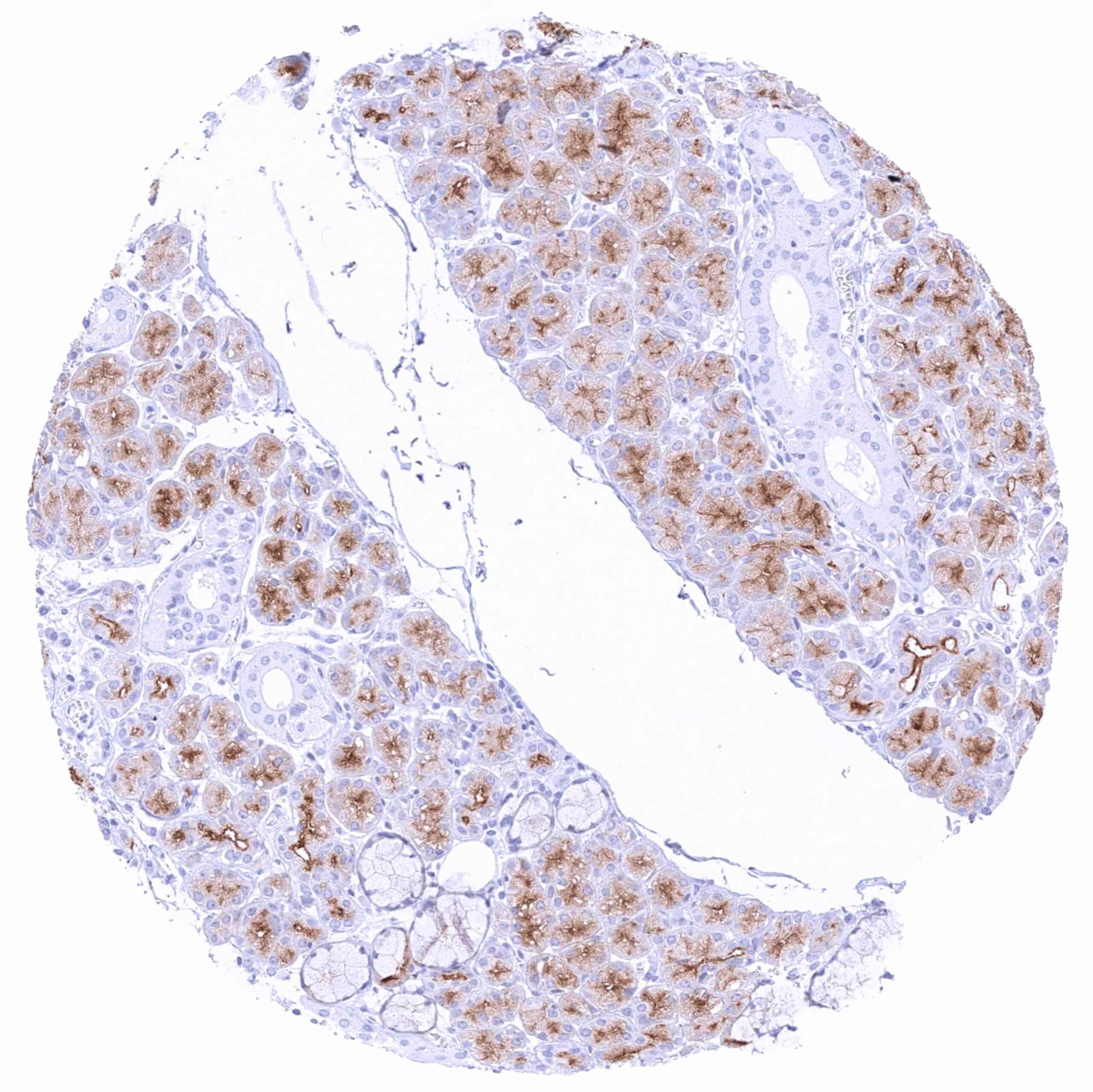
Parotid gland – Moderate to strong apical membranous and weak to moderate diffuse cytoplasmic PSMA staining of serous glands.
Pituitary gland, posterior lobe
Pituitary, anterior lobe
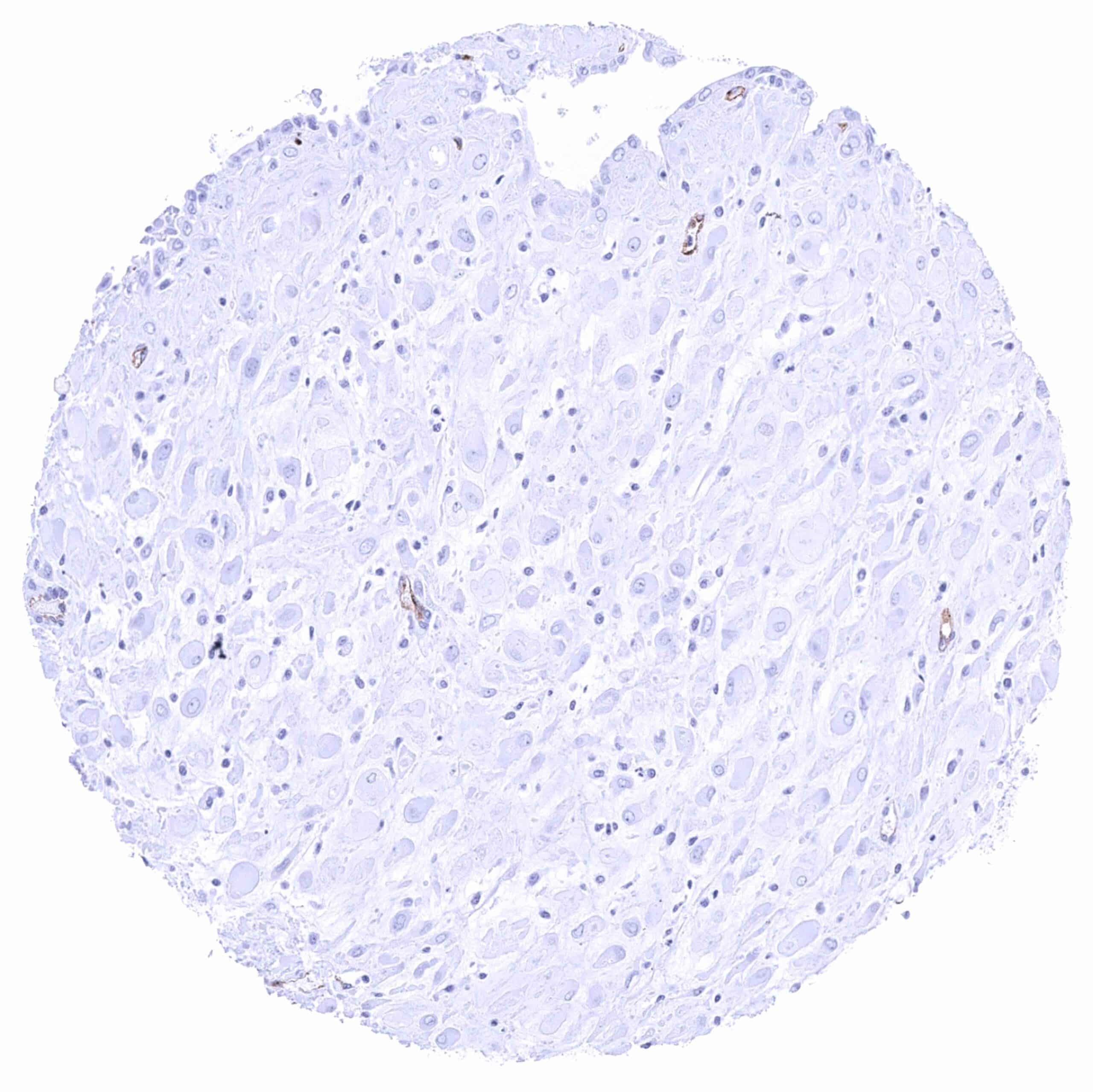
Placenta (chorion) – Endothelial PSMA staining in a small vessel.
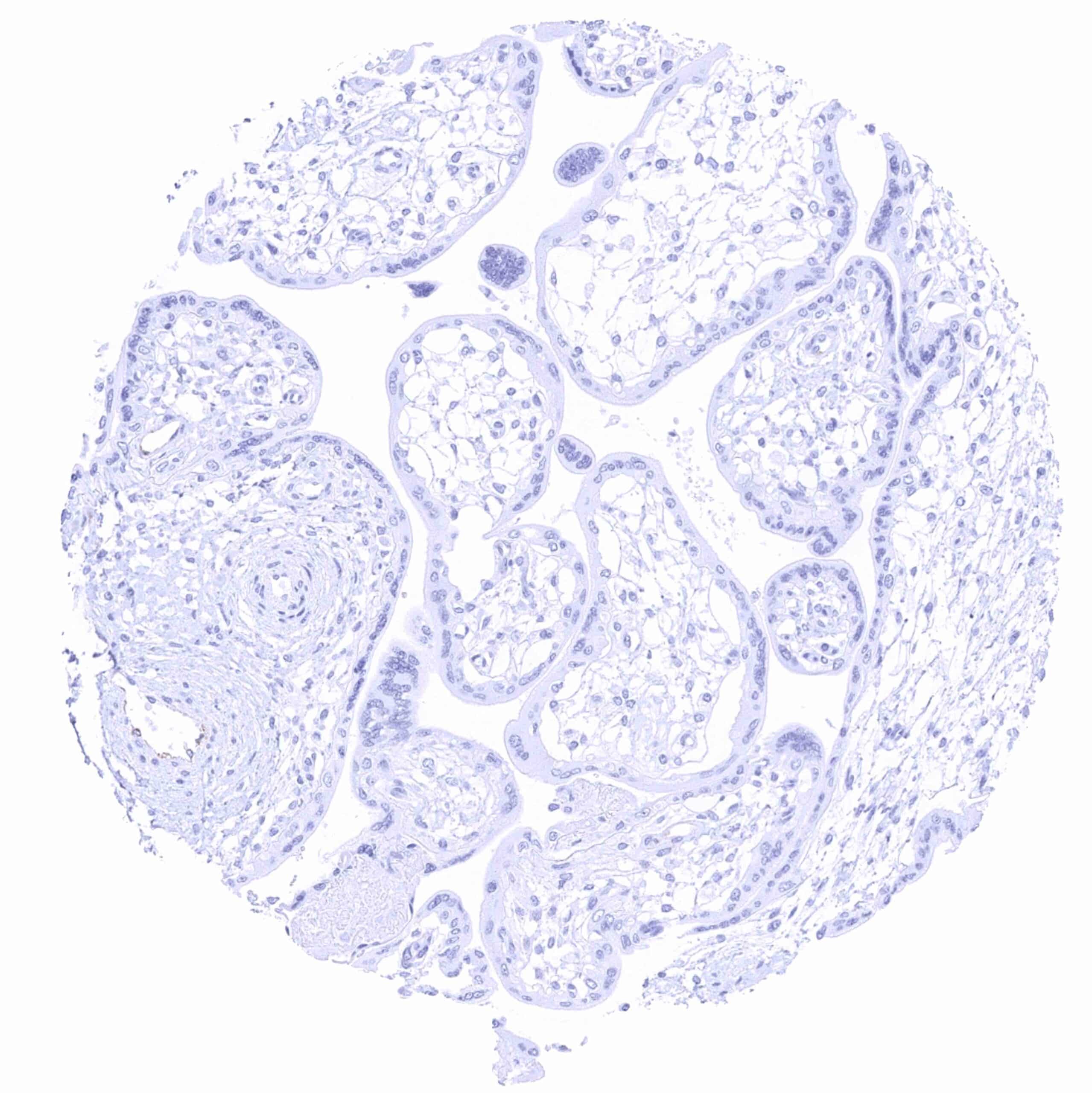
Placenta, early
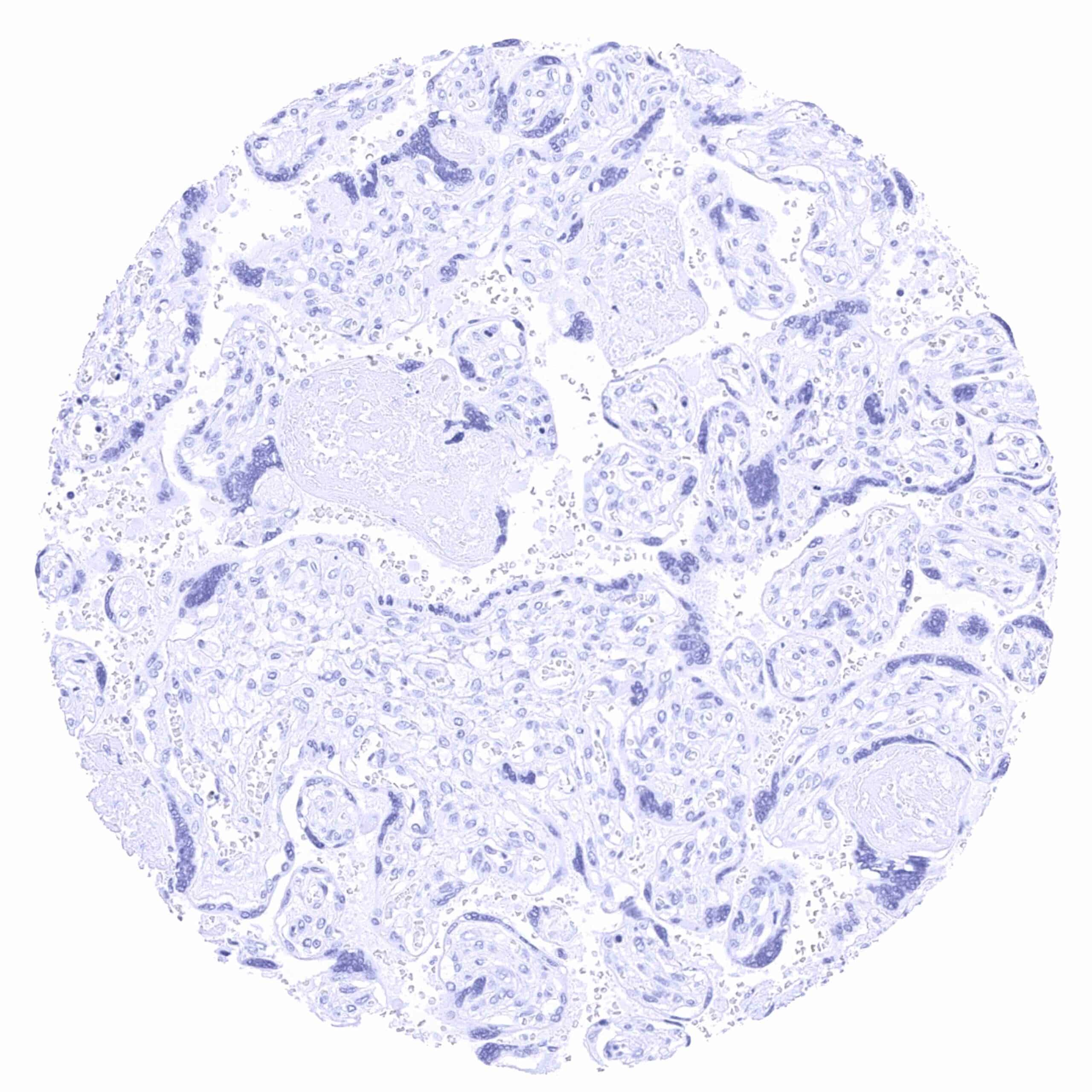
Placenta, mature
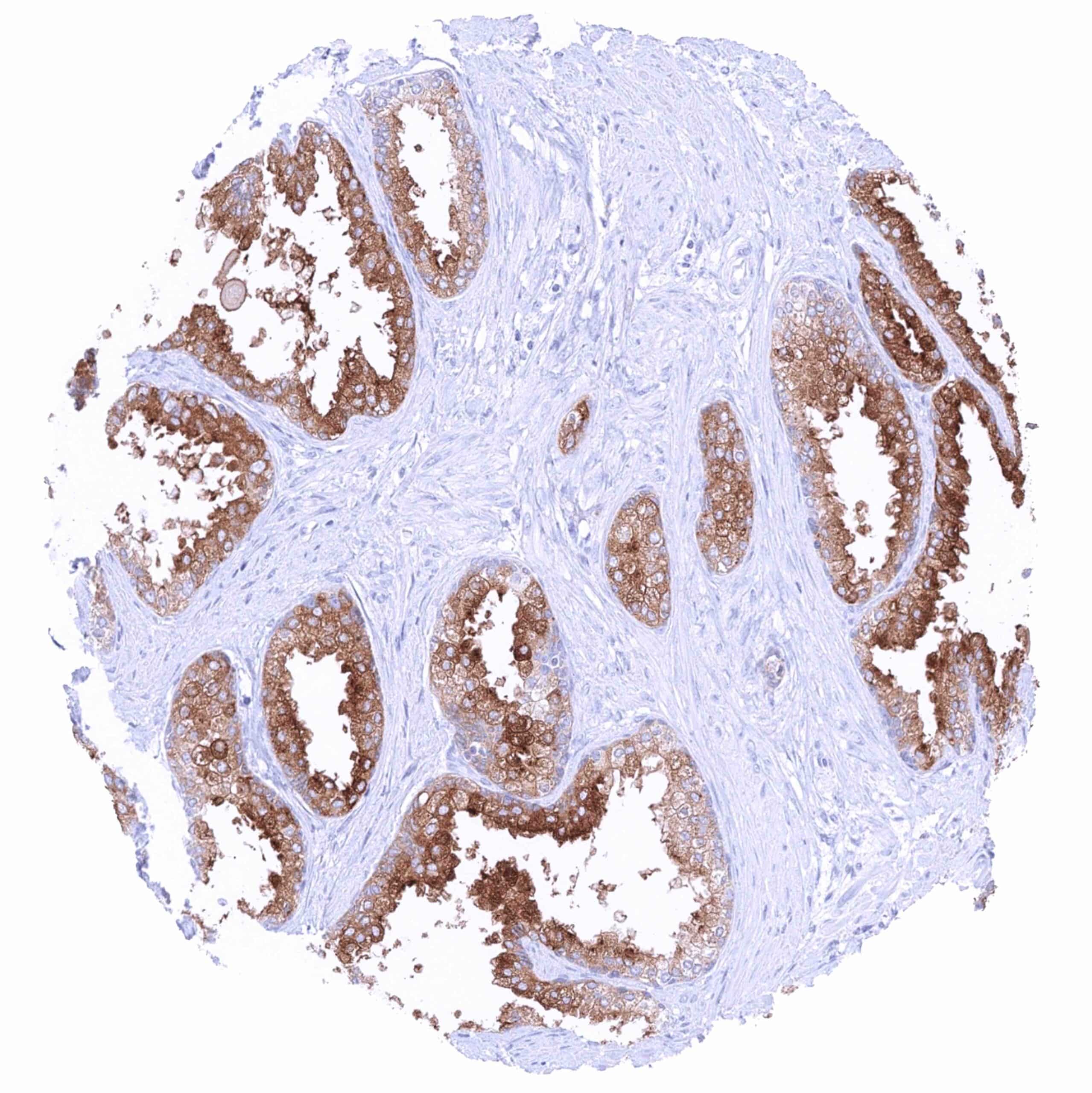
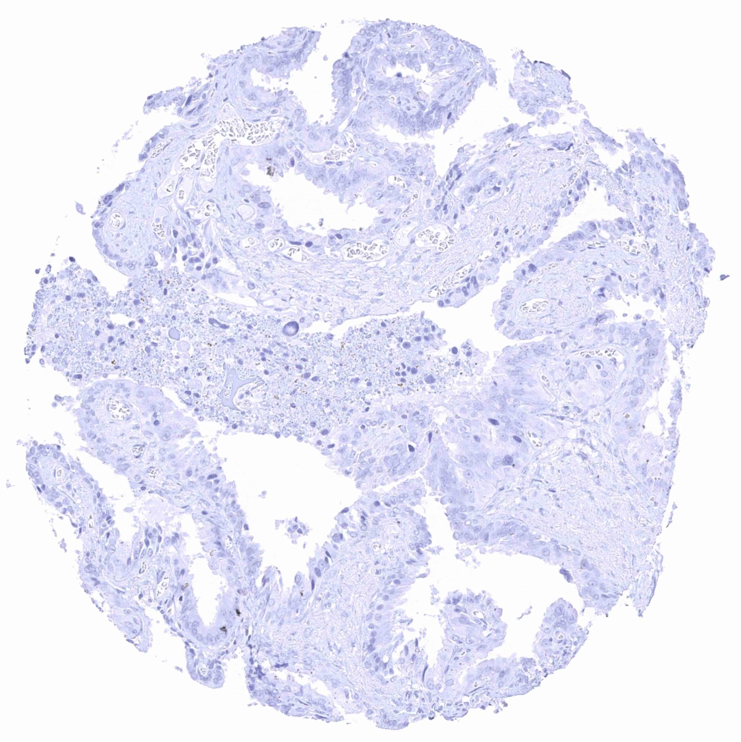
Prostate – Intense cytoplasmatic PSMA staining of all luminal epithelial cells.
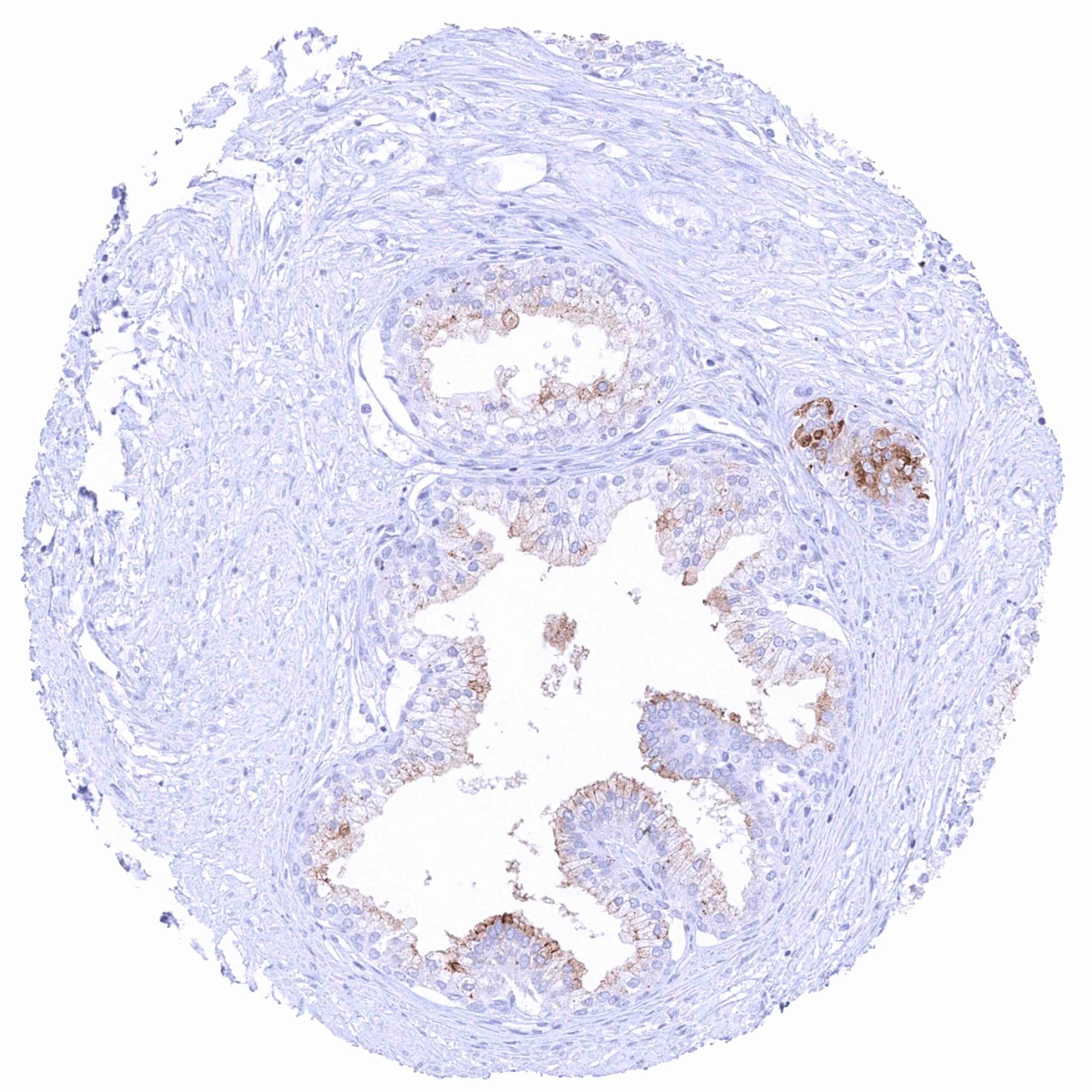
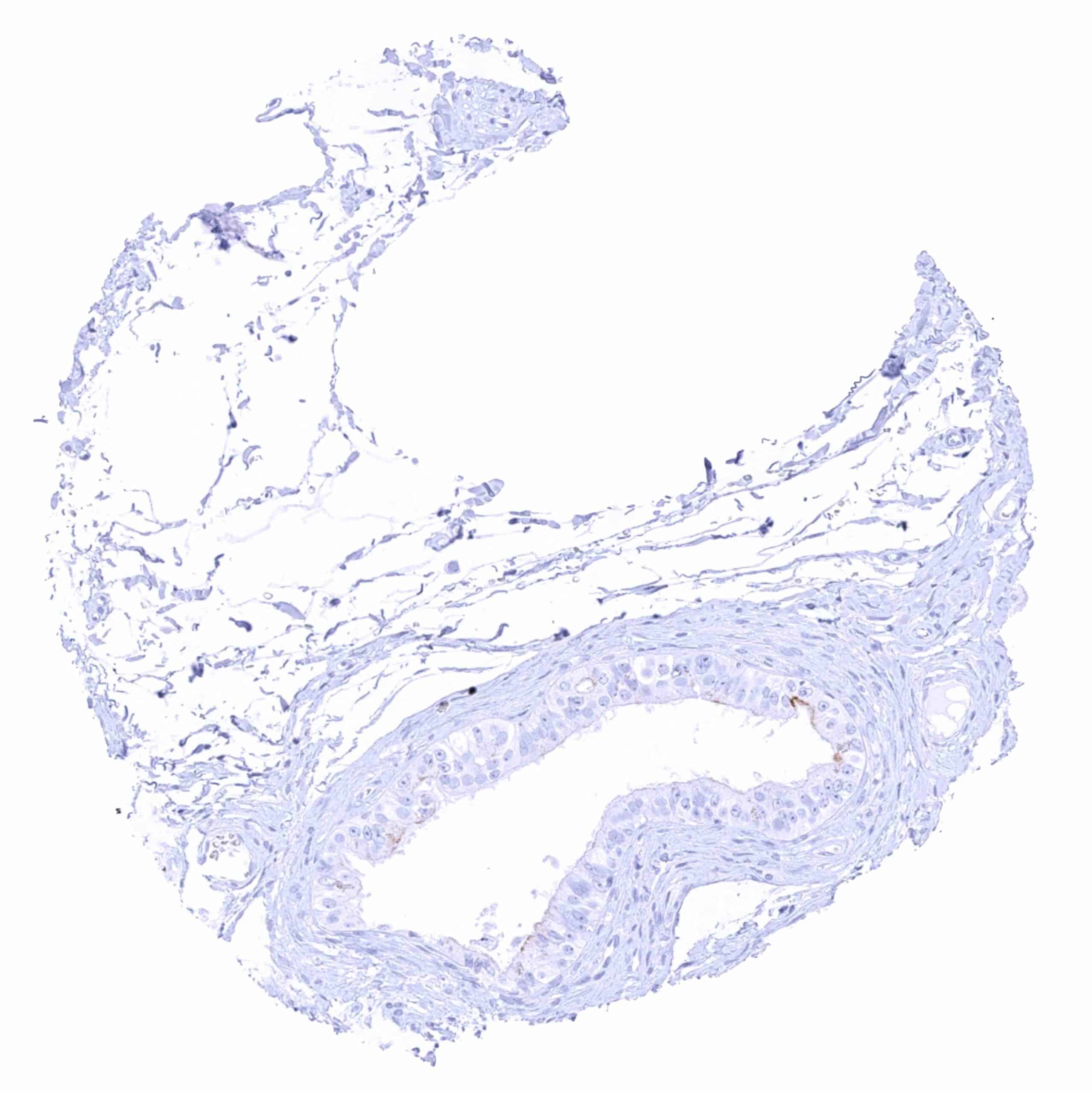
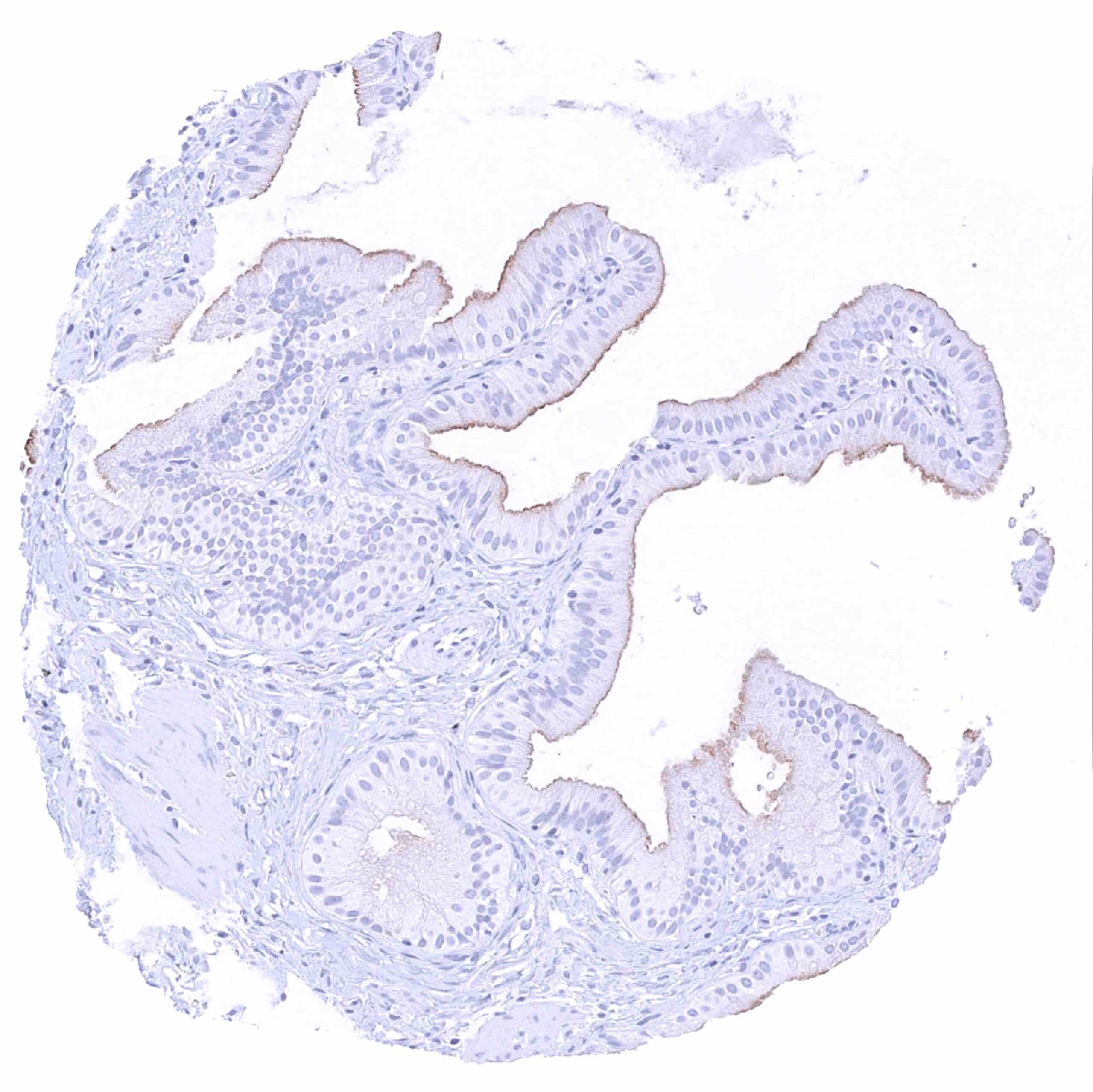
Prostate – Weak membranous (apical) and cytoplasmatic PSMA staining of most luminal cells in this sample.
Rectum, mucosa
Seminal vesicle
Sinus paranasales
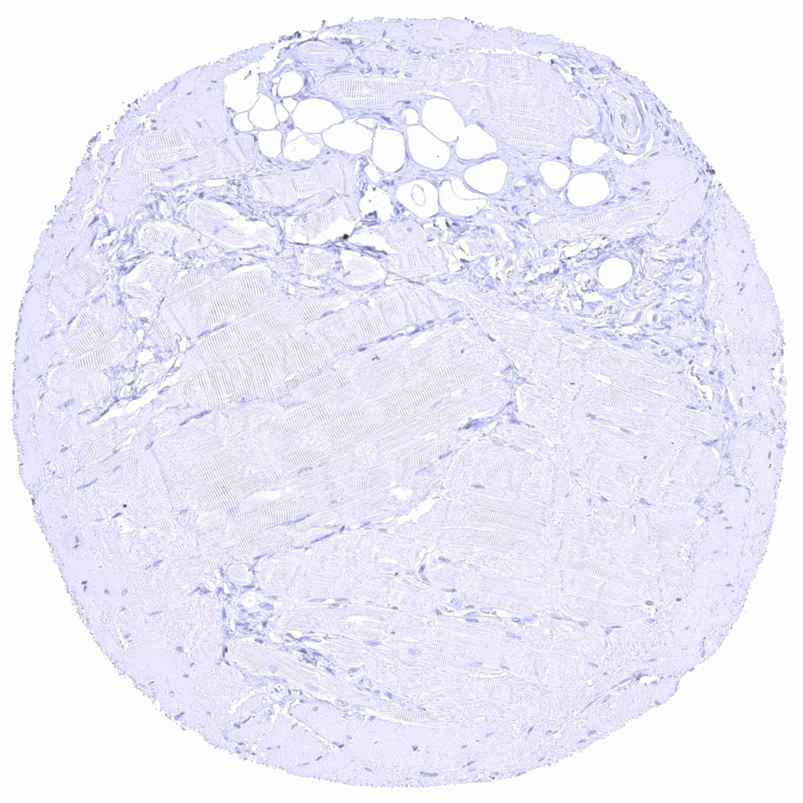
Skeletal muscle
Skin, hairfollicel and sebaceous glands
Skin
Spleen – Distinct PSMA staining of venous sinuses (littoral cells)
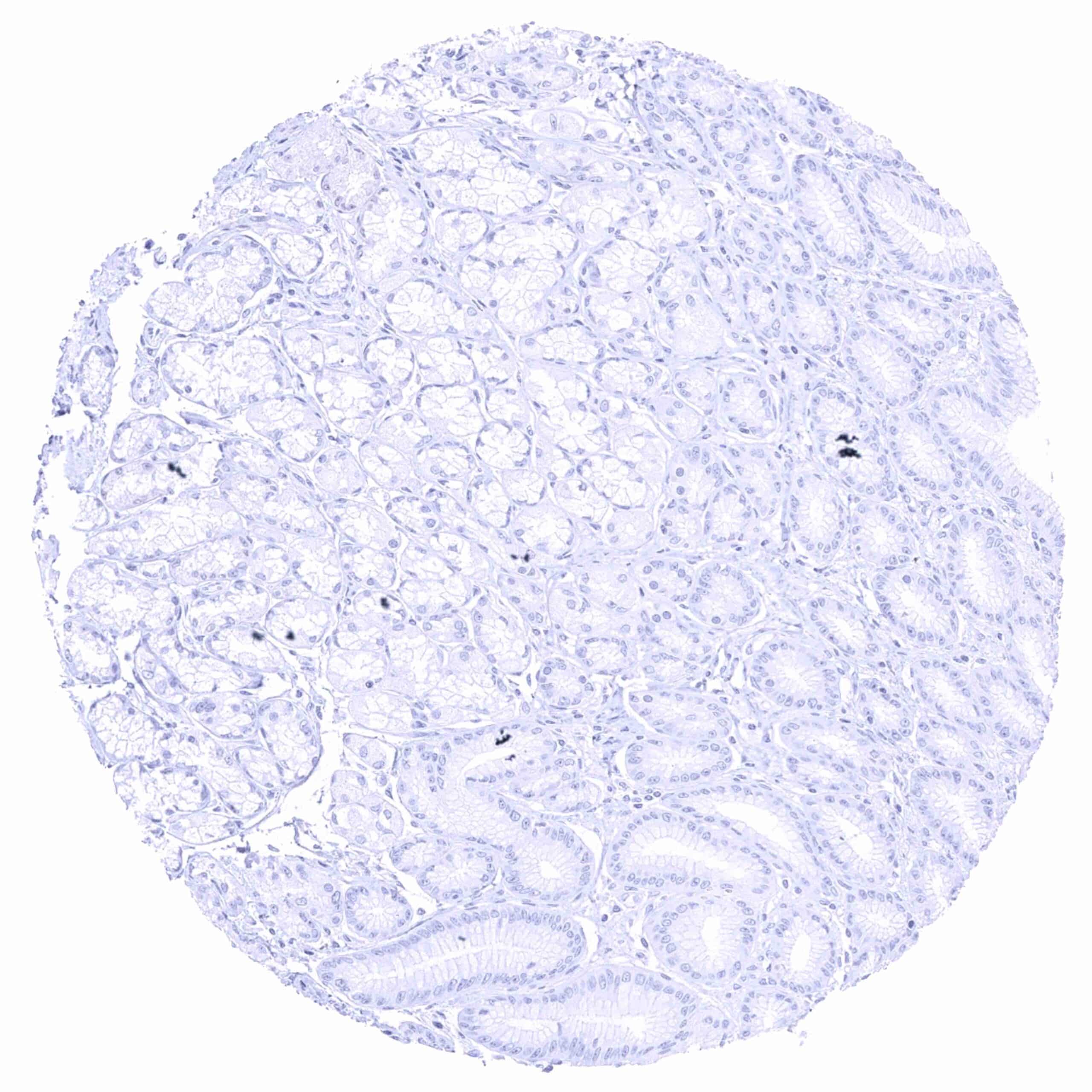
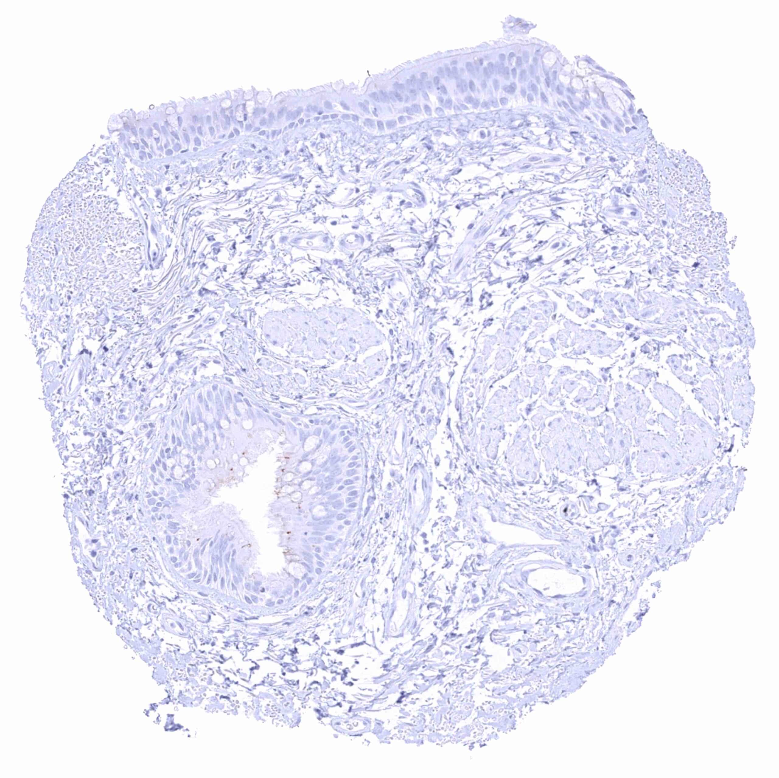
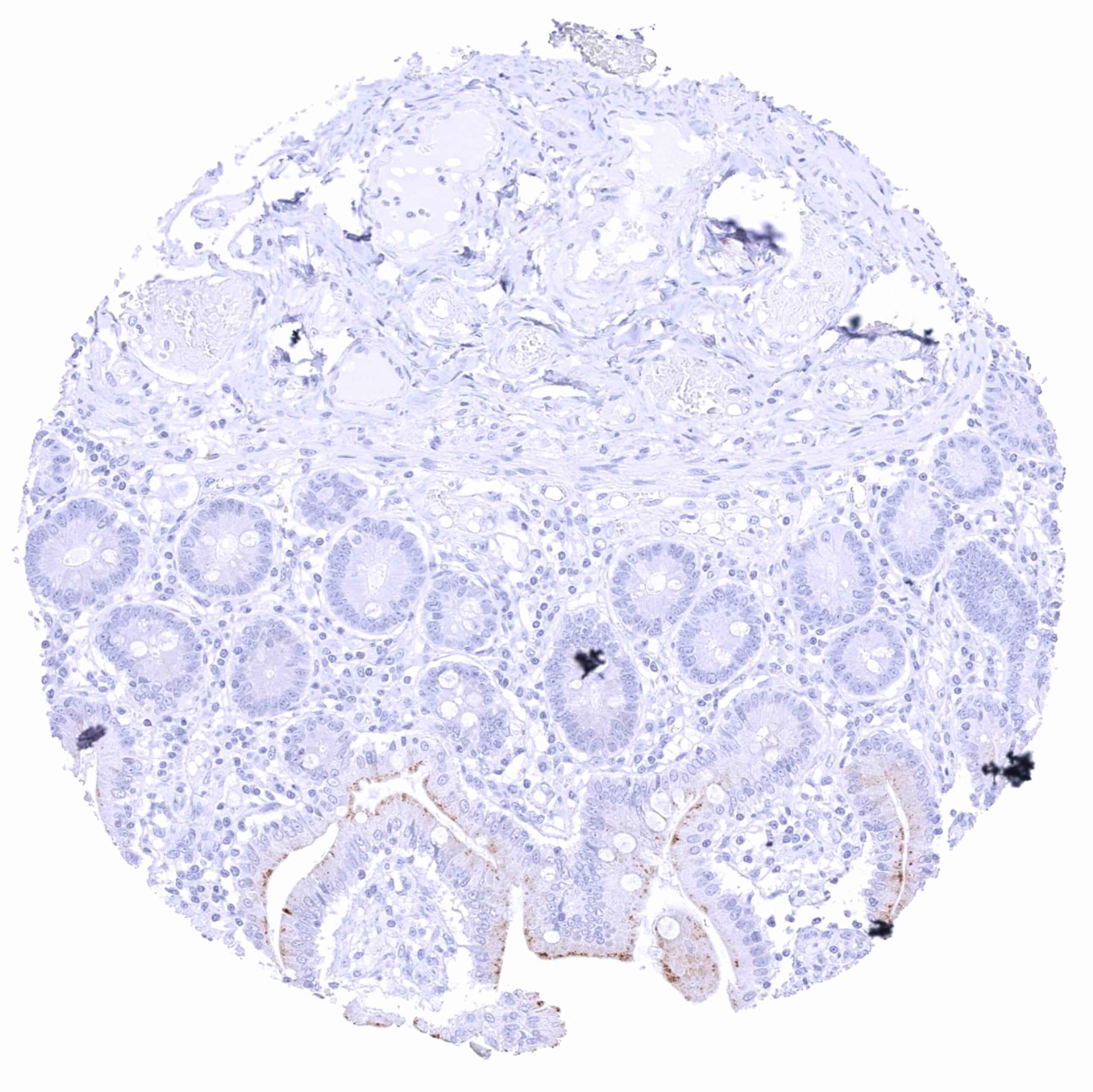
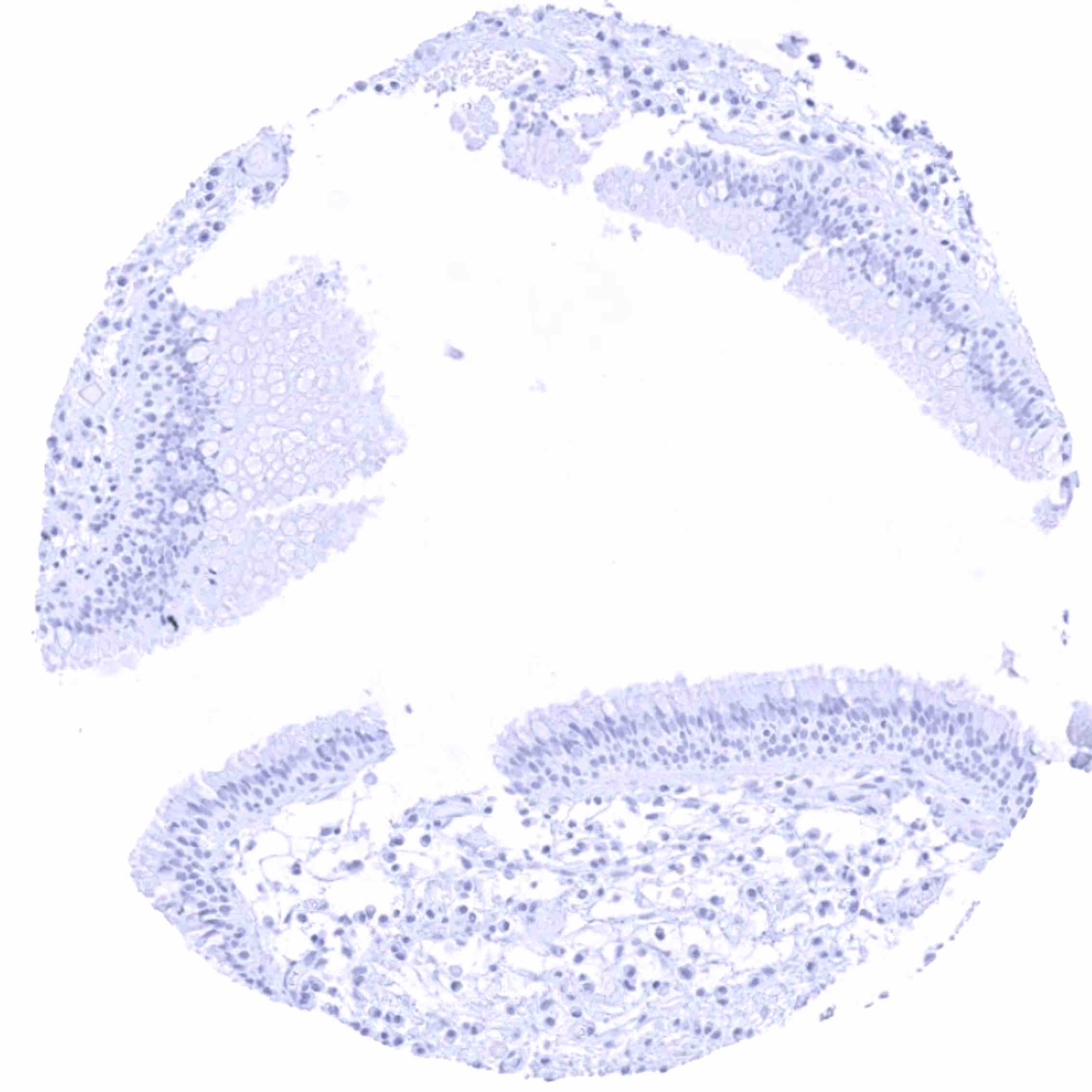
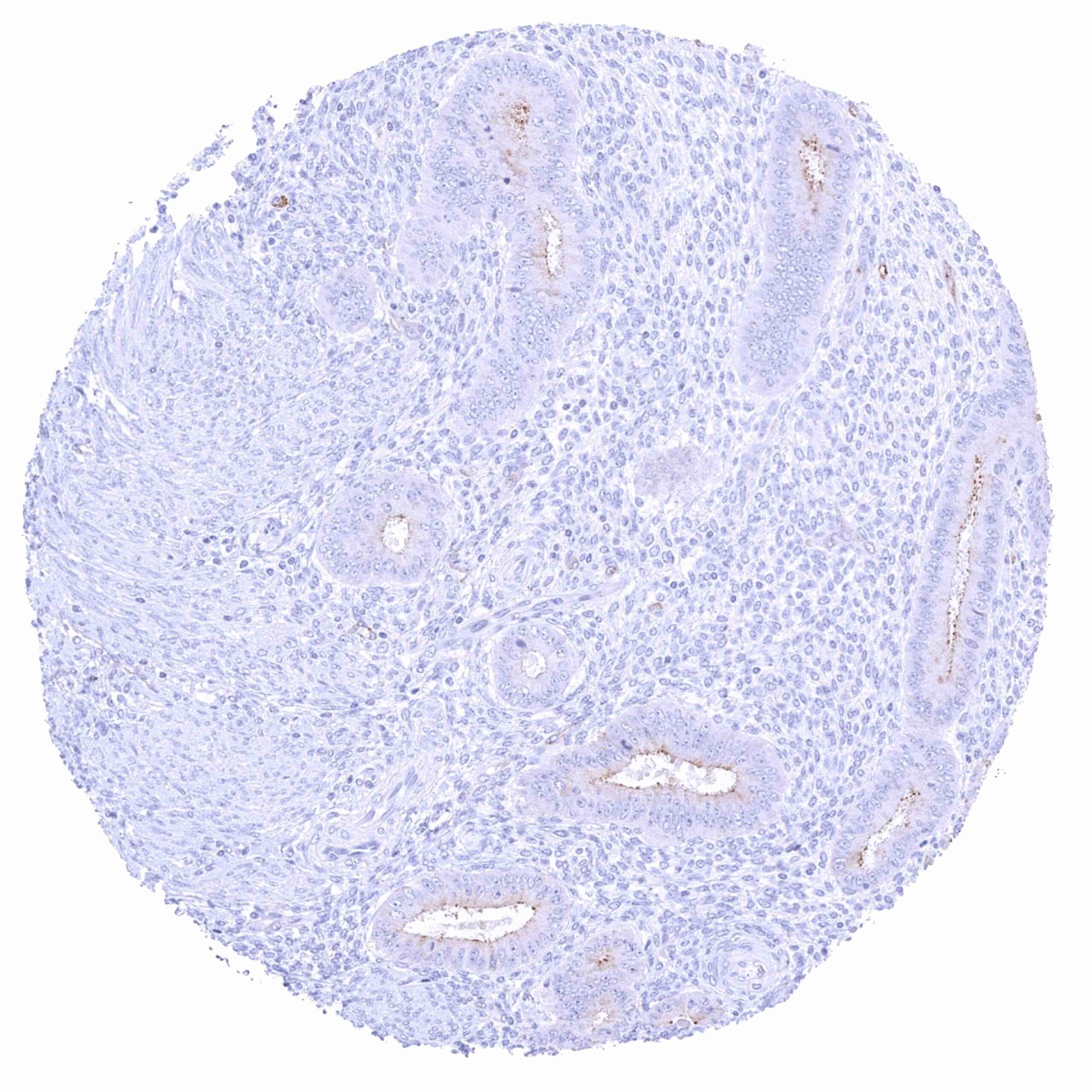
Stomach, antrum
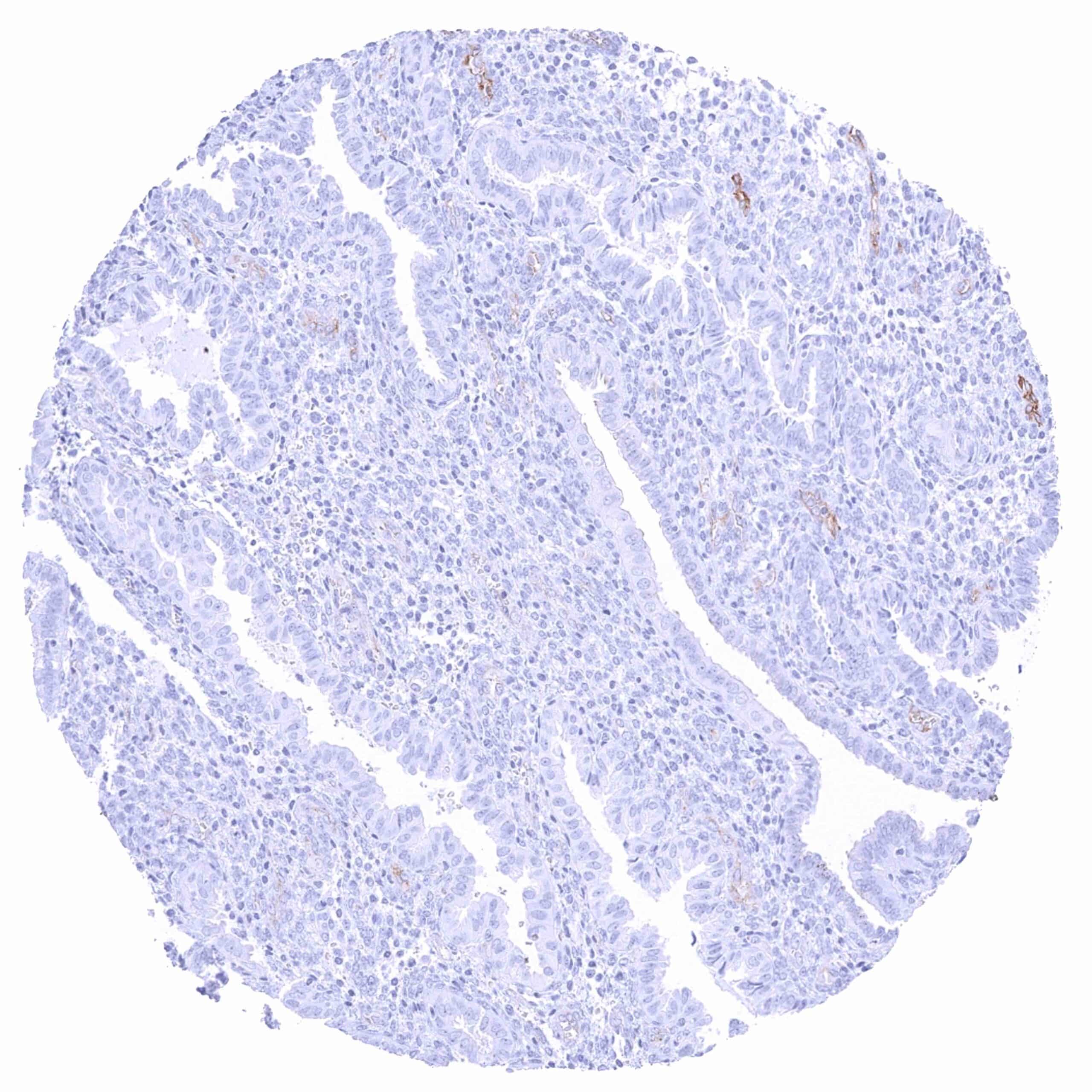
Stomach, corpus
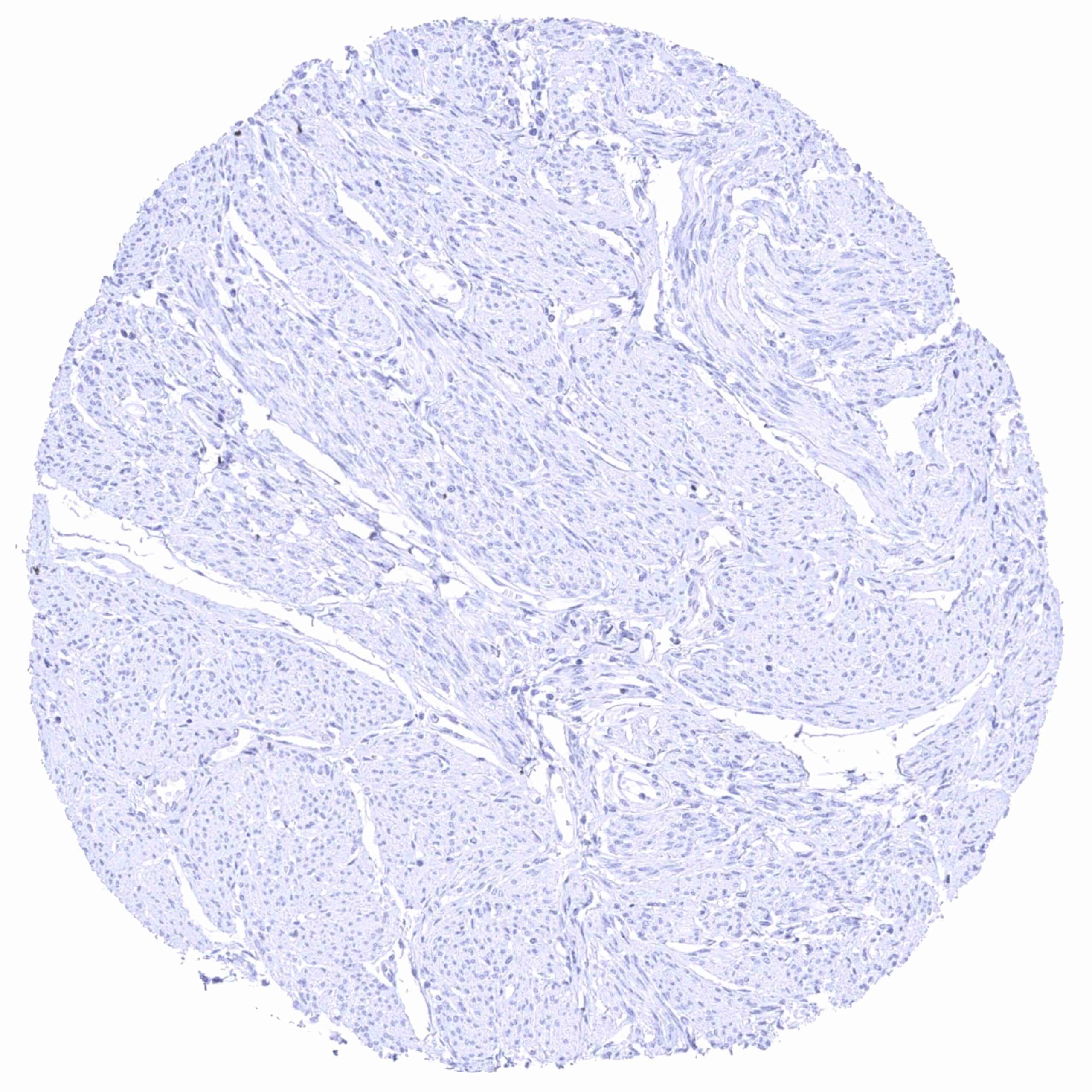
Stomach, muscular wall
Sublingual gland – Moderate apical membranous and weak cytoplasmic PSMA staining of glands.
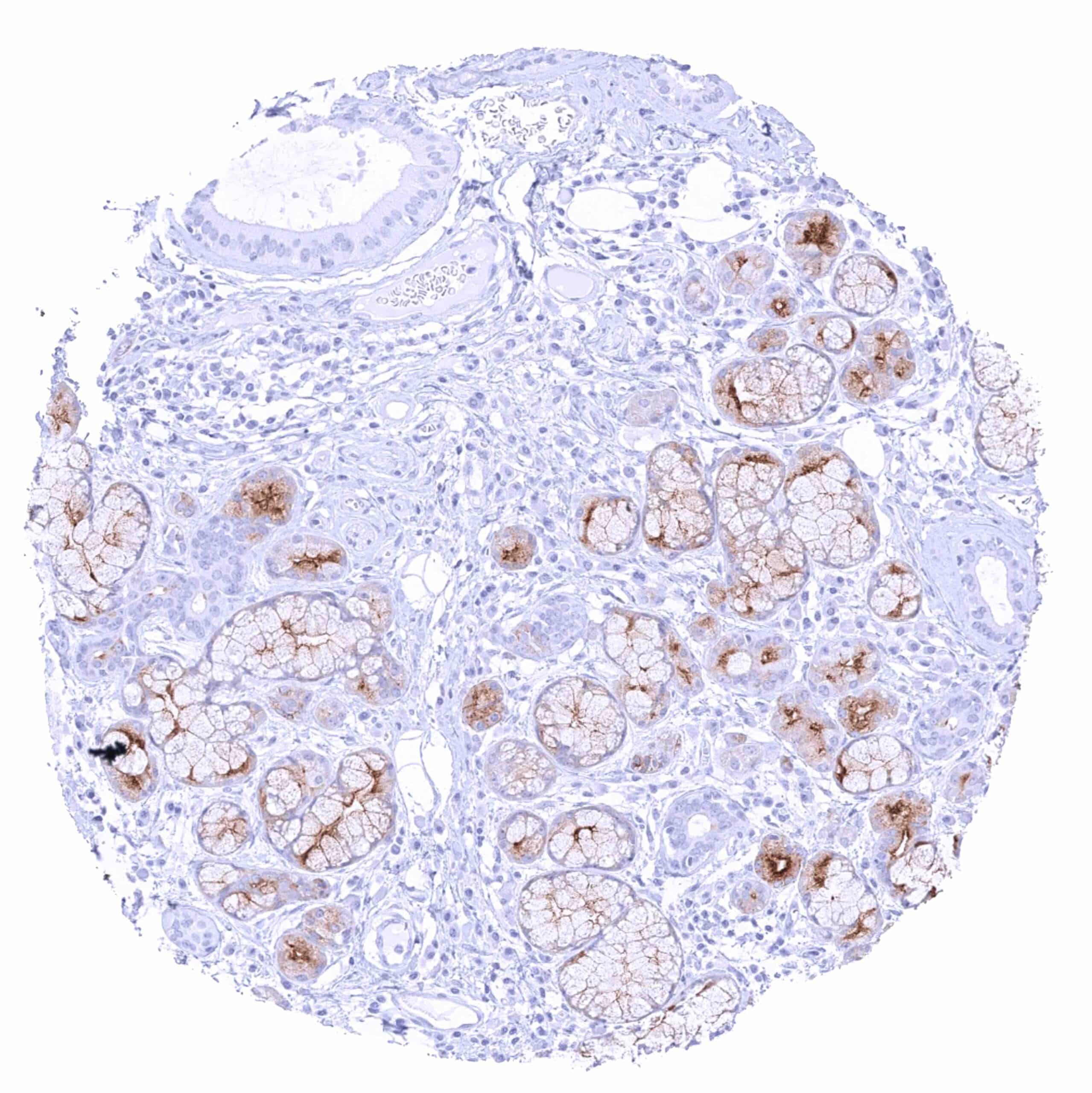
Submandibular gland – Strong apical membranous and weak to moderate cytoplasmic PSMA staining of glandular cells (more intense in serous than in mucinous glands).
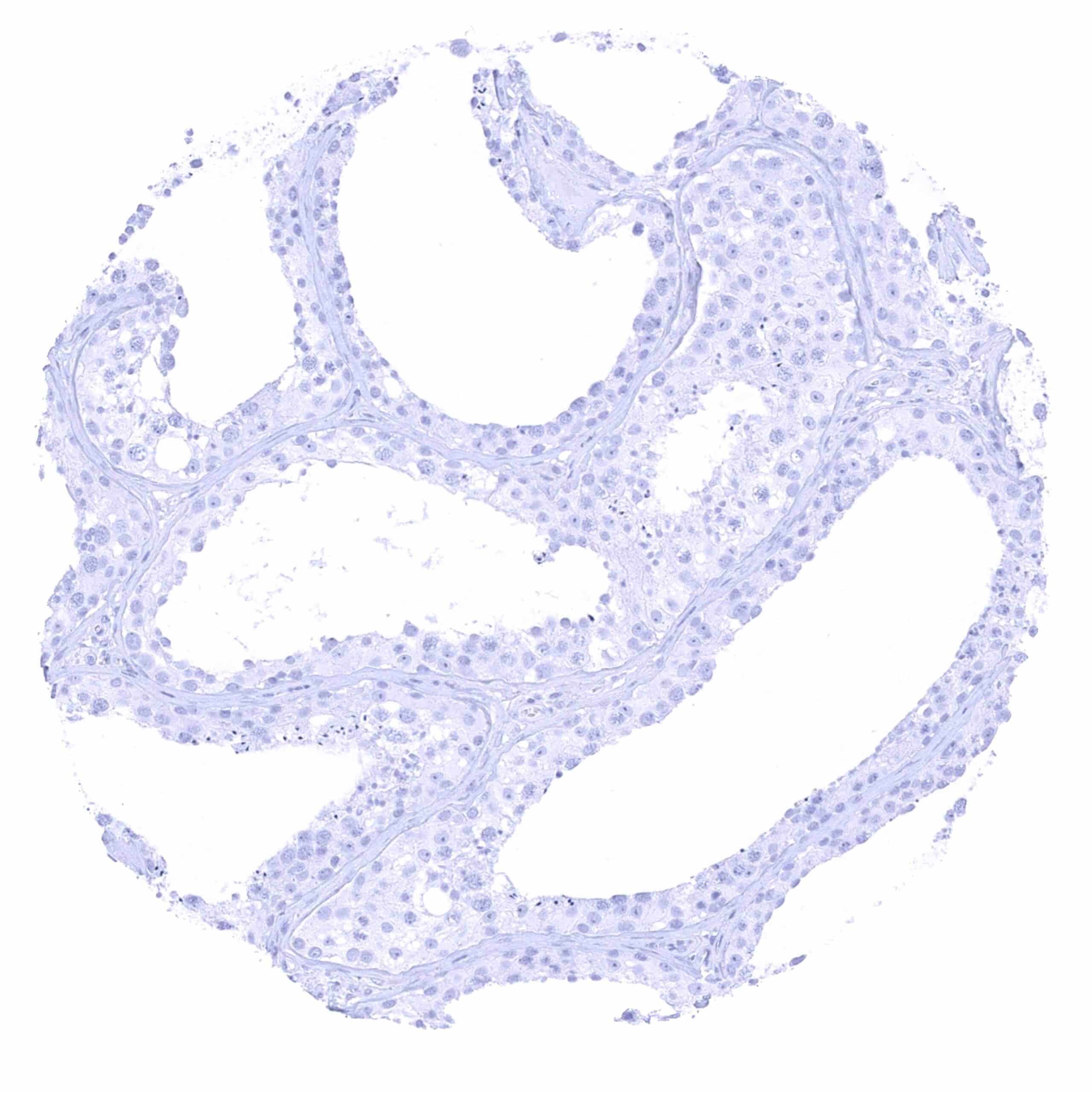
Testis
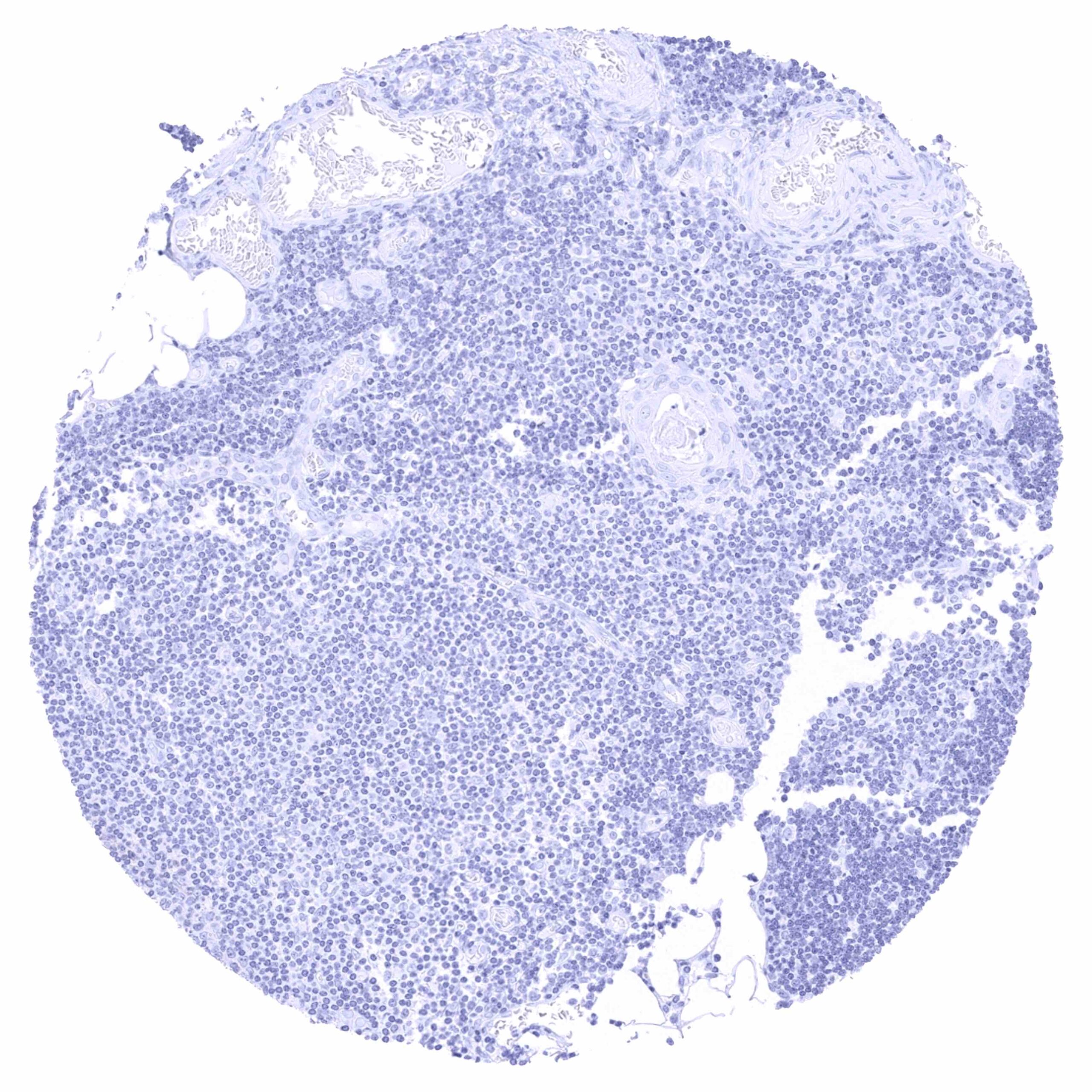
Thymus
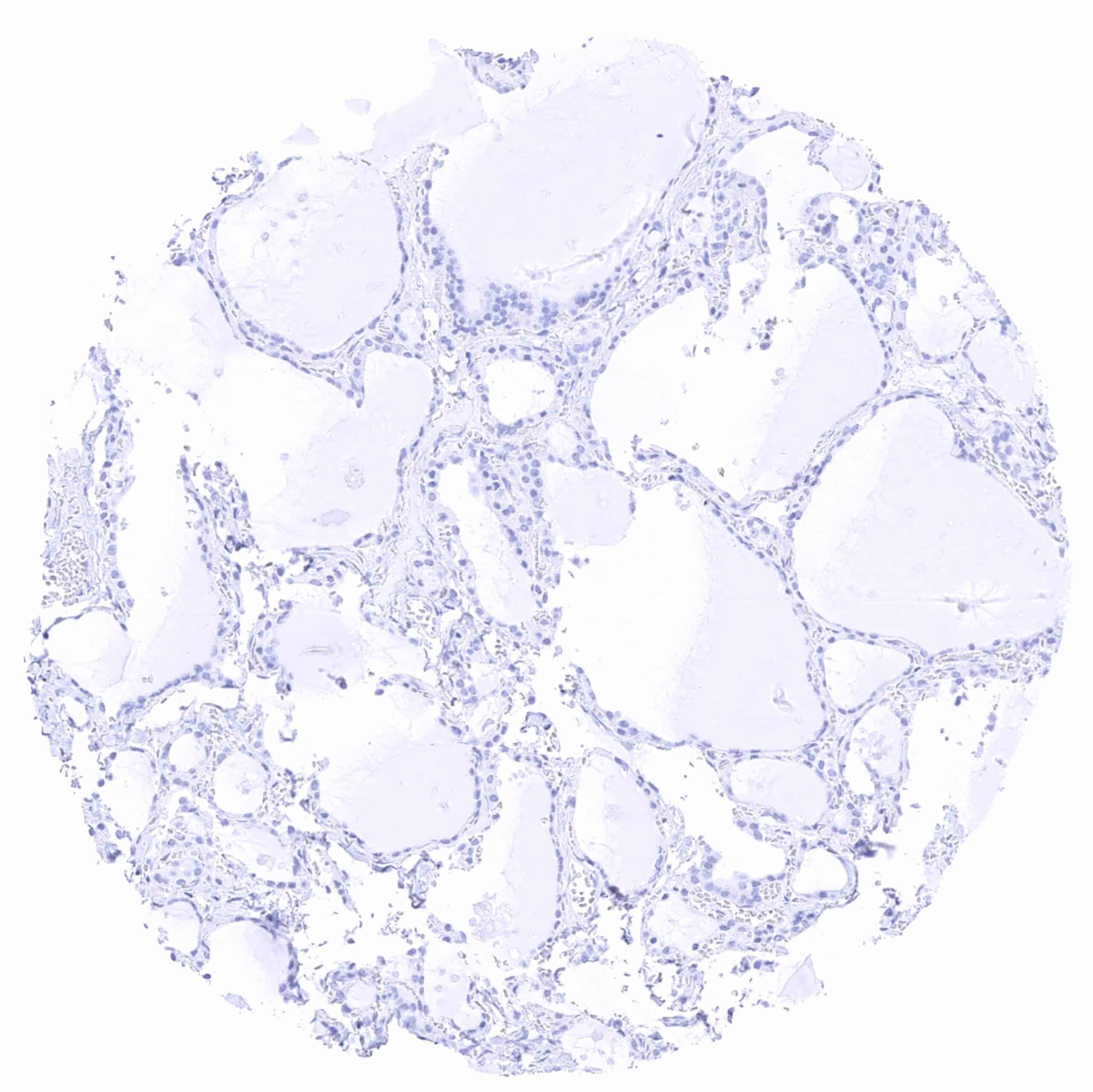
Thyroid gland
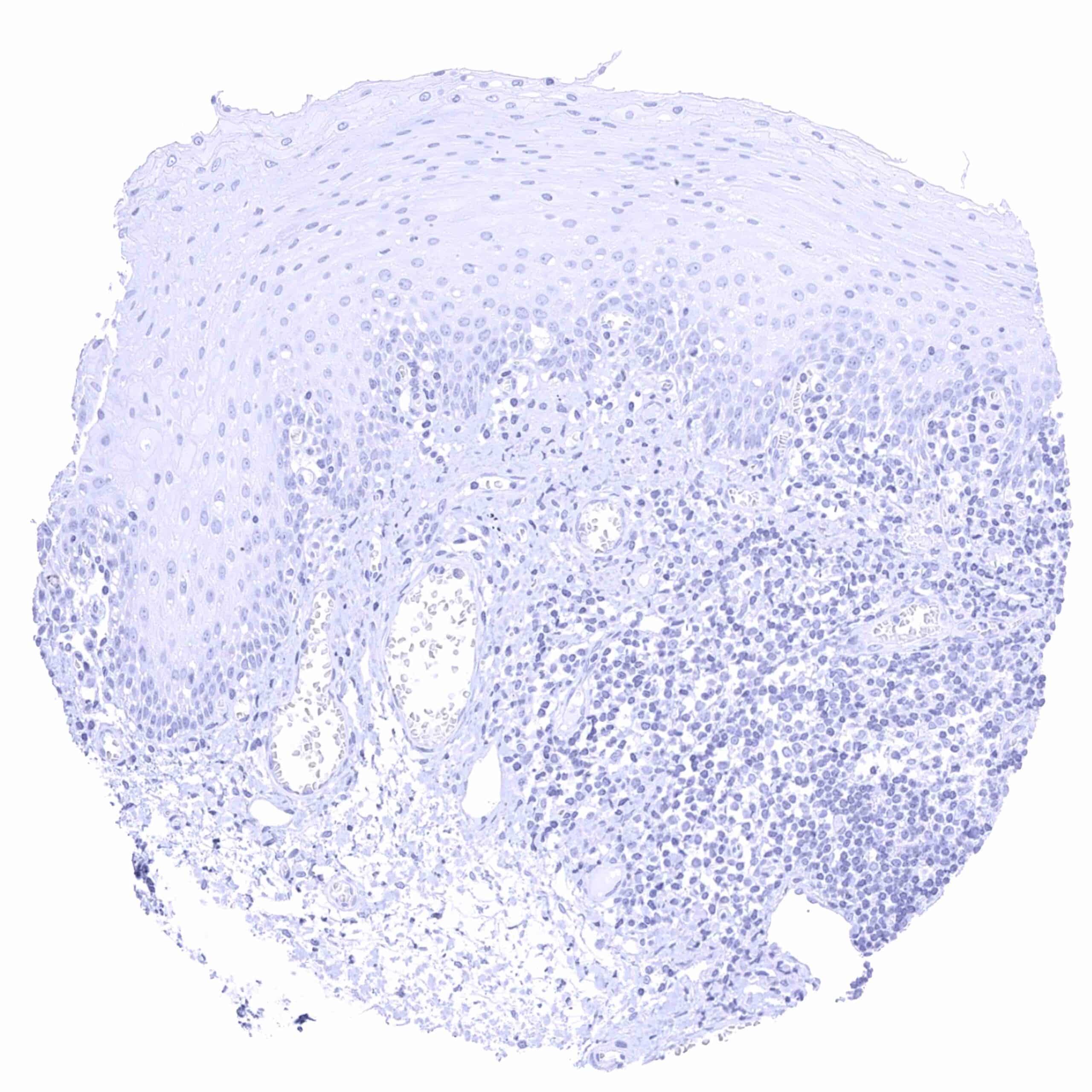
Tonsil, surface epithelium
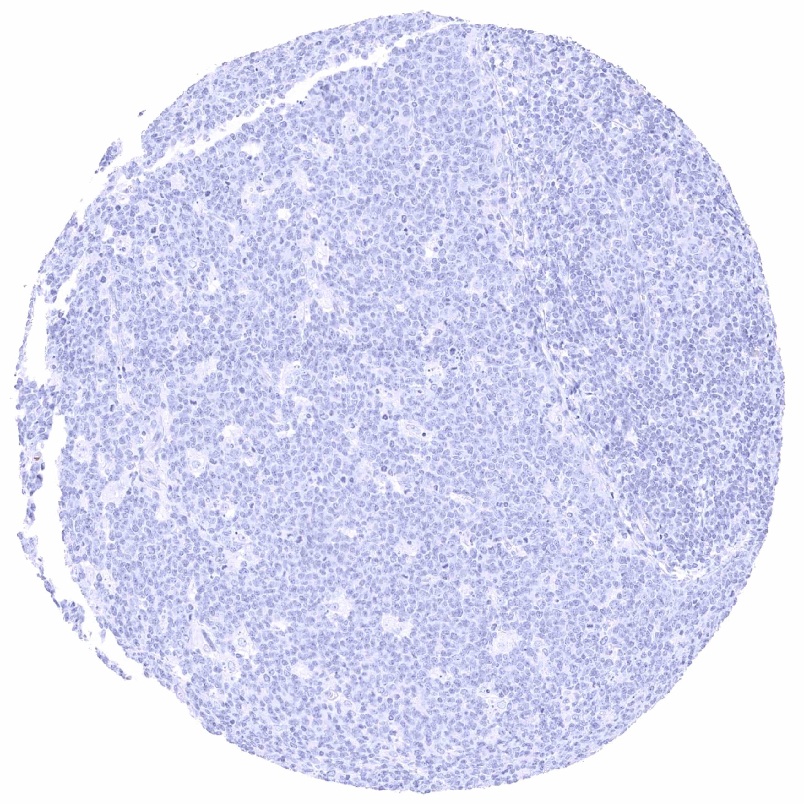
Tonsil
Urinary bladder, muscular wall
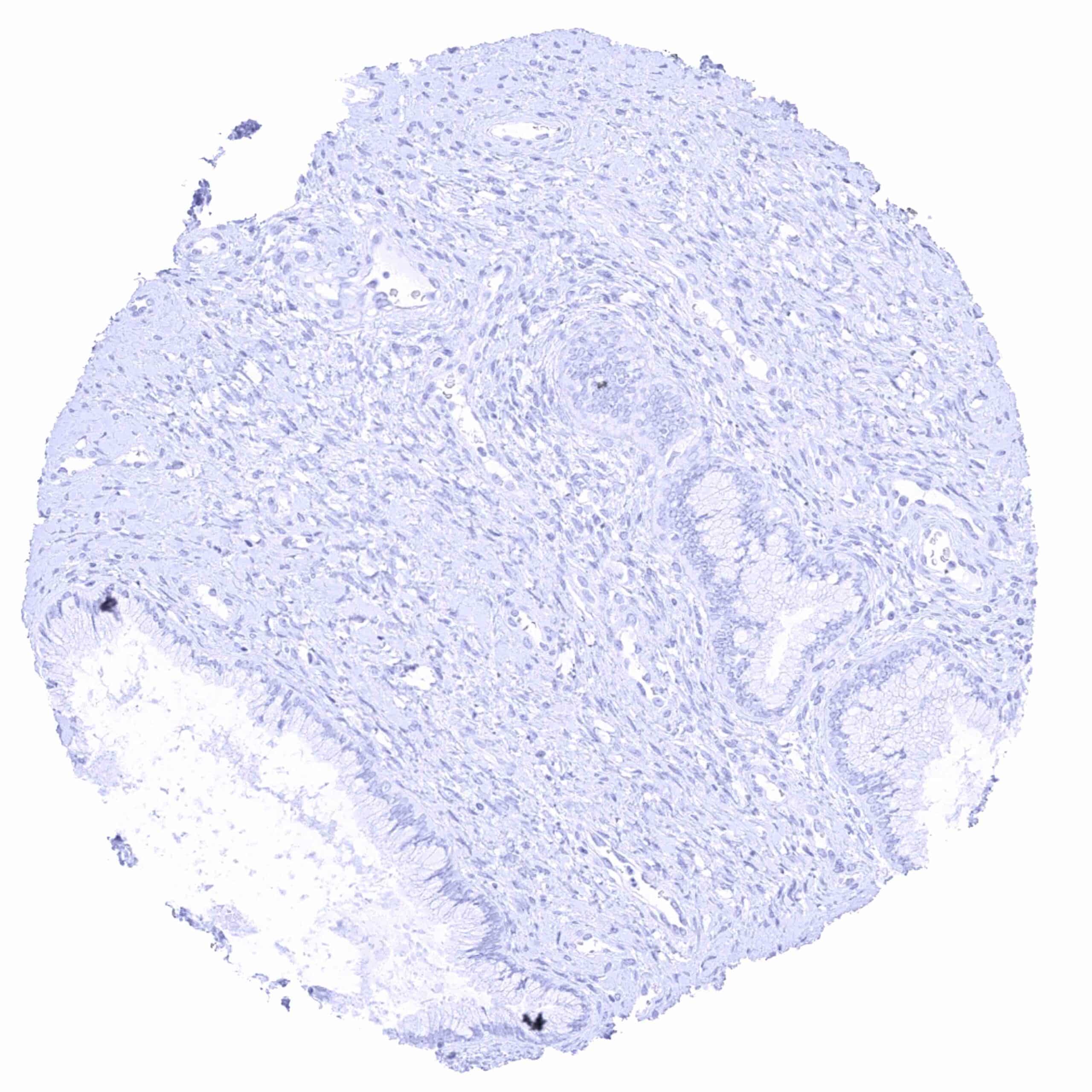
Uterus, ectocervix
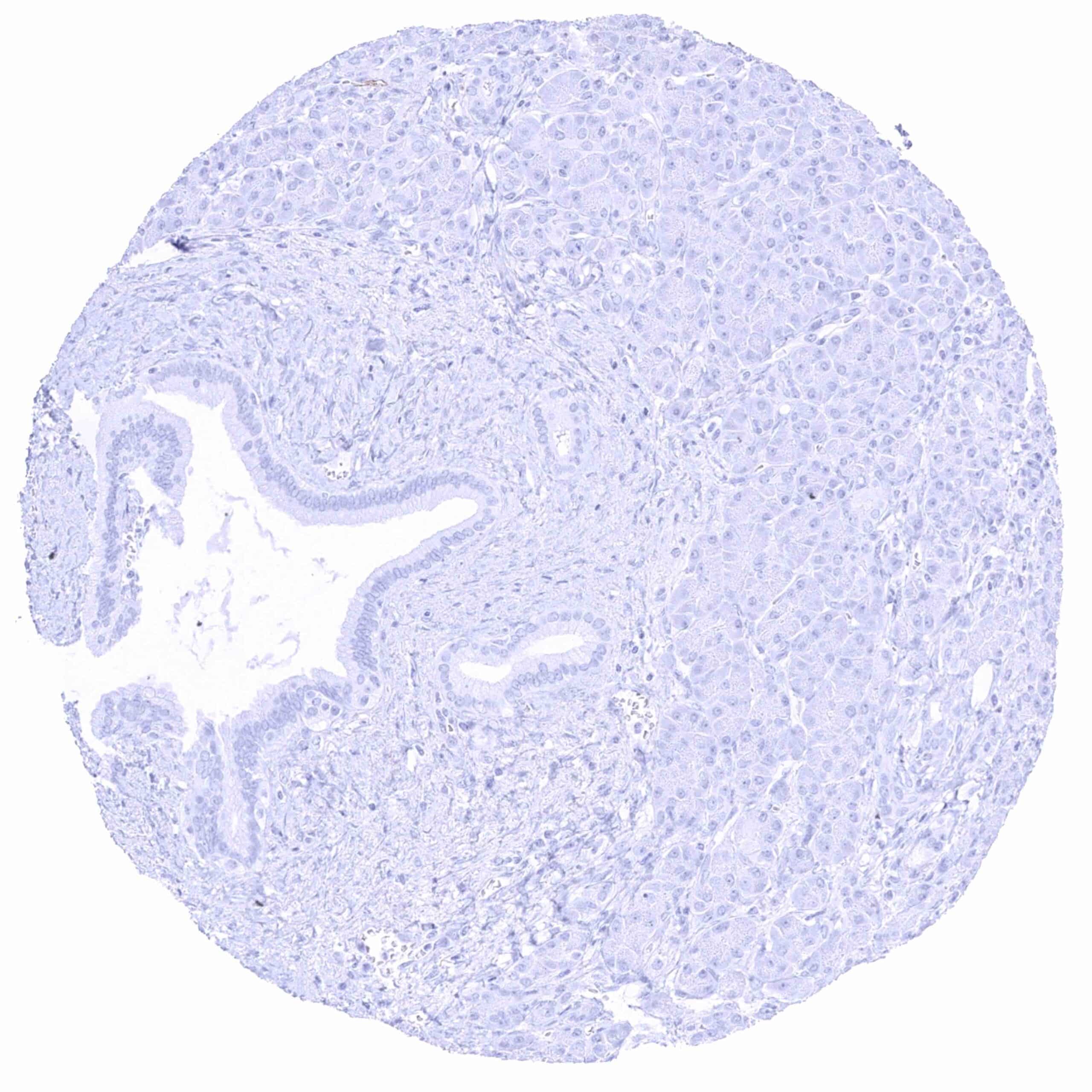
Uterus, endocervix
Uterus, endometrium (pregnancy)
Uterus, endometrium (proliferation)
Uterus, endometrium (secretion)
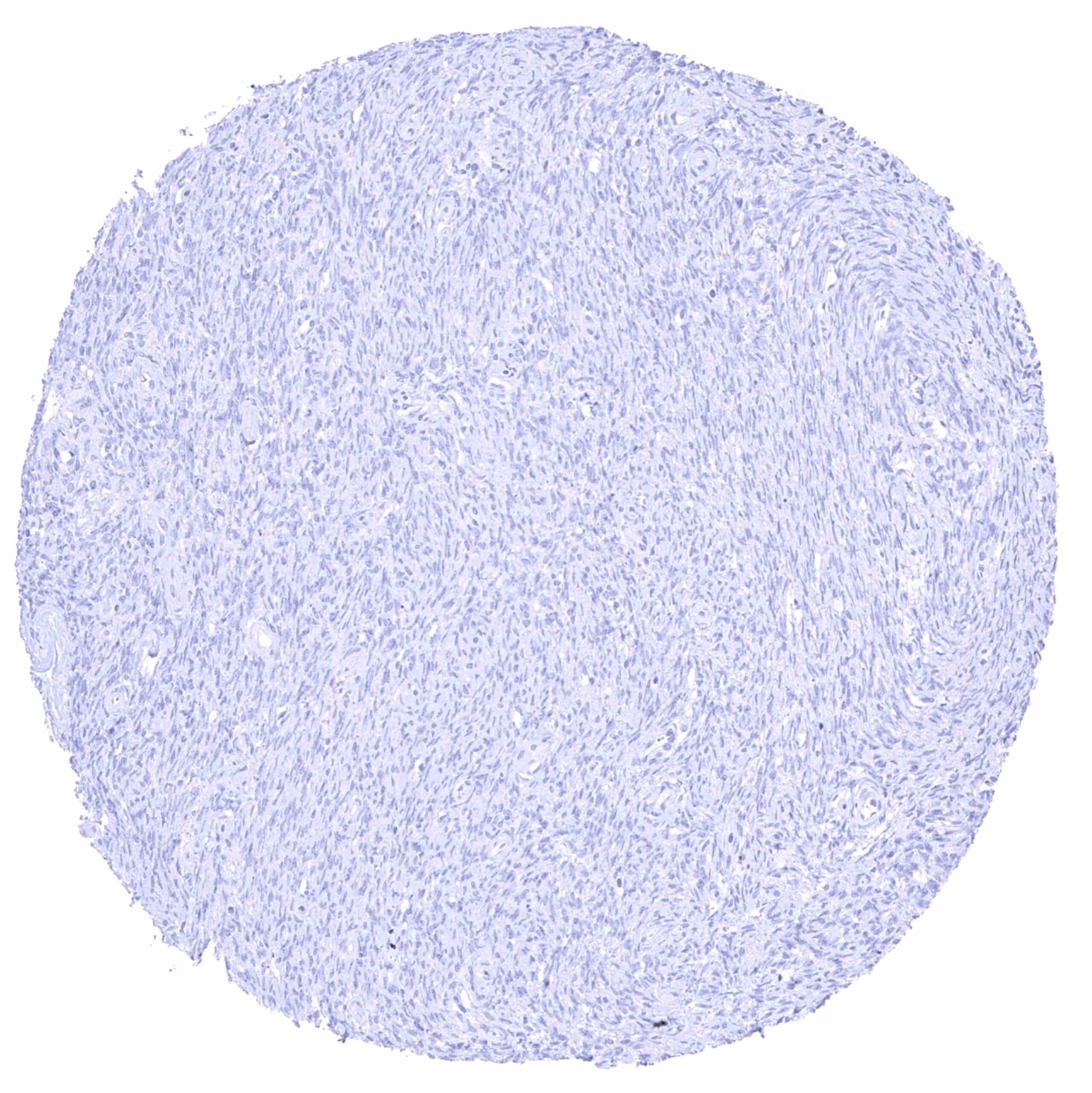
Uterus, myometrium