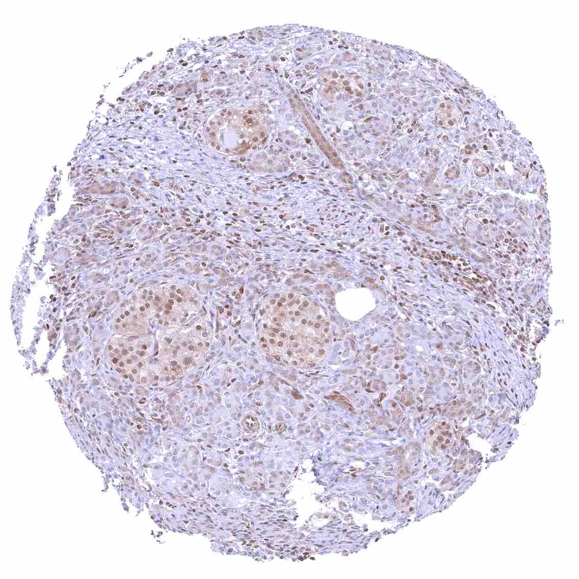
Adrenal gland – Distinct nuclear p27 positivity of a large fraction of cells.
Aorta, media – Strong nuclear p27 staining of muscle cells in the aorta.
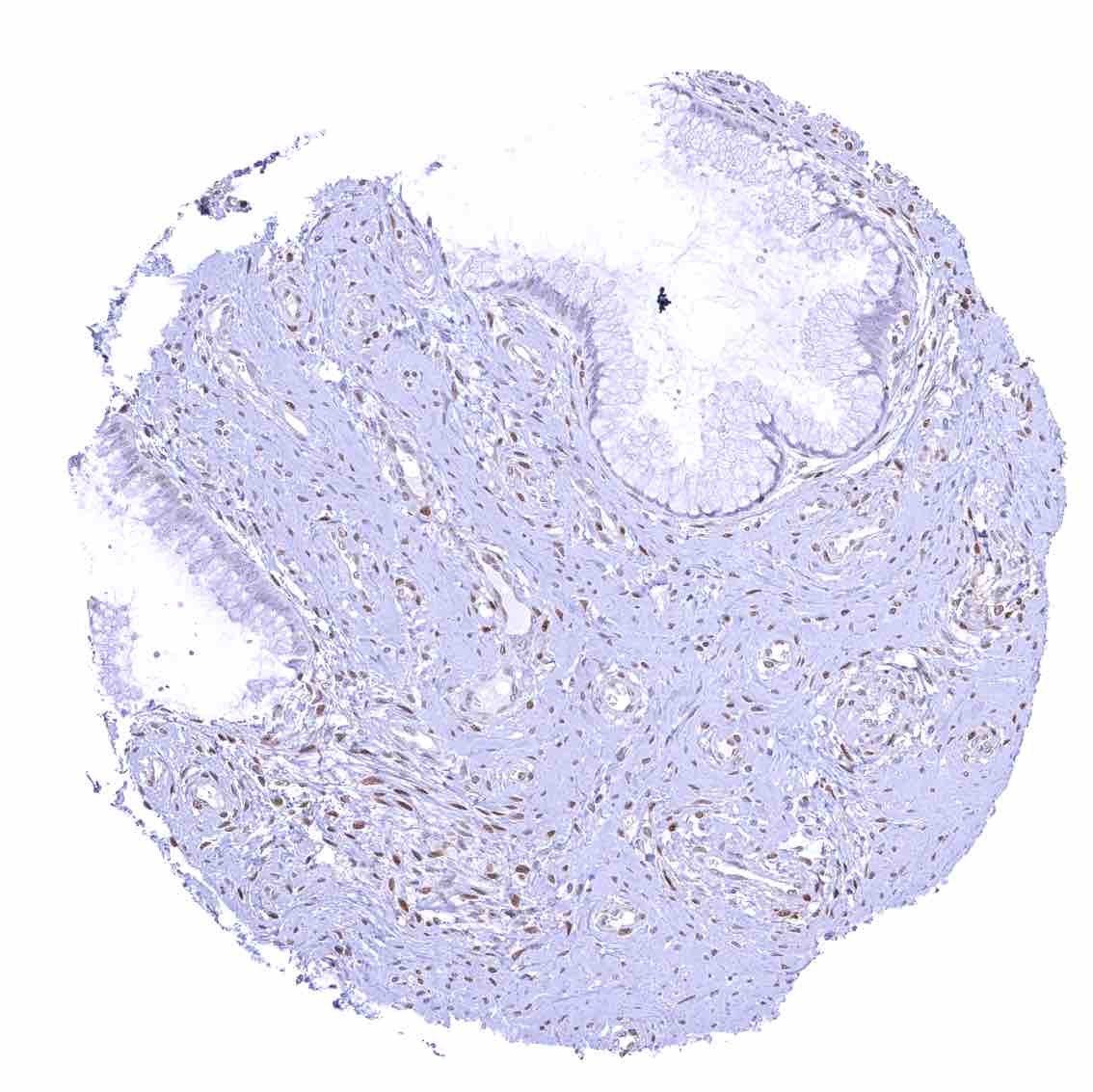
Appendix, mucosa – Among epithelial cells, nuclear p27 staining predominates in superficial epithelial cells. Most p27 staining occurs in lymphatic cells, however.
Appendix, muscular wall – Nuclear p27 staining of variable intensity in a fraction of smooth muscle and of neuronal cells.
Bone marrow – The number of p27 positive cells is rather low.
Breast – Moderate to strong nuclear p27 positivity of a large fraction of glandular epithelial cells.
Bronchus, mucosa – Strong nuclear p27 positivity of a large fraction of respiratory epithelial cells.
Cerebellum (molecular layer, Purkinje cell layer, granule cell layer) – Strong nuclear p27 positivity of all granular cells while Purkinje cells and most cells of the molecular layer are p27 negative.
Cerebellum (white matter) – Many strongly p27 positive glia cells.
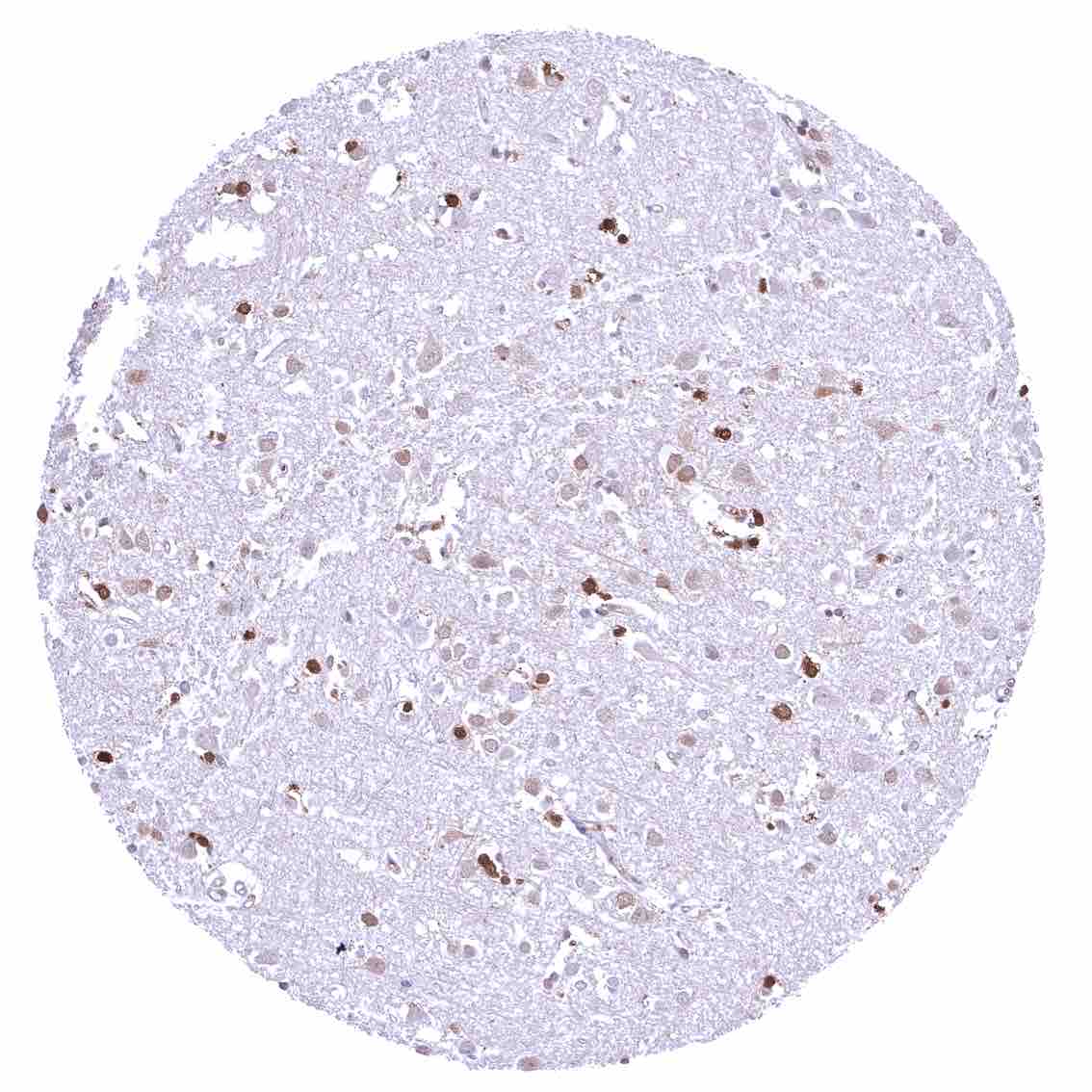
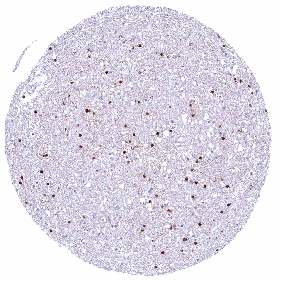
Cerebrum, grey matter –- Many strongly p27 positive glia cells.
Cerebrum, white matter
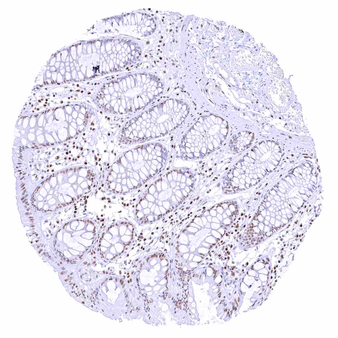
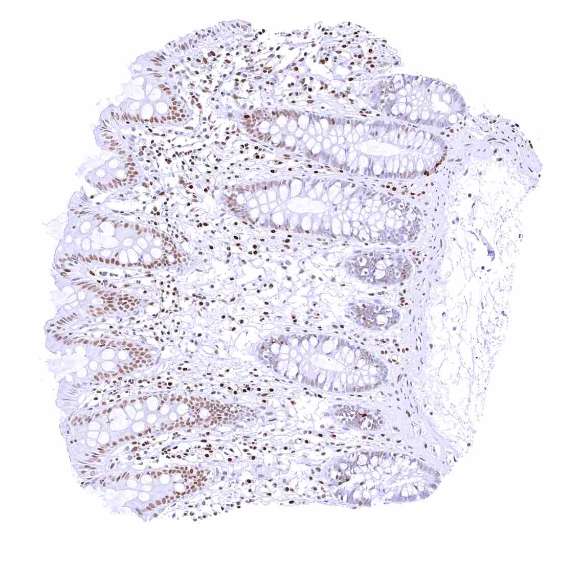
Colon descendens, mucosa
Colon descendens, muscular wall – Strong nuclear p27 staining of most smooth muscle and neuronal cells.
Colon descendens, muscular wall
Duodenum, Brunner gland
Duodenum, mucosa – Nuclear p27 positivity of a fraction of epithelial cells.
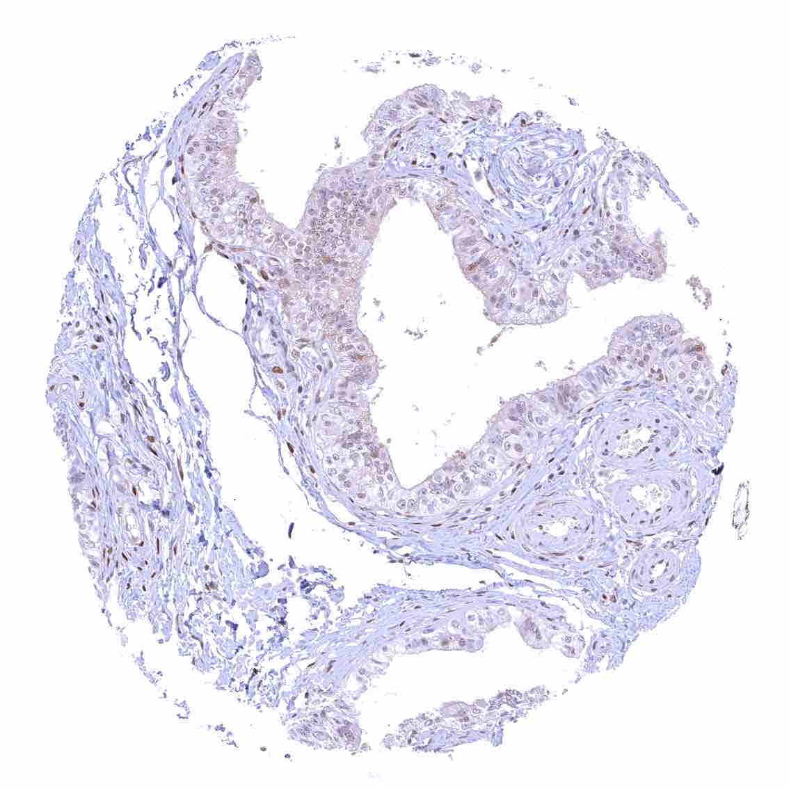
Epididymis (Cauda)
Epididymis (Corpus).jpeg
Esophagus, squamous epithelium – Nuclear staining of squamous epithelial cells predominates in the more mature cell layers (top 50_ of the epidermis).
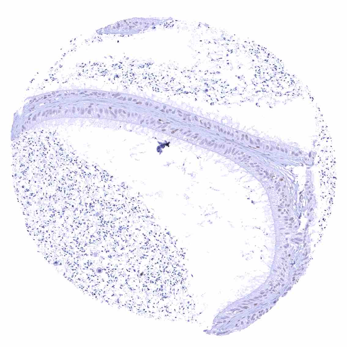
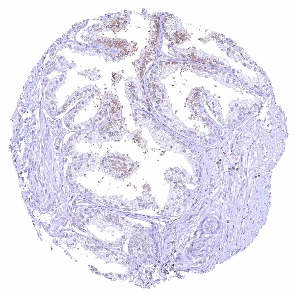
Fallopian tube, mucosa
Fat – Variable nuclear p27 staining of at least a fraction of fat cells.
Gallbladder, epithelium
Heart muscle – Distinct nuclear p27 staining of stroma cells while myocytes are largely negative.
Ileum, mucosa – Nuclear p27 positivity of a fraction of epithelial cells.
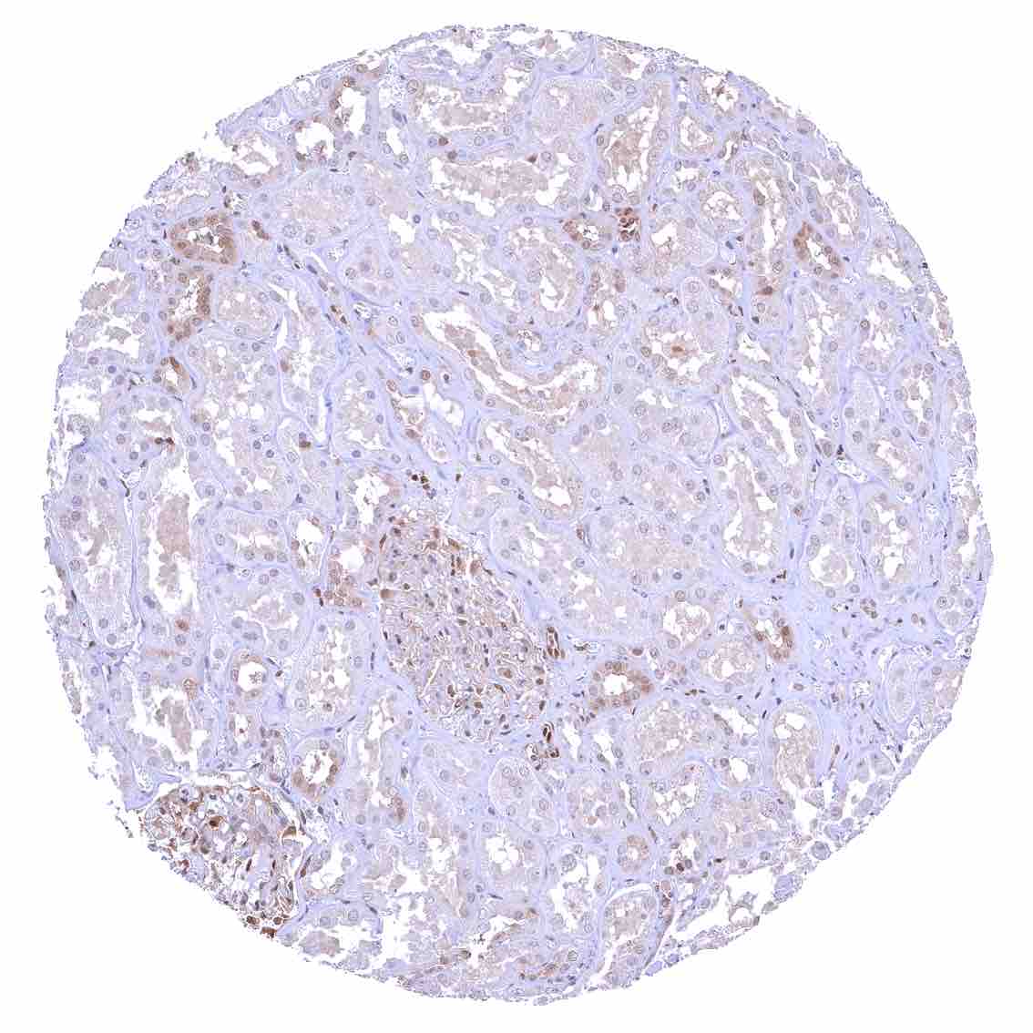
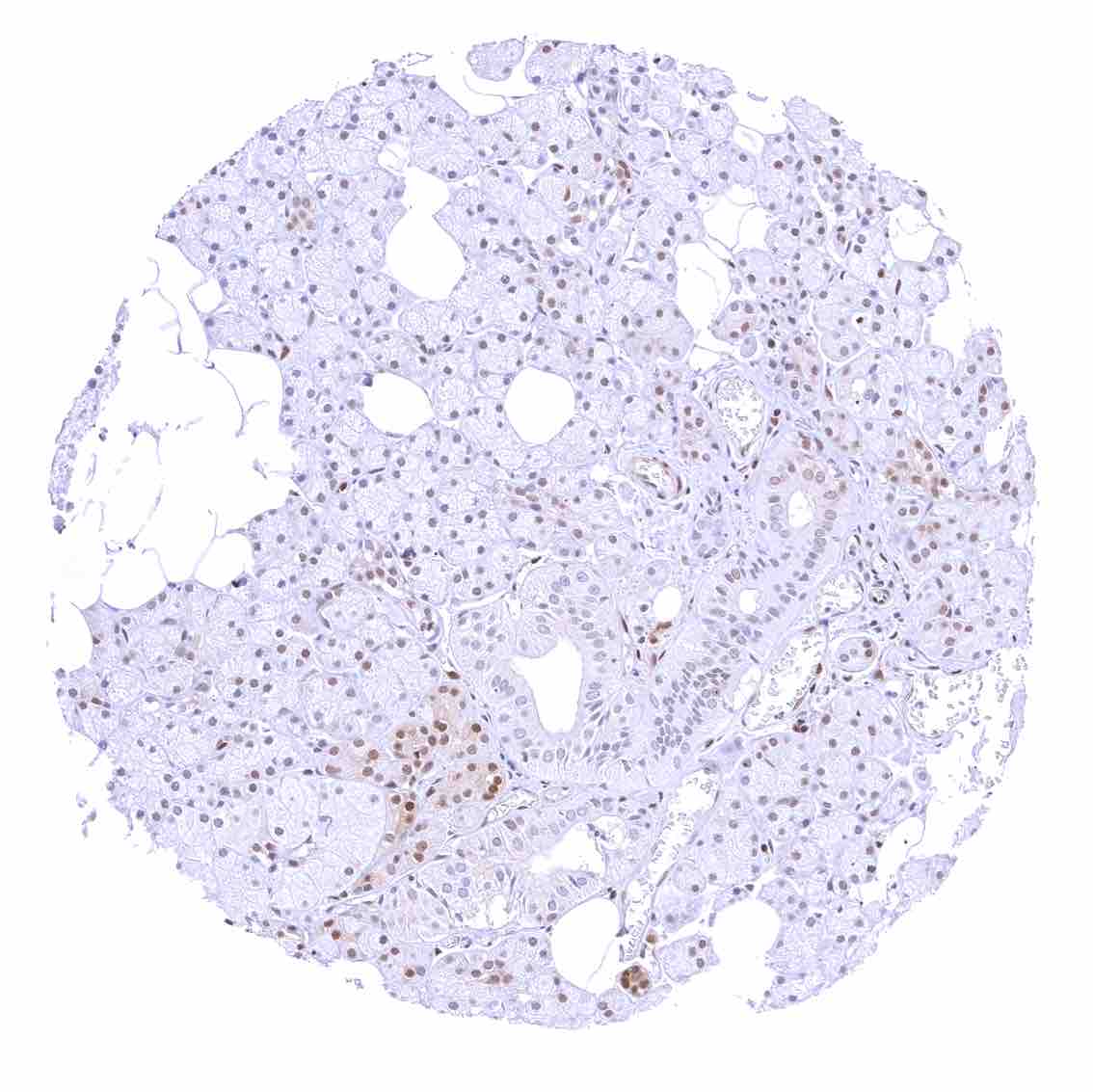
Kidney, cortex – Tubuli are usually p27 negative. The strongest p27 staining occurs in glomeruli and in stroma cells including small vessels.
Kidney, medulla
Kidney, pelvis, urothelium
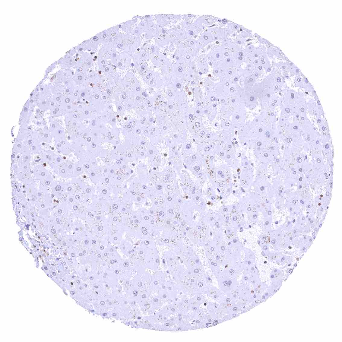
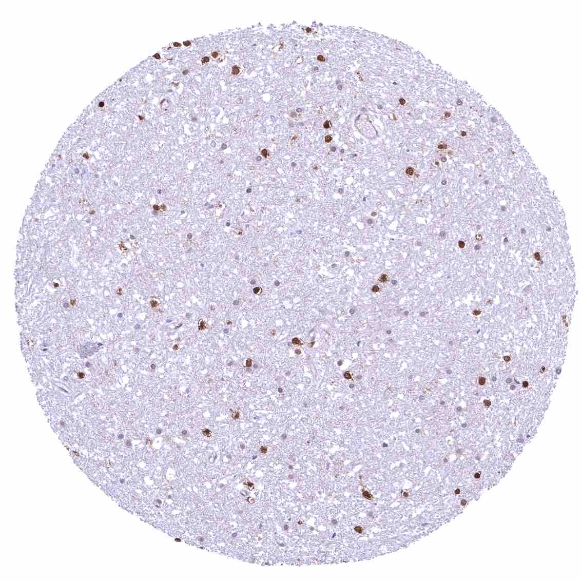
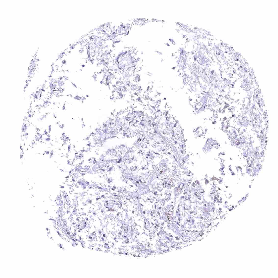
Liver – p27 staining is rare or absent in hepatocytes.
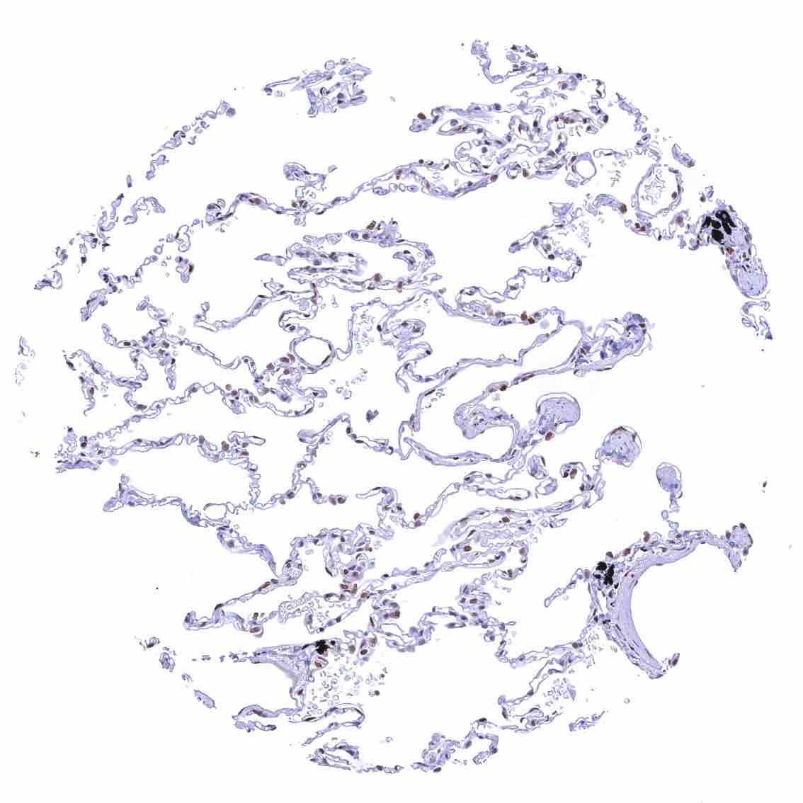
Lung
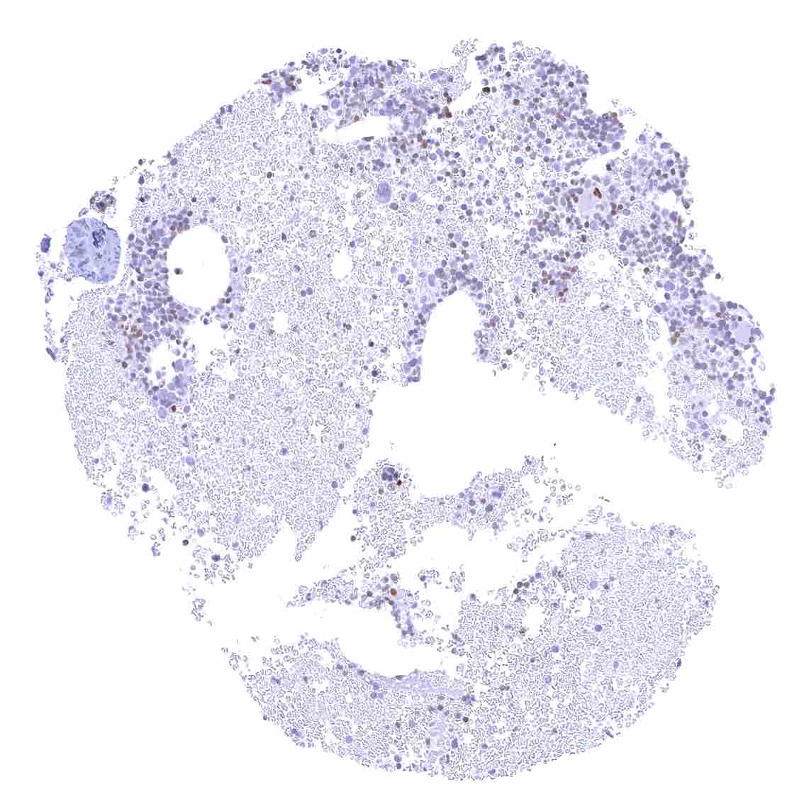
Lymph node – Strong nuclear p27 staining of a significant subset of cells, especially in the interfollicular area.
Ovary, corpus luteum – Weak to moderate nuclear p27 staining of all corpus luteum cells.
Ovary, stroma – Strong nuclear p27 staining of all stroma cells.
Pancreas – p27 staining is more common in islet cells than in acinar cells.
Parathyroid gland – Weak to moderate nuclear p27 positivity of most epithelial cells.
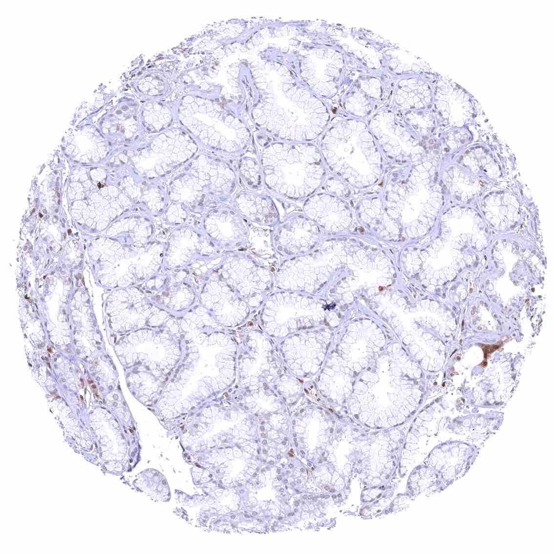
Parotid gland – p27 staining is more common in glandular cells than in excretory ducts.
Pituitary gland, posterior lobe.jpeg
Placenta (amnion and chorion) – Moderate to strong nuclear p27 staining of most or all chorion cells. A weak to moderate nuclear p27 staining also occurs in a subset of amnion cells. .jpeg
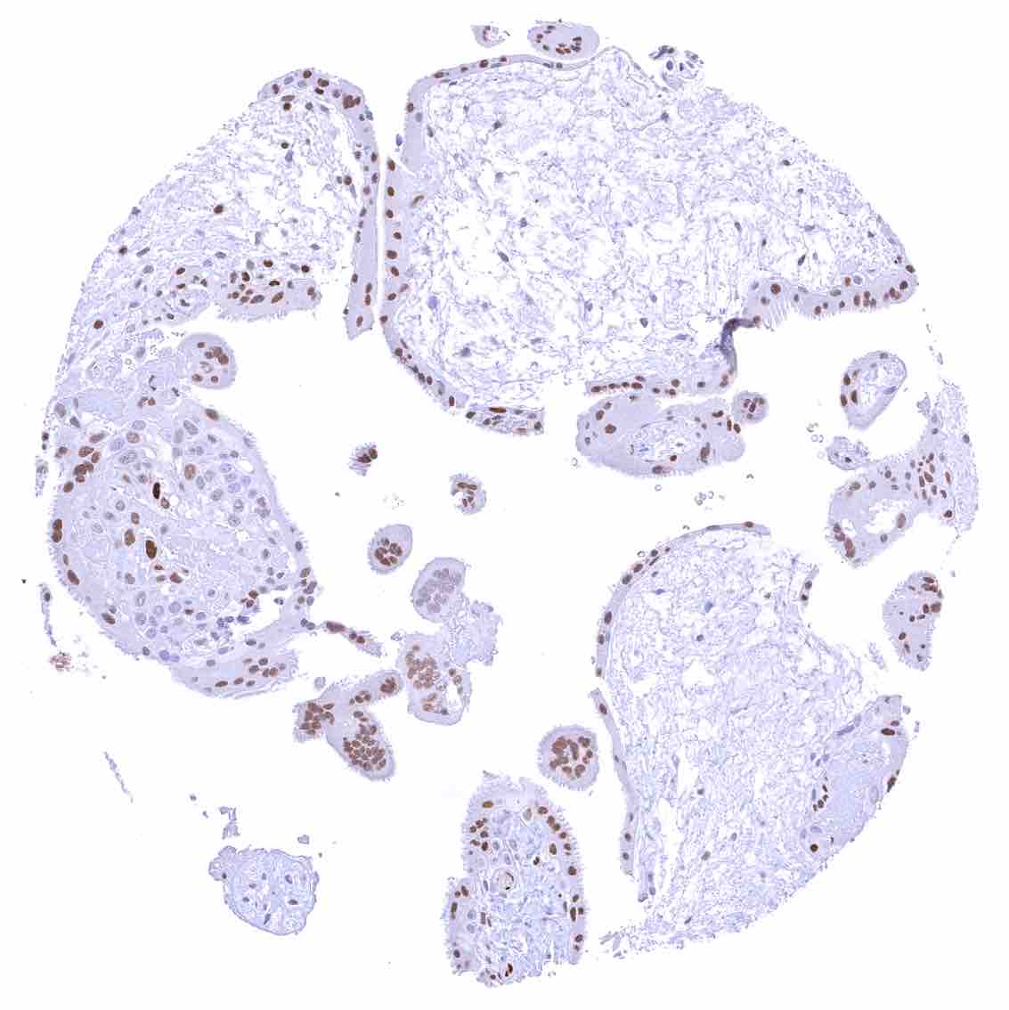
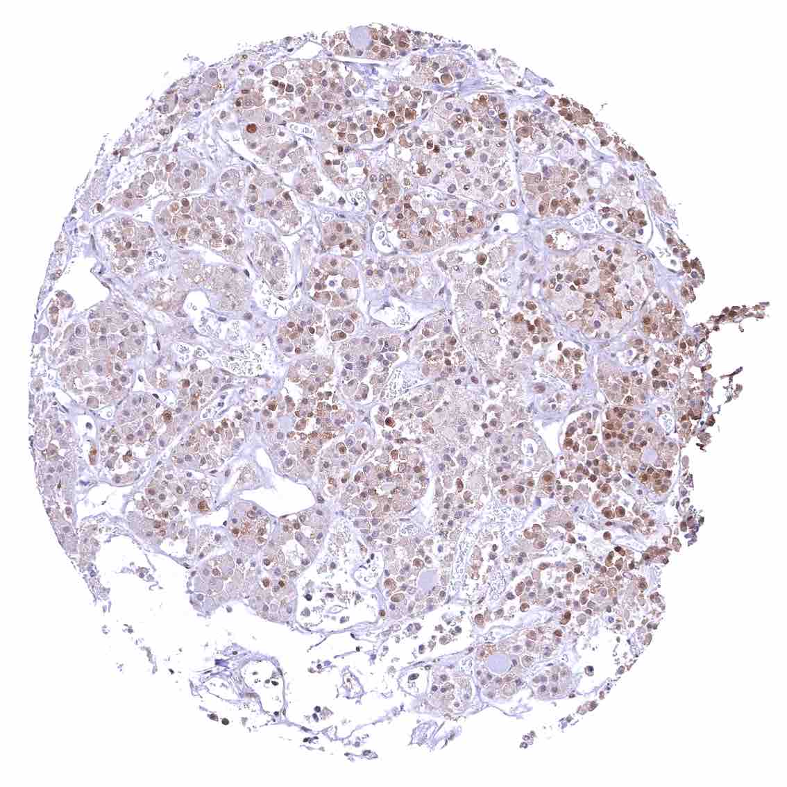
Placenta, early – Strong p27 staining of all nuclei of the syncytiotrophoblast.
Placenta, mature – Weak nuclear p27 staining of trophoblast and stroma cells.
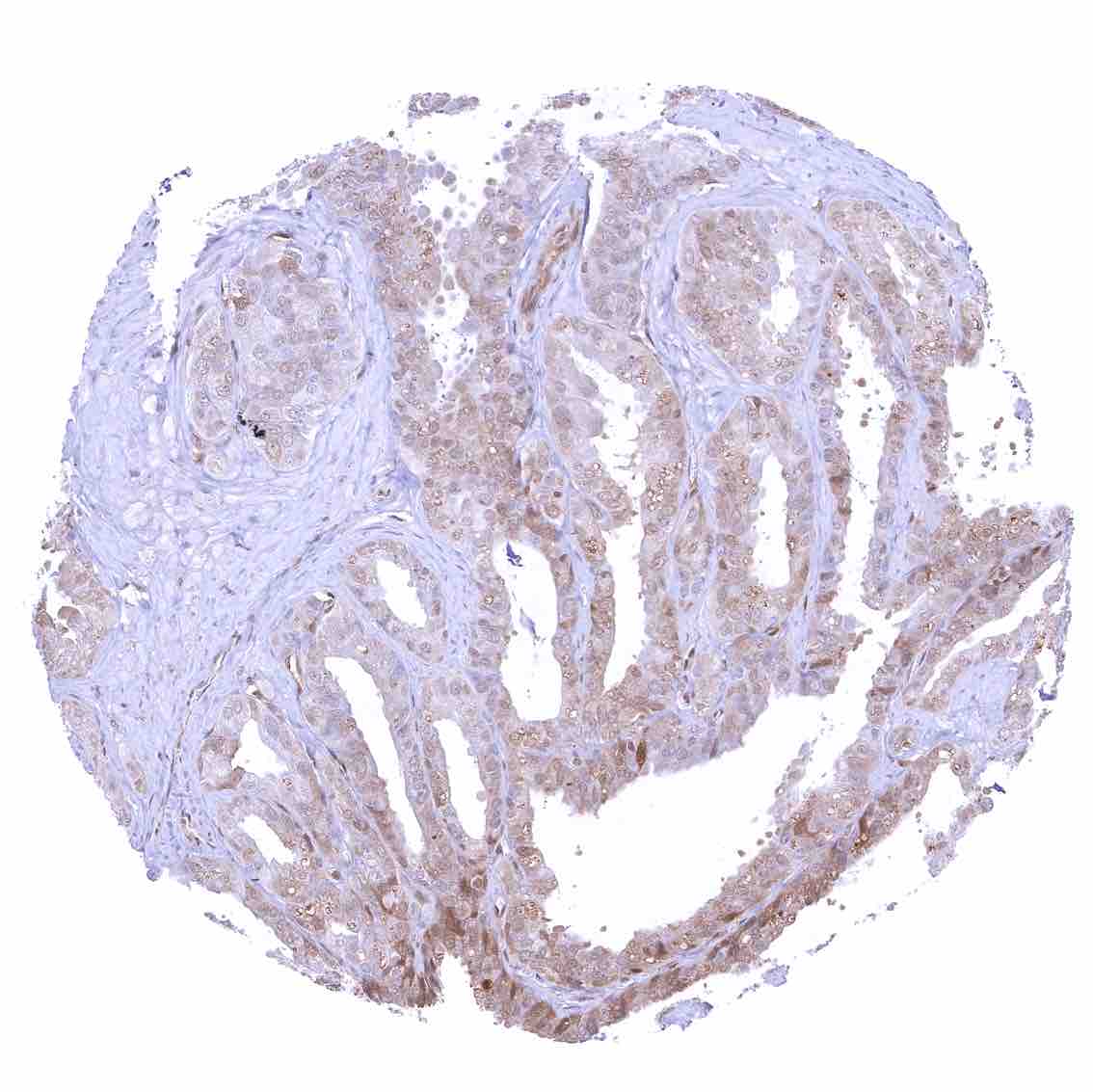
Prostate
Rectum, mucosa
Seminal vesicle
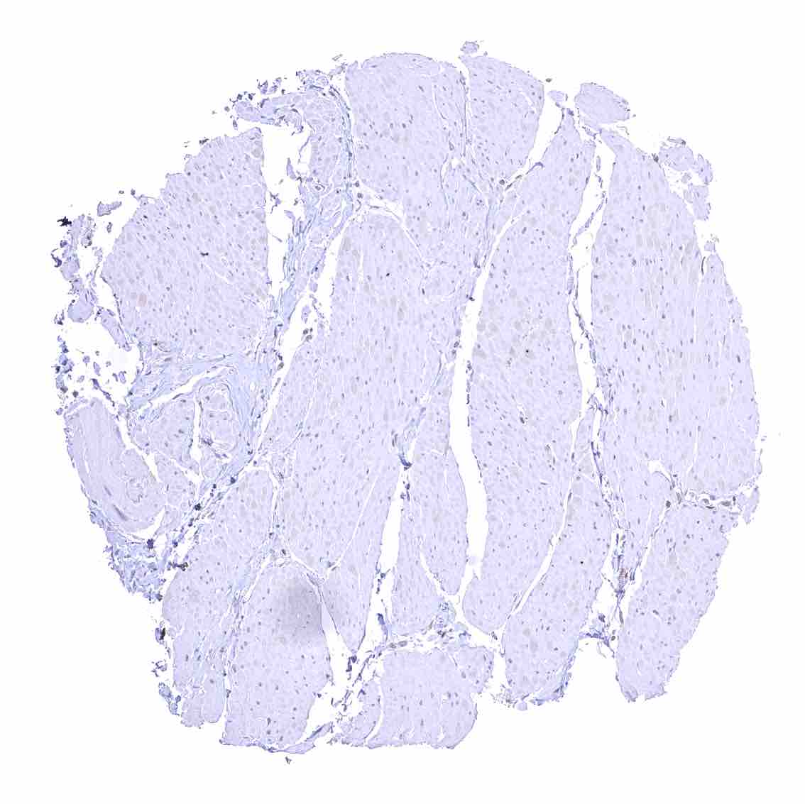
Skeletal muscle – Strong nuclear p27 staining of most but not all cells.
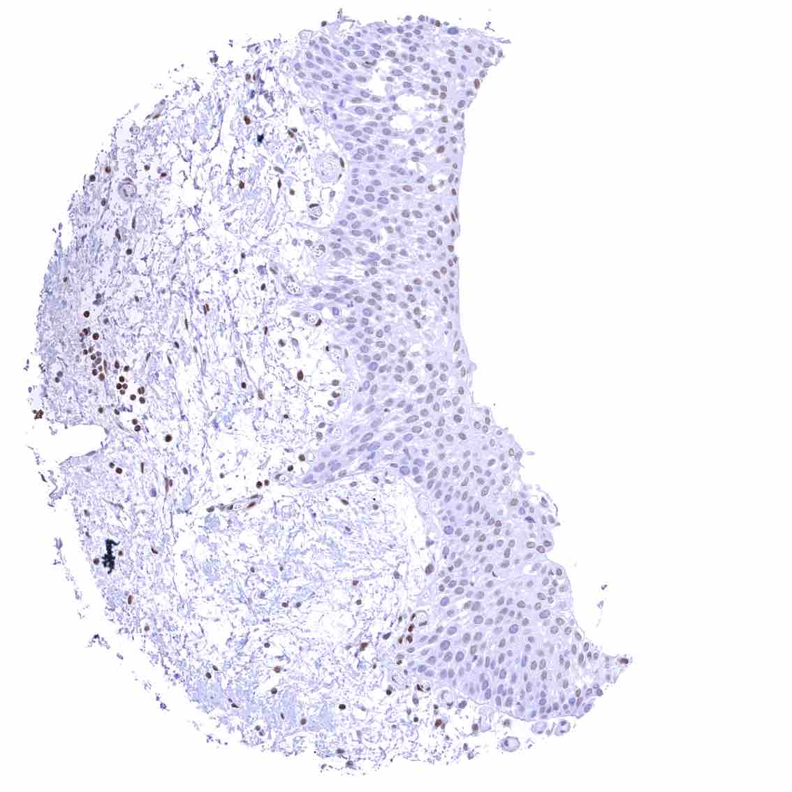
Skin – Moderate to strong nuclear p27 staining occurs in a large fraction of squamous epithelial cells and predominates in the more mature cell layers (top 50_ of the epidermis).
Skin, hairfollicel and sebaceous glands – Moderate to strong nuclear p27 staining in all cells of sebaceous glands. .jpeg
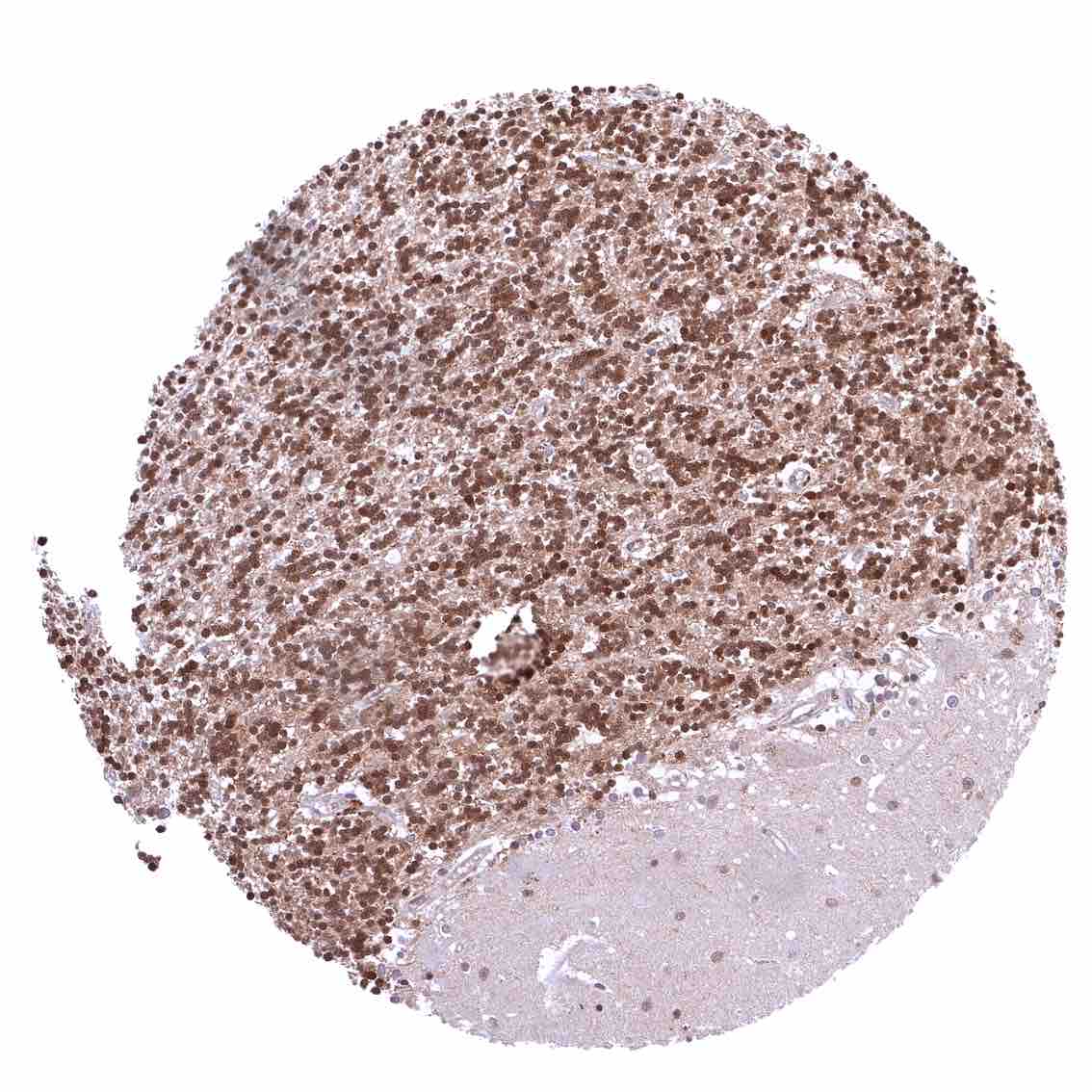
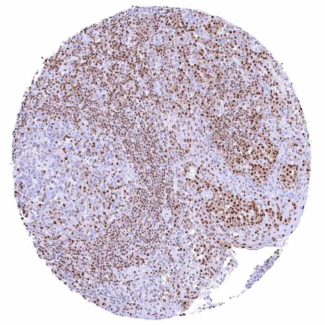
Spleen – Strong nuclear p27 staining of a large fraction of cells.
Stomach, corpus – Nuclear p27 positivity predominates in superficial epithelial cells and is rarely seen in neck cells.
Testis – Weak to moderate nuclear p27 staining of a subset of cells (predominantly Sertoli cells).
Thymus – Strong nuclear p27 staining of a large fraction of cells.
Thyroid gland – Weak to moderate nuclear p27 positivity of most follicular cells. .jpeg
Tonsil – Strong nuclear p27 staining of a significant subset of cells, especially in the interfollicular area. Nuclear staining of squamous epithelial cells predominates in the more mature cell layers (top 50_ of the epidermis)
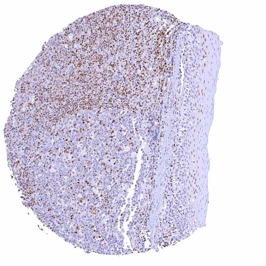
Tonsil, surface epithelium – Strong nuclear p27 staining of a significant subset of cells, especially in the interfollicular area. Nuclear staining of squamous epithelial cells predominates in the more mature cell layers (top 50_ of the epidermis)
Urinary bladder, muscular wall
Urinary bladder, urothelium
Uterus, ectocervix – Nuclear staining of squamous epithelial cells predominates in the more mature cell layers (top 50_ of the epidermis).
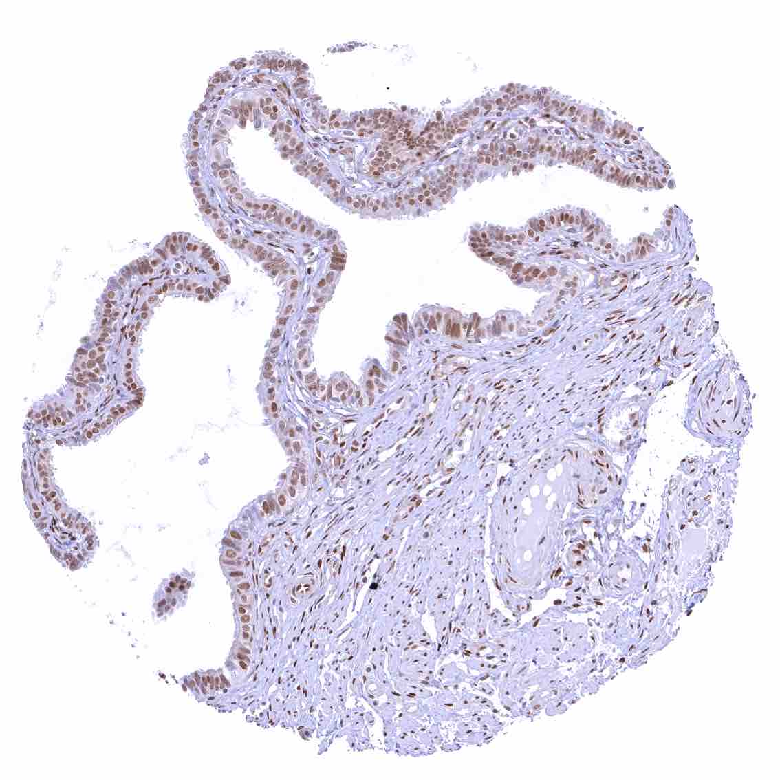
Uterus, endocervix – Nuclear p27 staining is variable in the endocervix. It is positive in all epithelial cells in this sample.
Uterus, endocervix.jpeg
Uterus, endometrium (pregnancy) – Distinct nuclear p27 staining of both endometrium and decidua cells.
Uterus, endometrium (proliferation) – Nuclear p27 staining is absent in endometrium epithelial cells in this sample.
Uterus, endometrium (proliferation) – Nuclear p27 staining is weak in endometrium epithelial cells in this sample.
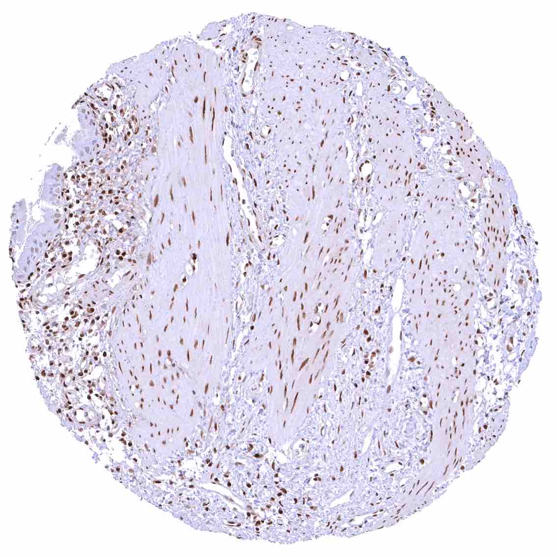
Uterus, myometrium – Nuclear p27 staining of variable intensity in most muscle cells. .jpeg